User login
Hemorrhage control system gets expanded approval
Control System
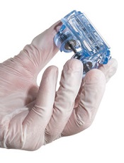
The US Food and Drug Administration has expanded the indication for the iTClamp® Hemorrhage Control System.
It is now approved to provide temporary control of severe bleeding of the neck. The product was already approved for use on the extremities, axilla, inguinal areas, and the scalp.
The iTClamp is a temporary wound closure device designed to control severe bleeding in seconds.
It seals the edges of a wound closed to create a temporary pool of blood under pressure. This forms a stable clot that mitigates further blood
loss until the wound can be surgically repaired.
Each iTClamp measures less than 2 by 2 inches and weighs less than 3 ounces. It requires only minimal training and gross motor skills to use, according to iTraumaCare, the company that makes the product.
“Addressing difficult-to-control hemorrhage in the neck has been a consistent problem with few solutions,” said Dennis Filips, MD, founder and chief medical officer of iTraumaCare.
“This expanded indication for the iTClamp will allow first responders, medical professionals, and tactical and battlefield medics to use the device in even more meaningful ways to improve patient care.” ![]()
Control System
The US Food and Drug Administration has expanded the indication for the iTClamp® Hemorrhage Control System.
It is now approved to provide temporary control of severe bleeding of the neck. The product was already approved for use on the extremities, axilla, inguinal areas, and the scalp.
The iTClamp is a temporary wound closure device designed to control severe bleeding in seconds.
It seals the edges of a wound closed to create a temporary pool of blood under pressure. This forms a stable clot that mitigates further blood
loss until the wound can be surgically repaired.
Each iTClamp measures less than 2 by 2 inches and weighs less than 3 ounces. It requires only minimal training and gross motor skills to use, according to iTraumaCare, the company that makes the product.
“Addressing difficult-to-control hemorrhage in the neck has been a consistent problem with few solutions,” said Dennis Filips, MD, founder and chief medical officer of iTraumaCare.
“This expanded indication for the iTClamp will allow first responders, medical professionals, and tactical and battlefield medics to use the device in even more meaningful ways to improve patient care.” ![]()
Group creates universal platelets using iPSCs

Credit: Salk Institute
Researchers say they can use induced pluripotent stem cells (iPSCs) to produce large-scale quantities of universal donor platelets.
The team generated megakaryocytes and platelets from iPSCs under feeder-free conditions.
They were able to produce universal platelets by removing a gene essential to expression of the major histocompatibility antigens.
The resulting platelets were functional and behaved like normal human platelets.
The researchers described this method of platelet production, owned by Advanced Cell Technology, Inc., in Stem Cell Reports.
“Unlike other sources of platelets, human induced pluripotent stem cells can be propagated indefinitely, providing a potentially unlimited source of cells for therapeutic purposes,” said Robert Lanza, MD, Chief Scientific Officer at Advanced Cell Technology.
“This study shows that platelets may be produced from [iPSCs] without the need for serum and feeders and, thus, removes potential risks associated with contaminants and pathogens.”
Dr Lanza and his colleagues used a 3-step protocol to differentiate human iPSCs into megakaryocytes and functional platelets in less than 20 days. The method incorporates several discrete intermediate cells, including proprietary hemogenic endothelium-like cells.
The technique allows for long-term storage of megakaryocyte progenitors so they can be available within a few days when needed to produce large quantities of platelets for transfusion.
In addition, by knocking out the β2-microglobulin gene, the researchers were able to generate platelets that are negative for the major histocompatibility antigens.
This suggests the platelets could be transfused into almost any patient, and the method might even prevent platelet refractoriness, according to the researchers.
The team found no major differences in the iPSC platelets and normal human platelets. The iPSC platelets formed aggregates, lamellipodia, and filopodia after activation, just like normal platelets.
Also like normal platelets, the iPSC platelets circulated for at least 8 hours in macrophage-depleted NOD/SCID mice, with a time to reach maximal accumulation of 30 minutes to an hour.
In another murine experiment, iPSC platelets incorporated into a growing thrombus just like normal human platelets, with an average number of 9.0 ± 1.8 platelets per thrombus.
“The platelets generated with our technology are functional and behave like normal human platelets,” Dr Lanza said. “This technology and these results represent an important step towards generating unlimited supplies of universal donor platelets for transfusion.” ![]()
Hospitalists and Liability
In this issue of the Journal of Hospital Medicine, Schaffer and colleagues report their analysis of malpractice claims against hospitalists compared to other physician specialties.[1] In contrast to previous work examining medical liability,[2, 3] Schaffer and colleagues have been able to identify hospitalists specifically.[2, 3]
Perhaps surprisingly, their main finding was that the rate of claims against hospitalists was significantly lower than for nonhospitalist internists, emergency medicine physicians, general surgeons, and obstetriciansgynecologists. We say surprisingly, because health systems utilizing hospitalists generally include features that might increase the risk for malpractice claims.
For example, new patients are typically assigned to whichever hospitalist is up for the next admission. Research shows that strained patientphysician relationships increase the risk for malpractice claims.[4, 5] Schaffer's data suggest that lack of a preexisting relationship is a challenge, but one to which most hospitalists have grown accustomed. Hospitalists develop and hone skills that allow them to quickly establish rapport with patients and families. Moreover, though patients seldom choose their hospitalist, they often do select the hospital in which they receive their care. The research group of 1 of the authors was recently surprised to find patients had high levels of trust with their hospital physicians, despite frequently being unable to name them or identify their role.[6] We suspect patients in the study had high levels of trust with the hospital and transferred this trust to their assigned physicians as representatives of the organization. Certainly, this hypothesis needs to be tested, and in no way does it minimize the importance of a strong patient‐physician relationship.
In addition, patientphysician continuity has long been felt to be paramount to safe and effective care; however, it is difficult to achieve in hospitalist systems. Hospitalized patients experience multiple handoffs, including those at admission, for night coverage, and at the time of service change (ie, end of rotation/stint). The potential for loss of information is enormous. Though increased attention has been dedicated to handoffs among housestaff, little work has been done to describe issues related to handoffs among practicing physicians. However, some discontinuity may be advantageous. A physician newly taking over patient care from another may not be anchored to the initial diagnosis and treatment plan established by the first. This free second look may actually prevent missed/delayed diagnoses and optimize plans of care, further reducing harm from care and risk of malpractice.[7]
Hospital discharge is another highly risky time, due to issues such as tests pending at the time of discharge and the need to manage ongoing workup and treatment of unresolved issues.[8, 9] The responsibility for tying up these loose ends may be unclear as patients are transitioned from the care of hospitalists to outpatient physicians. Prior research has shown that patients are at particularly high risk for preventable adverse events after hospital discharge.[10, 11] More recently, healthcare policy has focused on measuring and incentivizing the reduction of readmissions.[12] Although only a portion of readmissions are truly preventable,[13] and many patients who suffer adverse events after discharge are not readmitted,[11] the efforts resulting from these policy initiatives may have improved the overall safety of transitions of care.
A particularly important contribution of Schaffer and colleagues' study is that it helps us identify patient safety issues related to hospital medicine. Despite intense national efforts over the past 10 to 15 years, progress has been slow in reducing the rate of adverse events among hospitalized patients.[14, 15, 16] Although adverse events and medical liability do not always correlate,[17, 18] the contributing factors identified in Schaffer and colleagues' study help direct our patient safety efforts.
Clinical judgment was the most common factor associated with hospitalist malpractice claims, with examples including failure or delay in ordering a necessary diagnostic test or specialist consultation. These results may be misinterpreted by some to suggest that ordering more tests and services may reduce risk for malpractice claims. However, there is no evidence to support the belief that these defensive medicine behaviors actually reduce risk. In fact, the opposite may be true. Research shows that abnormal tests are frequently overlooked,[9, 19] and failure to act on abnormal results is a common cause of diagnostic error.[20] Experts have called for the development of diagnosis‐related quality measures and better strategies to enhance trainees' clinical reasoning skills.[21] We suggest that future research also clarify the effect of interruptions, distractions, and workload on cognitive errors in hospital settings.
Communication failures were the second most common contributing factor. As previously mentioned, communication failures may occur between hospitalists during handoffs. We also have major opportunities to improve interprofessional teamwork, especially between physicians and nurses.[22, 23] An increasing number of hospitalist groups are collaborating with other hospital‐based professionals to implement novel strategies to improve teamwork,[24, 25] many of which were recently summarized in a review published in this journal.[26]
Documentation was the third most common contributing factor. Most malpractice claims are filed long after the alleged injury has occurred.[18] Unless the clinicians involved and the hospital in which they work are aware of an event that might result in a malpractice claim, the investigation may be severely delayed. As time goes on, professionals are less able to recall details pertinent to understanding contributing factors to an event. Thus, documentation is critical. As the saying goes, if it wasn't documented, it didn't happen. The flipside of too little documentation is, of course, too much. The increasing use of electronic health records makes it easy to copy and paste outdated information, the sloppiness of which can only hurt when attempting to defend a malpractice claim.[27]
In conclusion, despite a model with inherent features that might contribute to medical malpractice risk, hospital medicine has a claim rate lower than many other specialties. Though reassuring, hospitalists should remember that the most productive way to approach malpractice risk is reframe the problem as one that attempts to reduce risk for patients, rather than for physicians. Improving patient safety is a core value for hospital medicine. Schaffer and colleagues' study identifies factors contributing to patient safety risk in hospital medicine, allowing us to renew our efforts in focused areas.
- , , , . Liability impact of the hospitalist model of care. J Hosp Med. 2014;9(12):750–755.
- , , . Paid malpractice claims for adverse events in inpatient and outpatient settings. JAMA. 2011;305(23):2427–2431.
- , , , . Malpractice risk according to physician specialty. N Engl J Med. 2011;365(7):629–636.
- , , , . The doctor‐patient relationship and malpractice. Lessons from plaintiff depositions. Arch Intern Med. 1994;154(12):1365–1370.
- , , , , . Physician‐patient communication. The relationship with malpractice claims among primary care physicians and surgeons. JAMA. 1997;277(7):553–559.
- , , , , , . The impact of facecards on patients' knowledge, satisfaction, trust, and agreement with hospital physicians: a pilot study. J Hosp Med. 2014;9(3):137–141.
- . Does continuity of care matter? No: discontinuity can improve patient care. West J Med. 2001;175(1):5.
- , , . Tying up loose ends: discharging patients with unresolved medical issues. Arch Intern Med. 2007;167(12):1305–1311.
- , , , et al. Patient safety concerns arising from test results that return after hospital discharge. Ann Intern Med. 2005;143(2):121–128.
- , , , et al. Adverse events among medical patients after discharge from hospital. CMAJ. 2004;170(3):345–349.
- , , , , . The incidence and severity of adverse events affecting patients after discharge from the hospital. Ann Intern Med. 2003;138(3):161–167.
- U.S. Department of Health 183(7):E391–E402.
- U.S. Department of Health 363(22):2124–2134.
- , , , et al. National trends in patient safety for four common conditions, 2005–2011. N Engl J Med. 2014;370(4):341–351.
- , , , et al. Relation between malpractice claims and adverse events due to negligence. Results of the Harvard Medical Practice Study III. N Engl J Med. 1991;325(4):245–251.
- , , , et al. Claims, errors, and compensation payments in medical malpractice litigation. N Engl J Med. 2006;354(19):2024–2033.
- , , , , , . “I wish I had seen this test result earlier!”: Dissatisfaction with test result management systems in primary care. Arch Intern Med. 2004;164(20):2223–2228.
- , , , et al. Diagnostic error in medicine: analysis of 583 physician‐reported errors. Arch Intern Med. 2009;169(20):1881–1887.
- , , . Bringing diagnosis into the quality and safety equations. JAMA. 2012;308(12):1211–1212.
- , , , , , . Teamwork on inpatient medical units: assessing attitudes and barriers. Qual Saf Health Care. 2010;19(2):117–121.
- , , , et al. Patterns of nurse‐physician communication and agreement on the plan of care. Qual Saf Health Care. 2010;19(3):195–199.
- , , , et al. Effects of a multicentre teamwork and communication programme on patient outcomes: results from the Triad for Optimal Patient Safety (TOPS) project. BMJ Qual Saf. 2012;21(2):118–126.
- , , , , , . Unit‐based interprofessional leadership models in six US hospitals. J Hosp Med. 2014;9(8):545–550.
- , , , . Interdisciplinary teamwork in hospitals: A review and practical recommendations for improvement. J Hosp Med. 2012;7(1):48–54.
- , . Legal, ethical, and financial dilemmas in electronic health record adoption and use. Pediatrics. 2011;127(4):e1042–e1047.
In this issue of the Journal of Hospital Medicine, Schaffer and colleagues report their analysis of malpractice claims against hospitalists compared to other physician specialties.[1] In contrast to previous work examining medical liability,[2, 3] Schaffer and colleagues have been able to identify hospitalists specifically.[2, 3]
Perhaps surprisingly, their main finding was that the rate of claims against hospitalists was significantly lower than for nonhospitalist internists, emergency medicine physicians, general surgeons, and obstetriciansgynecologists. We say surprisingly, because health systems utilizing hospitalists generally include features that might increase the risk for malpractice claims.
For example, new patients are typically assigned to whichever hospitalist is up for the next admission. Research shows that strained patientphysician relationships increase the risk for malpractice claims.[4, 5] Schaffer's data suggest that lack of a preexisting relationship is a challenge, but one to which most hospitalists have grown accustomed. Hospitalists develop and hone skills that allow them to quickly establish rapport with patients and families. Moreover, though patients seldom choose their hospitalist, they often do select the hospital in which they receive their care. The research group of 1 of the authors was recently surprised to find patients had high levels of trust with their hospital physicians, despite frequently being unable to name them or identify their role.[6] We suspect patients in the study had high levels of trust with the hospital and transferred this trust to their assigned physicians as representatives of the organization. Certainly, this hypothesis needs to be tested, and in no way does it minimize the importance of a strong patient‐physician relationship.
In addition, patientphysician continuity has long been felt to be paramount to safe and effective care; however, it is difficult to achieve in hospitalist systems. Hospitalized patients experience multiple handoffs, including those at admission, for night coverage, and at the time of service change (ie, end of rotation/stint). The potential for loss of information is enormous. Though increased attention has been dedicated to handoffs among housestaff, little work has been done to describe issues related to handoffs among practicing physicians. However, some discontinuity may be advantageous. A physician newly taking over patient care from another may not be anchored to the initial diagnosis and treatment plan established by the first. This free second look may actually prevent missed/delayed diagnoses and optimize plans of care, further reducing harm from care and risk of malpractice.[7]
Hospital discharge is another highly risky time, due to issues such as tests pending at the time of discharge and the need to manage ongoing workup and treatment of unresolved issues.[8, 9] The responsibility for tying up these loose ends may be unclear as patients are transitioned from the care of hospitalists to outpatient physicians. Prior research has shown that patients are at particularly high risk for preventable adverse events after hospital discharge.[10, 11] More recently, healthcare policy has focused on measuring and incentivizing the reduction of readmissions.[12] Although only a portion of readmissions are truly preventable,[13] and many patients who suffer adverse events after discharge are not readmitted,[11] the efforts resulting from these policy initiatives may have improved the overall safety of transitions of care.
A particularly important contribution of Schaffer and colleagues' study is that it helps us identify patient safety issues related to hospital medicine. Despite intense national efforts over the past 10 to 15 years, progress has been slow in reducing the rate of adverse events among hospitalized patients.[14, 15, 16] Although adverse events and medical liability do not always correlate,[17, 18] the contributing factors identified in Schaffer and colleagues' study help direct our patient safety efforts.
Clinical judgment was the most common factor associated with hospitalist malpractice claims, with examples including failure or delay in ordering a necessary diagnostic test or specialist consultation. These results may be misinterpreted by some to suggest that ordering more tests and services may reduce risk for malpractice claims. However, there is no evidence to support the belief that these defensive medicine behaviors actually reduce risk. In fact, the opposite may be true. Research shows that abnormal tests are frequently overlooked,[9, 19] and failure to act on abnormal results is a common cause of diagnostic error.[20] Experts have called for the development of diagnosis‐related quality measures and better strategies to enhance trainees' clinical reasoning skills.[21] We suggest that future research also clarify the effect of interruptions, distractions, and workload on cognitive errors in hospital settings.
Communication failures were the second most common contributing factor. As previously mentioned, communication failures may occur between hospitalists during handoffs. We also have major opportunities to improve interprofessional teamwork, especially between physicians and nurses.[22, 23] An increasing number of hospitalist groups are collaborating with other hospital‐based professionals to implement novel strategies to improve teamwork,[24, 25] many of which were recently summarized in a review published in this journal.[26]
Documentation was the third most common contributing factor. Most malpractice claims are filed long after the alleged injury has occurred.[18] Unless the clinicians involved and the hospital in which they work are aware of an event that might result in a malpractice claim, the investigation may be severely delayed. As time goes on, professionals are less able to recall details pertinent to understanding contributing factors to an event. Thus, documentation is critical. As the saying goes, if it wasn't documented, it didn't happen. The flipside of too little documentation is, of course, too much. The increasing use of electronic health records makes it easy to copy and paste outdated information, the sloppiness of which can only hurt when attempting to defend a malpractice claim.[27]
In conclusion, despite a model with inherent features that might contribute to medical malpractice risk, hospital medicine has a claim rate lower than many other specialties. Though reassuring, hospitalists should remember that the most productive way to approach malpractice risk is reframe the problem as one that attempts to reduce risk for patients, rather than for physicians. Improving patient safety is a core value for hospital medicine. Schaffer and colleagues' study identifies factors contributing to patient safety risk in hospital medicine, allowing us to renew our efforts in focused areas.
- , , , . Liability impact of the hospitalist model of care. J Hosp Med. 2014;9(12):750–755.
- , , . Paid malpractice claims for adverse events in inpatient and outpatient settings. JAMA. 2011;305(23):2427–2431.
- , , , . Malpractice risk according to physician specialty. N Engl J Med. 2011;365(7):629–636.
- , , , . The doctor‐patient relationship and malpractice. Lessons from plaintiff depositions. Arch Intern Med. 1994;154(12):1365–1370.
- , , , , . Physician‐patient communication. The relationship with malpractice claims among primary care physicians and surgeons. JAMA. 1997;277(7):553–559.
- , , , , , . The impact of facecards on patients' knowledge, satisfaction, trust, and agreement with hospital physicians: a pilot study. J Hosp Med. 2014;9(3):137–141.
- . Does continuity of care matter? No: discontinuity can improve patient care. West J Med. 2001;175(1):5.
- , , . Tying up loose ends: discharging patients with unresolved medical issues. Arch Intern Med. 2007;167(12):1305–1311.
- , , , et al. Patient safety concerns arising from test results that return after hospital discharge. Ann Intern Med. 2005;143(2):121–128.
- , , , et al. Adverse events among medical patients after discharge from hospital. CMAJ. 2004;170(3):345–349.
- , , , , . The incidence and severity of adverse events affecting patients after discharge from the hospital. Ann Intern Med. 2003;138(3):161–167.
- U.S. Department of Health 183(7):E391–E402.
- U.S. Department of Health 363(22):2124–2134.
- , , , et al. National trends in patient safety for four common conditions, 2005–2011. N Engl J Med. 2014;370(4):341–351.
- , , , et al. Relation between malpractice claims and adverse events due to negligence. Results of the Harvard Medical Practice Study III. N Engl J Med. 1991;325(4):245–251.
- , , , et al. Claims, errors, and compensation payments in medical malpractice litigation. N Engl J Med. 2006;354(19):2024–2033.
- , , , , , . “I wish I had seen this test result earlier!”: Dissatisfaction with test result management systems in primary care. Arch Intern Med. 2004;164(20):2223–2228.
- , , , et al. Diagnostic error in medicine: analysis of 583 physician‐reported errors. Arch Intern Med. 2009;169(20):1881–1887.
- , , . Bringing diagnosis into the quality and safety equations. JAMA. 2012;308(12):1211–1212.
- , , , , , . Teamwork on inpatient medical units: assessing attitudes and barriers. Qual Saf Health Care. 2010;19(2):117–121.
- , , , et al. Patterns of nurse‐physician communication and agreement on the plan of care. Qual Saf Health Care. 2010;19(3):195–199.
- , , , et al. Effects of a multicentre teamwork and communication programme on patient outcomes: results from the Triad for Optimal Patient Safety (TOPS) project. BMJ Qual Saf. 2012;21(2):118–126.
- , , , , , . Unit‐based interprofessional leadership models in six US hospitals. J Hosp Med. 2014;9(8):545–550.
- , , , . Interdisciplinary teamwork in hospitals: A review and practical recommendations for improvement. J Hosp Med. 2012;7(1):48–54.
- , . Legal, ethical, and financial dilemmas in electronic health record adoption and use. Pediatrics. 2011;127(4):e1042–e1047.
ECMO in Adults
As the distribution and utilization of technology in critical care medicine expands, patients experiencing respiratory failure, heart failure, or cardiac arrest are increasingly being treated with extracorporeal membrane oxygenation (ECMO). Although not customarily responsible for managing ECMO, hospitalists need to understand the rudiments of this technology and its associated ethical issues to assure that ECMO use is consistent with patient preferences and goals of care. This review aims to help prepare hospitalists for these clinical responsibilities. Following a brief review of modern‐day ECMO, including both venoarterial extracorporeal membrane oxygenation (VA‐ECMO) and venovenous extracorporeal membrane oxygenation (VV‐ECMO), we highlight special ethical considerations that may arise with VA‐ECMO and present an ethically grounded approach to the initiation, continuation, and discontinuation of treatment.
Many of the questions regarding the use of ECMO will be familiar. Certainly, similar questions arise with other life‐sustaining therapies; however, the general hospitalist may be a bit unfamiliar with ECMO and its unique ethical challenges. For example, ECMO is only provided transiently and generally while patients are in an intensive care unit. Unlike mechanical ventilation, which may be provided long‐term via tracheostomy, there is no comparable, enduring form of ECMO. Next, patients requiring ECMO are utterly dependent on the machine for their survival. If they do not recover and are not candidates for a ventricular assist device (VAD) or transplantation, there are no other therapies to offer. In this scenario, terminal discontinuation is the only option.
Informed hospitalists, who bring to counseling sessions both an understanding of the patient and family, and technical knowledge and background information on ECMO, will be far better equipped to help patients and families facing these difficult choices. As the use of ECMO becomes more prevalent, hospitalists must be prepared to address questions related to this evolving technology.
TECHNICAL AND HISTORICAL BACKGROUND
Extracorporeal life support (ECLS) involves the use of mechanical devices when native organ function fails.[1] ECMO involves the application of ECLS to provide a replacement form of cardiac and/or pulmonary function. An illustrative figure of the ECMO circuit may be seen at The Extracorporeal Life Support Organization (ELSO) (
Encouraging outcomes of clinical trials have ushered in enthusiasm for adult ECMO in the United States.[9] For example, the Conventional Ventilation or ECMO for Severe Adult Respiratory Failure (CESAR) trial, a prospective study of adult VV‐ECMO for respiratory failure conducted in the United Kingdom from 2001 to 2006, demonstrated a measurable survival benefit. Patients with severe adult respiratory failure randomized to an ECMO center (75% received ECMO) had a 63% 6‐month survival without severe disability, versus 47% for patients managed conventionally at a tertiary care center.[10] Similarly, data from the 2009 H1N1 flu virus epidemic in Australia and New Zealand suggested a benefit when patients with acute respiratory distress syndrome, who had failed mechanical ventilation, were treated with ECMO; 76% survived, which was an improvement over previously reported mortality rates of 30% to 48%.[11]
With respect to VA‐ECMO, recent studies and case reports out of Taiwan, Germany, and France propose a survival benefit when ECMO is used in patients with cardiac failure.[12, 13, 14, 15] Patients with in‐hospital cardiac arrest refractory to cardiopulmonary resuscitation (CPR) in Taiwan had close to a 20% increase in survival to hospital discharge when treated with VA‐ECMO.[12] A retrospective study of 1764 patients who had cardiac surgery from 2002 to 2006 in Taiwan demonstrated that, of the nearly 3% who required ECMO for postoperative cardiogenic shock, 53% were successfully weaned from ECMO and had a 1‐year survival approaching 30%.[13] A 2003 to 2006 study of 5750 patients undergoing cardiac surgery in Germany found that of the 0.8% of patients requiring VA‐ECMO for refractory cardiogenic shock, 29% survived to discharge, and 22% were alive at 1 year.[14] In France, among 81 patients who received ECMO for refractory cardiogenic shock from 2002 to 2006, 42% survived to hospital discharge.[15]
The survival benefit associated with adult ECMO is thought to stem both from improvements in circuit design (advancements in the pump and oxygenator), as well as from better patient selection. Further, antithrombotic circuit tubing has allowed for lower levels of anticoagulation and less risk of fatal bleeding.[16] According to the ELSO, a group that maintains an active registry of data from medical centers providing ECMO, in 2013 there were approximately 223 ECMO centers, a significant increase from the 83 centers present in 1990; there were nearly 4400 ECMO cases (all ages) in 2013.[17]
Although the number of physicians, patients, and families who consider ECMO as a treatment option have all expanded considerably in recent years and continue to rise, the use of the technology is often discretionary, and decisions as to whether and when to initiate and discontinue ECMO are not always clear‐cut either clinically or ethically.
TREATMENT WITH ECMO
Typically ECMO is initiated not as a treatment itself, but rather as a means to support a patient with cardiopulmonary failure, in order to buy time. Time for an intervention that may serve to fix the underlying organ defect, or time to allow the organ to heal on its own. As such, ECMO is often considered either a bridge to recovery or a bridge to a definitive and longer‐term treatment option (ie, VAD, heart or lung transplantation).[16, 18] ECMO is especially valuable given that the mechanical oxygenation and perfusion provide time for additional workup and intervention, which would not otherwise be feasible for a patient suffering from acute cardiopulmonary collapse.
There are 3 possible clinical outcomes for patients treated with ECMO: (1) native cardiopulmonary recovery and successful weaning off ECMO; (2) failure to recover, with ECMO serving as a bridge to a longer‐term circulatory support device or heart or lung transplantation; or (3) death.
Presently, ECMO may only be provided in an intensive care setting and only temporarily. Patients on VV‐ECMO may be maintained on the machine for weeks to months in some cases, and may be awake, walking, and talking, potentially allowing for these individuals to directly participate in discussions about goals of care.[19, 20] In contrast, adult patients on VA‐ECMO historically have only been maintained for days to weeks on the machine, intubated and typically sedated, making their participation in goals of care discussions generally more difficult, if not impossible.[7] As collective expertise in adult VA‐ECMO grows, however, patients awaiting heart or heart/lung transplants are similarly finding support for longer periods of time, enabling wakefulness and the ability to participate in decision making. Generally speaking, if a patient on ECMO neither recovers nor is a candidate for a longer‐term support device or transplantation, the risks of thromboembolic and infectious complications from continuing the treatment will eventually outweigh any real benefit. Accordingly, ELSO recommends that ECMO should be discontinued promptly if there is no hope for healthy survival (severe brain damage, no hope of heart or lung recovery, and no hope of organ replacement by VAD or transplant).[21]
Given that approximately 32% of adults treated with ECMO for cardiac failure and 47% treated for respiratory failure will survive to hospital discharge, many patients and families will be forced to make difficult, end‐of‐life decisions with ECMO.[22] ECMO is different from other life‐sustaining therapy (LST), such as mechanical ventilation, in that it may only be provided in an intensive care setting. Furthermore, unlike patients who cannot wean from a ventilator and thus are transitioned to a tracheostomy, there is no long‐term treatment option with ECMO. Terminal discontinuation is the sole option for patients on VA‐ECMO who do not recover and are not candidates for VAD or transplantation.
The remainder of this article will examine the ethical issues that emerge with ECMO. We will focus more specifically on VA‐ECMO, although certainly issues described and the guidance offered are relevant to VV‐ECMO. VA‐ECMO presents some unique issues, however, as patients are generally (although not uniformly) intubated, sedated, and thus incapacitated and unable to participate in goals of care discussions once treatment is initiated. Thus, the hospitalist can help ensure, preemptively, that the provision of VA‐ECMO is consistent with patient preferences and goals of care. In addition, VA‐ECMO is also unique in that some patients suffering from cardiac arrest refractory to cardiopulmonary resuscitation and advanced cardiac life support may be successfully oxygenated and perfused with VA‐ECMO; thus, VA‐ECMO extends the boundaries of what we commonly consider to be the limits of cardiac resuscitation, perhaps suggesting a need to reframe do not resuscitate (DNR) discussions.
VA‐ECMO: ETHICAL CONSIDERATIONS
Ethical concerns and difficult decisions may arise at any time during treatment with VA‐ECMO. For teaching purposes, we have conceptualized the treatment trajectory as consisting of 3 phases: (1) initiation, (2) continuation, and (3) discontinuation, each with its own set of issues (Table 1). Clinically, however, each phase of treatment is intrinsically linked to the others, and in reality clinicians must look forward, anticipate upcoming decisions to the extent possible, and prepare families for what lies ahead. Before we attend to each phase, we will briefly review who makes these decisions.
| Treatment Phase | Ethical Issues | Suggested Ethical Theories |
|---|---|---|
| Initiation | Informed consent | Emergency presumption |
| Goals of Care | ||
| Proportionality | ||
| Religious or cultural objection to terminal discontinuation of life‐sustaining therapy | Preventive ethics | |
| Justice | ||
| Proportionality | ||
| Goals of care | ||
| Continuation | On‐going consent | Proportionality |
| Autonomy | ||
| Goals of care | ||
| Discontinuation | Informed consent | Goals of care |
| Autonomy | ||
| Futility disputes | Preventive Ethics | |
| Respect for persons | ||
| Mediation | ||
| Religious or cultural objection to terminal discontinuation of life‐sustaining therapy | Proportionality | |
| Goals of care |
Who Decides?
Central to contemporary Western medicine is the principle of autonomy, manifested in most medical encounters as allowing patients to decide for themselves what should be done to and for them.[23] When patients are incapacitated, however, others must decide for them. Physicians must be prepared to guide families, with limited knowledge and familiarity with VA‐ECMO, through this process, providing information so that they truly can make informed decisions.[24]
In the absence of a patient‐designated healthcare agent or proxy, we turn to the surrogate of highest priority to assist with decision making. Although this may vary by jurisdiction, the typical hierarchy for surrogate decision making is as follows from highest to lowest priority: a court‐ appointed guardian or committee, a spouse or domestic partner, an adult son or daughter (>18 years old), a parent, a sibling, and then other relatives or close friends.[25] It should be noted that all adult children, regardless of age or birth order, should have equal standing as surrogate decision makers. In addition, if the surrogate of highest priority is unavailable or unwilling to make decisions, he or she may not simply delegate decision making to another person; we instead turn to the next individual in the hierarchy presented above.
Initiation of VA‐ECMO
VA‐ECMO is often initiated in emergencies, leaving little time for customary informed consent prior to treatment. Given that the need for VA‐ECMO might be anticipated earlier in the course of illness, however, in patients with chronic heart failure, those undergoing heart surgery, or those at risk for myocardial infarction, there may be an opportunity to initiate the consent process earlier. When possible, for patients or for families/surrogates, the consent process should include a full discussion of the risks, benefits, and goals of the VA‐ECMO, to allow for consideration of both the benefits and burdens of this treatment. This process should occur in conjunction with an exploration of goals of care and current or prior expressed wishes about medical and/or end‐of‐life care. As such, the hospitalist, particularly a hospitalist who may have had a longitudinal relationship with the patient, is integral to this process.
The hospitalist can help patients to clarify goals of care and elucidate whether a trial of VA‐ECMO, should it be medically indicated, is consistent with goals and wishes. Anticipating the need for ECMO and discussing it in advance will be advantageous, regardless of the ultimate decision, for if the patient loses capacity at any point during the course of treatment, documentation from these prior discussions about goals of care and attitudes toward various treatment modalities may serve as an advance directive to guide treatment decisions. Looking forward, as the use of VA‐ECMO becomes increasingly more commonplace, discussions about advance directives may expand accordingly, routinely integrating discussions of VA‐ECMO as a vital topic for consideration and reflection.
Continuation of VA‐ECMO
Once a patient is stabilized on VA‐ECMO, an opportunity emerges to engage in more comprehensive discussions about prognosis, treatment benefit and burdens, and goals of care. If VA‐ECMO was started emergently, there may not have been an opportunity to obtain informed consent prior to treatment initiation, and this vital task must now be assumed. Regardless of the circumstances, once VA‐ECMO is underway, we recommend that physicians regularly engage in discussions of on‐going consent.
We find this term to be helpful as a reminder that, although the patient is already receiving treatment, frequent discussions regarding prognosis, burdens and benefits of treatment, and goals of care remain essential. Clinically, it is important to monitor cardiopulmonary recovery and also renal function and neurologic status. As previously discussed, VA‐ECMO will not serve to fix the underlying cardiopulmonary pathology, and in fact, complications related to VA‐ECMO may be expected to grow over time.[7] Proportionality, a careful analysis of the benefits of continuing treatment, balanced with the risks and burdens imposed, will allow for thoughtful consideration about whether continuation is in the patient's best interest and consistent with the goals of care.
Discontinuation of VA‐ECMO
Three primary clinical indications may prompt the recommendation to discontinue VA‐ECMO: (1) there may be sufficient recovery and cardiopulmonary support is no longer needed, (2) there may be insufficent recovery with plans to transition to a VAD or transplantation, (3) or there may be insufficient recovery and recommendation for terminal discontinuation.
The procedure for discontinuing VA‐ECMO may vary with the clinical circumstances and institution. To anticipate the likely outcome with VA‐ECMO removed, prior to decannulation (the removal of the ECMO cannulas), support might be weaned weaned down with echocardiography used to assess cardiac function. Should indications point to decannulation, this process may take place in the operating room or catheters may be removed at the bedside.[7] In cases of terminal discontinuation, the VA‐ECMO may be stopped (assuming the patient is adequately sedated), the patient will then be allowed to die, with the cannulas subsequently removed.
Analogous to discontinuation of other cardiac devices, such as a pacemaker or defibrillator, ceasing VA‐ECMO may result in: (1) no clinical consequences, as the patient has recovered sufficiently; (2) immediate declaration of death; or (3) the emergence of new symptoms, for example symptoms of heart failure, which may precede death.[26] So as to prospectively account for this variability, a full discussion of the rationale behind discontinuation, as well as the range of expected outcomes, should precede cessation. Similarly, clinicians should implement a plan for symptom management and palliation. In cases of expected recovery, a contingency plan should be developed in case the patient unexpectedly decompensates upon or shortly after cessation. In sum, it remains essential to understand the prospective course, as the lack of anticipatory planning may precipitate confusion, distress, and conflict for patients, family members, and the clinical team.
DNR on VA‐ECMO?
Hospitalists accustomed to writing DNR orders may be distressed to find that, in our opinion, DNR orders are not appropriate for patients who are maintained on VA‐ECMO.[9] (It should also be noted that patients on VV‐ECMO, a device that only provides pulmonary function, could suffer a cardiac arrest necessitating CPR; thus, DNR may be relevant in this clinical context.) VA‐ECMO provides more effective oxygenation and perfusion than traditional advanced cardiac life support with CPR. Thus patients on VA‐ECMO will generally not receive CPR and, consequently, there is effectively no clinical meaning to a DNR order for a patient on VA‐ECMO. That said, when discontinuing VA‐ECMO (and at times VV‐ECMO), depending on the goals of care, a DNR may be useful to prevent further aggressive treatment should the patient arrest following cessation of ECMO.
The clinician will be wise to recognize that if families request a DNR order for a patient on VA‐ECMO, they are asking for something. Although a request not to resuscitate may not make medical sense in this context, clinicians must take the time to explore what is intended by this request. For many families, DNR is a stepping stone toward de‐escalation of treatment and a first move toward withdrawal of life‐sustaining therapies.[27, 28] A nuanced understanding of what a family hopes to accomplish by the suggested order, and specifically whether and how goals of care may have changed, is vital toward the maintenance of an appropriate, timely, and evolving treatment plan.
Terminal Discontinuation of VA‐ECMO
Among clinical ethicists, some of the most distressing conversations and meetings we have had with families have emerged in the context of terminal discontinuation of VA‐ECMO. Unlike mechanical ventilation, which theoretically may be continued indefinitely via tracheostomy, VA‐ECMO is only a temporary measure and, according to ELSO, should be discontinued promptly if healthy survival is not anticipated with the possibility of stopping for futility explained to the family before ECLS is begun.[21] Given the time constraints for what may have been an emergency procedure, and given the frequent reluctance of families and surrogates to discontinue life‐sustaining therapies, how does a clinician or institution ethically enact these guidelines? With respect to practical guidance, we offer 3 suggestions for directing these conversations.
First, we suggest physicians discuss the possibility and potential rationales of terminal discontinuation early and often, ideally as part of the initial consent process. Second, informed consent conversations should address potential complications (stroke, hemorrhage, and thrombosis) and their sequelae alongside discussions with patients and surrogates about their wishes in the context of such an event. Finally, we also recommend frequently revisiting the goals of care with the surrogate throughout the course of treatment.[28] Thus, when goals of care can no longer be achieved by continuing VA‐ECMO, either: (1) because the patient has no chance for recovery; (2) because VA‐ECMO no longer serves its intended purpose; or (3) owing to harm from complications, families may be able to appreciate that continuation of the intervention has become ethically disproportionate, and ECMO is now more burdensome than beneficial. Continuous and open dialogue should build a strong foundation of trust and knowledge that allows the surrogate to understand and accept the rationale behind a recommendation to terminally discontinue treatment, should the clinical course necessitate such.[29]
CONCLUSION
With indications for and utilization of ECMO in adult patients expanding, hospitalists may be expected to encounter these technologies with greater frequency and guide patients and families with medical decision‐making. Although the ethical issues reviewed are certainly not exclusive to ECMO, specific facets of ECMO, as discussed, may precipitate unique challenges or exacerbate common ones. Hospitalists can help to uphold patient autonomy by providing information that enables patients and surrogates to actively participate in goal setting and decision‐making. As the utilization of this technology grows, further research will need to address decision‐making in the context of ECMO to ensure that the process remains optimally patient‐ and family‐centered.
Acknowledgment
Disclosures: All work was performed at Weill Cornell Medical College of Cornell University, New York Presbyterian Weill Cornell Medical Center, and University of Pennsylvania. The authors report no conflicts of interest.
- , . Current status of extracorporeal life support (ECMO) for cardiopulmonary failure. Minerva Anestesiol. 2010;76(7):534–540.
- . . The first 20 years of the heart‐lung machine. Tex Heart Inst J. 1997;24(1):1–8.
- , , , , . Venovenous extracorporeal membrane oxygenation in adults: practical aspects of circuits, cannulae, and procedures. J Cardiothorac Vasc Anesth. 2012;26(5):893–909.
- , , , et al. Extracorporeal life support for adult cardiorespiratory failure. Surgery. 1993;114(2):161–172; discussion 172–163.
- , . Extracorporeal membrane oxygenation for ARDS in adults. N Engl J Med. 2011;365(20):1905–1914.
- , , , , . Extracorporeal life support. BMJ. 2010;341:c5317.
- , , , , , . Extracorporeal membrane oxygenation for treating severe cardiac and respiratory failure in adults: part 2‐technical considerations. J Cardiothorac Vasc Anesth. 2010;24(1):164–172.
- , . Mechanical circulatory support for bridge to decision: which device and when to decide. J Card Surg. 2010;25(4):425–433.
- , , . DNR on ECMO: a paradox worth exploring. J Clin Ethics. 2013;25(1):13–19.
- , , , et al. Randomised controlled trial and parallel economic evaluation of conventional ventilatory support versus extracorporeal membrane oxygenation for severe adult respiratory failure (CESAR). Health Technol Assess. 2010;14(35):1–46.
- , , , et al. Extracorporeal membrane oxygenation for 2009 influenza A (H1N1) acute respiratory distress syndrome. JAMA. 2009;302(17):1888–1895.
- , , , et al. Cardiopulmonary resuscitation with assisted extracorporeal life‐support versus conventional cardiopulmonary resuscitation in adults with in‐hospital cardiac arrest: an observational study and propensity analysis. Lancet. 2008;372(9638):554–561.
- , , , et al. Extracorporeal membrane oxygenation for refractory cardiogenic shock after cardiac surgery: predictors of early mortality and outcome from 51 adult patients. Eur J Cardiothorac Surg. 2010;37(2):328–333.
- , , , et al. Venoarterial extracorporeal membrane oxygenation for treatment of cardiogenic shock: clinical experiences in 45 adult patients. J Thorac Cardiovasc Surg. 2008;135(2):382–388.
- , , , et al. Outcomes and long‐term quality‐of‐life of patients supported by extracorporeal membrane oxygenation for refractory cardiogenic shock. Crit Care Med. 2008;36(5):1404–1411.
- , . Extracorporeal life support as a bridge to lung transplantation. Clin Chest Med. 2011;32(2):245–251.
- Extracoporeal Life Support Organization. General Guidelines for all ECLS Cases. 2012; http://www.elsonet.org/. Accessed August 5, 2014.
- , , , et al. Impact of extracorporeal life support on outcome in patients with idiopathic pulmonary arterial hypertension awaiting lung transplantation. J Heart Lung Transplant. 2011;30(9):997–1002.
- , , , . Ambulatory extracorporeal membrane oxygenation: a new approach for bridge‐to‐lung transplantation. J Thorac Cardiovasc Surg. 2010;139(6):e137–e139.
- , , , et al. Ambulatory veno‐venous extracorporeal membrane oxygenation: innovation and pitfalls. J Thorac Cardiovasc Surg. 2011;142(4):755–761.
- Extracoporeal Life Support Organization. General Guidelines for all ECLS Cases. 2012; http://www.elsonet.org/. Accessed August 5, 2014.
- Extracorporeal Life Support Organization. ECLS Registry Report United States Summary. August 2014; http://www.elsonet.org/. Accessed August 5, 2014.
- , . Principles of Biomedical Ethics. 6th ed. New York, NY: Oxford University Press; 2009.
- , , , et al. Decision making in advanced heart failure: a scientific statement from the American Heart Association. Circulation. 2012;125(15):1928–1952.
- , , . Ethics in Clinical Practice. 2nd ed. Sudbury, MA: Jones and Bartlett; 2005.
- . Pacemaker and defibrillator deactivation in competent hospice patients: an ethical consideration. Am J Hosp Palliat Care. 2005;22(1):14–19.
- , , , . Limitation of medical care: an ethnographic analysis. J Clin Ethics. 1993;4(2):134–145.
- . A Palliative Ethic of Care: Clinical Wisdom at Life's End. Sudbury, MA: Jones and Bartlett; 2006.
- , , , , , . Extracorporeal membrane oxygenation as a bridge to chemotherapy in an orthodox Jewish patient. Oncologist. 2014;19(9):985–989.
As the distribution and utilization of technology in critical care medicine expands, patients experiencing respiratory failure, heart failure, or cardiac arrest are increasingly being treated with extracorporeal membrane oxygenation (ECMO). Although not customarily responsible for managing ECMO, hospitalists need to understand the rudiments of this technology and its associated ethical issues to assure that ECMO use is consistent with patient preferences and goals of care. This review aims to help prepare hospitalists for these clinical responsibilities. Following a brief review of modern‐day ECMO, including both venoarterial extracorporeal membrane oxygenation (VA‐ECMO) and venovenous extracorporeal membrane oxygenation (VV‐ECMO), we highlight special ethical considerations that may arise with VA‐ECMO and present an ethically grounded approach to the initiation, continuation, and discontinuation of treatment.
Many of the questions regarding the use of ECMO will be familiar. Certainly, similar questions arise with other life‐sustaining therapies; however, the general hospitalist may be a bit unfamiliar with ECMO and its unique ethical challenges. For example, ECMO is only provided transiently and generally while patients are in an intensive care unit. Unlike mechanical ventilation, which may be provided long‐term via tracheostomy, there is no comparable, enduring form of ECMO. Next, patients requiring ECMO are utterly dependent on the machine for their survival. If they do not recover and are not candidates for a ventricular assist device (VAD) or transplantation, there are no other therapies to offer. In this scenario, terminal discontinuation is the only option.
Informed hospitalists, who bring to counseling sessions both an understanding of the patient and family, and technical knowledge and background information on ECMO, will be far better equipped to help patients and families facing these difficult choices. As the use of ECMO becomes more prevalent, hospitalists must be prepared to address questions related to this evolving technology.
TECHNICAL AND HISTORICAL BACKGROUND
Extracorporeal life support (ECLS) involves the use of mechanical devices when native organ function fails.[1] ECMO involves the application of ECLS to provide a replacement form of cardiac and/or pulmonary function. An illustrative figure of the ECMO circuit may be seen at The Extracorporeal Life Support Organization (ELSO) (
Encouraging outcomes of clinical trials have ushered in enthusiasm for adult ECMO in the United States.[9] For example, the Conventional Ventilation or ECMO for Severe Adult Respiratory Failure (CESAR) trial, a prospective study of adult VV‐ECMO for respiratory failure conducted in the United Kingdom from 2001 to 2006, demonstrated a measurable survival benefit. Patients with severe adult respiratory failure randomized to an ECMO center (75% received ECMO) had a 63% 6‐month survival without severe disability, versus 47% for patients managed conventionally at a tertiary care center.[10] Similarly, data from the 2009 H1N1 flu virus epidemic in Australia and New Zealand suggested a benefit when patients with acute respiratory distress syndrome, who had failed mechanical ventilation, were treated with ECMO; 76% survived, which was an improvement over previously reported mortality rates of 30% to 48%.[11]
With respect to VA‐ECMO, recent studies and case reports out of Taiwan, Germany, and France propose a survival benefit when ECMO is used in patients with cardiac failure.[12, 13, 14, 15] Patients with in‐hospital cardiac arrest refractory to cardiopulmonary resuscitation (CPR) in Taiwan had close to a 20% increase in survival to hospital discharge when treated with VA‐ECMO.[12] A retrospective study of 1764 patients who had cardiac surgery from 2002 to 2006 in Taiwan demonstrated that, of the nearly 3% who required ECMO for postoperative cardiogenic shock, 53% were successfully weaned from ECMO and had a 1‐year survival approaching 30%.[13] A 2003 to 2006 study of 5750 patients undergoing cardiac surgery in Germany found that of the 0.8% of patients requiring VA‐ECMO for refractory cardiogenic shock, 29% survived to discharge, and 22% were alive at 1 year.[14] In France, among 81 patients who received ECMO for refractory cardiogenic shock from 2002 to 2006, 42% survived to hospital discharge.[15]
The survival benefit associated with adult ECMO is thought to stem both from improvements in circuit design (advancements in the pump and oxygenator), as well as from better patient selection. Further, antithrombotic circuit tubing has allowed for lower levels of anticoagulation and less risk of fatal bleeding.[16] According to the ELSO, a group that maintains an active registry of data from medical centers providing ECMO, in 2013 there were approximately 223 ECMO centers, a significant increase from the 83 centers present in 1990; there were nearly 4400 ECMO cases (all ages) in 2013.[17]
Although the number of physicians, patients, and families who consider ECMO as a treatment option have all expanded considerably in recent years and continue to rise, the use of the technology is often discretionary, and decisions as to whether and when to initiate and discontinue ECMO are not always clear‐cut either clinically or ethically.
TREATMENT WITH ECMO
Typically ECMO is initiated not as a treatment itself, but rather as a means to support a patient with cardiopulmonary failure, in order to buy time. Time for an intervention that may serve to fix the underlying organ defect, or time to allow the organ to heal on its own. As such, ECMO is often considered either a bridge to recovery or a bridge to a definitive and longer‐term treatment option (ie, VAD, heart or lung transplantation).[16, 18] ECMO is especially valuable given that the mechanical oxygenation and perfusion provide time for additional workup and intervention, which would not otherwise be feasible for a patient suffering from acute cardiopulmonary collapse.
There are 3 possible clinical outcomes for patients treated with ECMO: (1) native cardiopulmonary recovery and successful weaning off ECMO; (2) failure to recover, with ECMO serving as a bridge to a longer‐term circulatory support device or heart or lung transplantation; or (3) death.
Presently, ECMO may only be provided in an intensive care setting and only temporarily. Patients on VV‐ECMO may be maintained on the machine for weeks to months in some cases, and may be awake, walking, and talking, potentially allowing for these individuals to directly participate in discussions about goals of care.[19, 20] In contrast, adult patients on VA‐ECMO historically have only been maintained for days to weeks on the machine, intubated and typically sedated, making their participation in goals of care discussions generally more difficult, if not impossible.[7] As collective expertise in adult VA‐ECMO grows, however, patients awaiting heart or heart/lung transplants are similarly finding support for longer periods of time, enabling wakefulness and the ability to participate in decision making. Generally speaking, if a patient on ECMO neither recovers nor is a candidate for a longer‐term support device or transplantation, the risks of thromboembolic and infectious complications from continuing the treatment will eventually outweigh any real benefit. Accordingly, ELSO recommends that ECMO should be discontinued promptly if there is no hope for healthy survival (severe brain damage, no hope of heart or lung recovery, and no hope of organ replacement by VAD or transplant).[21]
Given that approximately 32% of adults treated with ECMO for cardiac failure and 47% treated for respiratory failure will survive to hospital discharge, many patients and families will be forced to make difficult, end‐of‐life decisions with ECMO.[22] ECMO is different from other life‐sustaining therapy (LST), such as mechanical ventilation, in that it may only be provided in an intensive care setting. Furthermore, unlike patients who cannot wean from a ventilator and thus are transitioned to a tracheostomy, there is no long‐term treatment option with ECMO. Terminal discontinuation is the sole option for patients on VA‐ECMO who do not recover and are not candidates for VAD or transplantation.
The remainder of this article will examine the ethical issues that emerge with ECMO. We will focus more specifically on VA‐ECMO, although certainly issues described and the guidance offered are relevant to VV‐ECMO. VA‐ECMO presents some unique issues, however, as patients are generally (although not uniformly) intubated, sedated, and thus incapacitated and unable to participate in goals of care discussions once treatment is initiated. Thus, the hospitalist can help ensure, preemptively, that the provision of VA‐ECMO is consistent with patient preferences and goals of care. In addition, VA‐ECMO is also unique in that some patients suffering from cardiac arrest refractory to cardiopulmonary resuscitation and advanced cardiac life support may be successfully oxygenated and perfused with VA‐ECMO; thus, VA‐ECMO extends the boundaries of what we commonly consider to be the limits of cardiac resuscitation, perhaps suggesting a need to reframe do not resuscitate (DNR) discussions.
VA‐ECMO: ETHICAL CONSIDERATIONS
Ethical concerns and difficult decisions may arise at any time during treatment with VA‐ECMO. For teaching purposes, we have conceptualized the treatment trajectory as consisting of 3 phases: (1) initiation, (2) continuation, and (3) discontinuation, each with its own set of issues (Table 1). Clinically, however, each phase of treatment is intrinsically linked to the others, and in reality clinicians must look forward, anticipate upcoming decisions to the extent possible, and prepare families for what lies ahead. Before we attend to each phase, we will briefly review who makes these decisions.
| Treatment Phase | Ethical Issues | Suggested Ethical Theories |
|---|---|---|
| Initiation | Informed consent | Emergency presumption |
| Goals of Care | ||
| Proportionality | ||
| Religious or cultural objection to terminal discontinuation of life‐sustaining therapy | Preventive ethics | |
| Justice | ||
| Proportionality | ||
| Goals of care | ||
| Continuation | On‐going consent | Proportionality |
| Autonomy | ||
| Goals of care | ||
| Discontinuation | Informed consent | Goals of care |
| Autonomy | ||
| Futility disputes | Preventive Ethics | |
| Respect for persons | ||
| Mediation | ||
| Religious or cultural objection to terminal discontinuation of life‐sustaining therapy | Proportionality | |
| Goals of care |
Who Decides?
Central to contemporary Western medicine is the principle of autonomy, manifested in most medical encounters as allowing patients to decide for themselves what should be done to and for them.[23] When patients are incapacitated, however, others must decide for them. Physicians must be prepared to guide families, with limited knowledge and familiarity with VA‐ECMO, through this process, providing information so that they truly can make informed decisions.[24]
In the absence of a patient‐designated healthcare agent or proxy, we turn to the surrogate of highest priority to assist with decision making. Although this may vary by jurisdiction, the typical hierarchy for surrogate decision making is as follows from highest to lowest priority: a court‐ appointed guardian or committee, a spouse or domestic partner, an adult son or daughter (>18 years old), a parent, a sibling, and then other relatives or close friends.[25] It should be noted that all adult children, regardless of age or birth order, should have equal standing as surrogate decision makers. In addition, if the surrogate of highest priority is unavailable or unwilling to make decisions, he or she may not simply delegate decision making to another person; we instead turn to the next individual in the hierarchy presented above.
Initiation of VA‐ECMO
VA‐ECMO is often initiated in emergencies, leaving little time for customary informed consent prior to treatment. Given that the need for VA‐ECMO might be anticipated earlier in the course of illness, however, in patients with chronic heart failure, those undergoing heart surgery, or those at risk for myocardial infarction, there may be an opportunity to initiate the consent process earlier. When possible, for patients or for families/surrogates, the consent process should include a full discussion of the risks, benefits, and goals of the VA‐ECMO, to allow for consideration of both the benefits and burdens of this treatment. This process should occur in conjunction with an exploration of goals of care and current or prior expressed wishes about medical and/or end‐of‐life care. As such, the hospitalist, particularly a hospitalist who may have had a longitudinal relationship with the patient, is integral to this process.
The hospitalist can help patients to clarify goals of care and elucidate whether a trial of VA‐ECMO, should it be medically indicated, is consistent with goals and wishes. Anticipating the need for ECMO and discussing it in advance will be advantageous, regardless of the ultimate decision, for if the patient loses capacity at any point during the course of treatment, documentation from these prior discussions about goals of care and attitudes toward various treatment modalities may serve as an advance directive to guide treatment decisions. Looking forward, as the use of VA‐ECMO becomes increasingly more commonplace, discussions about advance directives may expand accordingly, routinely integrating discussions of VA‐ECMO as a vital topic for consideration and reflection.
Continuation of VA‐ECMO
Once a patient is stabilized on VA‐ECMO, an opportunity emerges to engage in more comprehensive discussions about prognosis, treatment benefit and burdens, and goals of care. If VA‐ECMO was started emergently, there may not have been an opportunity to obtain informed consent prior to treatment initiation, and this vital task must now be assumed. Regardless of the circumstances, once VA‐ECMO is underway, we recommend that physicians regularly engage in discussions of on‐going consent.
We find this term to be helpful as a reminder that, although the patient is already receiving treatment, frequent discussions regarding prognosis, burdens and benefits of treatment, and goals of care remain essential. Clinically, it is important to monitor cardiopulmonary recovery and also renal function and neurologic status. As previously discussed, VA‐ECMO will not serve to fix the underlying cardiopulmonary pathology, and in fact, complications related to VA‐ECMO may be expected to grow over time.[7] Proportionality, a careful analysis of the benefits of continuing treatment, balanced with the risks and burdens imposed, will allow for thoughtful consideration about whether continuation is in the patient's best interest and consistent with the goals of care.
Discontinuation of VA‐ECMO
Three primary clinical indications may prompt the recommendation to discontinue VA‐ECMO: (1) there may be sufficient recovery and cardiopulmonary support is no longer needed, (2) there may be insufficent recovery with plans to transition to a VAD or transplantation, (3) or there may be insufficient recovery and recommendation for terminal discontinuation.
The procedure for discontinuing VA‐ECMO may vary with the clinical circumstances and institution. To anticipate the likely outcome with VA‐ECMO removed, prior to decannulation (the removal of the ECMO cannulas), support might be weaned weaned down with echocardiography used to assess cardiac function. Should indications point to decannulation, this process may take place in the operating room or catheters may be removed at the bedside.[7] In cases of terminal discontinuation, the VA‐ECMO may be stopped (assuming the patient is adequately sedated), the patient will then be allowed to die, with the cannulas subsequently removed.
Analogous to discontinuation of other cardiac devices, such as a pacemaker or defibrillator, ceasing VA‐ECMO may result in: (1) no clinical consequences, as the patient has recovered sufficiently; (2) immediate declaration of death; or (3) the emergence of new symptoms, for example symptoms of heart failure, which may precede death.[26] So as to prospectively account for this variability, a full discussion of the rationale behind discontinuation, as well as the range of expected outcomes, should precede cessation. Similarly, clinicians should implement a plan for symptom management and palliation. In cases of expected recovery, a contingency plan should be developed in case the patient unexpectedly decompensates upon or shortly after cessation. In sum, it remains essential to understand the prospective course, as the lack of anticipatory planning may precipitate confusion, distress, and conflict for patients, family members, and the clinical team.
DNR on VA‐ECMO?
Hospitalists accustomed to writing DNR orders may be distressed to find that, in our opinion, DNR orders are not appropriate for patients who are maintained on VA‐ECMO.[9] (It should also be noted that patients on VV‐ECMO, a device that only provides pulmonary function, could suffer a cardiac arrest necessitating CPR; thus, DNR may be relevant in this clinical context.) VA‐ECMO provides more effective oxygenation and perfusion than traditional advanced cardiac life support with CPR. Thus patients on VA‐ECMO will generally not receive CPR and, consequently, there is effectively no clinical meaning to a DNR order for a patient on VA‐ECMO. That said, when discontinuing VA‐ECMO (and at times VV‐ECMO), depending on the goals of care, a DNR may be useful to prevent further aggressive treatment should the patient arrest following cessation of ECMO.
The clinician will be wise to recognize that if families request a DNR order for a patient on VA‐ECMO, they are asking for something. Although a request not to resuscitate may not make medical sense in this context, clinicians must take the time to explore what is intended by this request. For many families, DNR is a stepping stone toward de‐escalation of treatment and a first move toward withdrawal of life‐sustaining therapies.[27, 28] A nuanced understanding of what a family hopes to accomplish by the suggested order, and specifically whether and how goals of care may have changed, is vital toward the maintenance of an appropriate, timely, and evolving treatment plan.
Terminal Discontinuation of VA‐ECMO
Among clinical ethicists, some of the most distressing conversations and meetings we have had with families have emerged in the context of terminal discontinuation of VA‐ECMO. Unlike mechanical ventilation, which theoretically may be continued indefinitely via tracheostomy, VA‐ECMO is only a temporary measure and, according to ELSO, should be discontinued promptly if healthy survival is not anticipated with the possibility of stopping for futility explained to the family before ECLS is begun.[21] Given the time constraints for what may have been an emergency procedure, and given the frequent reluctance of families and surrogates to discontinue life‐sustaining therapies, how does a clinician or institution ethically enact these guidelines? With respect to practical guidance, we offer 3 suggestions for directing these conversations.
First, we suggest physicians discuss the possibility and potential rationales of terminal discontinuation early and often, ideally as part of the initial consent process. Second, informed consent conversations should address potential complications (stroke, hemorrhage, and thrombosis) and their sequelae alongside discussions with patients and surrogates about their wishes in the context of such an event. Finally, we also recommend frequently revisiting the goals of care with the surrogate throughout the course of treatment.[28] Thus, when goals of care can no longer be achieved by continuing VA‐ECMO, either: (1) because the patient has no chance for recovery; (2) because VA‐ECMO no longer serves its intended purpose; or (3) owing to harm from complications, families may be able to appreciate that continuation of the intervention has become ethically disproportionate, and ECMO is now more burdensome than beneficial. Continuous and open dialogue should build a strong foundation of trust and knowledge that allows the surrogate to understand and accept the rationale behind a recommendation to terminally discontinue treatment, should the clinical course necessitate such.[29]
CONCLUSION
With indications for and utilization of ECMO in adult patients expanding, hospitalists may be expected to encounter these technologies with greater frequency and guide patients and families with medical decision‐making. Although the ethical issues reviewed are certainly not exclusive to ECMO, specific facets of ECMO, as discussed, may precipitate unique challenges or exacerbate common ones. Hospitalists can help to uphold patient autonomy by providing information that enables patients and surrogates to actively participate in goal setting and decision‐making. As the utilization of this technology grows, further research will need to address decision‐making in the context of ECMO to ensure that the process remains optimally patient‐ and family‐centered.
Acknowledgment
Disclosures: All work was performed at Weill Cornell Medical College of Cornell University, New York Presbyterian Weill Cornell Medical Center, and University of Pennsylvania. The authors report no conflicts of interest.
- , . Current status of extracorporeal life support (ECMO) for cardiopulmonary failure. Minerva Anestesiol. 2010;76(7):534–540.
- . . The first 20 years of the heart‐lung machine. Tex Heart Inst J. 1997;24(1):1–8.
- , , , , . Venovenous extracorporeal membrane oxygenation in adults: practical aspects of circuits, cannulae, and procedures. J Cardiothorac Vasc Anesth. 2012;26(5):893–909.
- , , , et al. Extracorporeal life support for adult cardiorespiratory failure. Surgery. 1993;114(2):161–172; discussion 172–163.
- , . Extracorporeal membrane oxygenation for ARDS in adults. N Engl J Med. 2011;365(20):1905–1914.
- , , , , . Extracorporeal life support. BMJ. 2010;341:c5317.
- , , , , , . Extracorporeal membrane oxygenation for treating severe cardiac and respiratory failure in adults: part 2‐technical considerations. J Cardiothorac Vasc Anesth. 2010;24(1):164–172.
- , . Mechanical circulatory support for bridge to decision: which device and when to decide. J Card Surg. 2010;25(4):425–433.
- , , . DNR on ECMO: a paradox worth exploring. J Clin Ethics. 2013;25(1):13–19.
- , , , et al. Randomised controlled trial and parallel economic evaluation of conventional ventilatory support versus extracorporeal membrane oxygenation for severe adult respiratory failure (CESAR). Health Technol Assess. 2010;14(35):1–46.
- , , , et al. Extracorporeal membrane oxygenation for 2009 influenza A (H1N1) acute respiratory distress syndrome. JAMA. 2009;302(17):1888–1895.
- , , , et al. Cardiopulmonary resuscitation with assisted extracorporeal life‐support versus conventional cardiopulmonary resuscitation in adults with in‐hospital cardiac arrest: an observational study and propensity analysis. Lancet. 2008;372(9638):554–561.
- , , , et al. Extracorporeal membrane oxygenation for refractory cardiogenic shock after cardiac surgery: predictors of early mortality and outcome from 51 adult patients. Eur J Cardiothorac Surg. 2010;37(2):328–333.
- , , , et al. Venoarterial extracorporeal membrane oxygenation for treatment of cardiogenic shock: clinical experiences in 45 adult patients. J Thorac Cardiovasc Surg. 2008;135(2):382–388.
- , , , et al. Outcomes and long‐term quality‐of‐life of patients supported by extracorporeal membrane oxygenation for refractory cardiogenic shock. Crit Care Med. 2008;36(5):1404–1411.
- , . Extracorporeal life support as a bridge to lung transplantation. Clin Chest Med. 2011;32(2):245–251.
- Extracoporeal Life Support Organization. General Guidelines for all ECLS Cases. 2012; http://www.elsonet.org/. Accessed August 5, 2014.
- , , , et al. Impact of extracorporeal life support on outcome in patients with idiopathic pulmonary arterial hypertension awaiting lung transplantation. J Heart Lung Transplant. 2011;30(9):997–1002.
- , , , . Ambulatory extracorporeal membrane oxygenation: a new approach for bridge‐to‐lung transplantation. J Thorac Cardiovasc Surg. 2010;139(6):e137–e139.
- , , , et al. Ambulatory veno‐venous extracorporeal membrane oxygenation: innovation and pitfalls. J Thorac Cardiovasc Surg. 2011;142(4):755–761.
- Extracoporeal Life Support Organization. General Guidelines for all ECLS Cases. 2012; http://www.elsonet.org/. Accessed August 5, 2014.
- Extracorporeal Life Support Organization. ECLS Registry Report United States Summary. August 2014; http://www.elsonet.org/. Accessed August 5, 2014.
- , . Principles of Biomedical Ethics. 6th ed. New York, NY: Oxford University Press; 2009.
- , , , et al. Decision making in advanced heart failure: a scientific statement from the American Heart Association. Circulation. 2012;125(15):1928–1952.
- , , . Ethics in Clinical Practice. 2nd ed. Sudbury, MA: Jones and Bartlett; 2005.
- . Pacemaker and defibrillator deactivation in competent hospice patients: an ethical consideration. Am J Hosp Palliat Care. 2005;22(1):14–19.
- , , , . Limitation of medical care: an ethnographic analysis. J Clin Ethics. 1993;4(2):134–145.
- . A Palliative Ethic of Care: Clinical Wisdom at Life's End. Sudbury, MA: Jones and Bartlett; 2006.
- , , , , , . Extracorporeal membrane oxygenation as a bridge to chemotherapy in an orthodox Jewish patient. Oncologist. 2014;19(9):985–989.
Liability of Hospitalist Model of Care
The hospitalist model of care is becoming an increasingly prominent part of the inpatient clinical landscape. The percentage of hospitals in which hospitalists provide care has risen every year since 2003, and this trend is anticipated to continue.[1] In 2010, 59.8% of hospitals reported utilizing hospitalists to provide care, with a prevalence as high as 84.9% in New England.[1] Though the model started within internal medicine,[2] hospitalists can now be found in multiple medical disciplines including pediatrics, neurology, obstetrics‐gynecology, and orthopedics.[3] This model has many strengths, which include an improved provider presence in the hospital for acute issues, as well as a better understanding of hospital operations and knowledge of inpatient care. However, concerns have been raised that the hospitalist model, which does not usually involve longitudinal relationships with patients and introduces discontinuities in care, could carry a higher risk of malpractice claims.[4, 5, 6]
However, little is known about whether the hospitalist model actually leads to greater liability. Theoretical analyses suggest that failure to provide adequate follow up care, especially with regard to tests pending at discharge, may be a source of greater medical liability risk for hospitalists.[7] Coordination of care with consulting specialists and supervision of trainees may also be areas of increased liability risk.[7, 8] Prior research evaluating the difference in malpractice payments between the inpatient and outpatient settings found that the mean payment amounts were significantly higher in the inpatient setting.[9] Another study examined the rates of malpractice claims against physicians and determined that internal medicine physicians were at average risk of claims compared to other specialties.[10] However, none of the available data have provided direct information on liability risks specific to the hospitalist model.
METHODS
Design and Malpractice Claims Data
We conducted a retrospective observational analysis using closed claims data obtained from a liability insurer‐maintained database of over 52,000 coded medical malpractice claims. This database includes claims from 20 different insurance programs providing coverage to over 3000 different organizations, including academic medical centers, community hospitals, and physician groups. Approximately 30% of closed claims in the United States are included in the database. Claims in the database are categorized by allegation type, factors contributing to the error or injury, severity of injury, and claim outcome. Database categorization of claims was performed by trained registered nurses and performed according to prespecified criteria. Data on the number of physician coverage years (PCYs) were available for only one of the medical liability carriers, which covers a number of academic medical centers and community hospitals in New England. Therefore, claims rate analyses are based on information from this one insurer, which included 34,942 PCYs during the study period.
Claims with injury dates from 1997 to 2011 were used for analyses in this study. We chose 1997 as the starting year for the analysis because that was the first year the database formally included hospitalist claims as a separate category. For malpractice claims rates, the period analyzed was for injury dates from 1997 to 2008. We used 2008 as the cutoff for the analysis of claims rates to account for the time lag that can exist between the date of the alleged malpractice and the filing of a malpractice claim. Claims were classified by physician practice specialty, based on the attending physician's specialty at the time of the care that led to the claim. Hospitalists were defined as internal medicine physicians who spend >50% of their time practicing in the inpatient setting. This study was approved by the institutional review board at Brigham and Women's Hospital in Boston, Massachusetts.
Outcome Variables
Our primary outcome was the rate of malpractice claims, expressed as the number of malpractice claims per 100 PCYs. Other outcome variables, including major allegation types, contributing factors, and severity of injury, are reported as number of cases within a given category or subcategory and percentages of cases. The percentages are calculated as the percentage of the total number of claims against hospitalists. Severity of injury is ranked based on the National Association of Insurance Commissioners' Severity of Injury Scale, a standard scale for measuring the severity of injury in tort cases.[11, 12] Payment status refers to whether or not payment was made on a malpractice claim, regardless of whether payment resulted from a court judgment or a settlement. Compensation amounts are adjusted for inflation using the US Bureau of Labor Statistics Consumer Price Index, based on the year of payment and reported in 2011 dollars.[13]
Statistical Analysis
Comparisons between mean and median payment amounts were performed using the Wilcoxon rank sum test, as the distributions of the payment amounts were non‐normal. Comparisons for physician claims rates, severity of injury, and the percentage of cases in which payment was made were performed using Fisher's exact test. Confidence intervals (CIs) for proportions were calculated using the exact (Clopper‐Pearson) method. Tests performed were 2‐sided, with a P value <0.05 considered significant. Statistical analysis was performed using the SAS statistical software package, version 9.2 (SAS Institute Inc., Cary, NC).
RESULTS
We identified 272 medical malpractice claims against hospitalists. The mean age of the claimants was 56 years (standard deviation, 22 years). Claimants were 51.8% female and 44.5% male (gender not available for 3.7%).
The rate of claims against hospitalists (0.52 claims per 100 PCYs; 95% CI: 0.30‐0.85) was significantly lower than the rate of claims against nonhospitalist internal medicine physicians (1.91 claims per 100 PCYs; 95% CI: 1.73‐2.11), as well as the other physician types studied (P<0.001 for all claims rate comparisons) (Table 1). The rate of claims against nonhospitalist internal medicine physicians and emergency medicine physicians were approximately 3.5 times and 7 times, respectively, the rate of claims against hospitalists.
| Hospitalists (Internal Medicine Only) | All Other Internal Medicine Physicians | Emergency Medicine Physicians | General Surgeons | Obstetricians‐Gynecologists | |
|---|---|---|---|---|---|
| |||||
| No. of claims | 16 | 398 | 90 | 191 | 248 |
| Physician coverage years | 3,060 | 20,787 | 2,571 | 4,062 | 4,462 |
| Claims per 100 physician coverage years (95% CI) | 0.52 (0.30‐0.85) | 1.91a (1.73‐2.11) | 3.50a (2.82‐4.29) | 4.70a (4.07‐5.40) | 5.56a (4.90‐6.27) |
The most common types of allegations against hospitalists were for issues related to medical treatment (41.5%; 95% CI: 35.6%‐47.6%) and diagnosis‐related claims (36.0%; 95% CI: 30.3%‐42.0) (Table 2). The most common steps in the diagnostic process implicated in the diagnosis‐related allegations were errors in the ordering of diagnostic or lab tests (16.2%; 95% CI: 12.0%‐21.1%) and the performance of the history and physical (12.1%; 95% CI: 8.5%‐16.6%).
| Category | No. of Cases | % of Cases (95% CI) |
|---|---|---|
| ||
| Medical treatment | 113 | 41.5% (35.6%‐47.6%) |
| Diagnosis relatedb | 98 | 36.0% (30.3%‐42.0%) |
| Patient notes problem and seeks medical care | 2 | 0.7% (0.1%‐2.6%) |
| History/physical and evaluation of symptoms | 33 | 12.1% (8.5%‐16.6%) |
| Ordering of diagnostic/labs tests | 44 | 16.2% (12.0%‐21.1%) |
| Performance of tests | 8 | 2.9% (1.3%‐5.7%) |
| Interpretation of tests | 22 | 8.1% (5.1%‐12.0%) |
| Receipt or transmittal of test results | 8 | 2.9% (1.3%‐5.7%) |
| Physician follow‐up with patient | 6 | 2.2% (0.8%‐4.7%) |
| Referral management or consultation errors | 24 | 8.8% (5.7%‐12.8%) |
| Medication related | 26 | 9.6% (6.3%‐13.7%) |
| Patient monitoring | 12 | 4.4% (2.3%‐7.6%) |
| Surgical treatment | 9 | 3.3% (1.5%‐6.2%) |
The most common categories of contributing factors were errors in clinical judgment (54.4%; 95% CI: 48.3%‐60.4%) and lapses in communication (encompassing communication among clinicians and between the clinician and patient) (36.4%; 95% CI: 30.7%‐42.4%) (Table 3). Issues involving transitions of care were a factor in 37.9% of cases (95% CI: 32.1%‐43.9%). Supervision of housestaff was a factor in 1.5% of cases (95% CI: 0.4%‐3.7%).
| Contributing Factor | No. of Cases | % of Cases (95% CI) | Definition or Example |
|---|---|---|---|
| |||
| Clinical judgment | 148 | 54.4% (48.3%‐60.4%) | Problems with patient assessment or choice of therapy; failure/delay in obtaining consult/referral |
| Failure or delay in ordering a diagnostic test | 36 | 13.2% (9.4%‐17.8%) | |
| Failure or delay in obtaining a consult or referral | 35 | 12.9% (9.1%‐17.4%) | |
| Having too narrow a diagnostic focus | 34 | 12.5% (8.8%‐17.0%) | |
| Communication | 99 | 36.4% (30.7%‐42.4%) | Issues with communication among clinicians or between the clinicians and the patient or family |
| Inadequate communication among providers regarding the patient's condition | 61 | 22.4% (17.6%‐27.9%) | |
| Poor rapport with/lack of sympathy toward and patient and/or family | 15 | 5.5% (3.1%‐8.9%) | |
| Insufficient education of the patient and/or family regarding the risks of medications | 9 | 3.3% (1.5%‐6.2%) | |
| Documentation | 53 | 19.5% (14.9%‐24.7%) | Insufficient or lack of documentation |
| Administrative | 47 | 17.3% (13.0%‐22.3%) | Problems with staffing or hospital policies and protocols |
| Clinical systems | 44 | 16.2% (12.0%‐21.1%) | Failure or delay in scheduling a recommended test or failure to identify the provider coordinating care |
| Behavior related | 28 | 10.3% (7.0%‐14.5%) | Patient not following provider recommendations; seeking other providers due to dissatisfaction with care |
The percentage of claims involving a patient death was significantly higher among hospitalist cases (50.4%; 95% CI: 44.3%‐56.5%) compared to all other inpatient cases (29.1%; 95% CI: 28.4%‐29.8%) or outpatient cases (18.2%; 95% CI: 17.6%‐18.9%) (P<0.001 for both comparisons), but lower than nonhospitalist inpatient internal medicine cases (57.6%; 95% CI: 54.6%‐60.5%) (P=0.035) (Table 4).
| Severitya | Hospitalists Cases, Internal Medicine Only, n=272 | All Other Inpatient Internal Medicine Cases, n=1120 | All Other Inpatient Cases, n=14,386 | Outpatient Cases, n=15,039 | ||||
|---|---|---|---|---|---|---|---|---|
| No. of Cases | % of Cases (95% CI) | No. of Cases | % of Cases (95% CI) | No. of Cases | % of Cases (95% CI) | No. of Cases | % of Cases (95% CI) | |
| ||||||||
| Low | 19 | 7.0% (4.3%‐10.7%) | 61 | 5.4% (4.2%‐6.9%) | 1,180 | 8.2% (7.8%‐8.7%) | 2,279 | 15.2%b (14.6%‐15.7%) |
| Medium | 65 | 23.9% (19.0%‐29.4%) | 235 | 21.0% (18.6%‐23.5%) | 6,503 | 45.2%b (44.4%‐46.0%) | 7,803 | 51.9%b (51.1%‐52.7%) |
| High | 188 | 69.1% (63.3%‐74.6%) | 824 | 73.6% (70.9%‐76.1%) | 6,703 | 46.6%b (45.8%‐47.4%) | 4,957 | 33.0%b (32.2%‐33.7%) |
| Death | 137 | 50.4% (44.3%‐56.5%) | 645 | 57.6%c (54.6%‐60.5%) | 4,186 | 29.1%b (28.4%‐29.8%) | 2,744 | 18.2%b (17.6%‐18.9%) |
There were no significant differences in the percentage of hospitalist cases in which payment was made (32.0%; 95% CI: 26.5%‐37.9%) compared to any of the other 3 groups studied (Table 5). The median payment in hospitalist cases, $240,000 (interquartile range [IQR]: $100,000$524,245), was significantly higher than that in all other inpatient cases ($156,714; IQR: $39,188$488,996) (P=0.040) and in outpatient cases ($92,671; IQR: $20,895$325,461) (P<0.001), though not significantly different than the median payment in all other inpatient internal medicine cases ($206,314; IQR: $57,382$488,996).
| Hospitalist Cases, Internal Medicine Only | All Other Inpatient Internal Medicine Cases | All Other Inpatient Cases | Outpatient Cases | |||||
|---|---|---|---|---|---|---|---|---|
| No. of Cases | % of Cases (95% CI) | No. of Cases | % of Cases (95% CI) | No. of Cases | % of Cases (95% CI) | No. of Cases | % of Cases (95% CI) | |
| ||||||||
| Payment made | 87 | 32.0% (26.5%‐37.9%) | 330 | 29.5% (26.8%‐32.2%) | 5164 | 35.9% (35.1%‐36.7%) | 4632 | 30.8% (30.1%‐31.5%) |
| No payment made | 185 | 68.0% (62.1%‐73.5%) | 790 | 70.5% (67.8%‐73.2%) | 9222 | 64.1% (63.3%‐64.9%) | 10407 | 69.2% (68.5%‐69.9%) |
| Mean payment (95% CI) | $384,617 ($289,662‐$479,573) | $451,713 ($359,656‐$543,769) | $482,963 ($452,725‐$513,202) | $305,462b ($286,517‐$324,408) | ||||
| Median payment (IQR) | $240,000 ($100,000$524,245) | $206,314 ($57,382$488,996) | $156,714c ($39,188$488,996) | $92,671b ($20,895$325,461) | ||||
| Standard deviation | $445,531 | $850,086 | $1,108,404 | $657,707 | ||||
DISCUSSION
In our analysis of closed medical malpractice claims, we found that hospitalists have a significantly lower rate of claims compared to the other types of physicians studied, including other internal medicine physicians and emergency medicine physicians. Although hospitalists had a relatively low rate of claims, the severity of injury involved in those claims was high.
Prior research has found that the proportion of internal medicine physicians who face a malpractice claim each year is between 7% and 8%.[10] The rate of claims against internal medicine physicians in this prior study was similar to that of emergency medicine physicians, who, like hospitalists, are defined by their site of practice. In addition, both frequently work with acutely ill patients with whom they do not have a longitudinal relationship. However, this prior analysis did not assess for any difference in malpractice risk based on whether internal medicine physicians were practicing primarily as outpatient physicians or as hospitalists, and so the liability risk of hospitalists (as opposed to internal medicine physicians generally) remains undefined. Our analysis sought to determine whether there is a difference in claims rates when adopting a hospitalist model.
Notably, two factors have been raised as potentially increasing the risk that hospitalists will be subject to malpractice claims. The first is that hospitalists have only a brief relationship with their patients, thus limiting their ability to form the strong physician‐patient relationships that decrease the likelihood of a malpractice claim.[14, 15, 16, 17] Second, hospitalists face the challenge of transitions of care as patients move from the outpatient to the inpatient setting, and vice versa.[4, 7, 18, 19] Despite these theoretical concerns, we found that hospitalists face a relatively low rate of claims compared to other physicians. The reasons for this low liability risk remain uncertain.
One possible explanation for this relatively low rate of claims against hospitalists is that hospitalists are actually at lower risk of missing a diagnosis, the most common reason for a malpractice claim in the ambulatory setting.[20, 21, 22] In contrast to how patients may present in the clinic or the emergency department, when patients are admitted to the hospital, it is likely that they present to the hospitalist with a known problem, rather than a clinical symptom without an etiology. For example, when a patient is admitted to the hospital for chest pain, other physicians may have already been concerned enough to raise clinical suspicion of a myocardial infarction and order basic testing, making the diagnosis less likely to be missed when the hospitalist assumes care of that patient. Indeed, we found that, among the claims made against hospitalists, the leading type of allegation was an error in treatment rather than an error in diagnosis.
It is also possible that the lower rate of claims against hospitalists reflects the high quality of care provided by hospitalists, resulting from their clinical expertise and knowledge of hospital systems. High clinical volume is associated with better outcomes for multiple surgical procedures,[23] and, to a lesser degree, for certain medical conditions.[24] Because hospitalists are likely to see a high volume of those medical conditions that regularly require admission to an inpatient medical service, this high volume could translate into higher quality of care, both because of medical expertise in managing these conditions and because of proficiency in dealing with hospital systems. However, this theory must be tempered by the conclusion from earlier work that did not show a large difference in outcomes among patients cared for by hospitalists.[25]
Another reason for the lower claims rate could be a direct result of how hospitalist jobs are structured. In prior research, an inadequate physician‐patient relationship has been found to be a factor in patients deciding to file a malpractice claim.[14, 15, 16, 17] Although hospitalists usually only care for their patients during the few days of the hospital admission, hospitalists are on site all day and thus are able to frequently communicate with patients and families face to face. This level of interaction may allow for a sufficiently healthy, even if time‐limited, physician‐patient relationship that meets patients' expectations.
For the claims that occur, deficiencies in communication and transitions of care, both of which have been cited as a special concern for hospitalists, were in fact present in 37.9% of the hospitalist cases we evaluated.[7] This proportion appears to be higher than previous work in the ambulatory setting that showed communication generally to be a factor in 30% of cases, and problems related to handoffs specifically to be a factor in 20% of cases.[20] These findings highlight the risks associated with the discontinuities inherent in the hospitalist model, which can occur on admission, during the hospitalization (where a number of hospitalists may care for one patient), and on discharge. These findings also point to the need for ongoing efforts to address these concerns.
More than half of the claims against hospitalists (50.4%; 95% CI: 44.3%‐56.5%) involved the death of the patient. However, this high rate of claims involving the death of the patient did not appear to be specific to hospitalists. Rather, this appeared to be true for inpatient internal medicine cases generally, because the rate of claims in which the severity of injury was death was significantly higher among nonhospitalist inpatient internal medicine cases (57.6%; 95% CI: 54.6%‐60.5%).
Our study has several limitations. Though the database that we used includes hospitals and physician groups from 20 different liability carriers covering multiple regions across the country, it nonetheless may not be entirely representative, especially given the variation in the hospitalist models used at different institutions (for example, coverage of intensive care unit patients) and because of geographic variability. However, the sample did contain a large proportion (approximately 30%) of closed claims nationally. Claims rates are based on data from a single insurance carrier, albeit one with 23,847 PCYs among internal medicine physicians during the study period. Second, we defined hospitalist cases as those cases in which the hospitalist was the attending of record at the time of the clinical event that gave rise to the malpractice claim. It is possible that this definition captured claims in which the hospitalist, although the attending of record, may not have been directly involved in the care leading to the claim (for example, a problem with a surgery gave rise to the claim). Third, we assessed liability risk by years covered, which does not account for risk that may vary based on clinical volume.
Overall, our results suggest that liability fears should not impede the adoption of the hospitalist model in internal medicine. Not only do hospitalists have a lower rate of claims, but there is also no difference in the rate at which claims are paid or mean indemnity amounts for the claims that are paid for hospitalists. Previous analyses of the costs associated with care by hospitalists, compared to care by other types of physicians, have not taken into account the decreased liability costs that are likely associated with care provided by hospitalists.[25, 26]
In conclusion, contrary to concerns that have been raised, we found that hospitalists face a lower rate of malpractice claims when compared to other internal medicine physicians and specialties. However, we did find that care discontinuities may be resulting in liability risk due to communication and handoff‐related errors. Improvements in the hospitalist model of care targeted at improving communication and clinical judgment may not only further reduce claims against hospitalists, but also improve the safety of care associated with this model.
Disclosures
Dr. Kachalia has received honoraria from Quantia MD for presentations on patient safety. Dr. Schaffer, Ms. Raman, and Ms. Puopolo have no disclosures. The authors report no conflicts of interest.
- American Hospital Association. AHA Hospital Statistics. 2012 ed. Chicago, IL: Health Forum; 2012.
- , . The emerging role of "hospitalists" in the American health care system. N Engl J Med. 1996;335(7):514–517.
- , , . Specialty hospitalists: analyzing an emerging phenomenon. JAMA. 2012;307(16):1699–1700.
- , , , . Primary care physician attitudes regarding communication with hospitalists. Am J Med. 2001;111(9B):15S–20S.
- . Hospitalists and the doctor‐patient relationship. J Legal Stud. 2001;30(2):589–606.
- . Rapport and the hospitalist. Am J Med. 2001;111(9B):31S–35S.
- . Key legal principles for hospitalists. Am J Med. 2001;111(9B):5S–9S.
- , . Medical malpractice. In: McKean S, Ross J, Dressler D, Brotman D, Ginsberg J, eds. Principles and Practice of Hospital Medicine. New York, NY: McGraw Hill; 2012.
- , , . Paid malpractice claims for adverse events in inpatient and outpatient settings. JAMA. 2011;305(23):2427–2431.
- , , , . Malpractice risk according to physician specialty. N Engl J Med. 2011;365(7):629–636.
- Sowka MP, ed. NAIC Malpractice Claims, Final Compilation. Brookfield, WI: National Association of Insurance Commissioners; 1980.
- , . Medical Malpractice Insurance Claims in Seven States, 2000–2004. U.S. Department of Justice, Office of Justice Programs, Bureau of Justice Statistics Special Report; March 2007.
- Bureau of Labor Statistics. Available at: http://data.bls.gov/pdq/querytool.jsp?survey=cu. Accessed December 3, 2012.
- , , , . The doctor‐patient relationship and malpractice. Lessons from plaintiff depositions. Arch Intern Med. 1994;154(12):1365–1370.
- , , , . Factors that prompted families to file medical malpractice claims following perinatal injuries. JAMA. 1992;267(10):1359–1363.
- , , . Why do people sue doctors? A study of patients and relatives taking legal action. Lancet. 1994;343(8913):1609–1613.
- , , , , . Physician‐patient communication. The relationship with malpractice claims among primary care physicians and surgeons. JAMA. 1997;277(7):553–559.
- , , , . Medical errors related to discontinuity of care from an inpatient to an outpatient setting. J Gen Intern Med. 2003;18(8):646–651.
- , . The ethics of the hospitalist model. J Hosp Med. 2010;5(3):183–188.
- , , , et al. Missed and delayed diagnoses in the ambulatory setting: a study of closed malpractice claims. Ann Intern Med. 2006;145(7):488–496.
- , , , , , . Learning from malpractice claims about negligent, adverse events in primary care in the United States. Qual Saf Health Care. 2004;13(2):121–126.
- , , , et al. Primary care closed claims experience of Massachusetts malpractice insurers. JAMA Intern Med. 2013;173(22):2063–2068.
- , , , et al. Hospital volume and surgical mortality in the United States. N Engl J Med. 2002;346(15):1128–1137.
- , , , et al. Hospital volume and 30‐day mortality for three common medical conditions. N Engl J Med. 2010;362(12):1110–1118.
- , , , , , . Outcomes of care by hospitalists, general internists, and family physicians. N Engl J Med. 2007;357(25):2589–2600.
- , , , et al. Quality of care for decompensated heart failure: comparable performance between academic hospitalists and non‐hospitalists. J Gen Intern Med. 2008;23(9):1399–1406.
The hospitalist model of care is becoming an increasingly prominent part of the inpatient clinical landscape. The percentage of hospitals in which hospitalists provide care has risen every year since 2003, and this trend is anticipated to continue.[1] In 2010, 59.8% of hospitals reported utilizing hospitalists to provide care, with a prevalence as high as 84.9% in New England.[1] Though the model started within internal medicine,[2] hospitalists can now be found in multiple medical disciplines including pediatrics, neurology, obstetrics‐gynecology, and orthopedics.[3] This model has many strengths, which include an improved provider presence in the hospital for acute issues, as well as a better understanding of hospital operations and knowledge of inpatient care. However, concerns have been raised that the hospitalist model, which does not usually involve longitudinal relationships with patients and introduces discontinuities in care, could carry a higher risk of malpractice claims.[4, 5, 6]
However, little is known about whether the hospitalist model actually leads to greater liability. Theoretical analyses suggest that failure to provide adequate follow up care, especially with regard to tests pending at discharge, may be a source of greater medical liability risk for hospitalists.[7] Coordination of care with consulting specialists and supervision of trainees may also be areas of increased liability risk.[7, 8] Prior research evaluating the difference in malpractice payments between the inpatient and outpatient settings found that the mean payment amounts were significantly higher in the inpatient setting.[9] Another study examined the rates of malpractice claims against physicians and determined that internal medicine physicians were at average risk of claims compared to other specialties.[10] However, none of the available data have provided direct information on liability risks specific to the hospitalist model.
METHODS
Design and Malpractice Claims Data
We conducted a retrospective observational analysis using closed claims data obtained from a liability insurer‐maintained database of over 52,000 coded medical malpractice claims. This database includes claims from 20 different insurance programs providing coverage to over 3000 different organizations, including academic medical centers, community hospitals, and physician groups. Approximately 30% of closed claims in the United States are included in the database. Claims in the database are categorized by allegation type, factors contributing to the error or injury, severity of injury, and claim outcome. Database categorization of claims was performed by trained registered nurses and performed according to prespecified criteria. Data on the number of physician coverage years (PCYs) were available for only one of the medical liability carriers, which covers a number of academic medical centers and community hospitals in New England. Therefore, claims rate analyses are based on information from this one insurer, which included 34,942 PCYs during the study period.
Claims with injury dates from 1997 to 2011 were used for analyses in this study. We chose 1997 as the starting year for the analysis because that was the first year the database formally included hospitalist claims as a separate category. For malpractice claims rates, the period analyzed was for injury dates from 1997 to 2008. We used 2008 as the cutoff for the analysis of claims rates to account for the time lag that can exist between the date of the alleged malpractice and the filing of a malpractice claim. Claims were classified by physician practice specialty, based on the attending physician's specialty at the time of the care that led to the claim. Hospitalists were defined as internal medicine physicians who spend >50% of their time practicing in the inpatient setting. This study was approved by the institutional review board at Brigham and Women's Hospital in Boston, Massachusetts.
Outcome Variables
Our primary outcome was the rate of malpractice claims, expressed as the number of malpractice claims per 100 PCYs. Other outcome variables, including major allegation types, contributing factors, and severity of injury, are reported as number of cases within a given category or subcategory and percentages of cases. The percentages are calculated as the percentage of the total number of claims against hospitalists. Severity of injury is ranked based on the National Association of Insurance Commissioners' Severity of Injury Scale, a standard scale for measuring the severity of injury in tort cases.[11, 12] Payment status refers to whether or not payment was made on a malpractice claim, regardless of whether payment resulted from a court judgment or a settlement. Compensation amounts are adjusted for inflation using the US Bureau of Labor Statistics Consumer Price Index, based on the year of payment and reported in 2011 dollars.[13]
Statistical Analysis
Comparisons between mean and median payment amounts were performed using the Wilcoxon rank sum test, as the distributions of the payment amounts were non‐normal. Comparisons for physician claims rates, severity of injury, and the percentage of cases in which payment was made were performed using Fisher's exact test. Confidence intervals (CIs) for proportions were calculated using the exact (Clopper‐Pearson) method. Tests performed were 2‐sided, with a P value <0.05 considered significant. Statistical analysis was performed using the SAS statistical software package, version 9.2 (SAS Institute Inc., Cary, NC).
RESULTS
We identified 272 medical malpractice claims against hospitalists. The mean age of the claimants was 56 years (standard deviation, 22 years). Claimants were 51.8% female and 44.5% male (gender not available for 3.7%).
The rate of claims against hospitalists (0.52 claims per 100 PCYs; 95% CI: 0.30‐0.85) was significantly lower than the rate of claims against nonhospitalist internal medicine physicians (1.91 claims per 100 PCYs; 95% CI: 1.73‐2.11), as well as the other physician types studied (P<0.001 for all claims rate comparisons) (Table 1). The rate of claims against nonhospitalist internal medicine physicians and emergency medicine physicians were approximately 3.5 times and 7 times, respectively, the rate of claims against hospitalists.
| Hospitalists (Internal Medicine Only) | All Other Internal Medicine Physicians | Emergency Medicine Physicians | General Surgeons | Obstetricians‐Gynecologists | |
|---|---|---|---|---|---|
| |||||
| No. of claims | 16 | 398 | 90 | 191 | 248 |
| Physician coverage years | 3,060 | 20,787 | 2,571 | 4,062 | 4,462 |
| Claims per 100 physician coverage years (95% CI) | 0.52 (0.30‐0.85) | 1.91a (1.73‐2.11) | 3.50a (2.82‐4.29) | 4.70a (4.07‐5.40) | 5.56a (4.90‐6.27) |
The most common types of allegations against hospitalists were for issues related to medical treatment (41.5%; 95% CI: 35.6%‐47.6%) and diagnosis‐related claims (36.0%; 95% CI: 30.3%‐42.0) (Table 2). The most common steps in the diagnostic process implicated in the diagnosis‐related allegations were errors in the ordering of diagnostic or lab tests (16.2%; 95% CI: 12.0%‐21.1%) and the performance of the history and physical (12.1%; 95% CI: 8.5%‐16.6%).
| Category | No. of Cases | % of Cases (95% CI) |
|---|---|---|
| ||
| Medical treatment | 113 | 41.5% (35.6%‐47.6%) |
| Diagnosis relatedb | 98 | 36.0% (30.3%‐42.0%) |
| Patient notes problem and seeks medical care | 2 | 0.7% (0.1%‐2.6%) |
| History/physical and evaluation of symptoms | 33 | 12.1% (8.5%‐16.6%) |
| Ordering of diagnostic/labs tests | 44 | 16.2% (12.0%‐21.1%) |
| Performance of tests | 8 | 2.9% (1.3%‐5.7%) |
| Interpretation of tests | 22 | 8.1% (5.1%‐12.0%) |
| Receipt or transmittal of test results | 8 | 2.9% (1.3%‐5.7%) |
| Physician follow‐up with patient | 6 | 2.2% (0.8%‐4.7%) |
| Referral management or consultation errors | 24 | 8.8% (5.7%‐12.8%) |
| Medication related | 26 | 9.6% (6.3%‐13.7%) |
| Patient monitoring | 12 | 4.4% (2.3%‐7.6%) |
| Surgical treatment | 9 | 3.3% (1.5%‐6.2%) |
The most common categories of contributing factors were errors in clinical judgment (54.4%; 95% CI: 48.3%‐60.4%) and lapses in communication (encompassing communication among clinicians and between the clinician and patient) (36.4%; 95% CI: 30.7%‐42.4%) (Table 3). Issues involving transitions of care were a factor in 37.9% of cases (95% CI: 32.1%‐43.9%). Supervision of housestaff was a factor in 1.5% of cases (95% CI: 0.4%‐3.7%).
| Contributing Factor | No. of Cases | % of Cases (95% CI) | Definition or Example |
|---|---|---|---|
| |||
| Clinical judgment | 148 | 54.4% (48.3%‐60.4%) | Problems with patient assessment or choice of therapy; failure/delay in obtaining consult/referral |
| Failure or delay in ordering a diagnostic test | 36 | 13.2% (9.4%‐17.8%) | |
| Failure or delay in obtaining a consult or referral | 35 | 12.9% (9.1%‐17.4%) | |
| Having too narrow a diagnostic focus | 34 | 12.5% (8.8%‐17.0%) | |
| Communication | 99 | 36.4% (30.7%‐42.4%) | Issues with communication among clinicians or between the clinicians and the patient or family |
| Inadequate communication among providers regarding the patient's condition | 61 | 22.4% (17.6%‐27.9%) | |
| Poor rapport with/lack of sympathy toward and patient and/or family | 15 | 5.5% (3.1%‐8.9%) | |
| Insufficient education of the patient and/or family regarding the risks of medications | 9 | 3.3% (1.5%‐6.2%) | |
| Documentation | 53 | 19.5% (14.9%‐24.7%) | Insufficient or lack of documentation |
| Administrative | 47 | 17.3% (13.0%‐22.3%) | Problems with staffing or hospital policies and protocols |
| Clinical systems | 44 | 16.2% (12.0%‐21.1%) | Failure or delay in scheduling a recommended test or failure to identify the provider coordinating care |
| Behavior related | 28 | 10.3% (7.0%‐14.5%) | Patient not following provider recommendations; seeking other providers due to dissatisfaction with care |
The percentage of claims involving a patient death was significantly higher among hospitalist cases (50.4%; 95% CI: 44.3%‐56.5%) compared to all other inpatient cases (29.1%; 95% CI: 28.4%‐29.8%) or outpatient cases (18.2%; 95% CI: 17.6%‐18.9%) (P<0.001 for both comparisons), but lower than nonhospitalist inpatient internal medicine cases (57.6%; 95% CI: 54.6%‐60.5%) (P=0.035) (Table 4).
| Severitya | Hospitalists Cases, Internal Medicine Only, n=272 | All Other Inpatient Internal Medicine Cases, n=1120 | All Other Inpatient Cases, n=14,386 | Outpatient Cases, n=15,039 | ||||
|---|---|---|---|---|---|---|---|---|
| No. of Cases | % of Cases (95% CI) | No. of Cases | % of Cases (95% CI) | No. of Cases | % of Cases (95% CI) | No. of Cases | % of Cases (95% CI) | |
| ||||||||
| Low | 19 | 7.0% (4.3%‐10.7%) | 61 | 5.4% (4.2%‐6.9%) | 1,180 | 8.2% (7.8%‐8.7%) | 2,279 | 15.2%b (14.6%‐15.7%) |
| Medium | 65 | 23.9% (19.0%‐29.4%) | 235 | 21.0% (18.6%‐23.5%) | 6,503 | 45.2%b (44.4%‐46.0%) | 7,803 | 51.9%b (51.1%‐52.7%) |
| High | 188 | 69.1% (63.3%‐74.6%) | 824 | 73.6% (70.9%‐76.1%) | 6,703 | 46.6%b (45.8%‐47.4%) | 4,957 | 33.0%b (32.2%‐33.7%) |
| Death | 137 | 50.4% (44.3%‐56.5%) | 645 | 57.6%c (54.6%‐60.5%) | 4,186 | 29.1%b (28.4%‐29.8%) | 2,744 | 18.2%b (17.6%‐18.9%) |
There were no significant differences in the percentage of hospitalist cases in which payment was made (32.0%; 95% CI: 26.5%‐37.9%) compared to any of the other 3 groups studied (Table 5). The median payment in hospitalist cases, $240,000 (interquartile range [IQR]: $100,000$524,245), was significantly higher than that in all other inpatient cases ($156,714; IQR: $39,188$488,996) (P=0.040) and in outpatient cases ($92,671; IQR: $20,895$325,461) (P<0.001), though not significantly different than the median payment in all other inpatient internal medicine cases ($206,314; IQR: $57,382$488,996).
| Hospitalist Cases, Internal Medicine Only | All Other Inpatient Internal Medicine Cases | All Other Inpatient Cases | Outpatient Cases | |||||
|---|---|---|---|---|---|---|---|---|
| No. of Cases | % of Cases (95% CI) | No. of Cases | % of Cases (95% CI) | No. of Cases | % of Cases (95% CI) | No. of Cases | % of Cases (95% CI) | |
| ||||||||
| Payment made | 87 | 32.0% (26.5%‐37.9%) | 330 | 29.5% (26.8%‐32.2%) | 5164 | 35.9% (35.1%‐36.7%) | 4632 | 30.8% (30.1%‐31.5%) |
| No payment made | 185 | 68.0% (62.1%‐73.5%) | 790 | 70.5% (67.8%‐73.2%) | 9222 | 64.1% (63.3%‐64.9%) | 10407 | 69.2% (68.5%‐69.9%) |
| Mean payment (95% CI) | $384,617 ($289,662‐$479,573) | $451,713 ($359,656‐$543,769) | $482,963 ($452,725‐$513,202) | $305,462b ($286,517‐$324,408) | ||||
| Median payment (IQR) | $240,000 ($100,000$524,245) | $206,314 ($57,382$488,996) | $156,714c ($39,188$488,996) | $92,671b ($20,895$325,461) | ||||
| Standard deviation | $445,531 | $850,086 | $1,108,404 | $657,707 | ||||
DISCUSSION
In our analysis of closed medical malpractice claims, we found that hospitalists have a significantly lower rate of claims compared to the other types of physicians studied, including other internal medicine physicians and emergency medicine physicians. Although hospitalists had a relatively low rate of claims, the severity of injury involved in those claims was high.
Prior research has found that the proportion of internal medicine physicians who face a malpractice claim each year is between 7% and 8%.[10] The rate of claims against internal medicine physicians in this prior study was similar to that of emergency medicine physicians, who, like hospitalists, are defined by their site of practice. In addition, both frequently work with acutely ill patients with whom they do not have a longitudinal relationship. However, this prior analysis did not assess for any difference in malpractice risk based on whether internal medicine physicians were practicing primarily as outpatient physicians or as hospitalists, and so the liability risk of hospitalists (as opposed to internal medicine physicians generally) remains undefined. Our analysis sought to determine whether there is a difference in claims rates when adopting a hospitalist model.
Notably, two factors have been raised as potentially increasing the risk that hospitalists will be subject to malpractice claims. The first is that hospitalists have only a brief relationship with their patients, thus limiting their ability to form the strong physician‐patient relationships that decrease the likelihood of a malpractice claim.[14, 15, 16, 17] Second, hospitalists face the challenge of transitions of care as patients move from the outpatient to the inpatient setting, and vice versa.[4, 7, 18, 19] Despite these theoretical concerns, we found that hospitalists face a relatively low rate of claims compared to other physicians. The reasons for this low liability risk remain uncertain.
One possible explanation for this relatively low rate of claims against hospitalists is that hospitalists are actually at lower risk of missing a diagnosis, the most common reason for a malpractice claim in the ambulatory setting.[20, 21, 22] In contrast to how patients may present in the clinic or the emergency department, when patients are admitted to the hospital, it is likely that they present to the hospitalist with a known problem, rather than a clinical symptom without an etiology. For example, when a patient is admitted to the hospital for chest pain, other physicians may have already been concerned enough to raise clinical suspicion of a myocardial infarction and order basic testing, making the diagnosis less likely to be missed when the hospitalist assumes care of that patient. Indeed, we found that, among the claims made against hospitalists, the leading type of allegation was an error in treatment rather than an error in diagnosis.
It is also possible that the lower rate of claims against hospitalists reflects the high quality of care provided by hospitalists, resulting from their clinical expertise and knowledge of hospital systems. High clinical volume is associated with better outcomes for multiple surgical procedures,[23] and, to a lesser degree, for certain medical conditions.[24] Because hospitalists are likely to see a high volume of those medical conditions that regularly require admission to an inpatient medical service, this high volume could translate into higher quality of care, both because of medical expertise in managing these conditions and because of proficiency in dealing with hospital systems. However, this theory must be tempered by the conclusion from earlier work that did not show a large difference in outcomes among patients cared for by hospitalists.[25]
Another reason for the lower claims rate could be a direct result of how hospitalist jobs are structured. In prior research, an inadequate physician‐patient relationship has been found to be a factor in patients deciding to file a malpractice claim.[14, 15, 16, 17] Although hospitalists usually only care for their patients during the few days of the hospital admission, hospitalists are on site all day and thus are able to frequently communicate with patients and families face to face. This level of interaction may allow for a sufficiently healthy, even if time‐limited, physician‐patient relationship that meets patients' expectations.
For the claims that occur, deficiencies in communication and transitions of care, both of which have been cited as a special concern for hospitalists, were in fact present in 37.9% of the hospitalist cases we evaluated.[7] This proportion appears to be higher than previous work in the ambulatory setting that showed communication generally to be a factor in 30% of cases, and problems related to handoffs specifically to be a factor in 20% of cases.[20] These findings highlight the risks associated with the discontinuities inherent in the hospitalist model, which can occur on admission, during the hospitalization (where a number of hospitalists may care for one patient), and on discharge. These findings also point to the need for ongoing efforts to address these concerns.
More than half of the claims against hospitalists (50.4%; 95% CI: 44.3%‐56.5%) involved the death of the patient. However, this high rate of claims involving the death of the patient did not appear to be specific to hospitalists. Rather, this appeared to be true for inpatient internal medicine cases generally, because the rate of claims in which the severity of injury was death was significantly higher among nonhospitalist inpatient internal medicine cases (57.6%; 95% CI: 54.6%‐60.5%).
Our study has several limitations. Though the database that we used includes hospitals and physician groups from 20 different liability carriers covering multiple regions across the country, it nonetheless may not be entirely representative, especially given the variation in the hospitalist models used at different institutions (for example, coverage of intensive care unit patients) and because of geographic variability. However, the sample did contain a large proportion (approximately 30%) of closed claims nationally. Claims rates are based on data from a single insurance carrier, albeit one with 23,847 PCYs among internal medicine physicians during the study period. Second, we defined hospitalist cases as those cases in which the hospitalist was the attending of record at the time of the clinical event that gave rise to the malpractice claim. It is possible that this definition captured claims in which the hospitalist, although the attending of record, may not have been directly involved in the care leading to the claim (for example, a problem with a surgery gave rise to the claim). Third, we assessed liability risk by years covered, which does not account for risk that may vary based on clinical volume.
Overall, our results suggest that liability fears should not impede the adoption of the hospitalist model in internal medicine. Not only do hospitalists have a lower rate of claims, but there is also no difference in the rate at which claims are paid or mean indemnity amounts for the claims that are paid for hospitalists. Previous analyses of the costs associated with care by hospitalists, compared to care by other types of physicians, have not taken into account the decreased liability costs that are likely associated with care provided by hospitalists.[25, 26]
In conclusion, contrary to concerns that have been raised, we found that hospitalists face a lower rate of malpractice claims when compared to other internal medicine physicians and specialties. However, we did find that care discontinuities may be resulting in liability risk due to communication and handoff‐related errors. Improvements in the hospitalist model of care targeted at improving communication and clinical judgment may not only further reduce claims against hospitalists, but also improve the safety of care associated with this model.
Disclosures
Dr. Kachalia has received honoraria from Quantia MD for presentations on patient safety. Dr. Schaffer, Ms. Raman, and Ms. Puopolo have no disclosures. The authors report no conflicts of interest.
- American Hospital Association. AHA Hospital Statistics. 2012 ed. Chicago, IL: Health Forum; 2012.
- , . The emerging role of "hospitalists" in the American health care system. N Engl J Med. 1996;335(7):514–517.
- , , . Specialty hospitalists: analyzing an emerging phenomenon. JAMA. 2012;307(16):1699–1700.
- , , , . Primary care physician attitudes regarding communication with hospitalists. Am J Med. 2001;111(9B):15S–20S.
- . Hospitalists and the doctor‐patient relationship. J Legal Stud. 2001;30(2):589–606.
- . Rapport and the hospitalist. Am J Med. 2001;111(9B):31S–35S.
- . Key legal principles for hospitalists. Am J Med. 2001;111(9B):5S–9S.
- , . Medical malpractice. In: McKean S, Ross J, Dressler D, Brotman D, Ginsberg J, eds. Principles and Practice of Hospital Medicine. New York, NY: McGraw Hill; 2012.
- , , . Paid malpractice claims for adverse events in inpatient and outpatient settings. JAMA. 2011;305(23):2427–2431.
- , , , . Malpractice risk according to physician specialty. N Engl J Med. 2011;365(7):629–636.
- Sowka MP, ed. NAIC Malpractice Claims, Final Compilation. Brookfield, WI: National Association of Insurance Commissioners; 1980.
- , . Medical Malpractice Insurance Claims in Seven States, 2000–2004. U.S. Department of Justice, Office of Justice Programs, Bureau of Justice Statistics Special Report; March 2007.
- Bureau of Labor Statistics. Available at: http://data.bls.gov/pdq/querytool.jsp?survey=cu. Accessed December 3, 2012.
- , , , . The doctor‐patient relationship and malpractice. Lessons from plaintiff depositions. Arch Intern Med. 1994;154(12):1365–1370.
- , , , . Factors that prompted families to file medical malpractice claims following perinatal injuries. JAMA. 1992;267(10):1359–1363.
- , , . Why do people sue doctors? A study of patients and relatives taking legal action. Lancet. 1994;343(8913):1609–1613.
- , , , , . Physician‐patient communication. The relationship with malpractice claims among primary care physicians and surgeons. JAMA. 1997;277(7):553–559.
- , , , . Medical errors related to discontinuity of care from an inpatient to an outpatient setting. J Gen Intern Med. 2003;18(8):646–651.
- , . The ethics of the hospitalist model. J Hosp Med. 2010;5(3):183–188.
- , , , et al. Missed and delayed diagnoses in the ambulatory setting: a study of closed malpractice claims. Ann Intern Med. 2006;145(7):488–496.
- , , , , , . Learning from malpractice claims about negligent, adverse events in primary care in the United States. Qual Saf Health Care. 2004;13(2):121–126.
- , , , et al. Primary care closed claims experience of Massachusetts malpractice insurers. JAMA Intern Med. 2013;173(22):2063–2068.
- , , , et al. Hospital volume and surgical mortality in the United States. N Engl J Med. 2002;346(15):1128–1137.
- , , , et al. Hospital volume and 30‐day mortality for three common medical conditions. N Engl J Med. 2010;362(12):1110–1118.
- , , , , , . Outcomes of care by hospitalists, general internists, and family physicians. N Engl J Med. 2007;357(25):2589–2600.
- , , , et al. Quality of care for decompensated heart failure: comparable performance between academic hospitalists and non‐hospitalists. J Gen Intern Med. 2008;23(9):1399–1406.
© 2014 Society of Hospital Medicine
Fillers for men
Idealized masculine facial features tend to include an overhanging, horizontal brow with minimal arch, deeper-set eyes that look closer together, a somewhat larger nose, a wider mouth, a squared lower face, and a beard or coarser texture to the lower facial skin. A major component of aesthetic disharmony in the aging face in both men and women, however, is the loss or redistribution of subcutaneous fat. Detailed studies by Rohrich and Pessa have demonstrated that facial fat (unlike fat elsewhere in the body) is partitioned into discrete compartments that may age independently of one another. Redistribution and loss of these fat pads contribute to formation of the nasojugal fold, malar crease, nasolabial fold, prejowl sulcus and marionette lines, as well as wasting of the temples, superior brow, and buccal fat. These changes can be visualized most strikingly in cases of cachexia and severe HIV-associated lipodystrophy.
When using fillers to restore volume loss and wrinkles in men, care must be taken to not overfeminize the male face. Filler placement in the anteromedial cheek, submalar cheek, temples, tear trough area, nasolabial folds, and marionette areas are often similar to filler placement in women if the patient has an issue with volume loss in those areas. The main difference is placement in the zygomaticomalar region of the cheek (or the point where the maximal light reflection is off of the highest point of the zygoma, or cheek bone) and the lips. Care must be taken not to overvolumize this region in men.
Also, even within ethnic groups, the male lip is typically not as tall and curvy as the female lip; it often appears less full, with more of a shadow cast by the lower lip.
Men tend to prefer treatments with less downtime and more natural results and are less risk tolerant than women are. Keeping these points in mind can increase patient satisfaction when offering fillers to male patients.
Source: Rohrich R., Pessa J. The fat compartments of the face: anatomy and clinical implications for cosmetic surgery. J. Plast. Reconstr. Surg. 2007;119:2219-27.
Dr. Talakoub and Dr. Wesley are co-contributors to a monthly Aesthetic Dermatology column in Skin & Allergy News. Dr. Talakoub is in private practice in McLean, Va. Dr. Wesley practices dermatology in Beverly Hills, Calif. This month’s column is by Dr. Wesley.

Advanced practice registered nurses in cardiology
In case you didn’t notice, our clinical practice has been reinforced by a entirely new class of performers who have become essential to our daily activities.
They are Advanced Practice Registered Nurses, whose role has evolved over the last 50 years from the Coronary Care Unit nurse with special training in coronary care to an array of well-trained women and men who have received both Bachelor’s and Master’s degrees in Nursing.

As a further extension of their training, many are now going on to achieve a further doctorate degree in nursing after 90 hours of postgraduate classes at certified academic centers and additional clinical experience in a variety of fields, including cardiology. The nurses completing those programs will be certified and licensed as APRNs. They come to our practice with remarkable experience and expertise in arrhythmia and heart failure management, as well as interventional skills, on top of wide experience in the management of patients in critical care units. Their integration into comprehensive and collaborative cardiac care raises challenges to and opportunities for the cardiologist.
A recent survey of large cardiology clinics initiated by the Summit Medical Group assessed the role of the APRN in the care of patients both in hospitals and in clinics. It reported that in many of cardiology clinics surveyed, APRNs provide an expanding role in patient care leading to variable relationships between the patient and the cardiologist. In some settings, the APRNs often function independent of physician interaction, initiating the patients’ entrance into the clinic and managing their follow-up. In other situations, they may be supervised by a cardiologist and their future laboratory studies and therapy are completely managed by the APRN, who functions as a professional associate of a physician. However, in many situations the cardiologist may have little or no contact with the patient.
While the APRN is seeing the patient, the cardiologists often are seeing their own patients or, more likely, are involved with maintaining the electronic medical record or performing or interpreting tests that are usually reimbursed at a fee that is a multiple of that received for a patient visit alone. Much of this is carried out under the heading of “coordinated care” and is managed in a multidisciplinary matrix in which many specialists and APRNs play a role in the care of the patient. In this setting, the doctor has become manager of a diverse group of support staff, including APRNs, and may be increasingly remote from direct patient care. My medical colleagues find the process as a way to increase the patient “pass through.” Many patients presume that the APRN is a reasonable alternative to a busy cardiologist or are interacting with their cardiologist.
As doctors, we are divesting ourselves from the one thing that sets us apart and makes us unique in the health care system; the ability to interact with patients in a comprehensive way. We seem to be on the slippery slope to medical obsolescence, soon to be replaced by staff who can triage our patients to the next appropriate test. Both the patient and doctor seem to have slipped into a Walmart-like world where expedience dominates over skill and shortcuts are the alternative to thoughtful personal physician-patient interaction.
Dr. Goldstein, medical editor of Cardiology News, is professor of medicine at Wayne State University and division head emeritus of cardiovascular medicine at Henry Ford Hospital, both in Detroit. He is on data safety monitoring committees for the National Institutes of Health and several pharmaceutical companies.
In case you didn’t notice, our clinical practice has been reinforced by a entirely new class of performers who have become essential to our daily activities.
They are Advanced Practice Registered Nurses, whose role has evolved over the last 50 years from the Coronary Care Unit nurse with special training in coronary care to an array of well-trained women and men who have received both Bachelor’s and Master’s degrees in Nursing.
As a further extension of their training, many are now going on to achieve a further doctorate degree in nursing after 90 hours of postgraduate classes at certified academic centers and additional clinical experience in a variety of fields, including cardiology. The nurses completing those programs will be certified and licensed as APRNs. They come to our practice with remarkable experience and expertise in arrhythmia and heart failure management, as well as interventional skills, on top of wide experience in the management of patients in critical care units. Their integration into comprehensive and collaborative cardiac care raises challenges to and opportunities for the cardiologist.
A recent survey of large cardiology clinics initiated by the Summit Medical Group assessed the role of the APRN in the care of patients both in hospitals and in clinics. It reported that in many of cardiology clinics surveyed, APRNs provide an expanding role in patient care leading to variable relationships between the patient and the cardiologist. In some settings, the APRNs often function independent of physician interaction, initiating the patients’ entrance into the clinic and managing their follow-up. In other situations, they may be supervised by a cardiologist and their future laboratory studies and therapy are completely managed by the APRN, who functions as a professional associate of a physician. However, in many situations the cardiologist may have little or no contact with the patient.
While the APRN is seeing the patient, the cardiologists often are seeing their own patients or, more likely, are involved with maintaining the electronic medical record or performing or interpreting tests that are usually reimbursed at a fee that is a multiple of that received for a patient visit alone. Much of this is carried out under the heading of “coordinated care” and is managed in a multidisciplinary matrix in which many specialists and APRNs play a role in the care of the patient. In this setting, the doctor has become manager of a diverse group of support staff, including APRNs, and may be increasingly remote from direct patient care. My medical colleagues find the process as a way to increase the patient “pass through.” Many patients presume that the APRN is a reasonable alternative to a busy cardiologist or are interacting with their cardiologist.
As doctors, we are divesting ourselves from the one thing that sets us apart and makes us unique in the health care system; the ability to interact with patients in a comprehensive way. We seem to be on the slippery slope to medical obsolescence, soon to be replaced by staff who can triage our patients to the next appropriate test. Both the patient and doctor seem to have slipped into a Walmart-like world where expedience dominates over skill and shortcuts are the alternative to thoughtful personal physician-patient interaction.
Dr. Goldstein, medical editor of Cardiology News, is professor of medicine at Wayne State University and division head emeritus of cardiovascular medicine at Henry Ford Hospital, both in Detroit. He is on data safety monitoring committees for the National Institutes of Health and several pharmaceutical companies.
Bioengineered Brain Tissue: A Research Breakthrough
Bioengineers at Tufts University in Boston, Massachusetts have created 3-dimensional (3D), functional brainlike tissue that can be kept alive in a laboratory for more than 2 months. It is a major research achievement that promises to advance research into brain injury and disease.
The tissue was developed at the Tufts Tissue Engineering Resource Center, which is funded by the National Institute of Biomedical Imaging and Bioengineering (NIBIB). The researchers generated the brainlike tissue by creating a novel composite structure of 2 biomaterials: a spongy scaffold of silk protein that neurons can attach to and a softer, collagen-based gel to encourage axon growth.
The 3D aspect of the new tissue represents a step beyond the current research situation, in which scientists grow neurons in petri dishes. Neurons grown that way can’t duplicate the compartmentalization of gray and white matter in the brain, which is critical to research into brain injuries and diseases that affect those areas differently. Moreover, attempts to grow neurons in 3D gel environments have produced tissue models that don’t allow for tissue-level function, according to a NIBIB release. By contrast, neurons in the 3D tissue act more like those seen in a rat brain, with similar electrical activity and responsiveness to stimuli such as neurotoxins. The gel-based neurons begin to deteriorate within 24 hours.
The longevity and functionality of the new tissue allow researchers to track tissue response and repair in real time, over longer periods. David Kaplan, PhD, director of the Tufts Tissue Engineering Resource Center and lead investigator, said, “The fact that we can maintain this tissue for months in the lab means we can start to look at neurological diseases in ways that you can’t otherwise because you need long timeframes to study some of the key brain diseases.”
The discovery could bring new treatments for veterans with brain injuries. In early experiments, the researchers studied chemical and electrical changes that immediately follow traumatic brain injury and changes in the brain as it responds to a drug. Calling the work “an exceptional feat,” Rosemarie Hunziker, PhD, program director of Tissue Engineering at NIBIB, said, “The hope is that use of this model could lead to an acceleration of therapies for brain dysfunction.”
Megakaryocytes can control HSCs, team finds

to a megakaryocyte (red)
Credit: Meng Zhao
For the first time, researchers have shown that hematopoietic stem cells (HSCs) can be directly controlled by their own progeny, megakaryocytes.
Preclinical experiments revealed that megakaryocytes maintain HSC quiescence during homeostasis and promote HSC regeneration after chemotherapeutic stress.
The discovery suggests megakaryocytes might be used to treat patients with low blood cell counts and to expand HSCs for transplant.
The researchers described these findings in Nature Medicine.
The team examined the relationship between megakaryocytes and HSCs in mouse bone marrow. And they discovered that, as a terminally differentiated progeny, megakaryocytes regulate HSCs by performing two previously unknown functions.
“Megakaryocytes can directly regulate the amount of hematopoietic stem cells by telling the cells when they need to keep in the quiescent stage and when they need to start proliferating to meet increased demand,” said study author Meng Zhao, PhD, of the Stowers Institute for Medical Research in Kansas City, Missouri.
The researchers found that the protein transforming growth factor B1 (TGF-B1), contained in megakaryocytes, signaled quiescence.
And, when under stress from chemotherapy, megakaryocytes signaled fibroblast growth factor 1 (FGF1), to stimulate HSC proliferation.
“Our findings suggest that megakaryocytes are required for the recovery of hematopoietic stem cells post-chemotherapy,” said Linheng Li, PhD, also of the Stowers Institute.
The discovery could provide insight for using megakaryocyte-derived factors, such as TGF-B1 and FGF1, clinically to facilitate the regeneration of HSCs, he added.
Engineering a megakaryocyte niche that supports the growth of HSCs in culture is the next step for the researchers. They are also investigating whether a megakaryocyte niche can be used to help expand human HSCs in vitro for transplant.
These findings are supported by similar research also reported in Nature Medicine. ![]()
Trio-CES produces higher molecular diagnostic yield

Calvin, who was diagnosed
with Pitt-Hopkins Syndrome
via trio-CES
Credit: Lapidus family
A 3-pronged approach to clinical exome sequencing (CES) can provide a higher diagnostic yield than traditional molecular diagnostic methods, results of a new study suggest.
Investigators found that sequencing a patient’s exome together with his or her parents’—a method known as trio-CES—greatly improved the ability to reach a firm diagnosis in children with suspected genetic conditions.
This research was published in JAMA. It was released to coincide with a presentation at the American Society of Human Genetics Annual Meeting in San Diego.
The researchers performed CES on 814 patients with undiagnosed, suspected genetic conditions between January 2012 and August 2014. Sequencing was conducted as trio-CES or as proband-CES (only the affected individual sequenced) when parental samples were not available.
The team funneled the raw data through an informatics pipeline to identify variants from the standard human genome. Next, they applied a series of filters to the data based on the patient’s family history and other relevant aspects of his or her condition.
The investigators then hunted for all genes and mutations linked by medical literature to the patient’s symptoms. And a multidisciplinary team of experts reviewed the findings to reach a diagnosis.
Overall, 26% of patients (213/814) received a molecular diagnosis, with the causative variant(s) identified in a well-established clinical gene.
There was a significantly higher molecular diagnostic yield from cases performed as trio-CES relative to proband-CES—31% (127/410) and 22% (74/338), respectively.
In cases of developmental delay in children younger than 5 years (n=138), the molecular diagnosis rate was 41% (45/ 109) for trio-CES cases and 9% (2/23) for proband-CES cases.
The typical turnaround time for exome sequencing is less than 8 weeks, though test results have been returned to physicians within 10 days in medically urgent situations.
With preauthorization, many insurance providers cover the cost to sequence a child and both parents. If not, the out-of-pocket fee is about $6650.
“All families deserve a clear diagnosis of their child’s condition,” said study author Wayne Grody, MD, PhD, of the University of California, Los Angeles.
“Exome sequencing plays an important role in identifying the precise cause of a child’s illness. This is immediately useful to families and physicians in understanding how the disease occurred, preventing unnecessary testing, and developing the best strategies to treat it.”
The researchers noted, however, that the clinical implications of their findings should be better understood before trio- or proband-CES are routinely adopted. ![]()