User login
Insured cancer patients forgo care, scrimp to get by

cancer patient and her father
Credit: Rhoda Baer
Despite having insurance, many US cancer patients may forgo medical care and change their lifestyles due to treatment-related financial burdens, a new survey suggests.
Nearly 40% of the 174 patients surveyed adopted medical care-altering strategies, such as skipping recommended testing and treatment.
And nearly 9 out of 10 patients made at least one lifestyle change, such as spending less money on food and clothing or selling their possessions.
Prior research has suggested that about 13% of patients suffer from high out-of-pocket financial burden after they are diagnosed with cancer. According to the American Cancer Society, as many as 20% of Americans with cancer spend their life savings to pay for their care.
“We need a better, more open dialogue between patients and providers about the financial burden associated with cancer care costs,” said lead study author Ryan Nipp, MD, of Dana-Farber Cancer Institute in Boston.
“We found that people use a range of different cost-coping strategies, and we need to engage with patients on their choices and develop screening tools to identify patients who are likely to make potentially harmful decisions about their treatment.”
Dr Nipp presented these findings (abstract 161*) in a presscast in advance of the 2014 Palliative Care in Oncology Symposium, which is taking place October 24-25 at the Westin Boston Waterfront in Boston.
The researchers surveyed 174 patients (median age 67, 96% female, 83% white) undergoing treatment for cancer (85% breast, 4% colorectal, 11% other solid tumors). All patients were insured and had requested financial assistance through a national copay assistance program.
Overall, 89% of participants used at least one lifestyle-altering strategy, and 39% used at least one medical care-altering strategy.
The most common medical care-altering strategies were not filling a prescription (28%) and taking less medication than prescribed (22%). Patients also reported skipping tests (10%), forgoing procedures (8%), and missing doctor’s appointments (6%).
Lifestyle-altering strategies included spending less on leisure activities (78%), spending less on basics like food and clothing (57%), borrowing money (54%), spending savings (50%), selling possessions (18%), and having family members work more (15%).
Younger age, higher education, and shorter time on chemotherapy were all associated with a greater likelihood of adopting lifestyle coping strategies compared to older age, lower education, and longer time on chemotherapy.
Younger patients were also more likely to use care-altering strategies compared to older patients. And lower-income patients used more care-altering strategies than higher-income patients. ![]()
*Data in the abstract differ from data presented.
NICE supports use of rivaroxaban in ACS

Credit: Andre E.X. Brown
The UK’s National Institute for Health and Care Excellence (NICE) has issued a draft guidance recommending rivaroxaban (Xarelto) as an option for preventing atherothrombotic events in patients with acute coronary syndrome (ACS).
The agency is recommending rivaroxaban in combination with aspirin plus clopidogrel or aspirin alone to prevent atherothrombotic events in ACS patients with elevated cardiac biomarkers.
This includes patients who have had ST-segment-elevation myocardial infarctions (STEMIs) or non-ST-segment myocardial infarctions (NSTEMIs) but not unstable angina. In unstable angina, damage to the heart is not severe enough to result in the release of biomarkers into the blood, so this condition is not included in the draft guidance.
Because rivaroxaban increases the risk of bleeding, NICE recommends that clinicians undertake a careful assessment of a patient’s bleeding risk prior to treatment and ensure patients understand the benefits and risks associated with rivaroxaban.
Furthermore, clinicians should reassess the relative benefits and risks of continuing rivaroxaban treatment no later than 12 months after starting treatment.
Clinical and cost-effectiveness
An independent appraisal committee advising NICE concluded that rivaroxaban given at 2.5 mg twice daily in combination with aspirin plus clopidogrel or with aspirin alone was more effective than aspirin plus clopidogrel or aspirin alone for preventing further cardiovascular deaths and myocardial infarction in patients with ACS and raised cardiac biomarkers.
“The committee therefore recommended rivaroxaban as a cost-effective use of [National Health Service] resources,” said Carole Longson, NICE Health Technology Evaluation Centre Director.
The committee noted that, according to Bayer Healthcare (makers of rivaroxaban), the base case incremental cost-effectiveness ratio (ICER) was £6203 per quality-adjusted life-year (QALY) gained. The evidence review group’s preferred base case estimate was £5622 per QALY gained.
The committee acknowledged that there is uncertainty about the validity of the results, which were based on the ATLAS-ACS 2-TIMI 51 trial, because of the risk of bias resulting from missing trial data and informative censoring.
However, the committee considered that the ICERs presented were all within the range that could be considered cost-effective, and adjusting for the various types of bias that might have occurred was unlikely to increase the ICER to the extent that it would become unacceptable.
The list price of rivaroxaban is £58.88 per 2.5 mg, 56-capsule pack (excluding value-added tax). The license dose is 2.5 mg twice daily, which equates to a price of £2.10 per day.
Assuming a treatment duration of 12 months, total acquisition costs are £766.50. Costs may vary in different settings because of negotiated procurement discounts.
The draft guidance for rivaroxaban in ACS can be found on the NICE website. The closing date for comments is November 13, 2014. ![]()
Managing CNS relapse in cardiac lymphoma

Two cases of central nervous system (CNS) relapse in patients with primary cardiac lymphoma underline the importance of CNS evaluation in these patients, according to researchers.
Cardiac lymphoma is a rare condition, and doctors often overlook the potential of metastasis to the CNS, said study author Niccolò Frungillo, MD, of the European Institute of Oncology in Milan, Italy.
“In my opinion, it is very important to identify prognostic factors that predict the brain relapse of lymphoma,” he said. “It’s a rare—but often fatal—complication.”
In ecancermedicalscience, Dr Frungillo and his colleagues described the diagnosis and treatment of two women with cardiac lymphoma who achieved remission and later presented with isolated CNS relapse.
Doctors diagnosed the patients via endomyocardial biopsy, and results were consistent with diffuse large B-cell lymphoma in both cases. Immunochemotherapy produced complete remissions (CRs) in both women.
The patients then experienced isolated CNS relapses, one 8 weeks after achieving a CR, and one 5 weeks after CR.
In the first patient, MRI and cerebrospinal fluid confirmed her relapse, after she presented with a 3-day history of headache and vomiting.
The patient then received high-dose methotrexate and high-dose cytarabine, which prompted a second CR. She went on to receive an autologous stem cell transplant but died before engraftment due to a pulmonary infection.
The second patient received systemic CNS prophylaxis with high-dose methotrexate prior to her relapse. Nevertheless, one week after she was declared lymphoma-free, she presented with headache and a deterioration of motor skills.
MRI and lumbar puncture confirmed her relapse, and she received induction chemotherapy with high-dose methotrexate. Her disease progressed after two courses of treatment, so she was placed on salvage therapy with cytarabine and high-dose methotrexate.
Whole-brain radiotherapy prompted a CR, and the patient went on to receive an autologous stem cell transplant.
The researchers noted that isolated CNS relapse is very uncommon in cardiac lymphoma, but CNS evaluation should be considered. Additional studies are needed to determine the appropriate management of CNS relapse in cardiac lymphoma. ![]()
Patients in GDR trials were not properly informed

Credit: Esther Dyson
New research has shown that clinical trials carried out in the German Democratic Republic (GDR) in the second half of the 20th century were not always conducted with the full knowledge or understanding of participants.
A review of documents from that time suggests that, although questionable practices took place, the GDR attempted to conduct trials according to international ethical standards.
And there was no evidence to suggest that trial investigators intentionally hurt patients.
Nevertheless, these trials were hidden from the public, there was no record of patient consent, and some documents suggest patients did not receive adequate information.
Dr Rainer Erices, of Friedrich-Alexander-Universität Erlangen-Nuernberg in Germany, and his colleagues detailed these findings in the Journal of Medical Ethics.
The GDR, also known as East Germany, was a state within the Eastern Bloc during the Cold War period and between 1949 and 1990. Since the 1990s, the media has reported that unofficial clinical trials were conducted by Western pharmaceutical companies in East Germany from the 1960s onward.
Reports have suggested the GDR “sold” its patients as “guinea pigs” for experiments in exchange for hard currency; for example, for tests on doping effects in premature babies and on treating seriously ill patients with placebo instead of actual medicine.
However, there is still a lack of reliable data about the extent of studies taking place then, the contracts, the amount of money paid, and more moral issues, such as patient education and informed consent.
Dr Erices and his colleagues set out to uncover more information by evaluating the clinical trials based on archival material from the health system and the secret service.
The team found documents related to 220 trials carried out between 1983 and 1990, which involved more than 14,000 patients and 68 Western pharmaceutical companies.
However, there was no record of patient information forms or systematic documentation regarding the provision of patient consent.
A range of drugs were tested in these studies, including chemotherapeutic agents, heparin, insulin, anti-depressants, anti-allergy drugs, contrast agents, and toothpastes.
Between 1983 and 1990, the GDR’s health system received approximately DM 16.5 million for the trials, which were cost-effective for the drug firms, according to the researchers. The team also noted that the GDR agreed to these trials due to its desperate need for hard currency (impending bankruptcy).
Overall, the files the researchers studied suggested that the GDR attempted to conduct trials according to international ethical standards.
However, the trials were concealed from the public. And state legislation stipulated that patients had to consent to the trials, but no evidence was found to suggest that patients were systematically informed.
Some documents suggested that at least some of the trials were carried out without patients having a comprehensive understanding of what the trial involved. And it was unclear whether the patients themselves knew that they were participating in trials and were aware of all the risks.
The researchers concluded that further investigation of these trials is needed, and specific trials should be studied separately. ![]()
Antipsychotics in Hospitalized Elders
Antipsychotic (AP) medications are often used in the hospitalized geriatric population for the treatment of delirium.[1] Because of adverse events associated with APs, efforts have been made to reduce their use in hospitalized elders,[2] but it is not clear if these recommendations have been widely adopted. We studied the use of APs in a cohort of hospitalized elders to better understand why APs are started and how often they are continued on discharge.
METHODS
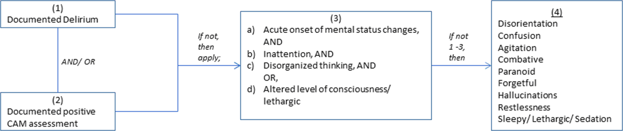
We conducted a retrospective cohort study of patients aged 65 years or older admitted to a tertiary care hospital between October 1, 2012 and September 31, 2013. Using Stata's (StataCorp., College Station, TX) sample command,[3] we included a subset of randomly selected inpatients who received more than 1 dose of oral APs (determined using the electronic medication administration summary). We excluded patients admitted under observation status or to the psychiatric service, those who were on APs prior to admission, and those who only received prochloperazine for nausea. Using prior literature to identify terms frequently used to describe delirium (Figure 1), we created an algorithm and a chart abstraction form (see Supporting Information, Appendix 1, in the online version of this article).[4] We tested these instruments in a preliminary chart review involving 30 patients. Disagreements were discussed with coauthors and resolved through consensus, resulting in some algorithm changes (eg, excluding a large number of patients who received only 1 dose of haloperidol postoperatively, because we hypothesized that this use could be a prophylactic measure).[5] Two investigators extracted the remaining charts independently. We used descriptive statistics and performed cross‐tabulations on the selected variables.

RESULTS
Of 12,817 geriatric hospitalizations during the study period, 1120 (9%) were treated with antipsychotics. We randomly selected 300 of these for extraction: 54% were male, and 67% were admitted to the medical service (Table 1). The inpatient mortality rate was 10% (30/300). The most frequent indication for AP use was delirium (83%, 249/300). Only 35% of delirious patients received a formal assessment with the Confusion Assessment Method (CAM). The most commonly used atypical antipsychotic was quetiapine (86%); 55% received more than 1 antipsychotic medication during hospitalization, and 48% (143/297) of patients were continued on APs at discharge (excluding 3 patients transferred to other acute care hospitals).
| Variable | N (%), Total=300 |
|---|---|
| |
| Gender | |
| Male | 161 (54) |
| Female | 139 (46) |
| Inpatient mortality rate | 30 (10) |
| Services | |
| Medicine | 202 (67) |
| Surgery | 98 (33) |
| Indication for APs use | |
| Delirium | 249 (83) |
| Hallucinations | 19 (6) |
| Anxiety | 20 (7) |
| Other | 38 (13) |
| Atypical APs | |
| Quetiapine | 257 (86) |
| Olanzapine | 29 (10) |
| Risperidone | 26 (9) |
| Typical APs | |
| Haloperidol | 166 (55) |
| Thorazine | 4 (1) |
| Use of CAM | 79 (32)a |
| Physical restraints | 89 (30) |
| Documented or suspected dementia | 134 (45) |
| Geriatrics consults | 120 (40) |
| Psychiatric consults | 29 (10) |
| ECG | |
| Prior to APs administration | 265 (88) |
| After APs administration | 157 (52) |
| QTc prolongation >500 ms | |
| Prior to APs administration | 41 (15)b |
| After APs administration | 39 (25)c |
| Admitted from SNF | 36 (12) |
| Discharge destination | |
| Home | 68 (23) |
| SNFs, short and long‐term rehabilitations | 199 (66) |
| Transfer to other acute care hospitals | 3 (1) |
| Continuation of APs at discharge | 143 (48)d |
Approximately 45% (134/300) had documented or suspected dementia, and 30% (89/300) were physically restrained during the hospital stay. Consultations with geriatrics were obtained in 40% (120/300) of the cases and with psychiatry in 10% (29/300) of the cases. Neurology is rarely consulted for delirium in our institution; thus, we did not collect data on those referrals. Electrocardiography (ECG) (recommended for patients at high cardiac risk[6]) was performed in 88% (265/300) of patients prior to AP administration and 52% (157/300) after. The corrected QT interval exceeded 500 ms in 15% (41/265) of patients prior to AP administration and 25% (39/157) after. Although few patients (12%) were admitted from nursing facilities, 66% (199/300) were eventually discharged to skilled nursing facilities (SNFs) or rehabilitation facilities; most of these patients (117/199, 59%) received AP treatment, compared to 38% of patients discharged to home (26/68).
DISCUSSION
In a cohort of hospitalized elders, we found that 9% were treated with APs. Most received APs for perceived delirium; in‐hospital ECG monitoring was suboptimal. Half of the patients started on APs remained on them at discharge; those discharged to SNFs were more likely to receive ongoing AP treatment.
Our study is limited by its retrospective, single‐center design, a lack of inter‐rater reliability measurement (although our training process was designed to standardize extraction methods), and the infrequent use of formal CAM assessment. Additionally, we were unable to determine how frequently APs were initiated in the intensive care unit. Any retrospective study is limited by the difficulty of distinguishing delirium from the behavioral and psychiatric symptoms of dementia, but we identified delirium using standard terms described in previous literature.
Our study also has a number of important implications. Because of a reported association between the use of APs and risk of death in the postacute setting,[7] national provider organizations have called for a reduction in AP initiation in hospitalized elders.[2] However, this study indicates that APs continue to be prescribed for delirium, which may be attributed to the lack of behavioral modification options in most hospitals, such as acute care for elders (ACE) units and hospital elder life programs (HELP).[8, 9] Our findings suggest that this problem would be further amplified in hospitals that lack access to geriatrics expertise.
Without alternative behavioral options, patients are at risk for prolonged delirium, which is associated with significant suffering and subsequent risk of further cognitive impairment and death.[10] Although evidence for the efficacy of APs in the treatment of delirium is limited and inconclusive, no better pharmacologic options exist. Hospitals that wish to reduce use of APs should therefore consider investing in environmental interventions (eg, ACE units, HELP), which lower the incidence of delirium and could, in turn, decrease the prescription and continuation of antipsychotics.[8, 9]
Acknowledgements
The authors acknowledge Mihaela Stefan, MD, FACP, for her comments on an earlier draft of this manuscript.
Disclosures: Drs. Lagu and Loh had full access to all of the data in the study. They take responsibility for the integrity of the data and the accuracy of the analysis. Drs. Loh, Brennan, Lindenauer, and Lagu conceived of the study. Drs. Loh and Ramdass acquired the data. Ms. Garb analyzed and interpreted the data. Dr. Loh drafted the manuscript. Drs Brennan, Lindenauer, and Lagu, and Ms. Garb critically reviewed the manuscript for important intellectual content. Dr. Lagu is supported by the National Heart, Lung, and Blood Institute of the National Institutes of Health under award number K01HL114745. The authors report no conflicts of interest.
- , , , , , . Delirium in elderly patients and the risk of postdischarge mortality, institutionalization, and dementia: a meta‐analysis. JAMA. 2010;304(4):443–451.
- . Off‐label use of antipsychotics for dementia patients discouraged. The Hospitalist. November 2012\http://www.the‐hospitalist.org/details/article/2785121/Off‐Label_Use_of_Antipsychotics_for_Dementia_Patients_Discouraged.html. Accessed June 29, 2014.
- STATA/MP [computer program]. Version 13.1 for Windows. College Station, TX: StataCorp; 2013.
- , , , , , . Association between sedating medications and delirium in older inpatients. J Am Geriatr Soc. 2013;61(6):923–930.
- , , , et al. Haloperidol prophylaxis decreases delirium incidence in elderly patients after noncardiac surgery: a randomized controlled trial. Crit Care Med. 2012;40(3):731–739.
- , , . QTc prolongation with antipsychotics: is routine ECG monitoring recommended? J Psychiatr Pract. 2014;20(3):196–206.
- , , , , . Risk of death associated with the use of conventional versus atypical antipsychotic drugs among elderly patients. CMAJ. 2007;176(5):627–632.
- , , , et al. Effectiveness of acute geriatric unit care using acute care for elders components: a systematic review and meta‐analysis. J Am Geriatr Soc. 2012;60(12):2237–2245.
- , , , , . The Hospital Elder Life Program: a model of care to prevent cognitive and functional decline in older hospitalized patients. J Am Geriatr Soc. 2000;48(12):1697–1706.
- , , , . Persistent delirium in older hospital patients: a systematic review of frequency and prognosis. Age Ageing. 2009;38(1):19–26.
Antipsychotic (AP) medications are often used in the hospitalized geriatric population for the treatment of delirium.[1] Because of adverse events associated with APs, efforts have been made to reduce their use in hospitalized elders,[2] but it is not clear if these recommendations have been widely adopted. We studied the use of APs in a cohort of hospitalized elders to better understand why APs are started and how often they are continued on discharge.
METHODS
We conducted a retrospective cohort study of patients aged 65 years or older admitted to a tertiary care hospital between October 1, 2012 and September 31, 2013. Using Stata's (StataCorp., College Station, TX) sample command,[3] we included a subset of randomly selected inpatients who received more than 1 dose of oral APs (determined using the electronic medication administration summary). We excluded patients admitted under observation status or to the psychiatric service, those who were on APs prior to admission, and those who only received prochloperazine for nausea. Using prior literature to identify terms frequently used to describe delirium (Figure 1), we created an algorithm and a chart abstraction form (see Supporting Information, Appendix 1, in the online version of this article).[4] We tested these instruments in a preliminary chart review involving 30 patients. Disagreements were discussed with coauthors and resolved through consensus, resulting in some algorithm changes (eg, excluding a large number of patients who received only 1 dose of haloperidol postoperatively, because we hypothesized that this use could be a prophylactic measure).[5] Two investigators extracted the remaining charts independently. We used descriptive statistics and performed cross‐tabulations on the selected variables.
RESULTS
Of 12,817 geriatric hospitalizations during the study period, 1120 (9%) were treated with antipsychotics. We randomly selected 300 of these for extraction: 54% were male, and 67% were admitted to the medical service (Table 1). The inpatient mortality rate was 10% (30/300). The most frequent indication for AP use was delirium (83%, 249/300). Only 35% of delirious patients received a formal assessment with the Confusion Assessment Method (CAM). The most commonly used atypical antipsychotic was quetiapine (86%); 55% received more than 1 antipsychotic medication during hospitalization, and 48% (143/297) of patients were continued on APs at discharge (excluding 3 patients transferred to other acute care hospitals).
| Variable | N (%), Total=300 |
|---|---|
| |
| Gender | |
| Male | 161 (54) |
| Female | 139 (46) |
| Inpatient mortality rate | 30 (10) |
| Services | |
| Medicine | 202 (67) |
| Surgery | 98 (33) |
| Indication for APs use | |
| Delirium | 249 (83) |
| Hallucinations | 19 (6) |
| Anxiety | 20 (7) |
| Other | 38 (13) |
| Atypical APs | |
| Quetiapine | 257 (86) |
| Olanzapine | 29 (10) |
| Risperidone | 26 (9) |
| Typical APs | |
| Haloperidol | 166 (55) |
| Thorazine | 4 (1) |
| Use of CAM | 79 (32)a |
| Physical restraints | 89 (30) |
| Documented or suspected dementia | 134 (45) |
| Geriatrics consults | 120 (40) |
| Psychiatric consults | 29 (10) |
| ECG | |
| Prior to APs administration | 265 (88) |
| After APs administration | 157 (52) |
| QTc prolongation >500 ms | |
| Prior to APs administration | 41 (15)b |
| After APs administration | 39 (25)c |
| Admitted from SNF | 36 (12) |
| Discharge destination | |
| Home | 68 (23) |
| SNFs, short and long‐term rehabilitations | 199 (66) |
| Transfer to other acute care hospitals | 3 (1) |
| Continuation of APs at discharge | 143 (48)d |
Approximately 45% (134/300) had documented or suspected dementia, and 30% (89/300) were physically restrained during the hospital stay. Consultations with geriatrics were obtained in 40% (120/300) of the cases and with psychiatry in 10% (29/300) of the cases. Neurology is rarely consulted for delirium in our institution; thus, we did not collect data on those referrals. Electrocardiography (ECG) (recommended for patients at high cardiac risk[6]) was performed in 88% (265/300) of patients prior to AP administration and 52% (157/300) after. The corrected QT interval exceeded 500 ms in 15% (41/265) of patients prior to AP administration and 25% (39/157) after. Although few patients (12%) were admitted from nursing facilities, 66% (199/300) were eventually discharged to skilled nursing facilities (SNFs) or rehabilitation facilities; most of these patients (117/199, 59%) received AP treatment, compared to 38% of patients discharged to home (26/68).
DISCUSSION
In a cohort of hospitalized elders, we found that 9% were treated with APs. Most received APs for perceived delirium; in‐hospital ECG monitoring was suboptimal. Half of the patients started on APs remained on them at discharge; those discharged to SNFs were more likely to receive ongoing AP treatment.
Our study is limited by its retrospective, single‐center design, a lack of inter‐rater reliability measurement (although our training process was designed to standardize extraction methods), and the infrequent use of formal CAM assessment. Additionally, we were unable to determine how frequently APs were initiated in the intensive care unit. Any retrospective study is limited by the difficulty of distinguishing delirium from the behavioral and psychiatric symptoms of dementia, but we identified delirium using standard terms described in previous literature.
Our study also has a number of important implications. Because of a reported association between the use of APs and risk of death in the postacute setting,[7] national provider organizations have called for a reduction in AP initiation in hospitalized elders.[2] However, this study indicates that APs continue to be prescribed for delirium, which may be attributed to the lack of behavioral modification options in most hospitals, such as acute care for elders (ACE) units and hospital elder life programs (HELP).[8, 9] Our findings suggest that this problem would be further amplified in hospitals that lack access to geriatrics expertise.
Without alternative behavioral options, patients are at risk for prolonged delirium, which is associated with significant suffering and subsequent risk of further cognitive impairment and death.[10] Although evidence for the efficacy of APs in the treatment of delirium is limited and inconclusive, no better pharmacologic options exist. Hospitals that wish to reduce use of APs should therefore consider investing in environmental interventions (eg, ACE units, HELP), which lower the incidence of delirium and could, in turn, decrease the prescription and continuation of antipsychotics.[8, 9]
Acknowledgements
The authors acknowledge Mihaela Stefan, MD, FACP, for her comments on an earlier draft of this manuscript.
Disclosures: Drs. Lagu and Loh had full access to all of the data in the study. They take responsibility for the integrity of the data and the accuracy of the analysis. Drs. Loh, Brennan, Lindenauer, and Lagu conceived of the study. Drs. Loh and Ramdass acquired the data. Ms. Garb analyzed and interpreted the data. Dr. Loh drafted the manuscript. Drs Brennan, Lindenauer, and Lagu, and Ms. Garb critically reviewed the manuscript for important intellectual content. Dr. Lagu is supported by the National Heart, Lung, and Blood Institute of the National Institutes of Health under award number K01HL114745. The authors report no conflicts of interest.
- , , , , , . Delirium in elderly patients and the risk of postdischarge mortality, institutionalization, and dementia: a meta‐analysis. JAMA. 2010;304(4):443–451.
- . Off‐label use of antipsychotics for dementia patients discouraged. The Hospitalist. November 2012\http://www.the‐hospitalist.org/details/article/2785121/Off‐Label_Use_of_Antipsychotics_for_Dementia_Patients_Discouraged.html. Accessed June 29, 2014.
- STATA/MP [computer program]. Version 13.1 for Windows. College Station, TX: StataCorp; 2013.
- , , , , , . Association between sedating medications and delirium in older inpatients. J Am Geriatr Soc. 2013;61(6):923–930.
- , , , et al. Haloperidol prophylaxis decreases delirium incidence in elderly patients after noncardiac surgery: a randomized controlled trial. Crit Care Med. 2012;40(3):731–739.
- , , . QTc prolongation with antipsychotics: is routine ECG monitoring recommended? J Psychiatr Pract. 2014;20(3):196–206.
- , , , , . Risk of death associated with the use of conventional versus atypical antipsychotic drugs among elderly patients. CMAJ. 2007;176(5):627–632.
- , , , et al. Effectiveness of acute geriatric unit care using acute care for elders components: a systematic review and meta‐analysis. J Am Geriatr Soc. 2012;60(12):2237–2245.
- , , , , . The Hospital Elder Life Program: a model of care to prevent cognitive and functional decline in older hospitalized patients. J Am Geriatr Soc. 2000;48(12):1697–1706.
- , , , . Persistent delirium in older hospital patients: a systematic review of frequency and prognosis. Age Ageing. 2009;38(1):19–26.
Shrink Rap News: Helping patients access outpatient psychiatric care
How often does someone ask you for help in getting an appointment with a psychiatrist? With the “double expansion” in access to mental health care – because of the Mental Health Parity and Addiction Equity Act, and the Affordable Care Act (ACA) – many more people are seeking help for mental health and substance use problems. But without more practitioners being produced, the waiting lists are getting longer.
A recent study found that 40% (146 of 360) of psychiatrists listed on insurance plans in three states could not be reached. Of the 214 who were reachable, 43% were unavailable, either because the psychiatrists were not accepting new patients or because they did not treat adult outpatients (e.g., inpatient only). Of the 123 psychiatrists left, the callers were able to schedule appointments with 93 (76%) of them. These results are similar to a 2007 study, where 44% of mental health professionals from seven health plans were unreachable.
Problems with access to psychiatric care is not a new problem. Mental health carve-outs are used by managed care organizations (MCOs) to manage the mental health benefits, but create inefficiency in claims management and clinical care coordination. While this model seems to be losing popularity, these managed behavioral health organizations (MBHOs) often do not integrate well with the MCO, may not share the same standards for network adequacy, and often have different provider directories from those of the MCO, making it more complex for patients to access providers.
While the Parity Act has somewhat improved the problem of rate disparities, the historically low rates paid to psychiatrists by MBHOs have led to high levels of insurance nonparticipation, as much as 50%. Compounding this problem is the fact that plan members often find it hard to access those practitioners who do participate with their insurance.
The Maryland Psychological Association conducted in 2007 with Open Minds a “secret shopper” survey of more than 900 behavioral health providers from seven different online carrier directories in Maryland. Their goal was to assess the extent to which problems with access to care were related to inadequate insurance provider directories.
They found that 44% of the listed providers were unreachable based on the contact information in the directories, and only a small proportion was actually able to see the new “patient” in an appropriate time frame. The average wait time for a psychiatrist was 20 days, for a psychologist was 15 days, and for other mental health professionals was 11 days.
The study, published (Psychiatric Serv. 2014 Oct. 15 [doi:10.1176/appi.ps.2014000051]), illustrates how hard it can be to get an appointment with a psychiatrist, regardless of who the payer is. The authors called 360 psychiatrists who were listed in the Blue Cross Blue Shield (BCBS) online directories in Boston, Chicago, and Houston. Callers posed as people with depressive symptoms seeking initial appointments. In each city, a third of the psychiatrists received a call from a caller saying they were either Medicare, BCBS, or self-pay. Voicemails were left when possible that included the type of payer. Callers followed up for a second round of calls if they did not hear back.
“Obtaining an outpatient appointment with a psychiatrist was difficult in the three cities we surveyed, and the appointments given were an average of 1 month away,” the authors wrote. Between 20%-27% of the phone numbers in the insurance company directories were wrong numbers, which led, instead, to places like McDonald’s and retail stores. In fact, they were able to reach only one-third of the psychiatrists.
The ACA requires Health Insurance Exchanges to have network provider directories that distinguish those providers who are currently accepting new outpatients. Unfortunately, the act left it up to the states to define network adequacy. For example, in Maryland, the definition of what constitutes an adequate network is left to each qualified health plan to define.

The problem with the health plans’ provider directories is that they are inaccurate, making it look like they have more available providers than they actually do. The plans have no incentive to expose the inadequacy of their network directories, while the regulators lack the staff to police them sufficiently. Meanwhile, provider groups benefit from being included on more lists, and plan members fail to effectively complain to the plans, employers, and regulators.
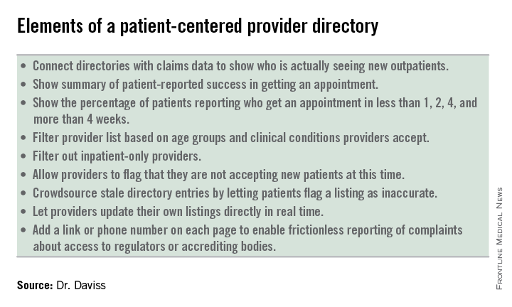
What the industry needs is to move away from these misleading, payer-centered provider directories and move toward transparent, patient-centered provider directories that include elements of crowdsourcing, provider control, and frictionless reporting. Making these changes will give consumers, employers, and regulators more information on provider availability, wait times, and true access to care.
Dr. Daviss is chair of the department of psychiatry at the University of Maryland’s Baltimore Washington Medical Center, chair of the APA Committee on Electronic Health Records, cochair of the CCHIT Behavioral Health Work Group, and coauthor of Shrink Rap: Three Psychiatrists Explain Their Work, published by Johns Hopkins University Press. He is found on Twitter @HITshrink, at [email protected], and on the Shrink Rap blog.
How often does someone ask you for help in getting an appointment with a psychiatrist? With the “double expansion” in access to mental health care – because of the Mental Health Parity and Addiction Equity Act, and the Affordable Care Act (ACA) – many more people are seeking help for mental health and substance use problems. But without more practitioners being produced, the waiting lists are getting longer.
A recent study found that 40% (146 of 360) of psychiatrists listed on insurance plans in three states could not be reached. Of the 214 who were reachable, 43% were unavailable, either because the psychiatrists were not accepting new patients or because they did not treat adult outpatients (e.g., inpatient only). Of the 123 psychiatrists left, the callers were able to schedule appointments with 93 (76%) of them. These results are similar to a 2007 study, where 44% of mental health professionals from seven health plans were unreachable.
Problems with access to psychiatric care is not a new problem. Mental health carve-outs are used by managed care organizations (MCOs) to manage the mental health benefits, but create inefficiency in claims management and clinical care coordination. While this model seems to be losing popularity, these managed behavioral health organizations (MBHOs) often do not integrate well with the MCO, may not share the same standards for network adequacy, and often have different provider directories from those of the MCO, making it more complex for patients to access providers.
While the Parity Act has somewhat improved the problem of rate disparities, the historically low rates paid to psychiatrists by MBHOs have led to high levels of insurance nonparticipation, as much as 50%. Compounding this problem is the fact that plan members often find it hard to access those practitioners who do participate with their insurance.
The Maryland Psychological Association conducted in 2007 with Open Minds a “secret shopper” survey of more than 900 behavioral health providers from seven different online carrier directories in Maryland. Their goal was to assess the extent to which problems with access to care were related to inadequate insurance provider directories.
They found that 44% of the listed providers were unreachable based on the contact information in the directories, and only a small proportion was actually able to see the new “patient” in an appropriate time frame. The average wait time for a psychiatrist was 20 days, for a psychologist was 15 days, and for other mental health professionals was 11 days.
The study, published (Psychiatric Serv. 2014 Oct. 15 [doi:10.1176/appi.ps.2014000051]), illustrates how hard it can be to get an appointment with a psychiatrist, regardless of who the payer is. The authors called 360 psychiatrists who were listed in the Blue Cross Blue Shield (BCBS) online directories in Boston, Chicago, and Houston. Callers posed as people with depressive symptoms seeking initial appointments. In each city, a third of the psychiatrists received a call from a caller saying they were either Medicare, BCBS, or self-pay. Voicemails were left when possible that included the type of payer. Callers followed up for a second round of calls if they did not hear back.
“Obtaining an outpatient appointment with a psychiatrist was difficult in the three cities we surveyed, and the appointments given were an average of 1 month away,” the authors wrote. Between 20%-27% of the phone numbers in the insurance company directories were wrong numbers, which led, instead, to places like McDonald’s and retail stores. In fact, they were able to reach only one-third of the psychiatrists.
The ACA requires Health Insurance Exchanges to have network provider directories that distinguish those providers who are currently accepting new outpatients. Unfortunately, the act left it up to the states to define network adequacy. For example, in Maryland, the definition of what constitutes an adequate network is left to each qualified health plan to define.
The problem with the health plans’ provider directories is that they are inaccurate, making it look like they have more available providers than they actually do. The plans have no incentive to expose the inadequacy of their network directories, while the regulators lack the staff to police them sufficiently. Meanwhile, provider groups benefit from being included on more lists, and plan members fail to effectively complain to the plans, employers, and regulators.
What the industry needs is to move away from these misleading, payer-centered provider directories and move toward transparent, patient-centered provider directories that include elements of crowdsourcing, provider control, and frictionless reporting. Making these changes will give consumers, employers, and regulators more information on provider availability, wait times, and true access to care.
Dr. Daviss is chair of the department of psychiatry at the University of Maryland’s Baltimore Washington Medical Center, chair of the APA Committee on Electronic Health Records, cochair of the CCHIT Behavioral Health Work Group, and coauthor of Shrink Rap: Three Psychiatrists Explain Their Work, published by Johns Hopkins University Press. He is found on Twitter @HITshrink, at [email protected], and on the Shrink Rap blog.

Delivering on the promise of cancer biomarkers in the clinic
Click on the PDF icon at the top of this introduction to read the full article.
Click on the PDF icon at the top of this introduction to read the full article.
Click on the PDF icon at the top of this introduction to read the full article.
JCSO 2014;12:381-388
Novel anticoagulants increase GI bleeds in elderly
PHILADELPHIA – Dabigatran and rivaroxaban, which are rapidly replacing warfarin for anticoagulation in patients with atrial fibrillation, appear to be associated with a greater risk of GI bleeding than the long-time standard in patients over the age of 65 years, according to an analysis presented at the American College of Gastroenterology (ACG).
The pattern of an increased GI bleeding risk with the newer oral anticoagulants relative to warfarin was consistent in older patients whether used for AF or for out-of-labeling indications, which were assessed separately, reported Dr. Neena S. Abraham, professor of medicine at Mayo Clinic, Scottsdale, Ariz.

“In all four cases, once patients were over the age of 65, the risk of GI bleeding increased significantly on the novel agents when compared to warfarin,” Dr. Abraham reported.
The propensity matches, based on such characteristics as GI bleeding risk factors, age, race, and concomitant medications, were drawn from 92,816 patients in a large database starting a new prescription of dabigatran, rivaroxaban, or warfarin over a recent 3-year period. With this matching, 9,860 new users of dabigatran and 20,619 new users of rivaroxaban were compared with equal number of new users of warfarin.
In the full dataset, before age stratification, the risk of total GI bleeding, particularly lower GI bleeding, appeared to be nonsignificantly lower for both dabigatran and rivaroxaban, relative to warfarin in patients with AF. In patients without AF receiving these drugs, the risk remained slightly lower on rivaroxaban but appeared to be slightly increased on dabigatran.
However, the hypothesis that bleeding risk was greater for newer agents in older patients was substantiated when the data were stratified by age. In the analysis, risk of bleeding started climbing more steeply with the newer agents as patients aged than with warfarin, with differences observed at about age 65 years.
By age 75, the hazard ratio for a GI bleed in dabigatran patients relative to warfarin in AF patients was 2.4 (95% confidence interval 1.5-3.8). In the rivaroxaban group, the HR for this risk at age 75 in AF patients was 4.0 (95% CI 2.1-7.4). In non-AF patients, the rates of GI bleeding were also significantly increased at a similar magnitude.
These data were anticipated by the initial trials that found dabigatran and rivaroxaban noninferior to warfarin for the prevention of stroke and systemic embolism, according to Dr. Abraham. Although there was heterogeneity in reported risk differences, she reported that GI bleeding was as much as 25% higher on the newer anticoagulants when compared to warfarin in older patients. These new data substantiate those findings.
“Our study is the first to evaluate GI safety in novel oral anticoagulants compared to warfarin in a real-world, multiage setting,” Dr. Abraham noted. She said these data “facilitate risk-benefit considerations” of these drugs. She further noted that the data on non-AF patients may be particularly pertinent “because this is the fastest-growing emerging market” for agents in this class.
Asked for a comment, Dr. Brian E. Lacy, chief of gastroenterology and hepatology at Dartmouth-Hitchcock Medical Center, Lebanon, N.H., said that the information about relative risk is potentially important, but he was particularly impressed by the substantial use of novel anticoagulants in non-AF patients.
“This tells me that we as gastroenterologists need to be asking more questions about exposure to anticoagulants,” Dr. Lacy said in an interview. “These data suggest that these newer drugs are being used frequently outside of labeling. We need to be aware of these changing patterns of use when trying to assess the risk of our patients for GI bleeds.”
Dr. Abraham had no financial disclosures.
PHILADELPHIA – Dabigatran and rivaroxaban, which are rapidly replacing warfarin for anticoagulation in patients with atrial fibrillation, appear to be associated with a greater risk of GI bleeding than the long-time standard in patients over the age of 65 years, according to an analysis presented at the American College of Gastroenterology (ACG).
The pattern of an increased GI bleeding risk with the newer oral anticoagulants relative to warfarin was consistent in older patients whether used for AF or for out-of-labeling indications, which were assessed separately, reported Dr. Neena S. Abraham, professor of medicine at Mayo Clinic, Scottsdale, Ariz.
“In all four cases, once patients were over the age of 65, the risk of GI bleeding increased significantly on the novel agents when compared to warfarin,” Dr. Abraham reported.
The propensity matches, based on such characteristics as GI bleeding risk factors, age, race, and concomitant medications, were drawn from 92,816 patients in a large database starting a new prescription of dabigatran, rivaroxaban, or warfarin over a recent 3-year period. With this matching, 9,860 new users of dabigatran and 20,619 new users of rivaroxaban were compared with equal number of new users of warfarin.
In the full dataset, before age stratification, the risk of total GI bleeding, particularly lower GI bleeding, appeared to be nonsignificantly lower for both dabigatran and rivaroxaban, relative to warfarin in patients with AF. In patients without AF receiving these drugs, the risk remained slightly lower on rivaroxaban but appeared to be slightly increased on dabigatran.
However, the hypothesis that bleeding risk was greater for newer agents in older patients was substantiated when the data were stratified by age. In the analysis, risk of bleeding started climbing more steeply with the newer agents as patients aged than with warfarin, with differences observed at about age 65 years.
By age 75, the hazard ratio for a GI bleed in dabigatran patients relative to warfarin in AF patients was 2.4 (95% confidence interval 1.5-3.8). In the rivaroxaban group, the HR for this risk at age 75 in AF patients was 4.0 (95% CI 2.1-7.4). In non-AF patients, the rates of GI bleeding were also significantly increased at a similar magnitude.
These data were anticipated by the initial trials that found dabigatran and rivaroxaban noninferior to warfarin for the prevention of stroke and systemic embolism, according to Dr. Abraham. Although there was heterogeneity in reported risk differences, she reported that GI bleeding was as much as 25% higher on the newer anticoagulants when compared to warfarin in older patients. These new data substantiate those findings.
“Our study is the first to evaluate GI safety in novel oral anticoagulants compared to warfarin in a real-world, multiage setting,” Dr. Abraham noted. She said these data “facilitate risk-benefit considerations” of these drugs. She further noted that the data on non-AF patients may be particularly pertinent “because this is the fastest-growing emerging market” for agents in this class.
Asked for a comment, Dr. Brian E. Lacy, chief of gastroenterology and hepatology at Dartmouth-Hitchcock Medical Center, Lebanon, N.H., said that the information about relative risk is potentially important, but he was particularly impressed by the substantial use of novel anticoagulants in non-AF patients.
“This tells me that we as gastroenterologists need to be asking more questions about exposure to anticoagulants,” Dr. Lacy said in an interview. “These data suggest that these newer drugs are being used frequently outside of labeling. We need to be aware of these changing patterns of use when trying to assess the risk of our patients for GI bleeds.”
Dr. Abraham had no financial disclosures.
AT THE ANNUAL SCIENTIFIC MEETING AND POSTGRADUATE COURSE OF THE AMERICAN COLLEGE OF GASTROENTEROLOGY
Key clinical point: The novel oral anticoagulants dabigatran and rivaroxaban were associated with an increased risk of GI bleeding in elderly patients, compared with warfarin, in data drawn from a real-world setting.
Major finding: When compared with warfarin in atrial fibrillation, the risk of upper and lower GI bleeding is nearly 2.5 times greater on dabigatran and 3 times greater on rivaroxaban at age 75 years.
Data source: A retrospective, propensity-matched study.
Disclosures: Dr. Abraham had no financial disclosures.

Drugs, Pregnancy, and Lactation: Herbs
Herbs are commonly consumed by pregnant and breast-feeding women, possibly because they believe that “natural products” are safer than drugs. However, even though some have been available for hundreds or thousands of years, little is known about their effects on the embryo, fetus, newborn, or nursing infant. Moreover, as unregulated products, the concentration, contents, and presence of contaminants cannot be easily determined. Detailed reviews of the 22 most commonly used herbs discussed here can be found in “Drugs in Pregnancy and Lactation,” Briggs GG, Freeman RK, 10th ed., Philadelphia: Wolters Kluwer Health, 2014).
In the following discussions, dose, one of the two key factors that determine the risk of developmental toxicity (abnormal growth, structural anomalies, functional and/or neurobehavioral deficits, or death), is rarely reported. In addition, all herbs contain multiple chemical compounds, few of which have been studied during pregnancy or lactation. Thus, with few exceptions, a woman who takes an herb in pregnancy should be informed that the risk to her developing baby is unknown.

Six herbs are considered contraindicated in pregnancy: arnica, black seed /kalanji, blue cohosh, feverfew, salvia divinorum, and valerian.
• Arnica. The dried flowers, and sometimes the roots and rhizomes, are the parts of this perennial plant that are used topically for their anti-inflammatory and analgesic effects. There is no clinical evidence to support this use. Occasional topical use probably represents a low risk, but absorption may occur when it is applied to broken skin. The Food and Drug Administration has classified arnica as an unsafe herb and, when used orally, it is considered a poison. It is a uterine stimulant and an abortifacient. Nevertheless, in homeopathic formulations, it has been promoted for use before and during labor for internal and external bruising of the mother and newborn. In Italy, it is one of the top 10 herbs taken by women (Pharmacoepidemiol. Drug Saf. 2006;15:354-9).
• Black seed/kalanji. This herb has been used for thousands of year as a medicine, food, or spice. Because of this, it is unlikely that it causes teratogenesis. Nevertheless, its use to stimulate menstruation and its potential contraceptive properties suggest that it is contraindicated in pregnancy.

• Blue cohosh. Some of the components of this herb have been shown to be teratogenic and toxic in various animal species, so it should be avoided in the first trimester. The herb has uterine stimulant properties that are used by nurse-midwives to stimulate labor. Blue cohosh was the most frequently used herbal preparation for this purpose. However, some sources believe that the potential fetal and newborn toxicity may outweigh any medical benefit (“PDR for Herbal Medicine,” 2nd ed., Montvale, N.J.: Medical Economics, 2000:109-10; “The Review of Natural Products,” St. Louis, MO: Facts and Comparisons, 2000).
• Feverfew. This herb has been used for labor, menstrual disorders, potential miscarriage, and morning sickness; as an abortifacient; and for several other indications. Because of its antipyretic properties, it has been known as “medieval aspirin.” The doses used for these indications have not been quantified. Because of its emmenagogic (capable of provoking menstruation) activity, the herb should not be used in pregnancy.
• Salvia divinorum. This herb has hallucinogenic effects and is used in certain regions of Mexico for healing and divinatory rituals. It is also thought to have antidiarrheal properties. The herb is either smoked or chewed, or its juices are ingested. When taken orally, systemic effects are dependent upon absorption across the oral mucosa as the active ingredient is destroyed in the GI tract. Persistent psychosis has been observed in people who smoked the herb, so it is contraindicated in pregnancy.
• Valerian. A large number of preparations containing valerian are available. It has been used as a sedative and hypnotic for anxiety, restlessness, and sleep disturbances, as well as several other pharmacologic claims. Because of the risk of cytotoxicity in the fetus and hepatotoxicity in the mother, the herb should be avoided during gestation.

For the remaining 16 herbs, small, infrequent doses probably cause no harm to the mother, embryo, fetus, or newborn. Nevertheless, as noted below, some of these herbs are best avoided during pregnancy.
• Chamomile. Excessive use of this herb should be avoided because it is thought to have uterine stimulant, emmenagogic, and abortifacient properties. Although controversial, some nurse-midwives prescribe chamomile teas for the treatment of morning sickness. Because the plant sources of the herb contain coumarin compounds, ingesting chamomile by pregnant women with coagulation disorders is a concern. However, the herb has been used for thousands of years, so the risk of harm, at least from occasional use, must be very rare.
• Echinacea. This herb is used topically to enhance wound healing and systemically as an immunostimulant. An IV formulation is used in Germany but is not available in the United States. It also has been recommended to assist in the prevention or treatment of viral upper respiratory tract infections. Its use in pregnancy is limited to one small study.
• Evening primrose oil. The oil contains two essential fatty acids: cis-linoleic and gamma-linolenic acid. In a national survey of nurse-midwives, it was the most frequently used herbal preparation for the induction of labor. No adverse effects have been reported in the fetus or newborn from this use. The doses used varied widely and included both oral and vaginal routes of administration. In addition, the oil has been used for rheumatoid arthritis and diabetic neuropathy, but there are no reports of these uses in pregnancy.
• Garlic. Garlic has been used for food flavoring since ancient times and appears to be safe during pregnancy. Some components cross the placenta, as shown by garlic odor in the amniotic fluid and on the newborn’s breath. Very high doses have the potential to induce menstruation or uterine contractions, but apparently these effects have not been reported.
• Ginger. No reports of ginger-induced developmental toxicity have been located. Ginger has been used as antiemetic for nausea and vomiting of pregnancy.

• Ginseng. The root is the most important part of this plant that is found throughout the world and has been used in medicine for more than 2,000 years. The herb has been promoted for multiple pharmacologic effects, including adaptogenic, CNS, cardiovascular, endocrine, ergogenic, antineoplastic, and immunomodulatory effects.
Hypertension and hypoglycemia have been reported in nonpregnant patients, but not in the limited human pregnancy data. A brief 1991 study compared 88 women who took the herb during pregnancy with 88 controls. No differences between the groups were found with regard to the mode of delivery, birth weight, low birth weight (< 2,500 ), preterm delivery (< 37 weeks), low Apgar score (< 7), stillbirths, neonatal deaths, or maternal complications (Asia Oceania J. Obstet. Gynaecol. 1991;17:379-80).
• Ginkgo biloba. The limited animal reproduction data suggest low risk, but there is no reported human pregnancy experience. Nevertheless, it is an ancient herbal preparation that is commonly used for organic brain syndrome, circulatory disorders, asthma, vertigo, and tinnitus. Because of its widespread use, it is doubtful that a major teratogenic effect would have escaped notice, but more subtle or low-incidence toxic effects may not have been detected.
• Kudzu. No human or animal data regarding pregnancy have been located. The herb has been used for more than 2,500 years for the treatment of alcohol hangover, drunkenness, alcoholism, muscle pain, and measles. Many of its chemical constituents can be found in foods. Nevertheless, high, frequent doses should be avoided.
• Nutmeg. This is a commonly used spice but, as with any herb, high doses can produce toxicity. The toxicity is caused by a chemical in the seeds, myristicin, which has anticholinergic properties. A woman at 30 weeks’ gestation misread a recipe and used a whole grated nutmeg rather than 1/8 teaspoon when making cookies. When she ate a cookie, she experienced sinus tachycardia, hypertension, and a sensation of impending doom. The fetus had tachycardia, and atropine-like poisoning was diagnosed. After about 12 hours, both mother and fetus made an uneventful recovery and a healthy infant was born at term.

• Passion flower. The name of this herb may refer to about 400 species of the genus Passiflora. It is available in both oral and topical forms and is used for nervousness, neuralgia, insomnia, pain, asthma, seizures, burns, hemorrhoids, and menopausal complaints. As with many herbs, it contains a large number of chemicals, none of which have undergone reproductive testing. No reports describing the use of this herb in human pregnancy have been located. However, because it has uterine stimulant properties, the oral formulation is best avoided in pregnancy.
• Peppermint. This popular flavoring appears to be harmless for the mother and developing baby when low, recommended doses are ingested. Peppermint oil is available in numerous topical and oral formulations. High oral doses, however, can cause significant toxicity, including death. During pregnancy, ingestion of more than the recommended doses is unsafe because of possible emmenagogic and abortifacient properties.
• Pumpkin seed. This herb, when used as a food, appears to be harmless for the mother and embryo-fetus, but no reports describing its use in pregnancy have been located. High doses, such as those used in traditional medicine or in eating disorders, should be avoided because of the potential for toxic effects from the many chemicals these seeds contain.
• Raspberry leaf. Raspberry leaf tea is commonly used by pregnant women. Nurse-midwives often prescribe the tea to treat nausea and vomiting and as a uterine tonic to shorten labor. A double-blind, randomized, placebo-controlled study evaluated the effect of raspberry leaf tablets (2 tablets/day) on pregnancy outcomes. Compared with controls, no differences were found for length of labor or stages of labor, mode of delivery, admission to the neonatal intensive care unit, Apgar score, and birth weight (J. Midwifery Womens Health 2001;46:51-9).
• Safflower. Safflower oil is commonly used in cooking and has been given for its laxative action. There are no reports describing the use of the herb in pregnancy. It is doubtful if such use would have any adverse effect on a pregnancy. Although abortifacient and emmenagogic effects have been suggested, there is no evidence supporting these effects when used as a food.
• St. John’s wort. No toxicity in pregnant humans has been reported. The use of the herb is widespread and dates back thousands of years. Thus, it is doubtful that the herb is a major teratogen or causes other elements of developmental toxicity. The herb has been used for the management of anxiety, depression, insomnia, inflammation, and gastritis. It is also used as a diuretic and, topically, for the treatment of hemorrhoids and enhanced wound healing.

• Yohimbine. The use of this herb in human pregnancies has not been reported. It has been used as an aphrodisiac and for weight loss, sexual dysfunction, and the treatment of orthostatic hypotension. Although it has no Food and Drug Administration–sanctioned indications, it is also available by prescription for male erectile dysfunction. Due to the lack of data regarding pregnancy, the herb is best avoided during pregnancy.
There are few data regarding the effects of the above herbs on a breast-feeding infant. Depending upon the herb, nursing infants will be exposed to many chemical compounds. For those herbs used as food, nursing is probably safe. The safety of the other herbs during lactation is unknown. However, toxicity has been reported in a 9-day-old term infant whose mother was taking arnica (Clin. Toxicol. 2009;47:726, abstract 120). The infant presented with lethargy, decreased milk intake, anemia, and jaundice but recovered with treatment. After the mother stopped the herb and resumed nursing, no further problems were noted in the infant.
Mr. Briggs is a pharmacist clinical specialist at the outpatient clinics of Memorial Care Center for Women at Miller Children’s Hospital in Long Beach, Calif.; clinical professor of pharmacy at the University of California, San Francisco; and adjunct professor of pharmacy at the University of Southern California, Los Angeles, and Washington State University, Spokane. He also is coauthor of “Drugs in Pregnancy and Lactation,” and coeditor of “Diseases, Complications, and Drug Therapy in Obstetrics.” He had no relevant financial disclosures. Contact him at [email protected].
Herbs are commonly consumed by pregnant and breast-feeding women, possibly because they believe that “natural products” are safer than drugs. However, even though some have been available for hundreds or thousands of years, little is known about their effects on the embryo, fetus, newborn, or nursing infant. Moreover, as unregulated products, the concentration, contents, and presence of contaminants cannot be easily determined. Detailed reviews of the 22 most commonly used herbs discussed here can be found in “Drugs in Pregnancy and Lactation,” Briggs GG, Freeman RK, 10th ed., Philadelphia: Wolters Kluwer Health, 2014).
In the following discussions, dose, one of the two key factors that determine the risk of developmental toxicity (abnormal growth, structural anomalies, functional and/or neurobehavioral deficits, or death), is rarely reported. In addition, all herbs contain multiple chemical compounds, few of which have been studied during pregnancy or lactation. Thus, with few exceptions, a woman who takes an herb in pregnancy should be informed that the risk to her developing baby is unknown.
Six herbs are considered contraindicated in pregnancy: arnica, black seed /kalanji, blue cohosh, feverfew, salvia divinorum, and valerian.
• Arnica. The dried flowers, and sometimes the roots and rhizomes, are the parts of this perennial plant that are used topically for their anti-inflammatory and analgesic effects. There is no clinical evidence to support this use. Occasional topical use probably represents a low risk, but absorption may occur when it is applied to broken skin. The Food and Drug Administration has classified arnica as an unsafe herb and, when used orally, it is considered a poison. It is a uterine stimulant and an abortifacient. Nevertheless, in homeopathic formulations, it has been promoted for use before and during labor for internal and external bruising of the mother and newborn. In Italy, it is one of the top 10 herbs taken by women (Pharmacoepidemiol. Drug Saf. 2006;15:354-9).
• Black seed/kalanji. This herb has been used for thousands of year as a medicine, food, or spice. Because of this, it is unlikely that it causes teratogenesis. Nevertheless, its use to stimulate menstruation and its potential contraceptive properties suggest that it is contraindicated in pregnancy.
• Blue cohosh. Some of the components of this herb have been shown to be teratogenic and toxic in various animal species, so it should be avoided in the first trimester. The herb has uterine stimulant properties that are used by nurse-midwives to stimulate labor. Blue cohosh was the most frequently used herbal preparation for this purpose. However, some sources believe that the potential fetal and newborn toxicity may outweigh any medical benefit (“PDR for Herbal Medicine,” 2nd ed., Montvale, N.J.: Medical Economics, 2000:109-10; “The Review of Natural Products,” St. Louis, MO: Facts and Comparisons, 2000).
• Feverfew. This herb has been used for labor, menstrual disorders, potential miscarriage, and morning sickness; as an abortifacient; and for several other indications. Because of its antipyretic properties, it has been known as “medieval aspirin.” The doses used for these indications have not been quantified. Because of its emmenagogic (capable of provoking menstruation) activity, the herb should not be used in pregnancy.
• Salvia divinorum. This herb has hallucinogenic effects and is used in certain regions of Mexico for healing and divinatory rituals. It is also thought to have antidiarrheal properties. The herb is either smoked or chewed, or its juices are ingested. When taken orally, systemic effects are dependent upon absorption across the oral mucosa as the active ingredient is destroyed in the GI tract. Persistent psychosis has been observed in people who smoked the herb, so it is contraindicated in pregnancy.
• Valerian. A large number of preparations containing valerian are available. It has been used as a sedative and hypnotic for anxiety, restlessness, and sleep disturbances, as well as several other pharmacologic claims. Because of the risk of cytotoxicity in the fetus and hepatotoxicity in the mother, the herb should be avoided during gestation.
For the remaining 16 herbs, small, infrequent doses probably cause no harm to the mother, embryo, fetus, or newborn. Nevertheless, as noted below, some of these herbs are best avoided during pregnancy.
• Chamomile. Excessive use of this herb should be avoided because it is thought to have uterine stimulant, emmenagogic, and abortifacient properties. Although controversial, some nurse-midwives prescribe chamomile teas for the treatment of morning sickness. Because the plant sources of the herb contain coumarin compounds, ingesting chamomile by pregnant women with coagulation disorders is a concern. However, the herb has been used for thousands of years, so the risk of harm, at least from occasional use, must be very rare.
• Echinacea. This herb is used topically to enhance wound healing and systemically as an immunostimulant. An IV formulation is used in Germany but is not available in the United States. It also has been recommended to assist in the prevention or treatment of viral upper respiratory tract infections. Its use in pregnancy is limited to one small study.
• Evening primrose oil. The oil contains two essential fatty acids: cis-linoleic and gamma-linolenic acid. In a national survey of nurse-midwives, it was the most frequently used herbal preparation for the induction of labor. No adverse effects have been reported in the fetus or newborn from this use. The doses used varied widely and included both oral and vaginal routes of administration. In addition, the oil has been used for rheumatoid arthritis and diabetic neuropathy, but there are no reports of these uses in pregnancy.
• Garlic. Garlic has been used for food flavoring since ancient times and appears to be safe during pregnancy. Some components cross the placenta, as shown by garlic odor in the amniotic fluid and on the newborn’s breath. Very high doses have the potential to induce menstruation or uterine contractions, but apparently these effects have not been reported.
• Ginger. No reports of ginger-induced developmental toxicity have been located. Ginger has been used as antiemetic for nausea and vomiting of pregnancy.
• Ginseng. The root is the most important part of this plant that is found throughout the world and has been used in medicine for more than 2,000 years. The herb has been promoted for multiple pharmacologic effects, including adaptogenic, CNS, cardiovascular, endocrine, ergogenic, antineoplastic, and immunomodulatory effects.
Hypertension and hypoglycemia have been reported in nonpregnant patients, but not in the limited human pregnancy data. A brief 1991 study compared 88 women who took the herb during pregnancy with 88 controls. No differences between the groups were found with regard to the mode of delivery, birth weight, low birth weight (< 2,500 ), preterm delivery (< 37 weeks), low Apgar score (< 7), stillbirths, neonatal deaths, or maternal complications (Asia Oceania J. Obstet. Gynaecol. 1991;17:379-80).
• Ginkgo biloba. The limited animal reproduction data suggest low risk, but there is no reported human pregnancy experience. Nevertheless, it is an ancient herbal preparation that is commonly used for organic brain syndrome, circulatory disorders, asthma, vertigo, and tinnitus. Because of its widespread use, it is doubtful that a major teratogenic effect would have escaped notice, but more subtle or low-incidence toxic effects may not have been detected.
• Kudzu. No human or animal data regarding pregnancy have been located. The herb has been used for more than 2,500 years for the treatment of alcohol hangover, drunkenness, alcoholism, muscle pain, and measles. Many of its chemical constituents can be found in foods. Nevertheless, high, frequent doses should be avoided.
• Nutmeg. This is a commonly used spice but, as with any herb, high doses can produce toxicity. The toxicity is caused by a chemical in the seeds, myristicin, which has anticholinergic properties. A woman at 30 weeks’ gestation misread a recipe and used a whole grated nutmeg rather than 1/8 teaspoon when making cookies. When she ate a cookie, she experienced sinus tachycardia, hypertension, and a sensation of impending doom. The fetus had tachycardia, and atropine-like poisoning was diagnosed. After about 12 hours, both mother and fetus made an uneventful recovery and a healthy infant was born at term.
• Passion flower. The name of this herb may refer to about 400 species of the genus Passiflora. It is available in both oral and topical forms and is used for nervousness, neuralgia, insomnia, pain, asthma, seizures, burns, hemorrhoids, and menopausal complaints. As with many herbs, it contains a large number of chemicals, none of which have undergone reproductive testing. No reports describing the use of this herb in human pregnancy have been located. However, because it has uterine stimulant properties, the oral formulation is best avoided in pregnancy.
• Peppermint. This popular flavoring appears to be harmless for the mother and developing baby when low, recommended doses are ingested. Peppermint oil is available in numerous topical and oral formulations. High oral doses, however, can cause significant toxicity, including death. During pregnancy, ingestion of more than the recommended doses is unsafe because of possible emmenagogic and abortifacient properties.
• Pumpkin seed. This herb, when used as a food, appears to be harmless for the mother and embryo-fetus, but no reports describing its use in pregnancy have been located. High doses, such as those used in traditional medicine or in eating disorders, should be avoided because of the potential for toxic effects from the many chemicals these seeds contain.
• Raspberry leaf. Raspberry leaf tea is commonly used by pregnant women. Nurse-midwives often prescribe the tea to treat nausea and vomiting and as a uterine tonic to shorten labor. A double-blind, randomized, placebo-controlled study evaluated the effect of raspberry leaf tablets (2 tablets/day) on pregnancy outcomes. Compared with controls, no differences were found for length of labor or stages of labor, mode of delivery, admission to the neonatal intensive care unit, Apgar score, and birth weight (J. Midwifery Womens Health 2001;46:51-9).
• Safflower. Safflower oil is commonly used in cooking and has been given for its laxative action. There are no reports describing the use of the herb in pregnancy. It is doubtful if such use would have any adverse effect on a pregnancy. Although abortifacient and emmenagogic effects have been suggested, there is no evidence supporting these effects when used as a food.
• St. John’s wort. No toxicity in pregnant humans has been reported. The use of the herb is widespread and dates back thousands of years. Thus, it is doubtful that the herb is a major teratogen or causes other elements of developmental toxicity. The herb has been used for the management of anxiety, depression, insomnia, inflammation, and gastritis. It is also used as a diuretic and, topically, for the treatment of hemorrhoids and enhanced wound healing.
• Yohimbine. The use of this herb in human pregnancies has not been reported. It has been used as an aphrodisiac and for weight loss, sexual dysfunction, and the treatment of orthostatic hypotension. Although it has no Food and Drug Administration–sanctioned indications, it is also available by prescription for male erectile dysfunction. Due to the lack of data regarding pregnancy, the herb is best avoided during pregnancy.
There are few data regarding the effects of the above herbs on a breast-feeding infant. Depending upon the herb, nursing infants will be exposed to many chemical compounds. For those herbs used as food, nursing is probably safe. The safety of the other herbs during lactation is unknown. However, toxicity has been reported in a 9-day-old term infant whose mother was taking arnica (Clin. Toxicol. 2009;47:726, abstract 120). The infant presented with lethargy, decreased milk intake, anemia, and jaundice but recovered with treatment. After the mother stopped the herb and resumed nursing, no further problems were noted in the infant.
Mr. Briggs is a pharmacist clinical specialist at the outpatient clinics of Memorial Care Center for Women at Miller Children’s Hospital in Long Beach, Calif.; clinical professor of pharmacy at the University of California, San Francisco; and adjunct professor of pharmacy at the University of Southern California, Los Angeles, and Washington State University, Spokane. He also is coauthor of “Drugs in Pregnancy and Lactation,” and coeditor of “Diseases, Complications, and Drug Therapy in Obstetrics.” He had no relevant financial disclosures. Contact him at [email protected].

Extended telepsychiatry outperformed primary care follow-up for ADHD
SAN DIEGO– Six telepsychiatry sessions cut symptoms by at least half for 46% of children with attention-deficit/hyperactivity disorder, compared with 13.6% of those who received one telepsychiatry session plus follow-up care by primary care providers, according to a randomized clinical trial.
The extended telepsychiatry intervention consistently outperformed primary care for attention-deficit/hyperactivity disorder (ADHD), including in subgroups of children with ADHD alone, comorbid anxiety disorders, oppositional defiant disorder, or both, said Dr. Carol M. Rockhill of Seattle Children’s Hospital. “We do think the results of this study justify a more extended consultation model. A single visit is not enough for a child to be stabilized,” she said at the annual meeting of the American Academy of Child and Adolescent Psychiatry.
Attention-deficit hyperactivity is one of the most common disorders of childhood, and children in rural areas often lack access to appropriate care. The Children’s ADHD Telemental Health Treatment Study (CATTS) included 223 children with ADHD and their primary caregivers at seven underserved sites in Washington and Oregon. The primary outcome was a 50% reduction in ADHD symptoms, “an ambitious goal,” Dr. Rockhill said. Average age of the patients was 9 years, and they did not have serious comorbid diagnoses such as autism, bipolar disorder, or conduct disorder, she said. In all, 18% of children had a diagnosis of ADHD alone, while the rest also had at least one comorbid psychiatric disorder, she said.
For the study, the intervention arm received a total of six telepsychiatry sessions provided by interactive televideo with psychiatrists at Seattle Children’s Hospital. All sites had high bandwidth connectivity, and equipment that could pan, tilt, and zoom, Dr. Rockhill said. “It was nice to really be able to see the parents and caregivers well,” she added. Children received medication management, and caregivers were trained on managing behaviors of ADHD.
The control arm received a single telepsychiatry session and follow-up care by primary care providers. Parents in both groups used the Vanderbilt Assessment Scale to rate children’s behavior throughout the study, Dr. Rockhill said.
The researchers also compared telepsychiatry strategies to those from the Texas Children’s Medication Algorithm Project, which provides consensus guidelines for children with ADHD alone or with comorbid anxiety, depression, tics, or aggression, Dr. Rockhill said. Telepsychiatrists most often used the first algorithm, suggesting that they focused on ADHD symptoms even if children had comorbidities, she reported. In more than 98% of cases, telepsychiatrists chose the same initial algorithm as did study reviewers. Psychiatrists most commonly prescribed methylphenidate alone, followed by amphetamine alone. Among 574 telepsychiatry sessions, there were 29 protocol violations, which most often consisted of changing the algorithm order or combining medications, she added.
Children with comorbidities were more likely to have their medications changed, but this did not translate to greater clinical improvement, Dr. Rockhill said. “The kids who did achieve a 50% reduction in symptoms and had two comorbidities had an average of 2.4 medication changes, compared with 3.2 changes for children who did not meet the treatment target,” she said. “Comorbidity makes achievement of a 50% improvement in symptoms more challenging, and is associated with more complex medication strategies, including more changes in medication and more use of polypharmacy.”
In fact, the rate of polypharmacy more than tripled during the course of the study, Dr. Rockhill said. At the beginning of the trial, 13% of children had been prescribed more than one medication, compared with 41.5% at the end. In most cases, polypharmacy consisted of prescribing one stimulant and one nonstimulant.
The National Institute of Mental Health funded the study. Dr. Rockhill did not report financial conflicts of interest.
Key clinical point: Six telepsychiatry sessions are far superior to a single session plus primary care follow-up in children with attention-deficit/hyperactivity disorder.
Major finding: The six-session intervention led to at least a 50% symptom reduction in 46% of children, compared with 13.6% of the control group (P < .001).
Data source: Randomized controlled trial of 223 children with ADHD and their primary caregivers in rural Washington and Oregon.
Disclosures: The National Institute of Mental Health funded the trial. Dr. Rockhill reported no conflicts of interest.