User login
Ketogenic diet, with variations, can help adult epilepsy
HOUSTON – The ketogenic diet is usually thought of as a solution of near-last resort for pediatric epilepsy, but some adolescents and adults with epilepsy can also benefit from a very low carbohydrate diet.
There are also limited data to suggest that more palatable adaptations of the diet may provide benefit while improving adherence, said Mackenzie C. Cervenka, MD, speaking at the annual meeting of the American Epilepsy Society.
“Ketogenic diets are a reasonable option for older adolescents and adults with drug-resistant epilepsy that’s not amenable to surgical intervention,” said Dr. Cervenka, director of the Adult Epilepsy Diet Center at Johns Hopkins University, Baltimore.

The antiepileptic benefit of a diet that induces ketogenesis, forcing the brain to utilize ketone bodies rather than glucose for energy, has been known since the 1920s, with benefit seen for adolescents and adults in studies completed in the 1930s. These diets mimic a starvation state, but provide enough calories through fat or protein to maintain weight. Calories in the traditional ketogenic diet, Dr. Cervenka said, are about 90% fat. Food for patients on this diet should be weighed on a gram scale, and those preparing meals should aim for a ratio of 3 to 4 grams of fat for each gram of carbohydrate and protein combined. A modified version uses a 1:1 or 2:1 ratio, a more appealing configuration for some patients.
Weighing each bite of food is cumbersome, and palatability can be a major problem as well, contributing to adherence problems with such a high-fat diet. However, for patients who are so ill that they are tube-fed, commercially available ketogenic formulas are available. Necessary supplementation on a traditional ketogenic diet includes calcium, vitamin D, multivitamins, and oral citrates to prevent kidney stone formation, she said.
However, ketogenic diets are known to be anti-inflammatory: Animal models have shown less inflammation, pain, and fever when rats are fed a ketogenic diet. Also, proinflammatory cytokines and chemokines are reduced on a ketogenic diet in a rodent model of Parkinson’s disease and multiple sclerosis. Particularly for patients with autoimmune encephalopathies, the ketogenic diet has been shown to be of benefit.
One option with promising, but limited, results is a low-carbohydrate diet rich in medium-chain triglycerides. Medium chain triglyceride (MCT) oil is available in a commercial preparation derived from coconut or palm kernel oil. On this diet, 30%-60% of calories should come from MCTs, which is usually sufficient to induce ketosis. However, gastrointestinal side effects such as bloating and diarrhea can be pronounced, especially if the diet is begun abruptly. It’s best to ramp up slowly with MCTs, so this diet would not be appropriate for the patient who needs quick improvement in seizure control, Dr. Cervenka said.
A modified Atkins diet provides 15-20 grams of net carbohydrates daily, after dietary fiber is subtracted. Using this strategy, adolescents and adults don’t have to weigh foods. Rather, food tables are used to track carbohydrates and fiber, and ketosis is assessed by measuring urine ketones on a test strip. The goal, Dr. Cervenka said, is to achieve moderate to large ketosis (40-160 ng/mL urine ketones).
Finally, low glycemic index treatment (LGIT) is an option worth considering. This diet takes advantage of certain carbohydrate-rich foods that do not raise blood sugar quickly, such as fiber-rich vegetables or legumes with some fat content. Patients on the LGIT diet can have from 40 to 60 grams of carbohydrate daily, and the diet has been used with some success in drug-resistant childhood epilepsy as well as Angelman syndrome, she said.
Though sample sizes are small and efficacy may be modest, Dr. Cervenka said, “the effect is quick.” Finding less-restrictive modifications of these diets may help patients stay on the diet over the long term, increasing real-world effectiveness.
But long-term adherence to a ketogenic or other high-fat, low-residue diet comes with a host of unknowns about cardiovascular, metabolic, and renal health; ongoing study of these patients may yield answers about whether theoretical concerns are borne out, and whether the risk is worth it in terms of seizure benefit.
Dr. Cervenka reported receiving grant support from Nutricia North America, Vitaflo, and the BrightFocus Foundation. She has also received an honorarium for speaking from LivaNova.
[email protected]
On Twitter @karioakes
HOUSTON – The ketogenic diet is usually thought of as a solution of near-last resort for pediatric epilepsy, but some adolescents and adults with epilepsy can also benefit from a very low carbohydrate diet.
There are also limited data to suggest that more palatable adaptations of the diet may provide benefit while improving adherence, said Mackenzie C. Cervenka, MD, speaking at the annual meeting of the American Epilepsy Society.
“Ketogenic diets are a reasonable option for older adolescents and adults with drug-resistant epilepsy that’s not amenable to surgical intervention,” said Dr. Cervenka, director of the Adult Epilepsy Diet Center at Johns Hopkins University, Baltimore.
The antiepileptic benefit of a diet that induces ketogenesis, forcing the brain to utilize ketone bodies rather than glucose for energy, has been known since the 1920s, with benefit seen for adolescents and adults in studies completed in the 1930s. These diets mimic a starvation state, but provide enough calories through fat or protein to maintain weight. Calories in the traditional ketogenic diet, Dr. Cervenka said, are about 90% fat. Food for patients on this diet should be weighed on a gram scale, and those preparing meals should aim for a ratio of 3 to 4 grams of fat for each gram of carbohydrate and protein combined. A modified version uses a 1:1 or 2:1 ratio, a more appealing configuration for some patients.
Weighing each bite of food is cumbersome, and palatability can be a major problem as well, contributing to adherence problems with such a high-fat diet. However, for patients who are so ill that they are tube-fed, commercially available ketogenic formulas are available. Necessary supplementation on a traditional ketogenic diet includes calcium, vitamin D, multivitamins, and oral citrates to prevent kidney stone formation, she said.
However, ketogenic diets are known to be anti-inflammatory: Animal models have shown less inflammation, pain, and fever when rats are fed a ketogenic diet. Also, proinflammatory cytokines and chemokines are reduced on a ketogenic diet in a rodent model of Parkinson’s disease and multiple sclerosis. Particularly for patients with autoimmune encephalopathies, the ketogenic diet has been shown to be of benefit.
One option with promising, but limited, results is a low-carbohydrate diet rich in medium-chain triglycerides. Medium chain triglyceride (MCT) oil is available in a commercial preparation derived from coconut or palm kernel oil. On this diet, 30%-60% of calories should come from MCTs, which is usually sufficient to induce ketosis. However, gastrointestinal side effects such as bloating and diarrhea can be pronounced, especially if the diet is begun abruptly. It’s best to ramp up slowly with MCTs, so this diet would not be appropriate for the patient who needs quick improvement in seizure control, Dr. Cervenka said.
A modified Atkins diet provides 15-20 grams of net carbohydrates daily, after dietary fiber is subtracted. Using this strategy, adolescents and adults don’t have to weigh foods. Rather, food tables are used to track carbohydrates and fiber, and ketosis is assessed by measuring urine ketones on a test strip. The goal, Dr. Cervenka said, is to achieve moderate to large ketosis (40-160 ng/mL urine ketones).
Finally, low glycemic index treatment (LGIT) is an option worth considering. This diet takes advantage of certain carbohydrate-rich foods that do not raise blood sugar quickly, such as fiber-rich vegetables or legumes with some fat content. Patients on the LGIT diet can have from 40 to 60 grams of carbohydrate daily, and the diet has been used with some success in drug-resistant childhood epilepsy as well as Angelman syndrome, she said.
Though sample sizes are small and efficacy may be modest, Dr. Cervenka said, “the effect is quick.” Finding less-restrictive modifications of these diets may help patients stay on the diet over the long term, increasing real-world effectiveness.
But long-term adherence to a ketogenic or other high-fat, low-residue diet comes with a host of unknowns about cardiovascular, metabolic, and renal health; ongoing study of these patients may yield answers about whether theoretical concerns are borne out, and whether the risk is worth it in terms of seizure benefit.
Dr. Cervenka reported receiving grant support from Nutricia North America, Vitaflo, and the BrightFocus Foundation. She has also received an honorarium for speaking from LivaNova.
[email protected]
On Twitter @karioakes
EXPERT ANALYSIS FROM AES 2016
Dementia prevalence increased in heart failure patients
NEW ORLEANS – Elderly patients with heart failure had a significantly increased prevalence of both dementia and mild cognitive impairment, compared with similar people without heart failure, in an analysis of data collected from more than 6,000 U.S. residents enrolled in a long-term observational study.
Patients diagnosed with either heart failure with reduced ejection fraction or heart failure with preserved ejection fraction had an 89% increased prevalence of dementia and a 41% increased prevalence of mild cognitive impairment (MCI), compared with people from the same cohort who did not develop heart failure, in an analysis that adjusted for several demographic and clinical variables, Lucy S. Witt, MD, reported at the American Heart Association scientific sessions. She speculated that the link between heart failure and dementia and MCI might result from impaired cerebral perfusion in heart failure patients or from effects from heart failure medications.
The analysis used data collected for the Atherosclerosis Risk in Communities (ARIC) study, which began in 1987 and enrolled a randomly selected representative cohort of nearly 16,000 women and men aged 45-64 years old who resided in any of four U.S. communities. She specifically focused on the data collected from 6,431 of the participants who returned for a fifth follow-up examination during 2011-2013, including 5,490 people without heart failure, whose average age was 76 years, and 941 participants with heart failure, whose average age was 78 years.
Dementia prevalence at the fifth follow-up visit occurred at an adjusted rate of 5.6% among those without heart failure and 7.0% in those with heart failure. The examinations also found MCI in an adjusted 21.5% of those without heart failure and in 26.2% of those with heart failure, Dr. Witt reported. Adjustments included age, sex, location, education, hypertension, diabetes, depression, alcohol and tobacco use, cerebral vascular disease, marital status, and several other factors.
The relative risk for having dementia among the heart failure patients was roughly similar, regardless of whether ARIC participants with heart failure had a reduced or preserved left ventricular ejection fraction, she said.
ARIC is funded by the National Heart, Lung, and Blood Institute. Dr. Witt had no disclosures.
[email protected]
On Twitter @mitchelzoler
AT THE AHA SCIENTIFIC SESSIONS
Key clinical point:
Major finding: Patients with heart failure had an adjusted 86% increased rate of dementia, compared with similar people without heart failure.
Data source: Analysis of 6,431 ARIC participants who returned for a fifth follow-up examination during 2011-2013.
Disclosures: The Atherosclerosis Risk in Commmunities (ARIC) study is funded by the National Heart, Lung, and Blood Institute. Dr. Witt had no disclosures.
Blood pressure rise follows halting CPAP
Continuous positive airway pressure (CPAP) therapy for obstructive sleep apnea (OSA) has a significant beneficial effect on blood pressure, according to an analysis of participants in three randomized controlled trials.
Previous meta-analyses suggested that CPAP treatment led to an average of improvement of 2-3 mm Hg, but the estimates relied on heterogeneous trials that often had low levels of CPAP adherence, and those factors might have led to an underestimation of the treatment effect. The new analysis showed that halting CPAP increases blood pressure between 5.0 and 9.0 mm Hg, compared with patients who continued using CPAP (Chest. 2016;150[6]:1202-10).
To get around the problem of adherence, researchers led by Malcolm Kohler, MD, at University Hospital of Zürich analyzed the results of three previous studies looking at the effects of CPAP withdrawal. The analysis included 153 OSA patients on CPAP therapy, who had been randomized to continue therapy or to withdraw from therapy for 2 weeks. Eighty-seven of these patients discontinued CPAP, and the remaining 66 patients continued the therapy. Blood pressure was measured at home and in hospital.
On average, those who discontinued CPAP had an increase in office systolic blood pressure of 5.4 mm Hg (95% confidence interval, 1.8-8.9 mm Hg; P = .003) and an increase in home systolic blood pressure of 9.0 mm Hg (95% CI, 5.7-12.3 mm Hg; P less than .001), compared with patients who continued CPAP. The effects of stopping CPAP, instead of continuing the therapy, on office diastolic blood pressure and home diastolic pressure were increases of 5.0 mm Hg (95% CI, 2.7-7.3 mm Hg; P less than .001) and 7.8 mm Hg (95% CI, 5.6-10.0 mm Hg; P less than .001), respectively.
Patients who discontinued CPAP also experienced a significant increase in apnea-hypopnea index, from 2.8/h to 33.2/h, while those who continued using CPAP, on average, experienced only a 0.3/h increase in apnea-hypopnea index from baseline.
“One clinical implication is that if you do not need to stop CPAP for obstructive sleep apnea, do not stop it. This study also suggests the importance of monitoring your blood pressure in a home setting, under usual conditions,” summed up Robert Kloner, MD, PhD, director of the Huntington Medical Research Institutes Cardiovascular Research Lab, Pasadena, Calif., who was not involved in the study.
Previous studies of CPAP, such as the SAVE study published in the New England Journal of Medicine in September (N Engl J Med. 2016;375:919-31), often find little or no connection between CPAP therapy and cardiovascular outcomes. That is probably because of inadequate adherence to CPAP therapy. “That’s always been the bane of sleep apnea studies,” said Krishna M. Sundar, MD, FCCP, who also did not participate in the study.
The current work got around the problem by looking at patients who had already established use of CPAP. “This is a very good study,” said Dr. Sundar, who is the medical director of the Sleep-Wake Center at the University of Utah, Salt Lake City.
The study was funded by the Swiss National Science Foundation and the University of Zürich. The analysis’ authors and the outside experts quoted in this story reported no financial disclosures.
FROM CHEST
Key clinical point: Interrupting CPAP therapy leads to a rise in blood pressure.
Major finding: Stopping CPAP was associated with 5.0-9.0 mm Hg blood pressure increase.
Data source: Analysis of 153 patients with moderate to severe OSA, who had participated in three randomized controlled trials.
Disclosures: The study was funded by the Swiss National Science Foundation and the University of Zürich. The authors of the analysis and the outside experts quoted in this story reported no financial disclosures.
Dr. Sundar and Dr. Kloner reported having no financial disclosures.
Genetics dictate interferon-alfa diarrhea risk
Genetics are probably to blame for why some patients have significant intestinal side effects from Roferon-A (interferon alfa-2a, recombinant – Roche) while others do not, according to a new French investigation reported in issue of Cellular and Molecular Gastroenterology and Hepatology.
They cultured intestinal wall samples taken from 20 colon cancer patients when they had surgery; none of them had been exposed to chemotherapy, radiation, or immunosuppressives. The team bathed the cells in IFN-alfa 2a and other biochemicals to see how they reacted. It was bench work, but the findings could eventually be useful if the team identifies the genetic risk factors for Roferon intestinal side effects and finds better options for patients at risk. Roferon is widely used for blood cancer, melanoma, viral hepatitis, and renal and hepatocellular carcinomas.
“IFN-alfa 2a elicited a rapid (24 hours) disruption of surface and crypt colonic epithelial cells via apoptosis that was variable in intensity among the 20 individuals studied. This apoptotic effect was dependent on the initiation of an IFN-gamma response … expressed in T cell–positive lamina propria cells,” the investigators said.
“IFN-alfa impairs human intestinal mucosa homeostasis by eliciting epithelial barrier disruption via apoptosis … The IFN-alfa–elicited impairment of intestinal mucosa homeostasis is heterogeneous among individuals,” Dr. Jarry and her associates wrote.
“This ex vivo finding parallels clinical observations of the interpatient variability of Roferon therapy side effects. … It has been reported that approximately 60% of patients with chronic hepatitis or cancer treated with Roferon have intestinal disorders, especially diarrhea,” they said.
The authors had no conflicts of interest. The work was funded by the University of Nantes.
Many studies have implicated cytokines in various human GI-tract disorders, but there is still limited information in the literature about their exact role in the maintenance and disturbance of tissue homeostasis and the molecular mechanisms involved.


Interestingly, IFN-alfa did not induce apoptosis in all human colonic fragments analyzed, showing that their culture model accounts for variability among individuals, recapitulating the heterogeneous response of cancer patients to IFN-alfa-based treatment who present with intestinal dysfunction as a side effect. The use of such models of human primary cell culture helps us develop a better understanding of how cytokines affect the GI mucosa, potentially leading to alternative targets for treatments. They also could be used to determine the individual patient differences underlying diverse responses to cytokines and drugs, which is particularly important to the advance of precision/personalized medicine.
Jason C. Mills, MD, PhD, and Luciana H. Osaki, PhD, are with the division of gastroenterology, departments of medicine, pathology and immunology, and developmental biology, Washington University, St. Louis. They have no conflicts of interest.
Many studies have implicated cytokines in various human GI-tract disorders, but there is still limited information in the literature about their exact role in the maintenance and disturbance of tissue homeostasis and the molecular mechanisms involved.
Interestingly, IFN-alfa did not induce apoptosis in all human colonic fragments analyzed, showing that their culture model accounts for variability among individuals, recapitulating the heterogeneous response of cancer patients to IFN-alfa-based treatment who present with intestinal dysfunction as a side effect. The use of such models of human primary cell culture helps us develop a better understanding of how cytokines affect the GI mucosa, potentially leading to alternative targets for treatments. They also could be used to determine the individual patient differences underlying diverse responses to cytokines and drugs, which is particularly important to the advance of precision/personalized medicine.
Jason C. Mills, MD, PhD, and Luciana H. Osaki, PhD, are with the division of gastroenterology, departments of medicine, pathology and immunology, and developmental biology, Washington University, St. Louis. They have no conflicts of interest.
Genetics are probably to blame for why some patients have significant intestinal side effects from Roferon-A (interferon alfa-2a, recombinant – Roche) while others do not, according to a new French investigation reported in issue of Cellular and Molecular Gastroenterology and Hepatology.
They cultured intestinal wall samples taken from 20 colon cancer patients when they had surgery; none of them had been exposed to chemotherapy, radiation, or immunosuppressives. The team bathed the cells in IFN-alfa 2a and other biochemicals to see how they reacted. It was bench work, but the findings could eventually be useful if the team identifies the genetic risk factors for Roferon intestinal side effects and finds better options for patients at risk. Roferon is widely used for blood cancer, melanoma, viral hepatitis, and renal and hepatocellular carcinomas.
“IFN-alfa 2a elicited a rapid (24 hours) disruption of surface and crypt colonic epithelial cells via apoptosis that was variable in intensity among the 20 individuals studied. This apoptotic effect was dependent on the initiation of an IFN-gamma response … expressed in T cell–positive lamina propria cells,” the investigators said.
“IFN-alfa impairs human intestinal mucosa homeostasis by eliciting epithelial barrier disruption via apoptosis … The IFN-alfa–elicited impairment of intestinal mucosa homeostasis is heterogeneous among individuals,” Dr. Jarry and her associates wrote.
“This ex vivo finding parallels clinical observations of the interpatient variability of Roferon therapy side effects. … It has been reported that approximately 60% of patients with chronic hepatitis or cancer treated with Roferon have intestinal disorders, especially diarrhea,” they said.
The authors had no conflicts of interest. The work was funded by the University of Nantes.
FROM CELLULAR AND MOLECULAR GASTROENTEROLOGY AND HEPATOLOGY
VIDEO: Point-of-care microsensor prototype beats conventional coagulopathy tests
SAN DIEGO – Using less than a drop of blood, a portable microsensor provided a comprehensive coagulation profile in minutes and perfectly distinguished various coagulopathies from normal blood samples – handily beating both activated partial thromboplastin time (aPTT) and prothrombin time (PT).
Dubbed ClotChip, the disposable device detects coagulation factors and platelet activity by using a technique called dielectric spectroscopy, Evi X. Stavrou, MD, of Case Western Reserve University, Cleveland, said in a video interview at the annual meeting of the American Society of Hematology. It points the way for comprehensive, rapid, point-of-care assessment of critically ill or severely injured patients and those who need ongoing monitoring to evaluate response to anticoagulant therapy, she added.
By plotting rates of true positives (patients with coagulopathies) against rates of true negatives (controls), the researchers obtained areas under the receiver operating curves of 100% for ClotChip, 78% for aPTT, and 57% for PT. In other words, ClotChip correctly identified all cases and controls in this small patient cohort, which neither aPTT or PT did.
Dr. Stavrou and her coinvestigators had no relevant financial disclosures.
The video associated with this article is no longer available on this site. Please view all of our videos on the MDedge YouTube channel
AT ASH 2016
Brentuximab vedotin beat methotrexate, bexarotene in cutaneous T-cell lymphoma
SAN DIEGO – For patients with CD30 expressing cutaneous T-cell lymphoma, antibody-drug conjugate therapy with brentuximab vedotin significantly outperformed two standard regimens in the phase III ALCANZA trial.
After a median of 17.5 months of follow-up, 56% of patients receiving brentuximab vedotin had an objective response lasting at least 4 months, versus 13% of patients treated with physician’s choice of methotrexate or bexarotene (P less than .0001), Youn H. Kim, MD, said during an oral presentation at the annual meeting of the American Society of Hematology.
As in past studies, brentuximab vedotin caused high rates of peripheral neuropathy, but more than 80% of cases improved or resolved over time, she said.
This is the first reported phase III trial to convincingly show that a new systemic agent outperformed standard therapies for cutaneous T-cell lymphoma (CTCL), which tend to have inadequate and short-lived efficacy, stated Dr. Kim, of Stanford (Calif.) University. Brentuximab vedotin not only met the primary endpoint, but all other predefined endpoints, including progression-free survival and a quality-of-life measure, she said.
“These compelling results have potential practice-changing implications,” she concluded.
Brentuximab vedotin (Adcetris) targets CD30, which is expressed in skin lesions of about half of patients with CTCL. A protease-cleavable linker attaches an anti-CD30 monoclonal antibody to monomethyl auristatin E, which disrupts microtubules when released into CD30-positive tumor cells (Blood. 2013;122:367). The agent showed clinical activity against CTCL in two previous phase II trials of CTCL.
Accordingly, the international, open-label phase III ALCANZA study enrolled 128 treatment-experienced patients with CD30-expressing mycosis fungoides or primary cutaneous anaplastic large cell lymphoma. Patients were randomly assigned to receive brentuximab vedotin (1.8 mg/kg once every 3 weeks) or physician’s choice of either methotrexate (5 to 50 mg once weekly) or bexarotene (300 mg/m2 once daily) for up to 16 3-week cycles, or until disease progression or unacceptable toxicity. Methotrexate or bexarotene were designated “physician’s choice” because they are used worldwide for treating CTCL, according to Dr. Kim.
To capture both the rate and duration of response, researchers defined objective response lasting at least 4 months as the primary endpoint. Brentuximab vedotin more than quadrupled the likelihood of this outcome when compared with the standard CTCL regimens, a trend that spanned key demographic and clinical subgroups, Dr. Kim said.
“All endpoints were highly [statistically] significant,” she further reported. For example, the objective response rate with brentuximab vedotin was 67%, versus 20% for methotrexate or bexarotene. Respective rates of complete response were 16% and 2%, and median durations of progression-free survival were 17 and 4 months, translating to a 73% lower risk of progression or death with brentuximab vedotin (95% confidence interval, 57%-83%). Patients who received brentuximab vedotin also reported about a three-fold greater improvement on the Skindex-29 symptom domain, compared with the physician’s choice group (–29 vs. –9 points; P less than .0001).
The safety profile of brentuximab vedotin resembled that seen in previous studies, Dr. Kim said. Most notably, 67% of patients developed peripheral neuropathy, and 9% developed grade 3 peripheral neuropathy. This usually improved or resolved over about the next 22 months. Diarrhea, fatigue, and vomiting affected about a third of patients on brentuximab vedotin, and about one in four stopped treatment because of adverse events, compared with 8% of the physician’s choice arm. Rates of serious adverse events were 41% and 47%, respectively. One brentuximab vedotin recipient died of multiple organ dysfunction syndrome that investigators attributed to treatment-associated necrosis of peripheral tumors. They identified no other treatment-related deaths.
Seattle Genetics and Takeda funded the trial. Dr. Kim disclosed ties to Takeda and Seattle Genetics, as well as several other pharmaceutical companies.
AT ASH 2016
Key clinical point: Brentuximab vedotin met all its endpoints but often caused peripheral neuropathy in a phase III trial of patients with CD30 expressing cutaneous T-cell lymphoma.
Major finding: After a median of 17.5 months of follow-up, 56% of patients receiving brentuximab vedotin had an objective response lasting at least 4 months, versus 13% of those receiving physician’s choice of methotrexate or bexarotene (P less than .0001).
Data source: A multicenter, open-label phase III trial of 128 patients with CD30-expressing mycosis fungoides or primary cutaneous anaplastic large cell lymphoma.
Disclosures: Seattle Genetics and Takeda funded the trial. Dr. Kim disclosed ties to Seattle Genetics and Takeda, as well as several other pharmaceutical companies.
Timed Sequential Therapy for Actinic Keratosis: Report From the Mount Sinai Winter Symposium
The video associated with this article is no longer available on this site. Please view all of our videos on the MDedge YouTube channel
The video associated with this article is no longer available on this site. Please view all of our videos on the MDedge YouTube channel
The video associated with this article is no longer available on this site. Please view all of our videos on the MDedge YouTube channel
Nonsurgical Treatment of Basal Cell Carcinoma: Report From the Mount Sinai Winter Symposium
The video associated with this article is no longer available on this site. Please view all of our videos on the MDedge YouTube channel
The video associated with this article is no longer available on this site. Please view all of our videos on the MDedge YouTube channel
The video associated with this article is no longer available on this site. Please view all of our videos on the MDedge YouTube channel

Daily moisturizing to prevent AD found cost effective
Daily full-body moisturizing of babies from birth to 6 months of age was cost effective and may prove to be a simple preventive strategy to reduce the burden of atopic dermatitis (AD), according to a report published online on Dec. 5 in JAMA Pediatrics.
The annual cost of AD in the United States is estimated at $364 million to $3.8 billion. Preliminary studies have suggested that applying moisturizers every day for the first several months of life to babies at high risk of developing AD reduces the cumulative incidence of the disorder by approximately 50%, said Shuai Xu, MD, of the department of dermatology, Northwestern University, Chicago, and his associates.
The average amount of moisturizer needed was 3.6 g/day at birth, increasing to 6.6 g/day at age 6 months. The cost for these amounts ranged from $0.13 per ounce to $2.96 per ounce for the seven moisturizers. Petroleum jelly was the most affordable product, costing just $7.30 for a 6-month supply, and Vaniply ointment was the most expensive, costing $173.39 for a 6-month supply. The costs of Aveeno Eczema Therapy moisturizing cream, Cetaphil moisturizing cream, CeraVe moisturizing cream, Aquaphor Baby Healing ointment, and sunflower-seed oil fell between the costs of these two products.
For preventing AD, petroleum jelly was the most cost-effective product at $353 per QALY and Vaniply ointment was the least cost effective at $8,386 per QALY. All the moisturizers easily met the widely accepted threshold for cost effectiveness of $38,000 per QALY, Dr. Xu and his associates said. “Beyond the direct cost savings in preventing atopic dermatitis, preserving the skin barrier early in life for high-risk individuals may theoretically reduce the risk of developing other atopic diseases. For instance, neonatal skin barrier dysfunction is associated with food allergies at 2 years of age,” they noted.
“Furthermore, prophylactic moisturization may mitigate the risk of the occurrence of a growing list of atopic dermatitis comorbidities, which include sleep disturbances, obesity, anemia, and attention-deficit/hyperactivity disorder.”
This study was limited in that it did not include any human participants and did not measure the actual development of AD throughout childhood, but instead relied on mathematical estimates and predictions. “Larger-scale studies with longer follow-up will determine whether prophylactic moisturization simply delays the onset of atopic dermatitis or alters the actual disease course,” Dr. Xu and his associates wrote.
No sponsor was cited for this study. Dr. Xu reported being the founder and an equity owner of a website providing safe product recommendations for patients with AD, which has no financial relationships with makers of any skin products. He also reported receiving a one-time travel award from Aquaphor manufacturer Beiersdorf to present research at a medical conference. One of his coauthors reported being a consultant and/or advisor for Anacor/Pfizer, Exeltis, Galderma, Johnson & Johnson, Pierre Fabre, Regeneron, Sanofi, Theraplex, and Valeant.
FROM JAMA PEDIATRICS
Key clinical point: Daily full-body moisturizing from birth to 6 months of age was cost effective and may prove to be a simple preventive strategy for atopic dermatitis (AD).
Major finding: For preventing AD, the seven moisturizers easily met the accepted threshold for cost effectiveness of $38,000 per QALY.
Data source: A cost-effectiveness analysis based on calculations of the body surface area of hypothetical babies, the price of seven common moisturizers, and previously reported estimates of risk reduction for AD.
Disclosures: No sponsor was cited for this study. Dr. Xu reported being the founder and an equity owner of a website providing safe product recommendations for patients with atopic dermatitis, which has no financial relationships with makers of any skin products. He also reported receiving a one-time travel award from Beiersdorf to present research at a medical conference. One of his coauthors reported being a consultant and/or advisor to Anacor/Pfizer, Exeltis, Galderma, Johnson & Johnson, Pierre Fabre, Regeneron, Sanofi, Theraplex, and Valeant.
VIDEO: Checkpoint inhibitors show few efficacy, safety differences
VIENNA – The two subclasses of immune checkpoint inhibitor drugs showed very little basis for choosing between them by either efficacy or toxicity in a systematic review of 23 trials run in patients with non–small cell lung cancer during 2013-2016.
For efficacy, inhibitors of the programmed death (PD-1) receptors had a 19% overall response rate when averaged from 12 different trials with 3,284 patients on one of these drugs. The PD-ligand 1 (PD-L1) inhibitors produced a 17% overall response rate in 11 trials with 2,615 patients on one of the drugs, a between-class efficacy difference that was not statistically significant, Rathi N. Pillai, MD, said at the World Conference on Lung Cancer, sponsored by the International Association for the Study of Lung Cancer.
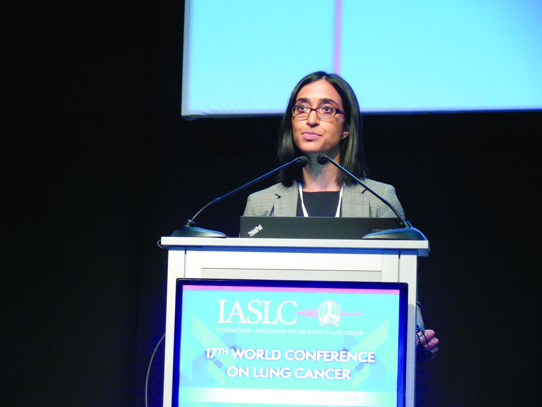
Immune-related adverse events were significantly more common in the patients treated with PD-1 inhibitors: 16%, compared with 11% in the PD-L1 inhibitor-treated patients (P = .04). The two subclasses also showed a trend toward a difference in the most common immune-related adverse event, hypothyroidism, with an incidence of 6.7% with PD-1 inhibitors and 4.2% with PD-L1 inhibitors (P = .07). The two sets of patients showed a statistically significant difference in the next most common immune-related adverse event, pneunomitis, 4.0% with PD-1 inhibitors and 2.0% with PD-L1 inhibitors (P = .02).
The trials with PD-1 inhibitors included nivolumab (Opdivo) and pembrolizumab (Keytruda). The trials with PD-L1 inhibitors included atezolizumab (Tecentriq), durvalumab, and avelumab. The total rate of all adverse events was highest among patients on nivolumab, 76%, and lowest among patients on durvalumab, 61%.
[email protected]
On Twitter @mitchelzoler
This very important systematic review with data from a total of nearly 6,000 patients shows that severe toxicity is unusual with the PD-1 and PD-L1 inhibitors, and they result in no meaningful difference in response rates. The toxicities seen with these drugs are milder and less frequent than we see with standard chemotherapy drugs. The severe autoimmune toxicities seen are a major concern but are manageable and occurred at low rates.
In general, efficacy and toxicity does not appear to form a basis by which to choose among these drugs. Fatigue was the most common adverse event, which is surprising to see with these drugs although we are accustomed to seeing it in patients on standard chemotherapy. Fatigue can be a major issue for patients, even if it is relatively mild, because they remain on these drugs for periods as long as 2 years.
If the PD-1 and PD-L1 inhibitors continue to perform with similar efficacy and safety profiles, clinicians will be forced to turn to other parameters when trying to decide which drug specifically to prescribe. This can include issues of cost, reimbursement, and dosing convenience. Nivolumab, for example, has been administered more often, every 2 weeks, than the other drugs in these classes. Oncologists are trying to develop effective regimens with these drugs that can be given once every 3 or every 4 weeks. Future investigations may also look at the possibility of treating patients with these drugs initially for 6 months, and then scaling back to retreatment only when there is disease progression. If this approach is successful, it would obviate concerns about causing long-term fatigue or the inconvenience of more frequent treatment schedules.
We also need to continue to monitor and compare the toxicities of these immune checkpoint inhibitors as we move into using them in combination regimens.
Paul Mitchell, MD, is a medical oncologist at the Olivia Newton-John Cancer and Wellness Centre in Heidelberg, Australia. He has served on advisory boards for AstraZeneca, Boehringer Ingelheim, BMS, Celgene, Merck/MSD, Merck Serono, and Roche, and he has received honoraria from Merck and Roche, and he has received travel grants from BMS and Roche. He made these comments as the designated discussant for the report and in a video interview.
The video associated with this article is no longer available on this site. Please view all of our videos on the MDedge YouTube channel
VIENNA – The two subclasses of immune checkpoint inhibitor drugs showed very little basis for choosing between them by either efficacy or toxicity in a systematic review of 23 trials run in patients with non–small cell lung cancer during 2013-2016.
For efficacy, inhibitors of the programmed death (PD-1) receptors had a 19% overall response rate when averaged from 12 different trials with 3,284 patients on one of these drugs. The PD-ligand 1 (PD-L1) inhibitors produced a 17% overall response rate in 11 trials with 2,615 patients on one of the drugs, a between-class efficacy difference that was not statistically significant, Rathi N. Pillai, MD, said at the World Conference on Lung Cancer, sponsored by the International Association for the Study of Lung Cancer.
Immune-related adverse events were significantly more common in the patients treated with PD-1 inhibitors: 16%, compared with 11% in the PD-L1 inhibitor-treated patients (P = .04). The two subclasses also showed a trend toward a difference in the most common immune-related adverse event, hypothyroidism, with an incidence of 6.7% with PD-1 inhibitors and 4.2% with PD-L1 inhibitors (P = .07). The two sets of patients showed a statistically significant difference in the next most common immune-related adverse event, pneunomitis, 4.0% with PD-1 inhibitors and 2.0% with PD-L1 inhibitors (P = .02).
The trials with PD-1 inhibitors included nivolumab (Opdivo) and pembrolizumab (Keytruda). The trials with PD-L1 inhibitors included atezolizumab (Tecentriq), durvalumab, and avelumab. The total rate of all adverse events was highest among patients on nivolumab, 76%, and lowest among patients on durvalumab, 61%.
[email protected]
On Twitter @mitchelzoler
AT WCLC 2016
Key clinical point:
Major finding: Overall response rates were 19% using a PD-1 inhibitor and 17% when using a PD-L1 inhibitor.
Data source: A systematic review of 23 trials in patients with non–small cell lung cancer published during 2013-2016.
Disclosures: Dr. Pillai had no disclosures.