User login
Special Considerations Needed in Applying Lupus Nephritis Guideline to Children
WASHINGTON — When the American College of Rheumatology (ACR) released its updated guideline for management of lupus nephritis (LN) at its 2024 Annual Meeting, they included recommendations for managing pediatric LN for the first time.
The pediatric recommendations use the same classification criteria, outcome measures, and treatments as in adults — including the first-line triple therapy recommendation — but there remain important differences between pediatric and adult LN, Mary Beth Son, MD, clinical chief of immunology and section chief of rheumatology at Boston Children’s Hospital in Massachusetts, and an associate professor of pediatrics at Harvard Medical School, also in Boston, told attendees.
“In general, kids and adolescents with lupus are sicker,” Son said. They are more likely to have renal manifestations and neuropsychiatric lupus at diagnosis, compared with adults. Further, “although the disease is the same, it’s happening to kids and adolescents who are undergoing critical periods of growth and development.”
Medication risk profiles also shift for younger patients, Son noted.
“Importantly, they’re at risk for higher cumulative dosing of both glucocorticoids and cyclophosphamide,” Son said. “When we give an adolescent a course of cyclophosphamide, we have to be aware that this might be the first of a few courses over the course of the lifetime disease, and with increasing numbers of cyclophosphamide courses, you have increased risk for infertility and malignancy.”
Son also acknowledged challenges of pediatric literature, including differences in definitions of pediatric lupus, very few randomized controlled trials, and fewer pediatric studies in general, with fewer participants. Given these research gaps, the guideline panels included pediatric rheumatologists and nephrologists, and the patient panel included several patients with childhood-onset disease.
Son also addressed differences in pediatric drug development. Dosing studies also do not always directly translate from adults to children because children have larger drug volume distribution and differences in drug clearance, and they may need different formulations, she said. Children tend to tolerate medications better than adults because they usually have fewer comorbidities, but the assessment of a drug’s safety must take its impact on growth and development into consideration.
During a press conference after the session where the guideline was presented, Linda Hiraki, MD, ScD, a clinician-scientist in rheumatology at the Hospital for Sick Children, Toronto, Ontario, Canada, said the panel took into consideration that pediatric patients receive their diagnosis during a critical time of development, so considerations of medication risks include the fact that children “have much more life to live.”
Triple Therapy Recommended
As with adults, the pediatric LN guideline recommends a triple therapy approach: glucocorticoids plus mycophenolate mofetil and belimumab, in addition to the usual renin-angiotensin-aldosterone system inhibitors and hydroxychloroquine. But Son acknowledged limitations of applying the new guideline to children. For one, voclosporin has not been studied in or approved for pediatric patients, although there exists modest evidence for other calcineurin inhibitors, mainly tacrolimus, in children.
“The other important consideration is that the lower dose of prednisone that’s being offered by the guidelines of 40 mg per day as a starting dose has not been studied in pediatric lupus nephritis patients,” Son said. “However, I would offer that, given that we know that kids get higher doses and longer courses, it’s even more important to consider a lower dose to begin with in the setting of other immunosuppressants.”
Good Practice Statements for Pediatric LN
Son also reviewed three good practice statements for pediatric LN. First, “glucocorticoid regimens should use pediatric-appropriate doses for children, as reduction of human glucocorticoid dosing is critically important given the early age of pediatric lupus onset and attendant comorbidities,” she said.
That statement is based on both common sense and some literature, including awareness that children are more likely to receive higher doses of steroids and that children’s higher damage scores are driven in part by steroid-related toxicity, such as avascular necrosis and cataracts. In addition, glucocorticoids can have profound effects on body mass index, mood, and height attainment.
“This is during a period of emerging self-identity and struggles with appearance; steroids exacerbate that” as well as mood issues already associated with puberty, Son said.
The second good practice statement recommends that clinicians monitor patients “for delayed pubertal onset and decreased growth velocity that can result from disease activity and glucocorticoid treatment and consider referral to pediatric endocrinology if indicated.” The third states that “a structured, intentional transition from pediatric to adult rheumatology care is indicated to avoid poor outcomes during this vulnerable period.”
During the press conference, Hiraki said that pediatric rheumatologists already recognize the need for discussions about transfer to adult care to begin very early, even years before patients are ready to transfer.
“The transition from being a pediatric patient to being an adult patient is very challenging for a number of reasons,” starting with loss of insurance coverage, added Bonnie Bermas, MD, a professor of internal medicine at UT Southwestern Medical Center in Dallas, Texas. When adult rheumatologists take on these patients, they may not have had care for 2 or 3 years, she said.
Rebecca Sadun, MD, PhD, an associate professor of pediatrics in rheumatology at Duke University School of Medicine, Durham, North Carolina, and vice-chair of the Systemic Lupus Erythematosus Committee for the Childhood Arthritis and Rheumatology Research Alliance, was not involved in the guideline development process but reviewed the new guideline.
“We appreciate that the ACR took care to involve pediatric rheumatologists, pediatric nephrologists, and patients with childhood-onset lupus in the development of the newest lupus nephritis treatment guidelines,” she said in an interview. She also noted, however, that “the dearth of pediatric-specific clinical trial data means that we continue to wonder when it is appropriate to extrapolate from adult data regarding the efficacy, safety, and dosing of certain medications, including steroids and voclosporin.” She also noted that voclosporin use can increase pill burden and therefore be difficult to use in pediatrics.
“Children, adolescents, and young adults are a unique population with unique challenges, including significant struggles with adherence to complex medication regimens,” she said. Sadun drew attention to two themes from the guideline that she found particularly applicable to management of pediatric LN.
“First, we must remain wary of the serious consequences of long-term, high-dose glucocorticoids, and we should continue to look towards steroid-sparing strategies that will reduce reliance on glucocorticoids,” Sadun said. “Second, we are likely to see better outcomes, including better renal response, when we take advantage of combination immunosuppression earlier in the disease course.”
Son, Bermas, and Sadun had no disclosures. Hiraki has consulted for Janssen. The guideline development did not involve outside funding.
A version of this article first appeared on Medscape.com.
FROM ACR 2024
Project’s Improvement in JIA Outcome Disparities Sets Stage for Further Interventions
WASHINGTON — A quality improvement project aimed at reducing racial disparities in juvenile idiopathic arthritis (JIA) led to a modest reduction in the overall clinical Juvenile Arthritis Disease Activity Score (cJADAS) and a 17% reduction in the disparity gap between Black and White patients, according to a study presented at the annual meeting of the American College of Rheumatology.
“Our work has led to initial progress in all groups, but we did not fully close the gap in outcomes,” Dori Abel, MD, MSHP, an attending rheumatologist at Children’s Hospital of Philadelphia in Pennsylvania, told attendees. But the project still revealed that it’s feasible to improve outcomes and reduce disparities with a “multipronged, equity-driven approach,” she said. “Stratifying data by demographic variables can reveal important differences in health care delivery and outcomes, catalyzing improvement efforts.”
Giya Harry, MD, MPH, MSc, an associate professor of pediatric rheumatology at Wake Forest University School of Medicine in Winston-Salem, North Carolina, was not involved in the study but praised both the effort and the progress made.
“The results are promising and suggest that with additional interventions targeting other key drivers, the team may be successful in completely eliminating the disparity in outcomes,” Harry said in an interview. “I applaud the hard work of Dr Abel and the other members of the team for doing the important work of characterizing the very complex issue of disparities in JIA outcomes across different race groups.”
It will now be important to build upon what the physicians learned during this process, said Harry, also the chair of the Diversity, Equity, Inclusion, and Accessibility committee of the Childhood Arthritis and Rheumatology Research Alliance.
“Patience is needed as they cycle through interventions with an emphasis on other key drivers” of disparities, Harry said.
Targeting Factors That Clinicians Can Potentially Influence
In her presentation, Abel discussed the various barriers that interfere with patients’ ability to move up the “JIA escalator” of getting referred and diagnosed, starting treatment and getting control of the disease, and monitoring and managing the disease and flares. These barriers include difficulties with access, trust, finances, insurance, caregivers’ missed work, medication burden, side effects, system barriers, and exhaustion and depression among caregivers and patients.
These barriers then contribute to disparities in JIA outcomes. In the STOP-JIA study, for example, Black children had greater polyarthritis disease activity in the first year and greater odds of radiographic damage, Abel noted. At her own institution, despite a mean cJADAS of 2.9 for the whole population of patients with JIA, the average was 5.0 for non-Hispanic Black patients, compared with 2.6 for non-Hispanic White patients.
The team therefore developed and implemented a quality improvement initiative aimed at improving the overall mean cJADAS and narrowing the gap between Black and White patients. The goal was to reduce the mean cJADAS to 2.7 by July 2024 and simultaneously reduce the cJADAS in Black patients by 1.2 units, or 50% of the baseline disparity gap, without increasing the existing gap.
The team first explored the many overlapping and interacting drivers of disparities within the realms of community characteristics, JIA treatment course, patient/family characteristics, organizational infrastructure, divisional infrastructure, and provider characteristics. While many of the individual factors driving disparities are outside clinicians’ control, “there are some domains clinicians may be able to directly influence, such as provider characteristics, JIA treatment course, and possibly divisional infrastructure,” Harry noted, and the team appeared to choose goals that fell under domains within clinicians’ potential influence.
The research team focused their efforts on four areas: Consistent outcome documentation, application of JIA best practices, providing access to at-risk patients, and team awareness and agency.
As part of improving consistent outcome documentation, they integrated outcome metrics into data visualization tools so that gaps were more evident. Applying JIA best practices included standardizing their approach to assessing medication adherence and barriers, with changes to the JIA note templates in the electronic health record and updates to medication adherence handouts.
Providing access to at-risk patients included several components:
- Creating a population management team
- Defining a target population to engage with for earlier follow-up
- Using a monthly batch outreach to defined patients
- Having a coordinator or social worker reach out to those who don’t make appointments
- Using a new JIA/high disease activity video follow-up program.
Finally, team awareness and agency involved giving physicians monthly access to mean cJADAS values for their own patients and at the division level. They also held quarterly disparity mitigation workshops.
Although the institution’s JIA population grew 13%, from 776 to 878 patients, over the course of the study, from January 2023 to May 2024, there was minimal change in the characteristics of the patient population. By May 2024, two thirds of patients (68%) were women, and 23% had public insurance. The population included 67% non-Hispanic White, 9% Hispanic/Latino, 7% non-Hispanic Black, and 4% Asian patients.
One third of the patients (32%) had the oligoarticular subtype, and other subtypes included enthesitis-related at 16%, polyarticular rheumatoid factor (RF)–negative at 15%, systemic at 7%, psoriatic at 6%, undifferentiated at 5%, and polyarticular RF-positive at 4%; data on subtype were unavailable for 14%. Most of their patients (71%) were in a high or very high quintile of the Childhood Opportunity Index, and 12% were in a low or very low quintile.
Results of the Quality Improvement Project
As of May 2024, the team had reached most of the goals they had set in terms of individual metrics. They met their goal of having a complete cJADAS calculated in more than 80% of JIA visits each month. With a goal of having over 90% of JIA monthly visits include disease activity target attestations, they reached 95% by May.
They aimed to have over half of JIA monthly visits include documentation of medication adherence/barrier assessment, and 75% of monthly visits had one. For their monthly outreach goal for overdue visits, they aimed to contact more than 75% of patients within 30 days if they were newly overdue for a follow-up visit but had only reached 47% by May 2024. The team had also completed 154 Maintenance of Certification assessments by May.
From initiation of project planning in January 2023 through May 2024, the overall JIA patient population experienced an improvement in cJADAS from 2.9 to 2.54. In individual cJADAS components, the mean patient global score improved from 1.71 to 1.47, the physician global score improved from 0.81 to 0.75, and the joint count score improved from 0.71 to 0.68.
In the non-Hispanic Black population, the mean cJADAS improved from 5.06 in January 2023 to 4.31 in May 2024. Mean cJADAS in the non-Hispanic White population fell from 2.63 to 2.29. With a difference of 0.4 points fewer between the Black and White populations, the disparity gap closed by 17%.
One of the team’s next steps will be to focus on the Hispanic population in 2024-2025 by optimizing language services, working toward greater family involvement to better understand barriers to care, and ongoing population management.
Abel and Harry had no disclosures. No external funding was noted.
A version of this article appeared on Medscape.com.
FROM ACR 2024
Could Biomarkers Help to Detect Lung Disease Earlier in Systemic JIA?
WASHINGTON — Children who have systemic juvenile idiopathic arthritis with lung disease (sJIA-LD) have distinct biomarker profiles that may hold potential in eventually detecting LD earlier and identifying personalized treatment, according to research presented at the American College of Rheumatology (ACR) 2024 Annual Meeting.
Established risk factors for LD, which affects up to 1 in every 20 patients with sJIA, include being of a younger age, having more macrophage activation syndrome (MAS) episodes, and having more adverse reactions to biologics, Esraa Eloseily, MD, MS, an assistant professor of pediatrics at UT Southwestern Children’s Medical Center, Dallas, told attendees.
“The pathophysiology remains unclear and debate centers around elevated IL-18 [interleukin 18] and T-cell activation in association with HLA-DRB1*15/DRESS [drug reaction with eosinophilia and systemic symptoms] reactions to biologics, and thus, we have urgent unmet needs to understand the prevalence, the pathogenesis, disease biomarkers, and influence of biologics,” Eloseily said.
Their study confirmed that patients with LD tended to be younger and have more MAS. The researchers also found lower hemoglobin and higher white blood cell counts and platelets in patients with sJIA-LD, as well as a higher medication burden, particularly with steroids, biologics, and Janus kinase (JAK) inhibitors.
Randy Cron, MD, PhD, director of the Division of Pediatric Rheumatology at the University of Alabama at Birmingham, attended the presentation and noted that every additional piece of information is helpful in filling out the picture of what we know and can predict about sJIA-LD development.
“We’re all learning as we go, so the more people that study this, the better,” Cron told Medscape Medical News. “Even if it’s just seeing things that other groups have seen or really solidifying the risk factors for the development of lung disease, I think, at this point, that’s one of the most clinically relevant things: Do we screen? Who do we screen? Certainly, kids who have very young age of onset, children who develop macrophage activation syndrome, children who have high IL-18 levels.”
Study Results
The study compared 37 patients with sJIA-LD from 16 Childhood Arthritis and Rheumatology Research Alliance (CARRA) Registry sites with 141 patients with sJIA but without LD who had similar follow-up durations in the CARRA Registry.
Disease duration for patients with sJIA-LD was defined as the time from their initial sJIA diagnosis to their baseline sJIA-LD cohort visit, which was considered their index visit. In patients without LD, duration was from their enrollment in the CARRA Registry to their last CARRA Registry visit, which was considered their index visit.
The patients with sJIA-LD were significantly younger — a median age of 1 year — at onset of sJIA than those without LD, who had a median age of 5 years (P < .0019). The patients with sJIA-LD were also younger (median age, 7 years) at their index visit than those without LD (median age, 10 years) (P < .0001).
There were also significant differences in medication usage between those with and without LD. While 40.5% of patients with sJIA-LD were using steroids at their index visit, only 11.4% of those without LD were using steroids (P < .0001). Yet the mean dose of steroids was significantly lower in those with LD (5.45 mg/d) than in those without (20.7 mg/d). In addition, nearly half the patients with sJIA-LD had ever used cyclosporin A (45.7%) compared with 2.8% of those without LD (P < .0001), and 17.1% of patients with sJIA-LD had used mycophenolate mofetil compared with 0.7% of those without LD (P = .0002).
Similar disparities were seen for usage of biologics and JAK inhibitors: Anakinra (82.9% vs 56.7%; P = .0036), abatacept (8.6% vs 1.4%; P = .053), tofacitinib (57.1% vs 5.7%; P < .0001), ruxolitinib (25.7% vs 0%; P < .0001), baricitinib (8.6% vs 0%; P = .007), and emapalumab (23% vs 0.7%; P < .0001). Further, 5.7% of those with sJIA-LD had taken etoposide and 22.9% had received intravenous immunoglobulin compared with 0% and 4.3%, respectively, in those without LD (P = .04 and P = .001).
Laboratory parameters of patients with sJIA-LD were also significantly different from those of patients without LD, including a higher white blood cell count (8.8 × 10⁹/L vs 8.1 × 10⁹/L; P = .01), higher platelets (316.5 × 10⁹/L vs 311.2 × 10⁹/L; P = .03), and lower hemoglobin (11.5 g/dL vs 12.6 g/dL; P = .007). Ferritin levels trended nonsignificantly higher in patients with sJIA-LD (506 ng/mL vs 173.2 ng/mL; P = .09), and aspartate aminotransferase levels were significantly higher (37 U/L vs 28.72 U/L; P < .0001).
Patients’ overall well-being was “unexpectedly” higher in patients with sJIA-LD (P = .007), Eloseily noted, including the parent/patient rating (P = .027). However, more of the patients without LD had an excellent (61%) or very good (20.4%) health-related quality of life compared with those with LD (13% and 39%), and nearly one third of patients with sJIA-LD (30.4%) had only fair health-related quality of life compared with 5.5% without LD (P = .0002).
In line with known risk factors, most of the patients with sJIA-LD had a prior MAS episode (67.6%) compared with 10.6% of those without LD (P < .0001). Mortality was also higher in those with LD, two of whom died, whereas none without LD died (P = .04).
While existing biomarkers have been reported, they lack independent validation, Eloseily told attendees. Among the known key biomarkers are IL-18/interferon (IFN)-gamma axis, which are known to drive MAS and pulmonary inflammation, especially in those with MAS and LD; ICAM-5 and MMP-7, which is linked to fibrosis and tissue remodeling; and CCL11, CCL17, and GDF-15, which is linked to fibrosis and inflammation.
Because the CARRA Registry has limited availability of biosamples for most patients, the researchers used plasma samples from the FROST study for 27 patients with sJIA-LD and 46 patients without LD. When they compared 23 biomarkers between the groups, most of the known key biomarkers, as well as several other biomarkers, were significantly elevated in those with LD compared with in those without:
- MMP-7 (P = .001)
- IFN gamma (P = .008)
- CCL11 (P < .0001)
- CCL17 (P = .002)
- CCL15 (P < .0001)
- MCP-1 (P = .0003)
- MCP-3 (P = .02)
- CCL25 (P < .0001)
- CD25 (P < .0001)
- GDF-15 (P < .0001)
- TRAIL (P < .0001)
- IL-23 (P = .002)
They found that IL-18 only trended higher (P = .07), and there were not adequate data for ICAM-5.
The study was limited by the differences in disease duration between the compared groups and the limited availability of biosamples, which they only had from patients enrolled in the FROST study.
The research was funded by CARRA and the Arthritis Foundation. Eloseily reported no disclosures. Cron reported serving as an adviser for AbbVie/Abbott and Sobi, receiving grant funding and speaking and consulting fees from Pfizer, and receiving royalties from Springer.
A version of this article appeared on Medscape.com.
FROM ACR 2024
Could a Urinary Biomarker Panel Be a ‘Game Changer’ for Lupus Nephritis Management?
WASHINGTON — An investigational 12-protein panel of urinary biomarkers predicted histologically active lupus nephritis (LN) with 86% accuracy, according to research presented at the American College of Rheumatology (ACR) 2024 Annual Meeting.
The noninvasive biomarker panel “robustly predicts meaningful and actionable histological findings” in patients with active proliferative LN, Andrea Fava, MD, assistant professor of medicine in the Division of Rheumatology at Johns Hopkins Medicine in Baltimore, told attendees.
“In contrast to proteinuria, which can’t differentiate inflammation from damage, this panel for histological activity includes a set of 12 proteins linked to intrarenal inflammation,” said Fava, director of Lupus Translational Research at Johns Hopkins. A decline in the biomarker score at 3 months predicted a clinical response at 1 year, and persistent elevation of the score at 1 year predicted permanent loss of kidney function, “which makes it tempting as a treatment endpoint,” Fava said. “Upon further validation, this biomarker panel could aid in the diagnosis of lupus nephritis and guide treatment decisions.”
Alfred Kim, MD, PhD, an associate professor of medicine at Washington University in St. Louis, was not involved in the research but noted the potential value of a reliable biomarker panel.
“If we have urinary biomarkers that strongly associate with histologic activity, this would be a game changer in the management of LN,” Kim told Medscape Medical News. “Right now, the gold standard is to perform another kidney biopsy to determine if therapy is working. But this is invasive, and many patients do not want to do another kidney biopsy. Conversely, the easiest way to assess lupus nephritis activity is through a urinalysis, focusing on urinary protein levels,” but relying on proteinuria has limitations as well.
“The most important [limitation] is that proteinuria cannot distinguish treatable inflammation from chronic damage,” Fava said. Persistent histologic activity in patients without proteinuria predicts flares, but tracking histologic activity, as Kim noted, requires repeat biopsies.
“So we need better biomarkers because biomarkers that can reflect tissue biology in real time can guide personalized treatment, and that’s one of the main goals of the Accelerating Medicines Partnership [AMP],” he said. The AMP is a public-private partnership between the National Institutes of Health (NIH), the US Food and Drug Administration (FDA), multiple biopharmaceutical and life science companies, and nonprofit and other organizations. Lupus is one of the AMP’s funded projects.
Kim agreed that “effective biomarkers are a huge unmet need in LN.” Further, he said, “imagine a world where the diagnosis of LN can be made just through urinary biomarkers and obviate the need for biopsy. Both patients and providers will be ecstatic at this possibility.”
Fava described the background for how his research team determined what biomarkers to test. They had previously enrolled 225 patients with LN undergoing a clinically indicated kidney biopsy and collected urine samples from them at baseline and at 12, 24, and 52 weeks after their biopsy.
Of the 225 patients included, 9% with only mesangial LN (class I-II), 25% with pure membranous LN (class V), 24% with mixed LN (class III or IV with or without V), 38% with proliferative LN (class III or IV), and 4% with advanced sclerosis LN (class VI). From these samples, they quantified 1200 proteins and looked at how they correlated with histologic activity.
“What was interesting was that in patients who were classified as responders after 1 year, there were many of these proteins that declined as early as 3 months, suggesting that effective immunosuppression is reducing intrarenal inflammation, and we can capture it in real time,” Fava said.
Biomarker Panel Predicts Histologically Active LN
So they set to determining whether they could develop a urinary biomarker for histologically active LN that could be useful in clinical decision-making. They focused on one that could detect active proliferative LN with an NIH activity index score > 2. Their 179 participants included 47.5% Black, 27.9% White, and 14.5% Asian participants, with 10.1% of other races. The predominantly female (86.6%) cohort had an average age of 37 years. Among the LN classes, about one third (34.6%) had pure proliferative disease, 17.9% had mixed proliferative, 27.9% had pure membranous, 11.7% had class I or II, and 5% had class VI. Just over half the participants (55.7%) had not responded to treatment at 12 months, whereas 25% had a complete response, and 19.3% had a partial response.
However, both the 78 participants with an NIH activity index score > 2 and the 101 with a score ≤ 2 had a median score of 3 on the NIH chronicity index. And the urine protein-to-creatinine ratio — 2.8 in the group with an NIH activity index score > 2 and 2.4 in the other group — was nearly indistinguishable between the two groups, Fava said.
They then trained multiple algorithms on 80% of the data to find the best performing set of proteins (with an area under the curve [AUC] of 90%) for predicting an NIH activity index score > 2. They reduced the number of proteins to maximize practicality and performance of the panel, Fava said, and ultimately identified a 12-protein panel that was highly predictive of an NIH activity index score > 2. Then, they validated that panel using the other 20% of the data. The training set had an AUC of 90%, and the test set was validated with an AUC of 93%.
The 12-protein panel score outperformed anti-dsDNA, C3 complement, and proteinuria, with a sensitivity of 81%, a specificity of 90%, a positive predictive value of 87%, a negative predictive value of 86%, and an accuracy of 86%. The proteins with the greatest relative importance were CD163, cathepsin S, FOLR2, and CEACAM-1.
“In contrast to proteinuria, these proteins were related to inflammatory processes found in the kidneys in patients with lupus nephritis, such as activation of macrophages, neutrophils and monocytes, lymphocytes, and complement,” Fava said.
When they looked at the trajectories of the probabilities from the biomarker panel at 3, 6, and 12 months, the probability of the NIH activity index score remaining > 2 stayed high in the nonresponders over 1 year, but the trajectory declined at 3 months in the responders, indicating a decrease in kidney inflammation (P < .001).
Can the Biomarker Panel Serve as a Treatment Endpoint?
Then, to determine whether the panel could act as a reliable treatment endpoint, the researchers followed the patients for up to 7 years. One third of the patients lost more than 40% of their kidney function during the follow-up. They found that a high urinary biomarker score at 12 months predicted future glomerular filtration rate loss, independent of proteinuria.
This panel was tested specifically for proliferative LN, so “we may need distinct panels for each [LN type] to capture most of these patients,” Kim said. “I think that’s where the gold mine is: A personalized medicine approach where a large biomarker panel identifies which smaller panel that patient best fits, then use that for monitoring.”
Kim did note an important potential limitation in the study regarding how samples are used in biomarker discovery and validation vs in clinical practice. “Most samples in research studies are frozen, then thawed, while urine is assayed within a couple hours after collection in the clinical setting,” he said. “Do sample processing differences create a situation where a biomarker works in a research project but not in the clinical setting?” But more likely, he said, the opposite may be the case, where frozen samples allow for more degradation of proteins and potentially useful LN biomarker candidates are never detected.
Another challenge, Kim added, albeit unrelated to the study findings, is that diagnostic companies are finding it difficult to get payers to cover new tests, so that could become a challenge if the panel undergoes further validation and then FDA qualification.
The research was funded by Exagen. Fava reported disclosures with Arctiva, AstraZeneca, Exagen, Novartis, UCB, Bristol Myers Squibb, Annexon Bio, and Bain Capital. His coauthors reported financial relationships with numerous pharmaceutical and life science companies, including Exagen, and some are employees of Exagen.
Kim reported research agreements with AstraZeneca, Bristol Myers Squibb, Novartis, and CRISPR Therapeutics; receiving royalties from Kypha; and receiving consulting/speaking fees from AbbVie, Amgen, Atara Bio, Aurinia, Cargo Tx, Exagen, GlaxoSmithKline, Hinge Bio, Kypha, and UpToDate.
A version of this article appeared on Medscape.com.
FROM ACR 2024
Triple Therapy Now Advised for Lupus Nephritis in Updated Guideline
WASHINGTON — A new guideline for management of lupus nephritis (LN) was unveiled at the annual meeting of the American College of Rheumatology (ACR), updating the 2012 LN guideline to recommend a more aggressive first-line approach to treating the disease.
“The biggest differences are that we are recommending what we’re calling triple therapy, where we incorporate the glucocorticoid therapy with baseline conventional immunosuppressants, usually mycophenolate with cyclophosphamide, and the addition of one of the newer agents more recently approved by the FDA [Food and Drug Administration] — belimumab, voclosporin, or another CNI [calcineurin inhibitor],” said Lisa Sammaritano, MD, director of the Rheumatology Reproductive Health Program of the Barbara Volcker Center for Women and Rheumatic Diseases at the Hospital for Special Surgery and professor of clinical medicine at Weill Cornell Medical College, both in New York City.
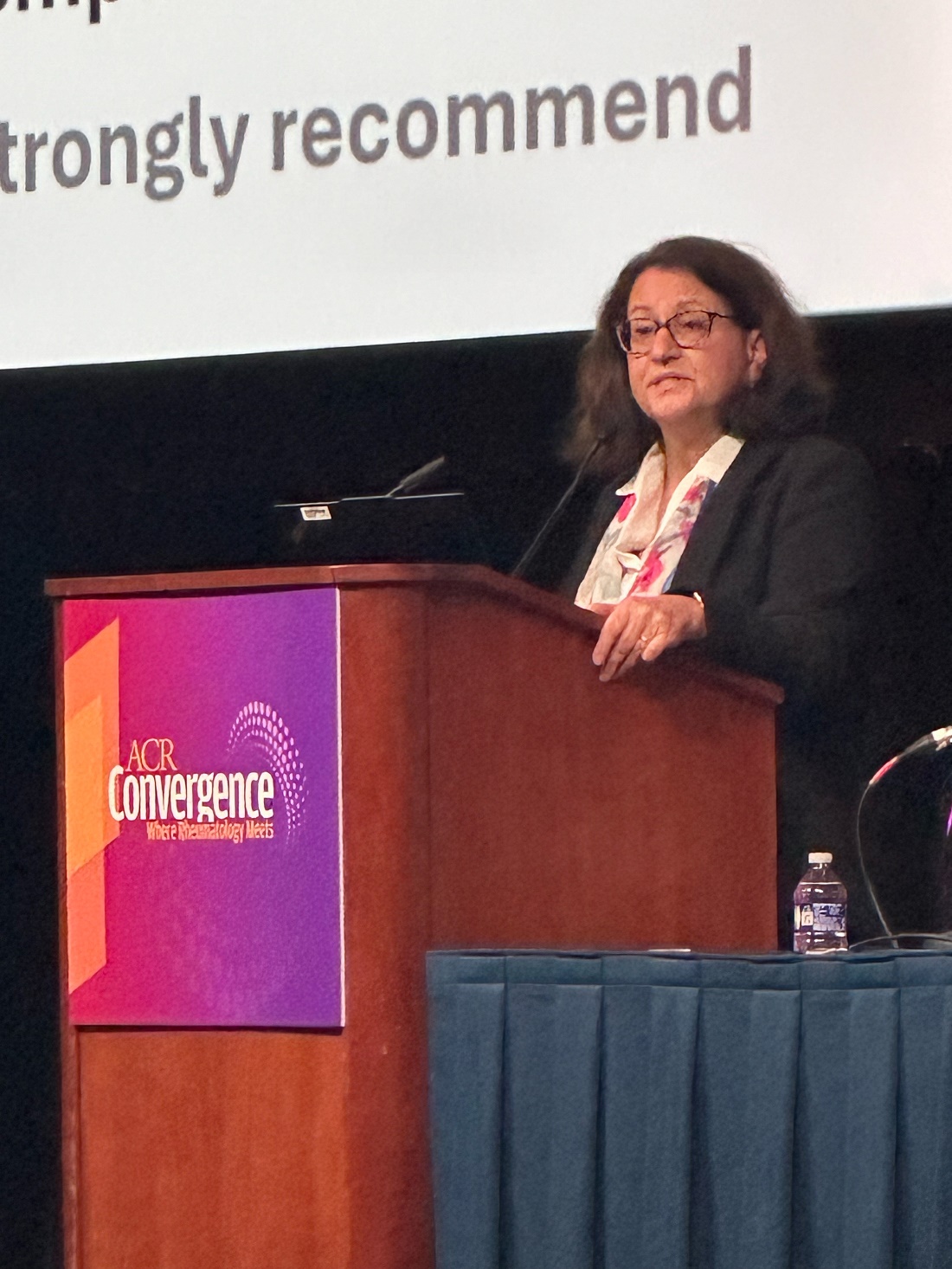
“This is a bit of a change from not only our previous guideline but some of the other guidelines out there, and it is based on the fact that we have very convincing evidence that starting with triple therapy yields to better long-term outcomes for our patients than starting with only two agents and waiting to see if they respond before escalating therapy,” she said. Other key updates include recommending use of pulse glucocorticoid therapy with a lower dose and more rapid steroid taper and treating patients with the recommended therapy for 3-5 years.
The guiding principles of the guideline are not only to preserve kidney function and minimize morbidity and mortality but also to ensure collaborative care with nephrology, to utilize shared decision-making that includes patients’ values and preferences, to reduce healthcare disparities, and to consider pediatric and geriatric populations. The guidelines are based on a quantitative synthesis of 105 studies that yielded 7 strong recommendations, 21 conditional recommendations, and 13 good practice statements — those commonly accepted as beneficial or practical advice even if there is little direct evidence to support them. The voting panel of 19 members included not only 3 nephrologists and 2 pediatric rheumatologists but also 2 patient representatives with LN.
The recommendations are just that, “a recommendation, not an order,” Sammaritano said, and strong recommendations are those “where we think, unequivocally, almost everybody should follow that recommendation. When we feel that we cannot make a strong recommendation, then we call our recommendation conditional, and it is conditional on looking at different things,” she said.
“Patients are different, especially lupus patients, and so one lupus nephritis patient may have different clinical characteristics, different thoughts about what therapy will work for them in their lives, or what therapy they really do not want to pursue,” Sammaritano said. “Maybe they can’t conceive of coming to the hospital once a month for intravenous therapy. Maybe they’re concerned about pill burden, which is something that our patient panel really emphasized to us. So, conditional recommendation means this voting panel thought that this was the best overall for most patients and most circumstances, recognizing there will still be a significant number of people, clinicians and patients, who may feel differently for that particular situation. So, that’s where you know the patient-clinician discussion can help with decision-making.”
What Are the Recommendations?
All patients with systemic lupus erythematosus (SLE) are strongly recommended to undergo proteinuria screening every 6-12 months or at the time of a flare. Those suspected of having LN should receive a prompt kidney biopsy and treatment with glucocorticoids while awaiting the biopsy and results. Two conditional recommendations for kidney biopsy include patients with SLE with unexplained impaired kidney function or a protein to creatinine ratio > 0.5 g/g, and patients with LN with a suspected flare after initial response or a lack of response or worsening after 6 months of therapy.
The guidelines include a strong recommendation for all patients with SLE to receive hydroxychloroquine and a conditional recommendation for all patients with elevated proteinuria (> 0.5 g/g) to receive renin-angiotensin-aldosterone system inhibitors (RAAS-I). Dosages in patients with LN with decreased glomerular filtration rate (GFR) should be adjusted as needed.
Sammaritano then reviewed the specifics on medication treatment. The glucocorticoid therapy in all patients with LN should begin with Pulse IV Therapy at 250-1000 mg/d for 1-3 days, followed by oral prednisone ≤ 0.5 mg/kg per day up to 40 mg/d, then tapered to a target dose > 5 mg/d within 6 months. The justification for this course comes from a 2024 systematic review finding pulse followed by oral glucocorticoids maximized complete renal response while minimizing toxicities, Sammaritano said.
“We have all become acutely aware of the very high risk of prolonged high dose of glucocorticoids for our patients,” she said, “and importantly, our patient panel participants strongly emphasized their preference for minimizing glucocorticoids dose.”
In addition to the recommendation of all patients receiving hydroxychloroquine and RAAS-I, first-line treatment of active, new-onset, or flaring LN should begin with triple therapy — glucocorticoids with two additional immunosuppressive agents. For patients with class III/IV LN, triple therapy includes the glucocorticoids course with a mycophenolic acid analog (MPAA) and either belimumab or a CNI. Conditional recommendations support MPAA with belimumab for significant extrarenal manifestations and MPAA with CNI for proteinuria ≥ 3 g/g.
An alternative triple therapy for class III/IV is glucocorticoids with low-dose cyclophosphamide and belimumab, but MPAA at 2-3 g/d is preferred over cyclophosphamide. The preferred regimen for cyclophosphamide is derived from the Euro-Lupus Nephritis Trial: Intravenous 500 mg every 2 weeks for six doses and then MPAA. Sammaritano noted that there are some limited data on using cyclophosphamide with belimumab, but “we do not specifically recommend cyclophosphamide with a CNI as one of our options because this combination has not been studied in randomized controlled trials.”
There are less data supporting class V recommendations, Sammaritano said, but for those with proteinuria of at least 1 g/g, the panel still recommends triple therapy with glucocorticoids, a MPAA, and a CNI. A CNI is preferred over belimumab because of its stabilizing effects on the podocyte cytoskeleton. Two alternative triple therapies for class V–only patients are glucocorticoids with belimumab and either low-dose cyclophosphamide or MPAA.
Dual therapy is only recommended if triple therapy is not available or not tolerated. The voting panel chose to recommend triple therapy over dual therapy with escalation for two reasons. First, the BLISS-LN and AURORA 1 trials showed improved outcomes with initial triple therapy over initial dual therapies.
Second, “nephron loss proceeds throughout a person’s lifetime even for those who do not have lupus nephritis, and every case of lupus nephritis or every period of time with uncontrolled lupus nephritis changes the course of that decline for the worse,” Sammaritano said. “So, we feel we can’t wait for nephron loss to implement what has been shown to be the most efficacious therapy. We want to gain rapid control of inflammation using the most effective regimen to prevent further damage and flare and maintain survival.”
Therapy is conditionally recommended for at least 3-5 years because “not only do we want to gain rapid control of disease activity [but we also] want to maintain control of disease activity until there’s sustained inactive disease,” Sammaritano said. “Repeat kidney biopsies show that immunologic activity persists in the kidneys for several years, and the withdrawal of immunosuppression when there is histologic activity predisposes patients to flare.” But immunosuppressive therapy can be tapered over time as determined by renal disease activity and medication tolerability.
For patients with refractory disease, consider additional factors that could be affecting the disease, such as adherence, the presence of other diagnoses, or advanced chronicity.
“If true refractory nephritis is present,” she said, “we recommend escalation to a more intensive regimen,” including the addition of anti-CD20 agents, combination therapy with three immunosuppressives, or referral for investigational therapy.
“We also emphasize the importance of other adjunctive therapies preventing comorbidities, such as cardiovascular disease, changes in bone health, or infection risk,” she said. In older patients, avoid polypharmacy as much as possible and be mindful of age-related GFR, she added.
A strong recommendation supported monitoring patients with LN and proteinuria at least every 3 months if they have not achieved complete renal response and every 3-6 months after sustained complete renal response.
Last, in patients with LN and end-stage kidney disease (ESKD), the voting panel strongly recommends transplant over dialysis and conditionally recommends proceeding to the transplant without requiring a complete clinical or serologic remission as long as no other organs are involved. In patients with LN at risk for ESKD, the guideline conditionally recommends consideration of a preemptive transplant, and patients on dialysis or post transplant are strongly recommended to regularly follow up with rheumatology.
Gabriel Kirsch, MD, a resident rheumatologist at the University of Florida College of Medicine, Jacksonville, said he found the guidelines helpful, “especially the guidance on the dichotomy between using belimumab and voclosporin and the clinical and patient preference that help you make that decision.”
Kirsch had hoped, however, to hear more about the impact of therapeutic drug monitoring of hydroxychloroquine on LN outcomes. He also noted a clinical scenario he’s come across that wasn’t addressed.
“When you’re checking GFR on these folks, a lot of our eGFR calculators are creatinine based, and creatinine at the extremes of muscle mass can be inaccurate,” such as getting artificially low creatinine readings from pediatric patients because of their low muscle mass or from patients with muscle atrophy caused by a lot of glucocorticoid exposure. “I was hoping for some more guidance on that,” he said.
Ellen Ginzler, MD, MPH, chief of rheumatology at SUNY Health Science Center in Brooklyn, New York, said the guidelines were pretty much what she expected them to be. She agreed with the panel’s advice that, when deciding between belimumab or voclosporin, “if it’s pure proteinuria, then you add voclosporin. If the patient has extra renal manifestations, you go with belimumab first.”
“They really made it quite clear that, despite the fact that people really want to reduce the amount of immunosuppression — and I agree you should taper steroids quickly — you really need to keep the immunosuppression for a prolonged period of time because all of the studies that have been done for years show that the longer you’re on immunosuppression after you achieve remission or a low disease activity state, the better your chance of not flaring,” Ginzler said. “Rapid tapering or discontinuation really increases the risk of flare.”
Sammaritano, Kirsch, and Ginzler had no disclosures. No external funding was used.
A version of this article appeared on Medscape.com.
WASHINGTON — A new guideline for management of lupus nephritis (LN) was unveiled at the annual meeting of the American College of Rheumatology (ACR), updating the 2012 LN guideline to recommend a more aggressive first-line approach to treating the disease.
“The biggest differences are that we are recommending what we’re calling triple therapy, where we incorporate the glucocorticoid therapy with baseline conventional immunosuppressants, usually mycophenolate with cyclophosphamide, and the addition of one of the newer agents more recently approved by the FDA [Food and Drug Administration] — belimumab, voclosporin, or another CNI [calcineurin inhibitor],” said Lisa Sammaritano, MD, director of the Rheumatology Reproductive Health Program of the Barbara Volcker Center for Women and Rheumatic Diseases at the Hospital for Special Surgery and professor of clinical medicine at Weill Cornell Medical College, both in New York City.
“This is a bit of a change from not only our previous guideline but some of the other guidelines out there, and it is based on the fact that we have very convincing evidence that starting with triple therapy yields to better long-term outcomes for our patients than starting with only two agents and waiting to see if they respond before escalating therapy,” she said. Other key updates include recommending use of pulse glucocorticoid therapy with a lower dose and more rapid steroid taper and treating patients with the recommended therapy for 3-5 years.
The guiding principles of the guideline are not only to preserve kidney function and minimize morbidity and mortality but also to ensure collaborative care with nephrology, to utilize shared decision-making that includes patients’ values and preferences, to reduce healthcare disparities, and to consider pediatric and geriatric populations. The guidelines are based on a quantitative synthesis of 105 studies that yielded 7 strong recommendations, 21 conditional recommendations, and 13 good practice statements — those commonly accepted as beneficial or practical advice even if there is little direct evidence to support them. The voting panel of 19 members included not only 3 nephrologists and 2 pediatric rheumatologists but also 2 patient representatives with LN.
The recommendations are just that, “a recommendation, not an order,” Sammaritano said, and strong recommendations are those “where we think, unequivocally, almost everybody should follow that recommendation. When we feel that we cannot make a strong recommendation, then we call our recommendation conditional, and it is conditional on looking at different things,” she said.
“Patients are different, especially lupus patients, and so one lupus nephritis patient may have different clinical characteristics, different thoughts about what therapy will work for them in their lives, or what therapy they really do not want to pursue,” Sammaritano said. “Maybe they can’t conceive of coming to the hospital once a month for intravenous therapy. Maybe they’re concerned about pill burden, which is something that our patient panel really emphasized to us. So, conditional recommendation means this voting panel thought that this was the best overall for most patients and most circumstances, recognizing there will still be a significant number of people, clinicians and patients, who may feel differently for that particular situation. So, that’s where you know the patient-clinician discussion can help with decision-making.”
What Are the Recommendations?
All patients with systemic lupus erythematosus (SLE) are strongly recommended to undergo proteinuria screening every 6-12 months or at the time of a flare. Those suspected of having LN should receive a prompt kidney biopsy and treatment with glucocorticoids while awaiting the biopsy and results. Two conditional recommendations for kidney biopsy include patients with SLE with unexplained impaired kidney function or a protein to creatinine ratio > 0.5 g/g, and patients with LN with a suspected flare after initial response or a lack of response or worsening after 6 months of therapy.
The guidelines include a strong recommendation for all patients with SLE to receive hydroxychloroquine and a conditional recommendation for all patients with elevated proteinuria (> 0.5 g/g) to receive renin-angiotensin-aldosterone system inhibitors (RAAS-I). Dosages in patients with LN with decreased glomerular filtration rate (GFR) should be adjusted as needed.
Sammaritano then reviewed the specifics on medication treatment. The glucocorticoid therapy in all patients with LN should begin with Pulse IV Therapy at 250-1000 mg/d for 1-3 days, followed by oral prednisone ≤ 0.5 mg/kg per day up to 40 mg/d, then tapered to a target dose > 5 mg/d within 6 months. The justification for this course comes from a 2024 systematic review finding pulse followed by oral glucocorticoids maximized complete renal response while minimizing toxicities, Sammaritano said.
“We have all become acutely aware of the very high risk of prolonged high dose of glucocorticoids for our patients,” she said, “and importantly, our patient panel participants strongly emphasized their preference for minimizing glucocorticoids dose.”
In addition to the recommendation of all patients receiving hydroxychloroquine and RAAS-I, first-line treatment of active, new-onset, or flaring LN should begin with triple therapy — glucocorticoids with two additional immunosuppressive agents. For patients with class III/IV LN, triple therapy includes the glucocorticoids course with a mycophenolic acid analog (MPAA) and either belimumab or a CNI. Conditional recommendations support MPAA with belimumab for significant extrarenal manifestations and MPAA with CNI for proteinuria ≥ 3 g/g.
An alternative triple therapy for class III/IV is glucocorticoids with low-dose cyclophosphamide and belimumab, but MPAA at 2-3 g/d is preferred over cyclophosphamide. The preferred regimen for cyclophosphamide is derived from the Euro-Lupus Nephritis Trial: Intravenous 500 mg every 2 weeks for six doses and then MPAA. Sammaritano noted that there are some limited data on using cyclophosphamide with belimumab, but “we do not specifically recommend cyclophosphamide with a CNI as one of our options because this combination has not been studied in randomized controlled trials.”
There are less data supporting class V recommendations, Sammaritano said, but for those with proteinuria of at least 1 g/g, the panel still recommends triple therapy with glucocorticoids, a MPAA, and a CNI. A CNI is preferred over belimumab because of its stabilizing effects on the podocyte cytoskeleton. Two alternative triple therapies for class V–only patients are glucocorticoids with belimumab and either low-dose cyclophosphamide or MPAA.
Dual therapy is only recommended if triple therapy is not available or not tolerated. The voting panel chose to recommend triple therapy over dual therapy with escalation for two reasons. First, the BLISS-LN and AURORA 1 trials showed improved outcomes with initial triple therapy over initial dual therapies.
Second, “nephron loss proceeds throughout a person’s lifetime even for those who do not have lupus nephritis, and every case of lupus nephritis or every period of time with uncontrolled lupus nephritis changes the course of that decline for the worse,” Sammaritano said. “So, we feel we can’t wait for nephron loss to implement what has been shown to be the most efficacious therapy. We want to gain rapid control of inflammation using the most effective regimen to prevent further damage and flare and maintain survival.”
Therapy is conditionally recommended for at least 3-5 years because “not only do we want to gain rapid control of disease activity [but we also] want to maintain control of disease activity until there’s sustained inactive disease,” Sammaritano said. “Repeat kidney biopsies show that immunologic activity persists in the kidneys for several years, and the withdrawal of immunosuppression when there is histologic activity predisposes patients to flare.” But immunosuppressive therapy can be tapered over time as determined by renal disease activity and medication tolerability.
For patients with refractory disease, consider additional factors that could be affecting the disease, such as adherence, the presence of other diagnoses, or advanced chronicity.
“If true refractory nephritis is present,” she said, “we recommend escalation to a more intensive regimen,” including the addition of anti-CD20 agents, combination therapy with three immunosuppressives, or referral for investigational therapy.
“We also emphasize the importance of other adjunctive therapies preventing comorbidities, such as cardiovascular disease, changes in bone health, or infection risk,” she said. In older patients, avoid polypharmacy as much as possible and be mindful of age-related GFR, she added.
A strong recommendation supported monitoring patients with LN and proteinuria at least every 3 months if they have not achieved complete renal response and every 3-6 months after sustained complete renal response.
Last, in patients with LN and end-stage kidney disease (ESKD), the voting panel strongly recommends transplant over dialysis and conditionally recommends proceeding to the transplant without requiring a complete clinical or serologic remission as long as no other organs are involved. In patients with LN at risk for ESKD, the guideline conditionally recommends consideration of a preemptive transplant, and patients on dialysis or post transplant are strongly recommended to regularly follow up with rheumatology.
Gabriel Kirsch, MD, a resident rheumatologist at the University of Florida College of Medicine, Jacksonville, said he found the guidelines helpful, “especially the guidance on the dichotomy between using belimumab and voclosporin and the clinical and patient preference that help you make that decision.”
Kirsch had hoped, however, to hear more about the impact of therapeutic drug monitoring of hydroxychloroquine on LN outcomes. He also noted a clinical scenario he’s come across that wasn’t addressed.
“When you’re checking GFR on these folks, a lot of our eGFR calculators are creatinine based, and creatinine at the extremes of muscle mass can be inaccurate,” such as getting artificially low creatinine readings from pediatric patients because of their low muscle mass or from patients with muscle atrophy caused by a lot of glucocorticoid exposure. “I was hoping for some more guidance on that,” he said.
Ellen Ginzler, MD, MPH, chief of rheumatology at SUNY Health Science Center in Brooklyn, New York, said the guidelines were pretty much what she expected them to be. She agreed with the panel’s advice that, when deciding between belimumab or voclosporin, “if it’s pure proteinuria, then you add voclosporin. If the patient has extra renal manifestations, you go with belimumab first.”
“They really made it quite clear that, despite the fact that people really want to reduce the amount of immunosuppression — and I agree you should taper steroids quickly — you really need to keep the immunosuppression for a prolonged period of time because all of the studies that have been done for years show that the longer you’re on immunosuppression after you achieve remission or a low disease activity state, the better your chance of not flaring,” Ginzler said. “Rapid tapering or discontinuation really increases the risk of flare.”
Sammaritano, Kirsch, and Ginzler had no disclosures. No external funding was used.
A version of this article appeared on Medscape.com.
FROM ACR 2024
Updated Guidance for Psoriatic Arthritis Ultrasound Comes at Time of Growing Use, New Technology
WASHINGTON — New draft guidance on the use of musculoskeletal ultrasound (MSUS) for diagnosis, monitoring, and prognosis of psoriatic arthritis was presented at the American College of Rheumatology (ACR) 2024 Annual Meeting. The new recommendations, intended to update 2012 guidance on rheumatologic use of MSUS, will go through another round of expert committee voting before being finalized and published.
“Even in the last 12 years, we’ve seen substantive advances, and there’s been significant improvements in musculoskeletal ultrasound technology,” Veena K. Ranganath, MD, professor of clinical medicine at the University of California, Los Angeles, and director of their Rheumatology Fellowship Musculoskeletal Ultrasound Training Program, told attendees. She noted that more than 30,000 articles on MSUS and arthritis have been published since the 2012 guidance. “We’ve seen mastery in teaching and really a wide distribution of this education to the next generation of rheumatologists, and this has led to significant increases in the use of musculoskeletal ultrasound in clinical practices.”
She also noted there have been significant improvements in therapeutic agents and strategies in psoriatic arthritis medications and that differences in today’s patients compared with those of a decade ago have influenced clinical questions related to the use of MSUS in rheumatology.
To develop the guidelines, a committee identified key domains and relevant clinical questions for ultrasonography using the PICO model (patient/population, intervention, comparison, and outcomes). A review of the literature published since 1993 in PubMed, Embase, and the Cochrane Database provided the evidence base, and a committee of 11 experts voted on the strength of the evidence for 22 statements. They rejected two that lacked consensus, and another round of voting will occur before the guidance is published.
Michael Stein, MD, assistant professor of medicine in rheumatology at McGill University in Montreal, Quebec, Canada, who was not involved in the guidance development, said he hopes and expects this new guidance will help persuade more clinicians to recognize the value of using MSUS in their practice.
“Number one, it’ll highlight the huge amount of data that exist that support using this technology for managing these groups of patients, among others, and I think it’ll also highlight the enormous number of questions that still exist that will hopefully be answered in the future, promoting new research,” Stein told this news organization.
“I do think it does allow people who are not comfortable with technology to adopt technology in a very gradual way and make it less threatening,” Stein added.
“Ultrasound is becoming part of the landscape, and so increasingly, we’re trying to promote it as being part of the standard of care, or at least an adjunct to care. I commend the committee for doing all this amazing work.”
Predicting and Diagnosing Early Psoriatic Arthritis
Catherine J. Bakewell, MD, a rheumatologist at Intermountain Health in Salt Lake City, Utah, reviewed the committee’s statements, starting with strong consensus that MSUS can help with diagnosing early psoriatic arthritis. Evidence has shown that patients with psoriasis who have subclinical synovitis, enthesitis, and other features have gone on to develop psoriatic arthritis, and researchers have documented the transition with ultrasonography.
“We can use it to enhance our CASPAR classification criteria” by using ultrasound to change how clinicians apply the classification criteria, Bakewell said. “For example, in order to go through those classification criteria, a patient has to have confirmed inflammatory articular disease, either the joint synthesis or spine, and ultrasound can help clarify that state for us.”
She also noted the potential for ultrasonography to help as a screening tool because studies have suggested that dermatologists’ use of handheld ultrasound transducers can help in screening appropriate patients to refer to rheumatologists.
Patients with psoriasis being evaluated for a potential early psoriatic arthritis diagnosis should undergo MSUS of the bilateral quadriceps tendon, patellar ligament, Achilles tendon, and plantar fascia entheses at a minimum, per moderate consensus.
“This truly is just designed to be the highest bang for your buck. This is designed for clinicians in practice,” Bakewell said. She noted criticism about the exclusion of upper extremities — something that will be discussed in the future published paper — but one reason that was excluded is because common findings have occurred in healthy individuals in some areas.
Moderate consensus also supported reliance on entheseal features — including hypoechogenicity, thickening, Doppler signal, bone erosions, enthesophytes/calcifications, and bursal enlargement — to support a diagnosis. Interpretation of entheseal changes in patients with psoriasis should take into account characteristics such as age, body mass index (BMI), and biomechanical stress.
“There are numerous articles already existing pointing out that people who are over the age of 50 with a BMI over 30 kg/m2 or who have higher levels of biomechanical stress will score more highly on endocytoscoring systems, even in the absence of an underlying disorder,” Bakewell said. Among the mitigating strategies proposed in the literature are to have at least three positive sites to qualify for an indication or to look at the specificity of each elementary lesion. “Whatever mitigating strategy the clinician chooses to use, they need to bear in mind some of these features are not exclusive to spondyloarthritis,” she said. “It has to be taken in the clinical context.”
Scanning the hand, wrist, foot, and relevant symptomatic joints with MSUS to diagnose early psoriatic arthritis in patients with psoriasis received strong consensus. Intracapsular findings of synovitis and erosions may help support an early diagnosis in patients with psoriasis. “These are not obviously specific to psoriatic arthritis but support the diagnosis” with moderate consensus, Bakewell said. “The more specific findings are these extracapsular findings — which did attain a strong level of consensus — which are enthesitis, tenosynovitis, and dactylitis, all supporting that diagnosis of early psoriatic arthritis.”
For patients with psoriatic arthritis, the cutoff for defining a positive joint received moderate consensus for grayscale (GS) of at least 2 or at least 1 with power Doppler (PD) of at least 1.
Strong consensus supported confirming the presence of dactylitis in patients with psoriasis or psoriatic arthritis through a combination of features including tenosynovitis, subcutaneous edema, soft tissue thickening, synovitis, paratenonitis, and pulley thickening.
“I will also note that enthesitis is missing from this definition of dactylitis,” Bakewell said. “It is, however, a feature that is detectable with those higher-frequency transducers, but this is a relatively early area of research and did not make it into this guidance statement.”
Moderate consensus supported determination of an increased risk of radiographic erosions in patients with a dactylitis PD score of at least 1.
“We know as far back as 2005, Brockbank et al taught us that the dactylitic digit is associated with radiographic erosion in that particular digit,” Bakewell said. “Flash forward all the way to 2021: Dubash et al published the paper, ‘Dactylitis is an indicator of a more severe phenotype independently associated with greater swollen joint counts, C-reactive protein, ultrasound synovitis, and erosive damage,’ showing us that this is more than just that particular digit. It is a more severe phenotype, and very minimal Doppler signal, just 1+, is associated with erosive damage.”
Progression of Psoriatic Arthritis and Shared Decision-Making
Strong consensus existed for all statements related to progression of psoriatic arthritis and the role of MSUS in shared decision-making. The first is that synovitis and enthesitis in MSUS can predict radiographic progression and worsening of patient-related outcomes. Second, sonographic features — including increased Doppler signal in synovitis, enthesitis, and tenosynovitis — and presence of bone erosions and dactylitis can help inform decisions regarding therapy escalation.
“This is the first treatment management–specific statement we have made, but we feel this to be justified because each of these ultrasonographic features is associated with overall inflammatory burden and worse outcomes, be it health assessment questionnaires, disability index, or patient-reported outcomes to harder endpoints, such as radiographic erosions or relapse of clinical remission,” Bakewell said.
Finally, MSUS can help inform patients of their disease activity to assist in shared decision-making regarding escalation or de-escalation of therapy.
“We’ve all had this in our practices. You’ve had the patient in front of you who is very inflamed, and they say, ‘Doctor, can’t I please use doTERRA oils? Do I really need to go on one of these toxic drugs? I’ve read the package insert,’” Bakewell said. “Aside from having that conversation about the relative risk–benefit of any individual medication that you recommend, it’s helpful to put the ultrasound transducer on the patient, show them the fire of the Doppler, show them the erosion, show them the damage that is being done. It comes to life for them, especially if they’re not suffering that much with pain or stiffness.”
Bakewell also addressed patients at the other end of the pain spectrum who are suffering more. “You’ve also probably had the patient with psoriatic arthritis and fibromyalgia who comes in and tells you, ‘Doctor, my psoriatic arthritis has been terrible. I’m flaring. I need more immune-suppressing medication,’” she said. “Their exam looks pretty good, and it’s helpful to put that transducer on them and show them the absence of Doppler signal, show them that you’re taking them very seriously. You didn’t just squeeze them and say they’re fine, but you looked more deeply. You looked underneath the skin, and that helps with that patient–provider understanding and communication. I use this every day.”
Clarifying Disease State and Defining Remission
As with patients with psoriasis undergoing evaluation, there was strong consensus for interpreting entheseal changes in psoriatic arthritis in the context of patient characteristics such as age, BMI, and biomechanical stress.
There was moderate consensus for confirming psoriatic arthritis flare with MSUS. Bakewell noted that many have seen in their practices how physical exams can be misleading, such as when a patient appears clinically normal but has ongoing synovitis, or on the flip side, the patient has a swollen joint but nothing is lighting up with Doppler on the ultrasound.
All of the statements on MSUS for remission received moderate consensus. These included defining MSUS remission as a PD score of 0 in entheses and synovial tissues and defining ultrasonographic remission as a total PD ultrasound score of 0, summing all analyzed joints and entheses, at a single given time point.
When using MSUS to evaluate for remission, it’s reasonable to screen the lower-extremity entheses, wrists, metacarpophalangeal joints, interphalangeal hand joints, metatarsophalangeal joints, and relevant symptomatic joints. The inflammatory features to evaluate to confirm ultrasound-defined remission include PD enthesitis, GS and PD synovitis, tenosynovitis, and dactylitis. Finally, for those in remission, subclinical inflammation detected by MSUS likely predicts a higher rate of flare.
During the discussion, Bakewell reiterated that MSUS should be regarded as a tool for patient subsets who can benefit from its use, rather than being used routinely across large patient groups without a clear purpose. “It’s used to answer a question,” she said. “If you’re going to demonstrate the efficacy of a tool, you have to use it appropriately, aka when there’s a question. We don’t need to ultrasound every patient every visit.”
No external funding for the development of the guidance was noted. Ranganath has reported receiving research support from Bristol Myers Squibb and Mallinckrodt. Bakewell has reported receiving speaking/consulting fees from AbbVie, UCB, Lilly, Janssen, Novartis, Sanofi/Regeneron/Genzyme, and Pfizer. Stein had no disclosures.
A version of this article first appeared on Medscape.com.
FROM ACR 2024
Expanded Ultrasound Use in RA, New Technology Spur Updated Guidance
WASHINGTON — After more than a decade, the American College of Rheumatology has developed new draft guidance for the use of musculoskeletal ultrasound (MSUS) to help with diagnosis, monitoring, and prognosis of rheumatoid arthritis (RA). Though not yet finalized, the statements that came out of a first round of committee voting were unveiled at the annual meeting of the American College of Rheumatology (ACR).
The committee was charged with updating the 2012 recommendations on using MSUS in rheumatology clinical practice, explained Veena K. Ranganath, MD, professor of clinical medicine at the University of California, Los Angeles, and director of their Rheumatology Fellowship Musculoskeletal Ultrasound Training Program.
More than 30,000 articles on MSUS and any arthritis have been published since 2012, and there have been significant advances and improvements in technology as well as more widespread education and use in rheumatologic clinical practice, Ranganath said.
“There’s also been advancements in therapeutic agents and therapeutic strategies in use of these medications in rheumatoid arthritis,” Ranganath said. “We all know that the patient of today is very different than the patient of 10 years ago or 20 years ago, so this really impacts the clinical questions we ask of how we need to incorporate musculoskeletal ultrasound into our rheumatology clinical practice.”
The process of developing the guidance involved determining key domains and then relevant clinical questions for ultrasonography using the PICO model (patient/population, intervention, comparison, and outcomes). Evidence came from a review of relevant literature published since 1993 in PubMed, Embase, and the Cochrane Database. A panel of 11 experts voted on the quality of the evidence as being moderate or strong for 33 statements, rejecting three that had no consensus. The committee will hold another round of voting before the guidance is published.
Erin Arnold, MD, a rheumatologist at Arnold Arthritis & Rheumatology in Skokie, Illinois, said in an interview she believes the new guidance will be “tremendously helpful,” particularly in getting “everybody on the same page” with similar practices and helping enhance diagnosis and response to therapy.
Having used MSUS for over 20 years, Arnold said watching it evolve and seeing “this type of manuscript being put together as a resource for physicians who are taking care of inflammatory arthritis is exciting.”
“There’s not a single way we really can assess disease activity in our patients, and so having a composite of things that you’re looking at really enhances our ability to understand people’s pain,” Arnold said.
“When you have a patient in front of you that is in so much pain but doesn’t have any active inflammation, it’s hard to want to further put them at risk with more medication,” she said. “It’s so meaningful to be able to have a conversation about ... what are other complementary interventions? How are they sleeping? How are they eating? What are they taking as far as supplements? What are they doing to decrease that kind of fear and fight-or-flight response that often can drive some of our pain?”
Use of MSUS for Diagnosis Confirmation and Treatment Decisions
Gurjit S. Kaeley, MBBS, professor of medicine, division chief of rheumatology and clinical immunology, and medical director of the Musculoskeletal Ultrasound Program at the University of Florida College of Medicine, Jacksonville, reviewed the final statements for MSUS use with RA.
He said there was strong consensus that adding MSUS to clinical examination can aid diagnosis of early RA in patients with suspected RA, particularly with detection of synovitis, tenosynovitis, and erosions. There was moderate consensus that MSUS detection of tenosynovitis could predict later development of RA.
“Furthermore, erosions do have a predictive prognostic value in telling us that these patients need more attention and more urgent attention to getting urgent care with disease-modifying medications,” Kaeley said. “Ultrasound scanning for bone erosions on a few target joints was found to be feasible in literature and provides information not available with clinical examination. Furthermore, ultrasound is more sensitive than plain radiography for the detection of erosions.”
Moderate consensus supported a cutoff of at least 2 mm for erosions when using MSUS for diagnostic purposes.
Strong consensus supported using MSUS of the wrist, second and third metacarpophalangeal (MCP) joints, and second and third interphalangeal (PIP) joints to aid early RA diagnosis, with moderate consensus that cutoffs of least 2 grayscale (GS) or at least 1 GS with at least 1 power Doppler (PD) at the joint level supports both an RA diagnosis and, in patients already diagnosed with RA, a positive joint.
“Grayscale-only definitions were included since equipment may not have sensitive Doppler,” Kaeley said.
Strong consensus supported scanning only a reduced set of representative or symptomatic joints to monitor disease activity with MSUS.
Inflammatory Signs, Disease Progression, and Flares
There was also strong consensus for using MSUS in patients with established RA and comorbidities to help distinguish between RA-related inflammation versus inflammation from other conditions, such as gout or calcium pyrophosphate deposition disease, or versus non–RA-related pain, such as that from fibromyalgia.
Patients with fibromyalgia, for example, “tend to have more steroid exposure and a high prevalence of biologic use because the composite disease scores tend to overestimate disease activity, especially when compared to ultrasound assessment,” Kaeley said.
Moderate consensus supported using MSUS in patients with established RA to objectively evaluate inflammation so as to eliminate age-related bias.
While MSUS signs of synovitis had only moderate consensus to be associated with radiographic progression and decline in patient-reported outcomes for patients with early RA, consensus was strong for this association in patients with established RA.
In terms of predicting disease progression with MSUS monitoring of RA disease activity, moderate consensus supported scanning the wrists and MCPs and PIPs of the hands and using the dorsal view. Kaeley emphasized that ultrasound is a clinical tool that should be used to answer a clinical question, so the sonographer or clinician needs to provide guidance on the areas to be scanned.
Multiple standardized scoring systems exist for predicting RA disease progression, but there is no consensus on which is the most effective, and there is only moderate consensus about the validity of using dichotomous scoring with an established cutoff for a positive joint.
The combination of MSUS with clinical examination appears to be more effective at confirming RA flares than using only clinical examination, and in certain patients with established RA, MSUS may provide insights into subclinical disease activity to help maintain remission and/or potentially guide treatment decisions, “especially when coming across de-escalation therapy decisions,” Kaeley said.
Despite the negative results of treat-to-target trials that tested MSUS as a routine tool in all patients, the committee achieved strong consensus on the potential value of using MSUS in early RA to clarify clinical status and/or help achieve low disease activity or remission in certain patient populations, “such as those with patient/provider discordance or difficult physical examinations,” Kaeley said.
Therapy Response, Remission, and Shared Decision-Making
Moderate consensus supported acknowledgment that using MSUS to assess response to therapy could be affected by obesity and that MSUS can distinguish active synovitis symptoms from other pain sources in difficult-to-treat RA.
In patients with established RA, the feasibility of scanning the wrists, MCPs, PIPs, and relevant symptomatic joints for remission evaluation received moderate consensus. Meanwhile, strong consensus supported the idea that increasing the number of joints scanned with MSUS could increase the certainty of the patient having achieved remission, though the guidance acknowledges that “this must be balanced against the feasibility within the context of clinical care.”
For confirming RA remission via MSUS, strong consensus supported using GS and PD synovitis and tenosynovitis findings. But consensus was moderate for using the combination of no PD signal and minimal synovial hypertrophy to define ultrasonographic remission and for the use of MSUS detection of subclinical inflammation to predict higher flare rates for those in clinical remission.
The committee moderately agreed that MSUS can enhance patient engagement and understanding of their disease to support personalized treatment decisions, such as adjusting disease-modifying antirheumatic drug regimens.
Finally, the committee broadly agreed that “the integration of musculoskeletal ultrasound presents significant advantages in shared decision-making between healthcare providers and patients,” Kaeley said. “Ultrasound, especially with Doppler technique, provides critical insights into disease activity and structural changes not always apparent during standard examination.”
Arnold said she particularly appreciated that the committee, rather than prescribing a specific exam, opted to be more generalizable so that people use the guidance in the context that makes the most sense for them clinically. She said it’s an incredible tool, without excluding the importance of a patient’s labs and physical examination.
“It’s helped us make diagnoses in patients who were difficult to diagnose. It’s helped us to understand response to therapy or no response to therapy,” she said. “It makes me question all the studies that I see done on medications where they’re not looking at some type of advanced imaging.”
No external funding was noted for the development of the guidance. Ranganath has reported receiving research support from Bristol-Myers Squibb and Mallinckrodt. Kaeley has reported receiving research funding from AbbVie, Bristol-Myers Squibb, Gilead/Galapagos, Janssen, and Novartis. Arnold had no disclosures.
A version of this article first appeared on Medscape.com.
FROM ACR 2024
Know the Ins and Outs of Prescribing Obesity Medications in Pediatric Patients
ORLANDO, FLORIDA — The rationale for using obesity medications in pediatric patients is that it’s using “a biological intervention to treat a biologically based disease,” according to Claudia Fox, MD, MPH, an associate professor of pediatrics and codirector of the Center for Pediatric Obesity Medicine at the University of Minnesota, Minneapolis. At the annual meeting of the American Academy of Pediatrics (AAP), Fox provided
“This field is changing so rapidly that even over the course of the last 3 or 4 months, the verbiage around what we should be calling these interventions has changed,” Fox noted. Instead of “anti-obesity” medications, “most of us are now using the term obesity medications to highlight or to reduce chances of stigma and bias that can come along with this topic.”
Jessica Ivers, MD, a pediatrician at Swedish Pediatrics in Seattle, Washington, said she found the session very informative, particularly because she doesn’t think many pediatricians currently feel very comfortable prescribing obesity medications.
“It answered questions that any general pediatrician would have, and it’s kind of a new field that people are learning about,” Ivers said. “I think we just need more education. It’s just too new, and people haven’t had the education and the support from colleagues to [use the medications].”
Fox first reminded attendees of precisely what obesity is: A chronic, relapsing, multifactorial, neurobehavioral disease that involves the accumulation and/or distribution of excess body fat that results in impaired health. AAP clinical practice guidelines currently advise that youth aged 12 years or older who have obesity be offered weight loss pharmacotherapy as an adjunct to lifestyle treatment, taking into consideration the indications, risks, and benefits of each medication.
That doesn’t necessarily mean every child aged 12 years or older with a body mass index (BMI) of at least the 95th percentile should be prescribed one of these medications, Fox said. But pediatricians should start becoming familiar with the options and recognize that part of reducing the stigma of this disease is emphasizing that these medications are prescribed not for “weight loss” but to treat the disease of obesity, Fox said. The guidelines advise “early, intensive care” and focusing on the whole child, “using a family-centered and nonstigmatizing approach that acknowledges obesity’s biologic, social, and structural drivers.”
Offer the Full Spectrum of Care Early On
Early intervention means starting obesity treatment at diagnosis, without watchful waiting or the previously recommended staged approach. Instead of trying lifestyle therapy for 3-6 months, then considering the addition of medication, and then considering bariatric surgery, “we should be offering the full spectrum of obesity care as appropriate for that individual patient,” Fox said.
Some children with severe obesity may need the combination of lifestyle therapy and pharmacotherapy right up front, whereas another might be able to try lifestyle therapy alone for a while first. “What we know is that, for most interventions, whether it is lifestyle therapy, a medication, or bariatric surgery, early response typically predicts longer-term response,” Fox said. A study conducted by her group, for example, found that a 3% BMI reduction after 1 month with lifestyle therapy was very predictive of clinically meaningful BMI reduction at 1 year.
As with any medical treatment, physicians need to weigh the risks of the medication — short-term side effects and unknown long-term risks (or benefits) — against the risks of not treating. Because obesity is a progressive disease, “if we don’t treat it, most will develop comorbid conditions, or worsening of their already present comorbid conditions, and this does indeed lead to shortened life expectancy,” Fox said. Those who should be treated with medication are obviously those in whom the benefits outweigh the risks, Fox said, which depends on their age, their comorbidities, the severity of obesity, and the safety and efficacy of medication options.
“If I have a patient who has maybe class 2 obesity but no other comorbid conditions, I may be less inclined to start an obesity medication than a kid who has class 1 obesity and obstructive sleep apnea, for instance,” Fox said. “Some of the medications are very, very potent and effective. If you have a kid who maybe has less severe forms of obesity, perhaps they don’t need something that’s so potent.”
BMI trajectory is also a factor to consider. She said she may not be too concerned about a 16-year-old who has always been at the 95th percentile and is otherwise healthy, but the situation is different for a 16-year-old who used to be in the 25th percentile and has rapidly progressed to the 50th and then 75th percentiles in a trajectory heading straight up.
Another factor that may come into play is the patient and family preferences, though Fox noted that weight bias and stigma often interfere here. If obesity medications are brought up, the family may bring up the need for more exercise and better meal prep at home.
“They have this sense that they just need to try harder, that if they did that, the obesity would somehow get better on its own,” Fox said. “That’s an internalized bias that it’s somehow their fault, rather than realizing that this is indeed a biological disorder.”
Finally, clinicians may want to consider the child’s response to lifestyle therapy and whether they have already had bariatric surgery because these medications can be prescribed in people who did not have an adequate response to surgery.
Overview of the Medications
There are currently six obesity medications approved by the Food and Drug Administration (FDA) for use in youth: Phentermine, orlistat, liraglutide, phentermine/topiramate, semaglutide, and setmelanotide.
Of these, orlistat is rarely used now because it results in the least amount of change in BMI (about a 3% loss change in BMI), has a lot of gastrointestinal side effects, often is not covered by insurance, and is expensive out of pocket. Setmelanotide is indicated only in those aged 6 years or older who have obesity because of Bardet-Biedl syndrome or one of three other rare genetic conditions: a POMC, LEPR, or PCSK1 deficiency. Fox therefore focused on the other medications besides these two.
While nearly all the currently available obesity medications are only approved in those aged 12 years or older, Fox noted that studies are ongoing at younger ages, so some of these medications may receive approval in younger populations in the future. The only one currently available for a younger age is liraglutide, which is approved down to 6 years old in children with type 2 diabetes.
“Very young kids who have very severe forms of obesity need intervention, and unfortunately, at this point, we really don’t have much to offer them,” Fox said.
Fox highlighted six key factors to consider in selecting a medication for those aged 12 years or older, though one of these, in the US healthcare system, can tend to trump all the others. Those factors are mechanism of action, side effect profile, effects on other diagnoses, patient/family preferences, provider comfort, and finally — the potentially overruling one — insurance coverage and access.
“These days, insurance coverage and access are really the No. 1 driver when I’m seeing a patient,” Fox said. “The first thing I do is look at their insurance and then also look at what kind of updates our pharmacist has given us about which medication is currently in stock.”
Each medication has different properties that should be considered with the child’s health profile. For example, topiramate is a carbonic anhydrase inhibitor so likely shouldn’t be prescribed in a child who is taking any other carbonic anhydrase inhibitor. Fox said she probably wouldn’t prescribe phentermine in a child with severe anxiety because it might enhance the anxiety effect. But if a child has migraines, she may be more inclined to try phentermine/topiramate first because the topiramate may help with the migraines. Similarly, if a child has type 2 diabetes or prediabetes, she may lean toward one of the glucagon-like peptide 1 (GLP-1) agonist drugs.
Liraglutide and Semaglutide
Liraglutide and semaglutide are both GLP-1 receptor agonists administered subcutaneously to reduce appetite, increase satiety, slow gastric emptying, and reduce the food reward response in the brain. Liraglutide can result in up to 4.5%-5% change in BMI, and semaglutide, the most potent of all the medications, can result in up to a 17% change in BMI.
Liraglutide and semaglutide are both approved for patients aged 12 years or older who weigh at least 60 kg and have a BMI of at least the 95th percentile. Liraglutide is also approved for those aged 10 years or older with type 2 diabetes. Both are contraindicated in those with a family history of medullary thyroid cancer or multiple endocrine neoplasia II. The risks to watch for include pancreatitis and gallbladder disease. Also keep in mind if you have a patient with type 1 diabetes and insulin resistance; prescribing a GLP-1 agonist is appropriate, but their insulin needs will decrease, necessitating close monitoring of their blood glucose, Fox noted.
These GLP-1 medications can be considered for those who have insurance coverage for them, who have diabetes or prediabetes, who are comfortable with daily (liraglutide) or weekly (semaglutide) injections, who have food cravings, and who have poor satiety or satiation. Without insurance, these medications are very expensive.
The most common side effects include injection site reactions and nausea, vomiting, and diarrhea, though all these usually fade and can be minimized with small portions and slower eating if needed. Less common possible side effects can include abdominal pain, constipation, headache, dizziness, fatigue, and hypoglycemia. If patients develop severe belly pain that radiates to their back, they should be assessed for pancreatitis.
It’s also important to demonstrate for patients how to do the injections, Fox said. Liraglutide dosing begins at 0.6 mg daily for a week, followed by a week at 1.2 mg, a week at 1.8 mg, a week at 2.4 mg, and then 3 mg daily. Semaglutide dosing starts at 0.25 mg weekly for 4 weeks, then going up each subsequent month as needed to 0.5 mg, then 1 mg, then 1.7 mg, and finally 2.4 mg. Though there’s no standard follow-up schedule for these medications, Fox suggested considering monthly visits for the first 3 months and then every 2-3 months to assess heart rate and blood pressure, the injection site, adherence, side effects, and the effect on BMI and eating.
“Are they getting appetite suppression, but not too much appetite suppression?” Fox said. “Just like in eating disorder treatment, we want our patients to eat regularly spaced meals. If their appetite is so suppressed that they are hardly eating anything, that’s a problem.”
Fox also offered the following additional pearls about these medications:
- Though manufacturers have struggled to keep up with demand, the shortages of these medications are improving. However, beware the compounding pharmacies filling the gap because compounded medications are not FDA approved, and quality control issues are a concern.
- Prior authorizations are usually needed, and common reasons for denial to anticipate include lack of documentation on not having contraindications, the patient not following a low-calorie diet or engaging in physical activity, and the patient not having seen a registered dietitian.
- Patients should expect gastrointestinal side effects, but ondansetron can be prescribed to lessen the intensity.
Phentermine/Topiramate
Phentermine/topiramate extended-release is a once-daily oral tablet, with the phentermine acting to reduce appetite (by simulating the release of norepinephrine) and the topiramate reducing caloric intake and food reward response (by increasing gamma-aminobutyric acid activity). It’s approved for those aged 12 years or older with a BMI of at least the 95th percentile and should be considered in those with strong hunger, low energy, binge eating disorder, or migraines, as well as those who have insurance coverage for it. It can result in up to a 10% change in BMI.
Contraindications include pregnancy, substance use, cardiovascular disease (though it’s okay in patients with controlled hypertension), hyperthyroidism, glaucoma, and monoamine oxidase inhibitor (MAOI) use. Fox emphasized the teratogenic effects, so patients capable of pregnancy need to be on reliable birth control. The most common side effects include paresthesia, dizziness, dysgeusia, insomnia, and constipation.
A risk of topiramate is kidney stones, so patients should drink a lot of water, especially in hot weather, Fox said. Other risks can include metabolic acidosis, suicidality, poor cognitive function, high blood pressure, and renal impairment.
“If your patient is struggling academically, I might use this medication a bit more cautiously, particularly when the dose gets above 100 mg a day,” Fox said. “That’s when the cognitive effects tend to emerge more strongly.”
Patients with congenital heart disease should meet with their cardiologist before starting this medication, and although patients taking selective serotonin reuptake inhibitors (SSRIs) can take this, there is a potential increased risk for serotonin syndrome because phentermine has a little bit of serotonergic activity, she said.
Before prescribing, do an exam to ensure the patient doesn’t have a heart murmur, isn’t hypertensive, isn’t pregnant, has normal kidney function, and has bicarbonate in a reasonable range. Dosing begins with a daily 3.75/23-mg capsule for 2 weeks, followed by 2 weeks at 7.5 mg/46 mg. As with the GLP-1 drugs, Fox advises considering monthly follow-ups for the first 3 months and then visits every 2-3 months. Each visit should include the assessment of cardiovascular health, heart rate, blood pressure, side effects, pregnancy risk, and the medication’s effect on BMI and eating. If the patient is tolerating a dose of 7.5 mg/46 mg, it can be increased to 11.25 mg/69 mg for 2 weeks and then to 15 mg/92 mg. Bicarbonate and creatinine should be checked every 6-12 months; if bicarbonate < 18 mEq/L, the dose should be reduced and then bicarbonate should be checked again a month later.
Fox noted that this drug is expected to go off patent in late 2024 or in 2025, which will substantially reduce the cost. It’s also possible to prescribe phentermine and topiramate separately, which may reduce costs or help with insurance coverage and can allow for evening dosing of topiramate.
Phentermine
Phentermine alone is only approved for those older than 16 years who have a BMI of at least 30, or at least 27 with weight-related comorbidities, and it’s not approved for use longer than 12 weeks. It results in a BMI change of up to 5%. It should be considered in those with strong hunger and low energy and in those who don’t have adequate insurance coverage because out-of-pocket costs can be as little as $5/mo.
Contraindications are the same as those for the combined pill above: Substance use, cardiovascular disease, hyperthyroidism, glaucoma, MAOI use, and agitation. Again, take caution with patients who have hypertension, have congenital heart disease, or take SSRIs or insulin.
Side effects can include palpitations, tachycardia, dry mouth, headache, insomnia, and anxiety. The dose starts at 15 mg daily, and Fox advises following a similar follow-up as with the other medications, at which clinicians should assess BMI, the medication’s effect on eating, cardiovascular health, and side effects and have a discussion about off-label use. Off-label use refers to prescriptions lasting longer than 12 weeks, but it’s arguably safer than attention-deficit/hyperactivity disorder stimulants because of the lower addiction potential, Fox said.
What Else to Know
Because obesity is a chronic disease, treatment will be ongoing, Fox noted. A lot of people will ask when or where the “off-ramp” for these medications is, but many people will need these medications long term just as someone with other chronic diseases requires lifetime pharmacotherapy. The treatment intensity will vary based on disease severity and individual characteristics, Fox said.
For those feeling overwhelmed by the options, Fox advises clinicians to start by picking one medication to learn and then spending the time to read the FDA package insert in full. Get samples and then closely follow patients to learn that medication well before moving on to learn another. She also noted the opportunity for pediatricians to see a pediatric obesity medicine fellowship.
No external funding was used for the presentation. Fox is a site principal investigator for clinical trials sponsored by Novo Nordisk and Eli Lilly. Ivers had no disclosures.
A version of this article first appeared on Medscape.com.
FROM AAP 2024
Chronic Cough in Children: Identifying Common and Uncommon Causes
ORLANDO, FLORIDA — , according to Rajeev Bhatia, MD, division chief of pulmonology at Phoenix Children’s in Phoenix, Arizona. Bhatia reviewed both the major causes of chronic cough as well as the rare zebras to watch out for in a presentation at the American Academy of Pediatrics (AAP) 2024 National Conference.
“When you see a patient for cough, the most important thing is, history, history, history,” Bhatia said. “There are many, many, many clues in the history, age of onset, and duration.” That includes starting at birth to ensure you don’t miss key details such as a preterm birth. It’s also important to discuss what the cough sounds like, how frequent it is, what makes it better, what makes it worse, and how it’s affecting the child and others around them — all of which can help narrow the diagnosis.
Jose Quijada, DO, a pediatrician with CommuniCare Pediatrics in San Antonio, found the session “incredibly useful” not only because of the specific pointers about each condition but also because of the realistic case studies he included throughout.
“Sometimes when you’re practicing, you focus on what’s most common,” Quijada said, so it was helpful to get a review of some of the key features and red flags that point to less common causes that may need to be considered. He particularly appreciated the discussion of habitual cough and potential treatments because those can be challenging patients and it can be tough to find a middle ground with how much workup to do.
Common Causes of Chronic Cough
The coughing from inadequately controlled asthma tends to be nonproductive and worse at night or in the early morning, Bhatia explained, and it’s often accompanied by wheezing, shortness of breath, and chest tightness. While fractional exhaled nitric oxide can be useful, “studies show that it is more useful for to monitor the symptoms rather than to diagnose the asthma,” Bhatia told attendees, but he also added that spirometry can be normal in patients with asthma. In young patients, the Asthma Predictive Index can be an invaluable tool, he also said.
Another common cause of chronic cough is a postinfectious cough, which lasts more than 3 weeks after resolution of acute upper respiratory infection. It occurs in about one in 10 children after a viral infection such as a rhinovirus or respiratory syncytial virus infection and results from extensive inflammation and disruption of the airway epithelial integrity. Chest x-rays are usually normal in these patients, and the cough will usually resolve on its own. Albuterol has not been shown to be any more effective than placebo for a postinfectious cough, and antibiotics similarly have no clinically useful role.
A wet cough that lasts for at least 4 weeks and appears to have no other specific cause may be protracted bacterial bronchitis (PBB). While the cause of PBB isn’t known, it could be due to frequent illnesses that cause airway inflammation and injury over time, thereby making it easier for bacteria to grow and cause infection. Risk factors include tracheomalacia/bronchomalacia and childcare attendance, and typical pathogens include Haemophilus influenzae, Streptococcus pneumonia, and Moraxella catarrhalis.
While an x-ray can be done, radiography is often normal in patients with PBB. Bronchoscopy will reveal purulent secretions. PBB should resolve with at least 2 weeks of antibiotics, including amoxicillin-clavulanate, but the course can be extended to 4 weeks if the cough persists. However, about 40% of patients will have recurrence, and those with recurrent PBB or with a chronic cough unresponsive to 4 weeks of antibiotics should be referred to a pediatric pulmonologist.
Upper airway cough syndrome is the updated name for postnasal drip, which can occur with both allergic and nonallergic rhinitis and chronic rhinosinusitis. This is often a dry, throat-clearing cough that can be accompanied by headaches, nasal congestion, and sinus or facial tenderness. An examination will reveal posterior pharynx cobblestoning, Bhatia said, and empirical use of intranasal steroids can be both diagnostic and therapeutic for upper airway cough syndrome. He also emphasized the importance of taking an environmental history and avoiding exposure of environmental triggers.
Uncommon Pulmonary Causes of Chronic Cough
After discussing frequent reasons patients may present with a chronic cough, Bhatia went on to discuss the less common things to consider if the provider has eliminated the other possibilities. These include both pulmonary causes as well as congenital malformations, gastrointestinal causes, and habit cough.
A child who presents with a sudden onset of cough or wheeze in the absence of an upper respiratory infection may have a retained foreign body. This cough can be mistaken for bronchiolitis, asthma, croup, and other infectious conditions, especially because a partial obstruction can make diagnosis confusing or challenging. Adding to the challenge is that most foreign bodies will be radiolucent. A decubitus chest x-ray could be useful, but bronchoscopy is necessary for diagnosis. Bhatia stressed that it’s easy to miss a foreign body in younger children and that the wheezing can be more prominent on one side or another.
Cystic fibrosis, another uncommon cause of chronic cough, is ideally diagnosed via newborn screening, but screening is imperfect and can involve missed diagnoses or false negatives. Over 75% of cystic fibrosis cases are diagnosed by age 2, but that means a substantial number of cases still are not diagnosed until older childhood or later. This cough will be a chronic productive/wet cough.
A family history of cystic fibrosis may be present but doesn’t have to be, so signs to look for include poor weight gain, sinusitis, nasal polyps, clubbing, and isolation of suspicious organisms from a respiratory culture, such as Pseudomonas aeruginosa or Burkholderia cepacia complex. Clubbing in the fingers is a particularly telltale symptom of undiagnosed cystic fibrosis, and bronchiectasis of unknown etiology will be present. Suspicion of cystic fibrosis should lead to a referral to a cystic fibrosis center for a sweat test.
Even rarer than cystic fibrosis is primary ciliary dyskinesia (PCD), an inherited autosomal recessive disease that occurs in about one in 20,000 live births and involves a structural or functional defect in the cilia. About half of all patients with PCD will have situs inversus — an arrangement of chest and abdominal organs that is a mirror image of typical human anatomical presentation — but most people with situs inversus do not have PCD. One type of PCD is Kartagener syndrome, identified by the triad of situs inversus totalis, chronic sinusitis, and bronchiectasis.
Children with PCD present with a chronic productive cough and recurrent pneumonias, and nearly all patients will have rhinosinusitis. About 60% of patients will develop respiratory symptoms such as mild distress or cough in their first month, and recurrent otitis media is common in these patients. PCD diagnosis is based on a combination of genetic testing, nasal nitric oxide, and evaluation of ciliary motion and structure. Clinical suspicion of PCD should lead to a specialist referral.
Nearly all people with PCD will eventually develop bronchiectasis, where the priority should be airway clearance using antibiotics for acute exacerbations and chronic azithromycin therapy for recurrent exacerbations. Patients with chronic rhinosinusitis, chronic otitis media, and nasal polyposis should be referred to an ENT specialist.
Other Uncommon Causes of Chronic Cough
A non-pulmonary, uncommon cause of chronic cough is a vascular ring, a congenital anomaly in which blood vessels encircle and potentially constrict the esophagus and/or trachea. The most common type is a double aortic arch, but a right aortic arch or pulmonary artery sling is also possible. These coughs sound harsh and are usually accompanied by stridor, dyspnea and feeding problems. Workup includes an echocardiogram, a CT angiogram, and possibly a bronchoscopy to determine the extent of the airway narrowing. In symptomatic patients, surgery is indicated for correction.
Another congenital malformation that can cause chronic cough is a tracheoesophageal fistula, which occurs in about one in 3500 live births, commonly linked to trisomy disorders and VACTERL. Several types of tracheoesophageal fistula exist, and H-type fistula is associated with late onset symptoms. The cough can be wet or dry and sometimes sounds like barking because of the associated airway collapse. Patients often have recurrent pneumonia, bronchitis, and cough or cyanosis with feeding. Workup should include an upper gastrointestinal series but not with barium, Bhatia said, because that can cause pneumonitis. Instead, the series should be done with a thickened water-soluble contrast material, and a bronchoscopy may be indicated as well.
Though common as a condition in adults, gastroesophageal reflux disease (GERD) is a rare, but possible, cause of chronic cough in children. More often, the reflux is the result of the cough rather than the cause. The most sensitive tool for assessing GERD is esophageal 24-hour pH/impedance reflux monitoring. However, treatment of the reflux for cough is not recommended unless the patient has clinical features of GERD, including dystonic neck posturing in infants, heartburn, regurgitation, or other symptoms. If the patient has clinical symptoms, then treatment is acid suppressive therapy for 4-8 weeks, followed by a clinical reassessment.
An uncommon cause of chronic cough with no biological mechanism is habit cough. Habit cough is most easily distinguished from other coughs by its sounds, a “large, loud, honking noise,” Bhatia said. It also lacks a clear trigger and is usually absent during sleep, but it can be continuous during the day. Frustratingly, the patients themselves often don’t seem bothered by the cough, but “it’s very disruptive in the school and everywhere else,” Bhatia said. Families and/providers will often have tried multiple treatments and seen no improvement with habit cough.
The first thing to do with habit cough is reassure the family that there’s nothing serious going on because they are often worried by this point. Several non-pharmaceutical treatments can be effective, such as suggestion therapy or the “warm water technique,” in which the patient takes a sip of warm water every time they feel the urge to cough. “If they’re able to break the cycle, most of the time, they are fine,” Bhatia said. In rarer cases, more involved behavioral interventions may be indicated, such as a psychology referral if an underlying anxiety or other behavioral disorder is contributing.
Newer Causes of Cough
Two more recent causes of cough to watch for are long COVID and e-cigarette or vaping product–associated lung injury (EVALI), Bhatia said. The clearest sign of EVALI is a history of e-cigarette/vaping exposure, but clinical symptoms include a dry cough that occurs with dyspnea and chest pain. A chest x-ray may show diffuse, hazy, or consolidative opacities. Sometimes antibiotics or steroids can be helpful, but the evidence isn’t strong, and the most effective treatment is stopping e-cigarette use. Less commonly, passive exposure to vaping can also be associated with EVALI.
The most recent research on long COVID suggests that about 10-20% of children with acute COVID develop long COVID, and about a quarter of these patients develop a chronic dry cough. It’s often associated with fatigue and shortness of breath, which can be assessed with cardiopulmonary exercise testing. Sometimes a short trial of inhaled steroids can help.
Bhatia also mentioned a handful of other uncommon causes of chronic cough that most American pediatricians are unlikely to see: Childhood interstitial lung disease, tuberculosis, use of Angiotensin-Converting Enzyme inhibitors, and a build-up of ear wax via the Arnold’s nerve reflex.
Evaluation and Workup
Bhatia also discussed what to cover while taking a history and questions to ask. The history should include the type of cough, the onset timing (sudden vs gradual), associated symptoms, the cough trajectory, medications the patient is taking, and the patient’s past medical history and environmental exposures. Those attributes are included in this more comprehensive list of questions to consider during evaluation, adapted from a list provided in a 2019 article in Paediatric Respiratory Reviews:
- Age of onset and duration?
- Was the onset sudden or associate with an illness?
- Is the cough wet or dry?
- What does the cough sound like?
- How often does the cough occur?
- Is it progressive?
- Is it present during sleep?
- Are there any other associated symptoms, such as wheeze, dyspnea, vomiting, chest pain, etc?
- Are there any exacerbating factors or known triggers?
- Are there any relieving factors, including a trial of bronchodilators?
- Has there been exposure to auto-irritants, such as secondhand smoke?
- What is the cough’s effect on the child and on others around the child?
- Does the child have any other underlying conditions such as neuromuscular disease or asthma?
- What medications is the child taking or has recently taken?
- Is there a family history of atopy and/or respiratory disease?
Bhatia also recommended paying special attention to the following red flags or key features that may help more quickly narrow the diagnosis and often require a specialist referral:
- Digital clubbing, failure to thrive, or low tone
- An abnormal cardiac exam
- Tachypnea, hypoxemia, chest retractions, or hemoptysis
- Abnormal breath sounds such as unilateral wheezing or coarse crackles
- Abnormal spirometry in those aged 5 and older showing reversible obstruction, which often indicates asthma
- An abnormal chest x-ray with, for example, bilateral infiltrates, hyperinflation, right middle lobe syndrome, situs inversus, unilateral hyperlucency, a right aortic arch, etc.
No external funding was used for the presentation. Bhatia and Quijada had no disclosures.
A version of this article appeared on Medscape.com.
FROM AAP 2024
Pediatricians Must Prepare for Impact on Allergies and Asthma From Climate Change
ORLANDO — It’s important for pediatricians not only to understand the causes and effects of climate change but also to know how to discuss this issue with families and make risk-based adjustments to their clinical practice based on the individual health and circumstances of each patient. That’s one of the key messages delivered at the annual meeting of the American Academy of Pediatrics (AAP) by Elizabeth C. Matsui, MD, MHS, professor of population health and pediatrics and director of the Center for Health and Environment Education and Research at the University of Texas at Austin Dell Medical School.
“Even though climate change has been here and has been affecting health already for a while, it’s just really impossible to ignore right now,” she told attendees in a session focused on climate change impacts on allergies and asthma. “The challenge is connecting the dots between something that is much larger, or feels much larger, than the patient and the family that’s in front of you.”
The reality, however, is that climate change is now impacting patients’ health on an individual level, and pediatricians have a responsibility to understand how that’s happening and to help their families prepare for it.
“From the perspective of someone who went into medicine to practice and take care of the individual patient, I think it has been more difficult to connect those dots, and for the people in this room, it’s our job to connect those dots,” Matsui said. She also acknowledged that many of the solutions are frustratingly limited to the policy level and challenging to implement, “but it doesn’t mean that we can’t make a difference for the patients who are in front of us.”
Charles Moon, MD, a pediatrician and Pediatric Environmental Health Fellow at the Children’s Environmental Health Center, Icahn School of Medicine at Mount Sinai, New York City, found the talk particularly helpful in providing information about both the broader issue and what it means on a local practice level.
“The biggest takeaway is that more people and more pediatricians are tuning in to this issue and realizing the dangers,” Moon said. “It’s clear that a larger community is forming around this, and I think we are at the cusp where more and more people will be coming in. We are really focusing on taking all the data and trying to figure out solutions. I think the solutions orientation is the most important part.”
Understanding the Big Picture
Matsui opened with a general discussion of the human causes of climate change and the effects on a global scale presently and in the future. For example, over the past 800,000 years, carbon dioxide levels have never been above 300 ppm, but they surpassed that threshold in 1911 and have reached 420 ppm today. The trapping of heat in Earth’s atmosphere caused by the increase in carbon dioxide and other greenhouse gases is leading to multiple phenomena that impact health, such as longer growing seasons; increased droughts, heat waves, and wildfire seasons; and higher temperatures. These changes, in turn, affect allergens and asthma.
, Matsui said. Childhood is a critical period for lung and immune development, and the Environmental Protection Agency’s 2023 Climate Change and Children’s Health and Well-Being report projects that an increase of 2° C in global warming will result in an additional 34,500 pediatric asthma cases and 228,000 allergic rhinitis cases per year, driven largely by predicted increases in ozone and 2.5-µm particulate matter. The report also forecasts an increase in 6240 asthma emergency department visits and 332 additional respiratory hospitalizations per year.
“We know that these associations that we see between climate change exposures and poor respiratory health outcomes in kids are biologically plausible,” Matsui said. “They’re not just correlation without causation. A lot of the mechanisms for how air pollution, allergies, and other factors directly affect the lungs of the airway epithelium have been worked out.”
An Increase in Allergens and Viral Infections
Pediatricians should prepare for anticipated growth in allergens and viral infections. The longer growing seasons mean that pollen seasons will also lengthen. Meanwhile, higher concentrations of carbon dioxide cause individual plants to produce more pollen.
“As the winters get warmer, mice that might not be able to survive during the winter are surviving, and mice reproduce at a very rapid rate,” she said. “The increase in moisture means that dust mites, which absorb their water — they drink by absorbing humidity that’s in the air — will be present in higher concentrations, and their range will expand.”
Fungal and mold exposures are also increasing, not just outdoors but also indoors, “and there are all sorts of allergic and respiratory health consequences of fungal exposure,” Matsui said. As hurricanes and flooding increase, storm damage can also make indoor environments more conducive to fungal and mold growth.
Extreme weather from climate change also affects infrastructure. “When there’s healthcare infrastructure disruption and other infrastructure disruption, it adds to the challenge,” she said. “It compounds all the other threat to health from climate change, so this overall problem of climate change and health is multidimensional and very complicated.”
Then there’s the impact of climate change on respiratory viruses, which are a major driver of asthma exacerbations, Matsui said. The greater variability in daytime temperatures affects environmental reservoirs, transmission patterns, geographical ranges, and seasonality of various respiratory pathogens. The prevalence of respiratory syncytial virus infections, for example, increases during humid periods.
“This is coupled with the fact that the projected increases in air pollution increase susceptibility to respiratory virus infections,” Matsui said. “In fact, climate change and air pollution are inextricably linked.”
Climate Change and Air Pollution
Climate disruption creates extreme weather patterns that then lead to worsening air quality due to high temperatures; heavier precipitation; and more forest fires, droughts, dust storms, thunderstorms, hurricanes, stagnation events, and other extreme weather. Matsui shared a map showing the substantial increase in days with stagnant air since 1973. During stagnation events, air pollution builds up in the atmosphere because of a stable air mass that remains over a region for several days, with low-level winds and no precipitation.
The pollutants can then contribute to rising temperatures. Black carbon particulate matter released from the burning of forests and other biomass absorbs more solar radiation, further contributing to temperature increases. Data from the National Bureau of Economic Research has shown that the US made big strides in reducing air pollution from 2009 through 2016, but it began to reverse in 2016 as severe weather events picked up.
Pediatricians need to be cognizant of the synergistic effect of these different impacts as well. “We oftentimes talk about these problems in a silo, so we may talk about air pollution and health effects, or allergens and health effects, or heat and health effects, but all of these interact with each other and further compound the health effects,” compared to just one of them in isolation, Matsui said.
For example, air pollution increases sensitivity to allergen exposure and increases reaction severity, which disrupts the immune tolerance to allergens. “Heat and air pollution also interact, and the combination of the two is more deadly than either one alone,” she said.
Air pollution from wildfire smoke is also more toxic to the lungs than air pollution from other sources, so if there’s wildfire-based air pollution, the impact on respiratory hospitalizations is significantly greater. Even in places that would not otherwise be at risk for wildfires, the threat remains of air pollution from more distant fires, as New York City experienced from Canadian wildfires last year.
“This is a problem that is not just isolated to the parts of the world where the wildfires are located,” Matsui said.
Moon, who practices in New York City, said he really appreciated Matsui’s perspectives and nuanced advice as a subspecialist “because it’s obvious that the way we deliver healthcare is going to have to change based on climate change.” He hopes to see more subspecialists from other pediatric areas getting involved in looking at climate impacts and providing nuanced advice about changing clinical care similar to the examples Matsui provided.
Air pollution can also be deadly, as a landmark case in the United Kingdom revealed a few years ago when the court ruled that a child’s death from an asthma attack was directly due to air pollution. In addition to causing worse asthma symptoms and exacerbations, air pollution also adds to the risk of developing asthma and impedes lung growth, all of which disproportionately affects disadvantaged and minoritized communities, she said.
Greater Impact on Disadvantaged Populations
Matsui called attention to the equity implications of climate change impacts on health.
“If you have a community that does not have the infrastructure and access to resources, and that same community has a prevalence of asthma that is double that of their more advantaged and white counterparts, then the impacts of climate change are going to be amplified even more,” she said.
For example, a 2019 study found that the biggest predictor of the location of ragweed plants has to do with vacant lots and demolition of housing. Ragweed plants being more common in neighborhoods with vacant lots will disproportionately affect disadvantaged neighborhoods, she said. Another study found in Baltimore that mouse allergens — specifically urine — were a bigger cause of asthma in low-income children than were cockroach allergens.
“It’s important to consider context,” including age, gender and social and behavioral context, she said. “We as pediatricians know that children are particularly vulnerable, and what happens to them has an effect across the lifespan.”
Furthermore, pediatricians are aware that disadvantaged and minoritized communities lack infrastructure; often live in areas with greater air pollution; often have heat islands in their communities without protection, such as tree canopy; and may be at greater flooding risk. “Poverty is also associated with increased vulnerability” because of poorer housing and infrastructure, less education, less access to care, more preexisting health conditions and greater discrimination, she said.
Three Cornerstone Interventions
Interventions fall into three main buckets, Matsui said: mitigation, adaption, and resilience.
“Mitigation means reducing greenhouse gas and air pollution production and trying to enhance sinks for greenhouse gases,” she said. Mitigation strategies primarily occur at the policy level, with improved regulation, treaties, and market-based approaches, such as carbon tax and cap and trade.
Adaptation includes actions that lessen the impact on health and environment, such as infrastructure changes and implementation of air conditioning. Examples of climate change adaptation strategies also mostly come from policy but largely at state and local levels, where individual pediatricians have a greater voice and influence. These can include changes in urban planning to address heat islands, flooding risk, and public transportation’s contribution to air pollution and climate change. It can also include changes in housing regulation and policy and investments in healthcare, such as expanded Medicaid and health insurance and investing in disaster planning and readiness.
“Resilience is a more holistic concept,” Matsui said, “which advocates for system-wide, multilevel changes and involves a range of strategies to enhance social, human, natural, physical, and financial capacities.”
What Pediatricians Can Do
Pediatricians have an important role to play when it comes to climate change and health impacts.
“The first step is sort of understanding the complexity of climate change in terms of its potential health effects, but also being prepared to talk with our patients and their families about it,” Matsui said. “The second step is advocacy.” She drew attention to the February policy statement in Pediatrics that discusses precisely the ways in which pediatricians can leverage their expertise and credibility.
“Pediatricians are ideal advocates with whom to partner and uplift youth and community voices working to advance zero-carbon energy policy and climate justice,” she said. “There are many opportunities to advocate for climate solution policies at the local, state, national, and even international level.”
These roles can include educating elected officials and health insurance entities about the risks that climate change poses to allergies, asthma, and child health more broadly, as well as the benefits of local solutions, including improved air quality, tree canopy, and green space. “There are lots of opportunities to engage with the community, including speaking at public hearings, serving as an expert testimony, and writing letters to the editor,” she said.
The impact of these efforts can be further maximized by working with other healthcare professionals. Lori Byron, MD, a pediatrician from Red Lodge, Montana, who heads the AAP Chapter Climate Advocates program, noted during Q&A that every AAP chapter in the country has climate advocates. She added that the AAP is the first medical board to have climate modules in their maintenance of certification specifically designed to incorporate climate change education into well visits.
Adjusting Clinical Care
Meanwhile, in patient care, Matsui acknowledged it can be frustrating to think about what a massive impact climate has and simultaneously challenging to engage families in discussions about it. However, a wide range of resources are available that can be provided to patients.
“For a patient in front of you, being informed and prepared to talk about it is the first step to being able to assess their climate change risk and provide tailored guidance,” she said. Tailored guidance takes into account the child’s specific health situation and the risks they’re most likely to encounter, such as wildfire smoke, air pollution, longer pollen seasons, environmental allergens, or disruption of infrastructure.
“If I am seeing a patient with asthma who is allergic to a particular pollen, I can anticipate that pollen may be present in higher levels of the future, and that the season for that pollen may be longer,” Matsui said. “So if I’m thinking about allergen immunotherapy for that patient, future risk may be something that would push the conversation and the shared decision-making” from possible consideration to more serious consideration, depending on the child’s age.
“Another example is a patient with asthma, thinking about wildfire risk and having them prepared, because we know from data that wildfire air pollution is going to be worse for that child than pollution from other sources, and there are ways for them to be prepared,” Matsui said. For instance, having an HVAC system with a high-grade air filter (at least a MERV 13) will filter the air better if a wildfire causes smoke to descend over an area. Portable, less expensive HEPA filters are also an option if a family cannot upgrade their system, and wearing an N95 or N95-equivalent mask can also reduce the impact of high air pollution levels.
An example of thinking about the impact of potential infrastructure disruption could be ensuring patients have enough of all their medications if they’re close to running out. “It’s important for them to always have think about their medications and get those refills ahead of a storm,” she said.
Additional Resources
Understanding that pediatricians may not have time to discuss all these issues or have broader conversations about climate change during visits, Matsui highlighted the AAP website of resources on climate change. In addition to resources for pediatricians, such as a basic fact sheet about climate change impacts on children’s health and the technical report that informed the policy statement, the site has multiple resources for families:
- Climate Change Impact: Safeguarding Your Family’s Health and Well-being (video), How to Talk With Children About Climate Change, Climate Change & Children’s Health: AAP Policy Explained, Climate Checkup for Children’s Health: Little Changes With Big Impact, How Climate Change Can Make Children Sick: What Parents Need to Know, Climate Change & Wildfires: Why Kids Are Most at Risk, Climate Change, Extreme Weather & Children: What Families Need to Know, Extreme Heat & Air Pollution: Health Effects on Babies & Pregnant People, and
The following resources can also be helpful to pediatricians and/or families:
- Ready.gov, AirNow, Patient Exposure and the Air Quality Index, Protecting Vulnerable Patient Populations from Climate Hazards: A Referral Guide for Health Professionals from the US Department of Health and Human Services, Low Income Home Energy Assistance Program (LIHEAP), Weatherization Assistance Program, and the Disaster Supplemental Nutrition Assistance Program (D-SNAP)
In some states, Medicaid will provide or cover the cost of air conditioning and/or air filters.
The presentation did not involve external funding. Drs. Matsui and Moon had no disclosures.
A version of this article first appeared on Medscape.com.
FROM AAP 2024