User login
VA to Adopt DoD Health Record System
The VA, which pioneered the electronic health record (EHR) system with the in-house development of VistA, will adopt a new EHR system based on the DoD’s MHS Genesis. “VA’s adoption of the same EHR system as DoD will ultimately result in all patient data residing in one common system and enable seamless care between the Departments without the manual and electronic exchange and reconciliation of data between two separate systems,” Secretary of Veteran Affairs David J. Shulkin, MD, explained in a press briefing.
To expedite the process, Dr. Shulkin issued a Determination and Findings, which allows the VA to circumvent the normal acquisition procedures and move forward more quickly. According to Dr. Shulkin, it took the DoD more than 2 years to complete the acquisition process. The MHS Genesis EHR system is based on the Cerner Millennium database. “It’s time to move forward, and as Secretary I was not willing to put this decision off any longer,” Dr. Shulkin said.
The move away from VistA and toward closer integration with the DoD has been a long time in coming. VistA was one of the first EHR systems, but the VA has struggled in recent years to maintain the system. It has been nearly 2 decades since Congress first asked VA and DoD to work more closely together, but integration of the 2 health systems has been expensive and incomplete.
“While we have established interoperability between VA and DoD for key aspects of the health record, seamless care is fundamentally constrained by ever-changing information sharing standards, separate chains of command, complex governance, separate implementation schedules that must be coordinated to accommodate those changes from separate program offices that have separate funding appropriations, and a host of related complexities requiring constant lifecycle maintenance,” Dr. Shulkin explained.
President Trump expressed his support for the move in a tweet. 
Even with the expedited acquisitions process, the process should still take 3 to 6 months to develop the specifications and determine the costs of the major technology overhaul. The DoD contract with Cerner is $4.3 billion, and the VA contract is expected to be larger. DoD officials involved in the MHS Genesis implementation will work with the VA, Dr. Shulkin noted, allowing the VA to leverage their experience in the EHR roll out.
The VA, which pioneered the electronic health record (EHR) system with the in-house development of VistA, will adopt a new EHR system based on the DoD’s MHS Genesis. “VA’s adoption of the same EHR system as DoD will ultimately result in all patient data residing in one common system and enable seamless care between the Departments without the manual and electronic exchange and reconciliation of data between two separate systems,” Secretary of Veteran Affairs David J. Shulkin, MD, explained in a press briefing.
To expedite the process, Dr. Shulkin issued a Determination and Findings, which allows the VA to circumvent the normal acquisition procedures and move forward more quickly. According to Dr. Shulkin, it took the DoD more than 2 years to complete the acquisition process. The MHS Genesis EHR system is based on the Cerner Millennium database. “It’s time to move forward, and as Secretary I was not willing to put this decision off any longer,” Dr. Shulkin said.
The move away from VistA and toward closer integration with the DoD has been a long time in coming. VistA was one of the first EHR systems, but the VA has struggled in recent years to maintain the system. It has been nearly 2 decades since Congress first asked VA and DoD to work more closely together, but integration of the 2 health systems has been expensive and incomplete.
“While we have established interoperability between VA and DoD for key aspects of the health record, seamless care is fundamentally constrained by ever-changing information sharing standards, separate chains of command, complex governance, separate implementation schedules that must be coordinated to accommodate those changes from separate program offices that have separate funding appropriations, and a host of related complexities requiring constant lifecycle maintenance,” Dr. Shulkin explained.
President Trump expressed his support for the move in a tweet.
Even with the expedited acquisitions process, the process should still take 3 to 6 months to develop the specifications and determine the costs of the major technology overhaul. The DoD contract with Cerner is $4.3 billion, and the VA contract is expected to be larger. DoD officials involved in the MHS Genesis implementation will work with the VA, Dr. Shulkin noted, allowing the VA to leverage their experience in the EHR roll out.
HCV Cases Have Been Underreported, CDC Says
The number of new hepatitis C virus (HCV) infections has tripled in just 5 years—to a 15-year high—but the true scale is only now being revealed, according to the CDC.
Limited surveillance resources have led to underreporting, the CDC says. The annual number of reported HCV cases does not reflect the reality of the epidemic. Although 850 cases were reported in 2010 and 2,436 cases in 2015, the CDC estimates 34,000 actual new infections in 2015.
About three-quarters of the 3.5 million Americans with HCV are baby boomers, born between 1945 and 1965. They’re 6 times more likely to be infected with HCV and are at greater risk of death due to HCV. But HCV infections are spreading most rapidly among young adults aged 20 to 29 years, the CDC says, primarily because of injection drug use associated with opioids.
Because of that dual threat of virus spread and the opioid epidemic, HHS-recommended strategies include using comprehensive syringe service programs (SSPs). One CDC study found that 80% of young people with HCV live > 10 miles from an SSP. Another study found that only 3 states have laws that support full access to SSPs and HCV-related treatment and preventive services for people who inject drugs.
Recombinant coagulation factor IX approved for hemophilia B

Nonacog beta pegol (N9-GP, Rebinyn®), a recombinant, GlycoPEGylated coagulation factor IX, has been approved by the US Food and Drug Administration (FDA) for on-demand treatment and control of bleeding episodes in adults and children with hemophilia B.
It is also indicated for the perioperative management of bleeding in these individuals. However, it is not indicated for routine prophylaxis or for immune tolerance induction.
NovoNordisk, the manufacturer of the drug, expects the launch of the coagulation factor in the United States to take place in the first half of 2018.
N9-GP earlier this year had been recommended for marketing authorization by the European Medicines Agency’s Committee for Medicinal Products for Human Use (CHMP) as Refixia.® On June 6 it was actually granted marketing authorization, which covers all 28 European Union member states.
The FDA based its approval on the efficacy and safety evaluation of 115 previously treated patients across the four paradigmTM clinical trials. Results from the paradigm 4 trial were published in Thrombosis Research in May 2016.
Of 597 new bleeding episodes in 79 of 105 patients, 551 (93%) were resolved successfully with good or excellent ratings by the patients or study site investigator. Forty (7%) were rated as moderate or poor.
Most (87%) were resolved with 1 injection, 60 (10%) with 2 injections, and 16 (3%) with more than 2 injections.
The median dose to treat a bleeding episode was 42.3 IU/kg.
On-demand treatment had a success rate of 95%, with 120 (84%) of the 143 bleeds treated with one injection.
For perioperative management, N9-GP was rated as excellent or good for 13 surgeries, for a success rate of 100%.
Patients received a preoperative dose of 80 IU/kg. No patient required additional doses on the day of surgery.
During the postoperative period, patients required additional 40 IU/kg doses. The mean total consumption of factor in the pre- and postoperative period was 241 IU/kg (range, 8 – 460 IU/kg.
No unexpected postoperative bleeding occurred.
For full prescribing information, see the package insert. ![]()
Addition of ublituximab to ibrutinib improves response in r/r CLL

Ibrutinib, the Bruton’s tyrosine kinase (BTK) inhibitor, has transformed the treatment landscape for patients with relapsed or refractory (r/r) chronic lymphocytic leukemia (CLL).
Yet for patients with high-risk molecular features, such as 11q deletion, 17p deletion, or TP53 mutation, relapse remains problematic.
Investigators evaluated whether the addition of ublituximab to ibrutinib would improve the outcome of patients with genetically high-risk CLL in the GENUINE (UTX-IB-301) phase 3 study.
Jeff P. Sharman, MD, of Willamette Valley Cancer Institute and Research Center in Springfield, Oregon, reported the results at the 2017 ASCO Annual Meeting (abstract 7504).*
Ublituximab is a glycoengineered, anti-CD20 type 1 monoclonal antibody that maintains complement-dependent cytotoxicity and enhances antibody-dependent cell-mediated cytotoxicity. In a phase 2 study in combination with ibrutinib, it achieved an ORR of approximately 88%.
Protocol design
Originally, the study had co-primary endpoints of overall response rate (ORR) and progression-free survival (PFS). To adequately power for both endpoints, the target enrollment was 330 patients.
Dr Sharman explained that after 22 months of open enrollment, the trial sponsor determined that the original enrollment goal could not be met in a timely manner and elected to redesign the protocol.
In the modified protocol, ORR became the primary response rate and PFS a secondary endpoint. This allowed for a reduced target enrollment of 120. However, the study was no longer powered to detect a change in PFS.
Investigators stratified the patients by lines of prior therapy and then randomized them to receive ibrutinib or ublituximab plus ibrutinib.
The ibrutinib dose was 420 mg daily in both arms. Ublituximab dose was 900 mg on days 1, 8, and 15 of cycle 1, day 1 of cycles 2 through 6 and every third cycle thereafter.
The primary endpoint was ORR as assessed by Independent Central Review (IRC) using the iwCLL 2008 criteria.
Secondary endpoints included PFS, the complete response (CR) rate and depth of response (minimal residual disease [MRD] negativity), and safety.
The investigators assessed patients for response on weeks 8, 16, 24, and every 12 weeks thereafter.
The primary endpoint was evaluated when all enrolled patients had at least 2 efficacy evaluations.
The median follow-up was 11.4 months.
Patient characteristics
Patients with relapsed or refractory high-risk CLL had their disease centrally confirmed for the presence of deletion 17p, deletion 11q, and/or TP53 mutation.
They had measurable disease, ECOG performance status of 2 or less, no history of transformation of CLL, and no prior BTK inhibitor therapy.
The investigators randomized 126 patients, and 117 received any dose of therapy.
“The dropout was because in part ibrutinib was via commercial supply and not every patient could get access,” Dr Sharman noted.
Fifty-nine patients were treated in the combination arm and 58 in the monotherapy arm.
All patients had at least one of the specified mutations, which were relatively balanced between the 2 arms.
Patients were a mean age of 67 (range, 43 – 87), had a median of 3 prior therapies (range, 1 – 8), and more than 70% were male.
Patient characteristics were similar in each arm except for bulky disease, with 45% in the combination arm having bulky disease of 5 cm or more at baseline, compared with 26% in the monotherapy arm.
Twenty percent of the patients were considered refractory to rituximab.
Safety
Infusion reactions occurred in 54% of patients in the combination arm and 5% had grade 3/4 reactions. None occurred in the ibrutinib arm, since the latter is an orally bioavailable drug.
Other adverse events of all grades occurring in 10% of patients or more for the combination and monotherapy arms, respectively, were: diarrhea (42% and 40%), fatigue (27% and 33%), insomnia (24% and 10%), nausea (22% and 21%), headache (20% and 28%), arthralgia (19% and 17%), cough (19% and 24%), abdominal pain (15% and 9%), stomatitis (15% and 9%), upper respiratory infection (15% and 12%), dizziness (15% and 22%), contusion (15% and 29%), anemia (14% and 17%), and peripheral edema (10% and 21%).
Neutropenia was higher in the experimental arm, 22% any grade, compared with 12% in the ibrutinib arm, although grade 3 or higher neutropenia was similar in the 2 arms. Other laboratory abnormalities were similar between the arms.
Efficacy
The best ORR in the combination arm was 78%, with 7% achieving CR compared with 45% in the monotherapy arm with no CRs (P<0.001).
Nineteen percent of the combination arm achieved MRD negativity in peripheral blood compared with 2% of the monotherapy arm (P<0.01).
The reduction in lymph node size was similar between the arms.
In contrast, lymphocytosis was very different between the arms.
“As has been reported multiple times with targeted B-cell receptor signaling inhibitors,” Dr Sharman said, “patients treated with ibrutinib experienced rapid increase in their lymphocytes, returning approximately to baseline by 3 months and decreasing thereafter.”
“By contrast,” he continued, “those patients treated with the additional antibody had much more rapid resolution of their lymphocytosis. This was true whether patients were considered rituximab refractory or not.”
The investigators performed an additional analysis of ORR, this time including patients who achieved partial response with lymphocytosis (PR-L). These patients were not included in the primary endpoint because the iwCLL 2008 criteria had not yet been updated to include PR-L.
The best overall response including active PR-L patients was 83% in the experimental arm and 59% in the ibrutinib monotherapy arm (P<0.01).
PFS showed a trend toward improvement in the patients treated with the combination, with a hazard ratio of 0.559, which was not of statistical significance at the time of analysis.
The investigators concluded that the study met its primary endpoint, with a greater response rate and a greater depth of response than ibrutinib alone.
And the addition of ublituximab did not alter the safety profile of ibrutinib monotherapy.
TG Therapeutics, Inc, funded the study. ![]()
*Data in the abstract differ from the meeting presentation.
Can His Heart Handle a Hip Operation?
ANSWER
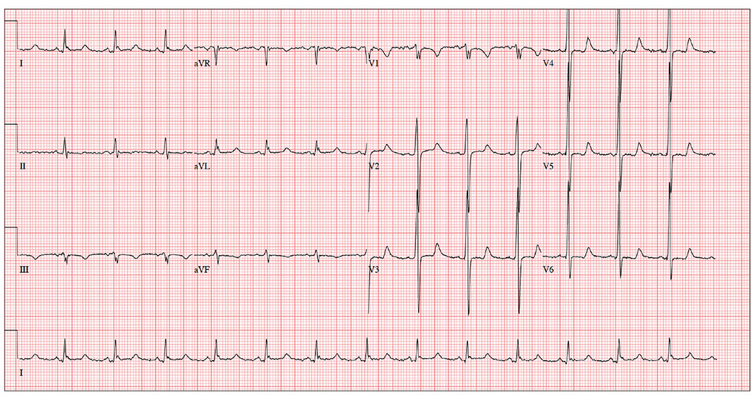
The ECG shows evidence of sinus rhythm, left ventricular hypertrophy, and a prolonged QT interval. Although it may be tempting to label the RR’ in lead V1 as right bundle branch block, recall that bundle branch block occurs with a QRS duration > 120 ms, which is not present here.
Left ventricular hypertrophy is demonstrated by high voltages in the precordial leads (S in lead V1 and R in lead V5 or V6 ≥ 35 mm); compare this with the ECG in the May issue of Clinician Reviews (2017;27(5):9, 13). A prolonged QT interval is suggested by a QTc interval of 500 m
The patient’s family history of sudden cardiac death makes these findings particularly concerning; genetic workup should be considered for both the patient and his children.
ANSWER
The ECG shows evidence of sinus rhythm, left ventricular hypertrophy, and a prolonged QT interval. Although it may be tempting to label the RR’ in lead V1 as right bundle branch block, recall that bundle branch block occurs with a QRS duration > 120 ms, which is not present here.
Left ventricular hypertrophy is demonstrated by high voltages in the precordial leads (S in lead V1 and R in lead V5 or V6 ≥ 35 mm); compare this with the ECG in the May issue of Clinician Reviews (2017;27(5):9, 13). A prolonged QT interval is suggested by a QTc interval of 500 m
The patient’s family history of sudden cardiac death makes these findings particularly concerning; genetic workup should be considered for both the patient and his children.
For the past three years, a now 62-year-old man has had pain in his left hip. His orthopedic surgeon recommends replacement, so he presents for preoperative assessment.
He has no history of cardiac disease but does have osteoarthritis, obesity, type 2 diabetes, hypertension, and hyperlipidemia. His surgical history is remarkable for a cholecystectomy performed when he was 48.
His medication list includes lisinopril, metoprolol, metformin, glyburide, naproxen, and atorvastatin. He is allergic to sulfa, which has caused anaphylaxis in the past.
The patient works as the warehouse supervisor of a local home improvement store. He is married with three adult children. His brother and uncle both succumbed to sudden cardiac death in their mid-40s. His mother died of a stroke, and his father of a myocardial infarction.
He reports joint pain consistent with osteoarthritis, occasional constipation, and urinary hesitancy. He is hard of hearing, wears corrective lenses, and walks with a limp.
Vital signs include a blood pressure of 148/88 mm Hg; pulse, 83 beats/min; respiratory rate, 12 breaths/min-1; and temperature, 99°F. His weight is 254 lb and his height, 69 in.
Physical exam reveals an obese man in no acute distress. Weber exam lateralizes to the left side. Dentition is in good repair, and his Mallampati score is II. The thyroid is normal, and there is no jugular venous distention. The lungs are clear bilaterally. The cardiac exam reveals a normal rhythm with a grade II/VI early systolic murmur best heard at the left upper sternal border. There are no extra heart sounds or rubs.
The abdomen is obese, with a well-healed surgical scar in the right upper quadrant. There are no masses. Peripheral pulses are strong bilaterally. There is no peripheral edema, and there are no lesions on either foot. Range of motion in the left hip is significantly limited due to pain.
An ECG reveals a ventricular rate of 83 beats/min; PR interval, 110 ms; QRS duration, 102 ms; QT/QTc interval, 426/500 ms; P axis, 4°; R axis, 5°; T axis, –21°. What is your interpretation?

Travel restrictions bar surgeon from U.S. meeting
A. Pieter Kappetein, MD, PhD, was looking forward to a busy week of meetings and activities at the annual meeting of the American Association for Thoracic Surgeons (AATS) in late April. Dr. Kappetein, a cardiothoracic surgeon at Erasmus MC in Rotterdam, the Netherlands, and secretary general for the European Association for Cardio-Thoracic Surgery, was giving two presentations at the meeting and had a full schedule of committee meetings, board meetings, and the like.
But on his way to the United States, Dr. Kappetein was pulled aside by U.S. immigration officials in Dublin and questioned about his travel background. After a long interrogation, he was informed he could not enter the United States and had to return home. The reason? His passport showed he had traveled to Iran 2 years earlier.

He was forced to give up his ticket to Boston, buy a new ticket home, and miss the work he was scheduled to do.
“I was angry,” Dr. Kappetein said. “They had to go get my luggage off the plane. I lost my ticket to Boston. It [was] a lot of money. I was very busy in Boston. I had meetings from early in the morning until late in the evening, and I had to give lectures as well. There were a lot of obligations that I could not fulfill.”
Restrictions hamper streamlined process
Dr. Kappetein’s ruined travel plans result from recent changes to the Visa Waiver Program (VWP). The program enables most citizens and nationals of participating countries to travel to the United States for tourism or business for 90 days or less without a visa; U.S. citizens and nationals get reciprocal travel access.
But changes to the program starting in 2015 mandated that most people who had traveled to or had been present in Iran, Iraq, Libya, Somalia, Sudan, Syria, or Yemen on March 1, 2011, or later, were no longer eligible to travel to under the VWP. The changes were part of the Visa Waiver Program Improvement and Terrorist Travel Prevention Act of 2015, signed by President Obama, which aimed to prevent travelers with ties to countries that pose terrorist threats from entering the country.
These new restrictions to the VWP have been enforced since January 2016, Jennifer Gabris, a spokesperson for U.S. Customs and Border Protection, said in an interview. She stressed that the eligibility requirements do not bar travel to the United States; rather, travelers who do not meet the requirements must obtain a visa.
Dr. Kappetein, however, said he has traveled to the United States from the Netherlands at least 10 times since his visit to Iran 2 years ago and encountered no problems until this year. He was under the assumption that travelers coming to the United States for business were exempt from the restrictions.
“I never had any issue at the border,” he said. “So the immigration officers, either they hadn’t seen [the passport stamp], or they had ignored it, or they didn’t care about it. I even got this prescreening [approval], so when I was in the States, I didn’t have to go in the line where you have to take off your shoes or your jacket.”
A Department of State spokeswoman referred questions about airport enforcement to the Department of Homeland Security, which declined to comment for this article.

“It takes time to implement, even after it’s signed,” Mr. Cohen said. “I don’t know to what degree it was being enforced, but the limited application means it probably just didn’t raise much attention.”
He noted there was little publicity surrounding the VWP restrictions until President Donald Trump called out the changes to justify his controversial travel ban, Mr. Cohen added. In his revised executive order released in March, the president noted that the countries named in his ban had already been “identified as presenting heightened concerns about terrorism and travel to the United States” through their exclusion from the VWP.
Harm to collaboration?
The VWP restrictions are among a number of increased security measures for foreigners and immigrants entering the country. President Trump’s March 6 Executive Order on immigration expanded uniform screening procedures for all visa classes and nationalities, while another provision suspended the Visa Interview Waiver Program. The interview program suspension means that certain applicants seeking to renew a visa must be interviewed in person by a consular officer. While a number of courts, including the 4th U.S. Circuit Court of Appeals, have blocked much of the Executive Order, the decisions did not halt the additional screening requirements or temporarily suspend the Visa Interview Waiver Program rollback. Both provisions remain in effect.
On May 4, the State Department proposed another regulation that would require more personal information from a subset of visa applicants, including 15 years of biographical information, employment history, addresses, prior passport numbers, information about family members including current and former spouses as well as travel histories and how trips were funded. Visa applicants would be required to provide phone numbers and email addresses used over the previous 5 years, according to the proposed rule. The subset of visa applicants would be determined by Department of State consular officers when resolving an applicant’s identity or when vetting for national security–related visa ineligibilities.
In a May 18 letter to the State Department, the American Association for the Advancement of Science and 17 other associations cautioned that the rule, if approved, would blunt scientific and academic collaborations, discourage foreign students from seeking to study and participate in research projects in the United States, and damage U.S. competitiveness.
“The notice, as proposed, is likely to have a chilling effect not only on those required to submit additional information, but indirectly on all international travelers to the United States,” the letter stated. “The uncertainties and confusion regarding supplemental questions will have a negative impact particularly on U.S. higher education and scientific collaborations.”
Since Dr. Kappetein’s travel mishap, he has obtained the required visa so that he may again travel to the United States. But he noted that entering this country will take at least 2 hours longer because of the visa entry protocols and additional airport screenings. The added restrictions and increased scrutiny of foreign travelers is unfortunate for U.S.-based medical conferences, he said.
“The interchange of information is really happening at those meetings,” he said. “[This] will block the exchange of information and it will block innovation. That’s a pity for [foreign physicians], but also for Americans.”
[email protected]
On Twitter @legal_med
A. Pieter Kappetein, MD, PhD, was looking forward to a busy week of meetings and activities at the annual meeting of the American Association for Thoracic Surgeons (AATS) in late April. Dr. Kappetein, a cardiothoracic surgeon at Erasmus MC in Rotterdam, the Netherlands, and secretary general for the European Association for Cardio-Thoracic Surgery, was giving two presentations at the meeting and had a full schedule of committee meetings, board meetings, and the like.
But on his way to the United States, Dr. Kappetein was pulled aside by U.S. immigration officials in Dublin and questioned about his travel background. After a long interrogation, he was informed he could not enter the United States and had to return home. The reason? His passport showed he had traveled to Iran 2 years earlier.
He was forced to give up his ticket to Boston, buy a new ticket home, and miss the work he was scheduled to do.
“I was angry,” Dr. Kappetein said. “They had to go get my luggage off the plane. I lost my ticket to Boston. It [was] a lot of money. I was very busy in Boston. I had meetings from early in the morning until late in the evening, and I had to give lectures as well. There were a lot of obligations that I could not fulfill.”
Restrictions hamper streamlined process
Dr. Kappetein’s ruined travel plans result from recent changes to the Visa Waiver Program (VWP). The program enables most citizens and nationals of participating countries to travel to the United States for tourism or business for 90 days or less without a visa; U.S. citizens and nationals get reciprocal travel access.
But changes to the program starting in 2015 mandated that most people who had traveled to or had been present in Iran, Iraq, Libya, Somalia, Sudan, Syria, or Yemen on March 1, 2011, or later, were no longer eligible to travel to under the VWP. The changes were part of the Visa Waiver Program Improvement and Terrorist Travel Prevention Act of 2015, signed by President Obama, which aimed to prevent travelers with ties to countries that pose terrorist threats from entering the country.
These new restrictions to the VWP have been enforced since January 2016, Jennifer Gabris, a spokesperson for U.S. Customs and Border Protection, said in an interview. She stressed that the eligibility requirements do not bar travel to the United States; rather, travelers who do not meet the requirements must obtain a visa.
Dr. Kappetein, however, said he has traveled to the United States from the Netherlands at least 10 times since his visit to Iran 2 years ago and encountered no problems until this year. He was under the assumption that travelers coming to the United States for business were exempt from the restrictions.
“I never had any issue at the border,” he said. “So the immigration officers, either they hadn’t seen [the passport stamp], or they had ignored it, or they didn’t care about it. I even got this prescreening [approval], so when I was in the States, I didn’t have to go in the line where you have to take off your shoes or your jacket.”
A Department of State spokeswoman referred questions about airport enforcement to the Department of Homeland Security, which declined to comment for this article.
“It takes time to implement, even after it’s signed,” Mr. Cohen said. “I don’t know to what degree it was being enforced, but the limited application means it probably just didn’t raise much attention.”
He noted there was little publicity surrounding the VWP restrictions until President Donald Trump called out the changes to justify his controversial travel ban, Mr. Cohen added. In his revised executive order released in March, the president noted that the countries named in his ban had already been “identified as presenting heightened concerns about terrorism and travel to the United States” through their exclusion from the VWP.
Harm to collaboration?
The VWP restrictions are among a number of increased security measures for foreigners and immigrants entering the country. President Trump’s March 6 Executive Order on immigration expanded uniform screening procedures for all visa classes and nationalities, while another provision suspended the Visa Interview Waiver Program. The interview program suspension means that certain applicants seeking to renew a visa must be interviewed in person by a consular officer. While a number of courts, including the 4th U.S. Circuit Court of Appeals, have blocked much of the Executive Order, the decisions did not halt the additional screening requirements or temporarily suspend the Visa Interview Waiver Program rollback. Both provisions remain in effect.
On May 4, the State Department proposed another regulation that would require more personal information from a subset of visa applicants, including 15 years of biographical information, employment history, addresses, prior passport numbers, information about family members including current and former spouses as well as travel histories and how trips were funded. Visa applicants would be required to provide phone numbers and email addresses used over the previous 5 years, according to the proposed rule. The subset of visa applicants would be determined by Department of State consular officers when resolving an applicant’s identity or when vetting for national security–related visa ineligibilities.
In a May 18 letter to the State Department, the American Association for the Advancement of Science and 17 other associations cautioned that the rule, if approved, would blunt scientific and academic collaborations, discourage foreign students from seeking to study and participate in research projects in the United States, and damage U.S. competitiveness.
“The notice, as proposed, is likely to have a chilling effect not only on those required to submit additional information, but indirectly on all international travelers to the United States,” the letter stated. “The uncertainties and confusion regarding supplemental questions will have a negative impact particularly on U.S. higher education and scientific collaborations.”
Since Dr. Kappetein’s travel mishap, he has obtained the required visa so that he may again travel to the United States. But he noted that entering this country will take at least 2 hours longer because of the visa entry protocols and additional airport screenings. The added restrictions and increased scrutiny of foreign travelers is unfortunate for U.S.-based medical conferences, he said.
“The interchange of information is really happening at those meetings,” he said. “[This] will block the exchange of information and it will block innovation. That’s a pity for [foreign physicians], but also for Americans.”
[email protected]
On Twitter @legal_med
Primary care involvement improves chance of meeting recommended surveillance intervals
CHICAGO – Only a small portion of patients with nondysplastic Barrett’s esophagus received appropriately timed endoscopic surveillance, a large database study showed.
But rather than being neglected, patients were more likely to be overassessed, with follow-up endoscopy performed more frequently than the recommended 3- to 5-year intervals, Anna Tavakkoli, MD, reported at the annual Digestive Disease Week.
“Very few patients entered our surveillance program with appropriate surveillance intervals,” said Dr. Tavakkoli, a gastroenterology fellow at the University of Michigan, Ann Arbor.

“We don’t have a formal program at the University of Michigan that drives coordination of care, but we do have great communication here between our primary care providers and our specialists. Our electronic medical records system makes quick messaging between providers easy, and primary care is very good about incorporating diagnoses into patients’ problem lists.”
Malignant transformation of nondysplastic Barrett’s is uncommon, with rates of no more than 4% per year. This understanding led three major societies – the American Gastroenterology Association, the American Society of Gastrointestinal Endoscopy, and the American College of Gastroenterology – to amend their surveillance recommendations in 2011 and 2012. All three societies now recommend a surveillance endoscopy every 3-5 years after the initial diagnosis of nondysplastic Barrett’s esophagus. In fact, the AGA has incorporated this suggestion into its five “Choosing Wisely” recommendations aimed at decreasing overutilization of testing and procedures.
Dr. Tavakkoli’s study examined surveillance timing in a cohort of 1,602 patients with nondysplastic Barrett’s who entered the University of Michigan Barrett’s Esophagus Registry from 1994 to 2016. All of these patents had at least three endoscopies or at least 5 years of follow-up data since their last endoscopy. The primary outcome was identification of trends in the appropriateness of surveillance of patients with nondysplastic Barrett’s esophagus at the University of Michigan. In her analysis, oversurveillance was defined as less than 3 years between the second and third endoscopy; undersurveillance was defined as more than 5 years between them. Dr. Tavakkoli and her colleagues also looked at patients who were lost to follow-up, defined as never receiving a second endoscopy after their initial diagnosis of nondysplastic Barrett’s esophagus and patients who were never surveilled, defined as never receiving their third endoscopy. All patients were compared with those who underwent appropriate surveillance, defined as 3-5 years between their second and third procedure.
The majority of patients were male, and the mean age was 59 years; 30% had long-segment Barrett’s, and 41% had a primary care provider in the university health care system. Most (90%) had their second endoscopy before 2012, when two of the three major societies issued their updated surveillance recommendations.
Of the entire cohort, 40% were lost to follow-up; 17% were never surveilled, and 3% were undersurveilled. Almost a third (31%) were oversurveilled, while just 8% had the appropriate surveillance, Dr. Tavakkoli said.
She then looked at several demographic and clinical factors associated with surveillance in each group, including sex, age, race, and income, comorbidities, length of Barrett’s, family history of esophageal cancer, and whether the patient had a University of Michigan primary care provider.
Having long-segment Barrett’s was associated with a 2.5-times increased risk of receiving a third endoscopy earlier than 3 years, which may be driven by studies that have shown that the risk of malignant transformation increases with Barrett’s length, she said.
The presence of a primary care physician significantly reduced the risk of inappropriate follow-up in every group, except patients who were undersurveilled, she said. The presence of a primary care physician at the University of Michigan decreased the risk of oversurveillance by 56%.
The positive influence of an in-system primary care physician was an important finding in this study, Dr Tavakkoli said. “The oncology data have shown us that poor coordination of care between oncologists and primary care providers contributes to avoidable patient morbidity and mortality, fragmented care, and increased costs. In 2005, the Institute of Medicine published a report emphasizing that coordination between specialists and primary care providers is one of the four key components to cancer survivorship care. There have been a number of GI studies looking at how primary care’s involvement in colorectal screening improves the rates of patients who undergo screening, but among Barrett’s patients, there have not been data showing that having a primary care physician at the center where endoscopic surveillance is done improves utilization patterns.”
Dr. Tavakkoli had no financial disclosures.
[email protected]
On Twitter @alz_gal
CHICAGO – Only a small portion of patients with nondysplastic Barrett’s esophagus received appropriately timed endoscopic surveillance, a large database study showed.
But rather than being neglected, patients were more likely to be overassessed, with follow-up endoscopy performed more frequently than the recommended 3- to 5-year intervals, Anna Tavakkoli, MD, reported at the annual Digestive Disease Week.
“Very few patients entered our surveillance program with appropriate surveillance intervals,” said Dr. Tavakkoli, a gastroenterology fellow at the University of Michigan, Ann Arbor.
“We don’t have a formal program at the University of Michigan that drives coordination of care, but we do have great communication here between our primary care providers and our specialists. Our electronic medical records system makes quick messaging between providers easy, and primary care is very good about incorporating diagnoses into patients’ problem lists.”
Malignant transformation of nondysplastic Barrett’s is uncommon, with rates of no more than 4% per year. This understanding led three major societies – the American Gastroenterology Association, the American Society of Gastrointestinal Endoscopy, and the American College of Gastroenterology – to amend their surveillance recommendations in 2011 and 2012. All three societies now recommend a surveillance endoscopy every 3-5 years after the initial diagnosis of nondysplastic Barrett’s esophagus. In fact, the AGA has incorporated this suggestion into its five “Choosing Wisely” recommendations aimed at decreasing overutilization of testing and procedures.
Dr. Tavakkoli’s study examined surveillance timing in a cohort of 1,602 patients with nondysplastic Barrett’s who entered the University of Michigan Barrett’s Esophagus Registry from 1994 to 2016. All of these patents had at least three endoscopies or at least 5 years of follow-up data since their last endoscopy. The primary outcome was identification of trends in the appropriateness of surveillance of patients with nondysplastic Barrett’s esophagus at the University of Michigan. In her analysis, oversurveillance was defined as less than 3 years between the second and third endoscopy; undersurveillance was defined as more than 5 years between them. Dr. Tavakkoli and her colleagues also looked at patients who were lost to follow-up, defined as never receiving a second endoscopy after their initial diagnosis of nondysplastic Barrett’s esophagus and patients who were never surveilled, defined as never receiving their third endoscopy. All patients were compared with those who underwent appropriate surveillance, defined as 3-5 years between their second and third procedure.
The majority of patients were male, and the mean age was 59 years; 30% had long-segment Barrett’s, and 41% had a primary care provider in the university health care system. Most (90%) had their second endoscopy before 2012, when two of the three major societies issued their updated surveillance recommendations.
Of the entire cohort, 40% were lost to follow-up; 17% were never surveilled, and 3% were undersurveilled. Almost a third (31%) were oversurveilled, while just 8% had the appropriate surveillance, Dr. Tavakkoli said.
She then looked at several demographic and clinical factors associated with surveillance in each group, including sex, age, race, and income, comorbidities, length of Barrett’s, family history of esophageal cancer, and whether the patient had a University of Michigan primary care provider.
Having long-segment Barrett’s was associated with a 2.5-times increased risk of receiving a third endoscopy earlier than 3 years, which may be driven by studies that have shown that the risk of malignant transformation increases with Barrett’s length, she said.
The presence of a primary care physician significantly reduced the risk of inappropriate follow-up in every group, except patients who were undersurveilled, she said. The presence of a primary care physician at the University of Michigan decreased the risk of oversurveillance by 56%.
The positive influence of an in-system primary care physician was an important finding in this study, Dr Tavakkoli said. “The oncology data have shown us that poor coordination of care between oncologists and primary care providers contributes to avoidable patient morbidity and mortality, fragmented care, and increased costs. In 2005, the Institute of Medicine published a report emphasizing that coordination between specialists and primary care providers is one of the four key components to cancer survivorship care. There have been a number of GI studies looking at how primary care’s involvement in colorectal screening improves the rates of patients who undergo screening, but among Barrett’s patients, there have not been data showing that having a primary care physician at the center where endoscopic surveillance is done improves utilization patterns.”
Dr. Tavakkoli had no financial disclosures.
[email protected]
On Twitter @alz_gal
AT DDW
Key clinical point:
Major finding: Only 8% of patients had the recommended interval of 3-5 years between surveillance endoscopies.
Data source: A retrospective database cohort of 1,602 patients.
Disclosures: Dr. Tavakkoli had no relevant financial disclosures.
Later school start tied to more sleep
BOSTON – Teenagers slept more hours when they started school later, a new study found.
With 73% of high schoolers reporting receiving less than the recommended 8 hours of sleep per night, teens are among the most sleep-deprived members of society.
In this study, the later a teenager started school, the later he or she woke up, with average wake-up times having been just after 6:00 a.m. for those starting school between 7:00 and 7:30 a.m. and about 7:00 a.m. for those starting school after 8:30 a.m. But only those teens who started after 8:30 a.m. achieved the 8-hour recommended sleep duration, said Nicole Nahmod, during a presentation at the annual meeting of the Associated Professional Sleep Societies.
The students with the later school start times averaged 32 minutes of extra sleep, when compared with their early-rising colleagues, noted Ms. Nahmod, who is one of the study’s authors, a research technician, and study coordinator at Pennsylvania State University, Hershey. Specifically, adolescents who started school after 8:30 a.m. had a mean sleep duration of 8.1 hours, while those who started school earlier slept for only 7.5 hours a night.*

Acknowledging the importance of adequate sleep on teen health, mood, and school performance, the American Academy of Pediatrics recommends that middle schools and high schools begin at 8:30 a.m. or later. However, most school days start earlier than that, as evidenced by this dataset. While 72% of the study’s participants started school between 7:00 a.m. and 8:30 a.m., as many as 15% of the study participants began school during the narrower 7:00-7:30 a.m. window.
In this study, researchers from Penn State used data from 413 adolescents (mean age, 15.4 years; 46% male), who participated in a substudy of the Fragile Families & Child Wellbeing Study conducted across 20 large American cities. The study oversampled nonmarried pregnant mothers, resulting in a racially diverse sample and a high proportion of low-income families. The household income for 29% of the sample was below the poverty line.*
For this substudy, sleep duration was calculated from app-based daily diary reports of bed times, wake times, and school start times on days when a teen attended school.
The study was limited by its cross-sectional nature, but was enriched by a diverse range of school start times sampled from 20 U.S. cities, the high proportion of at-risk teens, and the use of a daily sleep diary.
“Current literature shows associations between later school start times and academic success, mood, and health. It also shows a decrease in motor vehicle accidents, tardiness, school dropout, and daytime sleepiness,” Ms. Nahmod noted.
Her continued research in this area involves analyzing the relationships between actigraphically assessed sleep measures in students and school start times.
Ms. Nahmod reported having no financial disclosures.
*This article was updated on June 6, 2017.
BOSTON – Teenagers slept more hours when they started school later, a new study found.
With 73% of high schoolers reporting receiving less than the recommended 8 hours of sleep per night, teens are among the most sleep-deprived members of society.
In this study, the later a teenager started school, the later he or she woke up, with average wake-up times having been just after 6:00 a.m. for those starting school between 7:00 and 7:30 a.m. and about 7:00 a.m. for those starting school after 8:30 a.m. But only those teens who started after 8:30 a.m. achieved the 8-hour recommended sleep duration, said Nicole Nahmod, during a presentation at the annual meeting of the Associated Professional Sleep Societies.
The students with the later school start times averaged 32 minutes of extra sleep, when compared with their early-rising colleagues, noted Ms. Nahmod, who is one of the study’s authors, a research technician, and study coordinator at Pennsylvania State University, Hershey. Specifically, adolescents who started school after 8:30 a.m. had a mean sleep duration of 8.1 hours, while those who started school earlier slept for only 7.5 hours a night.*
Acknowledging the importance of adequate sleep on teen health, mood, and school performance, the American Academy of Pediatrics recommends that middle schools and high schools begin at 8:30 a.m. or later. However, most school days start earlier than that, as evidenced by this dataset. While 72% of the study’s participants started school between 7:00 a.m. and 8:30 a.m., as many as 15% of the study participants began school during the narrower 7:00-7:30 a.m. window.
In this study, researchers from Penn State used data from 413 adolescents (mean age, 15.4 years; 46% male), who participated in a substudy of the Fragile Families & Child Wellbeing Study conducted across 20 large American cities. The study oversampled nonmarried pregnant mothers, resulting in a racially diverse sample and a high proportion of low-income families. The household income for 29% of the sample was below the poverty line.*
For this substudy, sleep duration was calculated from app-based daily diary reports of bed times, wake times, and school start times on days when a teen attended school.
The study was limited by its cross-sectional nature, but was enriched by a diverse range of school start times sampled from 20 U.S. cities, the high proportion of at-risk teens, and the use of a daily sleep diary.
“Current literature shows associations between later school start times and academic success, mood, and health. It also shows a decrease in motor vehicle accidents, tardiness, school dropout, and daytime sleepiness,” Ms. Nahmod noted.
Her continued research in this area involves analyzing the relationships between actigraphically assessed sleep measures in students and school start times.
Ms. Nahmod reported having no financial disclosures.
*This article was updated on June 6, 2017.
AT SLEEP 2017
Key clinical point: Teenage students who start school after 8:30 a.m. are more likely to get the recommended 8 hours of sleep than are their counterparts with earlier start times.
Major finding: Adolescents who started school after 8:30 a.m. had a mean sleep duration of 8.1 hours, compared with 7.5 hours for students who started school earlier.
Data source: A national longitudinal birth cohort study of 413 adolescents.
Disclosures: Ms. Nahmod reported having no financial disclosures.
Antiangiogenesis in Small-Cell Lung Cancer: Is There a Path Forward?
Study Overview
Objective. To evaluate efficacy of adding bevacizumab to first-line chemotherapy for treatment of extensive-disease small-cell lung cancer (ED-SCLC).
Design. Phase III prospective multicenter randomized clinical trial.
Setting and participants. The study was conducted at 29 Italian centers and was supported by the Agenzia Italiana del Farmaco. Study entry was limited to patients with histologically or cytologically documented ED-SCLC who were previously untreated with systemic therapy, were 18 years of age or older, and had an Eastern Cooperative Oncology Group performance status (ECOG PS) of 0 to 2. Adequate bone marrow, renal, and liver functions were required. Patients with asymptomatic, treated brain metastases were eligible for trial participation. Exclusions included the following: mixed histologic diagnosis of SCLC and non–SCLC; history of grade 2 hemoptysis; evidence of lung tumor cavitation; significant traumatic injury within the 4 weeks before first dose of study treatment; other active malignancies (previous or current); and any underlying medical condition that might be aggravated by treatment.
Intervention. Patients received a combination of intravenous cisplatin (25 mg/m2 on days 1 to 3), etoposide (100 mg/m2 on days 1 to 3), and bevacizumab (7.5 mg/kg intravenously on day 1) administered every 3 weeks (experimental arm); or the same cisplatin and etoposide chemotherapy regimen alone given every 3 weeks (control arm). Carboplatin (area under the curve 5 on day 1) could be substituted for cisplatin in case of cisplatin contraindications or cisplatin-associated toxicity. Tumor response, on the basis of investigator-assessed Response Evaluation Criteria in Solid Tumors (RECIST; version 1.1), was evaluated every 3 cycles during chemotherapy treatment. After 6 cycles of chemotherapy, tumor assessment was performed every 9 weeks in both arms. In the absence of progression, patients in the treatment arm continued bevacizumab alone until disease progression or for a maximum of 18 courses. Survival follow-up information was collected every 6 months after treatment termination or last dose of study drug, until death or loss to follow-up.
Main outcome measure. The primary end point was overall survival (OS). Response rate, toxicity, and progression-free survival (PFS) were secondary end points.
Main results. 205 patients were randomized between November 2009 and October 2015. 204 patients were considered in the intent-to-treat analysis (103 in the control arm and 101 in the treatment arm). Most patients were male with ECOG PS of 0 to 1. Median age was 64 years. The median number of chemotherapy courses administered was 6 in both arms. Cisplatin was used in majority of the patients. Average relative dose intensities for all drugs were well balanced between 2 groups. A lower percentage of patients in the treatment arm (14.7%) than in the control arm (22.3%) discontinued treatment because of radiologic disease progression, which was the main reason for treatment discontinuation.
At a median follow-up of 34.9 months, the median PFS was 5.7 in the control arm and 6.7 months in the treatment arm (hazard ratio [HR], 0.72; 95% CI, 0.54 to 0.97; P = 0.30). Median OS times were 8.9 months and 9.8 months, and 1-year survival rates were 25% and 37% (HR, 0.78; 95% CI, 0.58 to 1.06; P = 0.113) in the control arm and treatment arm, respectively. A significant effect of the maintenance treatment on OS (HR, 0.60; 95% CI, 0.40 to 0.91, P = 0.011) was observed. A subgroup analysis revealed a statistically significant interaction for OS between treatment and sex; the addition of bevacizumab led to a significant survival benefit in men (HR, 0.55) and to a possible detrimental effect in women (HR, 1.55; interaction test, P = 0.003).
Addition of bevacizumab did not result in increase in hematologic toxicity such as anemia, neutropenia, or thrombocytopenia. Concerning the nonhematologic toxicity, only hypertension was more frequent in the bevacizumab arm (6.3%) compared to chemotherapy alone arm (1%). The rates of proteinuria and thrombosis were similar in both arms.
Conclusion. The addition of bevacizumab to cisplatin and etoposide in the first-line treatment of ED-SCLC had an acceptable toxicity profile and led to a statistically significant improvement in PFS, which, however, did not translate into a statistically significant increase in OS.
Commentary
SCLC currently accounts for approximately 12% to 15% of all lung cancers [1]. It is characterized by a rapid growth rate, metastasis at the time of diagnosis, sensitivity to first-line platinum-based chemotherapy, and invariable recurrence and progressive resistance to subsequent lines of therapy. A number of clinical trials over the past 2 decades have failed to produce outcomes superior to platinum-based doublet chemotherapy, leaving a significant unmet need [2]. Vascular endothelial growth factor (VEGF) is the most important proangiogenic factor, and it is implicated in tumor growth [3]. Bevacizumab, a humanized monoclonal antibody directed against VEGF, is now indicated in the treatment of several tumor types including non–SCLC and breast, colorectal, kidney, and ovarian cancer. Positive signal with bevacizumab was seen in phase II studies, providing rationale for this phase III trial [4,5] .
The study by Tiseo and colleagues reported the outcomes of a randomized study that added bevacizumab to standard combination therapy with platinum and etoposide for the treatment of ED-SCLC. A small statistically significant improvement was seen in PFS (6.7 months vs. 5.7 months, favoring the bevacizumab group). However, the study failed to meet the primary end point of improved OS.
So where do antiangiogenesis agents go from here? Alternative angiogenesis inhibitors with broader mechanism of action are being explored in clinical trials. One such trial (ClinicalTrials.gov identifier: NCT02945852) is evaluating the role of the tyrosine kinase inhibitor apatinib in combination with chemotherapy in ED-SCLC. Apatinib selectively inhibits the vascular growth factor receptor-2 (VEGFR2). In addition, this agent also inhibits c-kit and c-SRC tyrosine kinase. It would be interesting to see if antiangiogenic agents with broader mechanisms would be more effective in SCLC. Immunotherapy with checkpoint inhibitors such as nivolumab and pembrolizumab have revolutionized the lung cancer treatment paradigm. It would be interesting to see if bevacizumab could be safely added to these immunotherapy agents. The ongoing CheckMate 370 (ClinicalTrials.gov identifier: NCT02574078) is addressing this question, evaluating the safety of combining nivolumab with bevacizumab in non-SCLC.
Applications for Clinical Practice
The current study does not support the addition of bevacizumab as a standard therapeutic option in the first-line treatment of ED-SCLC. However, given that there was a trend towards improved OS, alternative strategies of incorporating antiangiogenesis agents should be considered in future clinical trials.
—Deval Rajyaguru, MD
1. Neal JW, Gubens MA, Wakelee HA. Current management of small cell lung cancer. Clin Chest Med 2011;32:853–63.
2. Bunn PA Jr, Minna JD, Augustyn A, et al. Small cell lung cancer. Can recent advances in biology and molecular biology be translated into improved outcomes? J Thorac Oncol 2016;11:453–74.
3. Ferrara N, Gerber HP, LeCouter J. The biology of VEGF and its receptors. Nat Med 2003:9:669–676.
4. Horn L, Dahlberg SE, Sandler AB, et al. Phase II study of cisplatin plus etoposide and bevacizumab for previously untreated, extensive-stage small-cell lung cancer: Eastern Cooperative Oncology Group Study E3501. J Clin Oncol 2009;27:6006–11.
5. Spigel DR, Townley PM, Waterhouse DM, et al. Randomized phase II study of bevacizumab in combination with chemotherapy in previously untreated extensive-stage small-cell lung cancer: Results from the SALUTE trial. J Clin Oncol 2011;29:2215–22.
Study Overview
Objective. To evaluate efficacy of adding bevacizumab to first-line chemotherapy for treatment of extensive-disease small-cell lung cancer (ED-SCLC).
Design. Phase III prospective multicenter randomized clinical trial.
Setting and participants. The study was conducted at 29 Italian centers and was supported by the Agenzia Italiana del Farmaco. Study entry was limited to patients with histologically or cytologically documented ED-SCLC who were previously untreated with systemic therapy, were 18 years of age or older, and had an Eastern Cooperative Oncology Group performance status (ECOG PS) of 0 to 2. Adequate bone marrow, renal, and liver functions were required. Patients with asymptomatic, treated brain metastases were eligible for trial participation. Exclusions included the following: mixed histologic diagnosis of SCLC and non–SCLC; history of grade 2 hemoptysis; evidence of lung tumor cavitation; significant traumatic injury within the 4 weeks before first dose of study treatment; other active malignancies (previous or current); and any underlying medical condition that might be aggravated by treatment.
Intervention. Patients received a combination of intravenous cisplatin (25 mg/m2 on days 1 to 3), etoposide (100 mg/m2 on days 1 to 3), and bevacizumab (7.5 mg/kg intravenously on day 1) administered every 3 weeks (experimental arm); or the same cisplatin and etoposide chemotherapy regimen alone given every 3 weeks (control arm). Carboplatin (area under the curve 5 on day 1) could be substituted for cisplatin in case of cisplatin contraindications or cisplatin-associated toxicity. Tumor response, on the basis of investigator-assessed Response Evaluation Criteria in Solid Tumors (RECIST; version 1.1), was evaluated every 3 cycles during chemotherapy treatment. After 6 cycles of chemotherapy, tumor assessment was performed every 9 weeks in both arms. In the absence of progression, patients in the treatment arm continued bevacizumab alone until disease progression or for a maximum of 18 courses. Survival follow-up information was collected every 6 months after treatment termination or last dose of study drug, until death or loss to follow-up.
Main outcome measure. The primary end point was overall survival (OS). Response rate, toxicity, and progression-free survival (PFS) were secondary end points.
Main results. 205 patients were randomized between November 2009 and October 2015. 204 patients were considered in the intent-to-treat analysis (103 in the control arm and 101 in the treatment arm). Most patients were male with ECOG PS of 0 to 1. Median age was 64 years. The median number of chemotherapy courses administered was 6 in both arms. Cisplatin was used in majority of the patients. Average relative dose intensities for all drugs were well balanced between 2 groups. A lower percentage of patients in the treatment arm (14.7%) than in the control arm (22.3%) discontinued treatment because of radiologic disease progression, which was the main reason for treatment discontinuation.
At a median follow-up of 34.9 months, the median PFS was 5.7 in the control arm and 6.7 months in the treatment arm (hazard ratio [HR], 0.72; 95% CI, 0.54 to 0.97; P = 0.30). Median OS times were 8.9 months and 9.8 months, and 1-year survival rates were 25% and 37% (HR, 0.78; 95% CI, 0.58 to 1.06; P = 0.113) in the control arm and treatment arm, respectively. A significant effect of the maintenance treatment on OS (HR, 0.60; 95% CI, 0.40 to 0.91, P = 0.011) was observed. A subgroup analysis revealed a statistically significant interaction for OS between treatment and sex; the addition of bevacizumab led to a significant survival benefit in men (HR, 0.55) and to a possible detrimental effect in women (HR, 1.55; interaction test, P = 0.003).
Addition of bevacizumab did not result in increase in hematologic toxicity such as anemia, neutropenia, or thrombocytopenia. Concerning the nonhematologic toxicity, only hypertension was more frequent in the bevacizumab arm (6.3%) compared to chemotherapy alone arm (1%). The rates of proteinuria and thrombosis were similar in both arms.
Conclusion. The addition of bevacizumab to cisplatin and etoposide in the first-line treatment of ED-SCLC had an acceptable toxicity profile and led to a statistically significant improvement in PFS, which, however, did not translate into a statistically significant increase in OS.
Commentary
SCLC currently accounts for approximately 12% to 15% of all lung cancers [1]. It is characterized by a rapid growth rate, metastasis at the time of diagnosis, sensitivity to first-line platinum-based chemotherapy, and invariable recurrence and progressive resistance to subsequent lines of therapy. A number of clinical trials over the past 2 decades have failed to produce outcomes superior to platinum-based doublet chemotherapy, leaving a significant unmet need [2]. Vascular endothelial growth factor (VEGF) is the most important proangiogenic factor, and it is implicated in tumor growth [3]. Bevacizumab, a humanized monoclonal antibody directed against VEGF, is now indicated in the treatment of several tumor types including non–SCLC and breast, colorectal, kidney, and ovarian cancer. Positive signal with bevacizumab was seen in phase II studies, providing rationale for this phase III trial [4,5] .
The study by Tiseo and colleagues reported the outcomes of a randomized study that added bevacizumab to standard combination therapy with platinum and etoposide for the treatment of ED-SCLC. A small statistically significant improvement was seen in PFS (6.7 months vs. 5.7 months, favoring the bevacizumab group). However, the study failed to meet the primary end point of improved OS.
So where do antiangiogenesis agents go from here? Alternative angiogenesis inhibitors with broader mechanism of action are being explored in clinical trials. One such trial (ClinicalTrials.gov identifier: NCT02945852) is evaluating the role of the tyrosine kinase inhibitor apatinib in combination with chemotherapy in ED-SCLC. Apatinib selectively inhibits the vascular growth factor receptor-2 (VEGFR2). In addition, this agent also inhibits c-kit and c-SRC tyrosine kinase. It would be interesting to see if antiangiogenic agents with broader mechanisms would be more effective in SCLC. Immunotherapy with checkpoint inhibitors such as nivolumab and pembrolizumab have revolutionized the lung cancer treatment paradigm. It would be interesting to see if bevacizumab could be safely added to these immunotherapy agents. The ongoing CheckMate 370 (ClinicalTrials.gov identifier: NCT02574078) is addressing this question, evaluating the safety of combining nivolumab with bevacizumab in non-SCLC.
Applications for Clinical Practice
The current study does not support the addition of bevacizumab as a standard therapeutic option in the first-line treatment of ED-SCLC. However, given that there was a trend towards improved OS, alternative strategies of incorporating antiangiogenesis agents should be considered in future clinical trials.
—Deval Rajyaguru, MD
1. Neal JW, Gubens MA, Wakelee HA. Current management of small cell lung cancer. Clin Chest Med 2011;32:853–63.
2. Bunn PA Jr, Minna JD, Augustyn A, et al. Small cell lung cancer. Can recent advances in biology and molecular biology be translated into improved outcomes? J Thorac Oncol 2016;11:453–74.
3. Ferrara N, Gerber HP, LeCouter J. The biology of VEGF and its receptors. Nat Med 2003:9:669–676.
4. Horn L, Dahlberg SE, Sandler AB, et al. Phase II study of cisplatin plus etoposide and bevacizumab for previously untreated, extensive-stage small-cell lung cancer: Eastern Cooperative Oncology Group Study E3501. J Clin Oncol 2009;27:6006–11.
5. Spigel DR, Townley PM, Waterhouse DM, et al. Randomized phase II study of bevacizumab in combination with chemotherapy in previously untreated extensive-stage small-cell lung cancer: Results from the SALUTE trial. J Clin Oncol 2011;29:2215–22.
Vigorous Physical Activity Associated with Greater Arterial Compliance in Both Large and Small Arteries
Study Overview
Objective. To investigate the association between habitually high levels of physical activity and the compliance of the large and small arteries in men and women throughout the life span.
Design. Cross-sectional study.
Setting and participants. 83 healthy men (n = 44) and women (n = 39) aged between 18 and 78 years were recruited to participate in the study. Potential participants were recruited via flyers designed to elicit responses from either very highly active (participate in regular, vigorous exercise more than 5 times per week) or less active/sedentary individuals (participate in light to moderate exercise less than 3 times per week or none at all). Both groups subjectively reported maintaining the specified activity level for at least the past 5 years. The highly active subjects performed regular vigorous swimming as their primary mode of exercise training as most were members of a varsity or masters swim team. All subjects were free of overt chronic diseases, nonsmokers, and none were taking vasoactive medications as assessed by a medical history questionnaire. All subjects provided written informed consent to participate. The study was reviewed and approved by the institutional review board at Indiana University.
Physical activity was self-assessed in all subject groups with a log detailing their activity over the previous 7 days. To ensure the older highly active population performed vigorous physical activity ≥ 5 days per week, the subjective activity log was verified by a 7-day previously validated, commercially available heart rate monitor and accelerometer (Actiheart, CamNtech, Cambridge, UK).
Main outcome measure. Compliance of the small and large arteries (inverse of stiffness) measured using a commercial pulse wave analyzer (Model CR-2000, Hypertension Diagnositics, Eagen, MN), which according to the manufacturer measures proximal capacitive compliance (C1, or estimate of large artery compliance) and distal oscillatory compliance (C2, or small artery compliance) [1].
Results. The study found a positive association between routine vigorous physical activity and arterial compliance. Specifically, the results suggest that vigorous physical activity is associated with greater compliance of the small and large arteries in both younger and older adults (P < 0.05). In addition, both the highly active and less active younger groups as well as the highly active older group demonstrated greater large arterial compliance compared to the less active older group (P < 0.008). No significant differences were found between men and women.
Conclusion. Researchers concluded that participation in habitual vigorous physical activity is associated with benefits to the compliance of the small and large arteries. Habitual vigorous physical activity over time may attenuate age-associated cardiovascular impairments.
Commentary
Arterial compliance declines with age, and increased arterial stiffness is associated with an increased risk of cardiovascular events [2]. Evidence suggests that physical activity may delay or prevent age-related increases in arterial stiffness [3]. Previous research regarding age-related arterial stiffness and exercise has focused primarily on the large arteries. For example, Tanaka found that regular aerobic-endurance exercise attenuates age-related reductions in central arterial compliance and restores levels in previously sedentary healthy middle-aged and older men [3]. More recently, a study by Duprez [4] found that small artery elasticity was superior to large artery elasticity with regard to predicting future CHD, stroke, and heart failure.
In this study, researchers cross-sectionally investigated the relationship of intense and continuous physical activity in young and older adults. The form of vigorous activity in this study was competitive swimming, as participants were recruited from a collegiate varsity and masters swim team. The study found a statistically strong association between routine vigorous physical activity and arterial compliance. These findings agree with several studies showing the benefits of vigorous exercise, but go beyond these by presenting findings on small artery compliance.
Methodologically, this study has some limitations. With the small sample, the study may not have been adequately powered. Further, physical activity assessment was by self-report in the main. Even though researchers had the participants keep a log, self-report measures may be inaccurate. Another limitation was the indirect method of measuring compliance, in which the radial waveform is calibrated to brachial blood pressure values. However, the researchers followed a valid model using the same BP level–based procedures reported in previous studies [1].
Applications for Clinical Practice
CVD is a major cause of disability and mortality in the United States. Health care professionals have a significant role to play in reducing cardiovascular risk factors in their patients, including encouraging aerobic exercise. The American Heart Association recommends at least 30 minutes of moderate-intensity aerobic activity at least 5 days per week or at least 25 minutes of vigorous aerobic activity at least 3 days per week, or a combination of moderate- and vigorous-intensity aerobic activity [4]. Patients can also be reminded that even modest levels of physical activity are associated with health benefits.
—Paloma Cesar de Sales, BS, RN, MS
1. Cohn JN, Finkelstein S, McVeigh G, et al. Noninvasive pulse wave analysis for the early detection of vascular disease. Hypertension 1995;26:503–8.
2. Strait JB, Lakatta EG. Aging-associated cardiovascular changes and their relationship to heart failure. Heart Failure Clin 2012;8:143–64.
3. Tanaka H, Dinenno FA, Monahan KD, et al. Aging, habitual exercise, and dynamic arterial compliance. Circulation 2000;102:1270–5.
4. Duprez DA, Jacobs DR Jr, Lutsey PL, et al. Association of small artery elasticity with incident cardiovascular disease in older adults: the multi-ethnic study of atherosclerosis. Am J Epidemiol 2011;174:528–36.
5. American Heart Association. Recommendations for physical activity in adults. Accessed at www.heart.org/HEARTORG/HealthyLiving/PhysicalActivity/FitnessBasics/American-Heart-Association-Recommendations-for-Physical-Activity-in-Adults_UCM_307976_Article.jsp#.WQx6ird77IU.
Study Overview
Objective. To investigate the association between habitually high levels of physical activity and the compliance of the large and small arteries in men and women throughout the life span.
Design. Cross-sectional study.
Setting and participants. 83 healthy men (n = 44) and women (n = 39) aged between 18 and 78 years were recruited to participate in the study. Potential participants were recruited via flyers designed to elicit responses from either very highly active (participate in regular, vigorous exercise more than 5 times per week) or less active/sedentary individuals (participate in light to moderate exercise less than 3 times per week or none at all). Both groups subjectively reported maintaining the specified activity level for at least the past 5 years. The highly active subjects performed regular vigorous swimming as their primary mode of exercise training as most were members of a varsity or masters swim team. All subjects were free of overt chronic diseases, nonsmokers, and none were taking vasoactive medications as assessed by a medical history questionnaire. All subjects provided written informed consent to participate. The study was reviewed and approved by the institutional review board at Indiana University.
Physical activity was self-assessed in all subject groups with a log detailing their activity over the previous 7 days. To ensure the older highly active population performed vigorous physical activity ≥ 5 days per week, the subjective activity log was verified by a 7-day previously validated, commercially available heart rate monitor and accelerometer (Actiheart, CamNtech, Cambridge, UK).
Main outcome measure. Compliance of the small and large arteries (inverse of stiffness) measured using a commercial pulse wave analyzer (Model CR-2000, Hypertension Diagnositics, Eagen, MN), which according to the manufacturer measures proximal capacitive compliance (C1, or estimate of large artery compliance) and distal oscillatory compliance (C2, or small artery compliance) [1].
Results. The study found a positive association between routine vigorous physical activity and arterial compliance. Specifically, the results suggest that vigorous physical activity is associated with greater compliance of the small and large arteries in both younger and older adults (P < 0.05). In addition, both the highly active and less active younger groups as well as the highly active older group demonstrated greater large arterial compliance compared to the less active older group (P < 0.008). No significant differences were found between men and women.
Conclusion. Researchers concluded that participation in habitual vigorous physical activity is associated with benefits to the compliance of the small and large arteries. Habitual vigorous physical activity over time may attenuate age-associated cardiovascular impairments.
Commentary
Arterial compliance declines with age, and increased arterial stiffness is associated with an increased risk of cardiovascular events [2]. Evidence suggests that physical activity may delay or prevent age-related increases in arterial stiffness [3]. Previous research regarding age-related arterial stiffness and exercise has focused primarily on the large arteries. For example, Tanaka found that regular aerobic-endurance exercise attenuates age-related reductions in central arterial compliance and restores levels in previously sedentary healthy middle-aged and older men [3]. More recently, a study by Duprez [4] found that small artery elasticity was superior to large artery elasticity with regard to predicting future CHD, stroke, and heart failure.
In this study, researchers cross-sectionally investigated the relationship of intense and continuous physical activity in young and older adults. The form of vigorous activity in this study was competitive swimming, as participants were recruited from a collegiate varsity and masters swim team. The study found a statistically strong association between routine vigorous physical activity and arterial compliance. These findings agree with several studies showing the benefits of vigorous exercise, but go beyond these by presenting findings on small artery compliance.
Methodologically, this study has some limitations. With the small sample, the study may not have been adequately powered. Further, physical activity assessment was by self-report in the main. Even though researchers had the participants keep a log, self-report measures may be inaccurate. Another limitation was the indirect method of measuring compliance, in which the radial waveform is calibrated to brachial blood pressure values. However, the researchers followed a valid model using the same BP level–based procedures reported in previous studies [1].
Applications for Clinical Practice
CVD is a major cause of disability and mortality in the United States. Health care professionals have a significant role to play in reducing cardiovascular risk factors in their patients, including encouraging aerobic exercise. The American Heart Association recommends at least 30 minutes of moderate-intensity aerobic activity at least 5 days per week or at least 25 minutes of vigorous aerobic activity at least 3 days per week, or a combination of moderate- and vigorous-intensity aerobic activity [4]. Patients can also be reminded that even modest levels of physical activity are associated with health benefits.
—Paloma Cesar de Sales, BS, RN, MS
1. Cohn JN, Finkelstein S, McVeigh G, et al. Noninvasive pulse wave analysis for the early detection of vascular disease. Hypertension 1995;26:503–8.
2. Strait JB, Lakatta EG. Aging-associated cardiovascular changes and their relationship to heart failure. Heart Failure Clin 2012;8:143–64.
3. Tanaka H, Dinenno FA, Monahan KD, et al. Aging, habitual exercise, and dynamic arterial compliance. Circulation 2000;102:1270–5.
4. Duprez DA, Jacobs DR Jr, Lutsey PL, et al. Association of small artery elasticity with incident cardiovascular disease in older adults: the multi-ethnic study of atherosclerosis. Am J Epidemiol 2011;174:528–36.
5. American Heart Association. Recommendations for physical activity in adults. Accessed at www.heart.org/HEARTORG/HealthyLiving/PhysicalActivity/FitnessBasics/American-Heart-Association-Recommendations-for-Physical-Activity-in-Adults_UCM_307976_Article.jsp#.WQx6ird77IU.