User login
The Search for Meaning After Surviving Cancer
Until now, research on meaning in cancer patients has focused mostly on patients with advanced cancer, who may be facing existential issues like the desire for hastened death. But as more people survive cancer, a sense of meaning is also an important issue for them, say researchers from VU University in Amsterdam. Those patients may be facing “fundamental uncertainties,” such as possible recurrence, long-term adverse effects of treatment, and physical, personal, and social losses. Helping them come to terms with those stressors can have benefits: higher psychological well-being, more successful adjustment, better quality of life.
Related: Social Interaction May Enhance Patient Survival After Chemotherapy
Noting the results of meaning-centered group psychotherapy (MCGP) for patients with advanced cancer, the researchers decided to compare MCGP with supportive group therapy (SGP) and usual care. Their study included 170 survivors who were diagnosed in the past 5 years, were treated with curative intent, and had completed their main treatment (surgery, radiotherapy, chemotherapy). Patients also had to have an expressed need for psychological care and at least 1 psychosocial condition, such as depressed mood, anxiety, or coping issues.
The researchers adapted the original MCGP intervention with different terminologies and topics more relevant for survivors (MCGP-CS [cancer survivors]). For instance, the topic “a good and meaningful death” was replaced by “carrying on in life despite limitations.” Topics included “The story of our life as a source of meaning: things we have done and want to do in the future.” The researchers also added mindfulness exercises to help patients with introspection.
Related: Women Living Longer With Metastatic Breast Cancer
The intervention consisted of 8 once-weekly sessions using didactics, group discussions, experimental exercises, and homework assignments. The SGP sessions, also 8 once-weekly meetings, did not pay specific attention to meaning. The psychotherapists leading the sessions, while maintaining an “unconditionally positive regard and empathetic understanding,” were trained to avoid group discussions on meaning-related topics. The primary outcome, measured before and after the intervention, then at 3 and 6 months, was personal meaning; secondary outcomes included psychological well-being, adjustment to cancer, optimism, and quality of life.
The researchers found “evidence for the efficacy of MCGP-CS to improve personal meaning among cancer survivors,” in both the short and longer terms. MCGP-CS participants scored significantly higher on goal-orientedness, psychological well-being, and adjustment to cancer. At 6 months, the intervention group also had lower scores for psychological distress and depressive symptoms.
Source:
van der Spek N, Vos J, van Uden-Kraan CF, et al. 2017;47(11):1990-2001.
doi: 10.1017/S0033291717000447.
Morning rituals
“How we spend our days is, of course, how we spend our lives.” – Annie Dillard
It’s 4:40 a.m. and I’ve got two items checked off my list. As I stir my coffee, made the same way each day, I’m engaged in my morning ritual. It begins at 4:30 a.m. and ends with me ready for whatever comes that day.
In our life-hacking world, morning rituals are hotter than my mug of Italian roast. Blog posts, magazine articles, podcasts, and books, such as the New York Times best-selling “Make Your Bed,” (New York: Hachette Book Group, 2017) written by a former Navy SEAL, all argue that the secret to a successful day, and life, lies in the start. But do morning rituals apply to us doctors?

Dr. William Osler, the father of modern medicine, had the answer a century ago: “The day [can] be predicted from the first waking hour. The start is everything,” he advised Yale medical students in his “Way of Life” address. “Live with day-tight compartments,” and focus on “what lies clearly at hand.” He encouraged them to develop focus so they might avoid “indecision and worry,” and fluster and flurry. Today, we call it “mindfulness,” so we might avoid “burnout.”
Dr. Osler, who read Ben Franklin, no doubt would have been familiar with Franklin’s recommendations: 5 a.m.: “Rise, wash, and address Powerful Goodness [prayer]! Contrive day’s business and take the resolution of the day; prosecute the present study, and breakfast.” Tested by over 200 years of self-help seekers, this is a good start. Through years of research and experimentation, I’ve refined this to the five morning activities that matter most:

2. Reflect on yesterday. Your brain is coming online in the few minutes after waking; while booting, review what happened yesterday. According to an article on-line in the Harvard Business Review (hbr.org), top CEOs make a habit of reviewing their actions and decisions to deconstruct both successes and failures. Replaying your day, like reviewing game film, is key to getting better.
3. Exercise. Physical activity improves memory, and cognition and aerobics are particularly effective. I vary both my activities and length of time in the gym. Ten minutes, if done all-out, might be all you need.
4. Preview and plan. In the excellent “How to Have a Good Day,” (New York: Penguin Random House, 2016) author Caroline Webb recommends an approach from three angles: “Aim, Attitude, and Attention.” Aim: What are the most important activities today? Who will you meet? What might you say to be successful? Attitude is key and often overlooked. Perhaps you have a patient you’d prefer not to see or a colleague with whom you need to have a difficult conversation. Reflect on how your attitude will impact the outcome. Lastly, attention must be paid. It’s as relevant today as when Dr. Osler recommended it. What must you focus on today to be successful?
5. Breathe deeply. Developing the habit of mindful breathing can help you become more resilient and focused. Spend 10-30 minutes breathing deeply and mindfully. You can take this time to pray as Franklin did or for priming as self-help guru Tony Robbins recommends today. Whichever you choose, be deliberate and consistent.
I’m invariably energized when I finish my morning routine. Even on my worst procrastination days, I have the satisfaction of getting at least five things done. Much of today will be out of my control: Patients will arrive late and surgeries might run over. But this morning was all mine. By faithfully carrying out this ritual I’m not only ready each day, I’m better each day.
What’s your morning ritual?
Dr. Benabio is director of Healthcare Transformation and chief of dermatology at Kaiser Permanente San Diego. The opinions expressed in this column are his own and do not represent those of Kaiser Permanente. Dr. Benabio is @Dermdoc on Twitter. Write to him at [email protected].
“How we spend our days is, of course, how we spend our lives.” – Annie Dillard
It’s 4:40 a.m. and I’ve got two items checked off my list. As I stir my coffee, made the same way each day, I’m engaged in my morning ritual. It begins at 4:30 a.m. and ends with me ready for whatever comes that day.
In our life-hacking world, morning rituals are hotter than my mug of Italian roast. Blog posts, magazine articles, podcasts, and books, such as the New York Times best-selling “Make Your Bed,” (New York: Hachette Book Group, 2017) written by a former Navy SEAL, all argue that the secret to a successful day, and life, lies in the start. But do morning rituals apply to us doctors?
Dr. William Osler, the father of modern medicine, had the answer a century ago: “The day [can] be predicted from the first waking hour. The start is everything,” he advised Yale medical students in his “Way of Life” address. “Live with day-tight compartments,” and focus on “what lies clearly at hand.” He encouraged them to develop focus so they might avoid “indecision and worry,” and fluster and flurry. Today, we call it “mindfulness,” so we might avoid “burnout.”
Dr. Osler, who read Ben Franklin, no doubt would have been familiar with Franklin’s recommendations: 5 a.m.: “Rise, wash, and address Powerful Goodness [prayer]! Contrive day’s business and take the resolution of the day; prosecute the present study, and breakfast.” Tested by over 200 years of self-help seekers, this is a good start. Through years of research and experimentation, I’ve refined this to the five morning activities that matter most:
2. Reflect on yesterday. Your brain is coming online in the few minutes after waking; while booting, review what happened yesterday. According to an article on-line in the Harvard Business Review (hbr.org), top CEOs make a habit of reviewing their actions and decisions to deconstruct both successes and failures. Replaying your day, like reviewing game film, is key to getting better.
3. Exercise. Physical activity improves memory, and cognition and aerobics are particularly effective. I vary both my activities and length of time in the gym. Ten minutes, if done all-out, might be all you need.
4. Preview and plan. In the excellent “How to Have a Good Day,” (New York: Penguin Random House, 2016) author Caroline Webb recommends an approach from three angles: “Aim, Attitude, and Attention.” Aim: What are the most important activities today? Who will you meet? What might you say to be successful? Attitude is key and often overlooked. Perhaps you have a patient you’d prefer not to see or a colleague with whom you need to have a difficult conversation. Reflect on how your attitude will impact the outcome. Lastly, attention must be paid. It’s as relevant today as when Dr. Osler recommended it. What must you focus on today to be successful?
5. Breathe deeply. Developing the habit of mindful breathing can help you become more resilient and focused. Spend 10-30 minutes breathing deeply and mindfully. You can take this time to pray as Franklin did or for priming as self-help guru Tony Robbins recommends today. Whichever you choose, be deliberate and consistent.
I’m invariably energized when I finish my morning routine. Even on my worst procrastination days, I have the satisfaction of getting at least five things done. Much of today will be out of my control: Patients will arrive late and surgeries might run over. But this morning was all mine. By faithfully carrying out this ritual I’m not only ready each day, I’m better each day.
What’s your morning ritual?
Dr. Benabio is director of Healthcare Transformation and chief of dermatology at Kaiser Permanente San Diego. The opinions expressed in this column are his own and do not represent those of Kaiser Permanente. Dr. Benabio is @Dermdoc on Twitter. Write to him at [email protected].
Nurse education boosts proper use of VTE prophylaxis
Online education programs for nurses can improve the administration of prophylaxis for venous thromboembolism (VTE), a new study suggests.
The research was spurred by a documented need to boost the administration of prescribed VTE prophylaxis in hospitalized patients.
Data had shown that patients’ refusal of VTE prophylaxis frequently resulted in nurses not administering the prescribed therapy.
The new research indicates that online education modules helped nurses communicate to patients the need for VTE prophylaxis and therefore improved rates of use.
“We teach in hopes of improving patient care, but there’s actually very little evidence that online professional education can have a measurable impact. Our results show that it does,” said Elliott Haut, MD, PhD, of The Johns Hopkins University School of Medicine in Baltimore, Maryland.
Dr Haut and his colleagues reported these results in PLOS ONE.
For this study, the researchers developed 2 online education modules about the importance of pharmacologic VTE prevention and tactics for better communicating its importance to patients.
One of the modules was “dynamic,” requiring nurses to select responses to clinical scenarios, such as how to respond to a patient who was refusing a prophylactic medication dose. The other module was “static,” involving a PowerPoint slide show with a traditional voice-over explaining the information.
The study included 933 permanently employed nurses on 21 medical or surgical floors at The Johns Hopkins Hospital.
Between April 1, 2014, and March 31, 2015, 445 nurses on 11 of the floors were randomized to the dynamic education arm of the study, and 488 nurses on 10 floors were enrolled in the static arm.
To track non-administration of VTE prophylaxis, the researchers retrieved data from the hospital’s electronic health record system. The team collected data for 1 year and divided it into 3 time periods: baseline, during the educational intervention, and post-education.
Over the entire study period, 214,478 doses of pharmacologic VTE prophylaxis were prescribed to patients on the 21 hospital floors.
After education, non-administration of prescribed VTE prophylaxis decreased from 12.4% to 11.1% (conditional odds ratio [cOR]=0.87, P=0.002).
Nurses who completed the dynamic education module saw a greater reduction in non-administration—from 10.8% to 9.2% (cOR=0.83)—than nurses who completed the static education module—14.5% to 13.5% (cOR=0.92). However, the difference between the study arms was not significant (P=0.26).
“Our study adds to evidence that the way something is taught to professionals has a great influence on whether they retain information and apply it,” said Brandyn Lau, of The Johns Hopkins University School of Medicine.
“Active learning seems to get better results than passive learning, showing that it’s not just what you teach, but also how you teach it.”
“Now that we’ve shown the modules can be effective in improving practice, we want to make [them] available to the more than 3 million nurses practicing in the US,” Dr Haut added. ![]()
Drug granted priority review for CTCL

The US Food and Drug Administration (FDA) has granted priority review to a supplemental biologics license application (sBLA) seeking approval for brentuximab vedotin (Adcetris) as a treatment for cutaneous T-cell lymphoma (CTCL).
The FDA grants priority review to applications for products that may provide significant improvements in the treatment, diagnosis, or prevention of serious conditions.
The agency’s goal is to take action on a priority review application within 6 months of receiving it, rather than the standard 10 months.
The FDA plans to make a decision on the sBLA for brentuximab vedotin by December 16, 2017.
The sBLA is supported by data from the phase 3 ALCANZA trial and a pair of phase 2 investigator-sponsored trials.
About brentuximab vedotin
Brentuximab vedotin is an antibody-drug conjugate directed to CD30, which is expressed on skin lesions in approximately 50% of patients with CTCL. The drug is being developed by Seattle Genetics and Takeda Pharmaceutical Company Limited.
Brentuximab vedotin is currently FDA-approved for 3 indications.
The drug is approved to treat patients with classical Hodgkin lymphoma after failure of autologous hematopoietic stem cell transplant (auto-HSCT) or after failure of at least 2 prior multi-agent chemotherapy regimens in patients who are not auto-HSCT candidates. Brentuximab vedotin initially had accelerated approval for this indication, but it was later converted to full approval.
Brentuximab vedotin also has full approval as consolidation for patients with classical Hodgkin lymphoma who have a high risk of relapse or progression after auto-HSCT.
And the drug has accelerated approval for the treatment of patients with systemic anaplastic large-cell lymphoma (sALCL) after failure of at least 1 prior multi-agent chemotherapy regimen. This accelerated approval is based on overall response rate. Continued approval for the sALCL indication may be contingent upon verification and description of clinical benefit in confirmatory trials.
Brentuximab vedotin previously received breakthrough therapy designation from the FDA for the treatment of patients with CD30-expressing mycosis fungoides (MF) and patients with primary cutaneous ALCL who require systemic therapy and have received 1 prior systemic therapy.
Brentuximab vedotin also has orphan drug designation from the FDA for the treatment of MF. ![]()
Thrombotic events prompt device recall

The US Food and Drug Administration (FDA) has announced that Cook Medical Inc. is recalling the Zenith Alpha Thoracic Endovascular Graft.
The company found that when the device is used for the treatment of blunt traumatic aortic injury (BTAI), thrombi can form and the device can become occluded. This can lead to serious adverse events, including death.
There have been 5 reports of thrombosis/occlusion with this product, all in patients treated for BTAI. In 1 case, a patient died.
Therefore, Cook Medical Inc. is recalling all lots of the Zenith Alpha Thoracic Endovascular Graft that were manufactured from April 10, 2015, to January 3, 2017, and distributed from October 29, 2015, to March 10, 2017.
On March 22, 2017, Cook Medical Inc. sent an “Urgent: Medical Device Correction and Removal” notification to all affected customers.
This recall notification included a description of the problem and reason for the recall, list of affected products, and customer actions to be taken in response to the recall notification.
On June 22, 2017, the company sent an updated notification to all affected customers.
This recall notification informed customers that the instructions for use (IFU) for the Zenith Alpha Thoracic Endovascular Graft were updated to remove the indication for BTAI.
Because of the IFU correction to remove BTAI from the indication, it is necessary to remove specific sizes of this device (grafts with a proximal or distal diameter of 18-22 mm) that would likely be used only for BTAI.
About 500 of these devices (18 to 22 mm) will be removed, and roughly 4500 will be relabeled. A Cook Medical sales representative will follow-up with affected customers and provide a corrected IFU.
The company recommends that patients already treated with the Zenith Thoracic Endovascular Graft for the BTAI indication be followed according the current IFU and with considerations outlined in Cook Medical’s March 22, 2017, medical device correction notification.
Customers with questions about this recall may contact Cook Medical Customer Relations at 800-457-4500 or 812-339-2235.
Healthcare professionals and patients are encouraged to report adverse events related to the Zenith Thoracic Endovascular Graft to the FDA’s MedWatch Safety Information and Adverse Event Reporting Program. ![]()
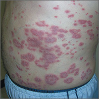
Rash on abdomen
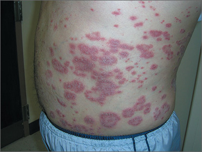
Based on the negative KOH and the nail pits, the FP made a diagnosis of plaque psoriasis. This condition can present in an annular pattern resembling tinea corporis. The combination of negative KOH and nail pits are enough to diagnose plaque psoriasis without a biopsy. Otherwise, a 4-mm punch biopsy of the area with erythema and scale would confirm the diagnosis.
Plaque psoriasis is typically treated using a mid- to high-potency topical steroid. Although repeated use of steroids in cases of atopic dermatitis can lead to skin atrophy, this is less common when treating psoriasis. If skin atrophy is still a concern, an alternative to topical steroids is a topical vitamin D preparation.
Vitamin D preparations are typically more expensive than steroids and require prior authorization, but there is one generic preparation (calcipotriene) that is more affordable than its brand-name counterparts. Another nonsystemic treatment to consider when treating plaque psoriasis without psoriatic arthritis is narrowband ultraviolet B therapy.
One risk factor for psoriasis is being overweight. In this case, the FP counseled the patient on weight loss. The FP then prescribed 0.1% triamcinolone ointment to be applied twice daily (especially after bathing).
At a follow-up appointment a month later, there was about 70% clearance of the lesions. For the stubborn areas, the FP prescribed a higher-potency steroid, 0.05% clobetasol ointment, to be applied twice daily. As the cost of clobetasol has risen over the past 2 years, alternatives that may be covered by insurance include augmented betamethasone and halobetasol.
Photos and text for Photo Rounds Friday courtesy of Richard P. Usatine, MD. This case was adapted from: Usatine R. Psoriasis. In: Usatine R, Smith M, Mayeaux EJ, et al, eds. Color Atlas of Family Medicine. 2nd ed. New York, NY: McGraw-Hill; 2013: 878-895.
To learn more about the Color Atlas of Family Medicine, see: www.amazon.com/Color-Family-Medicine-Richard-Usatine/dp/0071769641/
You can now get the second edition of the Color Atlas of Family Medicine as an app by clicking on this link: usatinemedia.com
Review of plant phenolics, part 1
Polyphenols are well known as the largest group of and most widely distributed phytochemicals among plants.1 These secondary plant metabolites, which are produced in response to environmental hazards that contribute to free-radical synthesis,2 are represented by more than 8,000 naturally occurring compounds. This family of widely divergent substances has gained increasing attention in recent years as polyphenols have been found – in vegetables, fruits, herbs, grains, tea, coffee beans, honey, and red wine – to be the most abundant sources of antioxidants in the human diet and are known to exert antioxidant, anti-inflammatory, and antimicrobial benefits to human health.3-7 The most prevalent and studied polyphenols are known as flavonoids, but nonflavonoid polyphenols are increasingly well investigated. This column will address the basic chemistry of these compounds. Subsequent columns will discuss the latest research on the cutaneous benefits of selected flavonoid and nonflavonoid polyphenols.

Chemistry and sources
Polyphenols share a common structural component: a phenol or an aromatic ring, usually two, with at least one hydroxyl, methyl, or acetyl group linked via a three-carbon bond to form a six-unit heterocyclic ring.8,9 When the “parent polyphenol” known as cinnamic acid is further catalytically transformed, scores of polyphenolic compounds result. These substances are divided into classes: glycosylated phenylpropanoids, flavonoids, isoflavonoids, stilbenoids, coumarins, curcuminoids, as well as phenolic polymers such as tannins, proanthocyanidins, suberin, lignins, and lignans. The flavonoids, which are the largest and most varied phenolic substances in plants, can be further divided into several categories: flavones (based on the 2-phenylchromen-4-one skeleton, such as apigenin and luteolin); flavonols (based on the 3-hydroxy-2-phenylchromen-4-one skeleton and functional group, such as quercetin, kaempferol, myricetin, and fisetin); flavanones (based on the 2,3-dihydro-2-phenylchromen-4-one skeleton and functional group, such as naringenin, hesperidin, and eriodictyol); isoflavones (based on the 3-phenylchromen-4-one skeleton, such as genistein and daidzein); flavanols – also known as flavan-3-ols or catechins – (based on the 2-phenyl-3,4-dihydro-2H-chromen-3-ol skeleton and functional groups, such as epicatechin, epicatechin 3-gallate, epigallocatechin, epigallocatechin 3-gallate (EGCG), catechin, and gallocatechin); and anthocyanins (based on the 2-phenylchromenylium ion skeleton, e.g., cyanidin and pelargonidin).5,10
The broader category of nonflavonoid polyphenols is rich and diverse, but is particularly noted for comprising the tannins, phenolic polymers of high molecular weight, which are divided into three classes, hydrolyzable tannins (such as ellagic acid, found in pomegranate, raspberries, strawberries, cranberries, and walnuts), derived tannins (created during food handling and processing and present in, for example, black and oolong teas), and condensed tannins (or proanthocyanidins, which are polymer chains of flavanols, such as catechins, and include pycnogenol, leukocyanidin, and leucoanthocyanin).1,4,5,8,10 There are a plethora of other nonflavonoid polyphenols, many of which confer health benefits, including stilbenes (such as resveratrol, found in red wine), lignans (such as enterodiol, found in flaxseed and flaxseed oil), lignins (found in green beans, carrots, peas, and Brazil nuts), and phenolic acids, such as hydroxybenzoic and hydroxycinnamic acids, among which caffeic and ferulic acids are often present in foods. In fact, hydroxycinnamic acids, which are the most common phenolic acids present in plant tissues, are present in numerous foods, such as apples, pears, plums, cherries, apricots, peaches, black currant, blueberries, potatoes, spinach, lettuce, cabbage, broccoli, asparagus, wine, and coffee.9

Broad health benefits have been associated with hundreds of polyphenolic substances. Notably, some of the best-known research results on polyphenols have reported on the success of various topical applications of green tea catechins, ferulic acid, and resveratrol, and other related compounds. Antioxidant, anti-inflammatory, and antimicrobial activities are the most common biologic properties associated with polyphenols, and antiaging activity has been widely reported.10
Conclusion
While the classification system for the 8,000 polyphenolic compounds may seem intimidating, the same essential activity is conferred by these abundant substances. Further, it is important to note the significant health benefits potentially derived from the oral consumption as well as topical application of polyphenols. The next two columns will delve into the research findings of flavonoid and nonflavonoid polyphenols.
Dr. Baumann is a private practice dermatologist, researcher, author and entrepreneur who practices in Miami. She founded the Cosmetic Dermatology Center at the University of Miami in 1997. Dr. Baumann wrote two textbooks: “Cosmetic Dermatology: Principles and Practice” (New York: McGraw-Hill, 2002), and “Cosmeceuticals and Cosmetic Ingredients,” (New York: McGraw-Hill, 2014), and a New York Times Best Sellers book for consumers, “The Skin Type Solution” (New York: Bantam Dell, 2006). Dr. Baumann has received funding for advisory boards and/or clinical research trials from Allergan, Evolus, Galderma, and Revance. She is the founder and CEO of Skin Type Solutions Franchise Systems LLC.
References
1. J Am Diet Assoc. 1999 Feb;99(2):213-8.
2. Ann N Y Acad Sci. 2012 Jul;1259:77-86.
3. Biomed Pap Med Fac Univ Palacky Olomouc Czech Repub. 2003 Dec;147(2):137-45.
4. J Nutr. 2000 Aug;130(8S Suppl):2073S-85S.
5. Annu Rev Nutr. 2002;22:19-34.
6. Pharmacol Ther. 2001 May-Jun;90(2-3):157-77.
7. Free Radic Biol Med. 2001 Jun 1;30(11):1213-22.
8. J Nutr. 2003 Oct;133(10):3248S-3254S.
9. Int J Mol Sci. 2016 Feb 18;17(2):160.
10. Asia Pac J Clin Nutr. 2004;13(Suppl):S72, 2004.
Polyphenols are well known as the largest group of and most widely distributed phytochemicals among plants.1 These secondary plant metabolites, which are produced in response to environmental hazards that contribute to free-radical synthesis,2 are represented by more than 8,000 naturally occurring compounds. This family of widely divergent substances has gained increasing attention in recent years as polyphenols have been found – in vegetables, fruits, herbs, grains, tea, coffee beans, honey, and red wine – to be the most abundant sources of antioxidants in the human diet and are known to exert antioxidant, anti-inflammatory, and antimicrobial benefits to human health.3-7 The most prevalent and studied polyphenols are known as flavonoids, but nonflavonoid polyphenols are increasingly well investigated. This column will address the basic chemistry of these compounds. Subsequent columns will discuss the latest research on the cutaneous benefits of selected flavonoid and nonflavonoid polyphenols.
Chemistry and sources
Polyphenols share a common structural component: a phenol or an aromatic ring, usually two, with at least one hydroxyl, methyl, or acetyl group linked via a three-carbon bond to form a six-unit heterocyclic ring.8,9 When the “parent polyphenol” known as cinnamic acid is further catalytically transformed, scores of polyphenolic compounds result. These substances are divided into classes: glycosylated phenylpropanoids, flavonoids, isoflavonoids, stilbenoids, coumarins, curcuminoids, as well as phenolic polymers such as tannins, proanthocyanidins, suberin, lignins, and lignans. The flavonoids, which are the largest and most varied phenolic substances in plants, can be further divided into several categories: flavones (based on the 2-phenylchromen-4-one skeleton, such as apigenin and luteolin); flavonols (based on the 3-hydroxy-2-phenylchromen-4-one skeleton and functional group, such as quercetin, kaempferol, myricetin, and fisetin); flavanones (based on the 2,3-dihydro-2-phenylchromen-4-one skeleton and functional group, such as naringenin, hesperidin, and eriodictyol); isoflavones (based on the 3-phenylchromen-4-one skeleton, such as genistein and daidzein); flavanols – also known as flavan-3-ols or catechins – (based on the 2-phenyl-3,4-dihydro-2H-chromen-3-ol skeleton and functional groups, such as epicatechin, epicatechin 3-gallate, epigallocatechin, epigallocatechin 3-gallate (EGCG), catechin, and gallocatechin); and anthocyanins (based on the 2-phenylchromenylium ion skeleton, e.g., cyanidin and pelargonidin).5,10
The broader category of nonflavonoid polyphenols is rich and diverse, but is particularly noted for comprising the tannins, phenolic polymers of high molecular weight, which are divided into three classes, hydrolyzable tannins (such as ellagic acid, found in pomegranate, raspberries, strawberries, cranberries, and walnuts), derived tannins (created during food handling and processing and present in, for example, black and oolong teas), and condensed tannins (or proanthocyanidins, which are polymer chains of flavanols, such as catechins, and include pycnogenol, leukocyanidin, and leucoanthocyanin).1,4,5,8,10 There are a plethora of other nonflavonoid polyphenols, many of which confer health benefits, including stilbenes (such as resveratrol, found in red wine), lignans (such as enterodiol, found in flaxseed and flaxseed oil), lignins (found in green beans, carrots, peas, and Brazil nuts), and phenolic acids, such as hydroxybenzoic and hydroxycinnamic acids, among which caffeic and ferulic acids are often present in foods. In fact, hydroxycinnamic acids, which are the most common phenolic acids present in plant tissues, are present in numerous foods, such as apples, pears, plums, cherries, apricots, peaches, black currant, blueberries, potatoes, spinach, lettuce, cabbage, broccoli, asparagus, wine, and coffee.9
Broad health benefits have been associated with hundreds of polyphenolic substances. Notably, some of the best-known research results on polyphenols have reported on the success of various topical applications of green tea catechins, ferulic acid, and resveratrol, and other related compounds. Antioxidant, anti-inflammatory, and antimicrobial activities are the most common biologic properties associated with polyphenols, and antiaging activity has been widely reported.10
Conclusion
While the classification system for the 8,000 polyphenolic compounds may seem intimidating, the same essential activity is conferred by these abundant substances. Further, it is important to note the significant health benefits potentially derived from the oral consumption as well as topical application of polyphenols. The next two columns will delve into the research findings of flavonoid and nonflavonoid polyphenols.
Dr. Baumann is a private practice dermatologist, researcher, author and entrepreneur who practices in Miami. She founded the Cosmetic Dermatology Center at the University of Miami in 1997. Dr. Baumann wrote two textbooks: “Cosmetic Dermatology: Principles and Practice” (New York: McGraw-Hill, 2002), and “Cosmeceuticals and Cosmetic Ingredients,” (New York: McGraw-Hill, 2014), and a New York Times Best Sellers book for consumers, “The Skin Type Solution” (New York: Bantam Dell, 2006). Dr. Baumann has received funding for advisory boards and/or clinical research trials from Allergan, Evolus, Galderma, and Revance. She is the founder and CEO of Skin Type Solutions Franchise Systems LLC.
References
1. J Am Diet Assoc. 1999 Feb;99(2):213-8.
2. Ann N Y Acad Sci. 2012 Jul;1259:77-86.
3. Biomed Pap Med Fac Univ Palacky Olomouc Czech Repub. 2003 Dec;147(2):137-45.
4. J Nutr. 2000 Aug;130(8S Suppl):2073S-85S.
5. Annu Rev Nutr. 2002;22:19-34.
6. Pharmacol Ther. 2001 May-Jun;90(2-3):157-77.
7. Free Radic Biol Med. 2001 Jun 1;30(11):1213-22.
8. J Nutr. 2003 Oct;133(10):3248S-3254S.
9. Int J Mol Sci. 2016 Feb 18;17(2):160.
10. Asia Pac J Clin Nutr. 2004;13(Suppl):S72, 2004.
Five-year outcomes favor on- versus off-pump CABG
Compared with adults who underwent off-pump coronary-artery bypass grafting surgery, those who underwent on-pump CABG had significantly lower rates of mortality and major adverse cardiovascular events at 5 years, results from a large randomized trial demonstrated.
“Given the results, it appears that innovative surgical approaches – such as the more technically demanding off-pump procedure – may not always provide superior clinical outcomes,” researchers led by A. Laurie Shroyer, PhD, wrote (N Engl J Med. 2017 Aug 17;377:623-32). “Additional long-term follow-up, evaluating these same outcomes rigorously at 10 years after CABG, appears to be warranted. Future research may identify the risk factors of the patients and the cardiac surgical processes of care that affect longer-term outcomes of coronary revascularization procedures, with the goal of increasing the rate of long-term event-free survival.”
Dr. Shroyer, of the Northport (N.Y.) VA Medical Center, and her associates conduced a 5-year follow-up study of patients who had participated in the original Randomized On/Off Bypass (ROOBY) trial, which compared the effectiveness of the two surgical approaches (N Engl J Med 2009 Nov 5;361:1827-37). During February 2002–June 2007, 2,203 patients at 18 medical centers were randomly assigned to either on-pump or off-pump CABG, with 1-year assessments completed by May 2008. The primary outcomes were the rates mortality and major adverse cardiovascular events at 5 years, while the secondary 5-year outcomes included death from cardiac causes, repeat revascularization, and nonfatal myocardial infarction.
The mean age of patients was 63 years, nearly all were male, 46% were between the ages of 55 and 64, and about 21% had chronic obstructive pulmonary disease. The researchers found that at 5 years, the rate of death was 15.2% in the off-pump group, compared with 11.9% in the on-pump group, which translated into a relative risk of 1.28 (P = .02). In addition, the rate of major cardiovascular events at 5 years was 31% in the off-pump group, compared with 27.1% in the on-pump group, which translated into a relative risk of 1.14 (P = .046). None of the secondary outcomes at 5 years met the prespecified threshold of a P value of .01 or less for statistical significance, when the off-pump and on-pump groups were compared. This included the rates of nonfatal myocardial infarction (12.1% vs. 9.6%, respectively; P = .05); death from cardiac causes (6.3% vs. 5.3%; P = .29); repeat vascularization (13.1% vs. 11.9%; P = .39), and repeat CABG (1.4% vs. 0.5%; P = .02).
“In combination with findings from other randomized trials and a 2012 Cochrane systematic review [Cochrane Database Syst Rev. 2012;14:CD007224], the 5-year outcomes in our study support the conclusion that off-pump CABG does not offer any substantial advantages over on-pump CABG except possibly in unusual situations such as, for example, in patients with an extensively calcified (porcelain) aorta, in whom the off-pump technique may result in less manipulation of the aorta, potentially decreasing the risk of aortic emboli or stroke,” the researchers wrote. “In light of the low rates of use of off-pump CABG in the United States, the findings in our trial may provide more of a real-world experience than those in the CORONARY and GOPCABE trials, which required surgeons with a very high volume of experience with off-pump procedures, as compared with the ROOBY trial and with most other surgeons who are based in the United States.”
They acknowledged certain limitations of the study, including the fact that the study population comprised mostly males who had multiple coexisting conditions, “so the findings may not be applicable to female patients or to patients who are not veterans.”
The study was supported by a grant from the Department of Veterans Affairs. Dr. Shroyer reported having received grants from the VA Cooperative Studies Program during the conduct of the study. Her coauthors reported having no financial disclosures.
FROM THE NEW ENGLAND JOURNAL OF MEDICINE
Key clinical point:
Major finding: At 5 years, the rate of death was 15.2% in the off-pump group, compared with 11.9% in the on-pump group, which translated into a relative risk of 1.28 (P = .02).
Data source: A 5-year follow-up study of 2,203 patients who had participated in the original Randomized On/Off Bypass (ROOBY) trial.
Disclosures: The study was supported by a grant from the Department of Veterans Affairs. Dr. Shroyer reported having received grants from the Department of Veterans Affairs Cooperative Studies Program during the study. The other coauthors reported having no financial disclosures.

Tele-neurology visits deemed as effective as office visits for Parkinson’s patients
A new study finds tele-neurology visits are just as effective as in-person visits for patients with Parkinson’s disease, while also saving time and mileage.
Lead author Christopher A. Beck, PhD, of the University of Rochester (N.Y.), and his colleagues studied 195 patients for 1 year who were either provided with their usual medical care or their usual medical care supplemented by four virtual visits via video conferencing from a remote specialist. Investigators evaluated the feasibility of telemedicine visits, as measured by the proportion of patients who completed at least one virtual visit and the proportion of virtual visits completed on time. Efficacy was also evaluated, as measured by the change in the Parkinson’s Disease Questionnaire–39, a quality of life scale. Secondary outcomes included quality of care, caregiver burden, and time and travel savings.
Eligible participants had a clinical diagnosis of Parkinson’s disease, had a private, Internet-enabled device, and lived in a state where a site investigator was licensed to practice. Patients randomized to the telemedicine group received up to four virtual visits over 12 months from a neurologist. Physicians and patients determined the specific content and frequency of each visit, but the format generally included a medical history, a Parkinson’s disease–specific examination (including assessment of tremor and gait), physician recommendations, and time to address patients’ concerns (Neurology. 2017 Aug 16. doi: 10.1212/WNL.0000000000004357).
The study findings showed that the virtual house calls were as effective as were in-person visits, with quality of life no better or worse for patients receiving care at home than for those receiving care in the office. Of telemedicine patients, 98% completed at least one virtual visit, and 91% of all virtual visits scheduled were completed. Researchers also found that participants’ overall quality of care and the burden felt by caregivers was no different whether they had virtual or in-person visits. Each virtual house call saved patients a median of 88 minutes and 38 miles per visit.
Ninety-seven percent of patients and 86% of participating neurologists said they were satisfied with the virtual visits, with 55% of patients stating they preferred virtual visits over in-person visits.
The study – the first national randomized controlled trial of telemedicine to connect remote specialists to patients at home – shows that tele-neurology is a practical and effective model of care for Parkinson patients, said study coauthor E. Ray Dorsey, MD, of the University of Rochester.
“People were very interested in taking part in this study, and the results showed that these virtual house calls were feasible for people with Parkinson’s disease,” Dr. Dorsey said in a statement. “People’s care was as effective as with the in-office visits, and the virtual house calls provided the participants with convenience and comfort.”
Dr. Dorsey noted that 73% of the study participants had visited a Parkinson’s disease specialist in the past year and 83% said they were satisfied with their care, which may have impacted the quality of life finding.
“The fact that adding the virtual house calls to people’s care did not improve their quality of life could be because a large proportion were already seeing a specialist and were satisfied with that care,” he said. “Of course, it’s also possible that virtual house calls are not enough to improve quality of life.”
Limitations of the study included that study participants were primarily well-educated and more familiar with the Internet than was the general population, so results may not be relevant for all people with Parkinson’s disease. In addition, the study population, of whom 96% were white with a mean age of 66 years, did not include people with the disease who live in nursing homes, who account for nearly 25% of all Medicare beneficiaries with Parkinson’s disease.
The study was supported by the Patient-Centered Outcomes Research Institute. Dr. Dorsey serves on the medical advisory board of, and has stock options in, Grand Rounds. No other relevant disclosures relevant were reported by study authors.
[email protected]
On Twitter @legal_med
Patients with Parkinson’s disease (PD) show more improvements in motor symptoms and less frequently receive inappropriate medications when treated by a specialist. However, reduced mobility due to PD, combined with the physical distance to the specialist’s office, poses substantial travel burdens and may limit access to care.
Telemedicine, utilizing video and audio conferencing technology to connect patients and clinicians, can mitigate this problem. However, the Centers for Medicare & Medicaid Services currently only covers telemedicine encounters under specific criteria: The patient lives in a rural area seen in a covered entity by a consulting physician licensed in the state in which the patient resides. Even if the rural designation were removed, requiring travel to a center that offers telemedicine consultation may preclude many patients from using this approach.
The study by Dr. Beck and his colleagues provides a good first step in demonstrating how neurologists can use available technology to reduce patient burdens while still maintaining high-quality neurologic care. If more evidence mounts, and the regulatory environment improves, home-based telemedicine has the potential to revolutionize modern health care for PD and other chronic debilitating diseases.
David Shprecher, DO, is with Banner Sun Health Research Institute in Sun City, Ariz. Jennifer Majersik, MD, is with the University of Utah, Salt Lake City. Dr. Shprecher reported financial relationships with a variety of companies that market or are developing drugs for Parkinson’s disease. Dr. Majersik reported receiving significant research support from the National Institute of Neurological Disorders and Stroke. These comments are adapted from an accompanying editorial (Neurology. 2017 Aug 16. doi: 10.1212/WNL.0000000000004366).
A new study finds tele-neurology visits are just as effective as in-person visits for patients with Parkinson’s disease, while also saving time and mileage.
Lead author Christopher A. Beck, PhD, of the University of Rochester (N.Y.), and his colleagues studied 195 patients for 1 year who were either provided with their usual medical care or their usual medical care supplemented by four virtual visits via video conferencing from a remote specialist. Investigators evaluated the feasibility of telemedicine visits, as measured by the proportion of patients who completed at least one virtual visit and the proportion of virtual visits completed on time. Efficacy was also evaluated, as measured by the change in the Parkinson’s Disease Questionnaire–39, a quality of life scale. Secondary outcomes included quality of care, caregiver burden, and time and travel savings.
Eligible participants had a clinical diagnosis of Parkinson’s disease, had a private, Internet-enabled device, and lived in a state where a site investigator was licensed to practice. Patients randomized to the telemedicine group received up to four virtual visits over 12 months from a neurologist. Physicians and patients determined the specific content and frequency of each visit, but the format generally included a medical history, a Parkinson’s disease–specific examination (including assessment of tremor and gait), physician recommendations, and time to address patients’ concerns (Neurology. 2017 Aug 16. doi: 10.1212/WNL.0000000000004357).
The study findings showed that the virtual house calls were as effective as were in-person visits, with quality of life no better or worse for patients receiving care at home than for those receiving care in the office. Of telemedicine patients, 98% completed at least one virtual visit, and 91% of all virtual visits scheduled were completed. Researchers also found that participants’ overall quality of care and the burden felt by caregivers was no different whether they had virtual or in-person visits. Each virtual house call saved patients a median of 88 minutes and 38 miles per visit.
Ninety-seven percent of patients and 86% of participating neurologists said they were satisfied with the virtual visits, with 55% of patients stating they preferred virtual visits over in-person visits.
The study – the first national randomized controlled trial of telemedicine to connect remote specialists to patients at home – shows that tele-neurology is a practical and effective model of care for Parkinson patients, said study coauthor E. Ray Dorsey, MD, of the University of Rochester.
“People were very interested in taking part in this study, and the results showed that these virtual house calls were feasible for people with Parkinson’s disease,” Dr. Dorsey said in a statement. “People’s care was as effective as with the in-office visits, and the virtual house calls provided the participants with convenience and comfort.”
Dr. Dorsey noted that 73% of the study participants had visited a Parkinson’s disease specialist in the past year and 83% said they were satisfied with their care, which may have impacted the quality of life finding.
“The fact that adding the virtual house calls to people’s care did not improve their quality of life could be because a large proportion were already seeing a specialist and were satisfied with that care,” he said. “Of course, it’s also possible that virtual house calls are not enough to improve quality of life.”
Limitations of the study included that study participants were primarily well-educated and more familiar with the Internet than was the general population, so results may not be relevant for all people with Parkinson’s disease. In addition, the study population, of whom 96% were white with a mean age of 66 years, did not include people with the disease who live in nursing homes, who account for nearly 25% of all Medicare beneficiaries with Parkinson’s disease.
The study was supported by the Patient-Centered Outcomes Research Institute. Dr. Dorsey serves on the medical advisory board of, and has stock options in, Grand Rounds. No other relevant disclosures relevant were reported by study authors.
[email protected]
On Twitter @legal_med
FROM NEUROLOGY
Key clinical point:
Major finding: Overall, 98% of tele-neurology patients completed at least one virtual visit, and 91% of all virtual visits scheduled were completed.
Data source: National randomized controlled trial of 195 patients with Parkinson’s disease.
Disclosures: E. Ray Dorsey, MD, serves on the medical advisory board of and has stock options in Grand Rounds. No other relevant disclosures were reported by the researchers. The study was supported by the Patient-Centered Outcomes Research Institute.
Identifying Gene Variants Linked to Epilepsy
Mutations in SCN1A, SCN2A, and SCN8A, which code for neuronal voltage-gated sodium channel alpha-subunits, have been associated with certain early onset epilepsy syndromes. Despite this association, many clinicians are uncertain about the value of missense genetic variants in the management of epilepsy because many mutations are classified as having unknown significance. A recent database analysis identified gene variants that are pathogenic and benign, giving clinicians a better understanding of how to interpret SCN test results. Details of the investigation include the following:
- Investigators used 8 algorithms to evaluate the pathogenicity of various genetic variants. They also used logistic regression to help determine if combining algorithms might improve their ability to predict pathogenicity.
- 440 variants were considered pathogenic or likely pathogenic.
- Most computer algorithms that attempt to determine the value of SCN test results are very sensitive but also suffer from low specificity.
- The Mendelian Clinically Applicable Pathogenicity algorithm proved most valuable, with an accuracy of 0.90.
Holland KD, Bouley TM, Horn PS. Comparison and optimization of in silico algorithms for predicting the pathogenicity of sodium channel variants in epilepsy. Epilepsia. 2017;58(7):1190-1198.