User login
‘Watch and wait’ good for most – but not all – rectal cancers
BOSTON –
was established over a decade ago, and widely adopted since then. The idea is to hold off on surgery after a complete response to chemotherapy and radiation, to see if the patient really needs it.
While most do not, tumors regrow in 20%-30%, and when they come back, they tend to be aggressive, with poor outcomes. Patients in those situations would have been better off with upfront surgery.
The problem right now is that there’s no way to predict who will be cured by neoadjuvant therapy and whose tumor will come back, said Philip Paty, MD. FACS, a colorectal surgeon at Memorial Sloan Kettering Cancer Center, New York.
“There are some who are probably harmed by the watch-and-wait paradigm. The risk of local regrowth is hardbaked into [the model]; we haven’t been able to eliminate it,” he said at the annual clinical congress of the American College of Surgeons..
Dr. Paty is one of many investigators working to identify those at risk. In the meantime, watch-and-wait patients need to be followed closely, particularly in the first 2 years. Surgery is the best option at the first sign of regrowth. Dr. Paty explained his follow-up protocol, as well as the current state of watch and wait for low rectal cancer, in an interview at the meeting.
He also talked about overcoming hurdles. The risk of surgery, including permanent bowel and sexual dysfunction, is so great “that many patients latch onto watch and wait and won’t let go. They don’t come back for follow-up, or resist the idea of surgery even if it’s needed,” he said.
EXPERT ANALYSIS FROM THE ACS CLINICAL CONGRESS

How to slash colorectal surgery infection rates
BOSTON – driven mostly by a reduction in deep organ space infections from 5.5% to 1.7%.
It was a remarkable finding that got the attention of attendees at the annual clinical congress of the American College of Surgeons. The Cleveland Clinic had been an outlier, in the wrong direction, compared with other centers, and administrators wanted a solution.
I. Emre Gorgun, MD, FACS, a colorectal surgeon and quality improvement officer at Cleveland Clinic, led the search for evidence-based interventions. Eventually, big changes were made to perioperative antibiotics, mechanical bowel prep, preop shower routines, and intraoperative procedures. The efforts paid off (Dis Colon Rectum. 2018 Jan;61[1]:89-98).
To help surgeons lower their own infection rates, Dr. Gorgun agreed to an interview at the meeting to explain exactly what was done.
There was resistance at first from surgeons who wanted to stick with their routines, but they came around once they were shown the data backing the changes. Eventually, “everyone was on board. We believe in this,” Dr. Gorgun said.
REPORTING FROM THE ACS CLINICAL CONGRESS
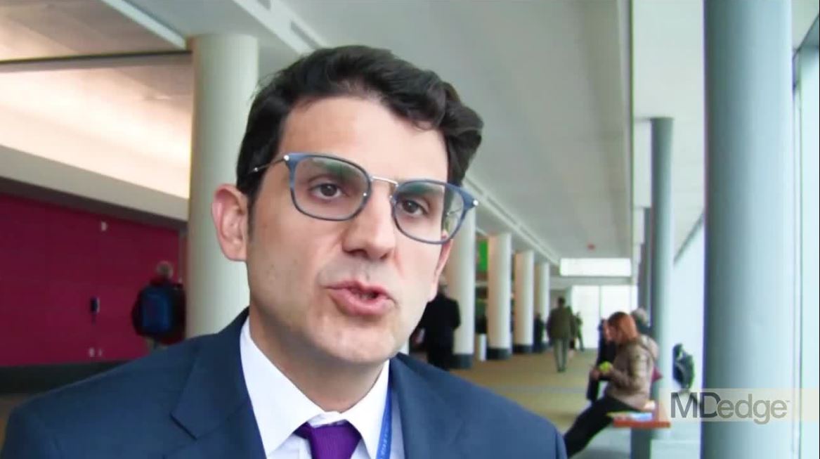
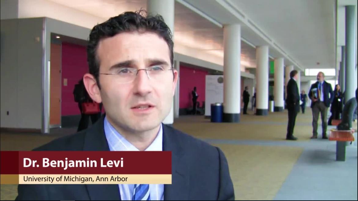
How, and when, to use fat grafting for scars
BOSTON – according to Benjamin Levi, MD, director of the burn/wound and regenerative medicine laboratory at the University of Michigan, Ann Arbor.
Some corners of the Internet tout it as “some sort of magic stem cell surgery,” Dr. Levi said, but in reality, for scar surgeons, it’s just another useful tool in the armamentarium, one that excels at filling skin depressions due to underlying tissue loss, whether from burns, trauma, or surgery. Unlike hyaluronic acid and other options, fat grafts last; about half of injected adipocytes remain indefinitely. Fat grafting might also help soften scars, he said.
To get the most out of the procedure, of course, it has to be done correctly, so Dr. Levi took a few minutes at the annual clinical congress of the American College of Surgeons to share his tips on harvesting and spinning down fat grafts, injecting adipocytes, and other matters. Although the concepts of fat grafting are straightforward, the techniques are a bit tricky. Dr. Levi hoped his treatment pearls would help other physicians, especially those considering adding fat grafting to their practice.
EXPERT ANALYSIS FROM THE ACS CLINICAL CONGRESS
Transcription factor plays key role in AML gene regulatory networks
The AP-1 transcription factor family, important in many tumor types, plays a major role in acute myeloid leukemia, according to researchers who conducted a comprehensive global analysis of gene regulatory networks involved in this disease.

This observation suggests new opportunities for targeted treatment of AML, according to the researchers, led by Peter N. Cockerill, PhD, and Constanze Bonifer, PhD, with the Institute of Cancer and Genomic Sciences, University of Birmingham, England.
“Induced and aberrantly expressed transcription factors are not bystanders, but are important for network maintenance and leukemic growth,” the investigators wrote in Nature Genetics.
Investigators combined data obtained via several different analytic techniques to construct transcription factor networks in normal CD34+ cells and cells from specific subgroups of subjects with defined mutations, including RUNX1 mutations, t(8;21) translocations, mutations of both alleles of the CEBPA gene, and FLT3-ITD with or without NPM1 mutation.
The AP-1 family network was of “high regulatory relevance” for all AML subtypes evaluated, the investigators reported.
Previous work revealed the existence of gene regulatory networks in different types of AML classified by gene expression and DNA-methylation patterns.
“Our work now defines these networks in detail, and shows that leukemic drivers determine the regulatory phenotype by establishing and maintaining specific gene regulatory and signaling networks that are distinct from those in normal cells,” the authors said in their report.
Follow-up in vitro and in vivo studies confirmed the importance of AP-1 for different AML subtypes.
In the in vitro study, investigators transduced AML cells with a doxycycline-inducible version of a dominant negative FOS protein.
“AP-1 is a heterodimer formed by members of the FOS, JUN, ATF, CREB and JDP families of transcription factors, thus it is challenging to target by defined RNA interference approaches,” the investigators explained.
Results of the in vitro study showed that induction of that protein, mediated by doxycycline, inhibited proliferation and colony-forming ability in AML cell lines.
To evaluate the relevance of AP-1 for leukemia propagation in vivo, they transplanted two different types of cells expressing inducible dominant negative FOS protein in immunodeficient mice.
For the first cell type, granulosarcomas developed in six out of seven mice in a control group, but in only two mice treated with doxycycline, neither of which expressed the inducible protein, suggesting that the transgene was silenced, according to the investigators. For the second cell type, doxycycline inhibited leukemia development, while untreated mice rapidly developed tumors.
“Taken together, these findings demonstrate the importance of AP-1 for several AML subtypes and emphasize the potential of transcriptional network analyses to predict transcription factors crucial for malignant propagation,” the investigators wrote.
They declared no competing interests related to their research, which was funded by Bloodwise, Cancer Research UK, a Kay Kendall Clinical Training Fellowship and a MRC/Leuka Clinical Training Fellowship.
SOURCE: Assi SA et al. Nat Genet. 2018 Nov 12. doi: 10.1038/s41588-018-0270-1.
The AP-1 transcription factor family, important in many tumor types, plays a major role in acute myeloid leukemia, according to researchers who conducted a comprehensive global analysis of gene regulatory networks involved in this disease.
This observation suggests new opportunities for targeted treatment of AML, according to the researchers, led by Peter N. Cockerill, PhD, and Constanze Bonifer, PhD, with the Institute of Cancer and Genomic Sciences, University of Birmingham, England.
“Induced and aberrantly expressed transcription factors are not bystanders, but are important for network maintenance and leukemic growth,” the investigators wrote in Nature Genetics.
Investigators combined data obtained via several different analytic techniques to construct transcription factor networks in normal CD34+ cells and cells from specific subgroups of subjects with defined mutations, including RUNX1 mutations, t(8;21) translocations, mutations of both alleles of the CEBPA gene, and FLT3-ITD with or without NPM1 mutation.
The AP-1 family network was of “high regulatory relevance” for all AML subtypes evaluated, the investigators reported.
Previous work revealed the existence of gene regulatory networks in different types of AML classified by gene expression and DNA-methylation patterns.
“Our work now defines these networks in detail, and shows that leukemic drivers determine the regulatory phenotype by establishing and maintaining specific gene regulatory and signaling networks that are distinct from those in normal cells,” the authors said in their report.
Follow-up in vitro and in vivo studies confirmed the importance of AP-1 for different AML subtypes.
In the in vitro study, investigators transduced AML cells with a doxycycline-inducible version of a dominant negative FOS protein.
“AP-1 is a heterodimer formed by members of the FOS, JUN, ATF, CREB and JDP families of transcription factors, thus it is challenging to target by defined RNA interference approaches,” the investigators explained.
Results of the in vitro study showed that induction of that protein, mediated by doxycycline, inhibited proliferation and colony-forming ability in AML cell lines.
To evaluate the relevance of AP-1 for leukemia propagation in vivo, they transplanted two different types of cells expressing inducible dominant negative FOS protein in immunodeficient mice.
For the first cell type, granulosarcomas developed in six out of seven mice in a control group, but in only two mice treated with doxycycline, neither of which expressed the inducible protein, suggesting that the transgene was silenced, according to the investigators. For the second cell type, doxycycline inhibited leukemia development, while untreated mice rapidly developed tumors.
“Taken together, these findings demonstrate the importance of AP-1 for several AML subtypes and emphasize the potential of transcriptional network analyses to predict transcription factors crucial for malignant propagation,” the investigators wrote.
They declared no competing interests related to their research, which was funded by Bloodwise, Cancer Research UK, a Kay Kendall Clinical Training Fellowship and a MRC/Leuka Clinical Training Fellowship.
SOURCE: Assi SA et al. Nat Genet. 2018 Nov 12. doi: 10.1038/s41588-018-0270-1.
FROM NATURE GENETICS
Key clinical point:
Major finding: The AP-1 factor family gene regulatory network was of high regulatory relevance in multiple subtypes of AML with defined mutations.
Study details: Analysis of normal CD34+ cells and cells from AML subjects.
Disclosures: Funding came from Bloodwise and Cancer Research UK, among other sources. The researchers reported having no competing financial interests.
Source: Assi SA et al. Nat Genet. 2018 Nov 12. doi: 10.1038/s41588-018-0270-1.
Concussion/TBI linked to suicide risk, meta-analysis suggests
Risk of suicide was doubled in persons who experienced a concussion or mild traumatic brain injury (TBI) earlier in life, according to results of a meta-analysis of 17 studies representing nearly 7 million patients.
However, the absolute risk of suicide remained quite low, according to Michael Fralick, MD, of the University of Toronto, and co-investigators.
“Nearly all patients diagnosed with concussion and/or mild TBI did not die by suicide,” Dr. Fralick and colleagues said in their report on the study, which appears in JAMA Neurology.
Nevertheless, the meta-analysis illustrates evidence for an increased risk of suicide, suicide attempts, and suicidal ideation for persons with a history of these injuries, they said in the report.
The meta-analysis included 10 cohort studies, 5 cross-sectional studies, and 2 case-control studies looking at the risk of suicide, suicide attempts, or suicidal ideation after a concussion or mild TBI. Those studies included a roughly 714,000 individuals with a concussion and/or TBI diagnosis, and 6,236,000 without a diagnosis.
For people diagnosed with at least one concussion and/or mild TBI, the risk of suicide was 2-fold higher (relative risk, 2.03; 95% CI, 1.47-2.80; P less than 0.001), according to the report.
The risk was “slightly stronger,” investigators said, when the analysis was limited to studies adjusting for factors associated with those brain injuries and with suicide (RR, 2.10; 95% CI, 1.40-3.13; P less than 0.01).
Four of the 5 cohort studies reported absolute risk of suicide, according to Dr. Fralick and coauthors. In one study with a median follow-up of 3.6 years, 0.50% of individuals with a concussion and/or TBI subsequently died of suicide, while similarly, 0.59% died in a study with 4.0 years of follow-up, 0.28% in a study with 9.3 years follow-up, and 0.49% in one with a 12.3 year median follow-up.
Most of the studies in the meta-analysis reported an increased risk of suicide attempt after concussion and/or mild TBI, according to Dr. Fralick and his collaborators, while the eight studies looking at suicidal ideation all reported heightened risk after those brain injuries.
The researchers acknowledged some limitations of their analysis. Recall bias could have led to an overestimation of the association between concussion and suicide risk, since suicide attempts may affect reporting of concussion history, they said.
Furthermore, most of the studies were retrospective, and did not include an active comparator group, such as individuals with non-neurologic injuries, they added.
“Until large prospective studies with sufficiently large durations of follow-up are available, we have to rely on the currently available data,” they said in the report.
Dr. Fralick and co-authors reported no conflict of interest disclosures related to the study.
SOURCE: Fralick M, et al. JAMA Neurol. 2018 Nov 12.
This meta-analysis provides a comprehensive review of medical science that suggests a significant association between concussions and later suicide, according to Donald A Redelmeier, MD, and Junaid A. Bhatti, MBBS.
In an editorial, Dr. Redelmeier and Dr. Bhatti noted “media speculation” on the link between concussion and suicide, and commented that medical science progresses more slowly than the news cycle.
“A meta-analysis always has limitations and these authors maintained a thoughtful approach to avoid overstatements,” they said in their editorial.
Although the absolute risks of suicide are modest, this meta-analysis highlights that a concussion could contribute to long-term neuropsychiatric illness, they added.
Health care should aim to prevent concussions, while clinicians need to avoid language such as “dinged” that trivializes the effects of concussion, according to the authors.
In particular, they said neurologists should be aware of the suicide risks highlighted in this meta-analysis, and may want to screen concussion patients for other factors such as mood disorders, substance use, or past suicide attempts, since there is some evidence that concussions may amplify latent psychiatric illnesses.
Likewise, they said, psychiatrists should look for a concussion history when evaluating a particular patient’s risk of suicide.
“We should all recognize that a concussion, in its own way, can be lethal,” the authors concluded.
Dr. Redelmeier and Dr. Bhatti are with the Departments of Medicine and of Surgery, University of Toronto. Their editorial was published in JAMA Neurology. Dr. Redelmeier reported support from the Canada Research Chair in Medical Decision Sciences, the Canadian Institutes of Health Research, and the BrightFocus Foundation, while Dr. Bhatti reported support from the Sunnybrook Research Institute.
Risk of suicide was doubled in persons who experienced a concussion or mild traumatic brain injury (TBI) earlier in life, according to results of a meta-analysis of 17 studies representing nearly 7 million patients.
However, the absolute risk of suicide remained quite low, according to Michael Fralick, MD, of the University of Toronto, and co-investigators.
“Nearly all patients diagnosed with concussion and/or mild TBI did not die by suicide,” Dr. Fralick and colleagues said in their report on the study, which appears in JAMA Neurology.
Nevertheless, the meta-analysis illustrates evidence for an increased risk of suicide, suicide attempts, and suicidal ideation for persons with a history of these injuries, they said in the report.
The meta-analysis included 10 cohort studies, 5 cross-sectional studies, and 2 case-control studies looking at the risk of suicide, suicide attempts, or suicidal ideation after a concussion or mild TBI. Those studies included a roughly 714,000 individuals with a concussion and/or TBI diagnosis, and 6,236,000 without a diagnosis.
For people diagnosed with at least one concussion and/or mild TBI, the risk of suicide was 2-fold higher (relative risk, 2.03; 95% CI, 1.47-2.80; P less than 0.001), according to the report.
The risk was “slightly stronger,” investigators said, when the analysis was limited to studies adjusting for factors associated with those brain injuries and with suicide (RR, 2.10; 95% CI, 1.40-3.13; P less than 0.01).
Four of the 5 cohort studies reported absolute risk of suicide, according to Dr. Fralick and coauthors. In one study with a median follow-up of 3.6 years, 0.50% of individuals with a concussion and/or TBI subsequently died of suicide, while similarly, 0.59% died in a study with 4.0 years of follow-up, 0.28% in a study with 9.3 years follow-up, and 0.49% in one with a 12.3 year median follow-up.
Most of the studies in the meta-analysis reported an increased risk of suicide attempt after concussion and/or mild TBI, according to Dr. Fralick and his collaborators, while the eight studies looking at suicidal ideation all reported heightened risk after those brain injuries.
The researchers acknowledged some limitations of their analysis. Recall bias could have led to an overestimation of the association between concussion and suicide risk, since suicide attempts may affect reporting of concussion history, they said.
Furthermore, most of the studies were retrospective, and did not include an active comparator group, such as individuals with non-neurologic injuries, they added.
“Until large prospective studies with sufficiently large durations of follow-up are available, we have to rely on the currently available data,” they said in the report.
Dr. Fralick and co-authors reported no conflict of interest disclosures related to the study.
SOURCE: Fralick M, et al. JAMA Neurol. 2018 Nov 12.
FROM JAMA NEUROLOGY
Key clinical point:
Major finding: For people diagnosed with at least one concussion and/or mild TBI, the risk of suicide was 2-fold higher.
Study details: A meta-analysis of 17 studies representing nearly 7 million individuals with or without a concussion diagnosis.
Disclosures: The authors reported no conflicts of interest.
Source: Fralick M, et al. JAMA Neurol. 2018 Nov 12.
RFM awareness program not effective at preventing stillbirths
Because reduced fetal movement is associated with higher stillbirth risk, asking women to be alert to RFM and report it immediately has emerged as a potential intervention to prevent stillbirth. But a large, randomized trial of one reporting and management protocol showed no reduction in stillbirths, only a rise in C-sections and preterm inductions.

Jane E. Norman, MD, of the University of Edinburgh, and her colleagues published results from a trial in the Lancet, in which 409,175 pregnant women (mean age, 30 years) across 33 hospitals in the United Kingdom and Ireland received either standard care or the experimental RFM care intervention before delivery. Women were seen during an experimental 3-month period, in which all were treated according to the protocol, or the 3-month control period that preceded it. A 2-month washout period occurred between allocations as institutions adapted to the study protocol.
The trial intervention consisted of training clinical staff on the implications and management of RFM, distributing written information on RFM to women at about 20 weeks’ gestation, and a management protocol aimed at quick action following a report of RFM at 24 or more weeks’ gestation. The protocol included cardiotocography within 2 hours of presentation followed by measurement of liquor volume and a growth scan, along with umbilical artery Doppler where available. Delivery was recommended for women at 37 or more weeks’ gestation with estimated fetal weight below the 10th centile, abdominal circumference below the 10th centile, a low liquor volume, an abnormal cardiotocograph, or recurrent RFM.
Incidence of stillbirth at or beyond 24 weeks was 4.40 per 1,000 births during the control period and 4.06 per 1,000 births in the intervention period (adjusted odds ratio, 0.90; 95% confidence interval, 0.75-1.07; P = .23), the researchers found. No differences were seen when stratifying for different gestational ages.
Meanwhile, induction of labor before 39 weeks was more frequent during intervention period at 40% of deliveries, compared with 34% during the control time (P less than .0001), and at term (41% vs. 36%; P = .0015). C-section was higher in the intervention group at 28% versus 26% (P = .0001). Neonatal ICU stays were not more common but were likely to be longer in the intervention period, with stays of 2 days or longer occurring in 6.7% of deliveries versus 6.2% (P =.0001).
The investigators concluded that their protocol, in its current design, was not effective and could not be recommended because of the significant increase in interventions.
Dr. Norman and her colleagues wrote that the findings would “reignite the controversy about the efficacy of RFM awareness to reduce stillbirth and the underlying mechanisms linking RFM and stillbirth.” However, the results do not mean that RFM is a sign of inevitable fetal death or that there is no role for RFM awareness as a stillbirth-prevention strategy. Other large trials testing RFM-based interventions are still underway, they noted.
“Further research to identify better predictive tests for stillbirth [to enable targeting of the only current treatment of earlier delivery] is urgently needed,” the investigators added.
In a related study also published in the Lancet, Lucy K. Smith, PhD, of the University of Leicester (England), and her colleagues reported that the real burden of stillbirth in Europe, while much lower than in the developing world, is still a third higher than reported using the current international cutoff established by the World Health Organization.
Dr. Smith and her colleagues examined national cohort data from 19 European countries between 2004 and 2015 for pregnancy outcomes from 22 completed weeks’ gestation. In 2015, they found more than 9,000 stillbirths occurred among more than 25 million births, and 3,022 of these (32%) occurred between 22 and 28 weeks’ gestation.
The WHO officially defines stillbirth as any baby born without life at 28 weeks or beyond, although it recommends that countries collect data on fetal death from 22 weeks. However, discrepancies between and even within countries in reporting laws and their implementation “inhibit reliable international comparisons” at those earlier gestational ages, Dr. Smith and her colleagues wrote.
The researchers, pooling data from the 19 countries, found that the stillbirth rate at 24-28 weeks’ gestation declined from 0.97 per 1,000 births (95% CI, 0.80-1.14) to 0.70 per 1,000 births (95% CI, 0.57-0.82) between 2004 and 2015, a reduction of 25% (risk ratio; 0.75; 95% CI, 0.65-0.85).
“The decrease of 25% in stillbirths at 24 weeks to less than 28 weeks is very similar to that seen globally for stillbirths of 28 weeks of gestation [25.5% worldwide and 24.5% in developed regions] and above for a similar time period of 2000-2015, suggesting consistent improvements over time in the reduction of stillbirths from 24 completed weeks of gestation,” the researchers wrote in their analysis.
Data from France, Spain, and Cyprus was not included in the analysis as these countries did not collect fetal death reports for the gestational periods in the study. Also, for a few countries in the study, late terminations of pregnancy could not be distinguished from spontaneous fetal death.
“The consistency in reporting of births over time at 24 weeks to less than 28 weeks of gestation and the similarity of reduction in the rate of stillbirth over time to births at 28 completed weeks of gestation and above suggest that stillbirths at 24 weeks to less than 28 weeks of gestation can be routinely included in rates of stillbirth for international comparisons from now on,” at least in high-income countries, the investigators wrote.
The study by Norman et al. was funded by the Scottish government, Tommy’s Health Center, and Sands, a U.K. stillbirth charity. The article presents research funded in part by the National Institute for Health Research. Several investigators, including the lead author, reported financial support from these entities. One author reported salary from National Health Service Lothian. All other authors reported no relevant financial disclosures. The study by Smith et al. was funded by the European Union and National Institute for Health Research. Dr. Smith received funding from a National Institute for Health Research Career Development Fellowship. All other authors reported no financial conflicts of interest.
SOURCES: Norman JE et al. Lancet. 2018;392:1629-38; Smith LK et al. Lancet. 2018;392:1639-46.
The well-conducted, randomized trial by Norman et al. enrolled over 400,000 women at 33 trials to see if reduced fetal movement reporting would significantly reduce stillbirths. It did not, but it did increase C-sections, Kate F. Walker, PhD, and Jim G. Thornton wrote in an invited commentary.
“Repeated episodes of reduced fetal movement can be so stressful to the mother that some doctors are persuaded to induce, even if further tests are normal. There also are anecdotes of women feigning reduced fetal movements to attain an ultrasound scan or induction of labor. The prevalence of women falsifying RFM is important because, although induction of birth at full term is unlikely to seriously harm the mother or the baby, preterm induction has risks,” they wrote.
“Failure of health care providers to respond to reported changes to fetal movement is probably impossible. However, discouraging campaigns that promote awareness preterm, improving induction guidelines, and not inducing delivery in response to perception of altered movement alone would seem to be sensible first steps,” Dr. Walker and Mr. Thornton concluded.
Dr. Walker and Mr. Thornton are with the division of child health, obstetrics, and gynecology at the University of Nottingham (England). They reported no financial interests related to their commentary (Lancet. 2018 Nov 3. doi: 10.1016/S0140-6736[18]31720-3).
Because reduced fetal movement is associated with higher stillbirth risk, asking women to be alert to RFM and report it immediately has emerged as a potential intervention to prevent stillbirth. But a large, randomized trial of one reporting and management protocol showed no reduction in stillbirths, only a rise in C-sections and preterm inductions.
Jane E. Norman, MD, of the University of Edinburgh, and her colleagues published results from a trial in the Lancet, in which 409,175 pregnant women (mean age, 30 years) across 33 hospitals in the United Kingdom and Ireland received either standard care or the experimental RFM care intervention before delivery. Women were seen during an experimental 3-month period, in which all were treated according to the protocol, or the 3-month control period that preceded it. A 2-month washout period occurred between allocations as institutions adapted to the study protocol.
The trial intervention consisted of training clinical staff on the implications and management of RFM, distributing written information on RFM to women at about 20 weeks’ gestation, and a management protocol aimed at quick action following a report of RFM at 24 or more weeks’ gestation. The protocol included cardiotocography within 2 hours of presentation followed by measurement of liquor volume and a growth scan, along with umbilical artery Doppler where available. Delivery was recommended for women at 37 or more weeks’ gestation with estimated fetal weight below the 10th centile, abdominal circumference below the 10th centile, a low liquor volume, an abnormal cardiotocograph, or recurrent RFM.
Incidence of stillbirth at or beyond 24 weeks was 4.40 per 1,000 births during the control period and 4.06 per 1,000 births in the intervention period (adjusted odds ratio, 0.90; 95% confidence interval, 0.75-1.07; P = .23), the researchers found. No differences were seen when stratifying for different gestational ages.
Meanwhile, induction of labor before 39 weeks was more frequent during intervention period at 40% of deliveries, compared with 34% during the control time (P less than .0001), and at term (41% vs. 36%; P = .0015). C-section was higher in the intervention group at 28% versus 26% (P = .0001). Neonatal ICU stays were not more common but were likely to be longer in the intervention period, with stays of 2 days or longer occurring in 6.7% of deliveries versus 6.2% (P =.0001).
The investigators concluded that their protocol, in its current design, was not effective and could not be recommended because of the significant increase in interventions.
Dr. Norman and her colleagues wrote that the findings would “reignite the controversy about the efficacy of RFM awareness to reduce stillbirth and the underlying mechanisms linking RFM and stillbirth.” However, the results do not mean that RFM is a sign of inevitable fetal death or that there is no role for RFM awareness as a stillbirth-prevention strategy. Other large trials testing RFM-based interventions are still underway, they noted.
“Further research to identify better predictive tests for stillbirth [to enable targeting of the only current treatment of earlier delivery] is urgently needed,” the investigators added.
In a related study also published in the Lancet, Lucy K. Smith, PhD, of the University of Leicester (England), and her colleagues reported that the real burden of stillbirth in Europe, while much lower than in the developing world, is still a third higher than reported using the current international cutoff established by the World Health Organization.
Dr. Smith and her colleagues examined national cohort data from 19 European countries between 2004 and 2015 for pregnancy outcomes from 22 completed weeks’ gestation. In 2015, they found more than 9,000 stillbirths occurred among more than 25 million births, and 3,022 of these (32%) occurred between 22 and 28 weeks’ gestation.
The WHO officially defines stillbirth as any baby born without life at 28 weeks or beyond, although it recommends that countries collect data on fetal death from 22 weeks. However, discrepancies between and even within countries in reporting laws and their implementation “inhibit reliable international comparisons” at those earlier gestational ages, Dr. Smith and her colleagues wrote.
The researchers, pooling data from the 19 countries, found that the stillbirth rate at 24-28 weeks’ gestation declined from 0.97 per 1,000 births (95% CI, 0.80-1.14) to 0.70 per 1,000 births (95% CI, 0.57-0.82) between 2004 and 2015, a reduction of 25% (risk ratio; 0.75; 95% CI, 0.65-0.85).
“The decrease of 25% in stillbirths at 24 weeks to less than 28 weeks is very similar to that seen globally for stillbirths of 28 weeks of gestation [25.5% worldwide and 24.5% in developed regions] and above for a similar time period of 2000-2015, suggesting consistent improvements over time in the reduction of stillbirths from 24 completed weeks of gestation,” the researchers wrote in their analysis.
Data from France, Spain, and Cyprus was not included in the analysis as these countries did not collect fetal death reports for the gestational periods in the study. Also, for a few countries in the study, late terminations of pregnancy could not be distinguished from spontaneous fetal death.
“The consistency in reporting of births over time at 24 weeks to less than 28 weeks of gestation and the similarity of reduction in the rate of stillbirth over time to births at 28 completed weeks of gestation and above suggest that stillbirths at 24 weeks to less than 28 weeks of gestation can be routinely included in rates of stillbirth for international comparisons from now on,” at least in high-income countries, the investigators wrote.
The study by Norman et al. was funded by the Scottish government, Tommy’s Health Center, and Sands, a U.K. stillbirth charity. The article presents research funded in part by the National Institute for Health Research. Several investigators, including the lead author, reported financial support from these entities. One author reported salary from National Health Service Lothian. All other authors reported no relevant financial disclosures. The study by Smith et al. was funded by the European Union and National Institute for Health Research. Dr. Smith received funding from a National Institute for Health Research Career Development Fellowship. All other authors reported no financial conflicts of interest.
SOURCES: Norman JE et al. Lancet. 2018;392:1629-38; Smith LK et al. Lancet. 2018;392:1639-46.
FROM THE LANCET
Key clinical point:
Major finding: Incidence of stillbirth at 24 weeks’ gestation or later was 4.06 per 1,000 in the intervention group and 4.40 per 1,000 with standard care (adjusted odds ratio, 0.90; 95% confidence interval, 0.75-1.07; P = .23).
Study details: Data from more than 400,000 pregnancies across 33 hospitals in the United Kingdom and Ireland; women were seen during a 3-month period of standard care or a 3-month intervention period.
Disclosures: The study by Norman et al. was funded by the Scottish government, Tommy’s Health Center, and Sands, a U.K. stillbirth charity. The article presents research funded in part by the National Institute for Health Research. Several investigators, including the lead author, reported financial support from these entities. One author reported salary from National Health Service Lothian. All other authors reported no relevant financial disclosures. The study by Smith et al. was funded by the European Union and National Institute for Health Research. Dr. Smith received funding from a National Institute for Health Research Career Development Fellowship. All other authors reported no financial conflicts of interest.
Sources: Norman JE et al. Lancet. 2018;392:1629-38; Smith LK et al. Lancet. 2018;392:1639-46.
Sibling abuse more common than child, domestic abuse combined
AUSTIN, TEX. – Sibling violence is the most common form of family violence – more prevalent than child abuse and domestic abuse combined – according to new research.
A review of the literature shows that it occurs in anywhere from 42% to 80%-90% of families, according to an abstract by Peter S. Martin, MD, MPH, of the University of Buffalo, New York.
Nearly 50% of siblings engaged in severe violence in the past year, though emotional aggression is more common than is physical aggression, Dr. Martin shared at the annual meeting of the American Academy of Psychiatry and the Law.
“Both perpetrators and victims are at risk for poor outcomes,” Dr. Martin wrote, listing distress, low self-esteem, developmental delays, depression, anxiety, posttraumatic stress disorder, substance use disorders, eating disorders, and suicidality, sometimes reaching into adulthood. Those symptoms typically can be as severe as those experienced by victims of peer bullying, he wrote.
Males involved in sibling violence tend to show more aggression and delinquency, while females experience more difficulties with psychological adjustment, he wrote. Sibling violence also is a predictor for college dating violence.
Siblings – whether biological, half, step, adoptive, foster or even fictive (like chosen family) – spend more time with each other than anyone else growing up. Those relationships provide companionship, support, and opportunities for play and engagement against an adversary, but they remain unique from other family relationships.
Healthy sibling relationships are linked to increased social competence, independence, self-control, companionship, general life skills, support, and overall social, cognitive, and emotional growth, Dr. Martin noted in his abstract.
On the flip side, “,” he wrote.
Yet, despite the prevalence of sibling aggression and the commonness of having a sibling in general, studying sibling violence is challenging because neither the academic research nor legal realms have a standardized definition for it.
To better understand the phenomenon, Dr. Martin conduced a literature review using Medline, Web of Sciences, PsycINFO, and Google Scholar. He identified 158 articles from peer-reviewed journals or textbooks.
Dr. Martin described sibling rivalry and sibling aggression and abuse separately, though overlap certainly occurs. Sibling rivalry – conflict over something the other sibling wants or a lack of balance between them – generally stems from resentment related to birth order and competition.
Common sources include favoritism or preferential treatment that one child perceives a parent to grant another sibling, problems with sharing possessions, and “fair” or “even” division of household chores.
“Usually the biggest problems is an impaired sibling relationship,” Dr. Martin wrote. But the experience can contribute to low self-esteem into adulthood if individuals believe themselves to be their parents’ less favored children, and sibling rivalry often can develop into sibling abuse.
Sibling aggression often is unrecognized with poor measures of prevalence, frequently relying on recall from college students. Yet, when paired with peer violence, sibling violence increases the likelihood of worse mental health outcomes, Dr. Martin found. Further, youth who fight with their siblings are 2.5 times more likely to fight with their peers.
The frequency of sibling violence is highest before age 9, but its “severity peaks in adolescence,” Dr. Martin wrote. Clinicians evaluating someone as a perpetrator or victim of sibling violence need to consider perception, intention, and severity in their assessments.
“Psychological aggression is often a precursor to physical aggression and often more damaging,” Dr. Martin wrote. Older siblings are more likely to be the aggressors, and males and females are equally likely to be victims and perpetrators of less severe abuse.
But “presence of a male child increases the likelihood of violence between siblings,” Dr. Martin found, and males are more likely to be perpetrators of more severe abuse – with one exception: Females are more likely to be perpetrators of sexual abuse. Although sexual abuse often is excluded from discussions of sibling violence, it is the most common form of familial sexual abuse.
Many psychological schools of thought can be used to explore causes from a theoretical perspective, but the list of interacting factors is long. It includes factors related to the parent-child relationship as well as individuals and the family as a whole.
Among the parent-child factors Dr. Martin lists are “parental differential treatment (particularly by fathers), active and direct judgmental comparison by parents, negative and conflictual parent-child relationships, lack of parental reinforcement of prosocial behavior, polarized definitions of good and bad children,” and rejecting or overcontrolling mothers. Other factors include coercive parenting, inadequate parental supervision, parental child abuse, parental approval of physical aggression between siblings, and lack of acknowledgment of children’s concerns.
In terms of the family unit, sibling violence is linked to domestic partner violence, marital conflict, poor family cohesion, living with a stepfamily, and lack of family resources and/or “lack of clear and consistent family rules,” Dr. Martin found.
While a “perpetrator’s lack of empathy, low self-esteem, and aggressive temperament” all are risk factors for sibling violence, protective factors include greater warmth in family relationships.
Sibling murder accounts for 1% of all homicide arrests and 8%-10% of all familial murders. The majority of these, about 75%, are brothers killing brothers. The remaining quarter include, in decreasing prevalence, brothers killing sisters, sisters killing brothers, and sisters killing sisters.
Though no evidence-based treatments exist for sibling violence, prevention strategies might include “secondary prevention, including family and individual approaches,” and “primary prevention with parenting programs for those at risk to abuse,” such as Successful Parenting, Systematic Training for Effective Parenting, and Parent Effectiveness Training.
Clinicians also have the option to modify existing tools, address sibling conflict through mediation, work to improve all family members’ communication skills, and establish rules for appropriate behaviors. Other treatment approaches may include “structured family therapy, task-centered approaches, utilizing social learning theory or nonviolent resistance,” Dr. Martin reported.
REPORTING FROM THE AAPL ANNUAL MEETING
Veterans are not ‘ticking time bombs’
Like all of us, I was very troubled by the recent mass shooting in Thousand Oaks, Calif. This shooting was on top of the massacre at Pittsburgh’s Tree of Life synagogue, the shootings in a yoga studio ... the sickening list goes on and on.

As both a veteran and a psychiatrist with expertise in posttraumatic stress disorder, I was especially dismayed by the assumption that the Thousand Oaks shooter, who had served in the Marine Corps, had PTSD, and that the PTSD had led to the shooting.
The overall effect of these assumptions is to reinforce the stigma against veterans as “ticking time bombs.”
No question, there are plenty of other stereotypes to go around, especially those of Muslims as terrorists. In reality, as reports from the GAO and independent news sources show, most “terrorist” attacks in the United States have been carried out by right-wing extremists, mainly white, and born in this country.
Back to veterans. It is true that there have been several mass shootings by service members and veterans, including the massacre at Fort Hood, Tex., in 2009 by an Army major, the 2017 shooting up of a church in Texas by someone who had served in the Air Force, and this most recent one by a former Marine.
But there have been many other shootings and acts of political violence by numerous others, including those for whom “life is going down the toilet.” When you look at these situations, the driving factors are usually anger, irritability, and a sense of being wronged. Often, delusions and paranoia emerge.
It is true that there are many barriers to treatment for both veterans and nonveterans, including stigma, lack of insurance, and the dearth of mental health providers.
Those factors have nothing to do with being a veteran, who are normally very proud of both their country and their military service.
Let us celebrate those who have given so much to this country, America’s sons and daughters.
Dr. Ritchie is chief of psychiatry at MedStar Washington Hospital Center.
Like all of us, I was very troubled by the recent mass shooting in Thousand Oaks, Calif. This shooting was on top of the massacre at Pittsburgh’s Tree of Life synagogue, the shootings in a yoga studio ... the sickening list goes on and on.
As both a veteran and a psychiatrist with expertise in posttraumatic stress disorder, I was especially dismayed by the assumption that the Thousand Oaks shooter, who had served in the Marine Corps, had PTSD, and that the PTSD had led to the shooting.
The overall effect of these assumptions is to reinforce the stigma against veterans as “ticking time bombs.”
No question, there are plenty of other stereotypes to go around, especially those of Muslims as terrorists. In reality, as reports from the GAO and independent news sources show, most “terrorist” attacks in the United States have been carried out by right-wing extremists, mainly white, and born in this country.
Back to veterans. It is true that there have been several mass shootings by service members and veterans, including the massacre at Fort Hood, Tex., in 2009 by an Army major, the 2017 shooting up of a church in Texas by someone who had served in the Air Force, and this most recent one by a former Marine.
But there have been many other shootings and acts of political violence by numerous others, including those for whom “life is going down the toilet.” When you look at these situations, the driving factors are usually anger, irritability, and a sense of being wronged. Often, delusions and paranoia emerge.
It is true that there are many barriers to treatment for both veterans and nonveterans, including stigma, lack of insurance, and the dearth of mental health providers.
Those factors have nothing to do with being a veteran, who are normally very proud of both their country and their military service.
Let us celebrate those who have given so much to this country, America’s sons and daughters.
Dr. Ritchie is chief of psychiatry at MedStar Washington Hospital Center.
VRIC Registration and Abtract Submission Site Now Open
Both registration and the abstract submission site for the 2019 Vascular Research Initiatives Conference are now open. This one-day meeting encourages interactive participation of all attendees and emphasizes emerging vascular science. To maximize researchers' and participants' travel dollars and time, VRIC will be held on May 13, 2019, in Boston, the day before the American Heart Association’s Vascular Discovery Scientific Sessions
Both registration and the abstract submission site for the 2019 Vascular Research Initiatives Conference are now open. This one-day meeting encourages interactive participation of all attendees and emphasizes emerging vascular science. To maximize researchers' and participants' travel dollars and time, VRIC will be held on May 13, 2019, in Boston, the day before the American Heart Association’s Vascular Discovery Scientific Sessions
Both registration and the abstract submission site for the 2019 Vascular Research Initiatives Conference are now open. This one-day meeting encourages interactive participation of all attendees and emphasizes emerging vascular science. To maximize researchers' and participants' travel dollars and time, VRIC will be held on May 13, 2019, in Boston, the day before the American Heart Association’s Vascular Discovery Scientific Sessions
CHEST Foundation awards grants to scholars, young investigators, and community service volunteers
Each year, the CHEST Foundation offers grants to worthy research candidates, generous community service volunteers, and distinguished scholars. More than 1,000 recipients worldwide have received more than $10 million in support and recognition of outstanding contributions to chest medicine.
In 2018, the Foundation awarded than $500,000 to researchers who were honored during Sunday’s Opening Session.

Robert C. Hyzy, MD, FCCP, director of the Critical Care Medicine Unit at the University of Michigan, was awarded the 2018 Eli Lilly and Company Distinguished Scholar in Critical Care Medicine grant for his research titled “The Use of Electrical Impedance Tomography to Assess Mechanical Ventilation in Acute Respiratory Distress Syndrome.” The grant, sponsored by Eli Lilly, will further Dr. Hyzy’s research into vetting electrical impedance tomography (EIT).
“EIT is essentially a belt that’s worn around a patient’s chest that creates an image through a low, imperceptible electronic current,” Dr. Hyzy said, noting a CAT scanner can also be used to see how air gets into the lungs, but those are not a practical tool in the ICU. “EIT creates some images, and the images change with regard to how air gets into the lungs, particularly when the patient has ARDS. So the idea here, with this generous grant, would be to build a better mousetrap—to explore this technology as a means to see various ways to push air through the lungs. It’s using the images you get to guide the way mechanical ventilation is provided.”
The Foundation’s grants have made a difference in patients’ lives by aiding young investigators. Many of the supported projects have led to breakthroughs in the treatment of chest diseases and patient care. The Foundation encourages members to apply for grants so that chest medicine will continue to improve and evolve.
Congratulations to all of our 2018 CHEST Foundation grant winners!
Watch for the list of all CHEST 2018 winners in the December issue of CHEST Physician.
Each year, the CHEST Foundation offers grants to worthy research candidates, generous community service volunteers, and distinguished scholars. More than 1,000 recipients worldwide have received more than $10 million in support and recognition of outstanding contributions to chest medicine.
In 2018, the Foundation awarded than $500,000 to researchers who were honored during Sunday’s Opening Session.
Robert C. Hyzy, MD, FCCP, director of the Critical Care Medicine Unit at the University of Michigan, was awarded the 2018 Eli Lilly and Company Distinguished Scholar in Critical Care Medicine grant for his research titled “The Use of Electrical Impedance Tomography to Assess Mechanical Ventilation in Acute Respiratory Distress Syndrome.” The grant, sponsored by Eli Lilly, will further Dr. Hyzy’s research into vetting electrical impedance tomography (EIT).
“EIT is essentially a belt that’s worn around a patient’s chest that creates an image through a low, imperceptible electronic current,” Dr. Hyzy said, noting a CAT scanner can also be used to see how air gets into the lungs, but those are not a practical tool in the ICU. “EIT creates some images, and the images change with regard to how air gets into the lungs, particularly when the patient has ARDS. So the idea here, with this generous grant, would be to build a better mousetrap—to explore this technology as a means to see various ways to push air through the lungs. It’s using the images you get to guide the way mechanical ventilation is provided.”
The Foundation’s grants have made a difference in patients’ lives by aiding young investigators. Many of the supported projects have led to breakthroughs in the treatment of chest diseases and patient care. The Foundation encourages members to apply for grants so that chest medicine will continue to improve and evolve.
Congratulations to all of our 2018 CHEST Foundation grant winners!
Watch for the list of all CHEST 2018 winners in the December issue of CHEST Physician.