FDA approves new treatment for chronic HCV genotypes 1 and 4

Article Type
Changed
Mon, 01/14/2019 - 14:07
Display Headline
FDA approves new treatment for chronic HCV genotypes 1 and 4

The US Food and Drug Administration (FDA) has approved Zepatier (elbasvir and grazoprevir) with or without ribavirin for the treatment of chronic hepatitis C virus (HCV) genotypes 1 and 4 infections in adults.

Zepatier, marketed by Merck, was granted breakthrough therapy designation for the treatment of chronic HCV genotype 1 infection in patients with end stage renal disease on hemodialysis and for the treatment of chronic HCV genotype 4 infection. This designation expedites the development and review of drugs that are intended to treat a serious condition when preliminary evidence indicates that the drug may demonstrate substantial improvement over an available therapy.

For more on Zepatier, see GI & Hepatology News: http://www.gihepnews.com/specialty-focus/liver-disease/single-article-page/fda-approves-new-treatment-for-chronic-hcv-genotypes-1-and-4/174b52697cbe2b7f82ce4ae71c9128b8.html.

References

Author and Disclosure Information

Publications
Topics
Legacy Keywords
HCV, hepatitis C, zepatier, FDA, hepatology, hepatitis C virus
Sections
Author and Disclosure Information

Author and Disclosure Information

The US Food and Drug Administration (FDA) has approved Zepatier (elbasvir and grazoprevir) with or without ribavirin for the treatment of chronic hepatitis C virus (HCV) genotypes 1 and 4 infections in adults.

Zepatier, marketed by Merck, was granted breakthrough therapy designation for the treatment of chronic HCV genotype 1 infection in patients with end stage renal disease on hemodialysis and for the treatment of chronic HCV genotype 4 infection. This designation expedites the development and review of drugs that are intended to treat a serious condition when preliminary evidence indicates that the drug may demonstrate substantial improvement over an available therapy.

For more on Zepatier, see GI & Hepatology News: http://www.gihepnews.com/specialty-focus/liver-disease/single-article-page/fda-approves-new-treatment-for-chronic-hcv-genotypes-1-and-4/174b52697cbe2b7f82ce4ae71c9128b8.html.

References

References

Publications
Publications
Topics
Article Type
Display Headline
FDA approves new treatment for chronic HCV genotypes 1 and 4
Display Headline
FDA approves new treatment for chronic HCV genotypes 1 and 4
Legacy Keywords
HCV, hepatitis C, zepatier, FDA, hepatology, hepatitis C virus
Legacy Keywords
HCV, hepatitis C, zepatier, FDA, hepatology, hepatitis C virus
Sections
Article Source

PURLs Copyright

Inside the Article

The intersection of obstructive lung disease and sleep apnea

Article Type
Changed
Mon, 01/14/2019 - 14:07
Display Headline
The intersection of obstructive lung disease and sleep apnea

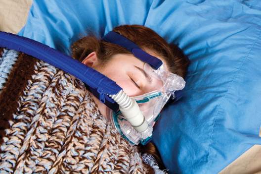
Many patients who have chronic obstructive pulmonary disease (COPD) or asthma also have obstructive sleep apnea (OSA)—and vice versa. This review from Cleveland Clinic Journal of Medicine, available at http://www.ccjm.org/topics/obesity-weight-management/single-article-page/the-intersection-of-obstructive-lung-disease-and-sleep-apnea/dff50621172ad1329c163560b7f1b19b.html, explores the shared risk factors for sleep-disordered breathing and obstructive lung diseases, describes potential pathophysiologic mechanisms explaining these associations, and highlights the importance of recognizing and individually treating the overlaps of OSA and COPD or asthma. 

References

Author and Disclosure Information

Publications
Topics
Legacy Keywords
chronic obstructive pulmonary disease, copd, asthma, sleep apnea, osa
Sections
Author and Disclosure Information

Author and Disclosure Information

Many patients who have chronic obstructive pulmonary disease (COPD) or asthma also have obstructive sleep apnea (OSA)—and vice versa. This review from Cleveland Clinic Journal of Medicine, available at http://www.ccjm.org/topics/obesity-weight-management/single-article-page/the-intersection-of-obstructive-lung-disease-and-sleep-apnea/dff50621172ad1329c163560b7f1b19b.html, explores the shared risk factors for sleep-disordered breathing and obstructive lung diseases, describes potential pathophysiologic mechanisms explaining these associations, and highlights the importance of recognizing and individually treating the overlaps of OSA and COPD or asthma. 

Many patients who have chronic obstructive pulmonary disease (COPD) or asthma also have obstructive sleep apnea (OSA)—and vice versa. This review from Cleveland Clinic Journal of Medicine, available at http://www.ccjm.org/topics/obesity-weight-management/single-article-page/the-intersection-of-obstructive-lung-disease-and-sleep-apnea/dff50621172ad1329c163560b7f1b19b.html, explores the shared risk factors for sleep-disordered breathing and obstructive lung diseases, describes potential pathophysiologic mechanisms explaining these associations, and highlights the importance of recognizing and individually treating the overlaps of OSA and COPD or asthma. 

References

References

Publications
Publications
Topics
Article Type
Display Headline
The intersection of obstructive lung disease and sleep apnea
Display Headline
The intersection of obstructive lung disease and sleep apnea
Legacy Keywords
chronic obstructive pulmonary disease, copd, asthma, sleep apnea, osa
Legacy Keywords
chronic obstructive pulmonary disease, copd, asthma, sleep apnea, osa
Sections
Article Source

PURLs Copyright

Inside the Article

Make the Diagnosis - March 2016

Article Type
Changed
Mon, 01/14/2019 - 09:34
Display Headline
Make the Diagnosis - March 2016

Diagnosis: Eruptive keratoacanthomas

Keratoacanthomas (KAs) most commonly affect people between the ages of 50 and 69 years old, although there have been reports in all age groups, including children. Studies have additionally revealed an equal distribution in prevalence between the sexes.

KAs are common, frequently self-limiting, epidermal tumors that consist of keratinizing squamous cells, thought to arise from the seboglandular part of the hair follicle. KAs have been divided into two general categories consisting of solitary and multiple types. Although the solitary type is most commonly observed, the multiple KAs category may be further subdivided to include the Ferguson-Smith type, which involves multiple self-healing KAs, generalized eruptive KA, which involves both skin and mucosa, multiple familial KA, multiple KA in association with Muir-Torre syndrome, and multiple KA centrifugum marginatum.

There are numerous factors implicated in the development of KAs, including trauma, light, exogenous carcinogens, impaired cell-mediated immunity, and immunosuppressive medications. A KA, which may be asymptomatic, slightly tender, or pruritic, initially forms as a small red macule and then evolves into a rapidly-growing (2 to 8 weeks) firm papule with scale. The papule then becomes a round, firm and raised skin-colored to pink nodule with a central keratin plug at the peak.

Histopathology varies depending upon the developmental stage of the lesion when biopsied. KA formation is comprised of 3 stages that may be recognized clinically and histologically, including the early-growing phase, the fully developed (stationary) phase and the senescent phase. Although not unique to KAs, histology may commonly show reactive proliferation of eccrine gland ducts beneath the tumor lobules. The ducts may adopt an adenomatoid appearance, as they lose their two-layer cellular construct.

The controversy regarding KA’s benign or malignant nature remains. Therefore, diagnosis is frequently confirmed through biopsy, in order to rule out squamous cell carcinoma. Although most KAs may resolve spontaneously, patients who find the lesions cosmetically unacceptable or painful may seek treatment. Nonsurgical modalities should be utilized before surgery, as surgical removal may leave scarring. Nonsurgical treatment options include local and systemic therapies, as well as electrodessication and curettage and laser therapy. A promising agent emerging in the treatment of KAs is 5-fluorouracil, which may be used as an intralesional injection, topically, or combined with lasers, leading to optimal cosmetic results with rapid clearance. The patient and family reported a noticeable improvement in appearance two weeks after discontinuing the new therapy.

Dr. Bilu Martin is in private practice at Premier Dermatology, MD, in Aventura, Fla. More diagnostic cases are available at edermatologynews.com. To submit your case for possible publication, send an email to [email protected].

References

Author and Disclosure Information

Publications
Sections
Author and Disclosure Information

Author and Disclosure Information

Diagnosis: Eruptive keratoacanthomas

Keratoacanthomas (KAs) most commonly affect people between the ages of 50 and 69 years old, although there have been reports in all age groups, including children. Studies have additionally revealed an equal distribution in prevalence between the sexes.

KAs are common, frequently self-limiting, epidermal tumors that consist of keratinizing squamous cells, thought to arise from the seboglandular part of the hair follicle. KAs have been divided into two general categories consisting of solitary and multiple types. Although the solitary type is most commonly observed, the multiple KAs category may be further subdivided to include the Ferguson-Smith type, which involves multiple self-healing KAs, generalized eruptive KA, which involves both skin and mucosa, multiple familial KA, multiple KA in association with Muir-Torre syndrome, and multiple KA centrifugum marginatum.

There are numerous factors implicated in the development of KAs, including trauma, light, exogenous carcinogens, impaired cell-mediated immunity, and immunosuppressive medications. A KA, which may be asymptomatic, slightly tender, or pruritic, initially forms as a small red macule and then evolves into a rapidly-growing (2 to 8 weeks) firm papule with scale. The papule then becomes a round, firm and raised skin-colored to pink nodule with a central keratin plug at the peak.

Histopathology varies depending upon the developmental stage of the lesion when biopsied. KA formation is comprised of 3 stages that may be recognized clinically and histologically, including the early-growing phase, the fully developed (stationary) phase and the senescent phase. Although not unique to KAs, histology may commonly show reactive proliferation of eccrine gland ducts beneath the tumor lobules. The ducts may adopt an adenomatoid appearance, as they lose their two-layer cellular construct.

The controversy regarding KA’s benign or malignant nature remains. Therefore, diagnosis is frequently confirmed through biopsy, in order to rule out squamous cell carcinoma. Although most KAs may resolve spontaneously, patients who find the lesions cosmetically unacceptable or painful may seek treatment. Nonsurgical modalities should be utilized before surgery, as surgical removal may leave scarring. Nonsurgical treatment options include local and systemic therapies, as well as electrodessication and curettage and laser therapy. A promising agent emerging in the treatment of KAs is 5-fluorouracil, which may be used as an intralesional injection, topically, or combined with lasers, leading to optimal cosmetic results with rapid clearance. The patient and family reported a noticeable improvement in appearance two weeks after discontinuing the new therapy.

Dr. Bilu Martin is in private practice at Premier Dermatology, MD, in Aventura, Fla. More diagnostic cases are available at edermatologynews.com. To submit your case for possible publication, send an email to [email protected].

References

References

Publications
Publications
Article Type
Display Headline
Make the Diagnosis - March 2016
Display Headline
Make the Diagnosis - March 2016
Sections
Questionnaire Body

A 67 year-old female with a past medical history significant for metastatic carcinoma of the lung and synovial sarcoma presented with a 6 week history of multiple verrucous scaly and acneiform papules scattered diffusely across her face and trunk. The lesions began one month post cancer treatment with a Notch inhibitor.
Article Source

PURLs Copyright

Inside the Article

Pediatric Dermatology Consult - March 2016

Article Type
Changed
Thu, 12/06/2018 - 17:50
Display Headline
Pediatric Dermatology Consult - March 2016

BY ELLEN S. HADDOCK AND LAWRENCE F. EICHENFIELD, M.D.

Pediatric Dermatology Consult: Pernio

Itchy localized areas of swelling on the bilateral hands with erythematous papules and crusting is consistent with pernio, as seen in the presentation of the teen boy described on p. 2. Pernio is a localized abnormal inflammatory response to cold and damp conditions, also known as chilblains, derived from the old English words for chill and sore.1 Damp air is thought to enhance the air conductivity of cold.2 Pernio typically presents with erythematous to blue-violet macules, papules, and nodules on the bilateral fingers and toes. When occurring on the feet, it is sometimes called trench foot or kibes.1 The nose and ears also can be affected.3 Lesions may develop 12-24 hours after exposure to damp or chilly weather, typically at temperatures above freezing.4

This patient’s pernio may have been triggered by recent stormy winter weather; being from within San Diego County, he had no exposure to snow. Lesions often are tender and may be accompanied by pruritus, pain, or a burning sensation, but they can be asymptomatic.3 Lesions may blister and ulcerate, and can become secondarily infected.5 Brownish or yellowish discoloration may be seen.1 Proposed diagnostic criteria requires localized erythema and swelling of the acral sites for more than 24 hours, as well as either onset during the cool months of the year or improvement with warming the affected area.3

Pernio is most common in adults, with a mean age of 38 years in one series.3 It also occurs in children but is uncommon, with only eight cases diagnosed at the University of Colorado over a 10-year period.6 Adult patients are primarily female,3 while the gender distribution in children is equal.6 Because pernio is triggered by cold and damp weather, it is not surprising that pernio occurs more often in cold climates.3 Raynaud’s phenomenon, smoking, and anorexia nervosa (due to lack of insulating fat) seem to be risk factors.1,3

Pernio typically is a benign primary disorder thought to result from cold-induced vasospasm, which leads to hypoxia and triggers a localized inflammatory reaction.7 Lesions of primary pernio usually resolve in a few weeks to months.4,6 However, pernio can be secondarily associated with systemic diseases including lupus (5% of patients in one of the largest series), non-lupus connective tissue disorders (4%), hematologic malignancy (3%), solid organ malignancy (2%), hepatitis, and Epstein-Barr virus.3 In these cases, pernio may be more persistent and hyperviscosity may contribute to its pathogenesis.8,9

Approximately a third of patients have laboratory abnormalities such as anemia, abnormal blood smear, autoantibodies, or serum monoclonal proteins,3 which may facilitate diagnosis of an underlying systemic disease. Not all pernio patients with connective tissue disease autoantibodies have clinical features of connective tissue disease, but these may manifest later.3,9 Laboratory abnormalities including cryoglobulinemia, cold agglutinins, rheumatoid factor, and antineutrophilic antibody also are seen in children;6,10 however, there are no reports of childhood pernio being associated with connective tissue disease or other systemic illness, although long-term studies are lacking.

When lesions are biopsied, histopathology shows nonspecific dermal edema with superficial and deep perivascular lymphocytic infiltrate.3,4

Differential diagnosis

The differential diagnosis for pernio includes Raynaud’s phenomenon, frostbite, herpetic whitlow, and purpura caused by cryoproteinemia. In Raynaud’s, pallor and cyanosis are followed by erythema, but the discoloration is more sharply demarcated and episodes are typically shorter, lasting hours rather than days.1,8 In this case, progression of the lesions over weeks and the lack of sudden skin color change when holding a cold drink make Raynaud’s unlikely. Frostbite, in which the tissue freezes and necroses, can be distinguished by history.11

When lesions have blistered, herpetic whitlow also may be on the differential, but herpetic whitlow vesicles typically cluster or coalesce into a single bulla while pernio lesions are more discrete. Cryoproteinemia causes lesions on acral sites exposed to the cold, but its onset is sudden and lesions are purpuric with a reticular (net-like) pattern.12 In adults, cutaneous thromboemboli also can present similarly to pernio,13 but thromboemboli are unlikely in children.

Clinical findings of pernio in the setting of lupus erythematosus is called chilblains lupus erythematosus. Confusingly, the condition called lupus pernio is actually a cutaneous manifestation of sarcoidosis, not lupus, and its erythematous or violaceous lesions occur on the nose and central face, not the hands and feet.13

Work-up

For pernio patients without systemic symptoms or signs of underlying systemic disease, laboratory workup or skin biopsy are not necessary.3,4 When history or physical exam is concerning for a systemic condition, preliminary workup should include complete blood count, peripheral blood smear, serum protein electrophoresis, cold agglutinins, and antinuclear antibody.3 Rheumatoid factor, antiphospholipid antibodies, and cryoglobulins also can be considered. Laboratory workup should be performed if pernio persists beyond the cold season, as persistent pernio may be associated with systemic illness.4,9


This patient’s recent weight loss was concerning for underlying systemic disease, so a laboratory workup including complete blood count, serum protein electrophoresis, cold agglutinins, antinuclear antibody, rheumatoid factor, and cryoglobulins was performed. Cryoglobulinemia was detected. All other lab values were within normal limits. Although cryoglobulinemia is rare in adults,3,14 it was detected in approximately 40% of the children in two pediatric series.6,10 Cryoglobulins, which can be produced in response to viral infection, may suggest a precipitating viral illness, with transient cryoproteinemia amplifying cold injury.6 Although the significance of laboratory abnormalities in pediatric pernio is unclear, because associated systemic disease has not been reported in children, some practitioners recommend long-term monitoring in light of the association between lab abnormalities and systemic disease in adults.10

Treatment

Most pernio (82% in one series) resolves when affected skin is warmed and dried, without additional treatment required.3 Corticosteroids, such as 0.1% triamcinolone cream, sometimes are given to hasten the healing of the lesions, but their benefit is unproven.6 The second-line treatment for persistent pernio is calcium channel blockers such as nifedipine.3,15 This patient’s pernio quickly improved after he began wearing gloves to keep his hands warm. On re-examination 2 weeks later, his hands were warm and the erythematous nodules had resolved, leaving only some scale at the sites of prior lesions (Figure 3). Some patients relapse annually during the cool months.11

References

  1. Pediatrics. 2005 Sep;116(3):e472-5. doi: 10.1542/peds.2004-2681.
  2. Journal of Medical Case Reports. 2014 Nov;8:381. doi: 10.1186/1752-1947-8-381.
  3. Mayo Clin Proc. 2014 Feb;89(2):207-15.
  4. Clin Exp Dermatol. 2012 Dec;37(8):844-9.
  5. J Paediatr Child Health. 2013 Feb;49(2):144-7.
  6. Pediatr Dermatol. 2000 Mar-Apr;17(2):97-9.
  7. Am J Med. 2009 Dec;122(12):1152-5.
  8. J Amer Acad Dermatol. 1990 Aug;23(Part 1):257-62.
  9. Medicine (Baltimore). 2001 May;80(3):180-8.
  10. Arch Dis Child. 2010 Jul;95(7):567-8.
  11. Br J Dermatol. 2010 Sep;163(3):645-6.
  12. Cutaneous manifestations of microvascular occlusion syndromes, in “Dermatology,” 3rd ed. (Philadelphia, 2012, pp 373-4).
  13. Environmental and sports-related skin diseases, in “Dermatology,” 3rd ed. (Philadelphia, 2012).
  14. J Am Acad Dermatol. 2010 Jun;62(6):e21-2.
  15. Br J Dermatol. 1989 Feb;120(2):267-75.

Ms. Haddock is a medical student at the University of California, San Diego, and a research associate at Rady Children’s Hospital, San Diego. Dr. Eichenfield is chief of pediatric and adolescent dermatology at Rady Children’s Hospital, San Diego, and Professor of Medicine and Pediatrics at the University of California, San Diego. Dr. Eichenfield and Ms. Haddock said they have no relevant financial disclosures.

References

Author and Disclosure Information

Publications
Sections
Author and Disclosure Information

Author and Disclosure Information

BY ELLEN S. HADDOCK AND LAWRENCE F. EICHENFIELD, M.D.

Pediatric Dermatology Consult: Pernio

Itchy localized areas of swelling on the bilateral hands with erythematous papules and crusting is consistent with pernio, as seen in the presentation of the teen boy described on p. 2. Pernio is a localized abnormal inflammatory response to cold and damp conditions, also known as chilblains, derived from the old English words for chill and sore.1 Damp air is thought to enhance the air conductivity of cold.2 Pernio typically presents with erythematous to blue-violet macules, papules, and nodules on the bilateral fingers and toes. When occurring on the feet, it is sometimes called trench foot or kibes.1 The nose and ears also can be affected.3 Lesions may develop 12-24 hours after exposure to damp or chilly weather, typically at temperatures above freezing.4

This patient’s pernio may have been triggered by recent stormy winter weather; being from within San Diego County, he had no exposure to snow. Lesions often are tender and may be accompanied by pruritus, pain, or a burning sensation, but they can be asymptomatic.3 Lesions may blister and ulcerate, and can become secondarily infected.5 Brownish or yellowish discoloration may be seen.1 Proposed diagnostic criteria requires localized erythema and swelling of the acral sites for more than 24 hours, as well as either onset during the cool months of the year or improvement with warming the affected area.3

Pernio is most common in adults, with a mean age of 38 years in one series.3 It also occurs in children but is uncommon, with only eight cases diagnosed at the University of Colorado over a 10-year period.6 Adult patients are primarily female,3 while the gender distribution in children is equal.6 Because pernio is triggered by cold and damp weather, it is not surprising that pernio occurs more often in cold climates.3 Raynaud’s phenomenon, smoking, and anorexia nervosa (due to lack of insulating fat) seem to be risk factors.1,3

Pernio typically is a benign primary disorder thought to result from cold-induced vasospasm, which leads to hypoxia and triggers a localized inflammatory reaction.7 Lesions of primary pernio usually resolve in a few weeks to months.4,6 However, pernio can be secondarily associated with systemic diseases including lupus (5% of patients in one of the largest series), non-lupus connective tissue disorders (4%), hematologic malignancy (3%), solid organ malignancy (2%), hepatitis, and Epstein-Barr virus.3 In these cases, pernio may be more persistent and hyperviscosity may contribute to its pathogenesis.8,9

Approximately a third of patients have laboratory abnormalities such as anemia, abnormal blood smear, autoantibodies, or serum monoclonal proteins,3 which may facilitate diagnosis of an underlying systemic disease. Not all pernio patients with connective tissue disease autoantibodies have clinical features of connective tissue disease, but these may manifest later.3,9 Laboratory abnormalities including cryoglobulinemia, cold agglutinins, rheumatoid factor, and antineutrophilic antibody also are seen in children;6,10 however, there are no reports of childhood pernio being associated with connective tissue disease or other systemic illness, although long-term studies are lacking.

When lesions are biopsied, histopathology shows nonspecific dermal edema with superficial and deep perivascular lymphocytic infiltrate.3,4

Differential diagnosis

The differential diagnosis for pernio includes Raynaud’s phenomenon, frostbite, herpetic whitlow, and purpura caused by cryoproteinemia. In Raynaud’s, pallor and cyanosis are followed by erythema, but the discoloration is more sharply demarcated and episodes are typically shorter, lasting hours rather than days.1,8 In this case, progression of the lesions over weeks and the lack of sudden skin color change when holding a cold drink make Raynaud’s unlikely. Frostbite, in which the tissue freezes and necroses, can be distinguished by history.11

When lesions have blistered, herpetic whitlow also may be on the differential, but herpetic whitlow vesicles typically cluster or coalesce into a single bulla while pernio lesions are more discrete. Cryoproteinemia causes lesions on acral sites exposed to the cold, but its onset is sudden and lesions are purpuric with a reticular (net-like) pattern.12 In adults, cutaneous thromboemboli also can present similarly to pernio,13 but thromboemboli are unlikely in children.

Clinical findings of pernio in the setting of lupus erythematosus is called chilblains lupus erythematosus. Confusingly, the condition called lupus pernio is actually a cutaneous manifestation of sarcoidosis, not lupus, and its erythematous or violaceous lesions occur on the nose and central face, not the hands and feet.13

Work-up

For pernio patients without systemic symptoms or signs of underlying systemic disease, laboratory workup or skin biopsy are not necessary.3,4 When history or physical exam is concerning for a systemic condition, preliminary workup should include complete blood count, peripheral blood smear, serum protein electrophoresis, cold agglutinins, and antinuclear antibody.3 Rheumatoid factor, antiphospholipid antibodies, and cryoglobulins also can be considered. Laboratory workup should be performed if pernio persists beyond the cold season, as persistent pernio may be associated with systemic illness.4,9


This patient’s recent weight loss was concerning for underlying systemic disease, so a laboratory workup including complete blood count, serum protein electrophoresis, cold agglutinins, antinuclear antibody, rheumatoid factor, and cryoglobulins was performed. Cryoglobulinemia was detected. All other lab values were within normal limits. Although cryoglobulinemia is rare in adults,3,14 it was detected in approximately 40% of the children in two pediatric series.6,10 Cryoglobulins, which can be produced in response to viral infection, may suggest a precipitating viral illness, with transient cryoproteinemia amplifying cold injury.6 Although the significance of laboratory abnormalities in pediatric pernio is unclear, because associated systemic disease has not been reported in children, some practitioners recommend long-term monitoring in light of the association between lab abnormalities and systemic disease in adults.10

Treatment

Most pernio (82% in one series) resolves when affected skin is warmed and dried, without additional treatment required.3 Corticosteroids, such as 0.1% triamcinolone cream, sometimes are given to hasten the healing of the lesions, but their benefit is unproven.6 The second-line treatment for persistent pernio is calcium channel blockers such as nifedipine.3,15 This patient’s pernio quickly improved after he began wearing gloves to keep his hands warm. On re-examination 2 weeks later, his hands were warm and the erythematous nodules had resolved, leaving only some scale at the sites of prior lesions (Figure 3). Some patients relapse annually during the cool months.11

References

  1. Pediatrics. 2005 Sep;116(3):e472-5. doi: 10.1542/peds.2004-2681.
  2. Journal of Medical Case Reports. 2014 Nov;8:381. doi: 10.1186/1752-1947-8-381.
  3. Mayo Clin Proc. 2014 Feb;89(2):207-15.
  4. Clin Exp Dermatol. 2012 Dec;37(8):844-9.
  5. J Paediatr Child Health. 2013 Feb;49(2):144-7.
  6. Pediatr Dermatol. 2000 Mar-Apr;17(2):97-9.
  7. Am J Med. 2009 Dec;122(12):1152-5.
  8. J Amer Acad Dermatol. 1990 Aug;23(Part 1):257-62.
  9. Medicine (Baltimore). 2001 May;80(3):180-8.
  10. Arch Dis Child. 2010 Jul;95(7):567-8.
  11. Br J Dermatol. 2010 Sep;163(3):645-6.
  12. Cutaneous manifestations of microvascular occlusion syndromes, in “Dermatology,” 3rd ed. (Philadelphia, 2012, pp 373-4).
  13. Environmental and sports-related skin diseases, in “Dermatology,” 3rd ed. (Philadelphia, 2012).
  14. J Am Acad Dermatol. 2010 Jun;62(6):e21-2.
  15. Br J Dermatol. 1989 Feb;120(2):267-75.

Ms. Haddock is a medical student at the University of California, San Diego, and a research associate at Rady Children’s Hospital, San Diego. Dr. Eichenfield is chief of pediatric and adolescent dermatology at Rady Children’s Hospital, San Diego, and Professor of Medicine and Pediatrics at the University of California, San Diego. Dr. Eichenfield and Ms. Haddock said they have no relevant financial disclosures.

References

References

Publications
Publications
Article Type
Display Headline
Pediatric Dermatology Consult - March 2016
Display Headline
Pediatric Dermatology Consult - March 2016
Sections
Questionnaire Body

A 13-year-old male presents with a rash that began as purplish spots on several fingers of the right hand and progressed over 2 months to involve all ten fingers. No other parts of his body are affected, and he had never experienced anything like this before. The fingers are intermittently painful and swollen. He is otherwise well, playing video games regularly and playing soccer with normal energy, although he states that a few of his soccer games have been canceled due to winter rains. He denies any sudden changes in the color of his hands with exposure to cold or holding cold drink bottles or cans (no “white, blue, and red changes”). He does not have any muscle or joint aches, but his mom reports that he has lost several pounds over the past 3 months. On physical exam, he has fifteen red, violaceous papules with surrounding swelling and erythema scattered on his dorsal fingers (Figure 1) and several similar lesions on his volar fingers. A few of the lesions are crusted. The skin of his volar fingers is dry, with some fine scale and several thickened, yellow-brown areas (Figure 2). His hands are very cold. His fingernails and feet are normal, and he has no lymphadenopathy.
Article Source

PURLs Copyright

Inside the Article

New and Noteworthy Information—March 2016

Article Type
Changed
Mon, 01/07/2019 - 10:16
Display Headline
New and Noteworthy Information—March 2016

Adults with a diagnosis of concussion have an increased long-term risk of suicide, particularly after concussions on weekends, according to a study published online ahead of print February 8 in the Canadian Medical Association Journal. Researchers performed a longitudinal cohort analysis of adults with a diagnosis of concussion from April 1, 1992 to March 31, 2012. Concussions that resulted in hospital admission were excluded. Investigators identified 235,110 patients with concussion, and 667 subsequent suicides occurring over a median follow-up of 9.3 years, which was three times the expected rate. Weekend concussions were associated with a one-third further increased risk of suicide, compared with weekday concussions. According to the researchers, the increased risk applied regardless of patients’ demographic characteristics, was independent of past psychiatric conditions, became accentuated with time, and exceeded the risk among military personnel.

Imaging with 3-T T2-weighted brain MRI distinguishes perivenous multiple sclerosis (MS) lesions from microangiopathic lesions, according to a study of 40 patients published online ahead of print December 10, 2015, in the Multiple Sclerosis Journal. Initially, a test cohort of 10 patients with MS and 10 patients with microangiopathic white matter lesions underwent T2-weighted brain imaging on a 3T MRI. Anonymized scans were analyzed blind to clinical data, and simple diagnostic rules were devised. These rules were applied to a validation cohort of 20 patients by a blinded observer. Within the test cohort, all patients with MS had central veins visible in more than 45% of brain lesions, while the rest had central veins visible in less than 45% of lesions. By applying diagnostic rules to the validation cohort, all remaining patients were correctly categorized.

In asymptomatic patients with severe carotid stenosis who are not at high risk for surgical complications, stenting is noninferior to endarterectomy with regard to the rates of stroke, death, or myocardial infarction at one year, according to a study published online ahead of print February 17 in the New England Journal of Medicine. Researchers compared carotid-artery stenting with embolic protection and carotid endarterectomy in 1,453 patients age 79 or younger. The rate of stroke or death within 30 days was 2.9% in the stenting group and 1.7% in the endarterectomy group. From 30 days to five years after the procedure, the rate of freedom from ipsilateral stroke was 97.8% in the stenting group and 97.3% in the endarterectomy group, and the overall survival rates were 87.1% and 89.4%, respectively.

Past exposure to marijuana is associated with worsened verbal memory, but does not appear to affect other domains of cognitive function, according to a study published online ahead of print February 1 in JAMA Internal Medicine. Researchers examined data for 5,115 African American and Caucasian men and women ages 18 to 30 at baseline. Participants were followed for 26 years to estimate cumulative exposure to marijuana. Among 3,385 participants with cognitive function measurements at the year 25 visit, 2,852 reported past marijuana use, and 392 continued to use marijuana into middle age. After excluding current users and adjusting for potential confounders, cumulative lifetime exposure to marijuana remained significantly associated with worsened verbal memory. For each five years of past exposure, verbal memory was 0.13 standardized units lower.

Frequent shifts in sleep timing may impair metabolic health among non-shift-working women of middle age, according to a study published in the February issue of Sleep. A total of 338 Caucasian, African American, and Chinese non-shift-working women ages 48 through 58 who were not taking insulin-related medications participated in the Study of Women’s Health Across the Nation Sleep Study and were examined approximately 5.39 years later. Daily diary-reported bedtimes were used to calculate four measures of sleep timing. BMI and insulin resistance were measured at two time points. In cross-sectional models, greater variability in bedtime and greater bedtime delay were associated with higher homeostatic model assessment-insulin resistance, and greater bedtime advance was associated with higher BMI. Prospectively, greater bedtime delay predicted increased homeostatic model assessment-insulin resistance.

Elderly people with high levels of depressive symptoms on several occasions over a 10-year period have substantially increased risk of coronary heart disease and stroke, according to a study published in the January issue of Journal of the American Geriatrics Society. Researchers examined 9,294 participants at baseline between 1999 and 2001, and during repeated study visits. There were 7,313 participants with an average age of 73.8, with no history of coronary heart disease, stroke, or dementia at baseline. After a median follow-up of 8.4 years, 629 first coronary heart disease or stroke events occurred. After adjustment for sociodemographic characteristics and vascular risk factors, the risk of coronary heart disease and stroke combined increased 1.15-fold per each additional study visit with high levels of depressive symptoms.


Submandibular gland needle biopsies identify phosphorylated alpha-synuclein staining in 74% of patients with early Parkinson’s disease, according to a study published in the February issue of Movement Disorders. Twenty-five patients with early Parkinson’s disease and 10 controls underwent transcutaneous needle core biopsies of the submandibular gland. Tissue was stained for phosphorylated alpha-synuclein and reviewed blind to clinical diagnosis. Only nerve element staining was considered positive. Mean age was 69.5 for the Parkinson’s disease group and 64.8 for controls, and disease duration was 2.6 years. Six people with Parkinson’s disease and one control subject had inadequate glandular tissue. Positive staining was found in 14 of 19 patients with Parkinson’s disease and two out of nine control subjects. Parkinson’s disease-positive and -negative cases did not differ clinically.

Lower cardiovascular fitness and exaggerated exercise blood pressure heart-rate responses in middle-aged adults are associated with smaller brain volume nearly two decades later, according to a study published online ahead of print February 10 in Neurology. In all, 1,094 people without dementia and cardiovascular disease underwent an exercise treadmill test at a mean age of 40. A second treadmill test and MRI scans of the brain were administered two decades later. Poor cardiovascular fitness and greater diastolic blood pressure and heart-rate response to exercise at baseline were associated with a smaller total cerebral brain volume almost two decades later in multivariable adjusted models. The effect of one standard deviation of lower fitness was equivalent to approximately one additional year of brain aging in individuals free of cardiovascular disease.

The increased risks of falling and hip fracture before the diagnosis of Parkinson’s disease may suggest the presence of clinically relevant neurodegenerative impairment years before the diagnosis of the disease, according to a study published February 2 in PLOS Medicine. Researchers compiled two nested case–control cohorts: In cohort one were individuals diagnosed with Parkinson’s disease; cohort two included individuals with an injurious fall. In cohort one, 18.0% of cases had at least one injurious fall before Parkinson’s disease diagnosis, whereas 11.5% of controls had an injurious fall. In cohort two, 0.7% of individuals with an injurious fall and 0.5% of controls were diagnosed with Parkinson’s disease during follow-up. The risk of Parkinson’s disease was increased for as long as 10 years after an injurious fall.

Increased amyloid β burden is observed in traumatic brain injury (TBI), according to a study published online ahead of print February 3 in Neurology. Patients age 11 months to 17 years with moderate to severe TBI underwent 11C-Pittsburgh compound B (11C-PiB)-PET, structural and diffusion MRI, and neuropsychologic examination. Healthy controls and patients with Alzheimer’s disease underwent PET and structural MRI. In TBI, fractional anisotropy was estimated and correlated with 11C-PiB binding potential. Increased 11C-PiB binding potential was found in TBI versus controls in the posterior cingulate cortex and cerebellum. Binding in the posterior cingulate cortex increased with decreasing fractional anisotropy of associated white matter tracts and increased with time since injury. Compared with Alzheimer’s disease, binding after TBI was lower in neocortical regions, but increased in the cerebellum.

Among participants in the Framingham Heart Study, the incidence of dementia has declined over three decades, according to a study published February 11 in the New England Journal of Medicine. In this analysis, which included 5,205 people age 60 and older, researchers used Cox proportional-hazards models adjusted for age and sex to determine the five-year incidence of dementia during each of four epochs. The five-year age- and sex-adjusted cumulative hazard rates for dementia were 3.6 per 100 persons during the first epoch, 2.8 per 100 persons during the second epoch, 2.2 per 100 persons during the third epoch, and 2.0 per 100 persons during the fourth epoch. During the second through fourth epochs, the incidence of dementia declined by 22%, 38%, and 44%, respectively, compared with the first epoch.

The FDA has approved Onzetra Xsail (sumatriptan nasal powder), formerly known as AVP-825, for the acute treatment of migraine with or without aura in adults. The approval is based on data from phase II and phase III trials, safety data from more than 300 patients, and reference data from the extensive clinical use of sumatriptan over the past 20 years. In one trial, 230 patients with migraine were randomized to self-administer either Onzetra Xsail or placebo, using the Xsail Breath Powered Delivery Device when they had moderate to severe migraine pain. A significantly greater proportion of patients on the study drug reported headache relief at 30 minutes and at every time point up to two hours post dose. Avanir Pharmaceuticals manufactures the drug.


The FDA has approved Medtronic Deep Brain Stimulation (DBS) Therapy for use in patients with Parkinson’s disease of at least four-years duration and with recent onset of motor complications, or motor complications of longer-standing duration that are not adequately controlled with medication. In 2002, the FDA initially approved Medtronic DBS Therapy for use in patients with advanced Parkinson’s disease. This new approval expands the population of patients who may benefit from this therapy. The approval is based on data from the EARLYSTIM study published in the New England Journal of Medicine in 2012.

Kimberly Williams

References

Author and Disclosure Information

Issue
Neurology Reviews - 24(3)
Publications
Page Number
6-7
Legacy Keywords
concussion, Alzheimer's disease, dementia, Parkinson's disease, depression, Kimberly Williams, Neurology Reviews
Sections
Author and Disclosure Information

Author and Disclosure Information

Adults with a diagnosis of concussion have an increased long-term risk of suicide, particularly after concussions on weekends, according to a study published online ahead of print February 8 in the Canadian Medical Association Journal. Researchers performed a longitudinal cohort analysis of adults with a diagnosis of concussion from April 1, 1992 to March 31, 2012. Concussions that resulted in hospital admission were excluded. Investigators identified 235,110 patients with concussion, and 667 subsequent suicides occurring over a median follow-up of 9.3 years, which was three times the expected rate. Weekend concussions were associated with a one-third further increased risk of suicide, compared with weekday concussions. According to the researchers, the increased risk applied regardless of patients’ demographic characteristics, was independent of past psychiatric conditions, became accentuated with time, and exceeded the risk among military personnel.

Imaging with 3-T T2-weighted brain MRI distinguishes perivenous multiple sclerosis (MS) lesions from microangiopathic lesions, according to a study of 40 patients published online ahead of print December 10, 2015, in the Multiple Sclerosis Journal. Initially, a test cohort of 10 patients with MS and 10 patients with microangiopathic white matter lesions underwent T2-weighted brain imaging on a 3T MRI. Anonymized scans were analyzed blind to clinical data, and simple diagnostic rules were devised. These rules were applied to a validation cohort of 20 patients by a blinded observer. Within the test cohort, all patients with MS had central veins visible in more than 45% of brain lesions, while the rest had central veins visible in less than 45% of lesions. By applying diagnostic rules to the validation cohort, all remaining patients were correctly categorized.

In asymptomatic patients with severe carotid stenosis who are not at high risk for surgical complications, stenting is noninferior to endarterectomy with regard to the rates of stroke, death, or myocardial infarction at one year, according to a study published online ahead of print February 17 in the New England Journal of Medicine. Researchers compared carotid-artery stenting with embolic protection and carotid endarterectomy in 1,453 patients age 79 or younger. The rate of stroke or death within 30 days was 2.9% in the stenting group and 1.7% in the endarterectomy group. From 30 days to five years after the procedure, the rate of freedom from ipsilateral stroke was 97.8% in the stenting group and 97.3% in the endarterectomy group, and the overall survival rates were 87.1% and 89.4%, respectively.

Past exposure to marijuana is associated with worsened verbal memory, but does not appear to affect other domains of cognitive function, according to a study published online ahead of print February 1 in JAMA Internal Medicine. Researchers examined data for 5,115 African American and Caucasian men and women ages 18 to 30 at baseline. Participants were followed for 26 years to estimate cumulative exposure to marijuana. Among 3,385 participants with cognitive function measurements at the year 25 visit, 2,852 reported past marijuana use, and 392 continued to use marijuana into middle age. After excluding current users and adjusting for potential confounders, cumulative lifetime exposure to marijuana remained significantly associated with worsened verbal memory. For each five years of past exposure, verbal memory was 0.13 standardized units lower.

Frequent shifts in sleep timing may impair metabolic health among non-shift-working women of middle age, according to a study published in the February issue of Sleep. A total of 338 Caucasian, African American, and Chinese non-shift-working women ages 48 through 58 who were not taking insulin-related medications participated in the Study of Women’s Health Across the Nation Sleep Study and were examined approximately 5.39 years later. Daily diary-reported bedtimes were used to calculate four measures of sleep timing. BMI and insulin resistance were measured at two time points. In cross-sectional models, greater variability in bedtime and greater bedtime delay were associated with higher homeostatic model assessment-insulin resistance, and greater bedtime advance was associated with higher BMI. Prospectively, greater bedtime delay predicted increased homeostatic model assessment-insulin resistance.

Elderly people with high levels of depressive symptoms on several occasions over a 10-year period have substantially increased risk of coronary heart disease and stroke, according to a study published in the January issue of Journal of the American Geriatrics Society. Researchers examined 9,294 participants at baseline between 1999 and 2001, and during repeated study visits. There were 7,313 participants with an average age of 73.8, with no history of coronary heart disease, stroke, or dementia at baseline. After a median follow-up of 8.4 years, 629 first coronary heart disease or stroke events occurred. After adjustment for sociodemographic characteristics and vascular risk factors, the risk of coronary heart disease and stroke combined increased 1.15-fold per each additional study visit with high levels of depressive symptoms.


Submandibular gland needle biopsies identify phosphorylated alpha-synuclein staining in 74% of patients with early Parkinson’s disease, according to a study published in the February issue of Movement Disorders. Twenty-five patients with early Parkinson’s disease and 10 controls underwent transcutaneous needle core biopsies of the submandibular gland. Tissue was stained for phosphorylated alpha-synuclein and reviewed blind to clinical diagnosis. Only nerve element staining was considered positive. Mean age was 69.5 for the Parkinson’s disease group and 64.8 for controls, and disease duration was 2.6 years. Six people with Parkinson’s disease and one control subject had inadequate glandular tissue. Positive staining was found in 14 of 19 patients with Parkinson’s disease and two out of nine control subjects. Parkinson’s disease-positive and -negative cases did not differ clinically.

Lower cardiovascular fitness and exaggerated exercise blood pressure heart-rate responses in middle-aged adults are associated with smaller brain volume nearly two decades later, according to a study published online ahead of print February 10 in Neurology. In all, 1,094 people without dementia and cardiovascular disease underwent an exercise treadmill test at a mean age of 40. A second treadmill test and MRI scans of the brain were administered two decades later. Poor cardiovascular fitness and greater diastolic blood pressure and heart-rate response to exercise at baseline were associated with a smaller total cerebral brain volume almost two decades later in multivariable adjusted models. The effect of one standard deviation of lower fitness was equivalent to approximately one additional year of brain aging in individuals free of cardiovascular disease.

The increased risks of falling and hip fracture before the diagnosis of Parkinson’s disease may suggest the presence of clinically relevant neurodegenerative impairment years before the diagnosis of the disease, according to a study published February 2 in PLOS Medicine. Researchers compiled two nested case–control cohorts: In cohort one were individuals diagnosed with Parkinson’s disease; cohort two included individuals with an injurious fall. In cohort one, 18.0% of cases had at least one injurious fall before Parkinson’s disease diagnosis, whereas 11.5% of controls had an injurious fall. In cohort two, 0.7% of individuals with an injurious fall and 0.5% of controls were diagnosed with Parkinson’s disease during follow-up. The risk of Parkinson’s disease was increased for as long as 10 years after an injurious fall.

Increased amyloid β burden is observed in traumatic brain injury (TBI), according to a study published online ahead of print February 3 in Neurology. Patients age 11 months to 17 years with moderate to severe TBI underwent 11C-Pittsburgh compound B (11C-PiB)-PET, structural and diffusion MRI, and neuropsychologic examination. Healthy controls and patients with Alzheimer’s disease underwent PET and structural MRI. In TBI, fractional anisotropy was estimated and correlated with 11C-PiB binding potential. Increased 11C-PiB binding potential was found in TBI versus controls in the posterior cingulate cortex and cerebellum. Binding in the posterior cingulate cortex increased with decreasing fractional anisotropy of associated white matter tracts and increased with time since injury. Compared with Alzheimer’s disease, binding after TBI was lower in neocortical regions, but increased in the cerebellum.

Among participants in the Framingham Heart Study, the incidence of dementia has declined over three decades, according to a study published February 11 in the New England Journal of Medicine. In this analysis, which included 5,205 people age 60 and older, researchers used Cox proportional-hazards models adjusted for age and sex to determine the five-year incidence of dementia during each of four epochs. The five-year age- and sex-adjusted cumulative hazard rates for dementia were 3.6 per 100 persons during the first epoch, 2.8 per 100 persons during the second epoch, 2.2 per 100 persons during the third epoch, and 2.0 per 100 persons during the fourth epoch. During the second through fourth epochs, the incidence of dementia declined by 22%, 38%, and 44%, respectively, compared with the first epoch.

The FDA has approved Onzetra Xsail (sumatriptan nasal powder), formerly known as AVP-825, for the acute treatment of migraine with or without aura in adults. The approval is based on data from phase II and phase III trials, safety data from more than 300 patients, and reference data from the extensive clinical use of sumatriptan over the past 20 years. In one trial, 230 patients with migraine were randomized to self-administer either Onzetra Xsail or placebo, using the Xsail Breath Powered Delivery Device when they had moderate to severe migraine pain. A significantly greater proportion of patients on the study drug reported headache relief at 30 minutes and at every time point up to two hours post dose. Avanir Pharmaceuticals manufactures the drug.


The FDA has approved Medtronic Deep Brain Stimulation (DBS) Therapy for use in patients with Parkinson’s disease of at least four-years duration and with recent onset of motor complications, or motor complications of longer-standing duration that are not adequately controlled with medication. In 2002, the FDA initially approved Medtronic DBS Therapy for use in patients with advanced Parkinson’s disease. This new approval expands the population of patients who may benefit from this therapy. The approval is based on data from the EARLYSTIM study published in the New England Journal of Medicine in 2012.

Kimberly Williams

References

References

Issue
Neurology Reviews - 24(3)
Issue
Neurology Reviews - 24(3)
Page Number
6-7
Page Number
6-7
Publications
Publications
Article Type
Display Headline
New and Noteworthy Information—March 2016
Display Headline
New and Noteworthy Information—March 2016
Legacy Keywords
concussion, Alzheimer's disease, dementia, Parkinson's disease, depression, Kimberly Williams, Neurology Reviews
Legacy Keywords
concussion, Alzheimer's disease, dementia, Parkinson's disease, depression, Kimberly Williams, Neurology Reviews
Sections
Article Source

PURLs Copyright

Inside the Article

Nerve Blocks May Provide Rapid Relief in Headache

Article Type
Changed
Mon, 01/07/2019 - 10:16
Display Headline
Nerve Blocks May Provide Rapid Relief in Headache

OJAI, CA—Peripheral nerve blocks are a reasonable therapeutic option for many patients with headache disorders, according to an overview provided at the Ninth Annual Headache Cooperative of the Pacific Winter Conference. Cluster headache is the indication for which nerve blocks have the best evidence. The data are mixed for other disorders, such as migraine.

“Therapeutic injections for headache can be performed effectively, safely, and efficiently after gaining a basic understanding of the literature and anatomic landmarks,” said Jack Schim, MD, Codirector of the Headache Center of Southern California and Chief of Neurology at Scripps Hospital in Encinitas.

Jack Schim, MD

A survey of members of the American Headache Society published in 2010 found that headache practitioners commonly use nerve blocks, but dosing regimens and the use of corticosteroids vary greatly. Neurologists most often use nerve blocks to treat occipital neuralgia and chronic migraine, although they also use nerve blocks for tension-type headache, hemicrania continua, and other disorders.

“Anesthetic blocks get the patients better pretty rapidly,” Dr. Schim said. “You do a nerve block … and they say, ‘Oh, my headache’s gone. How long is it going to feel good?’ That is the gratification to both us and the patient.”

A Two-Minute Procedure

Performing nerve blocks typically takes a few minutes. Some neurologists may choose the injection site based on the location of pain. Dr. Schim injects all of the potential targets—the auriculotemporal, zygomaticotemporal, supratochlear, supraorbital, and greater and lesser occipital nerves when treating migraine. For cluster headache, paroxysmal hemicrania, or hemicrania continua, he may block just the ipsilateral occipital nerve. If there is not sufficient pain relief, he may then block the trigeminal branches. “Local pain may not be the primary determinant of where we ought to inject,” he said. “Think about the usual cluster patient who has V1 orbital pain, and yet occipital block with steroids … can be very effective in transitional pain relief.”

Neurologists mainly inject lidocaine (concentration, 1–2%; maximum dose, 300 mg) or bupivacaine (concentration, 0.25–0.5%; maximum dose, 175 mg), and some neurologists inject both drugs. The effect of lidocaine lasts for one to three hours, whereas the effect of bupivacaine lasts for four to eight hours. Dr. Schim typically uses bupivacaine alone and finds that patients become numb in a couple of minutes.

Pain relief commonly lasts longer than the anesthetic effect. Virtually all patients seem to experience pain relief after receiving nerve blocks, but there is no way to predict whose headaches will return the next day or which patients will experience weeks of relief, Dr. Schim said.

Who Might Benefit

Nerve blocks may rescue patients who have failed their home medications. They also may be used to treat patients who need relief between onabotulinum toxin A injections. For example, in a patient who has good results for 10 weeks after receiving onabotulinum toxin A, a nerve block may provide relief during the two weeks until his or her next onabotulinum toxin A injection. Nerve blocks also may help to wean patients with medication overuse headache from their acute therapy. “If we get them to stop their acute medicine, things are going to get worse for a while. One of the options is to bring them in for blocks at whatever frequency you feel comfortable with,” he said.

Nerve blocks also may be appropriate for children and pregnant patients. In pregnancy, lidocaine is considered a category B drug. “It’s a reasonable migraine prophylaxis if it works for someone,” he said. “I have some patients who came in every two to three weeks, and that successfully got them through pregnancy.” Receiving nerve blocks with greater frequency might be uncomfortable for patients because the procedure usually is painful until the onset of numbness.

In 2014, Govindappagari et al published a case series of 13 pregnant women with migraine who received peripheral nerve blocks for status migrainosus or short-term prophylaxis of frequent headache attacks. In status migrainosus, average pain-score reduction was 4.0 immediately and at 24 hours after the procedure. For short-term prophylaxis, average immediate pain-score reduction was 3.0. The two patients who did not receive any acute pain reduction from the nerve blocks developed preeclampsia, and their headaches resolved post partum.

A 2014 study by Gelfand et al examined the efficacy of greater occipital nerve injections in 46 children with chronic primary headache disorders. They found that injections of lidocaine and methylprednisolone acetate benefited 53% of patients, including 62% of those with chronic migraine and 33% of those with new daily persistent headache. “In children, we have very few on-label options and we certainly have little kids who could benefit from some relief,” Dr. Schim said.


Add Steroids for Cluster Headache

In greater occipital nerve injections for cluster headache, steroids (ie, 40 mg of triamcinolone or 20 mg of methylprednisolone) should be used with the local anesthetic, according to randomized controlled trials. For migraine, studies have found that adding steroids to the local anesthetic is not beneficial.

Steroids can cause systemic and local effects, including fat atrophy and alopecia. Vasovagal attacks are another safety concern. Older patients on blood-pressure medication might be more susceptible to becoming hypotensive. In patients who have had a craniotomy, the anesthetic can diffuse through a prior craniotomy site and have direct intracranial effect, which could be hazardous. Injecting an anesthetic without steroids does not raise cosmetic concerns. Those administering nerve blocks must know the relevant anatomy of the nerves and local vasculature. For example, occipital blocks are often done above the skull base to reduce risk.

Jake Remaly

References

Suggested Reading
Ashkenazi A, Blumenfeld A, Napchan U. Peripheral nerve blocks and trigger point injections in headache management - a systematic review and suggestions for future research. Headache. 2010;50(6):943-952.
Blumenfeld A, Ashkenazi A, Grosberg B, et al. Patterns of use of peripheral nerve blocks and trigger point injections among headache practitioners in the USA: Results of the American Headache Society Interventional Procedure Survey (AHS-IPS). Headache. 2010;50(6):937-942.
Blumenfeld A, Ashkenazi A, Napchan U, et al. Expert consensus recommendations for the performance of peripheral nerve blocks for headaches--a narrative review. Headache. 2013;53(3):437-446.
Gelfand AA, Reider AC, Goadsby PJ. Outcomes of greater occipital nerve injections in pediatric patients with chronic primary headache disorders. Pediatr Neurol. 2014;50(2):135-139.
Govindappagari S, Grossman TB, Dayal AK, et al. Peripheral nerve blocks in the treatment of migraine in pregnancy. Obstet Gynecol. 2014;124(6):1169-1174.

Author and Disclosure Information

Issue
Neurology Reviews - 24(3)
Publications
Topics
Page Number
11
Legacy Keywords
peripheral nerve blocks, headache, migraine, injection, Jack Schim, Neurology Reviews
Sections
Author and Disclosure Information

Author and Disclosure Information

Related Articles

OJAI, CA—Peripheral nerve blocks are a reasonable therapeutic option for many patients with headache disorders, according to an overview provided at the Ninth Annual Headache Cooperative of the Pacific Winter Conference. Cluster headache is the indication for which nerve blocks have the best evidence. The data are mixed for other disorders, such as migraine.

“Therapeutic injections for headache can be performed effectively, safely, and efficiently after gaining a basic understanding of the literature and anatomic landmarks,” said Jack Schim, MD, Codirector of the Headache Center of Southern California and Chief of Neurology at Scripps Hospital in Encinitas.

Jack Schim, MD

A survey of members of the American Headache Society published in 2010 found that headache practitioners commonly use nerve blocks, but dosing regimens and the use of corticosteroids vary greatly. Neurologists most often use nerve blocks to treat occipital neuralgia and chronic migraine, although they also use nerve blocks for tension-type headache, hemicrania continua, and other disorders.

“Anesthetic blocks get the patients better pretty rapidly,” Dr. Schim said. “You do a nerve block … and they say, ‘Oh, my headache’s gone. How long is it going to feel good?’ That is the gratification to both us and the patient.”

A Two-Minute Procedure

Performing nerve blocks typically takes a few minutes. Some neurologists may choose the injection site based on the location of pain. Dr. Schim injects all of the potential targets—the auriculotemporal, zygomaticotemporal, supratochlear, supraorbital, and greater and lesser occipital nerves when treating migraine. For cluster headache, paroxysmal hemicrania, or hemicrania continua, he may block just the ipsilateral occipital nerve. If there is not sufficient pain relief, he may then block the trigeminal branches. “Local pain may not be the primary determinant of where we ought to inject,” he said. “Think about the usual cluster patient who has V1 orbital pain, and yet occipital block with steroids … can be very effective in transitional pain relief.”

Neurologists mainly inject lidocaine (concentration, 1–2%; maximum dose, 300 mg) or bupivacaine (concentration, 0.25–0.5%; maximum dose, 175 mg), and some neurologists inject both drugs. The effect of lidocaine lasts for one to three hours, whereas the effect of bupivacaine lasts for four to eight hours. Dr. Schim typically uses bupivacaine alone and finds that patients become numb in a couple of minutes.

Pain relief commonly lasts longer than the anesthetic effect. Virtually all patients seem to experience pain relief after receiving nerve blocks, but there is no way to predict whose headaches will return the next day or which patients will experience weeks of relief, Dr. Schim said.

Who Might Benefit

Nerve blocks may rescue patients who have failed their home medications. They also may be used to treat patients who need relief between onabotulinum toxin A injections. For example, in a patient who has good results for 10 weeks after receiving onabotulinum toxin A, a nerve block may provide relief during the two weeks until his or her next onabotulinum toxin A injection. Nerve blocks also may help to wean patients with medication overuse headache from their acute therapy. “If we get them to stop their acute medicine, things are going to get worse for a while. One of the options is to bring them in for blocks at whatever frequency you feel comfortable with,” he said.

Nerve blocks also may be appropriate for children and pregnant patients. In pregnancy, lidocaine is considered a category B drug. “It’s a reasonable migraine prophylaxis if it works for someone,” he said. “I have some patients who came in every two to three weeks, and that successfully got them through pregnancy.” Receiving nerve blocks with greater frequency might be uncomfortable for patients because the procedure usually is painful until the onset of numbness.

In 2014, Govindappagari et al published a case series of 13 pregnant women with migraine who received peripheral nerve blocks for status migrainosus or short-term prophylaxis of frequent headache attacks. In status migrainosus, average pain-score reduction was 4.0 immediately and at 24 hours after the procedure. For short-term prophylaxis, average immediate pain-score reduction was 3.0. The two patients who did not receive any acute pain reduction from the nerve blocks developed preeclampsia, and their headaches resolved post partum.

A 2014 study by Gelfand et al examined the efficacy of greater occipital nerve injections in 46 children with chronic primary headache disorders. They found that injections of lidocaine and methylprednisolone acetate benefited 53% of patients, including 62% of those with chronic migraine and 33% of those with new daily persistent headache. “In children, we have very few on-label options and we certainly have little kids who could benefit from some relief,” Dr. Schim said.


Add Steroids for Cluster Headache

In greater occipital nerve injections for cluster headache, steroids (ie, 40 mg of triamcinolone or 20 mg of methylprednisolone) should be used with the local anesthetic, according to randomized controlled trials. For migraine, studies have found that adding steroids to the local anesthetic is not beneficial.

Steroids can cause systemic and local effects, including fat atrophy and alopecia. Vasovagal attacks are another safety concern. Older patients on blood-pressure medication might be more susceptible to becoming hypotensive. In patients who have had a craniotomy, the anesthetic can diffuse through a prior craniotomy site and have direct intracranial effect, which could be hazardous. Injecting an anesthetic without steroids does not raise cosmetic concerns. Those administering nerve blocks must know the relevant anatomy of the nerves and local vasculature. For example, occipital blocks are often done above the skull base to reduce risk.

Jake Remaly

References

Suggested Reading
Ashkenazi A, Blumenfeld A, Napchan U. Peripheral nerve blocks and trigger point injections in headache management - a systematic review and suggestions for future research. Headache. 2010;50(6):943-952.
Blumenfeld A, Ashkenazi A, Grosberg B, et al. Patterns of use of peripheral nerve blocks and trigger point injections among headache practitioners in the USA: Results of the American Headache Society Interventional Procedure Survey (AHS-IPS). Headache. 2010;50(6):937-942.
Blumenfeld A, Ashkenazi A, Napchan U, et al. Expert consensus recommendations for the performance of peripheral nerve blocks for headaches--a narrative review. Headache. 2013;53(3):437-446.
Gelfand AA, Reider AC, Goadsby PJ. Outcomes of greater occipital nerve injections in pediatric patients with chronic primary headache disorders. Pediatr Neurol. 2014;50(2):135-139.
Govindappagari S, Grossman TB, Dayal AK, et al. Peripheral nerve blocks in the treatment of migraine in pregnancy. Obstet Gynecol. 2014;124(6):1169-1174.

Issue
Neurology Reviews - 24(3)
Issue
Neurology Reviews - 24(3)
Page Number
11
Page Number
11
Publications
Publications
Topics
Article Type
Display Headline
Nerve Blocks May Provide Rapid Relief in Headache
Display Headline
Nerve Blocks May Provide Rapid Relief in Headache
Legacy Keywords
peripheral nerve blocks, headache, migraine, injection, Jack Schim, Neurology Reviews
Legacy Keywords
peripheral nerve blocks, headache, migraine, injection, Jack Schim, Neurology Reviews
Sections
Article Source

PURLs Copyright

Inside the Article

March 2016 Quiz 2

Article Type
Changed
Tue, 12/13/2016 - 10:27
Display Headline
March 2016 Quiz 2

Q2: ANSWER: E

Critique

These results could be due to Lynch syndrome as a result of an EPCAM mutation. With hereditary nonpolyposis colorectal cancer (HNPCC), also known as Lynch syndrome, patients are at an increased risk for colorectal and several other cancers owing to inactivating germline mutations in mismatch repair genes (MMR), including MLH1, MSH2, MSH6, and PMS2. Germline EPCAM deletions in the 3’ region can cause HNPCC. The EPCAM deletions lead to methylation of the MSH2 promoter and ultimately silencing of MSH2 gene. Silencing of the MSH2 gene results in a pattern of MSH2 and MSH6 loss on immunohistochemistry. Since immunohistochemistry shows no loss of expression of MLH1 or PMS2 proteins, this indicates the absence of a mutation in MLH1 and PMS2 genes. The presence of high microsatellite instability and loss of expression of two mismatch repair proteins indicates the presence of Lynch syndrome and not a sporadic colorectal cancer. Therefore MLH1 hypermethylation and BRAF testing is not indicated.

Reference

1. Umar A., Boland C., Terdiman J.P., et al. Revised Bethesda guidelines for hereditary nonpolyposis colorectal cancer (Lynch syndrome) and microsatellite instability. J Natl Cancer Inst. 2004;96:261-8.

References

Author and Disclosure Information

Publications
Sections
Author and Disclosure Information

Author and Disclosure Information

Q2: ANSWER: E

Critique

These results could be due to Lynch syndrome as a result of an EPCAM mutation. With hereditary nonpolyposis colorectal cancer (HNPCC), also known as Lynch syndrome, patients are at an increased risk for colorectal and several other cancers owing to inactivating germline mutations in mismatch repair genes (MMR), including MLH1, MSH2, MSH6, and PMS2. Germline EPCAM deletions in the 3’ region can cause HNPCC. The EPCAM deletions lead to methylation of the MSH2 promoter and ultimately silencing of MSH2 gene. Silencing of the MSH2 gene results in a pattern of MSH2 and MSH6 loss on immunohistochemistry. Since immunohistochemistry shows no loss of expression of MLH1 or PMS2 proteins, this indicates the absence of a mutation in MLH1 and PMS2 genes. The presence of high microsatellite instability and loss of expression of two mismatch repair proteins indicates the presence of Lynch syndrome and not a sporadic colorectal cancer. Therefore MLH1 hypermethylation and BRAF testing is not indicated.

Reference

1. Umar A., Boland C., Terdiman J.P., et al. Revised Bethesda guidelines for hereditary nonpolyposis colorectal cancer (Lynch syndrome) and microsatellite instability. J Natl Cancer Inst. 2004;96:261-8.

References

References

Publications
Publications
Article Type
Display Headline
March 2016 Quiz 2
Display Headline
March 2016 Quiz 2
Sections
Questionnaire Body
A 49-year-old African American man presents to your outpatient gastroenterology clinic with a history of hematochezia for the past 5 months. To evaluate these symptoms you perform a colonoscopy that demonstrates a 5-cm cecal adenocarcinoma and one 5-mm adenoma in the transverse colon that is removed completely with snare polypectomy. He reports that his mother had three 5- to 6-mm colorectal adenomas on her only colonoscopy at age 60 years. He has no other family history of cancer or polyps in his other first- or second-degree relatives. Based on his history, you order tumor immunohistochemistry and microsatellite instability (MSI) testing. The tumor is MSI-high and demonstrates loss of MSH2 and MSH6 and intact expression of MLH1 and PMS2. No germline mutations in MSH2 or MSH6 are detected on genetic testing.
Article Source

PURLs Copyright

Inside the Article

Disallow All Ads

March 2016 Quiz 1

Article Type
Changed
Tue, 12/13/2016 - 10:27
Display Headline
March 2016 Quiz 1

Q1: ANSWER: C

Critique

The patient is morbidly obese, with persisting esophagitis despite twice-a-day PPI, and structural disruption of the gastroesophageal junction with a moderate-sized hiatus hernia and a patulous gastroesophageal junction. This is a setting in which a surgical approach is worthwhile. Since the patient is morbidly obese with hypertension and diabetes, the option of Roux-en-Y gastric bypass surgery is worth considering. This is a viable option for the surgical management of persisting reflux in the morbidly obese. Sucralfate has limited additive gain in persistent esophagitis despite PPI therapy. There is no net gain in switching the route of PPI administration to intravenous, and this is mostly indicated in the setting of gastrointestinal bleeding. Metoclopramide does not result in worthwhile augmentation of esophageal motility, and may not necessarily provide further symptom improvement or healing of esophagitis. Lifestyle measures complement pharmacologic management of reflux disease, but have not been shown to heal esophagitis.

Reference

1. De Groot N.L., Burgerhart J.S., Van De Meeberg P.C., et al. Systematic review: the effects of conservative and surgical treatment for obesity on gastro-oesophageal reflux disease. Aliment Pharmacol Ther. 2009;30:1091-102.

References

Author and Disclosure Information

Publications
Sections
Author and Disclosure Information

Author and Disclosure Information

Q1: ANSWER: C

Critique

The patient is morbidly obese, with persisting esophagitis despite twice-a-day PPI, and structural disruption of the gastroesophageal junction with a moderate-sized hiatus hernia and a patulous gastroesophageal junction. This is a setting in which a surgical approach is worthwhile. Since the patient is morbidly obese with hypertension and diabetes, the option of Roux-en-Y gastric bypass surgery is worth considering. This is a viable option for the surgical management of persisting reflux in the morbidly obese. Sucralfate has limited additive gain in persistent esophagitis despite PPI therapy. There is no net gain in switching the route of PPI administration to intravenous, and this is mostly indicated in the setting of gastrointestinal bleeding. Metoclopramide does not result in worthwhile augmentation of esophageal motility, and may not necessarily provide further symptom improvement or healing of esophagitis. Lifestyle measures complement pharmacologic management of reflux disease, but have not been shown to heal esophagitis.

Reference

1. De Groot N.L., Burgerhart J.S., Van De Meeberg P.C., et al. Systematic review: the effects of conservative and surgical treatment for obesity on gastro-oesophageal reflux disease. Aliment Pharmacol Ther. 2009;30:1091-102.

References

References

Publications
Publications
Article Type
Display Headline
March 2016 Quiz 1
Display Headline
March 2016 Quiz 1
Sections
Questionnaire Body
You are asked to see a patient in consultation for refractory GERD. The patient is a 44-year-old woman, admitted to the hospital for a urinary tract infection, who reports constant heartburn and regurgitation despite twice-a-day pantoprazole taken before breakfast and supper for the past 3 months. The patient is morbidly obese with a body mass index (in kg/m2) of 43, and suffers from diabetes and hypertension. Upper endoscopy demonstrates LA grade C erosive esophagitis on PPI therapy, a patulous gastroesophageal junction, and a 4-cm hiatus hernia.
Article Source

PURLs Copyright

Inside the Article

Readers weigh in on vaginal cleansing prior to cesarean delivery

Article Type
Changed
Tue, 08/28/2018 - 11:07
Display Headline
Readers weigh in on vaginal cleansing prior to cesarean delivery

“SHOULD YOU ADOPT THE PRACTICE OF VAGINAL CLEANSING WITH POVIDONE-IODINE PRIOR TO CESAREAN DELIVERY?”
ROBERT L. BARBIERI, MD (EDITORIAL; JANUARY 2016)

In his January 2016 Editorial, Editor in Chief Robert L. Barbieri, MD, presented evidence supporting the practice of vaginal cleansing with povidone-iodine prior to cesarean delivery (CD) to prevent postoperative endometritis. He then asked readers if they would consider adopting such a practice. More than 250 readers weighed in through the Quick Poll at obgmanagement.com, and many readers sent in letters with follow-up questions and comments on controlling bacterial contamination, vaginal seeding, etc. Here are some of the letters, along with Dr. Barbieri’s response and the Quick Poll results.

A contradiction in definitions?
There seems to be a contradiction in definitions. The second sentence of the article defines endometritis as the presence of fever plus low abdominal tenderness. However, the studies presented state that vaginal cleansing pre-CD decreased endometritis but did not decrease postpartum fever. Is this not a discrepancy?
Nancy Kerr, MD, MPH

Albuquerque, New Mexico

A question about povidone-iodine
Have any studies been done on newborn iodine levels after vaginal cleansing with povidone-iodine prior to CD?
G. Millard Simmons Jr, MD
Hilton Head, Bluffton, South Carolina

Additional tips for controlling bacterial contamination
Dr. Barbieri’s editorial on vaginal cleansing prior to CD is eye opening. I have a few additional suggestions to control bacterial contamination.

First, I examine my patients in labor as few times as necessary, and I ask the nurses (RNs) not to place their fingers in the patient’s vagina while she is pushing. I remove the Foley catheter when I feel progress (descent of fetal head) is being achieved. In addition, physicians as well as RNs should consider changing their scrubs between deliveries, as I believe that bacterial contamination is splattered all over the place, especially into the birth canal. These methods have worked for me in my over-20 years of practice.

I also firmly remind the RN circulator to perform a generous vaginal cleanse with povidone-iodine, in addition to the usual intravenous prophylaxis, before hysterectomy.
Luis Leyva Jr, MD
Miami, Florida

Mixed feelings
My first reaction to this Editorial was: Is this a solution in search of a problem? That is to say, how much of a clinical problem is endometritis after CD? Are we really treating the proposed problem, and does treatment affect long-term outcomes?

Upon reflection, I have concluded that vaginal cleansing pre-CD does intuitively make sense. What sways me in this direction is that the practice is simple, easy, and inexpensive. Since we typically have the patient positioned for Foley catheter insertion, performing vaginal cleansing as we put in the Foley would be easy. If vaginal cleansing were to be done, I definitely would be in favor of doing such practice liberally—for all CDs to make vaginal cleansing part of the “routine.”

Keep in mind that we are still chasing a problem of little clinical significance.

The biggest accomplishment has been to get everyone to give antibiotics preoperatively rather than after cutting the umbilical cord. We knew that this was best practice as early as the late 1980s/early 1990s, and I have been fighting this battle ever since. Believe it or not, there are still a few holdouts.
George H. Davis, DO
Johnson City, Tennessee

Would vaginal cleansing benefit all women in labor?
Vaginal cleansing before CD reminds me of my residency days when all women having hysterectomies were admitted early and given povidone-iodine (Betadine) douches the evening before surgery (unless an iodine allergy was present).

While reading your Editorial, I had several thoughts and questions. 1) Since vaginal cleansing seems to benefit CD patients, might it not benefit all laboring patients? 2) Is the timing of vaginal cleansing critical? 3) Should we do vaginal cleansing on all laboring patients if timing is not critical?

I plan to bring up the topic of vaginal cleansing for CD with my colleagues at our next department meeting, since it seems like such a simple, logical, inexpensive, and beneficial thing to do.
Douglas G. Tolley, MD
Yuba City, California

An early study on using povidone-iodine gel before CD
When I was a chief resident at Kings County Hospital in 1973, we had a very high rate of post-CD endometritis. I conducted a small study on the use of povidone-iodine gel in the last month of pregnancy. Before commencing, we confirmed that the gel did not interfere with diagnosing ruptured membranes.

Obstetric service patients were randomly divided into “A” and “B” groups. The A patients were asked to use povidone-iodine gel at night for the last 2 weeks before their estimated due date. When admitted in labor, they were asked to confirm its use. When a resident diagnosed post-CD endometritis, we kept track of which group the patient was in and whether or not that patient had used povidone-iodine. Approximately 100 infected patients were evaluated from each group.


As it turned out, there were about 3 times the number of infections among the patients who did not use povidone-iodine than among those who said they used it. It did not seem to matter how many times povidone-iodine was used. The “As” who did not use povidone-iodine had results similar to the “Bs.”

It was many years ago, and the study design was crude. However, it does seem to support the suggestion for vaginal cleansing.
Steve Ross, MD
Port Jefferson, New York

Two different ideas about the vaginal biome
This Editorial is timely in that Dr. Dominguez-Bello and colleagues recently published an article in Nature Medicine titled, “Partial restoration of the microbiota of cesarean-born infants via vaginal microbial transfer.”1 Dr. Dominguez-Bello is one of the founders of the idea of “vaginal seeding,” or using the natural biome of the vagina on a newborn immediately after CD by swabbing the baby with the bacteria from the vagina.

I find it interesting that there are two very different ideas about the biome at this time. Vaginal seeding is a new trend that a few patients have asked about during prenatal care. The jury is still out on seeding, but a larger study is currently underway at New York University. Of course, infection is one of the risks of seeding. I appreciate hearing both sides of the issue.
Deborah Herchelroath, DO
Harrisburg, Pennsylvania

Reference

  1. Dominguez-Bello MG, De Jesus-Labor KM, Shen N, et al. Partial restoration of the microbiota of cesarean-born infants via vaginal microbial transfer [published online ahead of print February 1, 2016]. Nat Med. doi:10.1038/nm.4039.

Dr. Barbieri responds
I would like to thank our readers for taking the time from their busy schedules to write about their clinical experiences and current practices for reducing infectious complications following CD.

Dr. Kerr raises the important issue of the apparent contradictory finding of the beneficial impact of vaginal cleansing on endometritis without a beneficial effect on the overall rate of fever. In the trial reported by Starr,1 fever was defined as a temperature above 38˚C at any time after CD and endometritis was defined as a temperature above 38.4˚C PLUS uterine tenderness occurring more than 24 hours after CD. Given these 2 definitions one can understand the differential effect of vaginal cleansing on fever versus endometritis.

Dr. Simmons raises the intriguing question of the impact of an iodine-containing surgical preparation on newborn thyroid function. There are few studies addressing this issue. One study reports a transient increase in thyroid-stimulating hormone (TSH) levels in a small percentage of newborns whose mothers received an iodine preparation.2 Another study reports no effect of an iodine surgical preparation on newborn thyroid function indices.3

I agree with the guidance of Drs. Leyva and Davis that we can help prevent postcesarean endometritis by minimizing the number of cervical examinations, changing scrubs between deliveries, and by ensuring that an intravenous anti‑ biotic is given before skin incision.

Dr. Tolley wonders if all women should receive vaginal cleansing, regardless of delivery route. It is possible that such an approach would be effective and it deserves study. Given the lower rate of endometritis following vaginal delivery compared with CD, many more women having a vaginal delivery would need to be treated to prevent one case of endometritis. Dr. Ross mentions his experience with the benefit of outpatient vaginal cleansing in the 2 weeks prior to delivery. Many general surgeons are recommending that their patients shower with chlorhexidine the day before surgery in order to reduce the rate of postoperative infection. Short-term and long-term outpatient vaginal cleansing prior to delivery deserves additional study.

Dr. Herchelroath raises the possibility that vaginal cleansing will decrease the ability of the newborn to develop a normal microbiome because it may not be exposed to sufficient vaginal bacteria. This possibility certainly deserves additional study.

The questions and guidance of our readers were incredibly helpful and stimulating. Thank you for sharing your perspective.

References

  1. Starr RV, Zurawski J, Ismail M. Preoperative vaginal preparation with povidone-iodine and the risk of postcesarean endometritis. Obstet Gynecol. 2005;105(5 pt 1):1024–1029.
  2. Nili F, Hantoushzadeh S, Alimohamadi A, et al. Iodine-containing disinfectants in preparation for cesarean section: impact on thyroid profile in cord blood. Postgrad Med J. 2015;91(1082):681–684.
  3. Ordookhani A, Pearce EN, Mirmiran P, Azizi F, Braverman LE. The effect of type of delivery and povidone-iodine application at delivery on cord dried-blood-specimen thyrotropin level and the rate of hyperthyrotropinemia in mature and normal-birth-weight neonates residing in an iodine-replete area. Thyroid. 2007;17(11):1097–1102.

“CELL-FREE DNA SCREENING FOR WOMEN AT LOW RISK FOR FETAL ANEUPLOIDY” MARY E. NORTON, MD (JANUARY 2016)


The price of cfDNA screening is dropping
I found Dr. Norton’s article on cell-free DNA (cfDNA) screening for women at low risk for fetal abnormalities to be enlightening and educational. The section addressing cost-effectiveness, however, was somewhat obsolete. The referenced study by Cuckle and colleagues,1 which estimated the cost of cfDNA per case of Down syndrome in low-risk patients at $3.6 million, was published in 2013. With 4 major companies in the market, the cost/benefit ratio has been changing rapidly. At least one company has dropped the cost of the cfDNA test nearly 80% from 2015 to 2016, making the above reference irrelevant. Recently, Ariosa dropped the price of their Harmony cfDNA test to just $119 in our area, regardless of a patient’s insurance or poverty level. This is significantly less than the cost of performing an early screen and is being welcomed by my patients even after substantial counseling on the test’s limitations in the low-risk population. Natera, another laboratory with a similar test, offers a low-cost option. However, patients must provide proof that their income is below a specified level.

Guidelines from the American College of Obstetricians and Gynecologists (ACOG) and the Society for Maternal-Fetal Medicine (SMFM) likely will have a hard time keeping up with the cost-effectiveness of noninvasive prenatal testing, as the price continues to be dynamic.
Samuel Wolf, DO
Panama City, Florida

Reference

  1. Cuckle H, Benn P, Pergament E. Maternal cfDNA screening for Down syndrome—a cost sensitivity analysis. Prenat Diagn. 2013;33(7):636–642.

“DOES THE DISCONTINUATION OF MENOPAUSAL HORMONE THERAPY AFFECT A WOMAN’S CARDIOVASCULAR RISK?”
ANDREW M. KAUNITZ, MD; JOANN E. MANSON, MD, DRPH; AND CYNTHIA A. STUENKEL, MD(EXAMINING THE EVIDENCE; DECEMBER 2015)

Disagrees with conclusion
In their expert commentary, Drs. Kaunitz, Manson, and Stuenkel state:

Although findings from this large observational study from Finland suggest that women stopping hormone therapy (HT) experienced elevations in cardiac and stroke mortality within the first year after discontinuation, these associations are not likely to be causal and contradict those of the Women’s Health Initiative, the largest randomized trial of HT, which found no elevated risks after discontinuation of HT.

They support this claim by citing Heiss 2008.1 In fact, however, the Women’s Health Initiative (WHI) data show opposite to their statement: In the WHI, all-cause mortality was increased among the women who were assigned to estrogen-progestin therapy (EPT) relative to those who were assigned to placebo within the 3 years of EPT cessation (hazard ratio [HR], 1.15; 95% confidence interval [CI], 0.95–1.39). More importantly, mortality was significantly increased among women who were originally assigned to EPT relative to those who were assigned to placebo and were at least 80% adherent with intervention (HR, 1.53; 95% CI, 1.04–2.24). Thus, the statement by Drs. Kaunitz, Manson, and Stuenkel is incorrect.

In addition to the WHI studies, data are available from at least 2 other randomized controlled trials addressing the issue of HT withdrawal. In the Heart and Estrogen/progestin Replacement Study (HERS) II,2 the unblinded 2.7-year follow-up to the HERS trial, women originally assigned to EPT had a 3.3-fold higher rate of ventricular arrhythmia requiring resuscitation than women assigned to placebo (HR, 3.30; 95% CI, 1.08–10.10). During the first 6 months of posttrial follow-up of the Women’s Estrogen for Stroke Trial (WEST),3 there were 3 fatal strokes and 18 nonfatal strokes among the women originally randomized to estradiol therapy; there were 9 strokes (1 fatal and 8 nonfatal) among the women originally assigned to placebo (HR, 2.3; 95% CI, 1.1–5.0; P = .03).

In our study we detected that women who stopped HT, compared with women who continued HT, had a 2.3-fold (95% CI, 2.12–2.50) greater risk of cardiac death within the first post-HT year and a 1.3-fold (95% CI, 1.21–1.31) greater risk of cardiac death more than 1 year after stopping HT.4 In addition, women who stopped HT, compared with women who continuedHT, had a 2.5-fold (95% CI, 2.28–2.77) greater risk of dying from stroke within the first post-HT year and a 1.3-fold (95% CI, 1.19–1.31) greater risk of dying from stroke more than 1 year after stopping HT. We believe that these data substantially further our understanding of the posttrial data from WHI, as well as HERS and WEST. Thus, cumulative data support that HT withdrawal potentially has detrimental implications for women. In total, the data are highly informative when counseling women regarding use or discontinuation of HT.
Tomi Mikkola, MD
Helsinki, Finland

References

  1. Heiss G, Wallace R, Anderson GL, et al; WHI investigators. Health risks and benefits 3 years after stopping randomized treatment with estrogen and progestin. JAMA. 2008;299(9):1036–1045.
  2. Grady D, Herrington D, Bittner V, et al; HERS Research Group. Cardiovascular disease outcomes during 6.8 years of hormone therapy: Heart and Estrogen/progestin Replacement Study follow-up (HERS II) [published correction appears in JAMA. 2002;288(9):1064]. JAMA. 2002;288(1):49–57. 
  3. Viscoli CM, Brass LM, Kernan WN, Sarrel PM, Suissa S, Horwitz RI. A clinical trial of estrogen-replacement therapy after ischemic stroke. N Engl J Med. 2001;345(17):1243–1249.
  4. Mikkola TS, Tuomikoski P, Lyytinen H, et al. Increased cardiovascular mortality risk in women discontinuing postmenopausal hormone therapy. J Clin Endocrinol Metab. 2015;100(12):4588–4594.


Drs. Kaunitz, Manson, and Stuenkel respond
We thank Dr. Mikkola for his response to our commentary, but we do not agree with his interpretation of the WHI reports or our conclusions. As we originally stated, the WHI trial of estrogen-only therapy (ET) and EPT provides an opportunity to observe outcomes in the largest randomized controlled trial of HT in healthy postmenopausal women. Our commentary was based on the most recent, 13-year follow-up of the WHI trials,1 and we are confident in the accuracy of our presentation of the results.

As the debate apparently focuses on the safety of stopping HT, we wish to reiterate, for those who may not be familiar with the data, that, in the ET trial, all-cause mortality declined (although not significantly) after stopping ET, as summarized here:

HR (95% CI)

Intervention phase

1.03 (0.88–1.21)

Postintervention phase (after stopping study medication)

0.96 (0.84–1.10)

Cumulative 13 years of follow-up

0.99 (0.90–1.10)

 Similarly, in the EPT trial, as the following findings indicate, stopping HT did not increase all-cause mortality:

HR (95% CI)

Intervention phase

0.97 (0.81–1.16)

Postintervention phase (afterstopping study medication)

1.01 (0.91–1.11)

Cumulative 13 years of follow-up

0.99 (0.91–1.08)

Again, these findings from the largest randomized trial of HT in healthy postmenopausal women are adequate for us to conclude that stopping HT does not elevate risk of mortality. Among all women participating in the WHI HT trials, HRs for coronary heart disease, pulmonary embolism, stroke, and cardiovascular disease mortality likewise were lower (better) after stopping treatment than during the intervention phase. The results for these outcomes in younger women followed similar patterns but, due to smaller numbers of events, could not be tested formally for differences in time trends.

Moreover, the data Dr. Mikkola cites from analyses conducted 3 years postcessation2 reflected a borderline increased risk of cancer mortality that emerged in the EPT trial after stopping treatment. This clearly was related to the prolonged effects of EPT on breast cancer and other cancers, given the known latency period for cancer, and was not observed in the ET trial postcessation. The risk elevation in the EPT trial became attenuated with longer follow-up and, as of 13 years, the HRs for cancer mortality were 1.07 (0.93–1.23) in the EPT trial and 0.95 (0.81–1.13) in the ET trial.

It is interesting that Dr. Mikkola now inculcates his interpretation of his findings3 with those from secondary prevention trials such as the Heart and Estrogen/progestin Replacement Study and the Women’s Estrogen for Stroke Trial, neither of which was included as corroborative evidence in the discussion section of his originally published manuscript, and neither of which is considered applicable to healthy postmenopausal women taking HT for treatment of menopausal symptoms. Based on these findings, we do not recommend that clinicians counsel women that stopping HT increases their risk of cardiovascular or overall mortality. Thank you for the opportunity to clarify the evidence and our position.

References

  1. Manson JE, Chlebowski RT, Stefanick ML, et al. Menopausal hormone therapy and health outcomes during the intervention and extended poststopping phases of the Women’s Health Initiative randomized trials. JAMA. 2013;310(13):1353–1368.
  2. Heiss G, Wallace R, Anderson GL, et al; WHI investigators. Health risks and benefits 3 years after stopping randomized treatment with estrogen and progestin. JAMA. 2008;299(9):1036–1045.
References

Article PDF
Author and Disclosure Information

Issue
OBG Management - 28(3)
Publications
Topics
Legacy Keywords
Robert Barbieri, Nancy Kerr, G. Millard Simmons, Luis Leyva, George Davis, Douglas Tolley, Steve Ross, Deborah Herchelroath, Mary Norton, Samuel Wolf, Andrew Kaunitz, Joann Manson, Cynthia Stuenkel, Tomi Mikkola, vaginal cleansing, cesarean delivery, cfDNA, hormone therapy
Sections
Author and Disclosure Information

Author and Disclosure Information

Article PDF
Article PDF
Related Articles

“SHOULD YOU ADOPT THE PRACTICE OF VAGINAL CLEANSING WITH POVIDONE-IODINE PRIOR TO CESAREAN DELIVERY?”
ROBERT L. BARBIERI, MD (EDITORIAL; JANUARY 2016)

In his January 2016 Editorial, Editor in Chief Robert L. Barbieri, MD, presented evidence supporting the practice of vaginal cleansing with povidone-iodine prior to cesarean delivery (CD) to prevent postoperative endometritis. He then asked readers if they would consider adopting such a practice. More than 250 readers weighed in through the Quick Poll at obgmanagement.com, and many readers sent in letters with follow-up questions and comments on controlling bacterial contamination, vaginal seeding, etc. Here are some of the letters, along with Dr. Barbieri’s response and the Quick Poll results.

A contradiction in definitions?
There seems to be a contradiction in definitions. The second sentence of the article defines endometritis as the presence of fever plus low abdominal tenderness. However, the studies presented state that vaginal cleansing pre-CD decreased endometritis but did not decrease postpartum fever. Is this not a discrepancy?
Nancy Kerr, MD, MPH

Albuquerque, New Mexico

A question about povidone-iodine
Have any studies been done on newborn iodine levels after vaginal cleansing with povidone-iodine prior to CD?
G. Millard Simmons Jr, MD
Hilton Head, Bluffton, South Carolina

Additional tips for controlling bacterial contamination
Dr. Barbieri’s editorial on vaginal cleansing prior to CD is eye opening. I have a few additional suggestions to control bacterial contamination.

First, I examine my patients in labor as few times as necessary, and I ask the nurses (RNs) not to place their fingers in the patient’s vagina while she is pushing. I remove the Foley catheter when I feel progress (descent of fetal head) is being achieved. In addition, physicians as well as RNs should consider changing their scrubs between deliveries, as I believe that bacterial contamination is splattered all over the place, especially into the birth canal. These methods have worked for me in my over-20 years of practice.

I also firmly remind the RN circulator to perform a generous vaginal cleanse with povidone-iodine, in addition to the usual intravenous prophylaxis, before hysterectomy.
Luis Leyva Jr, MD
Miami, Florida

Mixed feelings
My first reaction to this Editorial was: Is this a solution in search of a problem? That is to say, how much of a clinical problem is endometritis after CD? Are we really treating the proposed problem, and does treatment affect long-term outcomes?

Upon reflection, I have concluded that vaginal cleansing pre-CD does intuitively make sense. What sways me in this direction is that the practice is simple, easy, and inexpensive. Since we typically have the patient positioned for Foley catheter insertion, performing vaginal cleansing as we put in the Foley would be easy. If vaginal cleansing were to be done, I definitely would be in favor of doing such practice liberally—for all CDs to make vaginal cleansing part of the “routine.”

Keep in mind that we are still chasing a problem of little clinical significance.

The biggest accomplishment has been to get everyone to give antibiotics preoperatively rather than after cutting the umbilical cord. We knew that this was best practice as early as the late 1980s/early 1990s, and I have been fighting this battle ever since. Believe it or not, there are still a few holdouts.
George H. Davis, DO
Johnson City, Tennessee

Would vaginal cleansing benefit all women in labor?
Vaginal cleansing before CD reminds me of my residency days when all women having hysterectomies were admitted early and given povidone-iodine (Betadine) douches the evening before surgery (unless an iodine allergy was present).

While reading your Editorial, I had several thoughts and questions. 1) Since vaginal cleansing seems to benefit CD patients, might it not benefit all laboring patients? 2) Is the timing of vaginal cleansing critical? 3) Should we do vaginal cleansing on all laboring patients if timing is not critical?

I plan to bring up the topic of vaginal cleansing for CD with my colleagues at our next department meeting, since it seems like such a simple, logical, inexpensive, and beneficial thing to do.
Douglas G. Tolley, MD
Yuba City, California

An early study on using povidone-iodine gel before CD
When I was a chief resident at Kings County Hospital in 1973, we had a very high rate of post-CD endometritis. I conducted a small study on the use of povidone-iodine gel in the last month of pregnancy. Before commencing, we confirmed that the gel did not interfere with diagnosing ruptured membranes.

Obstetric service patients were randomly divided into “A” and “B” groups. The A patients were asked to use povidone-iodine gel at night for the last 2 weeks before their estimated due date. When admitted in labor, they were asked to confirm its use. When a resident diagnosed post-CD endometritis, we kept track of which group the patient was in and whether or not that patient had used povidone-iodine. Approximately 100 infected patients were evaluated from each group.


As it turned out, there were about 3 times the number of infections among the patients who did not use povidone-iodine than among those who said they used it. It did not seem to matter how many times povidone-iodine was used. The “As” who did not use povidone-iodine had results similar to the “Bs.”

It was many years ago, and the study design was crude. However, it does seem to support the suggestion for vaginal cleansing.
Steve Ross, MD
Port Jefferson, New York

Two different ideas about the vaginal biome
This Editorial is timely in that Dr. Dominguez-Bello and colleagues recently published an article in Nature Medicine titled, “Partial restoration of the microbiota of cesarean-born infants via vaginal microbial transfer.”1 Dr. Dominguez-Bello is one of the founders of the idea of “vaginal seeding,” or using the natural biome of the vagina on a newborn immediately after CD by swabbing the baby with the bacteria from the vagina.

I find it interesting that there are two very different ideas about the biome at this time. Vaginal seeding is a new trend that a few patients have asked about during prenatal care. The jury is still out on seeding, but a larger study is currently underway at New York University. Of course, infection is one of the risks of seeding. I appreciate hearing both sides of the issue.
Deborah Herchelroath, DO
Harrisburg, Pennsylvania

Reference

  1. Dominguez-Bello MG, De Jesus-Labor KM, Shen N, et al. Partial restoration of the microbiota of cesarean-born infants via vaginal microbial transfer [published online ahead of print February 1, 2016]. Nat Med. doi:10.1038/nm.4039.

Dr. Barbieri responds
I would like to thank our readers for taking the time from their busy schedules to write about their clinical experiences and current practices for reducing infectious complications following CD.

Dr. Kerr raises the important issue of the apparent contradictory finding of the beneficial impact of vaginal cleansing on endometritis without a beneficial effect on the overall rate of fever. In the trial reported by Starr,1 fever was defined as a temperature above 38˚C at any time after CD and endometritis was defined as a temperature above 38.4˚C PLUS uterine tenderness occurring more than 24 hours after CD. Given these 2 definitions one can understand the differential effect of vaginal cleansing on fever versus endometritis.

Dr. Simmons raises the intriguing question of the impact of an iodine-containing surgical preparation on newborn thyroid function. There are few studies addressing this issue. One study reports a transient increase in thyroid-stimulating hormone (TSH) levels in a small percentage of newborns whose mothers received an iodine preparation.2 Another study reports no effect of an iodine surgical preparation on newborn thyroid function indices.3

I agree with the guidance of Drs. Leyva and Davis that we can help prevent postcesarean endometritis by minimizing the number of cervical examinations, changing scrubs between deliveries, and by ensuring that an intravenous anti‑ biotic is given before skin incision.

Dr. Tolley wonders if all women should receive vaginal cleansing, regardless of delivery route. It is possible that such an approach would be effective and it deserves study. Given the lower rate of endometritis following vaginal delivery compared with CD, many more women having a vaginal delivery would need to be treated to prevent one case of endometritis. Dr. Ross mentions his experience with the benefit of outpatient vaginal cleansing in the 2 weeks prior to delivery. Many general surgeons are recommending that their patients shower with chlorhexidine the day before surgery in order to reduce the rate of postoperative infection. Short-term and long-term outpatient vaginal cleansing prior to delivery deserves additional study.

Dr. Herchelroath raises the possibility that vaginal cleansing will decrease the ability of the newborn to develop a normal microbiome because it may not be exposed to sufficient vaginal bacteria. This possibility certainly deserves additional study.

The questions and guidance of our readers were incredibly helpful and stimulating. Thank you for sharing your perspective.

References

  1. Starr RV, Zurawski J, Ismail M. Preoperative vaginal preparation with povidone-iodine and the risk of postcesarean endometritis. Obstet Gynecol. 2005;105(5 pt 1):1024–1029.
  2. Nili F, Hantoushzadeh S, Alimohamadi A, et al. Iodine-containing disinfectants in preparation for cesarean section: impact on thyroid profile in cord blood. Postgrad Med J. 2015;91(1082):681–684.
  3. Ordookhani A, Pearce EN, Mirmiran P, Azizi F, Braverman LE. The effect of type of delivery and povidone-iodine application at delivery on cord dried-blood-specimen thyrotropin level and the rate of hyperthyrotropinemia in mature and normal-birth-weight neonates residing in an iodine-replete area. Thyroid. 2007;17(11):1097–1102.

“CELL-FREE DNA SCREENING FOR WOMEN AT LOW RISK FOR FETAL ANEUPLOIDY” MARY E. NORTON, MD (JANUARY 2016)


The price of cfDNA screening is dropping
I found Dr. Norton’s article on cell-free DNA (cfDNA) screening for women at low risk for fetal abnormalities to be enlightening and educational. The section addressing cost-effectiveness, however, was somewhat obsolete. The referenced study by Cuckle and colleagues,1 which estimated the cost of cfDNA per case of Down syndrome in low-risk patients at $3.6 million, was published in 2013. With 4 major companies in the market, the cost/benefit ratio has been changing rapidly. At least one company has dropped the cost of the cfDNA test nearly 80% from 2015 to 2016, making the above reference irrelevant. Recently, Ariosa dropped the price of their Harmony cfDNA test to just $119 in our area, regardless of a patient’s insurance or poverty level. This is significantly less than the cost of performing an early screen and is being welcomed by my patients even after substantial counseling on the test’s limitations in the low-risk population. Natera, another laboratory with a similar test, offers a low-cost option. However, patients must provide proof that their income is below a specified level.

Guidelines from the American College of Obstetricians and Gynecologists (ACOG) and the Society for Maternal-Fetal Medicine (SMFM) likely will have a hard time keeping up with the cost-effectiveness of noninvasive prenatal testing, as the price continues to be dynamic.
Samuel Wolf, DO
Panama City, Florida

Reference

  1. Cuckle H, Benn P, Pergament E. Maternal cfDNA screening for Down syndrome—a cost sensitivity analysis. Prenat Diagn. 2013;33(7):636–642.

“DOES THE DISCONTINUATION OF MENOPAUSAL HORMONE THERAPY AFFECT A WOMAN’S CARDIOVASCULAR RISK?”
ANDREW M. KAUNITZ, MD; JOANN E. MANSON, MD, DRPH; AND CYNTHIA A. STUENKEL, MD(EXAMINING THE EVIDENCE; DECEMBER 2015)

Disagrees with conclusion
In their expert commentary, Drs. Kaunitz, Manson, and Stuenkel state:

Although findings from this large observational study from Finland suggest that women stopping hormone therapy (HT) experienced elevations in cardiac and stroke mortality within the first year after discontinuation, these associations are not likely to be causal and contradict those of the Women’s Health Initiative, the largest randomized trial of HT, which found no elevated risks after discontinuation of HT.

They support this claim by citing Heiss 2008.1 In fact, however, the Women’s Health Initiative (WHI) data show opposite to their statement: In the WHI, all-cause mortality was increased among the women who were assigned to estrogen-progestin therapy (EPT) relative to those who were assigned to placebo within the 3 years of EPT cessation (hazard ratio [HR], 1.15; 95% confidence interval [CI], 0.95–1.39). More importantly, mortality was significantly increased among women who were originally assigned to EPT relative to those who were assigned to placebo and were at least 80% adherent with intervention (HR, 1.53; 95% CI, 1.04–2.24). Thus, the statement by Drs. Kaunitz, Manson, and Stuenkel is incorrect.

In addition to the WHI studies, data are available from at least 2 other randomized controlled trials addressing the issue of HT withdrawal. In the Heart and Estrogen/progestin Replacement Study (HERS) II,2 the unblinded 2.7-year follow-up to the HERS trial, women originally assigned to EPT had a 3.3-fold higher rate of ventricular arrhythmia requiring resuscitation than women assigned to placebo (HR, 3.30; 95% CI, 1.08–10.10). During the first 6 months of posttrial follow-up of the Women’s Estrogen for Stroke Trial (WEST),3 there were 3 fatal strokes and 18 nonfatal strokes among the women originally randomized to estradiol therapy; there were 9 strokes (1 fatal and 8 nonfatal) among the women originally assigned to placebo (HR, 2.3; 95% CI, 1.1–5.0; P = .03).

In our study we detected that women who stopped HT, compared with women who continued HT, had a 2.3-fold (95% CI, 2.12–2.50) greater risk of cardiac death within the first post-HT year and a 1.3-fold (95% CI, 1.21–1.31) greater risk of cardiac death more than 1 year after stopping HT.4 In addition, women who stopped HT, compared with women who continuedHT, had a 2.5-fold (95% CI, 2.28–2.77) greater risk of dying from stroke within the first post-HT year and a 1.3-fold (95% CI, 1.19–1.31) greater risk of dying from stroke more than 1 year after stopping HT. We believe that these data substantially further our understanding of the posttrial data from WHI, as well as HERS and WEST. Thus, cumulative data support that HT withdrawal potentially has detrimental implications for women. In total, the data are highly informative when counseling women regarding use or discontinuation of HT.
Tomi Mikkola, MD
Helsinki, Finland

References

  1. Heiss G, Wallace R, Anderson GL, et al; WHI investigators. Health risks and benefits 3 years after stopping randomized treatment with estrogen and progestin. JAMA. 2008;299(9):1036–1045.
  2. Grady D, Herrington D, Bittner V, et al; HERS Research Group. Cardiovascular disease outcomes during 6.8 years of hormone therapy: Heart and Estrogen/progestin Replacement Study follow-up (HERS II) [published correction appears in JAMA. 2002;288(9):1064]. JAMA. 2002;288(1):49–57. 
  3. Viscoli CM, Brass LM, Kernan WN, Sarrel PM, Suissa S, Horwitz RI. A clinical trial of estrogen-replacement therapy after ischemic stroke. N Engl J Med. 2001;345(17):1243–1249.
  4. Mikkola TS, Tuomikoski P, Lyytinen H, et al. Increased cardiovascular mortality risk in women discontinuing postmenopausal hormone therapy. J Clin Endocrinol Metab. 2015;100(12):4588–4594.


Drs. Kaunitz, Manson, and Stuenkel respond
We thank Dr. Mikkola for his response to our commentary, but we do not agree with his interpretation of the WHI reports or our conclusions. As we originally stated, the WHI trial of estrogen-only therapy (ET) and EPT provides an opportunity to observe outcomes in the largest randomized controlled trial of HT in healthy postmenopausal women. Our commentary was based on the most recent, 13-year follow-up of the WHI trials,1 and we are confident in the accuracy of our presentation of the results.

As the debate apparently focuses on the safety of stopping HT, we wish to reiterate, for those who may not be familiar with the data, that, in the ET trial, all-cause mortality declined (although not significantly) after stopping ET, as summarized here:

HR (95% CI)

Intervention phase

1.03 (0.88–1.21)

Postintervention phase (after stopping study medication)

0.96 (0.84–1.10)

Cumulative 13 years of follow-up

0.99 (0.90–1.10)

 Similarly, in the EPT trial, as the following findings indicate, stopping HT did not increase all-cause mortality:

HR (95% CI)

Intervention phase

0.97 (0.81–1.16)

Postintervention phase (afterstopping study medication)

1.01 (0.91–1.11)

Cumulative 13 years of follow-up

0.99 (0.91–1.08)

Again, these findings from the largest randomized trial of HT in healthy postmenopausal women are adequate for us to conclude that stopping HT does not elevate risk of mortality. Among all women participating in the WHI HT trials, HRs for coronary heart disease, pulmonary embolism, stroke, and cardiovascular disease mortality likewise were lower (better) after stopping treatment than during the intervention phase. The results for these outcomes in younger women followed similar patterns but, due to smaller numbers of events, could not be tested formally for differences in time trends.

Moreover, the data Dr. Mikkola cites from analyses conducted 3 years postcessation2 reflected a borderline increased risk of cancer mortality that emerged in the EPT trial after stopping treatment. This clearly was related to the prolonged effects of EPT on breast cancer and other cancers, given the known latency period for cancer, and was not observed in the ET trial postcessation. The risk elevation in the EPT trial became attenuated with longer follow-up and, as of 13 years, the HRs for cancer mortality were 1.07 (0.93–1.23) in the EPT trial and 0.95 (0.81–1.13) in the ET trial.

It is interesting that Dr. Mikkola now inculcates his interpretation of his findings3 with those from secondary prevention trials such as the Heart and Estrogen/progestin Replacement Study and the Women’s Estrogen for Stroke Trial, neither of which was included as corroborative evidence in the discussion section of his originally published manuscript, and neither of which is considered applicable to healthy postmenopausal women taking HT for treatment of menopausal symptoms. Based on these findings, we do not recommend that clinicians counsel women that stopping HT increases their risk of cardiovascular or overall mortality. Thank you for the opportunity to clarify the evidence and our position.

References

  1. Manson JE, Chlebowski RT, Stefanick ML, et al. Menopausal hormone therapy and health outcomes during the intervention and extended poststopping phases of the Women’s Health Initiative randomized trials. JAMA. 2013;310(13):1353–1368.
  2. Heiss G, Wallace R, Anderson GL, et al; WHI investigators. Health risks and benefits 3 years after stopping randomized treatment with estrogen and progestin. JAMA. 2008;299(9):1036–1045.
References

References

Issue
OBG Management - 28(3)
Issue
OBG Management - 28(3)
Publications
Publications
Topics
Article Type
Display Headline
Readers weigh in on vaginal cleansing prior to cesarean delivery
Display Headline
Readers weigh in on vaginal cleansing prior to cesarean delivery
Legacy Keywords
Robert Barbieri, Nancy Kerr, G. Millard Simmons, Luis Leyva, George Davis, Douglas Tolley, Steve Ross, Deborah Herchelroath, Mary Norton, Samuel Wolf, Andrew Kaunitz, Joann Manson, Cynthia Stuenkel, Tomi Mikkola, vaginal cleansing, cesarean delivery, cfDNA, hormone therapy
Legacy Keywords
Robert Barbieri, Nancy Kerr, G. Millard Simmons, Luis Leyva, George Davis, Douglas Tolley, Steve Ross, Deborah Herchelroath, Mary Norton, Samuel Wolf, Andrew Kaunitz, Joann Manson, Cynthia Stuenkel, Tomi Mikkola, vaginal cleansing, cesarean delivery, cfDNA, hormone therapy
Sections
Article Source

PURLs Copyright

Inside the Article

Article PDF Media

Keep cancer prominent in differential diagnoses of pediatric rheumatic complaints

Article Type
Changed
Fri, 01/18/2019 - 15:44
Display Headline
Keep cancer prominent in differential diagnoses of pediatric rheumatic complaints

MAUI, HAWAII – The first thing nonpediatric rheumatologists need to understand about a child who presents with rheumatic complaints is the importance of ruling out malignancy, Dr. Anne M. Stevens stressed at the 2016 Rheumatology Winter Clinical Symposium.

“This is something I think we in pediatric rheumatology worry about a lot more than adult rheumatologists: malignancy and how to distinguish it from rheumatic diseases,” said Dr. Stevens, a pediatric rheumatologist at Seattle Children’s Hospital and the University of Washington.

Bruce Jancin/Frontline Medical News
Dr. Anne M. Stevens

And with there being only about 250 pediatric rheumatologists in the entire United States, and a handful of states having none at all, it’s important that physicians in other specialties be familiar with key differences between pediatric and adult rheumatic diseases, she added.

A diverse group of malignancies in children and teens can present with swollen joints or other rheumatic features. One of the biggest red flags suggestive of an underlying malignancy is disproportionate pain, especially nonarticular bone pain or tenderness or back pain as a major presenting feature.

The source of this bone or back pain may be a reactive arthritis in response to local bony changes caused by an osteosarcoma or neuroblastoma, or malignant effusions as a result of leukemia or lymphoma, Dr. Stevens explained.

Other atypical features that get her thinking about the possibility of underlying malignancy rather than juvenile idiopathic arthritis include weight loss, night sweats, fatigue, fever, and night pain. Overall, young patients with an undetected cancer just seem sicker than those with rheumatic disease, she continued.

In a classic retrospective study of 29 children and teens who initially presented to pediatric rheumatologists at the University of British Columbia and were ultimately found to have malignancy, the most common provisional rheumatologic diagnosis was juvenile rheumatoid arthritis in 12 of the 29. Five patients were thought by referring physicians to have a connective tissue disease, and three each were believed to have discitis or spondyloarthropathy. Other provisional diagnoses included systemic lupus erythematosus in two patients; Kawasaki disease in two; and Lyme disease, mixed connective tissue disease, and dermatomyositis in one each.

The final diagnoses included leukemia in 13 patients, neuroblastoma in 6, lymphoma in 3, Ewing sarcoma in 3, and single cases of ependymoma, thalamic glioma, epithelioma, and sarcoma (J Pediatr. 1999 Jan;134[1]:53-7).

Working backwards, the investigators developed a set of clinical clues helpful in detecting malignancy. Nonarticular bone pain was a prominent presenting complaint in 20 of the 29, bone tenderness in 8, and back pain in 9.

“Bone tenderness is not seen in juvenile idiopathic arthritis at all, and children under about age 10 just don’t get low back pain. That really alerts us to malignancy concern,” Dr. Stevens said.

Night sweats were present in four patients, severe constitutional symptoms in nine.

Two patients had true juvenile idiopathic arthritis, so that finding doesn’t rule out malignancy.

Surprisingly, the CBC was normal in three-quarters of patients. Antinuclear antibody testing is not helpful, as it can be strongly positive in the setting of pediatric malignancy, but lactate dehydrogenase and uric acid tests are important in making the differential diagnosis.

If there are any surprising findings raising concerns about possible malignancy, a bone marrow biopsy is essential.

“We have a lot of fights with our hematologists when we’re trying to get a bone marrow biopsy and they say, ‘No, the CBC is normal so you don’t need a bone marrow biopsy.’ But you have to get that bone marrow biopsy. A strategy that works is for us to say, ‘Could you please include a note in the chart that it’s okay for us to give steroids because you’re sure it’s not a lymphoma?’ Then we usually get it scheduled for the next day,” Dr. Stevens said.

She reported having no relevant financial disclosures.

[email protected]

References

Meeting/Event
Author and Disclosure Information

Publications
Topics
Legacy Keywords
juvenile idiopathic arthritis, pediatric malignancy
Sections
Author and Disclosure Information

Author and Disclosure Information

Meeting/Event
Meeting/Event

MAUI, HAWAII – The first thing nonpediatric rheumatologists need to understand about a child who presents with rheumatic complaints is the importance of ruling out malignancy, Dr. Anne M. Stevens stressed at the 2016 Rheumatology Winter Clinical Symposium.

“This is something I think we in pediatric rheumatology worry about a lot more than adult rheumatologists: malignancy and how to distinguish it from rheumatic diseases,” said Dr. Stevens, a pediatric rheumatologist at Seattle Children’s Hospital and the University of Washington.

Bruce Jancin/Frontline Medical News
Dr. Anne M. Stevens

And with there being only about 250 pediatric rheumatologists in the entire United States, and a handful of states having none at all, it’s important that physicians in other specialties be familiar with key differences between pediatric and adult rheumatic diseases, she added.

A diverse group of malignancies in children and teens can present with swollen joints or other rheumatic features. One of the biggest red flags suggestive of an underlying malignancy is disproportionate pain, especially nonarticular bone pain or tenderness or back pain as a major presenting feature.

The source of this bone or back pain may be a reactive arthritis in response to local bony changes caused by an osteosarcoma or neuroblastoma, or malignant effusions as a result of leukemia or lymphoma, Dr. Stevens explained.

Other atypical features that get her thinking about the possibility of underlying malignancy rather than juvenile idiopathic arthritis include weight loss, night sweats, fatigue, fever, and night pain. Overall, young patients with an undetected cancer just seem sicker than those with rheumatic disease, she continued.

In a classic retrospective study of 29 children and teens who initially presented to pediatric rheumatologists at the University of British Columbia and were ultimately found to have malignancy, the most common provisional rheumatologic diagnosis was juvenile rheumatoid arthritis in 12 of the 29. Five patients were thought by referring physicians to have a connective tissue disease, and three each were believed to have discitis or spondyloarthropathy. Other provisional diagnoses included systemic lupus erythematosus in two patients; Kawasaki disease in two; and Lyme disease, mixed connective tissue disease, and dermatomyositis in one each.

The final diagnoses included leukemia in 13 patients, neuroblastoma in 6, lymphoma in 3, Ewing sarcoma in 3, and single cases of ependymoma, thalamic glioma, epithelioma, and sarcoma (J Pediatr. 1999 Jan;134[1]:53-7).

Working backwards, the investigators developed a set of clinical clues helpful in detecting malignancy. Nonarticular bone pain was a prominent presenting complaint in 20 of the 29, bone tenderness in 8, and back pain in 9.

“Bone tenderness is not seen in juvenile idiopathic arthritis at all, and children under about age 10 just don’t get low back pain. That really alerts us to malignancy concern,” Dr. Stevens said.

Night sweats were present in four patients, severe constitutional symptoms in nine.

Two patients had true juvenile idiopathic arthritis, so that finding doesn’t rule out malignancy.

Surprisingly, the CBC was normal in three-quarters of patients. Antinuclear antibody testing is not helpful, as it can be strongly positive in the setting of pediatric malignancy, but lactate dehydrogenase and uric acid tests are important in making the differential diagnosis.

If there are any surprising findings raising concerns about possible malignancy, a bone marrow biopsy is essential.

“We have a lot of fights with our hematologists when we’re trying to get a bone marrow biopsy and they say, ‘No, the CBC is normal so you don’t need a bone marrow biopsy.’ But you have to get that bone marrow biopsy. A strategy that works is for us to say, ‘Could you please include a note in the chart that it’s okay for us to give steroids because you’re sure it’s not a lymphoma?’ Then we usually get it scheduled for the next day,” Dr. Stevens said.

She reported having no relevant financial disclosures.

[email protected]

References

References

Publications
Publications
Topics
Article Type
Display Headline
Keep cancer prominent in differential diagnoses of pediatric rheumatic complaints
Display Headline
Keep cancer prominent in differential diagnoses of pediatric rheumatic complaints
Legacy Keywords
juvenile idiopathic arthritis, pediatric malignancy
Legacy Keywords
juvenile idiopathic arthritis, pediatric malignancy
Sections
Article Source

EXPERT ANALYSIS FROM RWCS 2016

PURLs Copyright

Inside the Article