User login
HM16 Session Analysis: Maximizing Collaboration With PAs & NPs: Rules, Realities, Reimbursement
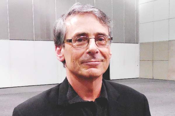
Presenter: Tricia Marriott, PA-C, MPAS, MJ Health Law
Summary: Ms. Marriott brought humor to a detailed #HospMed16 presentation on the rules of reimbursement and Medicare requirements for physician assistants (PAs) and nurse practitioners (NPs). The session was packed with information regarding the Medicare regulations relating to PAs and NPs, as well as information from state Medicaid programs and commercial payors. The presentation continued with focusing on myth busters and misperceptions about PAs and NPs. These topics were reviewed in depth:
- PAs and NPs have been recognized as providers by Medicare since 1998, as demonstrated by Medicare citations provided to the audience.
- Supervision/collaboration, as defined by Medicare requirements.
- Medicare payment policy: “incident to” vs. “split/shared visit,” reviewing unacceptable shared visit documentation and unintended consequences of fewer shared visits.
The discussion provided detailed insight into how to address the question, “What about the 15% reduced Medicare reimbursement for PAs and NPs?” An analytical approach to answering this question was provided as it relates to inpatient services, observation services, critical care services, and consultations. At the end of the talk, the audience was very engaged, and a lively Q&A ensued past the scheduled time. TH
HM16 Session Analysis: Health Information Technology Controversies
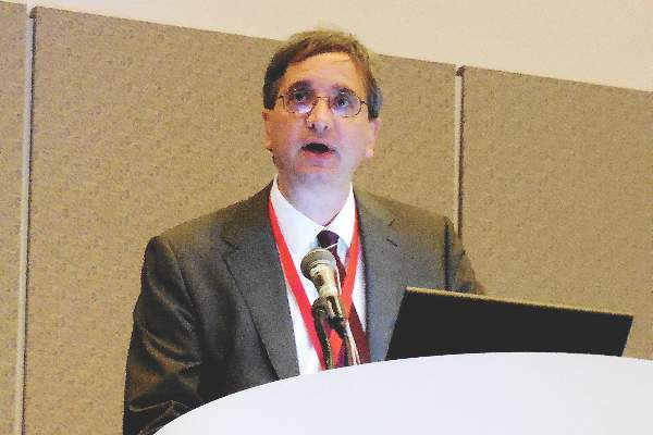
Presenter: Julie Hollberg, MD
Summary: Dr. Julie Hollberg, the chief medical information officer for Emory Healthcare, presented an overview of three pressing health information technology (IT) concerns at Hospital Medicine 2016, the “Year of the Hospitalist.” These issues are the use of copy-and-paste functions in electronic charting, alert fatigue, and patient access to electronic charts.
Dr. Hollberg states the key to leveraging healthcare IT to improve the patient and clinician experience is to coordinate people, technology, and the process. She relates that electronic note quality is poor due to lost narratives, “note bloat” (unnecessary text and data), and the use of copy-and-paste.
However, hospitalists themselves are essential in improving documentation. “We have 100% control of what goes into the note,” she describes. Some 90% of residents and attendings use copy-and-paste often. Most of the physicians agree the use of copy-and-paste increases inconsistencies, but 80% of physicians desire to continue the practice. The need for copy-and-paste should decrease as EMRs advance and expectations of note content is more broadly communicated.
Alerts are designed to improve patient safety and are a Meaningful Use initiative. The goal of clinical decision support is to provide the right information to the right person at the right time. However alert fatigue is a concern. Recommendations to address alert fatigue include making alerts non-interruptive, tier basing the alerts by severity, and decreasing the frequency of drug interaction alerts.
Dr. Hollberg also described the benefits of patient access to healthcare information on web portals. These benefits lead to improved patient engagement. Most physician concerns about open access has not been seen in actual practice. For example, only 1-8% of patients say that access to notes causes confusion, worry, or offense.
Key Takeaways:
- Use of copy-and-paste creates “note bloat” and inconsistencies. The practice is discouraged.
- Patients prefer access to healthcare information on portals. The benefit to improved access is greater patient engagement.
- While alert fatigue is a concern, clinicians should still read alerts! TH
Dr. Hale is a pediatric hospitalist at Floating Hospital for Children at Tufts Medical Center in Boston and a former member of Team Hospitalist.
VIDEO: Ischemic-stroke thrombectomy use widens and refines
LOS ANGELES – The use of endovascular thrombectomy in the United States to treat appropriate patients with acute ischemic stroke mushroomed during the past year, following several early-2015 reports that collectively documented the dramatic clinical benefit of the treatment.
As endovascular thrombectomy use grows, stroke centers are also refining and reshaping delivery of the treatment in concert with administration of intravenous tissue plasminogen activator (TPA; alteplase; Activase), which remains a key partner in producing best outcomes for acute ischemic-stroke patients with a proximal occlusion of a large cerebral artery. Collapsing delivery of the two treatments into a more seamless and streamlined process shaves critical minutes to treatment delivery, an approach called parallel processing. Recent findings have also emboldened stroke specialists to seriously consider simplifying the brain imaging that stroke patients receive prior to these treatments, a step that could further cut time to intervention while also making thrombectomy even more widely available.
Use of thrombectomy surges
The biggest endovascular thrombectomy news of the past year is how it has taken off for treating selected patients with acute ischemic stroke. “The rollout over the past year has been explosive. Everything pretty much shut down after the negative trial results in 2013, but now more hospitals are offering thrombectomy,” said Dr. Thomas A. Kent, professor of neurology and director of stroke research and education at Baylor College of Medicine in Houston, in an interview at the International Stroke Conference sponsored by the American Heart Association.
The best documentation of this surge came in a poster presented at the conference by researchers at the University of California, San Francisco. They analyzed data on treatment of 357,973 patients with acute ischemic stroke who were hospitalized at any one of 161 U.S. academic medical centers during October 2009-July 2015 and included in the University Healthsystem Consortium database. They tracked the percentage of patients treated endovascularly during each calendar quarter of the study period.
During 2009-2013, use of endovascular treatment rose steadily but gradually, from 1.5% of stroke patients in 2009 to 3.1% during the fourth quarter of 2012. Then, following three reports of no benefit from endovascular treatment presented at the International Stroke Conference in February 2013 – the IMS III, MR RESCUE, and SYNTHESIS trials – the endovascular rate dropped immediately and quickly bottomed out at a level of 2.6% that remained steady through the third quarter of 2014. But when the positive endovascular results from the MR CLEAN study became public in the final week of 2014, endovascular use began to quickly rise again, and then began to skyrocket during the first quarter of 2015 with three additional positive trial results reported during the Stroke Conference in February 2015. By the end of the second quarter of 2015, usage stood at 4.7%, representing a projected year-over-year increase of about 150% for all of 2015, compared with 2014, reported Dr. Anthony S. Kim, a vascular neurologist and medical director of the Stroke Center at the University of California, San Francisco, and his associates.
To put these percentages in perspective, experts estimate that roughly 10%-15% of all stroke patients qualify for thrombectomy intervention.
Their data also showed that the percentage of hospitals included in the database that performed endovascular therapies for stroke rose steadily from about 40% of centers in 2009 to nearly 60% by mid-2015.
“Endovascular therapy with newer-generation devices is increasingly part of standard treatment for acute ischemic stroke,” they said in their poster. In addition, they cited a “new urgency to evaluate regional access to embolectomy [another name for thrombectomy] nationally and to identify system-based solutions to improve access in underserved areas.”
Several stroke experts interviewed at the conference added their own anecdotal view of thrombectomy’s rapidly expanding use for appropriate acute ischemic stroke patients during 2015, and the need for continued effort to broaden its U.S. availability.
“The number of thrombectomies fell off after the negative 2013 trials and stayed flat until a year ago, but then jumped up. It has been very dramatic,” said Dr. Wade S. Smith, professor of neurology and director of the neurovascular service at the University of California, San Francisco.
“Thrombectomy use tremendously increased since February 2015,” said Dr. Mark J. Alberts, professor of neurology and medical director of the neurology service at the University of Texas Southwestern Medical Center in Dallas, in a video interview during the conference. But despite this growth, “the major challenge [today] is geography;” that is, reaching patients in suburban and rural areas who are not as close to the primarily urban medical centers that currently offer the procedure.
“We now have about 100 certified comprehensive stroke centers in the U.S.,” and by definition comprehensive stroke centers have the capability of treating patients with endovascular thrombectomy, noted Dr. Jeffrey Saver, professor of neurology and director of the stroke unit at the University of California, Los Angeles.
“Certification of these centers did not begin until about 2-3 years ago. But we probably need 300-400 of these centers” to provide thrombectomy to most U.S. stroke patients, he said. “A lot of additional hospitals are close to certification. I anticipate that over the next 1-2 years we will be in the neighborhood of having the number of centers we need,” Dr. Saver said in an interview.
Making thrombectomy better
In addition to expanding availability, the specifics of how endovascular thrombectomy gets delivered is evolving. A major trend is movement toward a “parallel processing” model, in which patients with an acute clinical presentation of a stroke amenable for endovascular treatment simultaneously undergo CT angiography to confirm and localize the large-artery clot causing their stroke, receive intravenous TPA, and undergo preparation for the endovascular access needed to remove the clot.
A pooled analysis of the recent, positive endovascular thrombectomy trials that was presented at the conference showed how quick you need to be to obtain a benefit from the procedures. “This gives us a starting point to further improve the target metrics for imaging and puncture times,” Dr. Saver said. “We want to shorten door-to-needle times for TPA and door-to-puncture times for thrombectomy, and the processes that need to be addressed for rapid delivery of both of these are very similar. We need for patients to only make a pit stop in the ED; we need to have the catheterization team ready to go in the thrombectomy suite within 30 minutes; and we need to emphasize speed in access to the target clot rather than time-consuming diagnostic angiography.”
“We now face the issue of how to best integrate TPA treatment and clot removal.” Dr. Kent said. “People are still trying to work that out. With parallel processing there is some overuse of resources: Some patients recover with TPA alone and don’t need thrombectomy. We are getting closer to the cardiology model of MI treatment. It’s now clear that there needs to be a simple, safe, effective way to do both TPA treatment and thrombectomy. We need to model ourselves on the cardiology experience.”
“If you can deal with the TPA decision in the same room without moving patients from room to room, from a scanner to a catheterization suite, you can really shorten the time to treatment,” Dr. Smith explained. “This is identical to the model that cardiologists have developed. We should now consider taking stroke patients directly to the angiography room in addition to administering TPA. We still need cross-sectional imaging, but the quality of the image from an angiography suite is probably sufficient to make a TPA decision. So you can start TPA while you are getting arterial access. The idea is simultaneous approaches to the patient instead of serial.”
“The whole system moves at the same time to eliminate wasted time,” Dr. Alberts summed up.
One of the big questions that has come up in this effort to speed up treatment and carve the quickest route to endovascular thrombectomy is whether TPA remains necessary. The skeptics’ position is, why waste time administering TPA if you’re also going to take out the offending clot?
The answer, at least for now, is that all signs indicate that giving TPA helps and is worth delivering.
“The 2015 thrombectomy trials had big differences among them in the dosage of TPA administered, and in the percentage of patients who received TPA. When 100% of patients received TPA they had the best outcomes,” Dr. Kent said. “There was a clear synergistic relationship between thrombectomy and TPA. There has been a trend to think about sending patients straight to thrombectomy and skipping TPA, but my colleagues and I think that we need to hold off on doing that. For now, if a patient is eligible to receive TPA they should get it and then quickly move to endovascular therapy. We are not yet ready to know it’s okay to go straight to endovascular treatment. In SWIFT-PRIME, it was pretty clear that the good outcomes were attributable to both [thrombectomy plus TPA]. Treating patients with TPA helps soften the clot to make it easier to remove, and improves flow through collateral arteries.”
“Our data in Memphis show that patients do better with thrombectomy plus intravenous TPA than on TPA alone,” agreed Dr. Lucas Elijovich, a neurologist at the University of Tennessee Health Science Center in Memphis, in an interview.
Simpler imaging also saves time
Although it’s not yet proven, another new wrinkle in working up acute ischemic-stroke patients for TPA and thrombectomy treatment is the idea that simpler and more widely available CT imaging, especially CT angiography of cerebral arteries, may suffice for confirming and localizing the culprit clot.
This concept received a significant boost at the International Stroke Conference in data reported from the Pragmatic Ischaemic Stroke Thrombectomy Evaluation (PISTE) trial, yet another study that compared treatment with TPA alone with TPA plus endovascular thrombectomy, this time in 65 randomized patients treated at any of 11 U.K. centers. PISTE had a low enrollment level because the trial stopped prematurely, in July 2015, following the news that several fully completed trials had collectively established the superiority of endovascular thrombectomy plus TPA, thereby making it unethical to continue yet another randomized study.
This premature stoppage prevented PISTE from itself producing a statistically significant difference for its primary efficacy endpoint in favor of the combined treatment, although the results did show a nominal advantage to using thrombectomy plus TPA over TPA alone that was fully consistent with the other studies, Dr. Keith W. Muir reported at the conference.
But what made the PISTE results especially notable was that the trial achieved this consistent outcome with a “simpler” imaging protocol for patients during their workup that used only CT angiography, avoiding the cerebral CT perfusion imaging or MRI used in several of the other TPA-plus-thrombectomy versus TPA-only trials, noted Dr. Muir, professor of neuroscience and head of the stroke imaging group at the University of Glasgow.
“PISTE raises the question of how much imaging is necessary,” Dr. Kent commented.
“The PISTE results are exciting. A lot of us believe that all we need to know is that there is a blockage in a target vessel,” Dr. Smith said. “If we have that information, then we can identify a population of patients who will benefit from [thrombectomy]. CT angiography is simple and can easily fit into work flows.”
“PISTE used a very simple imaging system that makes thrombectomy even more applicable and generalizable to less resourced health systems,” Dr. Saver said. “Although the results from PISTE were not internally statistically significant because the trial ended early, the results were consistent with the external studies of thrombectomy, so it provides further evidence for benefit from thrombectomy.” And because the consistent results were achieved with simpler imaging it suggests simpler imaging may be all that’s needed.
“That’s a major question to wrestle with,” Dr. Saver suggested. “We need addition trials with a head-to-head comparison of simpler and more sophisticated imaging so we can tailor treatment to patients who would benefit from simpler and faster imaging.”
Dr. Kent had no disclosures. Dr. Kim has received research funding from SanBio and Biogen. Dr. Smith served on the data safety and monitoring board for a trial funded by Stryker. Dr. Alberts has been a consultant to Genentech. Dr. Saver has been a consultant to Stryker, Neuravi, Cognition Medical, Boehringer Ingelheim, and Medtronic. Dr. Elijovich has been a consultant to Stryker and Codman and received research support from Siemens. Dr. Muir has received research support from ReNeuron and unrestricted grants from Codman and Covidien.
The video associated with this article is no longer available on this site. Please view all of our videos on the MDedge YouTube channel
On Twitter @mitchelzoler
EXPERT ANALYSIS FROM THE INTERNATIONAL STROKE CONFERENCE

HM16 Session Analysis: Reinforcing Practice Culture, Maximizing Engagement Through Effective Communication
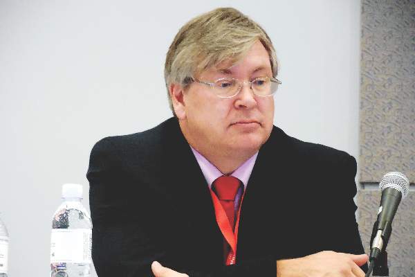
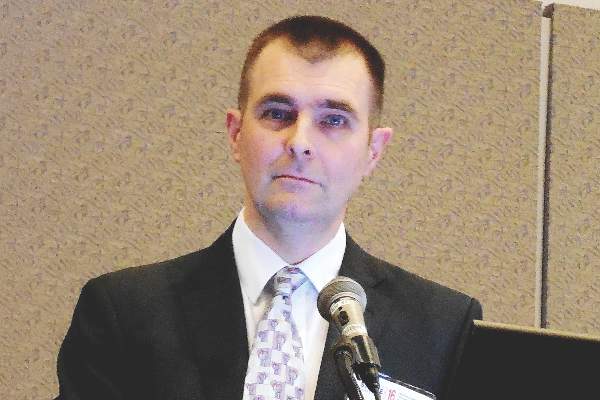
HM16 Presenters: Dr. Scott Rissmiller, Dr. Steve Deitelzweig, Dr. Jerome Siy, Dr. Thomas Mcllraith, and Dr. Michael Reitz
Summary: This session at #HospMed16 explored lessons learned from five hospitalist leaders across the country about improving hospitalist practice through enhancing hospitalist engagement, group communication, and leadership development. It was proposed that the “new” value equation is [Engagement * (quality/cost)] = Value. Engagement is the multiplier of value. The speakers highlighted the following:
Build a Plan : Approach engagement like any other business plan with metrics, accountability, and “S.M.A.R.T." goals.
Build Trust: Visibility breeds credibility. Credibility breeds Trust. Trust encourages Engagement.
Build Transparency: Keep communication simple and be sure that it’s helpful information.
Build Leaders: All hospitalists are leaders. Strong leadership skills promote effective communication across the system. Nurture leadership skills for the right level of leadership, to find the right seat on the bus.
Build Celebrations: Celebrate successes, and learn from failure. TH
Forgoing artificial nutrition and hydration
Question: Which of the following statements regarding artificial nutrition and hydration (ANH) is least supported in ethics and/or law?
A. One may invoke medical futility and discontinue ANH in noncognitive, terminally ill patients.
B. Forgoing ANH at the end of life is part of good palliative care.
C. Tube feeding is a way to provide food and sustenance and does not constitute medical treatment.
D. Many state statutes allow for the discontinuation of ANH under specified conditions.
E. Discontinuing ANH is most contentious when it affects a young, otherwise healthy person in a persistent vegetative state.
Answer: C.
Only a few decades ago, litigation over end-of-life care took the form of surrogates demanding to stop treatment, e.g., mechanical ventilation, against the orders of the hospital and doctors.1 The parties now tend to reverse their positions, with some families insisting on continuing all treatment modalities, especially artificial nutrition and hydration (ANH) such as tube feedings. Medical futility has been variously defined as an intervention that has no pathophysiologic rationale, where such intervention had already failed in the patient, where maximal treatment is already failing, or where the intervention will not achieve the goals of care. The AMA Code of Medical Ethics advises: “Physicians are not ethically obligated to deliver care that, in their best professional judgment, will not have a reasonable chance of benefiting their patients. Patients should not be given treatments simply because they demand them.”2 The AMA Code also clarifies that “life-sustaining treatment is any treatment that serves to prolong life without reversing the underlying medical condition” and “may include … artificial nutrition and hydration.”3

Although many palliative care specialists have deemed ANH to be of dubious medical benefit at the end of life,4 forgoing ANH nonetheless poses a particularly contentious dilemma. Opponents assert that ANH is a form of feeding rather than medical treatment, denial of which amounts to “starving” the patient to death. However in 1990, the U.S. Supreme Court in Cruzan v. Director5 declared that ANH could not be readily distinguished from other forms of medical treatment. The case involved a 25-year-old woman who lapsed into a persistent vegetative state (PVS) following an automobile accident. Her parents sought a court order to withdraw ANH when it became apparent that she had virtually no chance of recovering her cognitive faculties. Writing for a unanimous Supreme Court, Justice O’Connor opined: “Whether or not the techniques used to pass food and water into the patient’s alimentary tract are termed ‘medical treatment,’ it is clear they all involve some degree of intrusion and restraint. … Feeding a patient by means of a nasogastric tube requires a physician to pass a long flexible tube through the patient’s nose, throat, and esophagus and into the stomach. Because of the discomfort such a tube causes, ‘many patients need to be restrained forcibly and their hands put into large mittens to prevent them from removing the tube.’ A gastrostomy tube … or jejunostomy tube must be surgically implanted into the stomach or small intestine. … Requiring a competent adult to endure such procedures against her will burdens the patient’s liberty, dignity, and freedom to determine the course of her own treatment.”
The English High Court has likewise adopted this position in Airedale NHS Trust v. Bland.6 Tony Bland, a young man, was crushed in a Hillsborough football melee in 1989, which caused him to develop PVS. The court wrote: “The question is not whether it is in the best interests of the patient that he should die. The question is whether it is in the best interests of the patient that his life should be prolonged by the continuance of this form of medical treatment or care. The correct formulation of the question is of particular importance in a case such as the present, where the patient is totally unconscious or where there is no hope whatsoever of any amelioration of his condition.”
In 2005, the United States witnessed its most public and highly politicized case regarding the withdrawal of ANH.7 In 1990, Terri Schiavo, then age 26, sustained a cardiac arrest, possibly caused by an eating disorder and hypokalemia. Her husband, Michael, filed a malpractice suit and won damages of $1.5 million, but the arrest left her in a PVS. Terri had left no advance directive, and her husband said she would rather die than be kept alive artificially, and therefore directed that her tube feeding be stopped. On the other hand, her parents Bob and Mary Schindler wished to keep her alive, and volunteered to take care of her. After a long and bitter legal battle, an appellate court finally agreed to stop Terri’s tube feeding, as it found clear and convincing evidence that she would not have wanted to live in that manner. The U.S. Supreme Court refused to review the case. On Oct. 15, 2003, with the discontinuation of her tube feeding, Terri’s parents appealed to then Florida Gov. Jeb Bush, calling into question the diagnosis of PVS. They insisted Terri’s eyes were open, and she appeared to respond with smiles to the sound of her mother’s voice. Despite contrary expert opinion, 15 doctors testified she could and would improve. Both the Florida legislature and the U.S. Congress intervened but to no avail. Terri died at 10 a.m. on March 31, 2005, 13 days after tube withdrawal. An autopsy revealed severe brain atrophy.
In Cruzan, the U.S. Supreme Court had held that the liberty guaranteed by the Due Process Clause must protect an individual’s deeply personal decision to reject medical treatment, including the artificial delivery of food and water. However, where a patient’s wishes are not clear and convincing, a court will be reluctant to order cessation of treatment. In a 6-0 decision, the California Supreme Court ruled that a patient’s tube feedings could not be discontinued under the circumstances of the Wendland case.8 Robert Wendland regained consciousness after 14 months in a coma, but was left hemiparetic and incontinent, and could not feed by mouth or dress, bathe, and communicate consistently. His wife, Rose, refused to authorize reinsertion of his dislodged feeding tube, believing that Robert would not have wanted it replaced. The patient’s daughter and brother, as well as the hospital’s ethics committee, the county ombudsman, and a court-appointed counsel all agreed with the decision. Robert did not have an advance directive, but had made statements to the effect he would not want to live in a vegetative state. But the patient’s mother, Florence, went to court to block the action. The court determined that Robert’s statements were not clear and convincing because they did not address his current condition, were not sufficiently specific, and were not necessarily intended to direct his medical care. Further, the patient’s spouse had failed to provide sufficient evidence that her decision was in her husband’s best interests.
In some jurisdictions, a patient’s advance medical directive to discontinue ANH may require a specific opt-in or opt-out choice. And although medical directives typically spring into effect when a patient is terminally ill, many jurisdictions allow their applicability in nonterminal conditions such as irreversible unconscious states or where the likely risks and burdens of treatment would outweigh any expected benefits (Hawaii is such a state under HRS §327E-16). Physicians are legally bound to respect a patient’s wish regarding forgoing or continuing therapy including ANH. Given the wide variations in legal requirements governing this emotional matter, physicians must look closely at their individual state statute to determine the proper course of action under the circumstances.
References
1. In re Quinlan, 70 N.J. 10 (1976).
2. Code of Medical Ethics of the AMA, 2014-5 ed., section 2.035.
3. Code of Medical Ethics of the AMA, 2014-5 ed., section 2.20.
4. Danis M. Stopping artificial nutrition and hydration at the end of life. UpToDate, Nov 2, 2015.
5. Cruzan v. Director, Missouri Department of Health, 110 S. Ct. 2841 (1990).
6. Airedale NHS Trust v. Bland (1993) A.C. 789.
7. Available at https://en.wikipedia.org/wiki/Terri_Schiavo_case.
8. Wendland v. Wendland, 28 P.3d 151 (Cal., 2001).
Dr. Tan is emeritus professor of medicine and former adjunct professor of law at the University of Hawaii, and currently directs the St. Francis International Center for Healthcare Ethics in Honolulu. This article is meant to be educational and does not constitute medical, ethical, or legal advice. Some of the articles in this series are adapted from the author’s 2006 book, “Medical Malpractice: Understanding the Law, Managing the Risk,” and his 2012 Halsbury treatise, “Medical Negligence and Professional Misconduct.” For additional information, readers may contact the author at [email protected].
Question: Which of the following statements regarding artificial nutrition and hydration (ANH) is least supported in ethics and/or law?
A. One may invoke medical futility and discontinue ANH in noncognitive, terminally ill patients.
B. Forgoing ANH at the end of life is part of good palliative care.
C. Tube feeding is a way to provide food and sustenance and does not constitute medical treatment.
D. Many state statutes allow for the discontinuation of ANH under specified conditions.
E. Discontinuing ANH is most contentious when it affects a young, otherwise healthy person in a persistent vegetative state.
Answer: C.
Only a few decades ago, litigation over end-of-life care took the form of surrogates demanding to stop treatment, e.g., mechanical ventilation, against the orders of the hospital and doctors.1 The parties now tend to reverse their positions, with some families insisting on continuing all treatment modalities, especially artificial nutrition and hydration (ANH) such as tube feedings. Medical futility has been variously defined as an intervention that has no pathophysiologic rationale, where such intervention had already failed in the patient, where maximal treatment is already failing, or where the intervention will not achieve the goals of care. The AMA Code of Medical Ethics advises: “Physicians are not ethically obligated to deliver care that, in their best professional judgment, will not have a reasonable chance of benefiting their patients. Patients should not be given treatments simply because they demand them.”2 The AMA Code also clarifies that “life-sustaining treatment is any treatment that serves to prolong life without reversing the underlying medical condition” and “may include … artificial nutrition and hydration.”3
Although many palliative care specialists have deemed ANH to be of dubious medical benefit at the end of life,4 forgoing ANH nonetheless poses a particularly contentious dilemma. Opponents assert that ANH is a form of feeding rather than medical treatment, denial of which amounts to “starving” the patient to death. However in 1990, the U.S. Supreme Court in Cruzan v. Director5 declared that ANH could not be readily distinguished from other forms of medical treatment. The case involved a 25-year-old woman who lapsed into a persistent vegetative state (PVS) following an automobile accident. Her parents sought a court order to withdraw ANH when it became apparent that she had virtually no chance of recovering her cognitive faculties. Writing for a unanimous Supreme Court, Justice O’Connor opined: “Whether or not the techniques used to pass food and water into the patient’s alimentary tract are termed ‘medical treatment,’ it is clear they all involve some degree of intrusion and restraint. … Feeding a patient by means of a nasogastric tube requires a physician to pass a long flexible tube through the patient’s nose, throat, and esophagus and into the stomach. Because of the discomfort such a tube causes, ‘many patients need to be restrained forcibly and their hands put into large mittens to prevent them from removing the tube.’ A gastrostomy tube … or jejunostomy tube must be surgically implanted into the stomach or small intestine. … Requiring a competent adult to endure such procedures against her will burdens the patient’s liberty, dignity, and freedom to determine the course of her own treatment.”
The English High Court has likewise adopted this position in Airedale NHS Trust v. Bland.6 Tony Bland, a young man, was crushed in a Hillsborough football melee in 1989, which caused him to develop PVS. The court wrote: “The question is not whether it is in the best interests of the patient that he should die. The question is whether it is in the best interests of the patient that his life should be prolonged by the continuance of this form of medical treatment or care. The correct formulation of the question is of particular importance in a case such as the present, where the patient is totally unconscious or where there is no hope whatsoever of any amelioration of his condition.”
In 2005, the United States witnessed its most public and highly politicized case regarding the withdrawal of ANH.7 In 1990, Terri Schiavo, then age 26, sustained a cardiac arrest, possibly caused by an eating disorder and hypokalemia. Her husband, Michael, filed a malpractice suit and won damages of $1.5 million, but the arrest left her in a PVS. Terri had left no advance directive, and her husband said she would rather die than be kept alive artificially, and therefore directed that her tube feeding be stopped. On the other hand, her parents Bob and Mary Schindler wished to keep her alive, and volunteered to take care of her. After a long and bitter legal battle, an appellate court finally agreed to stop Terri’s tube feeding, as it found clear and convincing evidence that she would not have wanted to live in that manner. The U.S. Supreme Court refused to review the case. On Oct. 15, 2003, with the discontinuation of her tube feeding, Terri’s parents appealed to then Florida Gov. Jeb Bush, calling into question the diagnosis of PVS. They insisted Terri’s eyes were open, and she appeared to respond with smiles to the sound of her mother’s voice. Despite contrary expert opinion, 15 doctors testified she could and would improve. Both the Florida legislature and the U.S. Congress intervened but to no avail. Terri died at 10 a.m. on March 31, 2005, 13 days after tube withdrawal. An autopsy revealed severe brain atrophy.
In Cruzan, the U.S. Supreme Court had held that the liberty guaranteed by the Due Process Clause must protect an individual’s deeply personal decision to reject medical treatment, including the artificial delivery of food and water. However, where a patient’s wishes are not clear and convincing, a court will be reluctant to order cessation of treatment. In a 6-0 decision, the California Supreme Court ruled that a patient’s tube feedings could not be discontinued under the circumstances of the Wendland case.8 Robert Wendland regained consciousness after 14 months in a coma, but was left hemiparetic and incontinent, and could not feed by mouth or dress, bathe, and communicate consistently. His wife, Rose, refused to authorize reinsertion of his dislodged feeding tube, believing that Robert would not have wanted it replaced. The patient’s daughter and brother, as well as the hospital’s ethics committee, the county ombudsman, and a court-appointed counsel all agreed with the decision. Robert did not have an advance directive, but had made statements to the effect he would not want to live in a vegetative state. But the patient’s mother, Florence, went to court to block the action. The court determined that Robert’s statements were not clear and convincing because they did not address his current condition, were not sufficiently specific, and were not necessarily intended to direct his medical care. Further, the patient’s spouse had failed to provide sufficient evidence that her decision was in her husband’s best interests.
In some jurisdictions, a patient’s advance medical directive to discontinue ANH may require a specific opt-in or opt-out choice. And although medical directives typically spring into effect when a patient is terminally ill, many jurisdictions allow their applicability in nonterminal conditions such as irreversible unconscious states or where the likely risks and burdens of treatment would outweigh any expected benefits (Hawaii is such a state under HRS §327E-16). Physicians are legally bound to respect a patient’s wish regarding forgoing or continuing therapy including ANH. Given the wide variations in legal requirements governing this emotional matter, physicians must look closely at their individual state statute to determine the proper course of action under the circumstances.
References
1. In re Quinlan, 70 N.J. 10 (1976).
2. Code of Medical Ethics of the AMA, 2014-5 ed., section 2.035.
3. Code of Medical Ethics of the AMA, 2014-5 ed., section 2.20.
4. Danis M. Stopping artificial nutrition and hydration at the end of life. UpToDate, Nov 2, 2015.
5. Cruzan v. Director, Missouri Department of Health, 110 S. Ct. 2841 (1990).
6. Airedale NHS Trust v. Bland (1993) A.C. 789.
7. Available at https://en.wikipedia.org/wiki/Terri_Schiavo_case.
8. Wendland v. Wendland, 28 P.3d 151 (Cal., 2001).
Dr. Tan is emeritus professor of medicine and former adjunct professor of law at the University of Hawaii, and currently directs the St. Francis International Center for Healthcare Ethics in Honolulu. This article is meant to be educational and does not constitute medical, ethical, or legal advice. Some of the articles in this series are adapted from the author’s 2006 book, “Medical Malpractice: Understanding the Law, Managing the Risk,” and his 2012 Halsbury treatise, “Medical Negligence and Professional Misconduct.” For additional information, readers may contact the author at [email protected].

First sublingual immunotherapy tablet for house dust mite allergic rhinitis may be U.S.-bound
LOS ANGELES – A sublingual immunotherapy tablet for house dust mite–induced allergic rhinitis achieved clinically meaningful improvement in symptoms along with less use of rescue medications and a favorable safety profile in a pivotal phase III trial, Dr. Hendrik Nolte reported at the annual meeting of the American Academy of Allergy, Asthma, and Immunology.
The double-blind, 52-week randomized trial included 1,482 North American adults and adolescents.

“This was the largest clinical trial ever conducted with sublingual therapy in North America, and the first successful North American trial of a house dust mite sublingual immunotherapy tablet. It confirms results from previous large European trials,” noted Dr. Nolte of Merck, which is collaborating with the Danish company ALK in developing this therapy.
The sublingual immunotherapy (SLIT) tablet, known for now as the 12 SQ HDM SLIT-tablet, has been approved by European regulatory authorities for treatment of adults with house dust mite (HDM) allergic rhinitis or HDM allergic asthma. Based upon the positive findings in the phase III U.S. trial, Merck applied in February 2016 to the U.S. Food and Drug Administration for approval of the tablet as a biologic agent in patients aged 12 years or older.
HDM is the most common indoor allergen in the world. Unlike pollen and ragweed allergies, it’s not a seasonal problem. Moreover, HDM-induced allergic rhinitis is associated with increased risk of asthma. Although HDM allergic rhinitis can be treated symptomatically with oral antihistamines and nasal steroids, allergy immunotherapy has the appeal of addressing the underlying disease mechanism and potentially altering the long-term course. SLIT using a highly standardized HDM allergen extract offers a major advantage over traditional allergy immunotherapy via a lengthy program of subcutaneous injections – namely, the convenience of home self-administration.
The primary endpoint in the U.S. pivotal trial was the total combined rhinitis score (TCRS), which is the sum of the daily rhinitis symptom score and the daily rescue medication usage score averaged over the last 8 weeks of the year-long study. The FDA has set the bar for establishing clinically meaningful improvement: it requires demonstration of a reduction in the TCRS of at least 15%, compared with placebo. The 12 SQ HDM SLIT-tablet trial exceeded this standard, achieving a 17% reduction. The study also met its key prespecified secondary endpoints, including a 16% reduction in the daily rhinosinusitis symptom score and an 18% decrease in the daily medication score, compared with placebo.

Participants in the trial had a mean 18-year history of allergic rhinitis with or without conjunctivitis. Seventy-five percent of them were sensitized to other common allergens in addition to HDM, and 31% of subjects had comorbid asthma. The HDM SLIT therapy was equally effective in asthmatics and nonasthmatics, in patients allergic only to HDM and those who were polysensitized, and in subjects with and without conjunctivitis, according to Dr. Nolte.
There were no serious adverse events related to the 12 SQ HDM SLIT-tablet. A total of 9.8% of patients discontinued SLIT because of treatment-emergent adverse events, chiefly mild-to-moderate throat irritation, mouth swelling, or itchiness of the mouth or ears.
“Importantly, these events were very transient. They occurred typically within the first 8 days and lasted 14-67 hours, with a median 1-day duration of rescue medication,” he said.
Symptomatic improvement was typically seen beginning at 8-12 weeks. Adults were free to take the once-daily tablet anytime during the day. The pediatric patients were advised not to do so in the morning because they wouldn’t be under observation while on a school bus.
The tablet is based upon a formulation of allergen extracts from the two major species of HDM: Dermatophagoides pterornyssinus and D. farinae. More than 90% of HDM-sensitized patients are sensitized to both. A highly standardized manufacturing process ensures that the tablet contains 50 mcg of the four major HDM allergens in equal ratio – Der f1, Der p2, Der p1, and Der f 2 – plus other components.
In response to audience questions, Dr. Nolte said the company had tried incorporating an antihistamine into the tablet to block adverse reactions but it didn’t work. Adverse events typically occur within a couple of minutes after taking the tablet, and antihistamines are far too slow-acting to help.
“You can premedicate with antihistamines. We know European investigators and clinicians who are doing it. But I would not recommend it personally, because these tablets are taken at home after the first administration in the office, and I think it’s important that the patient has a good feel for what happens over the following days. There is a potential risk of masking side effects with premedication, which could be a concern,” Dr. Nolte said.
Merck already has two FDA-approved SLIT tablets developed with ALK on the U.S. market: Grastek, for treatment of grass pollen–induced allergic rhinitis in children and adults, and Ragwitek, for ragweed-induced allergic disease in adults.
The trial was sponsored by Merck and presented by a full-time company employee.
LOS ANGELES – A sublingual immunotherapy tablet for house dust mite–induced allergic rhinitis achieved clinically meaningful improvement in symptoms along with less use of rescue medications and a favorable safety profile in a pivotal phase III trial, Dr. Hendrik Nolte reported at the annual meeting of the American Academy of Allergy, Asthma, and Immunology.
The double-blind, 52-week randomized trial included 1,482 North American adults and adolescents.
“This was the largest clinical trial ever conducted with sublingual therapy in North America, and the first successful North American trial of a house dust mite sublingual immunotherapy tablet. It confirms results from previous large European trials,” noted Dr. Nolte of Merck, which is collaborating with the Danish company ALK in developing this therapy.
The sublingual immunotherapy (SLIT) tablet, known for now as the 12 SQ HDM SLIT-tablet, has been approved by European regulatory authorities for treatment of adults with house dust mite (HDM) allergic rhinitis or HDM allergic asthma. Based upon the positive findings in the phase III U.S. trial, Merck applied in February 2016 to the U.S. Food and Drug Administration for approval of the tablet as a biologic agent in patients aged 12 years or older.
HDM is the most common indoor allergen in the world. Unlike pollen and ragweed allergies, it’s not a seasonal problem. Moreover, HDM-induced allergic rhinitis is associated with increased risk of asthma. Although HDM allergic rhinitis can be treated symptomatically with oral antihistamines and nasal steroids, allergy immunotherapy has the appeal of addressing the underlying disease mechanism and potentially altering the long-term course. SLIT using a highly standardized HDM allergen extract offers a major advantage over traditional allergy immunotherapy via a lengthy program of subcutaneous injections – namely, the convenience of home self-administration.
The primary endpoint in the U.S. pivotal trial was the total combined rhinitis score (TCRS), which is the sum of the daily rhinitis symptom score and the daily rescue medication usage score averaged over the last 8 weeks of the year-long study. The FDA has set the bar for establishing clinically meaningful improvement: it requires demonstration of a reduction in the TCRS of at least 15%, compared with placebo. The 12 SQ HDM SLIT-tablet trial exceeded this standard, achieving a 17% reduction. The study also met its key prespecified secondary endpoints, including a 16% reduction in the daily rhinosinusitis symptom score and an 18% decrease in the daily medication score, compared with placebo.
Participants in the trial had a mean 18-year history of allergic rhinitis with or without conjunctivitis. Seventy-five percent of them were sensitized to other common allergens in addition to HDM, and 31% of subjects had comorbid asthma. The HDM SLIT therapy was equally effective in asthmatics and nonasthmatics, in patients allergic only to HDM and those who were polysensitized, and in subjects with and without conjunctivitis, according to Dr. Nolte.
There were no serious adverse events related to the 12 SQ HDM SLIT-tablet. A total of 9.8% of patients discontinued SLIT because of treatment-emergent adverse events, chiefly mild-to-moderate throat irritation, mouth swelling, or itchiness of the mouth or ears.
“Importantly, these events were very transient. They occurred typically within the first 8 days and lasted 14-67 hours, with a median 1-day duration of rescue medication,” he said.
Symptomatic improvement was typically seen beginning at 8-12 weeks. Adults were free to take the once-daily tablet anytime during the day. The pediatric patients were advised not to do so in the morning because they wouldn’t be under observation while on a school bus.
The tablet is based upon a formulation of allergen extracts from the two major species of HDM: Dermatophagoides pterornyssinus and D. farinae. More than 90% of HDM-sensitized patients are sensitized to both. A highly standardized manufacturing process ensures that the tablet contains 50 mcg of the four major HDM allergens in equal ratio – Der f1, Der p2, Der p1, and Der f 2 – plus other components.
In response to audience questions, Dr. Nolte said the company had tried incorporating an antihistamine into the tablet to block adverse reactions but it didn’t work. Adverse events typically occur within a couple of minutes after taking the tablet, and antihistamines are far too slow-acting to help.
“You can premedicate with antihistamines. We know European investigators and clinicians who are doing it. But I would not recommend it personally, because these tablets are taken at home after the first administration in the office, and I think it’s important that the patient has a good feel for what happens over the following days. There is a potential risk of masking side effects with premedication, which could be a concern,” Dr. Nolte said.
Merck already has two FDA-approved SLIT tablets developed with ALK on the U.S. market: Grastek, for treatment of grass pollen–induced allergic rhinitis in children and adults, and Ragwitek, for ragweed-induced allergic disease in adults.
The trial was sponsored by Merck and presented by a full-time company employee.
AT 2016 AAAAI ANNUAL MEETING
Key clinical point: A safe, effective, and convenient self-administered alternative to allergy shots or symptomatic medications for house dust mite allergic rhinitis may be in the works.
Major finding: A once-daily tablet containing house dust mite extract resulted in a clinically meaningful 17% improvement relative to placebo in a score combining symptomatic improvement and reduced use of rescue medications.
Data source: This pivotal phase III, double-blind, 52-week randomized trial included 1,482 North American adults and adolescents with house dust mite–induced allergic rhinitis with or without conjunctivitis.
Disclosures: The trial was sponsored by Merck and presented by a full-time company employee.
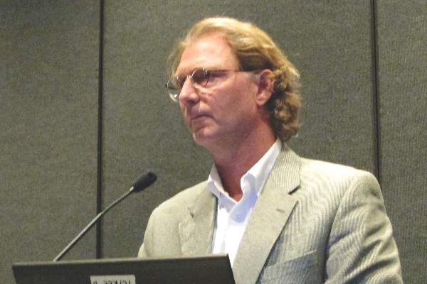
DoD and VA Enhance Complex Care Initiatives
The DoD and the VA unveiled a plan that synchronizes current processes to ease the transition of health care service members from the DoD to the VA. The new interagency coordination also aims to help afflicted service members and veterans who require multiple care specialties throughout both departments.
This effort comes as a result of the work of the DoD-VA Interagency Care Coordination Committee (IC3), which was established in 2012. The new plan’s goal is to align more than 250 subpolicies to 1 overarching policy that governs the coordination of complex care cases that transition between the departments.
Related: VA/DoD to Help Lead New Cancer Initiative
“More than a decade of combat has placed enormous demands on a generation of service members and veterans—particularly those who have suffered wounds, injuries, or illnesses that require a complex plan of care,” said Karen Guice, MD, MPP, Principal Deputy Assistant Secretary of Defense for Health Affairs and Co-chair of IC3. “These individuals require the complex coordination of medical and rehabilitative care, benefits, and other services to successfully transition from active duty to veteran status, and to optimally recover from their illnesses or injuries.”
To help maximize the plan’s chances to succeed, the IC3 developed a new role—Lead Coordinator—that makes current employees the primary coordinator for individual patients. The lead coordinator will offer personal guidance and help service members and their families understand the benefits and services to which they are entitled.
It is expected that a total of 1,500 DoD staff and 1,200 VA staff will serve as lead coordinators.
“Great attention has been made to developing a system that focuses on continuity of care, holistic support services and a ‘warm handoff’ for service members and veterans as they move from and between military, VA and community health care systems," said Linda Spoonster Schwartz, DrPH, assistant secretary for policy and planning for the VA and cochair of IC3. "Our care coordinators now have at their fingertips tools and processes that improve and simplify the lines of communication for our wounded, ill, and injured service members and veterans who require complex care coordination, their families, and those who provide their care in both departments."
Schwartz added that the process "will enhance and improve the quality of care and services for these Veterans and their families now and in the future.”
Better billing privacy protections needed for youth
Better protections of confidential health information of adolescents and young adults in billing and insurance statements are needed, according to a position paper endorsed by the American Academy of Pediatrics, Society for Adolescent Health and Medicine, and the American College of Obstetricians and Gynecologists.
The three societies are calling for policies and procedures to be established “to ensure that health care billing and insurance claims processes such as explanation of benefit (EOB) notifications do not impede the confidential provision of health care services to adolescents” (J Adolesc Health. 2016 Mar. doi: 10.1016/j.jadohealth.2015.12.009).

To reach that objective, the position paper is calling for the Department of Health & Human Services to issue guidance to clarify “endanger” and “endangerment” in the special confidentiality provisions of HIPAA to make it clear that they include “harms that result when access to important sensitive services, such as contraception and STI services, is impeded by fear of loss of confidentiality.”
AAP, ACOG, and SAHM also endorse the idea that sending EOBs and similar notices “should not be required when individuals insured as dependents obtain sensitive services.”
Dr. Lee Savio Beers, medical director for municipal and regional affairs, Child Health Advocacy Institute at Children’s National Medical Center in Washington, said the policy paper is “a nice summary of what has been best practices for a while and what we really should establish more clearly as best practice.”
She recalled experiences in practice where adolescents told her they did not come for treatment because of concerns that “information would show up on the billing statement to their parents, and they would know that they were seeking care for these issues and then surmise that they were sexually active.”
Dr. Beers added that the position paper could go further in establishing requirements for both parents and caregivers on the importance of confidentiality.
“I think that it is sometimes hard for parents to think about there might be something that my child is going to go to the doctor for that I am not going to know about,” she said in an interview.
“When we see patients for visits, when they get close to their teenage years, we start interviewing them, giving them an opportunity to talk without their parent in the room,” she continued. “Some families are surprised by it. Some are glad that you are asking them to step out. Others want to talk about it for a few minutes, but I think there is an important parent and caregiver education component to this as well, particularly when you start talking about things like insurance benefits, which I think parents typically think of as being really fully within their purview.”
Better protections of confidential health information of adolescents and young adults in billing and insurance statements are needed, according to a position paper endorsed by the American Academy of Pediatrics, Society for Adolescent Health and Medicine, and the American College of Obstetricians and Gynecologists.
The three societies are calling for policies and procedures to be established “to ensure that health care billing and insurance claims processes such as explanation of benefit (EOB) notifications do not impede the confidential provision of health care services to adolescents” (J Adolesc Health. 2016 Mar. doi: 10.1016/j.jadohealth.2015.12.009).
To reach that objective, the position paper is calling for the Department of Health & Human Services to issue guidance to clarify “endanger” and “endangerment” in the special confidentiality provisions of HIPAA to make it clear that they include “harms that result when access to important sensitive services, such as contraception and STI services, is impeded by fear of loss of confidentiality.”
AAP, ACOG, and SAHM also endorse the idea that sending EOBs and similar notices “should not be required when individuals insured as dependents obtain sensitive services.”
Dr. Lee Savio Beers, medical director for municipal and regional affairs, Child Health Advocacy Institute at Children’s National Medical Center in Washington, said the policy paper is “a nice summary of what has been best practices for a while and what we really should establish more clearly as best practice.”
She recalled experiences in practice where adolescents told her they did not come for treatment because of concerns that “information would show up on the billing statement to their parents, and they would know that they were seeking care for these issues and then surmise that they were sexually active.”
Dr. Beers added that the position paper could go further in establishing requirements for both parents and caregivers on the importance of confidentiality.
“I think that it is sometimes hard for parents to think about there might be something that my child is going to go to the doctor for that I am not going to know about,” she said in an interview.
“When we see patients for visits, when they get close to their teenage years, we start interviewing them, giving them an opportunity to talk without their parent in the room,” she continued. “Some families are surprised by it. Some are glad that you are asking them to step out. Others want to talk about it for a few minutes, but I think there is an important parent and caregiver education component to this as well, particularly when you start talking about things like insurance benefits, which I think parents typically think of as being really fully within their purview.”
FROM JOURNAL OF ADOLESCENT HEALTH

10 Questions You Should Consider for Specialist Consultations
Caring for patients in the inpatient setting is complex and often requires consultation from specialists. Yet the actual skill of obtaining a consult is rarely taught. Medical students and residents usually learn by trial and error, becoming targets of frustrated consultants and suffering humiliation and much anxiety. To facilitate communication between the primary team and the specialist, we propose that the student and/or resident start by asking the following questions.

1. Why Call This Consult?
To decide whether you need a consult, first determine the type. Consultations can be broken down into three different types: advice on diagnosis, advice on management, or arrangements for a specific procedure or test. Advice on diagnosis or management is typically required when a clinical issue has reached the bounds of knowledge, experience, or comfort zone of the team or physician (e.g., idiopathic leukocytosis). For procedures, a consultant who is licensed to perform the procedure may be required (e.g., endoscopy for GI bleed).
2. What Should Be Done before a Consult Is Requested?
First, ask yourself, “If I were the consultant, what would I want to know?” Before calling, put yourself in the shoes of the consultant and consider the available data carefully to develop your own hypotheses. For example, infectious disease consultants typically make judgments based on relevant culture data, current and/or past antibiotics, imaging, and signs or symptoms of active infection. Reading about the problem beforehand allows you to anticipate possible questions and consider additional studies that may be requested by the consultant. It also helps ascertain whether the consultation is actually necessary or targeted to the right team.
3. What Is the Clinical Question?
Bergus and colleagues found that a well-structured clinical question clearly identifies the treatment the primary doctor is proposing and the desired outcomes for the patient.1 For instance, rather than asking, “What should we do for this 75-year-old man with chest pain?”, a better question might be, “Will the addition of ranolazine increase exercise tolerance in our 75-year-old man with angina who is already taking a beta blocker and nitrates?” When both components are present, clinical questions are more likely to be answered.
4. How Do I Best Present the Case to My Consultant?
Requesting a consultation requires a succinct presentation that focuses on the aspects of the case most pertinent to the specialist. To do this, again put yourself in the shoes of the consultant. For example, a patient’s history of venous thromboembolism (VTE) will always be relevant to a hematologist, whereas a history of GERD may not be needed in your initial conversation. Limit the initial presentation to two to three minutes and organize using the four I’s:
- Introduction: “My name is X with blue medicine team; I am calling to request a consult.”
- Information: Patient name, location, medical record number, attending physician.
- Inquiry: “I am requesting evaluation for an EGD in a patient with an upper GI bleed.”
- Important items (the story): “Mr. X is a 55-year-old male with history of peptic ulcer disease presenting with abdominal pain.”
5. What Data Requests Should I Anticipate?
Have your clinical data easily accessible in case additional information is requested (i.e., keep the chart open when calling). If certain tests are predictably going to be needed by the specialist (e.g., renal ultrasound for a nephrologist), make sure that the results are available or in process. Also, be prepared to take notes if the consultant requests additional tests up front.
6. How Urgent Is the Consult?
Consultations can be emergent, urgent, or elective. Directly communicate any emergent or urgent consults in order to clarify the issues expeditiously. For more routine consults, consider delaying the call until enough laboratory data or imaging is available for the consultant to answer the question. Do not call a nonurgent consult at the end of the day or on a weekend.
7. Where Can I Meet with the Consultant to Discuss the Case?
Be available to your consultants by offering the fastest and most reliable means for them to get in touch with you. Take advantage of your consultants and learn from them. Be where they are: If looking at the blood smear, join them. If spinning the urine, ask to examine the sediment together. Discussing the case in person demonstrates your interest, engendering a more serious and perhaps expeditious consideration of your case. Finally, request seminal articles that have driven their decision to allow for more intelligent conversations in the future.
8. How Can I Nurture My Relationship with the Consulting Team?
The best relationships with consultants require give-and-take. Be a reliable source by providing accurate documentation of ongoing events, history and physical examination, and laboratory data in your notes. Understand consultant recommendations and summarize these in your plan. Avoid “Plan per Renal/GI/Cards/Heme, etc.” in your notes. Continue to think about the questions and issues and read on your own. If you are unclear about the recommendations, clarify them with the consulting team. Speaking with consultants is a learning opportunity; never forget to ask why they have made a certain recommendation. Avoid “chart wars” if there are points of disagreement with the plan or recommendations.
9. How Do I Close the Loop on the Consult?
Closing the communication loop is one of the most important aspects of the consult because it allows you to act on the recommendations. Remember that consultants are likely to be as busy as you are (if not busier). If the consult was urgent, call consultants directly for guidance. If it wasn’t urgent, look in the chart first for their note. Checking the chart later in the day could help to avoid unnecessary phone calls and increase your efficiency.
10. Am I Sure I Want a Curbside Consult?
In a curbside consult, you request advice of an expert who is neither in the presence of the patients nor has a therapeutic relationship with them. A study by Burden and colleagues in 2013 found that 55% of physicians offered different advice in formal consultation than in a curbside consultation, and 60% felt that formal consultation changed management.2 Similarly, Kuo and colleagues noted that 77% of subspecialists reported that important clinical findings were frequently missing from curbsides.3 Some recommend limiting curbsides to simple questions that don’t require consultants to assess multiple variables; as a courtesy, consider offering them the option of a formal consult. Ultimately, the decision to request a curbside consultation, and any consultation for that matter, should always be discussed with your attending physician.
Conclusion
Effective communication with consultants requires forethought and is an exercise in clinical reasoning of great educational value to students and residents. By considering the questions above, the consultative experience can be more productive for both the primary and consulting team and will enhance the care of the hospitalized patient. TH
Dr. Esquivel is a hospitalist in the Division of Hospital Medicine at Weill Cornell Medical College in New York City. Dr. Rendon is a hospitalist in the Division of Hospital Medicine at the University of New Mexico in Albuquerque.
References
1. Bergus GR, Randall CS, Sinift SD, Rosenthal DM. Does the structure of clinical questions affect the outcome of curbside consultations with specialty colleagues? Arch Fam Med. 2000;9(6):541-547.
2. Burden M, Sarcone E, Keniston A, et al. Prospective comparison of curbside versus formal consultations. J Hosp Med. 2013;8(1):31-35.
3. Kuo D, Gifford DR, Stein MD. Curbside consultation practices and attitudes among primary care physicians and medical subspecialists. JAMA. 1998;280(10):905-909.
Caring for patients in the inpatient setting is complex and often requires consultation from specialists. Yet the actual skill of obtaining a consult is rarely taught. Medical students and residents usually learn by trial and error, becoming targets of frustrated consultants and suffering humiliation and much anxiety. To facilitate communication between the primary team and the specialist, we propose that the student and/or resident start by asking the following questions.
1. Why Call This Consult?
To decide whether you need a consult, first determine the type. Consultations can be broken down into three different types: advice on diagnosis, advice on management, or arrangements for a specific procedure or test. Advice on diagnosis or management is typically required when a clinical issue has reached the bounds of knowledge, experience, or comfort zone of the team or physician (e.g., idiopathic leukocytosis). For procedures, a consultant who is licensed to perform the procedure may be required (e.g., endoscopy for GI bleed).
2. What Should Be Done before a Consult Is Requested?
First, ask yourself, “If I were the consultant, what would I want to know?” Before calling, put yourself in the shoes of the consultant and consider the available data carefully to develop your own hypotheses. For example, infectious disease consultants typically make judgments based on relevant culture data, current and/or past antibiotics, imaging, and signs or symptoms of active infection. Reading about the problem beforehand allows you to anticipate possible questions and consider additional studies that may be requested by the consultant. It also helps ascertain whether the consultation is actually necessary or targeted to the right team.
3. What Is the Clinical Question?
Bergus and colleagues found that a well-structured clinical question clearly identifies the treatment the primary doctor is proposing and the desired outcomes for the patient.1 For instance, rather than asking, “What should we do for this 75-year-old man with chest pain?”, a better question might be, “Will the addition of ranolazine increase exercise tolerance in our 75-year-old man with angina who is already taking a beta blocker and nitrates?” When both components are present, clinical questions are more likely to be answered.
4. How Do I Best Present the Case to My Consultant?
Requesting a consultation requires a succinct presentation that focuses on the aspects of the case most pertinent to the specialist. To do this, again put yourself in the shoes of the consultant. For example, a patient’s history of venous thromboembolism (VTE) will always be relevant to a hematologist, whereas a history of GERD may not be needed in your initial conversation. Limit the initial presentation to two to three minutes and organize using the four I’s:
- Introduction: “My name is X with blue medicine team; I am calling to request a consult.”
- Information: Patient name, location, medical record number, attending physician.
- Inquiry: “I am requesting evaluation for an EGD in a patient with an upper GI bleed.”
- Important items (the story): “Mr. X is a 55-year-old male with history of peptic ulcer disease presenting with abdominal pain.”
5. What Data Requests Should I Anticipate?
Have your clinical data easily accessible in case additional information is requested (i.e., keep the chart open when calling). If certain tests are predictably going to be needed by the specialist (e.g., renal ultrasound for a nephrologist), make sure that the results are available or in process. Also, be prepared to take notes if the consultant requests additional tests up front.
6. How Urgent Is the Consult?
Consultations can be emergent, urgent, or elective. Directly communicate any emergent or urgent consults in order to clarify the issues expeditiously. For more routine consults, consider delaying the call until enough laboratory data or imaging is available for the consultant to answer the question. Do not call a nonurgent consult at the end of the day or on a weekend.
7. Where Can I Meet with the Consultant to Discuss the Case?
Be available to your consultants by offering the fastest and most reliable means for them to get in touch with you. Take advantage of your consultants and learn from them. Be where they are: If looking at the blood smear, join them. If spinning the urine, ask to examine the sediment together. Discussing the case in person demonstrates your interest, engendering a more serious and perhaps expeditious consideration of your case. Finally, request seminal articles that have driven their decision to allow for more intelligent conversations in the future.
8. How Can I Nurture My Relationship with the Consulting Team?
The best relationships with consultants require give-and-take. Be a reliable source by providing accurate documentation of ongoing events, history and physical examination, and laboratory data in your notes. Understand consultant recommendations and summarize these in your plan. Avoid “Plan per Renal/GI/Cards/Heme, etc.” in your notes. Continue to think about the questions and issues and read on your own. If you are unclear about the recommendations, clarify them with the consulting team. Speaking with consultants is a learning opportunity; never forget to ask why they have made a certain recommendation. Avoid “chart wars” if there are points of disagreement with the plan or recommendations.
9. How Do I Close the Loop on the Consult?
Closing the communication loop is one of the most important aspects of the consult because it allows you to act on the recommendations. Remember that consultants are likely to be as busy as you are (if not busier). If the consult was urgent, call consultants directly for guidance. If it wasn’t urgent, look in the chart first for their note. Checking the chart later in the day could help to avoid unnecessary phone calls and increase your efficiency.
10. Am I Sure I Want a Curbside Consult?
In a curbside consult, you request advice of an expert who is neither in the presence of the patients nor has a therapeutic relationship with them. A study by Burden and colleagues in 2013 found that 55% of physicians offered different advice in formal consultation than in a curbside consultation, and 60% felt that formal consultation changed management.2 Similarly, Kuo and colleagues noted that 77% of subspecialists reported that important clinical findings were frequently missing from curbsides.3 Some recommend limiting curbsides to simple questions that don’t require consultants to assess multiple variables; as a courtesy, consider offering them the option of a formal consult. Ultimately, the decision to request a curbside consultation, and any consultation for that matter, should always be discussed with your attending physician.
Conclusion
Effective communication with consultants requires forethought and is an exercise in clinical reasoning of great educational value to students and residents. By considering the questions above, the consultative experience can be more productive for both the primary and consulting team and will enhance the care of the hospitalized patient. TH
Dr. Esquivel is a hospitalist in the Division of Hospital Medicine at Weill Cornell Medical College in New York City. Dr. Rendon is a hospitalist in the Division of Hospital Medicine at the University of New Mexico in Albuquerque.
References
1. Bergus GR, Randall CS, Sinift SD, Rosenthal DM. Does the structure of clinical questions affect the outcome of curbside consultations with specialty colleagues? Arch Fam Med. 2000;9(6):541-547.
2. Burden M, Sarcone E, Keniston A, et al. Prospective comparison of curbside versus formal consultations. J Hosp Med. 2013;8(1):31-35.
3. Kuo D, Gifford DR, Stein MD. Curbside consultation practices and attitudes among primary care physicians and medical subspecialists. JAMA. 1998;280(10):905-909.
Team finds precursors to HSPCs in mice

Image by Matthias Zepper
Researchers have reportedly identified precursor cells that can be matured into transplantable hematopoietic stem/progenitor cells (HSPCs) in the lab.
The investigators discovered the precursor cells in the placentas and embryos of mice, but the team believes their findings could aid the development of patient-specific HSPCs and more differentiated blood products for cell-replacement therapy in humans.
“To cure disease in the long-term, we need to be able to transplant something that can keep producing new blood cells and won’t be rejected by the patient’s body,” said study author Kateri Moore, DVM, of the Icahn School of Medicine at Mount Sinai in New York, New York.
“We are excited by the results of our study. The precursor cells can be matured in the lab to transplantable HSPCs. Our reprogramming process can inform developmental hematopoiesis and vice-versa.”
Dr Moore and her colleagues described this work in Developmental Cell.
With previous work, the researchers reprogrammed mouse fibroblasts to become HSPCs. They showed that this process occurred through hemogenic precursors that are Prom1+Sca1+CD34+CD45− (PS34CD45−).
So for the current study, the investigators examined mouse placentas and embryos, looking for cells with the same phenotype. They were able to find and analyze PS34CD45− cells.
Investigation revealed that PS34CD45− cells express endothelial and hematopoietic markers. And the cells originate in embryonic tissue and localize to the vascular labyrinth.
Furthermore, the researchers said global gene expression profiles of PS34CD45− cells correlate with reprogrammed precursors and establish a hemogenic precursor cell molecular signature.
In culture, PS34CD45− cells gave rise to multi-lineage hematopoietic colonies. PS34CD45− cells generated B and T lymphocytes and engrafted in primary and secondary immunodeficient mice.
The investigators said the next step is to test these findings in humans.
“Our ultimate goal is to grow blood-forming cells in the lab and improve efficiencies to generate patient-specific blood cells,” Dr Moore said. “This study brings us a step closer to reaching this goal.” ![]()