User login
Mortality may be reduced when surgical ablation for AF is done with mitral operations
HOUSTON – At the time of mitral valve repair or replacement operations, the addition of surgical ablation to treat atrial fibrillation (AF) can be performed without increased risk of mortality, results from a large cohort study has demonstrated.
“Surgical ablation for atrial fibrillation performed concomitant to mitral valve repair or replacement is accepted to reduce long-term AF rates and improve quality of life, and has recently attained a class I, level of evidence A recommendation in the recent Society of Thoracic Surgeons Guidelines,” Vinay Badhwar, MD, said in an interview. Dr. Badhwar is the lead author of the STS Guidelines and senior author of the current study on mortality following surgical ablation of AF. “Nevertheless, the direct impact of surgical ablation on operative mortality has not been studied on a large scale using robust registry data.”

In an effort to examine the impact of performing or not performing surgical ablation (SA) on mortality of contemporary mitral valve repair or replacement (MVRR) operations, Dr. Badhwar, Gordon F. Murray Professor and Chair, department of cardiovascular and thoracic surgery at West Virginia University, Morgantown, W.Va., and his associates at eight other leading centers nationwide evaluated the medical records of 88,765 MVRR patients in the STS Adult Cardiac Surgery Database between July 2011 and June 2014.
The research, presented as the Richard E. Clark award winning paper in adult cardiac surgery by study leader J. Scott Rankin, MD, professor of surgery in the department of cardiovascular and thoracic surgery at West Virginia University, included cases of tricuspid repair and coronary artery bypass grafting and assessed all STS comorbid risk variables and mitral etiology for predictors of mortality. After performing multivariable logistic regression, the researchers compared risk-adjusted odds ratios for mortality by AF type at operation and SA performance: group 1 (no AF plus no SA), group 2 (no AF plus SA), group 3 (AF plus no SA), group 4 (AF plus SA), group 5 (persistent AF plus no SA), and group 6 (persistent AF plus SA).
The median age of patients ranged from 63 to 72 years. Compared with their counterparts in other groups, patients in groups 3 and 5 were older (median age of 71 and 72 years, respectively), had worse symptoms (50% in each group had NYHA class III or IV disease), had more reoperations (30% vs. 32%), and had higher unadjusted mortalities (6.7% vs. 6.6%).
Following multivariable risk adjustment, groups 2-6 with AF history plus or minus SA were referenced to group 1 patients without AF or SA. Groups 2-4 represented the overall population, and groups 5 and 6 were patients with persistent AF. Patients with AF at the time of operation not receiving SA (groups 3 and 5) had odds ratios of 1.16 and 1.17, respectively, or an increase of 16% and 17% in relative risk of mortality (P = .004 and P = .01, respectively). In groups 4 and 6, concomitant SA reduced the AF-related mortality to a level that was statistically comparable to reference baseline group 1 without a history of AF (P = .13 and P = .21, respectively).
“The present study appears to clearly illustrate the adverse effects of AF on operative mortality following MVRR,” Dr. Badhwar said. “After thorough risk adjustment, we were impressed to learn that the relative mortality risk of mitral procedures increased by nearly 15% for either paroxysmal or persistent AF, and results were similar for the two AF types. A remote history of AF with preoperative sinus rhythm immediately prior to surgery had little effect on prognosis, and these patients appeared to have outcomes more like those without AF. In patients with active AF at the time of mitral operation, SA reduced the AF-related mortality and decreased operative mortality to levels not statistically different from patients without AF.” He added that the findings of this study support the recent STS Guidelines on Surgical Ablation published in the January 2017 edition of Annals of Thoracic Surgery (Ann Thorac Surg. 2017 Jan;103[1]:329-41). “However, the ultimate decision on whether or not to add surgical ablation must always remain between the surgeon and the patient,” he said.
Dr. Badhwar acknowledged that the study is limited by the information available in version 2.73 of the STS Adult Cardiac Surgery Database. “This does highlight the ongoing need for participants in the database to provide as much complete information as possible for all patients undergoing cardiac operations, especially those involving surgical ablation,” he said.
Dr. Badhwar reported having no financial disclosures. Dr. Rankin disclosed ties with Admedus, AtriCure, and BioStable Science and Engineering.
HOUSTON – At the time of mitral valve repair or replacement operations, the addition of surgical ablation to treat atrial fibrillation (AF) can be performed without increased risk of mortality, results from a large cohort study has demonstrated.
“Surgical ablation for atrial fibrillation performed concomitant to mitral valve repair or replacement is accepted to reduce long-term AF rates and improve quality of life, and has recently attained a class I, level of evidence A recommendation in the recent Society of Thoracic Surgeons Guidelines,” Vinay Badhwar, MD, said in an interview. Dr. Badhwar is the lead author of the STS Guidelines and senior author of the current study on mortality following surgical ablation of AF. “Nevertheless, the direct impact of surgical ablation on operative mortality has not been studied on a large scale using robust registry data.”
In an effort to examine the impact of performing or not performing surgical ablation (SA) on mortality of contemporary mitral valve repair or replacement (MVRR) operations, Dr. Badhwar, Gordon F. Murray Professor and Chair, department of cardiovascular and thoracic surgery at West Virginia University, Morgantown, W.Va., and his associates at eight other leading centers nationwide evaluated the medical records of 88,765 MVRR patients in the STS Adult Cardiac Surgery Database between July 2011 and June 2014.
The research, presented as the Richard E. Clark award winning paper in adult cardiac surgery by study leader J. Scott Rankin, MD, professor of surgery in the department of cardiovascular and thoracic surgery at West Virginia University, included cases of tricuspid repair and coronary artery bypass grafting and assessed all STS comorbid risk variables and mitral etiology for predictors of mortality. After performing multivariable logistic regression, the researchers compared risk-adjusted odds ratios for mortality by AF type at operation and SA performance: group 1 (no AF plus no SA), group 2 (no AF plus SA), group 3 (AF plus no SA), group 4 (AF plus SA), group 5 (persistent AF plus no SA), and group 6 (persistent AF plus SA).
The median age of patients ranged from 63 to 72 years. Compared with their counterparts in other groups, patients in groups 3 and 5 were older (median age of 71 and 72 years, respectively), had worse symptoms (50% in each group had NYHA class III or IV disease), had more reoperations (30% vs. 32%), and had higher unadjusted mortalities (6.7% vs. 6.6%).
Following multivariable risk adjustment, groups 2-6 with AF history plus or minus SA were referenced to group 1 patients without AF or SA. Groups 2-4 represented the overall population, and groups 5 and 6 were patients with persistent AF. Patients with AF at the time of operation not receiving SA (groups 3 and 5) had odds ratios of 1.16 and 1.17, respectively, or an increase of 16% and 17% in relative risk of mortality (P = .004 and P = .01, respectively). In groups 4 and 6, concomitant SA reduced the AF-related mortality to a level that was statistically comparable to reference baseline group 1 without a history of AF (P = .13 and P = .21, respectively).
“The present study appears to clearly illustrate the adverse effects of AF on operative mortality following MVRR,” Dr. Badhwar said. “After thorough risk adjustment, we were impressed to learn that the relative mortality risk of mitral procedures increased by nearly 15% for either paroxysmal or persistent AF, and results were similar for the two AF types. A remote history of AF with preoperative sinus rhythm immediately prior to surgery had little effect on prognosis, and these patients appeared to have outcomes more like those without AF. In patients with active AF at the time of mitral operation, SA reduced the AF-related mortality and decreased operative mortality to levels not statistically different from patients without AF.” He added that the findings of this study support the recent STS Guidelines on Surgical Ablation published in the January 2017 edition of Annals of Thoracic Surgery (Ann Thorac Surg. 2017 Jan;103[1]:329-41). “However, the ultimate decision on whether or not to add surgical ablation must always remain between the surgeon and the patient,” he said.
Dr. Badhwar acknowledged that the study is limited by the information available in version 2.73 of the STS Adult Cardiac Surgery Database. “This does highlight the ongoing need for participants in the database to provide as much complete information as possible for all patients undergoing cardiac operations, especially those involving surgical ablation,” he said.
Dr. Badhwar reported having no financial disclosures. Dr. Rankin disclosed ties with Admedus, AtriCure, and BioStable Science and Engineering.
AT THE STS ANNUAL MEETING
Key clinical point:
Major finding: Adding surgical ablation to treat AF at the time of MVRR reduced the AF-related mortality to a rate that was comparable to a reference baseline group that had no history of AF.
Data source: An analysis of medical records from 88,765 MVRR patients in the STS Adult Cardiac Surgery Database between July 2011 and June 2014.
Disclosures: Dr. Badhwar reported having no financial disclosures. Dr. Rankin disclosed ties with Admedus, AtriCure, and BioStable Science and Engineering.
Early, in-hospital shunt failure common among infants
HOUSTON – Among neonates and infants who underwent shunt construction as a source of pulmonary blood flow, early, in-hospital shunt failure occurred in 7.3% of cases, results from a large retrospective study showed.
“Approximately one in seven patients who experiences cardiac surgery in the first year of life undergoes construction of a systemic to pulmonary artery shunt of some type,” one of the study investigators, Marshall L. Jacobs, MD, said in an interview. The study was presented at the annual meeting of the Society of Thoracic Surgeons.
“Early failure of such shunts is an incompletely understood phenomenon which accounts for important morbidity and mortality among infants and neonates. Much of what is known about shunt failure is based on experiences reported from individual institutions. The few multicenter studies to date have been clinical trials that focused primarily on pharmacologic strategies intended to reduce the risk of shunt failure due to thrombosis. Their utility for guiding clinical decision making has been limited. Some have been underpowered; some have had limited risk adjustment of subjects.”

Dr. Do, who presented the findings at the meeting and is currently a Congenital Heart Surgery Fellow at the Children’s Hospital of Philadelphia, and a team of 11 other investigators utilized the STS Congenital Heart Surgery Database to identify 9,172 neonates and infants who underwent shunt construction as a source of pulmonary blood flow at 118 institutions from 2010 to 2015. Criteria for shunt failure included a documented diagnosis of in-hospital shunt failure, shunt revision, or catheter-based shunt intervention. The investigators used multivariable logistic regression to evaluate risk factors for in-hospital shunt failure.
Of the 9,172 at-risk neonates and infants, 674 (7.3%) experienced early, in-hospital shunt failure. “The observed rate of early shunt failure varied across the many specific types of shunts, and was lower with systemic ventricle to pulmonary artery shunts (as in the Sano modification of the Norwood procedure) than with the systemic artery to pulmonary artery shunts,” said Dr. Jacobs, who is a cardiothoracic surgeon at Johns Hopkins University, Baltimore.
In multivariable analysis, risk factors for in-hospital shunt failure included lower weight at operation for both neonates and infants, preoperative hypercoagulable state, and the collective presence of any other STS Congenital Heart Surgery Database preoperative risk factors. Neither cardiopulmonary bypass nor single ventricle diagnosis were risk factors for shunt failure. The investigators also observed that patients with in-hospital shunt failure had significantly higher rates of operative mortality (31.9% vs. 11.1%) and major morbidity (84.4% vs. 29.4%), and longer postoperative length of stay among survivors (a median of 45 vs. 22 days).
“Understanding the characteristics of the patient groups found to be at highest risk for early shunt failure is helpful in identifying individual patients that may warrant expectant surveillance, enhanced pharmacologic management, or other strategies to reduce the risk of shunt failure,” Dr. Jacobs concluded.
“But perhaps more importantly it provides key information that may be helpful in the design and development of future clinical trials and/or collaborative quality improvement initiatives designed to reduce the cost in lives and resources that is associated with early shunt dysfunction.”
He acknowledged certain limitations of the study, including its retrospective observational design and the voluntary nature of the STS Congenital Heart Surgery Database. “In addition, some potentially important variables, such as detailed data concerning preoperative test results of coagulation assays are not collected in the STS Congenital Heart Surgery Database,” he said.
The research was supported by the STS Access & Publications Research program. The investigators reported having no financial disclosures.
HOUSTON – Among neonates and infants who underwent shunt construction as a source of pulmonary blood flow, early, in-hospital shunt failure occurred in 7.3% of cases, results from a large retrospective study showed.
“Approximately one in seven patients who experiences cardiac surgery in the first year of life undergoes construction of a systemic to pulmonary artery shunt of some type,” one of the study investigators, Marshall L. Jacobs, MD, said in an interview. The study was presented at the annual meeting of the Society of Thoracic Surgeons.
“Early failure of such shunts is an incompletely understood phenomenon which accounts for important morbidity and mortality among infants and neonates. Much of what is known about shunt failure is based on experiences reported from individual institutions. The few multicenter studies to date have been clinical trials that focused primarily on pharmacologic strategies intended to reduce the risk of shunt failure due to thrombosis. Their utility for guiding clinical decision making has been limited. Some have been underpowered; some have had limited risk adjustment of subjects.”
Dr. Do, who presented the findings at the meeting and is currently a Congenital Heart Surgery Fellow at the Children’s Hospital of Philadelphia, and a team of 11 other investigators utilized the STS Congenital Heart Surgery Database to identify 9,172 neonates and infants who underwent shunt construction as a source of pulmonary blood flow at 118 institutions from 2010 to 2015. Criteria for shunt failure included a documented diagnosis of in-hospital shunt failure, shunt revision, or catheter-based shunt intervention. The investigators used multivariable logistic regression to evaluate risk factors for in-hospital shunt failure.
Of the 9,172 at-risk neonates and infants, 674 (7.3%) experienced early, in-hospital shunt failure. “The observed rate of early shunt failure varied across the many specific types of shunts, and was lower with systemic ventricle to pulmonary artery shunts (as in the Sano modification of the Norwood procedure) than with the systemic artery to pulmonary artery shunts,” said Dr. Jacobs, who is a cardiothoracic surgeon at Johns Hopkins University, Baltimore.
In multivariable analysis, risk factors for in-hospital shunt failure included lower weight at operation for both neonates and infants, preoperative hypercoagulable state, and the collective presence of any other STS Congenital Heart Surgery Database preoperative risk factors. Neither cardiopulmonary bypass nor single ventricle diagnosis were risk factors for shunt failure. The investigators also observed that patients with in-hospital shunt failure had significantly higher rates of operative mortality (31.9% vs. 11.1%) and major morbidity (84.4% vs. 29.4%), and longer postoperative length of stay among survivors (a median of 45 vs. 22 days).
“Understanding the characteristics of the patient groups found to be at highest risk for early shunt failure is helpful in identifying individual patients that may warrant expectant surveillance, enhanced pharmacologic management, or other strategies to reduce the risk of shunt failure,” Dr. Jacobs concluded.
“But perhaps more importantly it provides key information that may be helpful in the design and development of future clinical trials and/or collaborative quality improvement initiatives designed to reduce the cost in lives and resources that is associated with early shunt dysfunction.”
He acknowledged certain limitations of the study, including its retrospective observational design and the voluntary nature of the STS Congenital Heart Surgery Database. “In addition, some potentially important variables, such as detailed data concerning preoperative test results of coagulation assays are not collected in the STS Congenital Heart Surgery Database,” he said.
The research was supported by the STS Access & Publications Research program. The investigators reported having no financial disclosures.
AT THE STS ANNUAL MEETING
Key clinical point:
Major finding: Among neonates and infants who underwent shunt operations, 7.3% experienced early, in-hospital shunt failure.
Data source: A retrospective analysis of 9,172 neonates and infants who underwent shunt construction as a source of pulmonary blood flow at 118 institutions from 2010 to 2015.
Disclosures: The research was supported by the STS Access & Publications Research program. The investigators reported having no financial disclosures.
Flashback to April 2008
The April 2008 issue of GI & Hepatology News (GIHN) featured an article by Roy M. Soetikno, MD, MS, FASGE and his colleagues from the Palo Alto VA Medical Center in California. They drew our attention to nonpolypoid (flat) colonic lesions in an article from JAMA (2008;299:1027-35).
Coincidentally, the week before this article appeared, I was sitting with Roy in Kyoto, Japan, at a conference of international experts focused on flat colonic lesions. The Japanese definitions of flat and depressed lesions were markedly different from those used by Western physicians. We now know that most flat lesions seen by U.S.-based endoscopists are sessile serrated adenomas (SSAs). SSAs at that time also were a new and controversial classification. SSAs were first described by Torlakovic and Snover in 1996 (Gastroenterology 1996;110:748-55).
Dale Snover, MD, was my golfing partner and read pathology slides for our practice in Minneapolis, so we were the first gastroenterologists in the country to grapple with the clinical implications of SSAs. Roy’s article was accompanied by an excellent commentary by Jerome D. Waye, MD, FASGE, who emphasized the importance of a slow withdrawal time and meticulous visual technique during colonoscopy.
Key points in the JAMA article were a) prevalence of flat lesions was about 9% in a screening population, b) small flat polyps can harbor advanced histologic changes including cancers, and c) many physicians who perform colonoscopy missed these lesions putting patients at risk for interval colon cancers.

The GIHN piece, referencing Soetikno’s article, helped inform us about an important (and confusing) problem in our colon cancer prevention efforts. As numerous authors subsequently highlighted (see Gastroenterology 2016;151:870-8) most cancers, missed at initial colonoscopy, are proximal and frequently develop from SSAs. We continue to work to reduce missed cancers and thanks to this seminal article, we have better insights about how to achieve this goal.
John I. Allen, MD, MBA, AGAF, is professor of medicine in the division of gastroenterology and hepatology at the University of Michigan, Ann Arbor, and the Editor in Chief of GI & Hepatology News.
The April 2008 issue of GI & Hepatology News (GIHN) featured an article by Roy M. Soetikno, MD, MS, FASGE and his colleagues from the Palo Alto VA Medical Center in California. They drew our attention to nonpolypoid (flat) colonic lesions in an article from JAMA (2008;299:1027-35).
Coincidentally, the week before this article appeared, I was sitting with Roy in Kyoto, Japan, at a conference of international experts focused on flat colonic lesions. The Japanese definitions of flat and depressed lesions were markedly different from those used by Western physicians. We now know that most flat lesions seen by U.S.-based endoscopists are sessile serrated adenomas (SSAs). SSAs at that time also were a new and controversial classification. SSAs were first described by Torlakovic and Snover in 1996 (Gastroenterology 1996;110:748-55).
Dale Snover, MD, was my golfing partner and read pathology slides for our practice in Minneapolis, so we were the first gastroenterologists in the country to grapple with the clinical implications of SSAs. Roy’s article was accompanied by an excellent commentary by Jerome D. Waye, MD, FASGE, who emphasized the importance of a slow withdrawal time and meticulous visual technique during colonoscopy.
Key points in the JAMA article were a) prevalence of flat lesions was about 9% in a screening population, b) small flat polyps can harbor advanced histologic changes including cancers, and c) many physicians who perform colonoscopy missed these lesions putting patients at risk for interval colon cancers.
The GIHN piece, referencing Soetikno’s article, helped inform us about an important (and confusing) problem in our colon cancer prevention efforts. As numerous authors subsequently highlighted (see Gastroenterology 2016;151:870-8) most cancers, missed at initial colonoscopy, are proximal and frequently develop from SSAs. We continue to work to reduce missed cancers and thanks to this seminal article, we have better insights about how to achieve this goal.
John I. Allen, MD, MBA, AGAF, is professor of medicine in the division of gastroenterology and hepatology at the University of Michigan, Ann Arbor, and the Editor in Chief of GI & Hepatology News.

Multivessel PCI in STEMI gains traction
SNOWMASS, COLO. – The tide appears to have turned regarding the merits of percutaneous coronary intervention in non-infarct-related arteries in conjunction with primary PCI for ST-elevation MI in patients with multivessel disease, Douglas E. Drachman, MD, said at the Annual Cardiovascular Conference at Snowmass.
Previously, multivessel PCI in STEMI patients who are hemodynamically stable was believed harmful and was given a Class IIIb recommendation – meaning don’t do it – in the 2013 American College of Cardiology/American Heart Association STEMI guidelines. Just 2 years later, however, new evidence in the form of three randomized trials prompted a focused update of the joint guidelines in which the practice was upgraded to Class IIb status, meaning it could be considered and may be beneficial.
Roughly 50% of STEMI patients have significant lesions in non-infarct-related arteries (non-IRA). The question of how best to treat such patients is an important one because multivessel coronary disease in STEMI is associated with increased risks of both reinfarction and mortality, noted Dr. Drachman, an interventional cardiologist at Massachusetts General Hospital in Boston.
He offered several reasons why the findings of the three influential randomized trials differed from earlier negative retrospective observational studies: “I would argue there’s been significant improvement in our technique in doing PCI. We’re primarily doing transradial interventions now for our patients, so the risk associated with multiple accesses is reduced. Our ability to use more potent antithrombotic strategies is enhanced by our concern about bleeding risk. And the stent platforms that we use in our interventional strategies have improved to the point that we are tackling ever more challenging lesions with greater aplomb and less concern that we may cause harm. I think all these factors have enhanced the ability of the interventionalist to select and treat non-IRAs in a staged fashion and be less parsimonious at the point of care.”
The remaining questions are which non-IRA lesions should be treated, in whom, when relative to primary PCI, and what are the cost implications? These issues are being tackled in at least eight active randomized controlled trials. Depending upon the answers to come, multivessel PCI in STEMI patients could receive a further upgrade in the guidelines.
Since release of the 2015 focused guideline update, several large studies have provided further backing for multivessel PCI in STEMI patients with significant multivessel disease, although these weren’t randomized prospective studies and hence must be considered hypothesis-generating.
One of these major pieces of evidence was a meta-analysis of observational studies led by Eric R. Bates, MD, professor of internal medicine at the University of Michigan in Ann Arbor. He and his coinvestigators analyzed studies comparing culprit vessel-only primary PCI for STEMI patients with multivessel disease versus staged PCI in which primary PCI was done first, followed by PCI of a non-infarct-related vessel later during the same hospitalization or soon after. Staged PCI was the clear winner, with a 2.2-fold greater likelihood of freedom from mortality (J Am Coll Cardiol. 2016 Sep 6;68(10):1066-81).
When the investigators compared studies of staged PCI versus multivessel PCI in the same session as primary PCI, staged PCI was again the clear winner, with a 4-fold greater freedom from mortality.
Among the possible risks of performing PCI of a non-IRA in the same session as primary PCI are increased risks of thrombosis, contrast-induced nephropathy, stent undersizing due to vasospasm, and unintended jeopardy of distant viable myocardium due to microembolization or side branch occlusion, Dr. Drachman said.
“Maybe in certain circumstances it’s best to let the dust settle after the urgent vessel intervention. Wait a couple of days and then make your plan,” the cardiologist advised.
Another informative recent piece of evidence was provided by a Canadian retrospective observational study which compared revascularization strategies in 6,503 consecutive STEMI patients with multivessel disease. Staged multivessel PCI during the index hospitalization was performed in 658 patients, multivessel PCI during the primary PCI session in 1,325, and PCI limited to the infarct-related artery in 4,520. The study endpoints were 2-year all-cause mortality and repeat revascularization.
Staged multivessel PCI had the lowest mortality and repeat revascularization rates. The 2-year mortality rate associated with this strategy was 45% less than with multivessel intervention at the time of primary PCI and 35% lower than for culprit vessel-only PCI, which unsurprisingly had the highest repeat revascularization rate (JACC Cardiovasc Interv. 2017 Jan 9;10(1):11-23).
The first of the three randomized trials that led to a change in the guidelines was the UK PRAMI study (Preventive Angioplasty in Acute Myocardial Infarction). It showed at a mean 23-months followup that STEMI patients with multivessel disease had a 65% reduction in the relative risk of a composite endoint of cardiovascular death, MI, or refractory angina if they received non-IRA PCI at the same time as primary PCI compared with PCI limited to the IRA (N Engl J Med. 2013 Sep 19;369(12):1115-23).
Next came another UK trial: CvLPRIT (Complete vs. Culprit-Lesion Only Primary PCI) demonstrated a 65% reduction in the composite 12-month outcome of all-cause mortality, MI, heart failure, or ischemia-driven PCI with staged PCI during the index hospitalization compared with culprit vessel-only PCI (J Am Coll Cardiol. 2015 Mar 17;65(10):963-72).
Finally, DANAMI-3-PRIMULTI (the Third Danish Study of Optimal Acute Treatment of Patients with STEMI: Primary PCI in Multivessel Disease) showed a dramatic reduction in the risk of ischemia-driven PCI during a median 27 months of followup in patients who underwent staged multivessel PCI guided by the findings of fractional flow reserve measurement compared with primary PCI limited to the IRA (Lancet. 2015 Aug 15;386(9994):665-71). However, fractional flow reserve-guided multivessel PCI didn’t decrease the risk of death or nonfatal recurrent MI, leaving its role unsettled pending the results of ongoing clinical trials.
Dr. Drachman said it’s clear certain STEMI patients should not undergo non-IRA PCI. These include anyone in whom the procedure would be lengthy due to vessel tortuosity or chronic total occlusion, as well as patients with stable saphenous vein graft disease or heavily calcified lesions requiring atherectomy, since multivessel PCI in those settings would pose a high risk for additional left ventricular dysfunction.
“Be thoughtful about patients who have renal dysfunction,” he added.
Dr. Drachman reported having no financial conflicts of interest.
EXPERT ANALYSIS FROM THE CARDIOVASCULAR CONFERENCE AT SNOWMASS
Flu activity up slightly, but still down from seasonal peak
After rising to a high point for the season in the last week of 2016, influenza activity dropped a bit in the first week of the new year but then rose again in the second week, according to the Centers for Disease Control and Prevention.
As measured by outpatient visits for influenza-like illness (ILI), activity slipped from 3.4% at the end of 2016 to 3.2% for the week ending Jan. 7 but then ticked up to 3.3% for the week ending Jan. 14, the CDC reported. The national baseline level of outpatient visits is 2.2% for ILI, which is defined as fever (temperature of 100° F or greater) and cough and/or sore throat.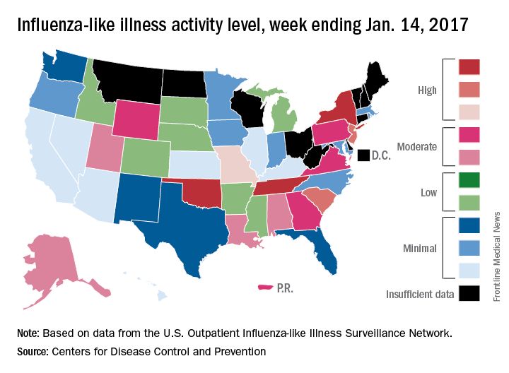
Two influenza-related pediatric deaths were reported for the week ending Jan. 14, although both occurred in earlier weeks: one during the week ending Dec. 10 and one during the week ending Jan. 7. So far for the 2016-2017 season, a total of five flu-related pediatric deaths have been reported, according to the CDC.
Resective epilepsy surgery found OK in septuagenarians
HOUSTON – With careful selection, patients in their 70s with refractory epilepsy may be offered resective epilepsy surgery, results from a small single-center study demonstrated.
The findings “were a surprise to us,” lead study author Ahmed Abdelkader, MD, said in an interview at the annual meeting of the American Epilepsy Society. “We expected that complications would be higher because this is a vulnerable age group with multiple comorbidities.”

Dr. Abdelkader and his associates searched the database of the Cleveland Clinic Epilepsy Center to identify patients aged 70 years and older who underwent respective epilepsy surgery between Jan. 1, 2000, and Sept. 30, 2015. They limited the analysis to seven patients who had at least one year of post-surgical follow-up. The mean age of the patients at surgery was 73 and the age of epilepsy onset ranged from 24-71 years, with a monthly frequency of 4.2 seizures. Their mean Charlson Combined Comorbidity Index score was 4, which translated into a 10-year mean survival probability of 53%. Four of the patients (57%) had a history of significant injuries due to seizures, while all but one had a positive MRI. Three of the patients had hippocampal sclerosis, “which is unique because most cases of hippocampal sclerosis are in younger age groups,” said Dr. Abdelkader, who is currently a research fellow at University Hospitals Case Medical Center, Cleveland.
All patients underwent anterior temporal lobectomy, four on the left side. None had a surgical complication. Six of the seven patients had a good surgical outcome, defined as a Class I or II on the Engel Epilepsy Surgery Outcome Scale, with four being completed free of seizures at one year of follow-up. One of the patients underwent two respective epilepsy surgeries: the first at age 72 and the second at age 75. He died of natural causes, 11 years after his first surgery, and was the only patient to pass away during the follow-up period.
In their abstract, the researchers called for future multi-center collaborative studies “to prospectively study factors influencing respective epilepsy surgery recommendation and its outcome in this rapidly growing population.”
Dr. Abdelkader reported having no financial disclosures.
HOUSTON – With careful selection, patients in their 70s with refractory epilepsy may be offered resective epilepsy surgery, results from a small single-center study demonstrated.
The findings “were a surprise to us,” lead study author Ahmed Abdelkader, MD, said in an interview at the annual meeting of the American Epilepsy Society. “We expected that complications would be higher because this is a vulnerable age group with multiple comorbidities.”
Dr. Abdelkader and his associates searched the database of the Cleveland Clinic Epilepsy Center to identify patients aged 70 years and older who underwent respective epilepsy surgery between Jan. 1, 2000, and Sept. 30, 2015. They limited the analysis to seven patients who had at least one year of post-surgical follow-up. The mean age of the patients at surgery was 73 and the age of epilepsy onset ranged from 24-71 years, with a monthly frequency of 4.2 seizures. Their mean Charlson Combined Comorbidity Index score was 4, which translated into a 10-year mean survival probability of 53%. Four of the patients (57%) had a history of significant injuries due to seizures, while all but one had a positive MRI. Three of the patients had hippocampal sclerosis, “which is unique because most cases of hippocampal sclerosis are in younger age groups,” said Dr. Abdelkader, who is currently a research fellow at University Hospitals Case Medical Center, Cleveland.
All patients underwent anterior temporal lobectomy, four on the left side. None had a surgical complication. Six of the seven patients had a good surgical outcome, defined as a Class I or II on the Engel Epilepsy Surgery Outcome Scale, with four being completed free of seizures at one year of follow-up. One of the patients underwent two respective epilepsy surgeries: the first at age 72 and the second at age 75. He died of natural causes, 11 years after his first surgery, and was the only patient to pass away during the follow-up period.
In their abstract, the researchers called for future multi-center collaborative studies “to prospectively study factors influencing respective epilepsy surgery recommendation and its outcome in this rapidly growing population.”
Dr. Abdelkader reported having no financial disclosures.
AT AES 2016
Key clinical point:
Major finding: Six of the seven patients achieved good surgical outcome, with four being completed free of seizures at one year of follow-up.
Data source: A retrospective review of seven patients who underwent resective epilepsy surgery in their 70s.
Disclosures: Dr. Abdelkader reported having no financial disclosures.
Age and disease stage predict long-term survival in elderly lung cancer patients
AT THE STS ANNUAL MEETING
HOUSTON – Although certain medical factors predict long-term survival in patients over age 65 years with lung cancer, advanced age and disease stage are especially strong predictors, results from a large analysis of national data demonstrated.
The findings, which were presented by Mark Onaitis, MD, at the annual meeting of the Society of Thoracic Surgeons, come from a novel effort to pair Medicare data with files from the STS General Thoracic Surgery Database (GTSD).

For the current study, he and his associates linked GTSD data to Medicare data on 29,899 patients who underwent lung cancer resection from 2002 to 2013. They used Cox proportional hazards modeling to create a long-term survival model and used statistically significant univariate factors and known clinical predictors of outcome to perform variable selection.
Dr. Onaitis reported that the median age of patients was 73 years and that 52% were female. Of the 29,899 patients, 805 had a missing pathologic stage. Of the 29,094 patients not missing a pathologic stage, 69% were stage I, 18% stage II, 11% stage III, and 2% stage IV. Two-thirds of patients (66%) underwent lobectomy, followed by wedge resection (17%), segmentectomy (7%), bilobectomy (3%), pneumonectomy (3%), and sleeve lobectomy (1%). A thoracoscopic approach was performed in nearly half of resections (47%).
Cox analysis revealed the following strong negative predictors of long-term survival: having stage III or IV-V disease (hazard ratio, 1.23 and 1.37, respectively), being age 70-74 (HR, 1.19), 75-80 (HR, 1.40), or 80 and older (HR, 1.90).
After controlling for disease stage, the following procedures were associated with increased hazard of death, compared with lobectomy: wedge resection (HR, 1.22), segmentectomy (HR, 1.10), bilobectomy (HR, 1.30), and pneumonectomy (HR, 1.58). In addition, video-assisted thoracoscopic surgery was associated with improved long-term survival, compared with thoracotomy (HR, 0.86).
“Given the large number of patients and the excellent quality of the data, it was not surprising that age and stage and known medical conditions affect long-term survival,” Dr. Onaitis commented. “The deleterious effects of sublobar operations and open [as opposed to thoracoscopic or VATS] approach were more pronounced than expected.”
Other modifiable predictive factors include being a past or current smoker (HR, 1.35 and HR, 1.54, respectively) and having a body mass index below 18.5 kg/m2 (HR, 1.58).
Dr. Onaitis acknowledged certain limitations of the study, including its retrospective design. “Because the study involves linkage of STS data to Medicare data, the findings may not be applicable to patients less than 65 years of age,” he added. He reported having no financial disclosures.
AT THE STS ANNUAL MEETING
HOUSTON – Although certain medical factors predict long-term survival in patients over age 65 years with lung cancer, advanced age and disease stage are especially strong predictors, results from a large analysis of national data demonstrated.
The findings, which were presented by Mark Onaitis, MD, at the annual meeting of the Society of Thoracic Surgeons, come from a novel effort to pair Medicare data with files from the STS General Thoracic Surgery Database (GTSD).
For the current study, he and his associates linked GTSD data to Medicare data on 29,899 patients who underwent lung cancer resection from 2002 to 2013. They used Cox proportional hazards modeling to create a long-term survival model and used statistically significant univariate factors and known clinical predictors of outcome to perform variable selection.
Dr. Onaitis reported that the median age of patients was 73 years and that 52% were female. Of the 29,899 patients, 805 had a missing pathologic stage. Of the 29,094 patients not missing a pathologic stage, 69% were stage I, 18% stage II, 11% stage III, and 2% stage IV. Two-thirds of patients (66%) underwent lobectomy, followed by wedge resection (17%), segmentectomy (7%), bilobectomy (3%), pneumonectomy (3%), and sleeve lobectomy (1%). A thoracoscopic approach was performed in nearly half of resections (47%).
Cox analysis revealed the following strong negative predictors of long-term survival: having stage III or IV-V disease (hazard ratio, 1.23 and 1.37, respectively), being age 70-74 (HR, 1.19), 75-80 (HR, 1.40), or 80 and older (HR, 1.90).
After controlling for disease stage, the following procedures were associated with increased hazard of death, compared with lobectomy: wedge resection (HR, 1.22), segmentectomy (HR, 1.10), bilobectomy (HR, 1.30), and pneumonectomy (HR, 1.58). In addition, video-assisted thoracoscopic surgery was associated with improved long-term survival, compared with thoracotomy (HR, 0.86).
“Given the large number of patients and the excellent quality of the data, it was not surprising that age and stage and known medical conditions affect long-term survival,” Dr. Onaitis commented. “The deleterious effects of sublobar operations and open [as opposed to thoracoscopic or VATS] approach were more pronounced than expected.”
Other modifiable predictive factors include being a past or current smoker (HR, 1.35 and HR, 1.54, respectively) and having a body mass index below 18.5 kg/m2 (HR, 1.58).
Dr. Onaitis acknowledged certain limitations of the study, including its retrospective design. “Because the study involves linkage of STS data to Medicare data, the findings may not be applicable to patients less than 65 years of age,” he added. He reported having no financial disclosures.
Key clinical point:
Major finding: Strong negative predictors of long-term survival included having stage III or IV-V disease (HR, 1.23 and 1.37, respectively), being age 70-74 (HR, 1.19), 75-80 (HR, 1.40), or 80 and older (HR, 1.90).
Data source: A retrospective analysis of 29,899 patients over age 65 who underwent lung cancer resection from 2002 to 2013.
Disclosures: Dr. Onaitis reported having no financial disclosures.
Study IDs risk factors for ideal timing of stage 2 palliation following Norwood
HOUSTON – The optimal timing of stage 2 palliation after the Norwood operation depends on certain patient-specific risk factors, but in most cases should be done around 3-4 months of age, results from a multi-center study show.
While previous studies have investigated whether early stage-2 palliation (S2P) can be performed without increased post-S2P mortality, the effect of the timing of S2P on post-Norwood mortality remains unknown, Robert “Jake” Jaquiss, MD, said in an interview in advance of the annual meeting of the Society of Thoracic Surgeons.
“There has been a lot of dispute about how early is too early for S2P,” said Dr. Jaquiss, the study’s senior author, who is professor and division chief of pediatric cardiothoracic surgery at the University of Texas Southwestern Medical Center. “That is one of the few things that is in the control of the doctor. Most of the rest of the decisions are based entirely on the condition of the patient and the patient’s specific anatomy. So the timing of S2P is something that we can truly define most always. What we want to find out is, what is the ideal timing? How early is too early? Is there such a thing as too late?”
In an effort to determine the optimal timing of S2P that both minimizes pre-S2P attrition and maximizes long-term post-S2P survival, Dr. Jaquiss and his associates at 19 other institutions evaluated data from 534 neonates diagnosed with left ventricular outflow tract obstruction that precluded adequate systemic cardiac output through the aortic valve who initially underwent a Norwood operation from 2005 to 2016.
S2P was performed in 377 patients (71%) at a mean age of 5.4 months, while 115 (22%) died after Norwood, and the rest underwent biventricular repair or heart transplantation. After S2P, 38 (10%) died, 248 (66%) underwent Fontan, and the rest were alive awaiting Fontan or underwent heart transplantation.
Risk factors for death after Norwood included requiring pre-Norwood extracorporeal membrane oxygenation (P less than .0001), birth weight of less than 2.5 kg (P less than .0001), modified Blalock-Taussig shunt vs. a right ventricle to pulmonary artery conduit (P = .0003), larger baseline right pulmonary artery diameter (P = .0002), smaller baseline mitral valve diameter (P = .0002), smaller baseline tricuspid valve diameter (P = .0001), and nonwhite race (P = .03).
Risk factors for death after S2P included lower oxygen saturation at pre-S2P clinic visit (P = .02), having moderate or severe pre-S2P right ventricular dysfunction (P = .007), younger age at S2P (P = .03), and longer post-Norwood hospital length of stay (P = .03).
The risk-adjusted, 4-year, post-Norwood survival was 72%, with a confidence interval of 67%-75%. When plotted vs. the age at S2P, risk-adjusted, 4-year, post-Norwood survival for the 534 patients was maximized by S2P at 3-6 months of age. At the same time, risk-adjusted, 4-year survival in low-risk infants was compromised only by undergoing S2P earlier than 3 months of age. In high-risk infants, survival was severely compromised, especially when undergoing S2P earlier than 6 months of age.
“The results reinforced intuitions or expectations that most of the investigators already had,” Dr. Jaquiss said. “But we are in an era where evidence-based medicine is much preferable to intuition-based medicine. I’m very confident in the findings we have. I feel more confident in suggesting that we should be planning these surgeries around 3-4 months of age in usual-risk children and also more confident in suggesting that we need to consider transplantation earlier in children who are perceived to be at high risk. There is some hope [by clinicians in] some centers that you can convert a high-risk prognosis to a lower or intermediate risk prognosis by doing the S2P earlier or at some alternative time. Our data suggests that would not be helpful.”
Dr. Jaquiss and Dr. Meza reported having no financial disclosures.
Key clinical point:
Major finding: When plotted vs. the age at stage 2 palliation, risk-adjusted, 4-year, post-Norwood survival for the 534 patients was maximized by S2P at 3-6 months of age.
Data source: A multi-institutional analysis of 534 neonates diagnosed with left ventricular outflow tract obstruction that precluded adequate systemic cardiac output through the aortic valve who initially underwent a Norwood operation from 2005 to 2016.
Disclosures: Dr. Jaquiss and Dr. Meza reported having no financial disclosures.
New thinking on septal myectomy vs. alcohol ablation for obstructive cardiomyopathy
SNOWMASS, COLO. – The first-ever national study of the impact of hospital volume on outcomes of septal myectomy versus alcohol septal ablation for treatment of obstructive hypertrophic cardiomyopathy deserves to be practice-changing, Rick A. Nishimura, MD, said at the Annual Cardiovascular Conference at Snowmass.
Prior to release of these eye-opening data, conventional thinking held that referral for percutaneous septal ablation was the preferred option for elderly, sedentary patients with lots of comorbid conditions and a limited remaining lifespan, while surgical septal myectomy was the best fix for young, active, relatively healthy patients because of its impressive durability of benefit.
Similarly, 80% of alcohol ablations took place at centers doing less than 20 cases over 9 years. But the success of the percutaneous procedure was less dependent upon large institutional volumes. Only at the lowest-volume centers, where a total of fewer than 10 of the procedures were done over 9 years, was procedural mortality significantly higher – indeed, three- to fourfold higher – than at mid- or high-volume institutions or centers of excellence, all of which had similar mortality rates. The same was true for rates of postoperative complete heart block requiring a permanent pacemaker: significantly higher only at the lowest-volume institutions, according to the investigators from Weill Cornell Medical College in New York (JAMA Cardiol. 2016 Jun 1;1[3]:324-32).
“I think the bottom line is this: for the patient who is severely symptomatic with obstruction on optimal medical therapy, septal myectomy probably offers the best chance of excellent long-term symptomatic improvement, but the mortality depends on the center and the surgical expertise there, and complications do, too. This is something good to know that we never had data on before, that if you can’t get to a center with an experienced surgeon doing myectomies, it’s reasonable to go to a center doing ablations as long as there is some experience with the procedure there,” said Dr. Nishimura, professor of cardiovascular diseases and hypertension at the Mayo Clinic in Rochester, Minn.
Of the 11,248 patients treated for obstructive hypertrophic cardiomyopathy identified by the Cornell investigators using the Agency for Healthcare Research and Quality National Inpatient Sample database, 57% got myectomy and 43% underwent ablation. During the study years ablation increased in popularity by about 50%, rising from an annual rate of 1.6 to 2.5 procedures per million per year, while myectomy declined from 2.0 to 1.5 cases per million population per year. But that’s not what’s happened at the Mayo Clinic and other hypertrophic cardiomyopathy centers of excellence.
At the Mayo Clinic, for example, the volume of septal myectomies climbed from roughly 50 procedures per year in 2000 to close to 250 in 2015. Meanwhile the rate of alcohol septal ablation procedures remained steady at fewer than 20 per year.
“With shared decision making at Mayo, surgery has gone way up,” said Dr. Nishimura. “In an experienced surgeon’s hands, operative mortality is 0.8%, the gradient improves to 3%, and 94% of patients are postoperative New York Heart Association class I or II. This lasts for decades. We have 20-, 30-, and 40-year follow-up data now showing that over 90% of patients will have an excellent symptomatic benefit and be able to return to a normal lifestyle. The septum doesn’t come back. They’re good for life. So it’s a wonderful operation.”
In contrast, catheter-based septal ablation has a 4-year rate of survival free of death, NYHA class III or IV, or myectomy of 76%.
“One in four treated patients will not benefit,” the cardiologist emphasized.
The percutaneous procedure entails instilling alcohol into the septal perforator artery supplying the area of obstruction in order to cause a localized MI. Over a period of several weeks this causes the septum to shrink, thereby relieving the outflow tract obstruction.
When the procedure fails to bring about improvement, it’s often because the patient had a very long septal perforator artery and instilling the alcohol caused a large MI, making things worse. Or the patient didn’t have a septal perforator artery, or had one with so many branches that the cardiologist couldn’t identify the right one to treat to target the septum.
Dr. Nishimura reported having no financial conflicts.
Tertiary center repeat CT scans find additional trauma injuries
HOLLYWOOD, FLA. – Imaging obtained at nontertiary trauma centers probably doesn’t tell the whole story of a trauma patent’s injuries, according to a new retrospective study.
Repeat scans done at a Level 1 trauma center identified new injuries in 76% of patients who were transferred, Morgan Bonds, MD, reported at the annual scientific assembly of the Eastern Association for the Surgery of Trauma. About half of these previously unobserved injuries were considered clinically significant, said Dr. Bonds, a surgical resident at the University of Oklahoma, Oklahoma City.
Her study examined imaging and clinical assessment of 203 trauma patients who were initially worked up at a nontertiary trauma center (NTC), and then transferred to the Level 1 University of Oklahoma tertiary trauma Center (TTC). The facility’s primary radiologist reviewed all of the initial CT scans while blinded to the NTC interpretation. The initial scans and interpretations were then compared with those done at the TTC.

The team split imaging and interpretation disconnects into four categories:
• Type A errors: A missed injury on the NTC scan. “This represents the expertise and experience of our primary radiologist,” Dr. Bonds said.
• Type B errors: Missed injuries on scans where NTC radiologists saw other injuries that the TTC radiologist did not confirm. “This represents the experience of our radiologist and also the inexperience and overreaction of the NTC radiologists.
• Type C errors: New injuries seen on additional TTC imaging of the same body area. “This represents the quality of the image.”
• Type D errors: New injuries found upon any new imaging, whether of a previously scanned or newly scanned body area. “This represents quality of work-up – the decision of the trauma team to more fully investigate the patient’s injuries, as well as the quality of the CT tech performing the scan.”
During the study period, 203 patients presented at the TTC with prior scans conducted at an NTC.
The mean age of the patients was 43 years; most (67%) were men. The mean Injury Severity Score was 16; 97% had experienced blunt trauma. Shock was present in 3% and a traumatic brain injury in 8%. Repeat scans were most common for neck and cervical spine injuries (54%) and thoracic/lumbar spine injuries (53%) and least common for chest injuries (32%).
An inadequate NTC work-up as judged by the TTC attending was the most common reason for getting new images (76%). Poor image quality was the next most common reason (31%).
Among the 203 patients, 99 (49%) had a type A error. Of these injuries missed on the initial scan, 90% were considered to be clinically significant.
Type B errors occurred in 15% of patients.
Type C errors (new injuries in different body area) occurred in 54% of patients and, of these, 76% were considered clinically significant. Type D errors (new injuries seen in any imaging of any area) occurred in 73% of patients.
“This study confirms that images are often repeated or completed after having images done at nontertiary trauma centers,” Dr. Bonds said. “Relying on NTC image interpretation can lead to undertreating our patients. One potential solution to this issue could be image sharing between NTCs and TTCs. This might reduce both the rate of missed injuries and the need for repeat scans.”
Dr. Bonds had no financial disclosures.
On Twitter @Alz_Gal
HOLLYWOOD, FLA. – Imaging obtained at nontertiary trauma centers probably doesn’t tell the whole story of a trauma patent’s injuries, according to a new retrospective study.
Repeat scans done at a Level 1 trauma center identified new injuries in 76% of patients who were transferred, Morgan Bonds, MD, reported at the annual scientific assembly of the Eastern Association for the Surgery of Trauma. About half of these previously unobserved injuries were considered clinically significant, said Dr. Bonds, a surgical resident at the University of Oklahoma, Oklahoma City.
Her study examined imaging and clinical assessment of 203 trauma patients who were initially worked up at a nontertiary trauma center (NTC), and then transferred to the Level 1 University of Oklahoma tertiary trauma Center (TTC). The facility’s primary radiologist reviewed all of the initial CT scans while blinded to the NTC interpretation. The initial scans and interpretations were then compared with those done at the TTC.
The team split imaging and interpretation disconnects into four categories:
• Type A errors: A missed injury on the NTC scan. “This represents the expertise and experience of our primary radiologist,” Dr. Bonds said.
• Type B errors: Missed injuries on scans where NTC radiologists saw other injuries that the TTC radiologist did not confirm. “This represents the experience of our radiologist and also the inexperience and overreaction of the NTC radiologists.
• Type C errors: New injuries seen on additional TTC imaging of the same body area. “This represents the quality of the image.”
• Type D errors: New injuries found upon any new imaging, whether of a previously scanned or newly scanned body area. “This represents quality of work-up – the decision of the trauma team to more fully investigate the patient’s injuries, as well as the quality of the CT tech performing the scan.”
During the study period, 203 patients presented at the TTC with prior scans conducted at an NTC.
The mean age of the patients was 43 years; most (67%) were men. The mean Injury Severity Score was 16; 97% had experienced blunt trauma. Shock was present in 3% and a traumatic brain injury in 8%. Repeat scans were most common for neck and cervical spine injuries (54%) and thoracic/lumbar spine injuries (53%) and least common for chest injuries (32%).
An inadequate NTC work-up as judged by the TTC attending was the most common reason for getting new images (76%). Poor image quality was the next most common reason (31%).
Among the 203 patients, 99 (49%) had a type A error. Of these injuries missed on the initial scan, 90% were considered to be clinically significant.
Type B errors occurred in 15% of patients.
Type C errors (new injuries in different body area) occurred in 54% of patients and, of these, 76% were considered clinically significant. Type D errors (new injuries seen in any imaging of any area) occurred in 73% of patients.
“This study confirms that images are often repeated or completed after having images done at nontertiary trauma centers,” Dr. Bonds said. “Relying on NTC image interpretation can lead to undertreating our patients. One potential solution to this issue could be image sharing between NTCs and TTCs. This might reduce both the rate of missed injuries and the need for repeat scans.”
Dr. Bonds had no financial disclosures.
On Twitter @Alz_Gal
Key clinical point:
Major finding: Overall, 76% of patients had missed injuries on their initial CT scans.
Data source: A study of 203 trauma patients.
Disclosures: Dr. Bonds had no financial disclosures.