User login
12 things pharmacists want hospitalists to know
It’s hard to rank anything in hospital medicine much higher than making sure patients receive the medications they need. When mistakes happen, the care is less than optimal, and, in the worst cases, there can be disastrous consequences. Yet, the pharmacy process – involving interplay between hospitalists and pharmacists – can sometimes be clunky and inefficient, even in the age of electronic health records (EHRs).
The Hospitalist surveyed a half-dozen experts, who touched on the need for extra vigilance, areas at high risk for miscues, ways to refine communications and, ultimately, how to improve the care of patients. The following are tips and helpful hints for front-line hospitalists caring for hospitalized patients.
1. Avoid assumptions and shortcuts when reviewing a patient’s home medication list.
“As the saying goes, ‘garbage in, garbage out.’ This applies to completing a comprehensive medication review for a patient at the time of admission to the hospital, to ensure the patient is started on the right medications,” said Lisa Kroon, PharmD, chair of the department of clinical pharmacy at the University of California, San Francisco.

The EHR “is often more of a record of which medications have been ordered by a provider at some point,” she notes.
Doug Humber, PharmD, clinical professor of pharmacy at the University of California, San Diego, said hospitalists should be sure to ask patients about over-the-counter drugs, herbals, and nutraceuticals.

Dr. Kroon encourages hospitalists to conduct a complete medication review, which helps determine what should be continued at discharge.
“Sometimes, not all medications a patient was taking at home need to be restarted, such as vitamins or supplements, so avoid just entering, ‘Restart all home meds,’ ” she said.
2. Pay close attention to adjustments based on liver and kidney function.
“A hospitalist may take a more hands-off approach and just make the assumption that their medications are dose-adjusted appropriately, and I think that might be a bad assumption. [Don’t assume] that things are just automatically going to be adjusted,” Dr. Humber said.

That said, hospitalists also need to be cognizant of adjustments for reasons that aren’t kidney or liver related.
“It is well known that patients with renal and hepatic disease often require dosage adjustments for optimal therapeutic response, but patients with other characteristics and conditions also may require dosage adjustments due to variations in pharmacokinetics and pharmacodynamics,” said Erika Thomas, MBA, RPh,, a pharmacist and director of the Inpatient Care Practitioners section of the American Society of Health-System Pharmacists. “Patients who are obese, elderly, neonatal, pediatric, and those with other comorbidities also may require dosage adjustment.”
Drug-drug interactions might call for unique dosage adjustments, too, she adds.
3. Carefully choose drug-information sources.

“Hospitalists can contact drug-information centers that answer complex clinical questions about drugs if they do not have the time to explore themselves,” he said.
Creighton University, Omaha, Neb., for example, has such a center that has been nationally recognized.
4. Carefully review patients’ medications when they transfer from different levels of care.
Certain medications are started in the ICU that may not need to be continued on the non-ICU floor or at discharge, said MacKenzie Clark, PharmD, program pharmacist at the University of California, San Francisco. One example is quetiapine, which is used in the ICU for delirium.
“Unfortunately, we are seeing patients erroneously continued on this [medication] on the floor. Some are even discharged on this [med],” Clark said, adding that a specific order set can be developed that has a 72-hour automatic stop date for all orders for quetiapine when used specifically for delirium.
“[The order set] can help reduce the chance that it be continued unnecessarily when a patient transfers out of the ICU,” she explains.
Another class of medication that is often initiated in the ICU is proton pump inhibitors for stress ulcer prophylaxis. Continuing these on the floor or at discharge, Clark said, should be carefully considered to avoid unnecessary use and potential adverse effects.
5. Seek opportunities to change from intravenous to oral medications – it could mean big savings.
Intravenous medications usually are more expensive than oral formulations. They also increase the risk of infection. Those are two good reasons to switch patients from IV to oral (PO) as early as possible.
“We find that physicians often don’t know how much drugs cost,” said Marilyn Stebbins, PharmD, vice chair of clinical innovation at University of California, San Francisco.
A common example, she said, is IV acetaminophen, the cost of which skyrocketed in 2014. Institutions can save significant dollars by limiting use of IV acetaminophen outside the perioperative area to patients unable to tolerate oral medications. For patients who are candidates for IV acetaminophen, consider setting an automatic expiration of the order at 24 hours.
Hospitalists can help reduce the drug budget by supporting IV-to-PO programs, in which pharmacists can automatically change an IV medication to PO formulation after verifying a patient is able to tolerate orals.
6. Consider a patient’s health insurance coverage when prescribing a drug at discharge.
“Don’t start the fancy drug that the patient can’t continue at home,” said Ian Jenkins, MD, SFHM, a hospitalist and health sciences clinical professor at the University of California, San Diego, and member of the UCSD pharmacy and therapeutics committee. “New anticoagulants are a great example. We run outpatient claims against their insurance before starting anything, as a policy to avoid this.”
7. Tell the pharmacist what you’re thinking.
Dr. Jenkins uses a case of sepsis as an example:
“If you make it clear that’s what’s happening, you can get a stat loading-dose infused and meet [The Joint Commission] goals for management and improve care, rather than just routine antibiotic starts,” he said.

“Why are you starting the anticoagulant? Recommendations could differ if it’s for acute PE (pulmonary embolism) versus just bridging, which pharmacists these days might catch as overtreatment,” he said. “Keep [the pharmacy] posted about upcoming changes, so they can do discharge planning and anticipate things like glucose management changes with steroid-dose fluctuations.”
8. Beware chronic medications that are not on the hospital formulary.
Your hospital likely has a formulary for chronic medications, such as ACE inhibitors, angiotensin receptor blockers, and statins, which might be different than what the patient was taking at home. So, changes might need to be made, Dr. Clark.
“Pharmacists can assist in this,” she said. “Often, a ‘therapeutic interchange program’ can be established whereby a pharmacist can automatically change the medication to a therapeutically equivalent one and ensure the appropriate dose conversion.”
At discharge, the reverse process is required.
“Be sure you are not discharging the patient on the hospital formulary drug [e.g., ramipril] ... when they already have lisinopril in their medicine cabinet at home,” Clark said. “This can lead to confusion by the patient about which medication to take and result in unintended duplicate drug therapy or worse. A patient may not take either medication because they aren’t sure just what to take.”
9. Don’t hesitate to rely on pharmacists’ expertise.
“To ensure that patients enter and leave the hospital on the right medications and [that they are] taken at the right dose and time, do not forget to enlist your pharmacists to provide support during care transitions,” Dr. Stebbins said.
Dr. Humber said pharmacists are “uniquely qualified” to be medication experts in a facility, and that “kind of experience and that type of expertise to the care of the hospitalized patient is paramount.”
Dr. Thomas said that pharmacists can save hospitalists time.
“Check with your pharmacist on available decision-support tools, available infusion devices, institutional medication-related protocols, and medications within a drug class.”Additionally, encourage pharmacists to join you for rounds, if they’re not already doing so. Dr. Humber also said hospitalists should consider more one-on-one communications, noting that it’s always better to chat “face to face than it is over the phone or with a text message. Things can certainly get misinterpreted.”
10. Consider asking a pharmacist for advice on how to administer complicated regimens.
“Drugs can be administered in a variety of ways, including nasogastric, sublingual, oral, rectal, IV infusion, epidural, intra-arterial, topical, extracorporeal, and intrathecal,” Dr. Thomas said. “Not all drug formulations can be administered by all routes for a variety of reasons. Pharmacists can assist in determining the safest and most effective route of administration for drug formulations.”
11. Not all patients need broad-spectrum antibiotics for a prolonged period of time.
According to the Centers for Disease Control and Prevention, 20%-50% of all antibiotics prescribed in U.S. acute care hospitals are either unnecessary or inappropriate, Dr. Kroon said.
“Specifying the dose, duration, and indication for all courses of antibiotics helps promote the appropriate use of antibiotics,” she noted.
Pharmacists play a large role in antibiotic dosing based on therapeutic levels, such as with vancomycin or on organ function, as with renal dose-adjustments; and in identifying drug-drug interactions that occur frequently with antibiotics, such as with the separation of quinolones from many supplements.
12. When ordering medications, a complete and legible signature is required.
With new computerized physician order entry ordering, it seems intuitive that what a physician orders is what they want, Dr. Kroon said. But, if medication orders are not completely clear, errors can arise at steps in the medication management process, such as when a pharmacist verifies and approves the medication order or at medication administration by a nurse. To avoid errors, she suggests that every medication order have the drug name, dose, route, and frequency. She also suggested that all “PRN” – as needed – orders need an indication and additional specificity if there are multiple medications.
For pain medications, an example might be: “Tylenol 1,000 mg PO q8h prn mild pain; Norco 5-325mg, 1 tab PO q4h prn moderate pain; oxycodone 5mg PO q4h prn severe pain.” This, Dr. Kroon explains, allows nurses to know when a specific medication should be administered to a patient. “Writing complete orders alleviates unnecessary paging to the ordering providers and ensures the timely administration of medications to patients,” she said.
It’s hard to rank anything in hospital medicine much higher than making sure patients receive the medications they need. When mistakes happen, the care is less than optimal, and, in the worst cases, there can be disastrous consequences. Yet, the pharmacy process – involving interplay between hospitalists and pharmacists – can sometimes be clunky and inefficient, even in the age of electronic health records (EHRs).
The Hospitalist surveyed a half-dozen experts, who touched on the need for extra vigilance, areas at high risk for miscues, ways to refine communications and, ultimately, how to improve the care of patients. The following are tips and helpful hints for front-line hospitalists caring for hospitalized patients.
1. Avoid assumptions and shortcuts when reviewing a patient’s home medication list.
“As the saying goes, ‘garbage in, garbage out.’ This applies to completing a comprehensive medication review for a patient at the time of admission to the hospital, to ensure the patient is started on the right medications,” said Lisa Kroon, PharmD, chair of the department of clinical pharmacy at the University of California, San Francisco.
The EHR “is often more of a record of which medications have been ordered by a provider at some point,” she notes.
Doug Humber, PharmD, clinical professor of pharmacy at the University of California, San Diego, said hospitalists should be sure to ask patients about over-the-counter drugs, herbals, and nutraceuticals.
Dr. Kroon encourages hospitalists to conduct a complete medication review, which helps determine what should be continued at discharge.
“Sometimes, not all medications a patient was taking at home need to be restarted, such as vitamins or supplements, so avoid just entering, ‘Restart all home meds,’ ” she said.
2. Pay close attention to adjustments based on liver and kidney function.
“A hospitalist may take a more hands-off approach and just make the assumption that their medications are dose-adjusted appropriately, and I think that might be a bad assumption. [Don’t assume] that things are just automatically going to be adjusted,” Dr. Humber said.
That said, hospitalists also need to be cognizant of adjustments for reasons that aren’t kidney or liver related.
“It is well known that patients with renal and hepatic disease often require dosage adjustments for optimal therapeutic response, but patients with other characteristics and conditions also may require dosage adjustments due to variations in pharmacokinetics and pharmacodynamics,” said Erika Thomas, MBA, RPh,, a pharmacist and director of the Inpatient Care Practitioners section of the American Society of Health-System Pharmacists. “Patients who are obese, elderly, neonatal, pediatric, and those with other comorbidities also may require dosage adjustment.”
Drug-drug interactions might call for unique dosage adjustments, too, she adds.
3. Carefully choose drug-information sources.
“Hospitalists can contact drug-information centers that answer complex clinical questions about drugs if they do not have the time to explore themselves,” he said.
Creighton University, Omaha, Neb., for example, has such a center that has been nationally recognized.
4. Carefully review patients’ medications when they transfer from different levels of care.
Certain medications are started in the ICU that may not need to be continued on the non-ICU floor or at discharge, said MacKenzie Clark, PharmD, program pharmacist at the University of California, San Francisco. One example is quetiapine, which is used in the ICU for delirium.
“Unfortunately, we are seeing patients erroneously continued on this [medication] on the floor. Some are even discharged on this [med],” Clark said, adding that a specific order set can be developed that has a 72-hour automatic stop date for all orders for quetiapine when used specifically for delirium.
“[The order set] can help reduce the chance that it be continued unnecessarily when a patient transfers out of the ICU,” she explains.
Another class of medication that is often initiated in the ICU is proton pump inhibitors for stress ulcer prophylaxis. Continuing these on the floor or at discharge, Clark said, should be carefully considered to avoid unnecessary use and potential adverse effects.
5. Seek opportunities to change from intravenous to oral medications – it could mean big savings.
Intravenous medications usually are more expensive than oral formulations. They also increase the risk of infection. Those are two good reasons to switch patients from IV to oral (PO) as early as possible.
“We find that physicians often don’t know how much drugs cost,” said Marilyn Stebbins, PharmD, vice chair of clinical innovation at University of California, San Francisco.
A common example, she said, is IV acetaminophen, the cost of which skyrocketed in 2014. Institutions can save significant dollars by limiting use of IV acetaminophen outside the perioperative area to patients unable to tolerate oral medications. For patients who are candidates for IV acetaminophen, consider setting an automatic expiration of the order at 24 hours.
Hospitalists can help reduce the drug budget by supporting IV-to-PO programs, in which pharmacists can automatically change an IV medication to PO formulation after verifying a patient is able to tolerate orals.
6. Consider a patient’s health insurance coverage when prescribing a drug at discharge.
“Don’t start the fancy drug that the patient can’t continue at home,” said Ian Jenkins, MD, SFHM, a hospitalist and health sciences clinical professor at the University of California, San Diego, and member of the UCSD pharmacy and therapeutics committee. “New anticoagulants are a great example. We run outpatient claims against their insurance before starting anything, as a policy to avoid this.”
7. Tell the pharmacist what you’re thinking.
Dr. Jenkins uses a case of sepsis as an example:
“If you make it clear that’s what’s happening, you can get a stat loading-dose infused and meet [The Joint Commission] goals for management and improve care, rather than just routine antibiotic starts,” he said.
“Why are you starting the anticoagulant? Recommendations could differ if it’s for acute PE (pulmonary embolism) versus just bridging, which pharmacists these days might catch as overtreatment,” he said. “Keep [the pharmacy] posted about upcoming changes, so they can do discharge planning and anticipate things like glucose management changes with steroid-dose fluctuations.”
8. Beware chronic medications that are not on the hospital formulary.
Your hospital likely has a formulary for chronic medications, such as ACE inhibitors, angiotensin receptor blockers, and statins, which might be different than what the patient was taking at home. So, changes might need to be made, Dr. Clark.
“Pharmacists can assist in this,” she said. “Often, a ‘therapeutic interchange program’ can be established whereby a pharmacist can automatically change the medication to a therapeutically equivalent one and ensure the appropriate dose conversion.”
At discharge, the reverse process is required.
“Be sure you are not discharging the patient on the hospital formulary drug [e.g., ramipril] ... when they already have lisinopril in their medicine cabinet at home,” Clark said. “This can lead to confusion by the patient about which medication to take and result in unintended duplicate drug therapy or worse. A patient may not take either medication because they aren’t sure just what to take.”
9. Don’t hesitate to rely on pharmacists’ expertise.
“To ensure that patients enter and leave the hospital on the right medications and [that they are] taken at the right dose and time, do not forget to enlist your pharmacists to provide support during care transitions,” Dr. Stebbins said.
Dr. Humber said pharmacists are “uniquely qualified” to be medication experts in a facility, and that “kind of experience and that type of expertise to the care of the hospitalized patient is paramount.”
Dr. Thomas said that pharmacists can save hospitalists time.
“Check with your pharmacist on available decision-support tools, available infusion devices, institutional medication-related protocols, and medications within a drug class.”Additionally, encourage pharmacists to join you for rounds, if they’re not already doing so. Dr. Humber also said hospitalists should consider more one-on-one communications, noting that it’s always better to chat “face to face than it is over the phone or with a text message. Things can certainly get misinterpreted.”
10. Consider asking a pharmacist for advice on how to administer complicated regimens.
“Drugs can be administered in a variety of ways, including nasogastric, sublingual, oral, rectal, IV infusion, epidural, intra-arterial, topical, extracorporeal, and intrathecal,” Dr. Thomas said. “Not all drug formulations can be administered by all routes for a variety of reasons. Pharmacists can assist in determining the safest and most effective route of administration for drug formulations.”
11. Not all patients need broad-spectrum antibiotics for a prolonged period of time.
According to the Centers for Disease Control and Prevention, 20%-50% of all antibiotics prescribed in U.S. acute care hospitals are either unnecessary or inappropriate, Dr. Kroon said.
“Specifying the dose, duration, and indication for all courses of antibiotics helps promote the appropriate use of antibiotics,” she noted.
Pharmacists play a large role in antibiotic dosing based on therapeutic levels, such as with vancomycin or on organ function, as with renal dose-adjustments; and in identifying drug-drug interactions that occur frequently with antibiotics, such as with the separation of quinolones from many supplements.
12. When ordering medications, a complete and legible signature is required.
With new computerized physician order entry ordering, it seems intuitive that what a physician orders is what they want, Dr. Kroon said. But, if medication orders are not completely clear, errors can arise at steps in the medication management process, such as when a pharmacist verifies and approves the medication order or at medication administration by a nurse. To avoid errors, she suggests that every medication order have the drug name, dose, route, and frequency. She also suggested that all “PRN” – as needed – orders need an indication and additional specificity if there are multiple medications.
For pain medications, an example might be: “Tylenol 1,000 mg PO q8h prn mild pain; Norco 5-325mg, 1 tab PO q4h prn moderate pain; oxycodone 5mg PO q4h prn severe pain.” This, Dr. Kroon explains, allows nurses to know when a specific medication should be administered to a patient. “Writing complete orders alleviates unnecessary paging to the ordering providers and ensures the timely administration of medications to patients,” she said.
Monitoring Programs Cut Down on Doctor Shopping
Prescription-drug monitoring programs (PDMPs), which require physicians to check drug registries before writing prescriptions, dramatically cut the odds of doctor shopping for opioid pain relievers, according to researchers from the Substance Abuse and Mental Health Services Administration (SAMHSA).
The PDMPs are electronic databases that track prescribing of controlled substances and identify people at high risk of misusing the drugs. The researchers analyzed annual nationwide surveys of drug use and health from 2004 to 2014, when 36 states implemented PDMPs. Their paper is the first to examine the role of PDMPs on individual-level opioid-related outcomes.
Every state except Missouri now has a PDMP. In some states it is mandatory to have a PDMP, but in some states it is voluntarily implemented. In states with mandatory checking, the odds that ≥ 2 doctors would be giving pain relievers for nonmedical purposes to a single patient were reduced by 80%. In states with voluntary monitoring, the odds dropped by 56%.
PDMPs also were associated with 10 to 20 fewer days of use of painkillers for nonmedical purposes in the previous year.
Novel inhibitor proves ‘potent’ in hematologic malignancies

BOSTON—A pair of preclinical studies suggest the FLT3/BTK inhibitor CG’806 is active in a range of hematologic malignancies.
In one of the studies, CG’806 proved particularly effective against acute myeloid leukemia (AML) cells harboring mutant forms of FLT3, and the compound was able to eradicate AML in mice.
In another study, researchers found CG’806 exhibited “broad potency” against leukemias, lymphomas, myelodysplastic syndromes (MDS), and myeloproliferative neoplasms (MPNs).
Both studies were presented as posters at Hematologic Malignancies: Translating Discoveries to Novel Therapies (poster 25 and poster 44).
Both studies involved researchers from Aptose Biosciences, the company developing CG’806.
Poster 25
Weiguo Zhang, MD, PhD, of The University of Texas MD Anderson Cancer Center in Houston, and his colleagues presented poster 25, “CG’806, a first-in-class FLT3/BTK inhibitor, exerts superior potency against AML cells harboring ITD, TKD and gatekeeper mutated FLT3 or wild-type FLT3.”
The researchers tested CG’806 and other FLT3 inhibitors in human or murine leukemia cell lines with wild-type (WT) FLT3, FLT3-ITD mutations, FLT3 TKD domain mutations, or ITD plus TKD mutations.
Compared to second-generation FLT3 inhibitors (quizartinib, gilteritinib, or crenolanib), CG’806 showed more pronounced anti-proliferative effects in leukemia cells with ITD mutations, D835 mutations, ITD plus F691I/Y842D/D835 mutations, or in FLT3 WT cells.
With CG’086, the IC50s in human AML cell lines were 0.17 nM for MV4-11 (FLT3-ITD) and 0.82 nM for MOLM13 (FLT3-ITD).
The IC50s in the murine leukemia cell lines were 9.49 nM for Ba/F3 (FLT3-WT), 0.30 nM for Ba/F3 (FLT3-ITD), 8.26 nM for Ba/F3 (FLT3-D835Y), 9.72 nM for Ba/F3 (FLT3-ITD+D835Y), and 0.43 nM for Ba/F3 (FLT3-ITD+F691L).
The researchers also found that CG’806 “triggers marked apoptosis” in FLT3-ITD-mutated primary AML samples but minimal apoptosis in normal bone marrow cells.
Another finding was that once-daily oral dosing of CG’806 in a murine model of AML (MV4-11) resulted in sustained micromolar plasma concentration over a 24-hour period.
This was accompanied by complete elimination of AML FLT3-ITD tumors without toxicity, the researchers said.
Poster 44
Stephen E. Kurtz, PhD, of Oregon Health & Science University in Portland, and his colleagues presented poster 44, “CG’806, a First-in-Class FLT3/BTK Inhibitor, Exhibits Potent Activity against AML Patient Samples with Mutant or Wild-Type FLT3, as well as Other Hematologic Malignancy Subtypes.”
The researchers tested CG’806 in samples from patients with AML (n=82), MDS/MPNs (n=15), acute lymphoblastic leukemia (ALL, n=17), chronic lymphocytic leukemia (CLL, n=58), and chronic myeloid leukemia (CML, n=4).
The team observed “broad sensitivity” to CG’806, with 59% (48/82) of AML, 53% (8/15) of MDS/MPN, 40% (23/58) of CLL, 29% (5/17) of ALL, and 25% (1/4) of CML cases exhibiting an IC50 of less than 100 nM.
Among the 38 tested AML samples with known FLT3 mutational status, the FLT3-ITD+ AML samples tended to have enhanced sensitivity to CG’806 (median IC50 = 20 nM, n=8) relative to the FLT3-WT samples (median IC50 = 120 nM, n=30).
The researchers also found that CG’806 exerted potent anti-proliferative activity against human AML, B-ALL, mantle cell lymphoma, Burkitt lymphoma, and diffuse large B-cell lymphoma cell lines.
“The analyses of CG’806 against primary hematologic malignancy patient samples and cultured cell lines show evidence of potent and broad drug activity in AML and other disease subtypes and support further development of this agent for hematologic malignancies,” Dr Kurtz said. ![]()
IV and SC rituximab produce similar results in FL

In a phase 3 trial, subcutaneous (SC) and intravenous (IV) rituximab produced comparable results as part of a first-line treatment regimen for follicular lymphoma (FL).
Overall response rates (ORR) were similar in patients who received SC rituximab and those who received IV rituximab, first in combination with chemotherapy and then alone as maintenance therapy.
Although patients who received SC rituximab had administration-related reactions that weren’t observed in the IV rituximab group, these events were largely mild-to-moderate local injection-site reactions.
Andrew Davies, PhD, of the University of Southampton in the UK, and his colleagues reported these results in The Lancet Haematology.
Data from stage 1 of this study, known as SABRINA, were previously published in The Lancet Oncology. The current publication includes stage 2 data.
The study was funded by Roche, which markets rituximab as Rituxan and MabThera.
The trial enrolled 410 patients with previously untreated, grade 1-3a, CD20-positive FL.
Patients were randomized to receive IV rituximab at 375 mg/m2 (n=205) or SC rituximab at 1400 mg (n=205) plus chemotherapy.
Chemotherapy consisted of 6 to 8 cycles of cyclophosphamide, doxorubicin, vincristine, and prednisone (CHOP) or 8 cycles of cyclophosphamide, vincristine, and prednisone (CVP) every 3 weeks during induction.
Patients then received rituximab maintenance every 8 weeks.
The researchers said baseline characteristics were balanced between the treatment arms, although there were more females in the SC arm than the IV arm—120 (59%) and 99 (48%), respectively.
Efficacy
In stage 1 of this study, the primary endpoint was the ratio of observed rituximab serum trough concentrations (Ctrough) between the treatment arms at cycle 7.
The results suggested SC rituximab was non-inferior to the IV formulation. The geometric mean Ctrough was 83.13 μg/mL in the IV arm and 134.58 μg/mL in the SC arm (ratio=1.62).
In stage 2, the primary endpoint was efficacy, or ORR, at the end of induction based on the researchers’ assessments and confirmed by an independent review panel of radiologists.
At the end of induction, the ORR was 84.9% (174/205) in the IV arm and 84.4% (173/205) in the SC arm. The complete response rate was 32.2% (n=66) in both arms.
At the end of maintenance therapy, the ORR was 78.1% (139/178) in the IV arm and 77.9% (134/172) in the SC arm. The complete response rates were 56.2% (n=100) and 50.6% (n=87), respectively.
At a median follow-up of 37 months, there was no significant difference between the arms with regard to progression-free survival (hazard ratio[HR]=0.84), event-free survival (HR=0.91), or overall survival (HR=0.81).
Safety
The incidence of adverse events (AEs) was similar between the treatment arms—95% in the IV arm and 96% in the SC arm. The incidence of grade 3 or higher AEs was 55% and 56%, respectively, and the incidence of serious AEs was 34% and 37%, respectively.
Overall, the most common AEs were gastrointestinal disorders (60% in the IV arm and 66% in the SC arm), infections and infestations (64% and 67%, respectively), and general or administration site conditions (50% and 60%, respectively).
Administration-related reactions were more common in the SC arm than the IV arm—48% and 35%, respectively. The most common of these reactions were chills (7%) and pruritus (6%) in the IV arm and injection-site erythema (11%), pruritus (6%), rash (5%), and injection-site pain (5%) in the SC arm.
Neutropenia was the most common grade 3 or higher AE, occurring in 34% of patients in the IV arm and 37% in the SC arm. Febrile neutropenia was the most frequent serious AE, occurring in 5% and 6%, respectively.
The researchers said these results suggest the SC formulation of rituximab has similar efficacy and a similar safety profile as IV rituximab in the first-line treatment of FL. ![]()
Study provides new insight into HSC dormancy

Preclinical research has revealed new insights regarding hematopoietic stem cells’ (HSCs) transition from a dormant state to an active one.
Investigators said they found the transition to be “a continuous developmental path” that is different for each individual cell, rather than a stepwise progression, as researchers previously believed.
The team also found the transition was characterized by low levels of Myc and high expression of retinoic acid.
In fact, the group’s experiments suggested that a diet lacking vitamin A can have a detrimental effect on HSCs.
Nina Cabezas-Wallscheid, PhD, of the German Cancer Research Center (DKFZ) in Heidelberg, Germany, and her colleagues conducted this research and reported their findings in Cell.
The investigators used single-cell RNA sequencing to show that HSCs’ transition from dormant to active cells is “a continuous stream-like progression of steadily increasing metabolic activity and preparation for cell-cycle entry without the apparent presence of accumulating cellular intermediates.”
The team said they observed robust downregulation of biosynthetic processes in dormant HSCs (compared to active HSCs) that was associated with the downregulation of Myc target genes.
The investigators also found that retinoic acid-induced signaling was “highly enriched” in dormant HSCs, and all-trans retinoic acid (ATRA) treatment maintained dormant HSCs in vitro.
In mice, ATRA protected dormant HSCs from activation.
The investigators exposed mice to a few different HSC activation conditions, including bacterial liposaccharide, the double-stranded RNA analog polyI:polyC, and the chemotherapeutic agent 5-fluorouracil. But pre-treatment with ATRA allowed HSCs to maintain a quiescent state in all 3 cases.
Finally, the investigators evaluated the effects of a vitamin A-free diet on HSCs. Adult mice fed a vitamin A-free diet for 14 to 17 weeks lost HSCs, particularly dormant HSCs. And without vitamin A, active HSCs were unable to return to a dormant state.
“Thus, we can prove, for the first time, that vitamin A has a direct impact on blood stem cells,” Dr Cabezas-Wallscheid said. “This shows how vitally important it is to have a sufficient intake of vitamin A from a balanced diet.”
The investigators also believe these findings could be applied in cancer research, as there is evidence to suggest that cancer cells rest in a state of dormancy, which makes them resistant to chemotherapy.
“Once we understand in detail how vitamin A or retinoic acid, respectively, sends normal and malignant stem cells into dormancy, we can try to turn the tables,” said study author Andreas Trumpp, PhD, also of DKFZ.
“If we could make cancer cells temporarily enter an active state, we could thus make them vulnerable to modern therapies.” ![]()
Woman’s Weakness is Worsening
ANSWER
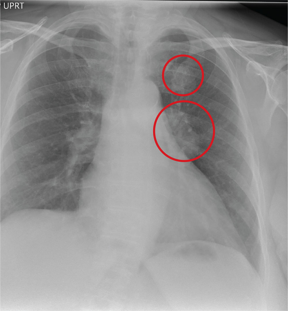
The radiograph shows a large, hyperdense mass within the left hilum. A second hyperdense mass is seen within the left upper lobe. Both are concerning for neoplastic processes and warrant further evaluation with contrast-enhanced CT.
Although thorough work-up and biopsy is needed, the presumptive diagnosis is a primary lung mass with likely metastasis to the brain.
ANSWER
The radiograph shows a large, hyperdense mass within the left hilum. A second hyperdense mass is seen within the left upper lobe. Both are concerning for neoplastic processes and warrant further evaluation with contrast-enhanced CT.
Although thorough work-up and biopsy is needed, the presumptive diagnosis is a primary lung mass with likely metastasis to the brain.

A 65-year-old woman is transferred to your facility for evaluation of left-side weakness she has been experiencing for more than two months. She states that it is worsening with time but denies any other symptoms. Outpatient MRI of the brain, obtained by the referring provider, is reported to show a right parietal mass with surrounding edema.
Medical history is significant for hypertension, diabetes, and hypercholesterolemia, which are controlled with medication. The patient reports smoking nearly two packs of cigarettes daily for at least 30 years.
Physical examination reveals normal vital signs and no apparent distress. The patient does have left hemiparesis; her left upper extremity is approximately 4/5 throughout, and her left lower extremity is approximately 2/5 throughout. The exam is otherwise normal.
As you review her admission lab results, you note that a chest radiograph was obtained (shown). What is your impression?

Using shock index in the ED to predict hospital admission and inpatient mortality
CLINICAL QUESTION: Can shock index (SI) in the ED predict the likelihood for hospital admission and inpatient mortality?
STUDY DESIGN: Retrospective chart review.
SETTING: Academic tertiary care center.
SYNOPSIS: All ED patients over 18 years of age over a 12-month period were included in the study for a total of 58,633 charts. Charts were excluded if the patient presented in cardiac arrest, left prior to full evaluation in the ED, or had an incomplete or absent first set of vital signs. Likelihood ratio (LR) values of greater than 5 and 10 were considered moderate and large increases in the outcomes, respectively. Authors found SI greater than 1.2 had a positive LR of 11.69 for admission to the hospital and a positive LR of 5.82 for inpatient mortality.
This study identified potential thresholds for SI but did not validate them. Whether SI would be a useful tool for triage remains unanswered.
BOTTOM LINE: Initial SI greater than 1.2 at presentation to the ED was associated with increased likelihood of hospital admission and inpatient mortality.
CITATIONS: Balhara KS, Hsieh YH, Hamade B, et al. Clinical metrics in emergency medicine: the shock index and the probability of hospital admission and inpatient mortality. Emerg Med J. 2017 Feb;34(2):89-94.
Dr. Dietsche is a clinical instructor, Division of Hospital Medicine, University of Colorado School of Medicine, Aurora.
Perioperative statin associated with reduction in all-cause perioperative mortality in noncardiac surgery
CLINICAL QUESTION: Does perioperative statin use reduce 30-day mortality in noncardiac surgery?
BACKGROUND: Current perioperative guidelines focus on continuation of existing therapy in long-term statin users with weak recommendations of potential efficacy in reducing perioperative complications.
STUDY DESIGN: Retrospective, observational cohort analysis.

SYNOPSIS: Using the Veterans Affairs Surgical Quality Improvement Program database, 96,486 patients were studied who were undergoing elective or emergent noncardiac surgery (vascular, general, orthopedic, neurosurgery, otolaryngology, and urology). 96.3% were men. Patients who died the day of the surgery or the day after were excluded, as were patients with multiple surgeries during the assessment period. Statin exposure on the day of or the day after surgery was compared with no statin use. The primary outcome was 30-day mortality and the secondary outcomes were significant reduction in any other complication.
Statin exposure was associated with reduced 30-day all-cause mortality with a marginally favorable effect with longer-term statin use (6 months to 1 year before admission). For the secondary outcomes, there was significant risk reduction in cardiac, infectious, respiratory, and renal complications but no significant change in central nervous system or nonatherosclerotic thrombotic complications.
Statin exposure may be associated with adherence to medical treatment and follow-up thus causing a selection bias.
BOTTOM LINE: Perioperative statin use was associated with a reduction in 30-day mortality and other complications.
CITATIONS: London MJ, Schwartz GG, Hur K, Henderson WG. Association of perioperative statin use with mortality and morbidity after major noncardiac surgery. JAMA Intern Med. 2017 Feb 1;177(2):231-42.
Dr. Dietsche is a clinical instructor, Division of Hospital Medicine, University of Colorado School of Medicine, Aurora.
CLINICAL QUESTION: Does perioperative statin use reduce 30-day mortality in noncardiac surgery?
BACKGROUND: Current perioperative guidelines focus on continuation of existing therapy in long-term statin users with weak recommendations of potential efficacy in reducing perioperative complications.
STUDY DESIGN: Retrospective, observational cohort analysis.
SYNOPSIS: Using the Veterans Affairs Surgical Quality Improvement Program database, 96,486 patients were studied who were undergoing elective or emergent noncardiac surgery (vascular, general, orthopedic, neurosurgery, otolaryngology, and urology). 96.3% were men. Patients who died the day of the surgery or the day after were excluded, as were patients with multiple surgeries during the assessment period. Statin exposure on the day of or the day after surgery was compared with no statin use. The primary outcome was 30-day mortality and the secondary outcomes were significant reduction in any other complication.
Statin exposure was associated with reduced 30-day all-cause mortality with a marginally favorable effect with longer-term statin use (6 months to 1 year before admission). For the secondary outcomes, there was significant risk reduction in cardiac, infectious, respiratory, and renal complications but no significant change in central nervous system or nonatherosclerotic thrombotic complications.
Statin exposure may be associated with adherence to medical treatment and follow-up thus causing a selection bias.
BOTTOM LINE: Perioperative statin use was associated with a reduction in 30-day mortality and other complications.
CITATIONS: London MJ, Schwartz GG, Hur K, Henderson WG. Association of perioperative statin use with mortality and morbidity after major noncardiac surgery. JAMA Intern Med. 2017 Feb 1;177(2):231-42.
Dr. Dietsche is a clinical instructor, Division of Hospital Medicine, University of Colorado School of Medicine, Aurora.
Anti-TNF drugs reduce mortality in Crohn’s disease
CHICAGO – As compared with prolonged use of corticosteroids, the use of anti–tumor necrosis factor (TNF) drugs was associated with reduced mortality in patients with Crohn’s disease, according to new findings presented here at Digestive Disease Week®.
The reduced mortality seen in this population may be secondary to the lower rates of major adverse cardiovascular events and hip fracture that are associated with anti-TNF use as compared to corticosteroid use. However, the same reduction in mortality risk was not observed in patients with ulcerative colitis using anti-TNF drugs.
Anti-TNF therapy has become a cornerstone in the management of inflammatory bowel disease (IBD), and Dr. Lewis noted that these agents have been shown to be useful for induction, maintenance, and remission, and to reduce surgical and hospitalization rates.
“However, fear of adverse events and cost has deterred greater use of these agents,” he told attendees.
In their study, Dr. Lewis and his colleagues compared the mortality risk with prolonged corticosteroids use versus anti-TNF drugs in patients with IBD.
They conducted a retrospective cohort study using data from 2006-2013 of a population of Medicaid and Medicare beneficiaries in the United States. The cohort included individuals who had received treatment with corticosteroids within the prior year and subsequently had been treated with either additional corticosteroid therapy for a total of greater than 3,000 mg of prednisone or equivalent within 12 months or newly initiated anti-TNF therapy.
The primary outcome of the study was all-cause mortality and secondary outcomes included common causes of death.
Dr. Lewis explained that 57 potential confounding variables were thought to be associated with the choice between corticosteroid use or anti-TNF therapy. These variables, which included demographic characteristics, medications, diagnostic tests, and comorbidities, were measured.
Among Crohn’s disease patients, 7,694 who were prolonged corticosteroid users and 1,879 were new to anti-TNF therapy. Among patients with ulcerative colitis, 3,224 were long-term corticosteroid users and 459 were new anti-TNF users.
The researchers found that the weighted annual incidences of death per 1,000 Crohn’s disease treated patients were 21.4 for those using anti-TNF therapy and 30.1 for those with prolonged corticosteroid use. For those with ulcerative colitis, these figures were 23.0 and 30.9, respectively.
The risk of death was statistically significantly reduced in Crohn’s disease patients who used anti-TNF therapy (odds ratio, 0.78; 95% confidence interval, 0.65-0.93). However, the benefit was not as pronounced in ulcerative colitis.
“We did not see the same effect for ulcerative colitis but for mortality, it was in the same direction, with a hazard ratio of 0.87,” he said.
Among the Crohn’s disease patients, anti-TNF therapy was associated with lower rates of major adverse cardiovascular events (OR, 0.68; 95% CI, 0.55-0.85) and hip fracture (OR, 0.5; 95% CI, 0.34-0.83), which were statistically significant. The use of anti-TNF therapy also reduced the risk of stroke in Crohn’s disease patients (OR, 0.72; 95% CI, 0.51-1.03), but there was also an increase in the risk of cancer (OR, 0.27; 95% CI, 0.98-1.65). Both of these findings nearly reached statistical significance.
In the model that censored for any of the secondary outcomes, the lower mortality risk was attenuated and very close to a null result (OR, 0.97; 95% CI 0.63-1.47).
Dr. Lewis also pointed out that in some of their models, the magnitude of benefit with anti-TNF therapy appears to be greatest in the patients with the most comorbidities.
Digestive Disease Week® is jointly sponsored by the American Association for the Study of Liver Diseases (AASLD), the American Gastroenterological Association (AGA) Institute, the American Society for Gastrointestinal Endoscopy (ASGE), and the Society for Surgery of the Alimentary Tract (SSAT).
Dr. Lewis has disclosed financial relationships with Takeda, Pfizer, Lilly, Gilead, Johnson and Johnson, Samsung Bioepis, AbbVie, and Dark Canyon Laboratories.
AT DDW
Key clinical point:
Major finding: Anti-TNF drugs were associated with reduced mortality in patients with Crohn’s disease (OR, 0.78) but to a much lesser extent in ulcerative colitis (OR, 0.87).
Data source: Database of Medicare and Medicaid recipients with 9,573 patients with Crohn’s disease and 3,683 with ulcerative colitis.
Disclosures: Dr. Lewis has disclosed financial relationships with Takeda, Pfizer, Lilly, Gilead, Johnson and Johnson, Samsung Bioepis, AbbVie, and Dark Canyon Laboratories.
VIDEO: Registry study will follow 4,000 fecal transplant patients for 10 years
CHICAGO – A 10-year registry study aims to gather clinical and patient-reported outcomes on 4,000 adult and pediatric patients who undergo fecal microbiota transplant in the United States, officials of the American Gastroenterological Association announced during Digestive Disease Week®.
The AGA Fecal Microbiota Transplantation National Registry will be the first study to assess both short- and long-term patient outcomes associated with fecal microbiota transplant (FMT) in both adults and children, Colleen Kelly, MD, said in an video interview. Most subjects will have received FMT for recurrent or refractory Clostridium difficile infections – the only indication for which Food and Drug Administration currently allows independent clinician action. But the investigational uses of FMT are expanding rapidly, and patients who undergo the procedure during any registered study will be eligible for enrollment, said Dr. Kelly, co-chair of the study’s steering committee.
The video associated with this article is no longer available on this site. Please view all of our videos on the MDedge YouTube channel
The study’s primary objectives are short- and long-term safety outcomes, said Dr. Kelly of Brown University, Providence, R.I. While generally considered quite safe, short-term adverse events have been reported with FMT, and some of them have been serious – including one death from aspiration pneumonia in a patient who received donor stool via nasogastric tube (Clin Infect Dis. 2015 Mar;61[1]:136-7). Other adverse events are usually self-limited but can include low-grade fever, abdominal pain, distention, bloating, and diarrhea.
Researchers seek to illuminate many of the unknowns associated with this relatively new procedure. Scientists are only now beginning to unravel the myriad ways the human microbiome promotes both health and disease. Specific alterations, for example, have been associated with obesity and other conditions; there is concern that transplanting a new microbial population could induce a disease phenotype in a recipient who might not have otherwise been at risk.
With the planned cohort size and follow-up period, the study should be able to detect any unanticipated adverse events that occur in more than 1% of the population, Dr. Kelly said. It will include a comparator group of patients with recurrent or refractory C. difficile infection from a large insurance claims database to allow comparison between patients treated with FMT and those treated with antibiotics only.
The registry study also aims to discover which method or methods of transplant material delivery are best, she said. Right now, there are a number of methods (colonoscopy/sigmoidoscopy, enema, upper gastrointestinal endoscopy, nasogastric or nasoduodenal tube, and capsules), and no consensus on which is the best. As indications for FMT expand, there may be no single best method. The approach will probably be matched to the disorder being treated, and the study may help illuminate this as well.
For the first 2 years after a transplant, clinicians will follow patients and enter data into the registry. After that, an electronic patient-reported outcomes system will automatically contact the patient annually for follow-up information by email or text message. When patients enter their data, they can access educational material that will help keep them up-to-date on potential adverse events.
The study will also include a biobank of stool samples obtained during the procedures, hosted by the American Gut Project and the Microbiome Initiative at the University of California, San Diego. This arm of the project will analyze the microbiome of 3,000 stool samples from recipients, both before and after their transplant, as well as the corresponding donors whose material was used in the fecal transplant.
The registry study, a project of the AGA Center for Gut Microbiome Research and Education, is funded by a $3.3 million grant from the National Institute of Allergy and Infectious Diseases. It will be conducted in partnership with the Crohn’s and Colitis Foundation, Infectious Diseases Society, and the North American Society for Pediatric Gastroenterology, Hepatology, and Nutrition.
The registry study currently is accepting applications. Physicians who perform FMT for C. difficile infections, and centers that conduct FMT research for other potential indications, can fill out a short survey to indicate their interest.
Digestive Disease Week® is jointly sponsored by the American Association for the Study of Liver Diseases (AASLD), the American Gastroenterological Association (AGA) Institute, the American Society for Gastrointestinal Endoscopy (ASGE) and the Society for Surgery of the Alimentary Tract (SSAT).
This article was updated June 8, 2017.
[email protected]
On Twitter @Alz_gal
AT DDW