User login
FDA approves avelumab for advanced urothelial carcinoma
The Food and Drug Administration has granted accelerated approval to avelumab for the treatment of patients with locally advanced or metastatic urothelial carcinoma whose disease progressed during or following platinum-containing chemotherapy or within 12 months of neoadjuvant or adjuvant platinum-containing chemotherapy.
Approval was based on a confirmed overall response rate (ORR) of 16.1% in 26 patients who had been followed for at least 6 months in a single-arm, multicenter study that enrolled 242 patients with locally advanced or metastatic urothelial carcinoma whose disease progressed following platinum-containing neoadjuvant or adjuvant chemotherapy. The ORR was 13.3% among 30 patients who had been followed for at least 13 weeks. Median time to response was 2.0 months (range 1.3-11.0). The median response duration had not been reached in patients followed for at least 13 weeks or at least 6 months, but ranged from 1.4+ to 17.4+ months in both groups, the FDA said in a statement.![]()
Serious adverse reactions, including urinary tract infection/urosepsis, abdominal pain, musculoskeletal pain, creatinine increased/renal failure, dehydration, hematuria/urinary tract hemorrhage, intestinal obstruction/small intestinal obstruction, and pyrexia, were reported in 41% of patients, causing death in 6% of patients. The most common adverse reactions were infusion-related reaction, musculoskeletal pain, nausea, decreased appetite, and urinary tract infection.
The FDA recommended an intravenous infusion of 10 mg/kg over 60 minutes every 2 weeks, and premedication with an antihistamine and acetaminophen prior to the first four infusions of avelumab.
Full prescribing information is available here.
The drug is being marketed as Bavencio by EMD Serono.
Dalbavancin proves highly effective in osteomyelitis
VIENNA – Two infusions of the long-acting lipoglycopeptide antibiotic dalbavancin showed a favorable clinical benefit for treatment of adult osteomyelitis in a phase II study.
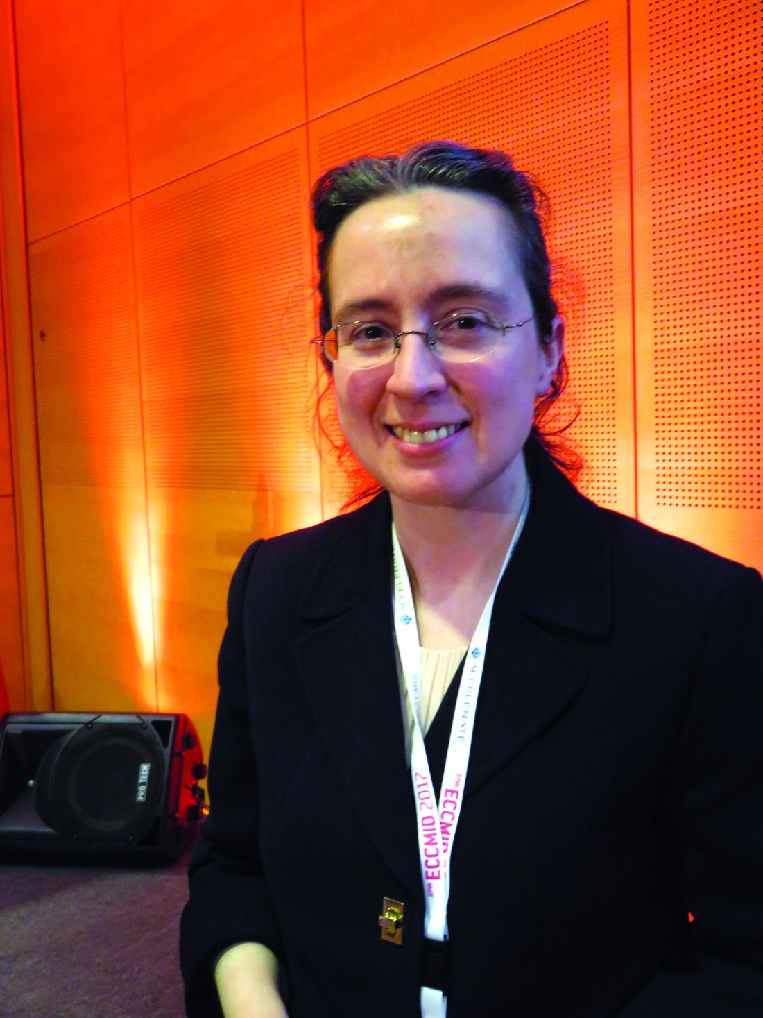
Dalbavancin (Dalvance) showed positive results while avoiding the complexities of standard therapies that require longer, more frequent dosing, Urania Rappo, MD, said at the European Society of Clinical Microbiology and Infectious Diseases annual congress.
Dalbavancin already is approved for acute bacterial skin and skin structure infections, and its long terminal half-life of 14 days and high bone penetration made it a natural candidate for evaluation in the treatment of osteomyelitis, said Dr. Rappo, director of clinical development (anti-infectives) at Allergan, which markets the drug and sponsored the study.
Dalbavancin is a glycopeptide antibiotic with a lipid tail that prolongs its half-life, compared with other drugs in this category, such as vancomycin and teicoplanin, to which it is structurally related. It is highly potent against gram-positive bacterial infections, including methicillin-resistant Staphylococcus aureus.
The drug’s MIC90 for S. aureus is 0.06 mcg/mL; vancomycin’s, in comparison, is 1 mcg/mL. A 2015 bone penetration study found that the bone level 12 hours after a 1,000-mg infusion was 6.3 mcg/g. This remained elevated for 14 days; the concentration at 2 weeks was 4 mcg/g (Antimicrob Agents Chemother. 2015 Apr;59[4]:1849-55).
“The mean bone-to-plasma penetration ratio was 13%, and drug levels in bone were very similar to free drug levels in serum, so we expect that much of this is free drug in the bone, available for antimicrobial activity,” Dr. Rappo noted. “It’s not only long lasting, but highly potent, meaning we need less drug to kill the infecting organism.”
Dalbavancin was administered as two 1,500-mg IV infusions, 1 week apart in Dr. Rappo’s randomized, open-label, phase II study – the first clinical trial to examine the drug’s effect in osteomyelitis in adults. The study is ongoing; she presented interim results on 68 patients, 59 of whom took dalbavancin. The nine patients in the standard-of-care arm were treated according to the investigator’s clinical judgment. Vancomycin was the most commonly employed therapy. Three patients received vancomycin infusions for 4 weeks. Four received a regimen of 4-16 days of intravenous vancomycin followed by intravenous linezolid or levofloxacin to complete a 4- to 6-week course of therapy. Adjunctive aztreonam was permitted for presumed coinfection with a gram-negative pathogen and a switch to oral antibiotic for gram-negative coverage was allowed after clinical improvement.
The primary endpoint was clinical cure at 42 days in the clinically evaluable population, defined as recovery without need for further antibiotic therapy. Failure was defined as the need for additional antibiotics; more than 6 weeks of treatment in the comparator arm; new purulence; amputation due to infection progression; or death. Indeterminate response was defined as loss to follow-up or amputation due to vascular insufficiency.
There were several secondary endpoints: clinical improvement at day 21, including changes in C-reactive protein level and clinical response in patients who had follow-up at days 42, 180, and 365.
In the dalbavancin arm, patients had a mean age of 51 years. All had undergone surgical debridement and bone culture. The most common site of infection was the foot or leg (about 83%). The baseline mean CRP level was 41.8 mg/L. About half of the patients had methicillin-susceptible S. aureus on bone culture. Coagulase-negative staphylococci were present in 20%. About 22% had gram-negative pathogens, mostly present in a mixed infection along with gram-positive pathogens. Five patients (three on dalbavancin, two on standard of care) discontinued the study drug early because they were solely infected with gram-negative pathogens.
At day 42, clinical cure was seen in all the dalbavancin patients and six of the standard treatment patients. In the group with treatment data out to 180 days (54 on dalbavancin and 5 on standard therapy), clinical cure rates were similar.
At 180 days, clinical cure continued in 93% of the dalbavancin patients (50 of 54). Two patients were defined as failures, and two patients were indeterminate because of loss to follow-up. In the standard therapy group at 180 days, four of five standard therapy patients maintained clinical cure; none of these patients has reached the 365-day outcome.
There were no treatment-emergent adverse events (TEAEs) in the standard therapy group. In the dalbavancin group, 10 patients experienced TEAEs, only one of whom had TEAEs related to the study drug, which were not serious, Dr. Rappo said.
This study was performed at a single center in the Ukraine, and an additional phase II study in the United States has begun, she added.
[email protected]
On Twitter @alz_gal
VIENNA – Two infusions of the long-acting lipoglycopeptide antibiotic dalbavancin showed a favorable clinical benefit for treatment of adult osteomyelitis in a phase II study.
Dalbavancin (Dalvance) showed positive results while avoiding the complexities of standard therapies that require longer, more frequent dosing, Urania Rappo, MD, said at the European Society of Clinical Microbiology and Infectious Diseases annual congress.
Dalbavancin already is approved for acute bacterial skin and skin structure infections, and its long terminal half-life of 14 days and high bone penetration made it a natural candidate for evaluation in the treatment of osteomyelitis, said Dr. Rappo, director of clinical development (anti-infectives) at Allergan, which markets the drug and sponsored the study.
Dalbavancin is a glycopeptide antibiotic with a lipid tail that prolongs its half-life, compared with other drugs in this category, such as vancomycin and teicoplanin, to which it is structurally related. It is highly potent against gram-positive bacterial infections, including methicillin-resistant Staphylococcus aureus.
The drug’s MIC90 for S. aureus is 0.06 mcg/mL; vancomycin’s, in comparison, is 1 mcg/mL. A 2015 bone penetration study found that the bone level 12 hours after a 1,000-mg infusion was 6.3 mcg/g. This remained elevated for 14 days; the concentration at 2 weeks was 4 mcg/g (Antimicrob Agents Chemother. 2015 Apr;59[4]:1849-55).
“The mean bone-to-plasma penetration ratio was 13%, and drug levels in bone were very similar to free drug levels in serum, so we expect that much of this is free drug in the bone, available for antimicrobial activity,” Dr. Rappo noted. “It’s not only long lasting, but highly potent, meaning we need less drug to kill the infecting organism.”
Dalbavancin was administered as two 1,500-mg IV infusions, 1 week apart in Dr. Rappo’s randomized, open-label, phase II study – the first clinical trial to examine the drug’s effect in osteomyelitis in adults. The study is ongoing; she presented interim results on 68 patients, 59 of whom took dalbavancin. The nine patients in the standard-of-care arm were treated according to the investigator’s clinical judgment. Vancomycin was the most commonly employed therapy. Three patients received vancomycin infusions for 4 weeks. Four received a regimen of 4-16 days of intravenous vancomycin followed by intravenous linezolid or levofloxacin to complete a 4- to 6-week course of therapy. Adjunctive aztreonam was permitted for presumed coinfection with a gram-negative pathogen and a switch to oral antibiotic for gram-negative coverage was allowed after clinical improvement.
The primary endpoint was clinical cure at 42 days in the clinically evaluable population, defined as recovery without need for further antibiotic therapy. Failure was defined as the need for additional antibiotics; more than 6 weeks of treatment in the comparator arm; new purulence; amputation due to infection progression; or death. Indeterminate response was defined as loss to follow-up or amputation due to vascular insufficiency.
There were several secondary endpoints: clinical improvement at day 21, including changes in C-reactive protein level and clinical response in patients who had follow-up at days 42, 180, and 365.
In the dalbavancin arm, patients had a mean age of 51 years. All had undergone surgical debridement and bone culture. The most common site of infection was the foot or leg (about 83%). The baseline mean CRP level was 41.8 mg/L. About half of the patients had methicillin-susceptible S. aureus on bone culture. Coagulase-negative staphylococci were present in 20%. About 22% had gram-negative pathogens, mostly present in a mixed infection along with gram-positive pathogens. Five patients (three on dalbavancin, two on standard of care) discontinued the study drug early because they were solely infected with gram-negative pathogens.
At day 42, clinical cure was seen in all the dalbavancin patients and six of the standard treatment patients. In the group with treatment data out to 180 days (54 on dalbavancin and 5 on standard therapy), clinical cure rates were similar.
At 180 days, clinical cure continued in 93% of the dalbavancin patients (50 of 54). Two patients were defined as failures, and two patients were indeterminate because of loss to follow-up. In the standard therapy group at 180 days, four of five standard therapy patients maintained clinical cure; none of these patients has reached the 365-day outcome.
There were no treatment-emergent adverse events (TEAEs) in the standard therapy group. In the dalbavancin group, 10 patients experienced TEAEs, only one of whom had TEAEs related to the study drug, which were not serious, Dr. Rappo said.
This study was performed at a single center in the Ukraine, and an additional phase II study in the United States has begun, she added.
[email protected]
On Twitter @alz_gal
Key clinical point:
Major finding: At 42 days, 100% of patients taking the drug were clear of infection.
Data source: A phase II trial involving 68 patients, 59 of whom were randomized to the study drug; the remainder were on standard treatment.
Disclosures: Allergan sponsored the study; Dr. Rappo is a company employee.
Changing gloves before closure cut wound-related cesarean complications
SAN DIEGO – Changing gloves before closure during cesarean deliveries can reduce overall wound morbidity, a study showed.
Glove changing during the study had a number needed to treat (NNT) of 14 to see benefit, no demonstrated risk, and a total cost per procedure of about $5.
Researchers led by Buvana Reddy, MD, an ob.gyn. in group practice in Woodbury, Minn., used a randomized trial design to determine whether donning a fresh pair of gloves makes a difference in the rate of surgical complications during cesarean delivery. .
Just one previous study had shown a reduced wound infection rate when gloves were changed after placental delivery (J Reprod Med. 2004 Jan;49[1]:13-6). The study showed a statistically significant drop in wound infections, but Dr. Scrafford said that the sample size was small, and the point at which glove changing was performed is not very practical in terms of the flow of surgery.
Building on this study, Dr. Scrafford and his colleagues designed a trial that was meant to be larger, incorporated a composite wound outcome score rather than just focusing strictly on infection, and switched the timing of glove change to just before abdominal closure.
The randomized controlled trial included all women who underwent nonemergent cesarean delivery at the investigators’ home facility during a 15-month period. The patient allocation was not known until the presurgical “time-out.” Patients allocated to the control condition did not have any change in operative procedures; the surgical team of patients allocated to the intervention arm changed their outer gloves immediately before peritoneal or fascial closure.
The composite primary outcome measure assessed wound seroma, hematoma, or dehiscence (defined as a separation of at least 1 cm), as well as wound infection and any other wound abnormality. Secondary outcome measures included whether patients had a fever or peri-incisional cellulitis.
A total of 553 patients were randomized. Follow-up was high, so Dr. Scrafford and his colleagues were able to analyze data from a total of 250 patients in the control arm and 236 patients who received the glove-changing intervention.
Patient demographics and comorbidities were similar between groups, as was the surgical time and estimated blood loss. Dr. Scrafford noted that significantly more patients in the glove-changing group than in the control group received presurgical vaginal preparation with antiseptic solution (20.3% vs. 10.5%; P = .001). However, he reported, statistical analysis showed that this difference had no effect on the results.
Overall, 15 (6.4%) of the patients in the glove-changing group met the criteria for wound complication, compared with 34 (13.6%) in the control group (P = .008). Of the individual components of the primary outcome measure, the difference in incidence of skin separation was also significant; separation was experienced by five patients (2.1%) in the glove-changing group, compared with 14 (5.6%) in the control group (P = .01).
The rate of wound infections was numerically lower in the intervention group, compared with the control group (8 vs. 14), but the difference was not statistically significant.
There were no significant differences between the groups when secondary outcomes were analyzed, Dr. Scrafford said.
The lack of block randomization in the study left open the potential for historical bias, Dr. Scrafford said. Since surgeons couldn’t be blinded as to study arm, the potential for observer bias also existed. Still, he said, the study’s strengths included its large size, and the real-world applicability of the broad inclusion criteria and diverse patient population.
The study authors reported no outside sources of funding and no conflicts of interest.
[email protected]
On Twitter @karioakes
Key clinical point:
Major finding: The number needed to treat to see benefit from glove changing was 14; the cost per procedure was about $5.
Data source: A randomized controlled trial of 553 patients undergoing nonemergent cesarean delivery.
Disclosures: The study authors reported no outside sources of funding and no conflicts of interest.
Antacid use in infants linked to increased fracture risk
SAN FRANCISCO – Children were more likely to experience a fracture if they were prescribed antacids before age 1 year, according to a study of military families.
The large study revealed that use of proton pump inhibitors (PPIs) before age 1 year was linked to a 22% increased risk of fracture, compared with those not prescribed antacids. Similarly, children prescribed both PPIs and H2 blockers before age 1 year were 31% more likely to have a fracture compared to those not taking the drugs.
“A lot of data are coming out that proton pump inhibitors are not quite as benign as we used to think, and we are seeing that fracture risk is increased with use,” U.S. Air Force Capt. Laura Malchodi, MD, a pediatrics resident at Walter Reed National Military Medical Center in Bethesda, Md., told colleagues at the Pediatric Academic Societies meeting.
Antacid use has been increasing among both adults and children, but the biggest rise has been in children under age 1 year, she said. Previous research into adult use of antacids has revealed an increased incidence of fractures, so Dr. Malchodi investigated the incidence of fractures in children under age 1 year among those who had taken PPIs, H2 blockers, neither, or both.
“What this means for doctors is that when you do start to think of using proton pump inhibitors or any antacid therapy in children, we should really think of limiting it to one type if possible – H2 blockers are now preferable – and for the shortest amount of time as possible,” Dr. Malchodi said of her findings.
The retrospective study’s cohort comprised 874,447 children born between 2001 and 2013 who had been in the U.S. Military Health System for at least 2 years. Children who took antacids after age 1, spent more than a week in a neonatal intensive care unit, or had nonaccidental trauma (abuse) or osteogenesis imperfecta were excluded.
Ninety percent of the cohort had not received prescriptions for any antacids (789,631 children) in their first year of life, and 1.2% had received prescriptions for both PPIs and H2 blockers before age 1 year. Of the remaining children, 7.7% had received prescriptions for H2 blockers, and 0.8% for PPIs.
The children who had and had not been prescribed antacids were similar in median years enrolled in the system, but nearly twice as many who received antacid prescription had been preterm (6.4% vs. 3.5%, P less than .05). Similarly, 3.7% of those prescribed antacids had a low birth weight, compared with 2.2% of those not prescribed antacids (P less than .05). The median age of fracture also differed for the two groups: 3.9 years for those prescribed antacids and 4.5 years for those not (P less than .05).
In using medical records during their analysis, the researchers excluded follow-up visits for the same fracture within the previous 6 months. Before adjustment for covariates, boys had a slightly increased risk of fracture (hazard ratio [HR], 1.08), and those with a previous fracture had an 85% increased risk (HR, 1.85). Compared with children not prescribed antacids, those prescribed PPIs had a 23% increased risk of fracture (HR, 1.23), and those prescribed H2 blockers had a 13% increased risk (HR, 1.13). Those prescribed combination antacid therapy had a 32% increased risk of fractures (HR, 1.32).
Adjustment for preterm birth, low birth weight, sex, and a previous fracture barely reduced those risks: 22% increased risk for PPI use, 4% increased risk for H2 blocker use, and 31% increased risk for using both. The vast majority of children who took antacids had been prescribed them in their first 6 months, so the researchers calculated adjusted risk by age of exposure. For H2 blockers, no statistically significant increased risk of fracture existed in those taking them before or after 6 months old.
Those taking PPIs, however, had a 25% increased risk of fracture if they took them before 6 months old, compared with a 20% increased risk if prescribed PPIs between 6 and 12 months. Likewise, children taking both PPIs and H2 blockers before 6 months old had a 32% increased risk of fracture, compared with a 23% increased risk between 6 and 12 months old.
Analysis of the duration of children’s use of antacids revealed a dose-response relationship, with an increasing risk alongside increasing days taking the medication. For example, those on PPIs for a month or less had a 19% increased risk of fracture, compared with children not prescribed antacids, but that rose to a 23% increased risk for those taking PPIs from 60 to 150 days and to a 42% increased risk for taking them longer than 150 days.
Similarly, the risk of fracture after having taken H2 blockers for up to a month was 14%, which increased to 22% for medication durations over 120 days. Children on combination therapy took the medication for much longer than did children prescribed either antacid. The risk of fracture was 17% greater for those taking them for up to 4 months, but that increased to a 50% greater risk for children taking both antacids for longer than 338 days.
“A couple of decades ago, we thought these medications were super safe, that there could be no problem with them,” Dr. Malchodi said, suggesting that their availability over the counter for adults may contribute to that perception. “With this growing evidence, there’s at least a lot more caution about using them,” she said.
Because the study relied on prescriptions for antacids, the researchers could not take into account which children actually took the antacids. Another limitation was their inability to consider other potential confounders, such as socioeconomic status or comorbidities that may later increase the risk of fracture. Further, exclusion of 6 months of follow-up after one fracture may have missed new fractures in that time period. Using a military cohort, on the other hand, meant having a geographically and socioeconomically diverse population with less risk of care bias because all the children had universal health care coverage.
No external funding was used. Dr. Malchodi reported having no disclosures.
*The View on the News was added on 6/13/17.
Acid suppression is frequently prescribed in infants for the treatment of symptoms such as fussiness, arching, and poor feeding, despite randomized controlled trials showing no benefit for these symptoms over placebo. These medications are often prescribed because physicians think they are useful; families are frustrated, exhausted, and worried about the infant’s symptoms; and these medications are considered safe and well tolerated. Recent adult studies have raised the possibility that these medications may not be as safe as once thought, with case-controlled studies linking them to increased risk of infectious, renal, cardiac, neurologic, and orthopedic complications. While there are pediatric studies supporting an increased infectious risk from both PPI and H2 antagonist use, there are no pediatric studies that address other complications. In this study by Dr. Malchodi et al., acid suppression use in infants under the age of 1 year was associated with an increased risk of fractures over the duration of enrollment in the U.S. Military Health System. They also found a dose-dependent effect, which further strengthens the conclusions that acid suppression may predispose patients to fractures. This research is a critical first step in elucidating the relationship of acid suppression and fracture risk in infants.
As with all database studies, there are some limitations to this study. First, patients taking acid suppression often have more comorbidities than do patients who are not taking the medications; because these patients are sicker, they may have more risk factors including compromised nutritional status and malabsorption predisposing them to fractures. The authors controlled for some of these comorbidities, but future studies should address additional ones. Second, as with all case-control studies, proving causality, not just association, is difficult so any future prospective acid suppression trials should include an assessment of bone health. Third, because the dosing per kilogram is not included, it is difficult to determine if there is a safe level of acid suppression for those children who need it. Fourth, because this is a database review, it is not clear if patients actually took the prescribed medication.
Because of the safety concerns regarding acid suppression as well as the lack of benefit in reducing symptoms in infants, nonpharmacologic therapies should be considered as first-line therapy for the treatment of bothersome symptoms. In the fussy, arching, or irritable child, changing the frequency or volume of feeds, thickening feeds, or changing to partially hydrolyzed formulas or eliminating dairy from the maternal diet (for breastfed infants) should be considered before starting acid suppression therapy. Other diagnoses besides gastroesophageal reflux disease, such as colic and cow’s milk protein allergy, need to be considered as well to ensure that the therapy matches the diagnosis. For those patients in whom acid suppression is required, using the lowest dose possible for the shortest amount of time is critical. Finally, for patients on multiple medications that may impact fracture risk (such as acid suppression, steroids), extra vigilance is needed to stop unnecessary medications as soon as possible.
SAN FRANCISCO – Children were more likely to experience a fracture if they were prescribed antacids before age 1 year, according to a study of military families.
The large study revealed that use of proton pump inhibitors (PPIs) before age 1 year was linked to a 22% increased risk of fracture, compared with those not prescribed antacids. Similarly, children prescribed both PPIs and H2 blockers before age 1 year were 31% more likely to have a fracture compared to those not taking the drugs.
“A lot of data are coming out that proton pump inhibitors are not quite as benign as we used to think, and we are seeing that fracture risk is increased with use,” U.S. Air Force Capt. Laura Malchodi, MD, a pediatrics resident at Walter Reed National Military Medical Center in Bethesda, Md., told colleagues at the Pediatric Academic Societies meeting.
Antacid use has been increasing among both adults and children, but the biggest rise has been in children under age 1 year, she said. Previous research into adult use of antacids has revealed an increased incidence of fractures, so Dr. Malchodi investigated the incidence of fractures in children under age 1 year among those who had taken PPIs, H2 blockers, neither, or both.
“What this means for doctors is that when you do start to think of using proton pump inhibitors or any antacid therapy in children, we should really think of limiting it to one type if possible – H2 blockers are now preferable – and for the shortest amount of time as possible,” Dr. Malchodi said of her findings.
The retrospective study’s cohort comprised 874,447 children born between 2001 and 2013 who had been in the U.S. Military Health System for at least 2 years. Children who took antacids after age 1, spent more than a week in a neonatal intensive care unit, or had nonaccidental trauma (abuse) or osteogenesis imperfecta were excluded.
Ninety percent of the cohort had not received prescriptions for any antacids (789,631 children) in their first year of life, and 1.2% had received prescriptions for both PPIs and H2 blockers before age 1 year. Of the remaining children, 7.7% had received prescriptions for H2 blockers, and 0.8% for PPIs.
The children who had and had not been prescribed antacids were similar in median years enrolled in the system, but nearly twice as many who received antacid prescription had been preterm (6.4% vs. 3.5%, P less than .05). Similarly, 3.7% of those prescribed antacids had a low birth weight, compared with 2.2% of those not prescribed antacids (P less than .05). The median age of fracture also differed for the two groups: 3.9 years for those prescribed antacids and 4.5 years for those not (P less than .05).
In using medical records during their analysis, the researchers excluded follow-up visits for the same fracture within the previous 6 months. Before adjustment for covariates, boys had a slightly increased risk of fracture (hazard ratio [HR], 1.08), and those with a previous fracture had an 85% increased risk (HR, 1.85). Compared with children not prescribed antacids, those prescribed PPIs had a 23% increased risk of fracture (HR, 1.23), and those prescribed H2 blockers had a 13% increased risk (HR, 1.13). Those prescribed combination antacid therapy had a 32% increased risk of fractures (HR, 1.32).
Adjustment for preterm birth, low birth weight, sex, and a previous fracture barely reduced those risks: 22% increased risk for PPI use, 4% increased risk for H2 blocker use, and 31% increased risk for using both. The vast majority of children who took antacids had been prescribed them in their first 6 months, so the researchers calculated adjusted risk by age of exposure. For H2 blockers, no statistically significant increased risk of fracture existed in those taking them before or after 6 months old.
Those taking PPIs, however, had a 25% increased risk of fracture if they took them before 6 months old, compared with a 20% increased risk if prescribed PPIs between 6 and 12 months. Likewise, children taking both PPIs and H2 blockers before 6 months old had a 32% increased risk of fracture, compared with a 23% increased risk between 6 and 12 months old.
Analysis of the duration of children’s use of antacids revealed a dose-response relationship, with an increasing risk alongside increasing days taking the medication. For example, those on PPIs for a month or less had a 19% increased risk of fracture, compared with children not prescribed antacids, but that rose to a 23% increased risk for those taking PPIs from 60 to 150 days and to a 42% increased risk for taking them longer than 150 days.
Similarly, the risk of fracture after having taken H2 blockers for up to a month was 14%, which increased to 22% for medication durations over 120 days. Children on combination therapy took the medication for much longer than did children prescribed either antacid. The risk of fracture was 17% greater for those taking them for up to 4 months, but that increased to a 50% greater risk for children taking both antacids for longer than 338 days.
“A couple of decades ago, we thought these medications were super safe, that there could be no problem with them,” Dr. Malchodi said, suggesting that their availability over the counter for adults may contribute to that perception. “With this growing evidence, there’s at least a lot more caution about using them,” she said.
Because the study relied on prescriptions for antacids, the researchers could not take into account which children actually took the antacids. Another limitation was their inability to consider other potential confounders, such as socioeconomic status or comorbidities that may later increase the risk of fracture. Further, exclusion of 6 months of follow-up after one fracture may have missed new fractures in that time period. Using a military cohort, on the other hand, meant having a geographically and socioeconomically diverse population with less risk of care bias because all the children had universal health care coverage.
No external funding was used. Dr. Malchodi reported having no disclosures.
*The View on the News was added on 6/13/17.
AT PAS 17
Key clinical point:
Major finding: Risk of fracture increased 22% among children who took proton pump inhibitors in their first year of life and increased 31% among children taking both PPIs and H2 blockers.
Data source: A retrospective cohort study of 874,447 children born between 2001 and 2013 and who were in the U.S. Military Health System for at least 2 years.
Disclosures: No external funding was used. Dr. Malchodi reported having no relevant financial disclosures.
RIV spotlights HM-focused research in real-time
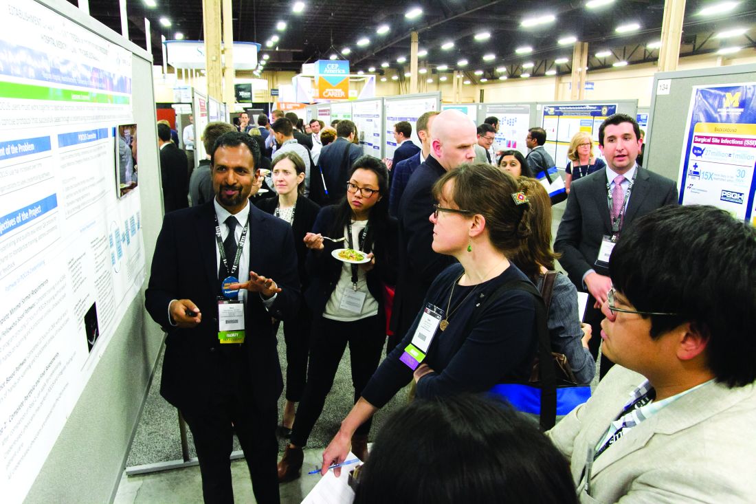
LAS VEGAS – Masih Shinwa, MD, stood beside a half-circle of judges at SHM’s annual Research, Innovations, and Clinical Vignettes poster competition and argued why his entry, already a finalist, should win.
To think, his work, “Please ‘THINK’ Before You Order: A Multidisciplinary Approach to Decreasing Overutilization of Daily Labs,” was borne simply of a group of medical students who incredulously said that they were amazed patients would be woken up in the night for tests.

Dr. Shinwa’s project shows just how an idea can blossom into a recognized poster.
Some 18 months ago, the students he works with at Mount Sinai Hospital in New York just couldn’t understand why so many tests had to be done overnight while a patient slept. So, Dr. Shinwa and his colleagues looked at ways to reduce unnecessary lab tests and chemistry testing.
Now, Dr. Shinwa was humbled to think his work and that of his colleagues could be a pathway to eliminating tests that don’t need to happen across the country, a focal point of SHM and the American Board of Internal Medicine Foundation’s Choosing Wisely Campaign.
“This is a way to make it national,” he said. “You may have affected the lives of the patients in your hospital, but, unless you attend these types of national meetings, it’s hard to get that perspective across (the country).”
That level of personal and professional collaboration is the purpose of the RIV, said Margaret Fang, MD, MPH, FHM, program chair for the HM17 competition.
“One of the amazing things is, everyone has their own poster. They’re doing their work,” she added. “But then they start up conversations with the people next to them. ... Seeing the organic networking and discussion that arise from that is really exciting. RIV serves as a way of connecting people who might not have know the other person was doing that kind of work.”
Dr. Fang said that the intergenerational aspect of the RIV, where early-career hospitalists mingle with the field’s founders and leaders, creates an environment where research is encouraged.
“Just seeing the intense interest that more senior hospitalists have in mentoring and guiding the next generation is delightful,” she added.
Dr. Shinwa said that the specialty’s focus on both clinical research and systems-level change is important, as the work positions the field to be leaders not just in patient care but for hospitals as a whole.
“We are physicians,” he said. “Our role is taking care of patients. Knowing that there are people who are not just focusing on taking care of specific patients but are actually there to improve the entire system and the process – that’s really gratifying.”
That’s the word that Merideth Prevost, MD, of New Mexico VA Health Care System, Albuquerque also used to describe presenting her poster, “Improving Accuracy in Measuring Fluid Balance on a General Medicine Ward.”
“If we can improve our little microcosm, then spread it to other folks, then patients all over the country can be helped by what we do,” she said. “And that’s a really cool thought.”
The RIV also has the unique advantage of letting people have immediate and direct access to lead researchers at the exact moment of reading their research. HM17 attendees had conversations that usually went beyond just the results, which can be downloaded at www.shmabstracts.com.
Dr. Prevost believes that the chats can helpfully highlight the behind-the-scenes pitfalls and mistakes of research that can sometimes be just as valuable as the published results.
“The things that don’t make it to the posters are all the challenges that people experienced on the way to get to this particular work,” she added. “Like ‘Oh, well, I’ve tried this before, and it didn’t work at all.’ Or, ‘Oh yeah we tried this and it didn’t work at all, but we tried this other thing that worked really great.’ Or, ‘This was the key to our success.’ You can brainstorm with every poster that you’re interested in, which is really exciting.”
LAS VEGAS – Masih Shinwa, MD, stood beside a half-circle of judges at SHM’s annual Research, Innovations, and Clinical Vignettes poster competition and argued why his entry, already a finalist, should win.
To think, his work, “Please ‘THINK’ Before You Order: A Multidisciplinary Approach to Decreasing Overutilization of Daily Labs,” was borne simply of a group of medical students who incredulously said that they were amazed patients would be woken up in the night for tests.
Dr. Shinwa’s project shows just how an idea can blossom into a recognized poster.
Some 18 months ago, the students he works with at Mount Sinai Hospital in New York just couldn’t understand why so many tests had to be done overnight while a patient slept. So, Dr. Shinwa and his colleagues looked at ways to reduce unnecessary lab tests and chemistry testing.
Now, Dr. Shinwa was humbled to think his work and that of his colleagues could be a pathway to eliminating tests that don’t need to happen across the country, a focal point of SHM and the American Board of Internal Medicine Foundation’s Choosing Wisely Campaign.
“This is a way to make it national,” he said. “You may have affected the lives of the patients in your hospital, but, unless you attend these types of national meetings, it’s hard to get that perspective across (the country).”
That level of personal and professional collaboration is the purpose of the RIV, said Margaret Fang, MD, MPH, FHM, program chair for the HM17 competition.
“One of the amazing things is, everyone has their own poster. They’re doing their work,” she added. “But then they start up conversations with the people next to them. ... Seeing the organic networking and discussion that arise from that is really exciting. RIV serves as a way of connecting people who might not have know the other person was doing that kind of work.”
Dr. Fang said that the intergenerational aspect of the RIV, where early-career hospitalists mingle with the field’s founders and leaders, creates an environment where research is encouraged.
“Just seeing the intense interest that more senior hospitalists have in mentoring and guiding the next generation is delightful,” she added.
Dr. Shinwa said that the specialty’s focus on both clinical research and systems-level change is important, as the work positions the field to be leaders not just in patient care but for hospitals as a whole.
“We are physicians,” he said. “Our role is taking care of patients. Knowing that there are people who are not just focusing on taking care of specific patients but are actually there to improve the entire system and the process – that’s really gratifying.”
That’s the word that Merideth Prevost, MD, of New Mexico VA Health Care System, Albuquerque also used to describe presenting her poster, “Improving Accuracy in Measuring Fluid Balance on a General Medicine Ward.”
“If we can improve our little microcosm, then spread it to other folks, then patients all over the country can be helped by what we do,” she said. “And that’s a really cool thought.”
The RIV also has the unique advantage of letting people have immediate and direct access to lead researchers at the exact moment of reading their research. HM17 attendees had conversations that usually went beyond just the results, which can be downloaded at www.shmabstracts.com.
Dr. Prevost believes that the chats can helpfully highlight the behind-the-scenes pitfalls and mistakes of research that can sometimes be just as valuable as the published results.
“The things that don’t make it to the posters are all the challenges that people experienced on the way to get to this particular work,” she added. “Like ‘Oh, well, I’ve tried this before, and it didn’t work at all.’ Or, ‘Oh yeah we tried this and it didn’t work at all, but we tried this other thing that worked really great.’ Or, ‘This was the key to our success.’ You can brainstorm with every poster that you’re interested in, which is really exciting.”
Adalimumab is good first-line anti-TNF therapy for pediatric Crohn’s disease
Adalimumab (ADA) as a first-line anti–tumor necrosis factor therapy induced and maintained clinical remission in children with Crohn’s disease, said Víctor Manuel Navas-López, MD, PhD, of the Hospital Materno Infantil, Málaga, Spain, and his associates.
Infliximab is the usual first-line anti–tumor necrosis factor treatment given to children with Crohn’s disease, with ADA used in patients who don’t respond or who develop tolerance to infliximab.

Dose escalation was necessary for 26% of the 62 patients. Thirty-nine percent of patients had growth retardation.
“ADA treatment significantly improved z-score growth rate in children with Crohn’s disease, especially in those with severe growth failure at baseline,” the researchers said. Only 13% of patients reported adverse events, none of them severe.
Read more in the Anales de Pediatría (2017 Apr 14. doi: 10.1016/j.anpedi.2017.01.013).
Adalimumab (ADA) as a first-line anti–tumor necrosis factor therapy induced and maintained clinical remission in children with Crohn’s disease, said Víctor Manuel Navas-López, MD, PhD, of the Hospital Materno Infantil, Málaga, Spain, and his associates.
Infliximab is the usual first-line anti–tumor necrosis factor treatment given to children with Crohn’s disease, with ADA used in patients who don’t respond or who develop tolerance to infliximab.
Dose escalation was necessary for 26% of the 62 patients. Thirty-nine percent of patients had growth retardation.
“ADA treatment significantly improved z-score growth rate in children with Crohn’s disease, especially in those with severe growth failure at baseline,” the researchers said. Only 13% of patients reported adverse events, none of them severe.
Read more in the Anales de Pediatría (2017 Apr 14. doi: 10.1016/j.anpedi.2017.01.013).
FROM ANALES DE PEDIATRIA
Tubed out
I’m a little concerned about my grandchildren. I worry that when they are in their twenties, no one will want them as trivia teammates. Or when they are hanging out with other 40-something couples, they will fade into the wallpaper when the conversation turns to, “Remember that episode of Big Bang Theory when ... .”
The 5½-year-old and the 8-year-old have grown up in a household that has never had a TV, and the 10- and 12-year-old are surviving with a cable connection so basic that it barely gets more than a few shopping channels and the local school board meetings.

Our children were just too busy doing things to watch much TV. Now as adults they have been paying attention to what they have heard and read about the potential negative influence that TV may have on their own children, and imposed restrictions far more severe than those under which they were raised. It has been interesting to watch how their children are responding to these TV-deprived environments.
For the most part, there has been no whining or begging to turn on the TV. The younger two have no other option and don’t realize what they are missing. The older two, who watched some Sesame Street as toddlers, have been similarly disinterested, although my 10-year-old grandson enjoys watching some sports when the opportunity arises.
So what do my grandchildren do with the 28 hours each week that their peers are spending in front of a TV (“Television and Children,” University of Michigan Medical School/Michigan Medicine website)? The two older girls are voracious readers. One spends hours drawing, and with her younger sister, always has a craft project or two going. The older two are skillful board and card game players, and they play musical instruments. All four are involved in at least one sport per season, and when asked, they would prefer to be playing something outside. And they go to bed at a healthy hour.
In a recent article in AAP News (“How to provide evidence-based pediatric care for the digital age,” May 2017), Michael O. Rich, MD, a member of the American Academy of Pediatrics Council on Communications and Media, writes, “Our traditional advice to limit screen time and restrict content is no longer relevant and often unheard by families.” I agree that for many years that AAP advice had been too focused on content. However, seeing my grandchildren thrive in an environment of what many might consider an extreme screen time restriction has further reinforced my previous observations that the critical issue with screen time is that it replaces health-promoting active alternatives. Even screen time that requires some interaction relegates the child to the role of a sedentary spectator.
Although Dr. Rich is to be commended for suggesting that we look at evidence-based studies as we decide how to counsel parents about screen time, I am always skeptical about the validity of short-term “evidence.” I fear that some of the evidence-based studies are being used to excuse or rationalize an already unhealthy situation. At some point we need to step back and take the longer look. Would you rather see your grandchildren hunched over a screen or couched in front of a television watching other people doing things, or would you prefer that they be physically active doers and creators themselves?
Dr. Wilkoff practiced primary care pediatrics in Brunswick, Maine, for nearly 40 years. He has authored several books on behavioral pediatrics, including “How to Say No to Your Toddler.”
I’m a little concerned about my grandchildren. I worry that when they are in their twenties, no one will want them as trivia teammates. Or when they are hanging out with other 40-something couples, they will fade into the wallpaper when the conversation turns to, “Remember that episode of Big Bang Theory when ... .”
The 5½-year-old and the 8-year-old have grown up in a household that has never had a TV, and the 10- and 12-year-old are surviving with a cable connection so basic that it barely gets more than a few shopping channels and the local school board meetings.
Our children were just too busy doing things to watch much TV. Now as adults they have been paying attention to what they have heard and read about the potential negative influence that TV may have on their own children, and imposed restrictions far more severe than those under which they were raised. It has been interesting to watch how their children are responding to these TV-deprived environments.
For the most part, there has been no whining or begging to turn on the TV. The younger two have no other option and don’t realize what they are missing. The older two, who watched some Sesame Street as toddlers, have been similarly disinterested, although my 10-year-old grandson enjoys watching some sports when the opportunity arises.
So what do my grandchildren do with the 28 hours each week that their peers are spending in front of a TV (“Television and Children,” University of Michigan Medical School/Michigan Medicine website)? The two older girls are voracious readers. One spends hours drawing, and with her younger sister, always has a craft project or two going. The older two are skillful board and card game players, and they play musical instruments. All four are involved in at least one sport per season, and when asked, they would prefer to be playing something outside. And they go to bed at a healthy hour.
In a recent article in AAP News (“How to provide evidence-based pediatric care for the digital age,” May 2017), Michael O. Rich, MD, a member of the American Academy of Pediatrics Council on Communications and Media, writes, “Our traditional advice to limit screen time and restrict content is no longer relevant and often unheard by families.” I agree that for many years that AAP advice had been too focused on content. However, seeing my grandchildren thrive in an environment of what many might consider an extreme screen time restriction has further reinforced my previous observations that the critical issue with screen time is that it replaces health-promoting active alternatives. Even screen time that requires some interaction relegates the child to the role of a sedentary spectator.
Although Dr. Rich is to be commended for suggesting that we look at evidence-based studies as we decide how to counsel parents about screen time, I am always skeptical about the validity of short-term “evidence.” I fear that some of the evidence-based studies are being used to excuse or rationalize an already unhealthy situation. At some point we need to step back and take the longer look. Would you rather see your grandchildren hunched over a screen or couched in front of a television watching other people doing things, or would you prefer that they be physically active doers and creators themselves?
Dr. Wilkoff practiced primary care pediatrics in Brunswick, Maine, for nearly 40 years. He has authored several books on behavioral pediatrics, including “How to Say No to Your Toddler.”
Novel evaluation, treatment of NAS decreases medication use
AT PAS 2017
SAN FRANCISCO – A nonpharmacologic approach to neonatal abstinence syndrome (NAS) appears to reduce the use of morphine and may shorten hospital stay, compared with the conventional evaluation that looks at symptoms of opioid withdrawal, a study showed.
“If you focus on the well-being of these infants rather than a list of symptoms, you are much less likely to start medication. Our approach inherently destigmatizes the parents of these infants by allowing them to focus on the same things that any other parent focuses on,” said Matthew Lipshaw, MD, a pediatrician at Yale–New Haven (Conn.) Children’s Hospital.

The novel approach aims instead to avoid drug use. According to Dr. Lipshaw, the nonintrusive approach “assesses infants’ ability to function as infants during their withdrawal.” The approach provides a low-stimulation environment featuring rooming-in by mothers, and frequent feeding of their infants. Dubbed ESC, the approach gauges the ability of an infant to eat 1 ounce or more or breastfeed well, sleep undisturbed for an hour or longer, and be consolable within 10 minutes.
The ESC approach replaced the FNASS at Yale–New Haven Children’s Hospital in 2013. While patient management decisions since then have been based on ESC, FNASS scores have continued to be collected every 2-6 hours. This provided the researchers with the means to conduct a head-to-head comparison of the two systems on the same patients.
The records of 50 consecutive newborns born from March 2014 to August 2015 who had been exposed to opioids for at least 30 days prior to birth were reviewed. The primary outcome was the proportion of infants treated with morphine. Secondary outcomes included disagreements between the two approaches on a daily basis, seizures, 30-day readmissions, and need for more intensive care.
The neonates (56%, female) were mostly white. All were born at greater than 36 weeks’ gestation. Opioid exposure was methadone in 80% of cases and buprenorphine in 14%, with the remaining 6% exposed to hydrocodone, Percocet (acetaminophen/oxycodone), and/or OxyContin (oxycodone).
Morphine was started in 6 (12%) of the 50 patients. If the FNASS protocol had been followed, 31 (62%) of the infants would have been started on morphine (P less than .01). Over a span of 296 hospital days, when the ESC protocol was used, morphine was not used 87% of the time, morphine use was increased 3% of the time, use was decreased 7% of the time, and use was maintained 3% of the time. If decisions had been made based on the FNASS protocol, the frequency of nonuse, increased use, decreased use, and maintained use of morphine would have been 53%, 26%, 12%, and 10%, respectively (all P less than .01).
The use of morphine was less than the FNASS recommendation on 78 days (26% of the total days). Moreover, the FNASS scores on the days following the decreased use of morphine were lower by an average of 0.9 points and were decreased in 69% of cases. The ESC protocol led to greater morphine use than recommended by the FNASS protocol on only 2 days. Both times, the FNASS score was increased the following day.
No adverse events occurred during the study.
“These findings are significant because nearly all institutions use the Finnegan score to guide management, and most research has used Finnegan-based medication thresholds to evaluate new medical therapies. Our point is that if you base your assessment on function, many of these infants may not need medication at all. We have had dramatic reductions in length of stay, which allows these infants to get home and minimize the interruption in this crucial period for maternal-child bonding in these high-risk patients,” Dr. Lipshaw said at the Pediatric Academic Societies meeting.
So far, only the Boston Medical Center has implemented the new system. This does not surprise Dr. Lipshaw: “Most places have been using a symptom-based approach for decades. It requires major buy in from physicians and nurses who have been doing things differently for a long time.”
He said is not deterred, however, and pointed to ongoing efforts by colleagues at Yale–New Haven Hospital and Boston Medical Center that are underway that could led to the ESC’s use in a network of hospitals in New Hampshire and Vermont.
The study was sponsored by Yale–New Haven Children’s Hospital and was not funded. Dr. Lipshaw reported having no relevant financial disclosures.
AT PAS 2017
SAN FRANCISCO – A nonpharmacologic approach to neonatal abstinence syndrome (NAS) appears to reduce the use of morphine and may shorten hospital stay, compared with the conventional evaluation that looks at symptoms of opioid withdrawal, a study showed.
“If you focus on the well-being of these infants rather than a list of symptoms, you are much less likely to start medication. Our approach inherently destigmatizes the parents of these infants by allowing them to focus on the same things that any other parent focuses on,” said Matthew Lipshaw, MD, a pediatrician at Yale–New Haven (Conn.) Children’s Hospital.
The novel approach aims instead to avoid drug use. According to Dr. Lipshaw, the nonintrusive approach “assesses infants’ ability to function as infants during their withdrawal.” The approach provides a low-stimulation environment featuring rooming-in by mothers, and frequent feeding of their infants. Dubbed ESC, the approach gauges the ability of an infant to eat 1 ounce or more or breastfeed well, sleep undisturbed for an hour or longer, and be consolable within 10 minutes.
The ESC approach replaced the FNASS at Yale–New Haven Children’s Hospital in 2013. While patient management decisions since then have been based on ESC, FNASS scores have continued to be collected every 2-6 hours. This provided the researchers with the means to conduct a head-to-head comparison of the two systems on the same patients.
The records of 50 consecutive newborns born from March 2014 to August 2015 who had been exposed to opioids for at least 30 days prior to birth were reviewed. The primary outcome was the proportion of infants treated with morphine. Secondary outcomes included disagreements between the two approaches on a daily basis, seizures, 30-day readmissions, and need for more intensive care.
The neonates (56%, female) were mostly white. All were born at greater than 36 weeks’ gestation. Opioid exposure was methadone in 80% of cases and buprenorphine in 14%, with the remaining 6% exposed to hydrocodone, Percocet (acetaminophen/oxycodone), and/or OxyContin (oxycodone).
Morphine was started in 6 (12%) of the 50 patients. If the FNASS protocol had been followed, 31 (62%) of the infants would have been started on morphine (P less than .01). Over a span of 296 hospital days, when the ESC protocol was used, morphine was not used 87% of the time, morphine use was increased 3% of the time, use was decreased 7% of the time, and use was maintained 3% of the time. If decisions had been made based on the FNASS protocol, the frequency of nonuse, increased use, decreased use, and maintained use of morphine would have been 53%, 26%, 12%, and 10%, respectively (all P less than .01).
The use of morphine was less than the FNASS recommendation on 78 days (26% of the total days). Moreover, the FNASS scores on the days following the decreased use of morphine were lower by an average of 0.9 points and were decreased in 69% of cases. The ESC protocol led to greater morphine use than recommended by the FNASS protocol on only 2 days. Both times, the FNASS score was increased the following day.
No adverse events occurred during the study.
“These findings are significant because nearly all institutions use the Finnegan score to guide management, and most research has used Finnegan-based medication thresholds to evaluate new medical therapies. Our point is that if you base your assessment on function, many of these infants may not need medication at all. We have had dramatic reductions in length of stay, which allows these infants to get home and minimize the interruption in this crucial period for maternal-child bonding in these high-risk patients,” Dr. Lipshaw said at the Pediatric Academic Societies meeting.
So far, only the Boston Medical Center has implemented the new system. This does not surprise Dr. Lipshaw: “Most places have been using a symptom-based approach for decades. It requires major buy in from physicians and nurses who have been doing things differently for a long time.”
He said is not deterred, however, and pointed to ongoing efforts by colleagues at Yale–New Haven Hospital and Boston Medical Center that are underway that could led to the ESC’s use in a network of hospitals in New Hampshire and Vermont.
The study was sponsored by Yale–New Haven Children’s Hospital and was not funded. Dr. Lipshaw reported having no relevant financial disclosures.
Key clinical point: Evaluation and treatment of neonatal abstinence syndrome that focuses on feeding, quality of sleep, and ability to be consoled significantly reduces morphine use, compared with the established system.
Major finding: The novel ESC approach decreased morphine use, compared with the established FNASS approach (3% vs. 26%, P less than .01).
Data source: A retrospective examination of patient medical records.
Disclosures: The study was sponsored by Yale–New Haven Children’s Hospital and was not funded. Dr. Lipshaw reported having no relevant financial disclosures.
Dining alone
I have a repertoire of about a dozen soups that I enjoy preparing, but I certainly don’t consider myself a gourmet chef. However, I can legitimately claim to be a master of the microwave. Hand me a potentially edible substance, and I will nuke it to a palatable temperature in one step. This skill comes from 30 years of practice and requires a sixth sense that includes factoring in the object’s water content, shape, and density, and knowing whether I am starting from the frozen state, refrigerator cool, or room temperature.
Sadly, our 30-some-year-old microwave nuked its last leftover in a shower of sparks a few weeks ago, and I have been forced to recalibrate my technique with a new machine. Not to worry, I am just one or two more rewarmed meals away from returning to my old “nukelear” mastery.
Unfortunately, as with any new technology, the ubiquity of countertop microwave ovens has come with some downsides. While they do offer the cooking challenged among us a broad choice of foods we can prepare in minutes or seconds, the choices we make are not always nutritiously sound.
The microwave oven and single-serving prepared frozen meals have been a great boon to people who live alone or live or work on schedules out of sync with their families’ meal schedule. However, there is a point when this technologically-enabled nutritional independence begins to take precedence over communal dining. The family meal slips on to the endangered species list. Although there is some debate about whether family meals are any more valuable as character-building exercises than other shared family experiences, there is no question that children in families who dine together on a regular basis enjoy substantial health benefits, such as less depressive symptoms, more healthy foods, fewer weight problems, and less delinquency.
The forces that have driven the family meal into decline are numerous and powerful. However, we should not underestimate the role that the microwave oven has had in greasing this path toward extinction. Even if the family has one member with the time, skills, and commitment to create nutritious and complete meals, the microwave oven offers even the youngest member an easy way to opt out of sharing it with the rest of his family. A parent who must work late can rewarm his serving at 9:00 p.m. when he or she gets home. The high school thespian can nuke her own prepared frozen dinner at 5 p.m. so she can get to a rehearsal at 6 p.m. And, the 4-year-old picky eater who won’t touch anything green can have his treasured mac ‘n cheese warmed to his taste while everyone else is enjoying fish tacos. And, there you have it. Poof! With the touch of a couple buttons, the opportunity for a family to enjoy a meal together and share their experiences of the day has vanished into thin air along with a valuable lesson in cooperation and compromise.
But, we needn’t worry about those family members who are dining separately getting lonely because more than likely they each have their own electronic companion to keep them company while they eat their microwaved meal.
Dr. Wilkoff practiced primary care pediatrics in Brunswick, Maine, for nearly 40 years. He has authored several books on behavioral pediatrics, including “How to Say No to Your Toddler.”
Three developmental screens differ in outcomes in comparative study
SAN FRANCISCO – The use of three different screening instruments to gauge behavioral development in children up to 5 years of age has yielded results that vary within a single practice and between different practices. This heterogeneity complicates the accurate and early identification of developmental disorders children in the primary care setting.
“The burden of diagnostic services that go along with developmental screening depends on the number of positive screens and the referral completion rate. These rates may vary markedly across practices that from the outside seem relatively homogeneous. This differential burden may help explain the variation between practices that has been observed,” said Radley Sheldrick, PhD, of Boston University School of Public Health.
The American Academy of Pediatrics has recommended the use of developmental screening instruments that have a track record in prior studies of a sensitivity and specificity of at least 70% each. Children who score positive can receive further services. The aim is laudable, Dr. Sheldrick said, but little is known of how different screens compare to one another in the results obtained, and the consistency of their performance in different practice settings.
A few years ago, Dr. Sheldrick and his colleagues at Tufts Medical Center, Boston, initiated the Screen Early, Screen Accurately for Child Well-Being (SESAW) head-to-head comparison of the effectiveness of three sets of developmental behavioral screening instruments used in the pediatric primary care setting: the Ages and Stages Questionnaire, 2nd edition (ASQ-2), Parent’s Evaluation of Developmental Status (PEDS), and the Survey of Well-Being of Young Children (SWYC).
The ASQ-2 and PEDS instruments have been in use for some time. Differences in their sensitivity and specificity of developmental concerns have been noted, although both can be used at the discretion of the physician. SWYC is a more recent instrument, which was developed at Tufts Medical Center. It was designed to be easy to read and quickly completed.
In the study, 1,000 parents of children aged 9 months to 5.5 years were enrolled at six pediatric practices in Massachusetts. About 50% of the children were boys, 10% were Hispanic, and 10% were African American. About one-quarter of the parents were receiving some form of public assistance. The parents completed the three screens. Children scoring positive on any screen were assessed further.
The researchers were especially interested in the agreement between the three screens and the variance across the six practices in the performance of the screens and the proportion of children who tested positive and actually received referral care.
Overall, about 44% of the children scored positive on at least one screen. Of these, 72% were assessed more comprehensively. A closer look at those who were assessed revealed agreement between all three screens in only 16% of the children.
The performance of the three screens was not consistent from practice to practice. Variations were evident with each screen in the different practices, and between the three screens in individual practices. The differences in the performance of the screens in the individual practices were not significantly different. However, considerable difference was noted between practices, the extreme being a 70% higher difference in one practice compared to another.
Referral completion rates also displayed variation between practices, although no significant difference was evident. Still, the extreme case was a 30% higher rate of completion of one of the practices, compared with another.
“As I’ve gotten further into this research, I’ve become struck by the number of things we don’t know about developmental screens [compared to] what we do know. Whether, for example, the sensitivity and specificity of a screen in one population carries over to other populations is an assumption we have made, but which we don’t really know,” said Dr. Sheldrick.
Another unknown is whether a developmental disorder identified by a screen at one age can be identified at a later age in someone who has not received specialized care.
Finally, the issue of false positive results is vexing. While a false positive might be suspected, not to do anything sends the wrong message.
“What to do when there is a problem between a clinical result and a screening result is one of the most important clinical questions we have right now. Clinicians have to make up their minds on this issue every day, and there is not a lot of research on it. The results need to be evaluated while recognizing that there are still some uncertainties with screening results, and recognizing other forms of information, such as parent reporting and observations of the child, that can be informative,” explained Dr. Sheldrick.
Tufts Medical Center sponsored the study, which was funded by the National Institutes of Health. Dr. Sheldrick reported having no relevant financial disclosures.
AT PAS 17
Key clinical point: There were significant differences in the results of different developmental screening instruments within and between practices in a comparative study.
Major finding: The PEDS, ASQ-2, and SWYC developmental screens coidentified only 16% of 317 children aged 9 months to 5.5 years, with significant differences in the results of each developmental screen between practices.
Data source: The Screen Early, Screen Accurately for Child Well-Being (SESAW) head-to-head comparative effectiveness trial of three sets of developmental behavioral screening instruments used in pediatric primary care.
Disclosures: Tufts Medical Center sponsored the study, which was funded by the National Institutes of Health. Dr. Sheldrick reported having no relevant financial disclosures.