User login
Medical abortions provided via telemedicine appear safe
Medical abortions using mifepristone and misoprostol to terminate a pregnancy up to 10 weeks of gestation were associated with similar rates of clinically significant adverse events whether the patient encounters occurred via telemedicine or in-office appointments, according to a retrospective cohort study published online in Obstetrics & Gynecology.
Among the 8,765 medical abortions associated with telemedicine visits and the 10,405 associated with in-person visits at Planned Parenthood of the Heartland clinics in Iowa during the 7-year study period, a total of 49 clinically significant adverse events (hospital admission, blood transfusion, treatment given in an emergency department) were reported. There were no surgeries or deaths in the cohort. Of telemedicine patients, 0.18% had any clinically adverse event (95% confidence interval, 0.11%-0.29%), compared with 0.32% of in-person patients (95% CI, 0.23%-0.45%), wrote Daniel Grossman, MD, of Advancing New Standards in Reproductive Health (ANSIRH), Bixby Center for Global Reproductive Health, Department of Obstetrics, Gynecology and Reproductive Sciences, University of California, San Francisco, and Kate Grindlay, MSPH, of IBIS Reproductive Health, Cambridge, Mass.
“In the 2 years after telemedicine was introduced at [these] clinics, women had an almost 50% higher adjusted odds of obtaining a first trimester abortion instead of a second-trimester abortion, [which] is associated with a higher risk of complications,” Dr. Grossman and Dr. Grindlay wrote. “Rather than increasing risks of abortion, it may be that telemedicine provision of medical abortion helps to reduce such risks by improving access to early abortion.”
Dr. Grossman has served as a consultant to the Planned Parenthood Federation of America, providing input on the implementation of telemedicine services. Dr. Grindlay reported no financial disclosures.
FROM OBSTETRICS AND GYNECOLOGY
Key clinical point:
Major finding: Of patients seen via telemedicine, 0.18% had any clinically significant adverse event (95% confidence interval, 0.11%-0.29%), compared with 0.32% of in-person patients (95% CI, 0.23%-0.45%).
Data source: A retrospective cohort study of 19,170 women who had received medical abortions at Planned Parenthood of the Heartland clinics in Iowa.
Disclosures: Dr. Grossman has served as a consultant to the Planned Parenthood Federation of America, providing input on the implementation of telemedicine services. Dr. Grindlay reported no financial disclosures.
For interstitial cystitis, restrictive diet pays off
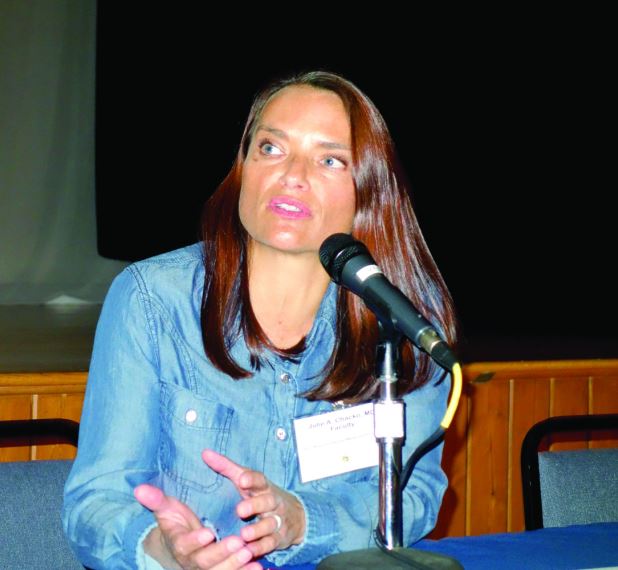
ESTES PARK, CO. – When patients with interstitial cystitis (IC) learn that first-line therapy is a rigorous diet designed to eliminate common bladder irritants, they tend to react in one of two ways, according to Julie A. Chacko, MD, a urologist in private practice in Santa Barbara, Calif.
Some “are just so grateful that they’re not crazy, which is what they’ve been told after 15 negative urine cultures. (Others) “look at the diet and think I’m sentencing them to death,” she said.
The sole medication approved by the Food and Drug Administration for IC is pentosan polysulfate sodium (Elmiron), and it should be reserved for the minority of patients who don’t experience significant improvement after giving the diet a reasonable shot, Dr. Chako advised. “When Elmiron works it’s great, but it’s not usually my go-to agent because it’s very expensive, you have to take it for 3-6 months to know for sure if it’s efficacious, and it has to be taken on an empty stomach. It’s a difficult medication.”
She advises patients to work with the diet. “Over time, they’re going to be able to find what I call their island – a point where they know very well their limitations and become quite comfortable with them,” she said at a conference on internal medicine sponsored by the University of Colorado.
A poorly understood yet common disorder, IC has a prevalence estimated at 0.5%-4% in women, less in men. Although typically diagnosed in the fourth decade or later, IC occurs at all ages. In some studies, the delay from first appearance of symptoms to arrival at a diagnosis is up to 8 years.
Interstitial cystitis is increasingly being called bladder pain syndrome in the literature, said Dr. Chako, who added, “I personally don’t love bladder pain syndrome as a description for this process. This syndrome has variable symptoms, and patients can have no pain at all.”
The mechanisms that result in IC are a mystery. The leading theory is that a bladder permeability problem allows urinary irritants to reach the interstitium. Nearly 80% of patients with IC can, with coaxing, identify dietary triggers for their symptoms, thereby basically establishing the diagnosis.
Other proposed mechanisms include an infectious agent that’s yet to be identified, allergic reaction, and neuromodulatory dysfunction. Common triggers other than foods include menses, copulation, emotional distress, and bladder trauma, including transvaginal ultrasound.
Conditions commonly associated with IC include fibromyalgia, irritable bowel syndrome, chronic fatigue, vulvodynia, migraines, depression, and anxiety.
The most common symptoms of IC are urinary urgency and frequency. Many affected patients have dysuria. Some have pain, which is typically suprapubic. However, pain can be present anywhere in a band circumscribing the whole central section of the torso, including the lower back, lower abdomen, urethra, vagina, and vulva. Patients describe a range of pain – burning, aching, stabbing, itching, buzzing, or a feeling of pressure.
“Most women who come in with IC are married to the idea that they’re having recurrent UTIs. They’re going to get antibiotics any way they can for their UTIs: over the phone, at urgent care. You need to get them to buy into the idea that even though UTIs are common, maybe not all of their flares are infections. They ask, ‘Then why do I feel better when I’m on antibiotics for recurrent UTI even though the cultures are negative?’ I say, ‘You feel less stress and anxiety because you think you’re on effective treatment,” Dr. Chacko said.
The diagnosis of IC is one of exclusions. Diagnoses to rule out before arriving at IC include recurrent UTI; overactive bladder, which should present with pure urge frequency and respond to medications for that condition; kidney stone disease present at the end of the ureter where it enters the bladder; gastrointestinal pathology; bladder cancer; and ovarian or uterine pathology.
Referral to a urologist for cystoscopy and cytology is appropriate in patients with microscopic hematuria, a significant smoking history predisposing to bladder cancer, or severe pain with severe frequency, which raises the possibility of Hunner’s ulcers, considered pathognomic for IC, respond “beautifully” to fulguration, she said.
Otherwise, IC can readily be managed by interested primary care physicians. The IC diet initially calls for 2 weeks of strict avoidance of all high-risk foods, most of which are acidic foods. These include fruits and fruit juices, especially citrus and cranberry juices; tomatoes and tomato products, including ketchup; yogurt; chocolate; coffee and tea, including decaf; vinegar; spicy foods; and carbonated beverages, water included.
These foods can later be added back one at a time to the diet while watching for IC flares, which typically occur within hours to several days of re-introducing the food. The return to coffee consumption, if that’s something important to the patient, should be with low-acid coffee. If that triggers an IC flare, try decaf. In time, many patients find they can consume some trigger foods in modest amounts.
“I tell patients it will take 12-18 months to get a good handle on their IC,” Dr. Chacko noted.
The use of OTC alkalizing agents such as Prelief may diffuse dietary triggers. A teaspoon of baking soda in water is also effective.
Second-line treatments include oral hydroxyzine 10-20 mg at bedtime; amitriptyline 10-20 mg at bedtime, mainly for patients with predominant pain symptoms; cimeditine; and pentosan polysulfate at 100 mg TID.
For IC patients with pelvic muscle tightness on pelvic examination, referral to a physical therapist adept at pelvic floor trigger point release can work wonders, she added.
One second-line option is bladder instillations of dimethyl sulfoxide weekly for 6 weeks, cutting back to once monthly maintenance therapy if the more intensive regimen is effective. Instillation of “heparin with lidocaine is a rescue solution. If it’s going to work, it kicks in within a few hours and usually lasts for 24-72 hours. It gets patients through a weekend, a wedding, or a funeral. A response can help make the IC diagnosis, too,” Dr. Chacko said.
She reported having no financial conflicts of interest regarding her presentation.
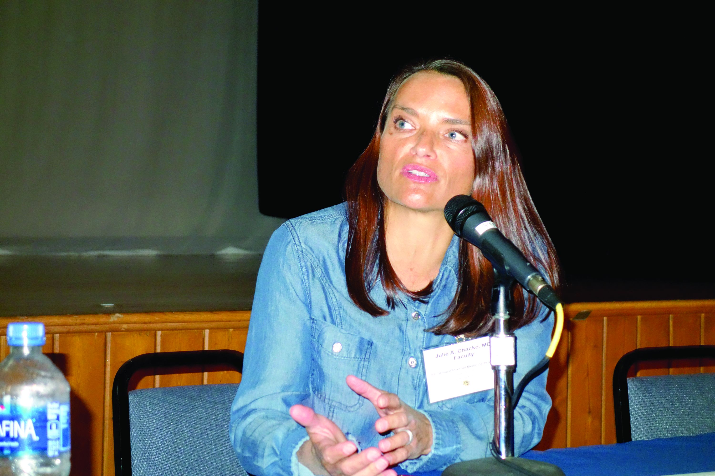
EXPERT ANALYSIS FROM THE ANNUAL INTERNAL MEDICINE PROGRAM
Opioid use disorder: Simplifying diagnosis and treatment in primary care
ESTES PARK, COLO. – Have a low threshold for diagnosing opioid use disorder in chronic pain patients, Joshua Blum, MD, advised at a conference on internal medicine sponsored by the University of Colorado.
Be alert to so-called ‘chemical copers’ as they skate through your practice, he said. “Maybe they call in here and there for an early refill; maybe they go to the ER and get a few pills here and there. But they never really surface to the level where we recognize them as an opioid use disorder patient.
“Some of them cross that line where they go from use and maybe intermittent misuse to meeting the criteria for opioid use disorder. I think we underdiagnose this in our chronic pain patients. I spend a lot of time trying to convince patients who tell me I just need to take their pain seriously that, yes, you’re in pain, and you have a pain diagnosis, but you also have an opioid use disorder. Medicalizing it makes things a lot easier for them; it helps take away the blame,” said Dr. Blum, program coordinator for the HIV primary care clinic at Denver Health.

Chemical copers, a well-established term in addiction medicine, are not the glaringly obvious substance abusers or addicts. “They’re the ones in your practice who are on four or five different psychoactive drugs. They’ve never met a psychoactive drug they didn’t like. When they hurt their back, they’re on Flexoril [cyclobenzaprine]; they’re on an antidepressant, a neuropathic pain agent; they’re on a sedative or sleeping agent or trazadone. If you took the five pills they’re on, you’d be knocked out for 2 days. And they always go to the maximum dose,” he said.
As newly described in the the Diagnostic and Statistical Manual of Mental Disorders, DSM-5, the diagnosis of opioid use disorder (OUD) requires that any 2 of the following 11 criteria be met:
- Taking more opioids than intended.
- Unsuccessful efforts to control opioid use.
- Spending a great deal of time in activities aimed at obtaining, using, or recovering from the effects of opioids.
- Craving opioids.
- Failure to fulfill major work, school, or home obligations because of opioid use.
- Worsening interpersonal problems related to opioid use.
- Giving up or reducing involvement in important social, recreational, or occupational activities because of opioid use.
- Recurrent use in situations where it’s physically hazardous, such as driving under the influence.
- Continued use despite physical or psychological problems stemming from opioid use.
- And finally, two special criteria applicable only if the opioid wasn’t prescribed and therefore isn’t being used under medical supervision: tolerance for opioids and withdrawal symptoms when they aren’t taken.
“Even if you can’t remember all these criteria, all you basically have to remember is ‘control.’ Many of these criteria describe situations where the patient is losing control of the drug. When they’re not controlling their drug use, the drug use is controlling them – and that’s addiction. All you need is for a patient to tell you ‘I tried to cut back on these drugs and I couldn’t,’ and that they’re experiencing some health consequences related to use yet still want to stay on the drugs, and, boom, they meet criteria for at least mild opioid use disorder,” he explained.
The standard treatment for OUD is opioid replacement therapy using methadone or buprenorphine (Subutex).
This approach is evidence-based therapy, Dr. Blum said, citing a recent meta-analysis of studies totaling nearly 123,000 opioid-dependent patients treated long-term with methadone and 16,000 treated with buprenorphine. The risk of all-cause mortality dropped by two-thirds when patients went on methadone, from 36.1% while out of treatment to 11.3% while on treatment. Similarly, all-cause mortality was 4.3% in patients on buprenorphine, compared with 9.5% in those out of treatment (BMJ. 2017 Apr 26;357:j1550. doi: 10.1136/bmj.j1550).
Access to methadone for treatment of OUD is available only through authorized methadone clinics, typically found only in big cities. But buprenorphine is a very useful alternative to methodone, Dr. Blum said.
“Buprenorphine is a schedule III drug that’s safe to prescribe in an office-based setting. It’s a partial mu opioid agonist with a ceiling effect, so people using buprenorphine can’t die from taking excessive amounts of it. You can even write refills on the prescription. And in head-to-head studies, it looks about as effective as moderate-dose methodone at 60 mg/day,” he said.
Opioid replacement therapy reduces euphoria and extinguishes craving. When methadone or buprenorphine is on board, saturating the opioid receptors, patients can use prescription or illicit opioids, but they won’t get high.
“People can really get their brains back online again,” Dr. Blum said.
Opioid maintenance therapy is consistent with the principles of harm reduction, a philosophy Dr. Blum said he embraces. Harm reduction can be summarized as “a set of practical strategies that reduce negative consequences of drug use, incorporating a spectrum of strategies from safer use, to managed use, to abstinence,” according to the Harm Reduction Coalition.
“Primary care doctors are very used to meeting patients where they are. We don’t require perfectionism from our patients. We don’t withhold insulin from diabetic patients because they’re not exercising, for example. In the case of drug use, there is complete abstinence on one end and really severe misuse on the other, and there’s a whole lot of life that happens in the middle. We’re addressing that part in the middle,” Dr. Blum said.
Dr. Blum reported having no financial conflicts of interest regarding his presentation.
ESTES PARK, COLO. – Have a low threshold for diagnosing opioid use disorder in chronic pain patients, Joshua Blum, MD, advised at a conference on internal medicine sponsored by the University of Colorado.
Be alert to so-called ‘chemical copers’ as they skate through your practice, he said. “Maybe they call in here and there for an early refill; maybe they go to the ER and get a few pills here and there. But they never really surface to the level where we recognize them as an opioid use disorder patient.
“Some of them cross that line where they go from use and maybe intermittent misuse to meeting the criteria for opioid use disorder. I think we underdiagnose this in our chronic pain patients. I spend a lot of time trying to convince patients who tell me I just need to take their pain seriously that, yes, you’re in pain, and you have a pain diagnosis, but you also have an opioid use disorder. Medicalizing it makes things a lot easier for them; it helps take away the blame,” said Dr. Blum, program coordinator for the HIV primary care clinic at Denver Health.
Chemical copers, a well-established term in addiction medicine, are not the glaringly obvious substance abusers or addicts. “They’re the ones in your practice who are on four or five different psychoactive drugs. They’ve never met a psychoactive drug they didn’t like. When they hurt their back, they’re on Flexoril [cyclobenzaprine]; they’re on an antidepressant, a neuropathic pain agent; they’re on a sedative or sleeping agent or trazadone. If you took the five pills they’re on, you’d be knocked out for 2 days. And they always go to the maximum dose,” he said.
As newly described in the the Diagnostic and Statistical Manual of Mental Disorders, DSM-5, the diagnosis of opioid use disorder (OUD) requires that any 2 of the following 11 criteria be met:
- Taking more opioids than intended.
- Unsuccessful efforts to control opioid use.
- Spending a great deal of time in activities aimed at obtaining, using, or recovering from the effects of opioids.
- Craving opioids.
- Failure to fulfill major work, school, or home obligations because of opioid use.
- Worsening interpersonal problems related to opioid use.
- Giving up or reducing involvement in important social, recreational, or occupational activities because of opioid use.
- Recurrent use in situations where it’s physically hazardous, such as driving under the influence.
- Continued use despite physical or psychological problems stemming from opioid use.
- And finally, two special criteria applicable only if the opioid wasn’t prescribed and therefore isn’t being used under medical supervision: tolerance for opioids and withdrawal symptoms when they aren’t taken.
“Even if you can’t remember all these criteria, all you basically have to remember is ‘control.’ Many of these criteria describe situations where the patient is losing control of the drug. When they’re not controlling their drug use, the drug use is controlling them – and that’s addiction. All you need is for a patient to tell you ‘I tried to cut back on these drugs and I couldn’t,’ and that they’re experiencing some health consequences related to use yet still want to stay on the drugs, and, boom, they meet criteria for at least mild opioid use disorder,” he explained.
The standard treatment for OUD is opioid replacement therapy using methadone or buprenorphine (Subutex).
This approach is evidence-based therapy, Dr. Blum said, citing a recent meta-analysis of studies totaling nearly 123,000 opioid-dependent patients treated long-term with methadone and 16,000 treated with buprenorphine. The risk of all-cause mortality dropped by two-thirds when patients went on methadone, from 36.1% while out of treatment to 11.3% while on treatment. Similarly, all-cause mortality was 4.3% in patients on buprenorphine, compared with 9.5% in those out of treatment (BMJ. 2017 Apr 26;357:j1550. doi: 10.1136/bmj.j1550).
Access to methadone for treatment of OUD is available only through authorized methadone clinics, typically found only in big cities. But buprenorphine is a very useful alternative to methodone, Dr. Blum said.
“Buprenorphine is a schedule III drug that’s safe to prescribe in an office-based setting. It’s a partial mu opioid agonist with a ceiling effect, so people using buprenorphine can’t die from taking excessive amounts of it. You can even write refills on the prescription. And in head-to-head studies, it looks about as effective as moderate-dose methodone at 60 mg/day,” he said.
Opioid replacement therapy reduces euphoria and extinguishes craving. When methadone or buprenorphine is on board, saturating the opioid receptors, patients can use prescription or illicit opioids, but they won’t get high.
“People can really get their brains back online again,” Dr. Blum said.
Opioid maintenance therapy is consistent with the principles of harm reduction, a philosophy Dr. Blum said he embraces. Harm reduction can be summarized as “a set of practical strategies that reduce negative consequences of drug use, incorporating a spectrum of strategies from safer use, to managed use, to abstinence,” according to the Harm Reduction Coalition.
“Primary care doctors are very used to meeting patients where they are. We don’t require perfectionism from our patients. We don’t withhold insulin from diabetic patients because they’re not exercising, for example. In the case of drug use, there is complete abstinence on one end and really severe misuse on the other, and there’s a whole lot of life that happens in the middle. We’re addressing that part in the middle,” Dr. Blum said.
Dr. Blum reported having no financial conflicts of interest regarding his presentation.
EXPERT ANALYSIS AT THE ANNUAL INTERNAL MEDICINE PROGRAM
Poor smell predicts 6-year risk of Parkinson’s
Poor olfaction is a predictor of Parkinson’s disease (PD) in both the short and immediate term, with accuracy out to at least 6 years, especially in white men, according to a retrospective case adjudication of the Health ABC study published in Neurology.
Of 2,462 study participants, 42 were found to have PD during an average of 9.8 years of follow-up, Honglei Chen, MD, PhD, of the department of epidemiology and biostatistics at Michigan State University, East Lansing, and his coauthors wrote. Of those cases, 26 (62%) were associated with a poor Brief Smell Identification Test (BSIT) score of 0-8 out of 12 (hazard ratio, 5.1; 95% confidence interval, 2.1-11.9), supporting other studies that indicate that olfactory impairment is one of the early symptoms of PD.

In contrast to previous studies, however, “the current study has longer follow-up and showed that poor olfaction was associated with PD beyond the first [4] years of follow-up, although the association was somewhat attenuated,” the researchers said. Both before and after the first 5 years of follow-up, 13 people in the lowest BSIT tertile were diagnosed with PD (HR, 4.2; 95% CI, 1.7-10.8 vs. HR, 4.1; 95% CI, 1.7-9.8, respectively),
In addition, poor sense of smell was more clearly associated with PD diagnosis in white patients, Dr. Chen and her coauthors said. Of 30 PD diagnoses in white participants, 19 were associated with poor smell (HR, 4.9; 95% CI, 2.3-10.5), compared with 7 out of 12 diagnoses in black participants (HR, 2.5; 95% CI, 0.8-8.1; P = .3). Similarly, 19 out of 26 diagnoses in men were associated with poor smell (HR, 5.4; 95% CI, 2.3-12.9), compared with 7 out of 16 diagnoses in women (HR, 2.9; 95% CI, 1.1-7.8; P = .4).
Future studies should confirm these stratified findings. Olfactory dysfunction’s “potential usefulness as a screening tool for prodromal or undiagnosed PD in other general population awaits further investigations,” the authors said.
Dr. Chen reported editorial roles for a number of medical journals. None of the authors reported any financial disclosures.
email address
Olfactory dysfunction is high in Parkinson’s disease, and the study by Chen et al. is the first to describe the natural history of olfactory dysfunction in the evolution of PD prior to diagnosis in diverse populations with extended periods of follow-up.
The length of follow-up is the longest to date, and includes important data on a large cohort of black participants. The results are a start for creating a much needed preventional paradigm, but more granularity will be needed to determine how poor smell, especially as stratified by race and sex, might help in prevention and treatment of PD.
Gene L. Bowman, ND, MPH, is a physician in the department of neurology at Oregon Health & Science University, Portland. His editorial comments accompanied the Chen et al. article in Neurology. He reported no financial disclosures.
Poor olfaction is a predictor of Parkinson’s disease (PD) in both the short and immediate term, with accuracy out to at least 6 years, especially in white men, according to a retrospective case adjudication of the Health ABC study published in Neurology.
Of 2,462 study participants, 42 were found to have PD during an average of 9.8 years of follow-up, Honglei Chen, MD, PhD, of the department of epidemiology and biostatistics at Michigan State University, East Lansing, and his coauthors wrote. Of those cases, 26 (62%) were associated with a poor Brief Smell Identification Test (BSIT) score of 0-8 out of 12 (hazard ratio, 5.1; 95% confidence interval, 2.1-11.9), supporting other studies that indicate that olfactory impairment is one of the early symptoms of PD.
In contrast to previous studies, however, “the current study has longer follow-up and showed that poor olfaction was associated with PD beyond the first [4] years of follow-up, although the association was somewhat attenuated,” the researchers said. Both before and after the first 5 years of follow-up, 13 people in the lowest BSIT tertile were diagnosed with PD (HR, 4.2; 95% CI, 1.7-10.8 vs. HR, 4.1; 95% CI, 1.7-9.8, respectively),
In addition, poor sense of smell was more clearly associated with PD diagnosis in white patients, Dr. Chen and her coauthors said. Of 30 PD diagnoses in white participants, 19 were associated with poor smell (HR, 4.9; 95% CI, 2.3-10.5), compared with 7 out of 12 diagnoses in black participants (HR, 2.5; 95% CI, 0.8-8.1; P = .3). Similarly, 19 out of 26 diagnoses in men were associated with poor smell (HR, 5.4; 95% CI, 2.3-12.9), compared with 7 out of 16 diagnoses in women (HR, 2.9; 95% CI, 1.1-7.8; P = .4).
Future studies should confirm these stratified findings. Olfactory dysfunction’s “potential usefulness as a screening tool for prodromal or undiagnosed PD in other general population awaits further investigations,” the authors said.
Dr. Chen reported editorial roles for a number of medical journals. None of the authors reported any financial disclosures.
email address
FROM NEUROLOGY
Key clinical point:
Major finding: Both before and after the first 5 years of follow-up, 13 people in the lowest BSIT tertile were diagnosed with PD (HR, 4.2; 95% CI, 1.7-10.8 vs. HR, 4.1; 95% CI, 1.7-9.8, respectively).
Data source: A retrospective case adjudication of PD cases diagnosed in the Health ABC study.
Disclosures: Dr. Chen reported editorial roles for a number of medical journals. None of the authors reported any financial disclosures.
Study finds low risk for jaw osteonecrosis with denosumab for postmenopausal osteoporosis
AT ASBMR
DENVER – Osteonecrosis of the jaw (ONJ) was a rare adverse event in women taking denosumab for postmenopausal osteoporosis, with a 0.7% rate for women who reported an invasive oral procedure or event while taking the drug and a 0.05% rate for women who did not have such procedures, Nelson Watts, MD, reported at the annual meeting of the American Society of Bone and Mineral Research.
The finding comes from a new analysis of a 7-year extension study of denosumab use in 4,550 women who participated in the 3-year, double-blind, phase 3 FREEDOM trial (NCT00089791) that compared denosumab 60 mg and placebo every 6 months. Those who missed 1 dose or fewer and completed visits through year 3 of the initial study were eligible to continue in the 7-year, open-label extension study. Those who had received placebo in the initial trial were crossed over to denosumab for the extension study.
Extension study participants were instructed to chronicle invasive oral procedures and events that had occurred in the initial trial and completed an oral event questionnaire once every 6 months of the extension trial.
All surveys were completed by 3,591 (79%) of the extension study participants, and 45.1% reported at least one invasive oral procedure or event during that time. The frequency of events was similar for the crossover and long-term denosumab groups; these events included scaling or root planing (29.1% and 28.5%), tooth extraction (25.1% and 24.6%), dental implant (5.8% and 6.0%), natural tooth loss (4.2% and 4.0%), and jaw surgery (0.9% and 0.9%). ONJ occurred at a rate of 5.2 cases per 10,000 patient-years of denosumab use, said Nelson Watts, MD, director of osteoporosis and bone health services at Mercy Health Services in Cincinnati, Ohio.
Of the 12 ONJ cases identified in the study, 11 occurred in women who reported an invasive oral procedure or event. This translated to a 0.7% risk of ONJ in women who reported an invasive oral procedure or event (11 in 1,621) and a 0.05% risk in women who did not (1 in 1,970).
The most common inciting event for ONJ appeared to be dental extractions, often of two or three teeth. The next most common dental issue associated with ONJ seemed to be poorly-fitted dentures.

ONJ resolved with treatment in 10 of 12 cases; one case was ongoing at the end of the study and one had an unknown outcome because the subject had withdrawn from the study. “With effective dental therapy, healing is the most likely outcome,” said Dr. Watts.
In clinical trials, ONJ occurred at a rate between 1 and 10 per 10,000 patient-years. A report in 2003, however, described severe ONJ in 36 cancer patients who received bisphosphonates (https://www.ncbi.nlm.nih.gov/pubmed/12966493).
The denosumab doses that cancer patients receive can be 10 to 12 times higher than the typical dose given to a postmenopausal woman being treated for osteoporosis.
“I can’t tell you how many phone calls I get from patients who are worried somehow or worried in situations created by their dentists that whatever procedure they’re going to have is going to end horribly,” Dr. Watts said. “In some cases dentists are telling my patients to either stop the drug that I’m giving them or to wait to get the next dose, and there’s absolutely nothing to support that.”
The study was funded by Amgen, the maker of denosumab (Prolia). Dr. Watts has received research support from Shire and has consulted for Abbvie, Amgen, and Radius. He is on the speakers’ bureau for Amgen, Radius, and Shire.
AT ASBMR
DENVER – Osteonecrosis of the jaw (ONJ) was a rare adverse event in women taking denosumab for postmenopausal osteoporosis, with a 0.7% rate for women who reported an invasive oral procedure or event while taking the drug and a 0.05% rate for women who did not have such procedures, Nelson Watts, MD, reported at the annual meeting of the American Society of Bone and Mineral Research.
The finding comes from a new analysis of a 7-year extension study of denosumab use in 4,550 women who participated in the 3-year, double-blind, phase 3 FREEDOM trial (NCT00089791) that compared denosumab 60 mg and placebo every 6 months. Those who missed 1 dose or fewer and completed visits through year 3 of the initial study were eligible to continue in the 7-year, open-label extension study. Those who had received placebo in the initial trial were crossed over to denosumab for the extension study.
Extension study participants were instructed to chronicle invasive oral procedures and events that had occurred in the initial trial and completed an oral event questionnaire once every 6 months of the extension trial.
All surveys were completed by 3,591 (79%) of the extension study participants, and 45.1% reported at least one invasive oral procedure or event during that time. The frequency of events was similar for the crossover and long-term denosumab groups; these events included scaling or root planing (29.1% and 28.5%), tooth extraction (25.1% and 24.6%), dental implant (5.8% and 6.0%), natural tooth loss (4.2% and 4.0%), and jaw surgery (0.9% and 0.9%). ONJ occurred at a rate of 5.2 cases per 10,000 patient-years of denosumab use, said Nelson Watts, MD, director of osteoporosis and bone health services at Mercy Health Services in Cincinnati, Ohio.
Of the 12 ONJ cases identified in the study, 11 occurred in women who reported an invasive oral procedure or event. This translated to a 0.7% risk of ONJ in women who reported an invasive oral procedure or event (11 in 1,621) and a 0.05% risk in women who did not (1 in 1,970).
The most common inciting event for ONJ appeared to be dental extractions, often of two or three teeth. The next most common dental issue associated with ONJ seemed to be poorly-fitted dentures.
ONJ resolved with treatment in 10 of 12 cases; one case was ongoing at the end of the study and one had an unknown outcome because the subject had withdrawn from the study. “With effective dental therapy, healing is the most likely outcome,” said Dr. Watts.
In clinical trials, ONJ occurred at a rate between 1 and 10 per 10,000 patient-years. A report in 2003, however, described severe ONJ in 36 cancer patients who received bisphosphonates (https://www.ncbi.nlm.nih.gov/pubmed/12966493).
The denosumab doses that cancer patients receive can be 10 to 12 times higher than the typical dose given to a postmenopausal woman being treated for osteoporosis.
“I can’t tell you how many phone calls I get from patients who are worried somehow or worried in situations created by their dentists that whatever procedure they’re going to have is going to end horribly,” Dr. Watts said. “In some cases dentists are telling my patients to either stop the drug that I’m giving them or to wait to get the next dose, and there’s absolutely nothing to support that.”
The study was funded by Amgen, the maker of denosumab (Prolia). Dr. Watts has received research support from Shire and has consulted for Abbvie, Amgen, and Radius. He is on the speakers’ bureau for Amgen, Radius, and Shire.
Meningitis B vaccine’s high price tag poses a health care conundrum
Four years ago, when meningitis B, an extremely rare but potentially lethal form of the infection, sickened a small number of college students at Princeton and the University of California–Santa Barbara, there was no vaccine against the disease sold in the U.S. Despite its availability abroad, it had never been licensed in the country due to its limited marketability.
Scientific evidence supporting an absolute need to immunize against meningitis B still falls short. The risk of contracting it is smaller than that of being involved in a car crash.
But the headlines prompted by those 13 campus cases – which resulted in one death and one double amputation – helped reshape the financial prospects for a vaccine.
Today, two brand-name vaccines, both with price tags of more than $300, are widely advertised on television and touted as a smart investment for parents who love their college-bound kids.
“As moms, we send our kids out into the world, full of hope,” says a mother in the ad for Bexsero, sold by pharmaceutical giant GlaxoSmithKline, as her son loads up the car to go off to college.
Says another voice, “And we don’t want something like meningitis B getting in their way.”
Analysts expect the two vaccines to generate annually at least hundreds of millions of dollars in global sales.
As new crops of students head to college, some physicians and other industry experts, though, are growing uneasy about the role of marketing in leveraging parental fears to sell the MenB vaccine – as well as ever more expensive vaccines that prevent quite rare illnesses. A complete Bexsero series costs $320; a competing vaccine, Trumenba, costs $345.
“Parents believe their children are susceptible to this terrible condition, and [drugmakers] use that fear to get parents to take action,” said Adrienne Faerber, a lecturer at the Dartmouth Institute for Health Policy and Clinical Practice who researches drug marketing.
The advertising, especially when coupled with news coverage, puts parents in a quandary left unresolved by federal vaccination guidelines and university requirements.
The Centers for Disease Control and Prevention recommends doctors consider the meningitis B vaccine for people ages 16-23 years on an individual basis. This recommendation – ranked as a category B – is not as universal as the approach applied to illnesses such as measles or human papillomavirus vaccines or even the “quadrivalent” vaccine for meningitis A, C, W, and Y, which all students must get.
Meanwhile, insurers generally cover it as part of preventive care. Still, most universities don’t require the vaccine but simply list it as an option for families to consider.
The resulting messages can confuse parents.
“There is perhaps, with all the marketing and advertising, some bending of the truth, and perhaps a little bit of creating fear – again recognizing that meningitis disease is a very severe disease,” said William Moss, a professor at Johns Hopkins Bloomberg School of Public Health who specializes in vaccines and global children’s health. “[The risk] is not a large enough problem to warrant routine vaccination.”
In recent years, drugmakers’ interests have begun to expand beyond the relatively cheap, broadly used immunizations – such as a tetanus shot or the children’s hepatitis A vaccine – to new, much pricier ones for less common infections.
These newer treatments have the potential to transform what’s long been a less lucrative side of drug production, manufacturing vaccines, into a major cash cow. But since the newer vaccines are regarded as less crucial than, say, preventing measles – and are often not required – marketing has become a big part of the sales equation.
Bexsero and its competitor, Trumenba, offer clues into how this scenario plays out.
Both vaccines got accelerated approval by the Food and Drug Administration in 2015 and 2014 respectively, following the Princeton and UCSB outbreaks.
Meningitis B does not spread readily from person to person. It requires close physical contact, like kissing or sharing utensils. It can be fatal but is treatable with antibiotics if caught early. Caused by the B serogroup of the meningococcal infection, it tends to appear in rare-scatter, slowed, self-limited outbreaks on college campuses. The standard meningitis vaccine doesn’t prevent it.
After new cases at Princeton and UC Santa Barbara kept appearing over many months, the CDC arranged for an emergency import of Bexsero. All students on those campuses got the shots, and there were no more cases.
Now the drugmakers are urging all parents to be proactive. Last year, Pfizer put more than $21 million into paid advertisements for the vaccine, according to figures kept by Kantar Media, a firm that tracks multimedia advertising. GlaxoSmithKline put in just about $79,000.
Those figures don’t account for other efforts such as meningitis awareness and ongoing social media campaigns done by GlaxoSmithKline, a “substantial effort” that “wasn’t cheap,” said Sriram Jambunathan, who heads GlaxoSmithKline’s meningococcal franchise in the United States. They also don’t include Pfizer’s investments in similar activities.
Already, industry analysts forecast Bexsero could bring in global revenue north of $1 billion per year by 2022. Trumenba is expected to earn Pfizer $880 million by that time.
But the industry’s gain may come at the expense of efficient health care spending and inflated consumer concern.
First, there’s the relative rarity of meningitis B. The CDC has estimated fewer than 300 cases occur in the United States per year, and some medical experts interviewed suggested the number may be closer to 50 or 60.
“As a mom, I would say, if my kid got this disease, and I had had the opportunity to prevent it, and I didn’t, I would kill myself,” said Martha Arden, a practicing physician and the medical director of Mount Sinai Adolescent Health Center’s school-based health program in New York City. “But the odds are small. It’s much more dangerous to send a kid out skiing than it is to not give the vaccine.”
Jambunathan said the price tag is warranted given the resources needed to bring Bexsero to market. Similar vaccines, he added, are comparably priced, and firms won’t necessarily want to develop these pharmaceuticals if they aren’t sure they can recoup their investment.
For parents who opt for the vaccines, there are caveats. Researchers don’t know, for instance, how long its immunity lasts. Many noted it also doesn’t cover all strains of the infection, so its efficacy in the United States is uncertain (there are different strains in different parts of the world).
And the cost of vaccination, while substantial, isn’t immediately felt by consumers because the treatment usually is covered without having to pay out-of-pocket. But the price tag may contribute to increasing premium costs.
In a world where there already aren’t enough health care dollars to address every possible harm, many experts noted, other health concerns might be a smarter investment.
Still, the price tag might not cause parents to blink. “When it’s your child or one case you know about, suddenly the health economic arguments feel difficult to have,” Jambunathan said.
Kaiser Health News is a national health policy news service that is part of the nonpartisan Henry J. Kaiser Family Foundation.
In October 2014 and January 2015, the Food and Drug Administration licensed two meningococcal serogroup B vaccines for administration in adolescents and young adults aged 10-25 years based on each vaccine's ability to elicit bactericidal antibody against the majority of invasive serogroup B strains and demonstrated safety. Each vaccine represented novel technology that overcame the challenge of both the poor immunogenicity of serogroup B polysaccharide protein conjugates and the potential cross reactivity with fetal brain tissue. In the United States, the vaccine was recommended (category A) for individuals in this age grouping with complement deficiency, anatomic or functional asplenia, outbreaks (when indicated), and for microbiologists. The Centers for Disease Control and Prevention also recommends that physicians consider the MenB vaccine for individuals aged 16-23 years who wish to obtain short-term protection against diverse strains of serogroup B meningococcal disease (category B). The American Academy of Pediatrics encouraged pediatricians to discuss the availability of the MenB vaccines with families.
The annual incidence of meningococcal disease varied between approximately 0.5-1.5 cases per 100,000 population between 1950 and 1990 - approximately 3,000 cases annually. Between 1990 and 2010, disease caused by the three common serogroups in the United States (B, C, and Y) declined to approximately 0.35 cases per 100,000. Subsequent to the introduction of a tetravalent meningococcal conjugate vaccine (MCV4) further declines - sustained over a longer time period than previously observed - have occurred, reaching a nadir of approximately 400 annual cases. Despite the absence of serogroup B component in MCV4, declines in serogroup B disease were reported in addition to vaccine serogroups C and Y. The biological explanations for the substantial decline in the overall rate of meningococcal disease are unknown. This decline in meningococcal serogroup B disease has created a controversy about implementation of Advisory Committee of Immunization Practices and the AAP and American Academy of Family Physician recommendations reflected in Shefali Luthra's writings.
There is widespread agreement about the severity of invasive meningococcal disease, the peaks of incidence in infancy and late adolescence, a 10% case fatality rate, an additional 10%-15% morbidity, and the limited number of cases (in the United States) to be prevented by adolescent immunization despite serogroup B being the most common. The effectiveness (greater than 80%) of at least one of these vaccines (MenB-4C) has been established in the United Kingdom, where it is recommended for all infants as part of a three-dose series at 2, 4, and 12 months. The value proposition (number of people immunized to prevent one death), however, is estimated at 1 million vaccinees for each death prevented.
Some experts believe the small burden of disease that might be prevented by these expensive vaccines requires thoughtful consideration in this era of increasingly limited resources. Others (as cited in the accompanying article) believe the marketing and advertising bend the truth and create fear in the public and conclude the risk is not great enough to warrant universal immunization (called category A by ACIP designation). In contrast, parent groups (especially those including parents of children who had meningococcal serogroup B disease) advocate strongly for a universal approach. For example, Alicia Stillman, director of the Emily Stillman Foundation, feels the current recommendation is "irresponsible" because it leaves so many teens and young adults vulnerable to the disease. The group believes that ACIP has made the menB vaccine to be an "optional item," but there is no requirement to provide the education to the parent and/or patient so they are aware of this option.
For me, the question is this: Who should decide how we use limited resources? I am reminded of an editorial in the New England Journal of Medicine titled, "The Meningococcal Vaccine - Public Policy and Individual Choices" by Paul Offit, MD, and Georges Peter, MD, that examined this question (N Engl J Med. 2003;349[24]:2353-6). They advocated that parents, if aware, may choose vaccination to protect adolescents and young adults from devastating infection, even if they were required to pay. I believe the CDC foresaw this as likely and moved to recommend individual choice. The wisdom of this was that category B status required MenB vaccine to be covered by insurers, thus preventing a potential divergent uptake, where families that could afford the price would recognize its value and those unable to pay for the vaccine would have no choice but to decline. This is especially relevant as there are not specific risk factors among healthy adolescents to warrant prioritizing one group over another.
MenB vaccines are valuable but costly tools for the prevention of life-threatening infectious disease. The use of increasingly limited resources, as raised by Dr. Moss and others, is a relevant and important question, and a call for a national dialogue.
As new medical breakthroughs increase, seemingly exponentially, how do we resolve the individual versus societal benefit of costly new treatments or preventions? How do we value prevention of life-threatening illness and death in mostly healthy adolescents, compared with treatment of end-stage diseases? These are important conversations that are only in their infancy.
Abraham Verghese wrote, in his book "Cutting for Stone," that American ambulance crews "salvaged people we'd never see in Missing [fictional hospital in Addis Ababa], because no one would have tried to bring them to a hospital [in Addis Ababa]. Judging someone to be beyond help never crossed the minds of police, firemen, or doctors here" in the United States. We need transparency and a national dialogue to develop consensus about priorities. We need to make sure the discussions are comprehensive and civil - not about pushing grandmothers over cliffs or death panels. Currently, ACIP and AAP have advocated for individual choice and to empower the parent and adolescent to choose after we (clinicians) communicate disease severity, the risk to the adolescent, and adverse events associated with MenB vaccine.
Stephen I. Pelton, MD, is chief of pediatric infectious disease and coordinator of the maternal-child HIV program at Boston Medical Center. Dr. Pelton disclosed that he has participated in advisory boards on meningitis B vaccines for GlaxoSmithKline and Pfizer, has research grants from Pfizer and Merck, and has spoken at CME events on meningitis B vaccines.
Four years ago, when meningitis B, an extremely rare but potentially lethal form of the infection, sickened a small number of college students at Princeton and the University of California–Santa Barbara, there was no vaccine against the disease sold in the U.S. Despite its availability abroad, it had never been licensed in the country due to its limited marketability.
Scientific evidence supporting an absolute need to immunize against meningitis B still falls short. The risk of contracting it is smaller than that of being involved in a car crash.
But the headlines prompted by those 13 campus cases – which resulted in one death and one double amputation – helped reshape the financial prospects for a vaccine.
Today, two brand-name vaccines, both with price tags of more than $300, are widely advertised on television and touted as a smart investment for parents who love their college-bound kids.
“As moms, we send our kids out into the world, full of hope,” says a mother in the ad for Bexsero, sold by pharmaceutical giant GlaxoSmithKline, as her son loads up the car to go off to college.
Says another voice, “And we don’t want something like meningitis B getting in their way.”
Analysts expect the two vaccines to generate annually at least hundreds of millions of dollars in global sales.
As new crops of students head to college, some physicians and other industry experts, though, are growing uneasy about the role of marketing in leveraging parental fears to sell the MenB vaccine – as well as ever more expensive vaccines that prevent quite rare illnesses. A complete Bexsero series costs $320; a competing vaccine, Trumenba, costs $345.
“Parents believe their children are susceptible to this terrible condition, and [drugmakers] use that fear to get parents to take action,” said Adrienne Faerber, a lecturer at the Dartmouth Institute for Health Policy and Clinical Practice who researches drug marketing.
The advertising, especially when coupled with news coverage, puts parents in a quandary left unresolved by federal vaccination guidelines and university requirements.
The Centers for Disease Control and Prevention recommends doctors consider the meningitis B vaccine for people ages 16-23 years on an individual basis. This recommendation – ranked as a category B – is not as universal as the approach applied to illnesses such as measles or human papillomavirus vaccines or even the “quadrivalent” vaccine for meningitis A, C, W, and Y, which all students must get.
Meanwhile, insurers generally cover it as part of preventive care. Still, most universities don’t require the vaccine but simply list it as an option for families to consider.
The resulting messages can confuse parents.
“There is perhaps, with all the marketing and advertising, some bending of the truth, and perhaps a little bit of creating fear – again recognizing that meningitis disease is a very severe disease,” said William Moss, a professor at Johns Hopkins Bloomberg School of Public Health who specializes in vaccines and global children’s health. “[The risk] is not a large enough problem to warrant routine vaccination.”
In recent years, drugmakers’ interests have begun to expand beyond the relatively cheap, broadly used immunizations – such as a tetanus shot or the children’s hepatitis A vaccine – to new, much pricier ones for less common infections.
These newer treatments have the potential to transform what’s long been a less lucrative side of drug production, manufacturing vaccines, into a major cash cow. But since the newer vaccines are regarded as less crucial than, say, preventing measles – and are often not required – marketing has become a big part of the sales equation.
Bexsero and its competitor, Trumenba, offer clues into how this scenario plays out.
Both vaccines got accelerated approval by the Food and Drug Administration in 2015 and 2014 respectively, following the Princeton and UCSB outbreaks.
Meningitis B does not spread readily from person to person. It requires close physical contact, like kissing or sharing utensils. It can be fatal but is treatable with antibiotics if caught early. Caused by the B serogroup of the meningococcal infection, it tends to appear in rare-scatter, slowed, self-limited outbreaks on college campuses. The standard meningitis vaccine doesn’t prevent it.
After new cases at Princeton and UC Santa Barbara kept appearing over many months, the CDC arranged for an emergency import of Bexsero. All students on those campuses got the shots, and there were no more cases.
Now the drugmakers are urging all parents to be proactive. Last year, Pfizer put more than $21 million into paid advertisements for the vaccine, according to figures kept by Kantar Media, a firm that tracks multimedia advertising. GlaxoSmithKline put in just about $79,000.
Those figures don’t account for other efforts such as meningitis awareness and ongoing social media campaigns done by GlaxoSmithKline, a “substantial effort” that “wasn’t cheap,” said Sriram Jambunathan, who heads GlaxoSmithKline’s meningococcal franchise in the United States. They also don’t include Pfizer’s investments in similar activities.
Already, industry analysts forecast Bexsero could bring in global revenue north of $1 billion per year by 2022. Trumenba is expected to earn Pfizer $880 million by that time.
But the industry’s gain may come at the expense of efficient health care spending and inflated consumer concern.
First, there’s the relative rarity of meningitis B. The CDC has estimated fewer than 300 cases occur in the United States per year, and some medical experts interviewed suggested the number may be closer to 50 or 60.
“As a mom, I would say, if my kid got this disease, and I had had the opportunity to prevent it, and I didn’t, I would kill myself,” said Martha Arden, a practicing physician and the medical director of Mount Sinai Adolescent Health Center’s school-based health program in New York City. “But the odds are small. It’s much more dangerous to send a kid out skiing than it is to not give the vaccine.”
Jambunathan said the price tag is warranted given the resources needed to bring Bexsero to market. Similar vaccines, he added, are comparably priced, and firms won’t necessarily want to develop these pharmaceuticals if they aren’t sure they can recoup their investment.
For parents who opt for the vaccines, there are caveats. Researchers don’t know, for instance, how long its immunity lasts. Many noted it also doesn’t cover all strains of the infection, so its efficacy in the United States is uncertain (there are different strains in different parts of the world).
And the cost of vaccination, while substantial, isn’t immediately felt by consumers because the treatment usually is covered without having to pay out-of-pocket. But the price tag may contribute to increasing premium costs.
In a world where there already aren’t enough health care dollars to address every possible harm, many experts noted, other health concerns might be a smarter investment.
Still, the price tag might not cause parents to blink. “When it’s your child or one case you know about, suddenly the health economic arguments feel difficult to have,” Jambunathan said.
Kaiser Health News is a national health policy news service that is part of the nonpartisan Henry J. Kaiser Family Foundation.
CB-5083 showed significant activity against B-ALL
A first-in-class oral inhibitor of valosin-containing protein (VCP) p97 showed significant activity against human B acute lymphoblastic leukemia (B-ALL) cells, including those with the most common fusion genes seen in pediatric and adult B-ALL, researchers reported.
The drug, dubbed CB-5083 (Cleave Biosciences), induces cell death by causing endoplasmic reticulum stress, to which B-ALL cells are “distinctly vulnerable,” wrote Gabriele Gugliotta, MD, PhD, of Cedars-Sinai Medical Center in Los Angeles, Calif., and her coinvestigators. In their study, CB-5083 significantly reduced the viability of 10 B-ALL cell lines tested, and did so at blood concentrations that caused no significant toxicities in mice. Human B-ALL cells also showed no signs of upfront drug resistance, the researchers wrote in Neoplasia.
CB-5083 received an FDA orphan drug designation in 2015 for treating multiple myeloma, and also has shown activity against solid tumors. In the study, exposing BALL1 and OP1 B-ALL cell lines to CB-5083 triggered “early and strong” apoptosis, along with a “robust” cleavage of PARP, the researchers reported (Neoplasia. 2017 Aug 24. doi: 10.1016/j.neo.2017.08.001). http://www.neoplasia.com/article/S1476-5586(17)30258-0/fulltext
The drug also reduced the viability of human B-ALL cells without genes for GRP78, GRP94, or XBP1, suggesting that CB-5083 does not require the presence of these proteins to work, the investigators said. Deficiency of X-box binding protein 1 (XBP1) predicted greater sensitivity to CB-5083, which might mean that XBP1 splicing counteracts drug activity by mitigating endoplasmic reticulum stress, they added. “Finally, vincristine was synergistic with CB-5083 in both BALL1 and OP1 [cell lines],” they wrote. “In summary, the targeting of p97 with CB-5083 is a novel promising therapeutic approach that should be further evaluated in B-ALL.”
The investigators acknowledged support from the Melamed family, Reuben Yeroushalmi, National Research Foundation Singapore, Singapore Ministry of Education, Leukemia Lymphoma Society of America, and University of Bologna, Italy. They did not report having conflicts of interest.
FROM NEOPLASIA
Key clinical point: A novel oral inhibitor of valosin-containing protein (VCP) p97 showed significant activity against human B acute lymphoblastic leukemia (B-ALL) cells.
Major finding: After exposure, BALL1 and OP1 cell lines showed early and strong indicators of apoptosis and robust cleavage of PARP.
Data source: In vitro studies of human B-ALL cells and in vivo studies in mice.
Disclosures: The investigators acknowledged support from the Melamed family, Reuben Yeroushalmi, National Research Foundation Singapore, Singapore Ministry of Education, Leukemia Lymphoma Society of America, and University of Bologna, Italy. They did not report having conflicts of interest.
CCSs have higher burden of chronic conditions

Adult survivors of childhood cancer have a greater cumulative burden of chronic health conditions than the general public, according to research published in The Lancet.
The study showed that, by age 50, childhood cancer survivors (CCSs) had experienced, on average, 17.1 chronic health conditions, and matched control subjects had experienced 9.2.
“The cumulative burden of chronic disease revealed in this analysis, along with the complexity and severity of chronic conditions some survivors experience, found childhood cancer survivors to be a vulnerable, medically complex population,” said study author Nickhill Bhakta, MD, of St. Jude Children’s Research Hospital in Memphis, Tennessee.
For this study, Dr Bhakta and his colleagues assessed the lifelong impact of 168 chronic health conditions—such as hepatic, thyroid, ocular, and reproductive disorders—on CCSs and control subjects.
The 3010 evaluable CCSs had survived 10 years or longer from their initial cancer diagnosis and were 18 years or older as of June 30, 2015. The 272 controls had no history of pediatric cancer and were matched to CCSs by age and sex.
At age 50, the cumulative incidence of chronic health conditions (of any grade) was 99.9% in CCSs and 96.0% in controls (P<0.0001). The cumulative incidence of grade 3 to 5 chronic health conditions was 96.0% and 84.9%, respectively (P<0.0001).
The cumulative burden for CCSs was 17.1 chronic health conditions, including 4.7 that were grade 3 to 5. For controls, the cumulative burden was 9.2 chronic health conditions, including 2.3 that were grade 3 to 5 (P<0.0001 for both comparisons).
The researchers said second neoplasms, spinal disorders, and pulmonary disease were major contributors to the excess total cumulative burden observed in CCSs. However, there was “notable heterogeneity” in burden according to the patients’ primary cancer diagnosis.
For instance, growth hormone deficiency was in the top 10th percentile of chronic health conditions for survivors of acute lymphoblastic leukemia but not for controls.
And pulmonary function deficits were in the top 10th percentile for survivors of acute myeloid leukemia and Hodgkin lymphoma but not for controls or survivors of acute lymphoblastic leukemia or non-Hodgkin lymphoma.
“This study found that the average childhood cancer survivor has a cumulative burden of chronic disease that requires a significant time investment by healthcare providers to disentangle and manage—time that community providers are unlikely to have,” Dr Bhakta said.
“The results suggest that childhood cancer survivors may benefit from the integrated, specialized healthcare delivery that is being tried for individuals infected with HIV or those with other complex, chronic health problems.” ![]()
How to tackle telangiectasias with light
AT MOAS 2017
SAN DIEGO – Multiple light-based devices can be used to treat telangiectasias, often with little or no down time.
During a presentation at the annual Masters of Aesthetics Symposium, Kristen M. Kelly, MD, listed her preferred light-based treatment options for telangiectasias as the pulsed dye laser (PDL), the 532 nm Nd:YAG laser, and the diode laser. The PDL has been around the longest and “is one of the greatest devices for treating vasculature, because it’s very specific,” she said. Purpura can result, but for cosmetic indications, pulse durations of 6 milliseconds or more work well. “Keep in mind, though, that when you’re using longer pulse durations, patients will often require more than one treatment,” she said. “Most people are okay with that, knowing that they’re going to have minimal to no down time with these procedures.”

Dr. Kelly, professor of dermatology and surgery at the University of California, Irvine, noted that erythematotelangiectatic rosacea generally responds well to light-based devices, while papulopustular/inflammatory forms of the condition are best treated with anti-inflammatory agents or antibiotics. Multiple passes or judicious pulse stacking can be used with PDL for treating telangiectases, “but always know what your endpoint is,” Dr. Kelly advised. “You generally don’t want to pulse over purpura when treating telangiectasias; that just increases the risk of adverse effects.”
Settings to consider vary, depending on the patient and the area to be treated. The desired endpoints for telangiectasia are vessel blanching or diminution, or a darker red/purple change to the vessel. “You should not see graying or whitening of the skin; that’s when you’re getting epidermal damage, so you want to watch for that,” she said. For a first pass, Dr. Kelly will often treat with a spot size of 10 mm or 12 mm with a 6-millisecond pulse duration with cryogen spray cooling set at 30 milliseconds with a 30-millisecond delay. If a second pass is required, she will often treat with a spot size of 7 mm and a 6-millisecond pulse duration and slightly higher fluence, again using cryogen spray cooling. “It is important to know your specific device to determine settings,” she said.
The frequency-doubled Nd:YAG laser features a wavelength of 532 nm but requires cautious use in patients with darker skin types or photodamaged skin. The desired endpoint is a darkening of the vessel or vessel disappearance. “Many of these devices have contact cooling, which is an excellent method of cooling, but you have to make sure you keep the contact,” she said. Other cooling options include cryogen spray and the application of cold air. “When you’re doing cold-air cooling, you want to be careful not to point it toward someone’s nose,” she said. “It sounds funny, but this is uncomfortable for the patient.”
Diode lasers with wavelengths of 800-980 nm allow the user to treat larger blood vessels, such as nasal telangiectasias. “Some devices with small spot sizes allow you to trace along the vessel, and you can see it disappear before your eyes, and when the patient leaves, they’re generally very happy,” she said.
Intense pulsed light works well in patients with both pigmentary and vascular changes, especially for poikiloderma with brown and red pigment. “Multiple treatments are required, and there is significant melanin absorption, so you don’t want to treat people with tanned skin,” she said.
Dr. Kelly emphasized the importance of being familiar with the device you use and knowing the desired tissue endpoint. Decisions depend on the patient’s skin type, their tolerance for down time and multiple treatments, and the devices at your disposal. “Patients should be aware of all options, even if you may not have a certain device in your office,” she said. “You can refer them.”
She disclosed having drugs or devices donated by Light Sciences Oncology, Solta Medical, Syneron Candela, ThermiRF, and Novartis. She is also a consultant for MundiPharma, Allergan, and Syneron Candela.
[email protected]
AT MOAS 2017
SAN DIEGO – Multiple light-based devices can be used to treat telangiectasias, often with little or no down time.
During a presentation at the annual Masters of Aesthetics Symposium, Kristen M. Kelly, MD, listed her preferred light-based treatment options for telangiectasias as the pulsed dye laser (PDL), the 532 nm Nd:YAG laser, and the diode laser. The PDL has been around the longest and “is one of the greatest devices for treating vasculature, because it’s very specific,” she said. Purpura can result, but for cosmetic indications, pulse durations of 6 milliseconds or more work well. “Keep in mind, though, that when you’re using longer pulse durations, patients will often require more than one treatment,” she said. “Most people are okay with that, knowing that they’re going to have minimal to no down time with these procedures.”
Dr. Kelly, professor of dermatology and surgery at the University of California, Irvine, noted that erythematotelangiectatic rosacea generally responds well to light-based devices, while papulopustular/inflammatory forms of the condition are best treated with anti-inflammatory agents or antibiotics. Multiple passes or judicious pulse stacking can be used with PDL for treating telangiectases, “but always know what your endpoint is,” Dr. Kelly advised. “You generally don’t want to pulse over purpura when treating telangiectasias; that just increases the risk of adverse effects.”
Settings to consider vary, depending on the patient and the area to be treated. The desired endpoints for telangiectasia are vessel blanching or diminution, or a darker red/purple change to the vessel. “You should not see graying or whitening of the skin; that’s when you’re getting epidermal damage, so you want to watch for that,” she said. For a first pass, Dr. Kelly will often treat with a spot size of 10 mm or 12 mm with a 6-millisecond pulse duration with cryogen spray cooling set at 30 milliseconds with a 30-millisecond delay. If a second pass is required, she will often treat with a spot size of 7 mm and a 6-millisecond pulse duration and slightly higher fluence, again using cryogen spray cooling. “It is important to know your specific device to determine settings,” she said.
The frequency-doubled Nd:YAG laser features a wavelength of 532 nm but requires cautious use in patients with darker skin types or photodamaged skin. The desired endpoint is a darkening of the vessel or vessel disappearance. “Many of these devices have contact cooling, which is an excellent method of cooling, but you have to make sure you keep the contact,” she said. Other cooling options include cryogen spray and the application of cold air. “When you’re doing cold-air cooling, you want to be careful not to point it toward someone’s nose,” she said. “It sounds funny, but this is uncomfortable for the patient.”
Diode lasers with wavelengths of 800-980 nm allow the user to treat larger blood vessels, such as nasal telangiectasias. “Some devices with small spot sizes allow you to trace along the vessel, and you can see it disappear before your eyes, and when the patient leaves, they’re generally very happy,” she said.
Intense pulsed light works well in patients with both pigmentary and vascular changes, especially for poikiloderma with brown and red pigment. “Multiple treatments are required, and there is significant melanin absorption, so you don’t want to treat people with tanned skin,” she said.
Dr. Kelly emphasized the importance of being familiar with the device you use and knowing the desired tissue endpoint. Decisions depend on the patient’s skin type, their tolerance for down time and multiple treatments, and the devices at your disposal. “Patients should be aware of all options, even if you may not have a certain device in your office,” she said. “You can refer them.”
She disclosed having drugs or devices donated by Light Sciences Oncology, Solta Medical, Syneron Candela, ThermiRF, and Novartis. She is also a consultant for MundiPharma, Allergan, and Syneron Candela.
[email protected]
Absorb bioresorbable scaffold folds
The first bioresorbable vascular scaffold to reach clinical practice is no longer available because of poor sales, following nearly a year of bad news for the innovative device.
"We took this decision for commercial reasons, not for safety," Kristina Becker, director of communications at Abbott's Vascular business. Abbott announced that sales of the Absorb scaffold would cease worldwide as of Sept. 14, 2017, adding that the company would continue to follow patients in existing clinical trials according to their protocols.
The device was approved by the Food and Drug Administration in July 2016, but soon after, long-term trial results took an ominous turn. In March 2017, the agency issued a safety alert regarding the Absorb scaffold after release of the 2-year data from the 2,008-patient ABSORB III trial showing a significantly higher rate of target-lesion failure than with the everolimus-eluting metallic Xience stent, also marketed by Abbott.
Meanwhile, 3-year results of the ABSORB II trial, reported last October, revealed a 1% per year rate of late stent thrombosis during both the second and third years following Absorb placement in coronary arteries, the period when the Absorb scaffold was in the process of disappearing. More bad news came in July, when device thrombosis occurred nearly four times more frequently in recipients of the Absorb scaffold than with the Xience stent during 2 years of prospective follow-up in the randomized, investigator-initiated AIDA trial.
Abbott is not giving up on the idea of nonpermanent stents. “We recognize that [bioresorbable scaffold] technologies offer patients the possibility of life without permanent metallic implants, and we will continue to work on a next generation device while monitoring long-term outcomes after stent resorption in current Absorb trials,” the announcement said.
This article was updated 9/8/17.