User login
AML candidate drug back in the pipeline
The Food and Drug Administration has given the biopharmaceutical company Cellectis permission to resume phase 1 trials of UCART123, a gene-edited T-cell investigational drug that targets CD123, as a potential treatment for acute myeloid leukemia (AML) and blastic plasmacytoid dendritic cell neoplasm (BPDCN), according to a press release from the company.
UCART123 is the first allogeneic, “off-the-shelf” gene-edited chimeric antigen receptor (CAR) T-cell product candidate that the FDA has approved for clinical trials. The agency had placed a clinical hold on phase 1 trials of the gene-edited CAR T-cell drug on Sept. 4, following a patient death in the BPDCN clinical study. In order to proceed with the trials, Cellectis agreed to several changes in the study protocols.
The changes include decreasing the dose of the UCART123 therapy to 6.25x10⁴ cells/kg and lowering the dose of the lympho-depleting regimen of cyclophosphamide to 750 mg/m2 per day over 3 days with a maximum daily dose of 1.33 g. Additionally, there can be no uncontrolled infection after receipt of the lympho-depleting preconditioning regimen. Patients must be afebrile at the start of treatment, off all but a replacement dose of corticosteroids, and have no organ dysfunction. Plus, the next three patients treated in each study must be under age 65.
There’s also a condition that patient enrollments be staggered by at least 28 days.
The drug sponsor is working with investigators and each clinical site to obtain the Institutional Review Board’s approval of the revised protocols.
The hold followed the death of a 78-year-old man with relapsed/refractory BPDCN with 30% blasts in his bone marrow and cutaneous lesions. The first dose of UCART123 at 6.25x10⁵ cells/kg was administered without complication, but at day 5 the patient began experiencing side effects, including cytokine release syndrome and a lung infection. At day 8, the cytokine release syndrome had worsened and the patient had also developed capillary leak syndrome. He died on day 9 of the study.
In the AML phase 1 study, a 58-year-old woman with AML and 84% blasts in her bone marrow received the same dose of UCART123. She also developed cytokine release syndrome and capillary leak syndrome but both resolved with treatment.
Both patients also received the same preconditioning treatment: 30 mg/m2 per day fludarabine for 4 days and 1g/m2 per day cyclophosphamide for 3 days.
[email protected]
On Twitter @maryellenny
Late-Breaking Science preview: Tuesday, Nov. 14
On the third day of the American Heart Association scientific sessions, antithrombotic therapy is the focus of the Late-Breaking Science 5 presentation, at 10:45 a.m.-12 p.m. on Tuesday, Nov. 14, followed by innovative investigations in evaluating quality improvement and patient-centered care interventions in the Late-Breaking Science 6 session, to be held at 3:45-5:15 p.m. Here are some highlights.
Late-Breaking Science 5, antithrombotic therapy

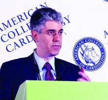
- RE-DUAL PCI: Jonas Oldgren, MD, of Uppsala (Sweden) University, will present an unspecified subgroup analysis from the RE-DUAL PCI (Dual Antithrombotic Therapy With Dabigatran in Patients With Atrial Fibrillation Undergoing Percutaneous Coronary Intervention) trial, focusing on one arm of the study. The main results, presented in August, showed patients with atrial fibrillation who had undergone percutaneous coronary intervention had a lower risk of bleeding if they received dual therapy with dabigatran plus clopidogrel or ticagrelor than did those treated with warfarin, clopidogrel, or ticagrelor.
- POISE-2 PCI: Michelle M. Graham, MD, of the University of Alberta, Edmonton, will present a substudy of POISE-2 focusing on the patients in undergoing noncardiac surgery who had a previous percutaneous coronary intervention. POISE-2, published in 2014, showed that administering aspirin periprocedurally had no effect on the rate of a composite of death or nonfatal MI, but increased the risk of major bleeding.

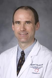
Dr. Jonas Oldgren - GEMINI-ACS-1: In the main study, the risk of major bleeding was similar between ACS patients treated with a combination of low-dose rivaroxaban and a P2Y12 inhibitor and those treated with aspirin and P2Y12 inhibitor. Matthew T. Roe, MD, Duke Clinical Research Institute, Durham, N.C., will present the substudy focusing on P2Y12 inhibitor switching in response to routine notification of CYP2C19 clopidogrel metabolizer status following ACS.
- PRAGUE-18: Zuzana Motovska, MD, of Charles University in Prague, will present an unspecified substudy of PRAGUE-18, which in August 2016 showed no difference in safety or efficacy between prasugrel and ticagrelor in AMI patients undergoing primary angioplasty.

Late-Breaking Session 6, quality improvement and patient-centered care
The seven presentations in this session range from findings from the enormous SWEDEHEART registry on how treatments have improved for ST-elevation MI over 20 years, to STIC2IT, a cluster randomized, controlled trial to test whether a novel telepharmacist-based intervention for patients with metabolic syndrome improves medication adherence and disease control.
Other presentations include evaluation of a quality improvement toolkit on AMI in India called ACS QUIK; a trial of a decision support intervention for patients and caregivers offered a heart assist device as destination therapy called DECIDE-LVAD; a national rollout of a clinical guidance framework for the assessment of patients with possible ACS in emergency departments (iCARE-ACS); and a report from the American College of Cardiology’s Mission: Lifeline STEMI ACCELERATOR-2 study.
[email protected]
On Twitter @cardionews
On the third day of the American Heart Association scientific sessions, antithrombotic therapy is the focus of the Late-Breaking Science 5 presentation, at 10:45 a.m.-12 p.m. on Tuesday, Nov. 14, followed by innovative investigations in evaluating quality improvement and patient-centered care interventions in the Late-Breaking Science 6 session, to be held at 3:45-5:15 p.m. Here are some highlights.
Late-Breaking Science 5, antithrombotic therapy
- RE-DUAL PCI: Jonas Oldgren, MD, of Uppsala (Sweden) University, will present an unspecified subgroup analysis from the RE-DUAL PCI (Dual Antithrombotic Therapy With Dabigatran in Patients With Atrial Fibrillation Undergoing Percutaneous Coronary Intervention) trial, focusing on one arm of the study. The main results, presented in August, showed patients with atrial fibrillation who had undergone percutaneous coronary intervention had a lower risk of bleeding if they received dual therapy with dabigatran plus clopidogrel or ticagrelor than did those treated with warfarin, clopidogrel, or ticagrelor.
- POISE-2 PCI: Michelle M. Graham, MD, of the University of Alberta, Edmonton, will present a substudy of POISE-2 focusing on the patients in undergoing noncardiac surgery who had a previous percutaneous coronary intervention. POISE-2, published in 2014, showed that administering aspirin periprocedurally had no effect on the rate of a composite of death or nonfatal MI, but increased the risk of major bleeding.
Dr. Jonas Oldgren - GEMINI-ACS-1: In the main study, the risk of major bleeding was similar between ACS patients treated with a combination of low-dose rivaroxaban and a P2Y12 inhibitor and those treated with aspirin and P2Y12 inhibitor. Matthew T. Roe, MD, Duke Clinical Research Institute, Durham, N.C., will present the substudy focusing on P2Y12 inhibitor switching in response to routine notification of CYP2C19 clopidogrel metabolizer status following ACS.
- PRAGUE-18: Zuzana Motovska, MD, of Charles University in Prague, will present an unspecified substudy of PRAGUE-18, which in August 2016 showed no difference in safety or efficacy between prasugrel and ticagrelor in AMI patients undergoing primary angioplasty.
Late-Breaking Session 6, quality improvement and patient-centered care
The seven presentations in this session range from findings from the enormous SWEDEHEART registry on how treatments have improved for ST-elevation MI over 20 years, to STIC2IT, a cluster randomized, controlled trial to test whether a novel telepharmacist-based intervention for patients with metabolic syndrome improves medication adherence and disease control.
Other presentations include evaluation of a quality improvement toolkit on AMI in India called ACS QUIK; a trial of a decision support intervention for patients and caregivers offered a heart assist device as destination therapy called DECIDE-LVAD; a national rollout of a clinical guidance framework for the assessment of patients with possible ACS in emergency departments (iCARE-ACS); and a report from the American College of Cardiology’s Mission: Lifeline STEMI ACCELERATOR-2 study.
[email protected]
On Twitter @cardionews
Psych evaluation identifies bariatric surgery patients who do less well
NATIONAL HARBOR, MD. – Psychological screening prior to bariatric surgery is not effective for identifying who will do poorly after the procedure, but it can identify patients who are at risk for less weight loss and likely to benefit from additional support, according to one of the largest studies designed to evaluate the predictive accuracy of this screening. The results were presented at Obesity Week 2017.
“Psychological evaluations should not be used to exclude patients, because we do not appear to be very good at predicting who does well,” said Nina E. Boulard, PhD, a psychologist who currently performs preoperative psychological screening of bariatric surgery candidates at Eastern Maine Medical Center, Bangor. Rather, “our evaluations identify those who need to be followed more closely so we can intervene early when patients struggle,” she said at an annual meeting presented by the American Society for Metabolic and Bariatric Surgery and The Obesity Society.

The two primary outcomes of interest were the ability to predict postsurgical mental health issues and the ability to predict difficulty in complying with behavioral changes. Percent excess weight loss (EWL) was also monitored. For mental health screening, the protocol included clinical interviews augmented with standardized testing, such as the Minnesota Multiphasic Personality Inventory-2-Restuctured Form (MMPI-2-RF). Patients were placed into five risk categories: low, low-moderate, moderate, moderate-high, and high.
For compliance, the patient evaluation was based primarily on interviews capturing self-reports of adherence to presurgical dietary and other lifestyle recommendations. Patients were also asked about eating behaviors, such as binge eating, and history of sexual abuse and attention-deficit/hyperactivity disorder and other neurodevelopmental issues.
The psychological screening report is provided to the surgeon, who ultimately decides whether patients go on to surgery. In this analysis, relatively few high-risk patients were included because these patients, such as those with active psychosis, uncontrolled binge eating, or severe depression, were excluded from surgery and therefore did not generate postsurgical follow-up data.
As has been reported by others, Dr. Boulard reported that no single standardized test or clinical variable was an effective predictor of “who will and will not struggle” psychologically after surgery. However, after adjusting for age, gender, presurgery weight, diabetes history, and surgery type, several variables were associated with reduced proportion EWL. These included anxiety (P = .02), ADHD (P = .01), and prior hospitalization of a psychological indication (P less than .05). History of sexual abuse was predictive of reduced percentage EWL 1 year but not 2 years after surgery.
“So, what this tells us is the clinical interpretation of the big picture might be important,” Dr. Boulard commented.
The presurgical psychological screening was not predictive of compliance, a result that Dr. Boulard labeled as “surprising.” She speculated that because weight loss in the first 2 years after bariatric surgery is relatively uniform, longer follow-up, such as 5 years, might be needed to capture the impact of patients predicted to have poor compliance.
Importantly, percent EWL remained high even among patients with risk factors associated with mental health challenges after surgery, according to Dr. Boulard. She stressed that even those with less weight loss “did not do poorly.” Rather, the relative differences in postsurgical weight loss for those who were predicted to struggle versus those who were not was “a matter of degrees.”
While Dr. Boulard acknowledged a prospective study using a standardized psychological screening protocol would be appropriate to validate the findings of this study, the study results have already changed practice at her institution.
“We are following the at-risk patients more closely, so we can intervene quickly if there are issues after surgery,” Dr. Boulard explained. The question raised by this study is, “can we bring them [patients at higher risk] to a higher level of response so they are just as successful” as those without risk factors identified in psychological screening.
Dr. Boulard reports no financial relationships relevant to this topic.
NATIONAL HARBOR, MD. – Psychological screening prior to bariatric surgery is not effective for identifying who will do poorly after the procedure, but it can identify patients who are at risk for less weight loss and likely to benefit from additional support, according to one of the largest studies designed to evaluate the predictive accuracy of this screening. The results were presented at Obesity Week 2017.
“Psychological evaluations should not be used to exclude patients, because we do not appear to be very good at predicting who does well,” said Nina E. Boulard, PhD, a psychologist who currently performs preoperative psychological screening of bariatric surgery candidates at Eastern Maine Medical Center, Bangor. Rather, “our evaluations identify those who need to be followed more closely so we can intervene early when patients struggle,” she said at an annual meeting presented by the American Society for Metabolic and Bariatric Surgery and The Obesity Society.
The two primary outcomes of interest were the ability to predict postsurgical mental health issues and the ability to predict difficulty in complying with behavioral changes. Percent excess weight loss (EWL) was also monitored. For mental health screening, the protocol included clinical interviews augmented with standardized testing, such as the Minnesota Multiphasic Personality Inventory-2-Restuctured Form (MMPI-2-RF). Patients were placed into five risk categories: low, low-moderate, moderate, moderate-high, and high.
For compliance, the patient evaluation was based primarily on interviews capturing self-reports of adherence to presurgical dietary and other lifestyle recommendations. Patients were also asked about eating behaviors, such as binge eating, and history of sexual abuse and attention-deficit/hyperactivity disorder and other neurodevelopmental issues.
The psychological screening report is provided to the surgeon, who ultimately decides whether patients go on to surgery. In this analysis, relatively few high-risk patients were included because these patients, such as those with active psychosis, uncontrolled binge eating, or severe depression, were excluded from surgery and therefore did not generate postsurgical follow-up data.
As has been reported by others, Dr. Boulard reported that no single standardized test or clinical variable was an effective predictor of “who will and will not struggle” psychologically after surgery. However, after adjusting for age, gender, presurgery weight, diabetes history, and surgery type, several variables were associated with reduced proportion EWL. These included anxiety (P = .02), ADHD (P = .01), and prior hospitalization of a psychological indication (P less than .05). History of sexual abuse was predictive of reduced percentage EWL 1 year but not 2 years after surgery.
“So, what this tells us is the clinical interpretation of the big picture might be important,” Dr. Boulard commented.
The presurgical psychological screening was not predictive of compliance, a result that Dr. Boulard labeled as “surprising.” She speculated that because weight loss in the first 2 years after bariatric surgery is relatively uniform, longer follow-up, such as 5 years, might be needed to capture the impact of patients predicted to have poor compliance.
Importantly, percent EWL remained high even among patients with risk factors associated with mental health challenges after surgery, according to Dr. Boulard. She stressed that even those with less weight loss “did not do poorly.” Rather, the relative differences in postsurgical weight loss for those who were predicted to struggle versus those who were not was “a matter of degrees.”
While Dr. Boulard acknowledged a prospective study using a standardized psychological screening protocol would be appropriate to validate the findings of this study, the study results have already changed practice at her institution.
“We are following the at-risk patients more closely, so we can intervene quickly if there are issues after surgery,” Dr. Boulard explained. The question raised by this study is, “can we bring them [patients at higher risk] to a higher level of response so they are just as successful” as those without risk factors identified in psychological screening.
Dr. Boulard reports no financial relationships relevant to this topic.
AT OBESITY WEEK 2017
Key clinical point: Psychological screening prior to bariatric surgery selects patients at risk for reduced postoperative weight loss.
Major finding: Prior psychological hospitalization (P less than .05) and number of previous psychological diagnoses (P = .04) are among markers of less postop weight loss.
Data source: Retrospective analysis.
Disclosures: Dr. Boulard reports no financial relationships relevant to this topic.
Pregnant women’s access to vaccination website, social media improved infants’ up-to-date immunizations
than were those receiving usual care, said Jason M. Glanz, PhD, of the Institute for Health Research, Kaiser Permanente Colorado, Denver, and his associates.
“These results suggest that interactive, informational interventions administered outside of the physician’s office can improve vaccine acceptance,” the investigators said. “The information appears to be effective when presented to parents before their children are born.”
The women in the VSM group had access to vaccine content, social media technologies that included a blog, discussion forum, chat rooms with experts, and “Ask a Question” portal through which the women could directly ask experts questions about vaccination. The experts were a pediatrician, a vaccine safety researcher, and a risk communication specialist.
Monthly, one to two blog posts were created on topics such as new vaccine safety research, vaccine-preventable disease outbreaks, changes in immunization policy, and the importance of adhering to the recommended immunization schedule, the researchers said. The women also could submit questions privately through e-mail, and receive personalized responses within 2 business days.
At the end of 200 days of follow-up, the proportion of infants up to date with their vaccinations were 93% for the VSM group, 91% for the VI group, and 87% for the UC arms. Infants in the VSM group were more likely to be up to date at age 200 days, compared with infants in the UC group (odds ratio, 1.92; 95% confidence interval, 1.07-3.47).
“Up-to-date status did not differ significantly between the VI and UC arms or the VSM and VI arms,” Dr. Glanz and his associates wrote.
Of 739 women in the VSM and VI groups, 35% visited the website at least once, with a mean of 1.8 visits (range, 1-15 visits). Of 75 vaccine-hesitant women, 44% visited the website, compared with 34% of 664 nonhesitant women. Median vaccine hesitancy scores were 13 for the VSM group, 17 for the VI group, and 15 for the UC group.
The study authors had no relevant disclosures.
Read more in Pediatrics (2017 Nov. doi: 10.1542/peds.2017-1117).
FROM PEDIATRICS
Late-Breaking Science preview: Monday, Nov. 13
Analyses of landmark, practice-changing trials will pepper the three late-breaking science sessions.
Late-Breaking Science 2
The Late-Breaking Science 2 session will focus on prevention and will include analyses from the two most illuminating and perhaps practice-changing trials presented this year, FOURIER and CANTOS. REVEAL, another large and important trial of 2017, also spawned a subanalysis in this deep-diving session at 9:00 a.m.-10:15 a.m.:

- REAL-CAD: This study evaluated the prevention of cardiovascular disease with pitavastatin 1 mg/day or 4 mg/day in patients with stable coronary artery disease followed for 3-6 years. In the REAL-CAD (Randomized Evaluation of Aggressive or Moderate Lipid Lowering Therapy With Pitavastatin in Coronary Artery Disease) trial, presented by Takeshi Kimura, MD, of Kyoto (Japan) University, the primary endpoint was the occurrence of cardiovascular death, nonfatal MI, nonfatal cerebral infarction, or unstable angina.
- REVEAL: Presented in August at the European Society of Cardiology Congress (ESC), the primary results of REVEAL (Randomized Evaluation of the Effects of Anacetrapib Through Lipid-Modification), a multicenter, pivotal trial with more than 30,000 patients treated for about 4 years, showed that patients treated with anacetrapib had a statistically significant 9% decrease in major coronary events, compared with placebo-treated controls. The analysis to be presented at AHA by Martin Landrey, MD of the University of Oxford, England, examined the effects of anacetrapib on the incidence of new-onset diabetes and on vascular events in people with diabetes.
 Frontline Medical News
Dr. Paul M. Ridker - FOURIER: This blockbuster trial in more than 27,000 patients showed that the PCSK9 inhibitor evolocumab significantly improved cardiovascular outcomes in high-risk patients already on statin therapy and has already started spawning important subanalyses. Two will be presented in this session. First, Marc P. Bonaca, MD, of Brigham & Women’s Hospital in Boston, will present outcomes in patients with peripheral artery disease. Second, Marc S. Sabatine, MD, also of Brigham & Women’s, will present the clinical benefit of evolocumab in patients with a history of MI.
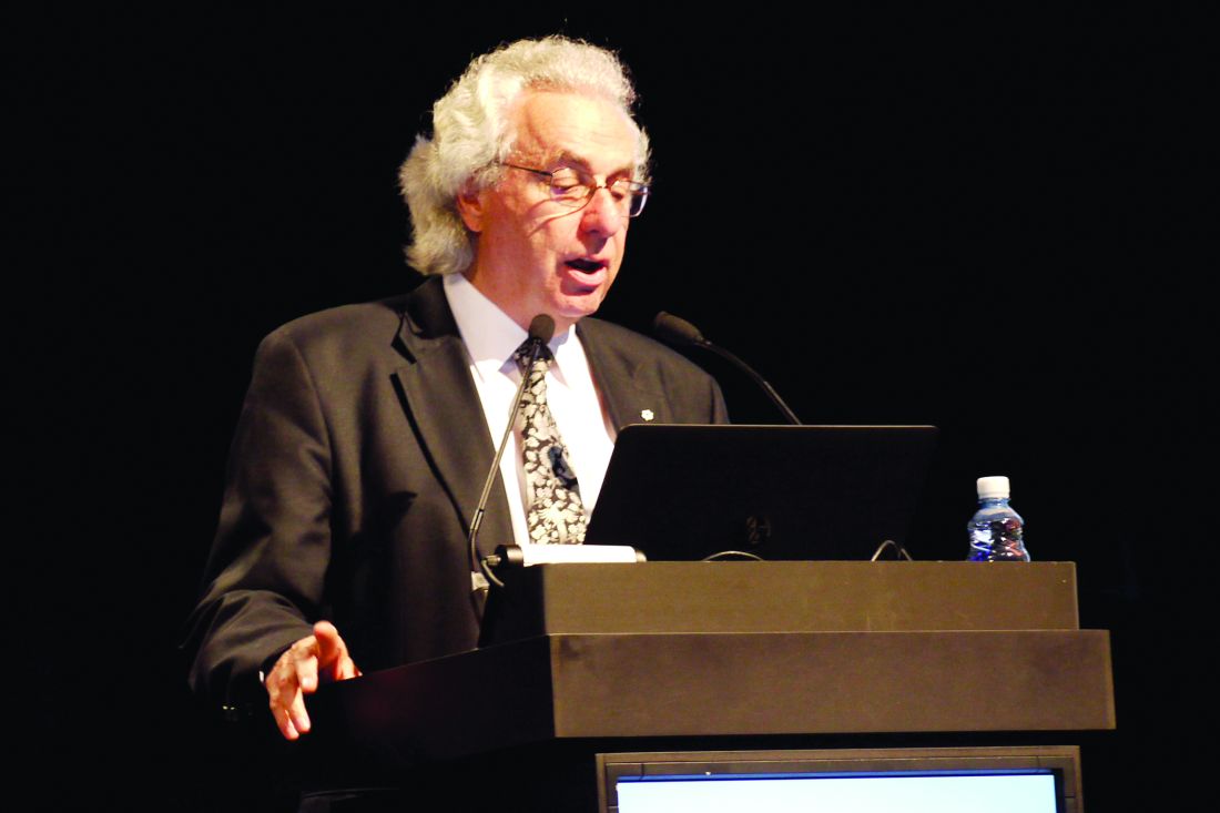
Dr. Marc S. Sabatine - CANTOS: The findings of CANTOS (Canakinumab Anti-Inflammatory Thrombosis Outcome Study) stunned the cardiovascular world and piqued interest in their oncology colleagues. “These data provide the first proof that inflammation inhibition in the absence of lipid lowering can improve atherogenic outcomes and potentially alter progression of some fatal cancers,” declared Paul M. Ridker, MD, of Brigham and Women’s Hospital and professor of medicine at Harvard Medical School, both in Boston, at ESC. In CANTOS, the fully human monoclonal antibody canakinumab, which targets IL-1B, reduced the risk of recurrent cardiovascular events by 15% in a very-high-risk population, compared with placebo, while cutting incident lung cancer by 67%.

Late-Breaking Science 3
This session promises insights into hypertension management and will be held at 10:45 a.m.-12:00 p.m.:
- Chinese BP trial: Presented by Mar Pujades-Rodriguez, PhD, of the University of Leeds, (England), this study examined time at blood pressure target and the risk of cardiovascular diseases and mortality in a Chinese population.
- SPRINT: Yes, the trial that upended hypertension guidelines is bearing fruit again. This analysis of SPRINT (Systolic Blood Pressure Intervention Trial) looked at blood pressure measurement. Karen C. Johnson, MD, of the University of Tennessee, Memphis, will present the results.

Dr. Robert J. Mentz - GATEWAY: As the benefits of bariatric surgery seem to pile up, the GATEWAY (Gastric Bypass to Treat Obese Patients With Steady Hypertension) trial focused on reducing the need for antihypertensive drugs and decreasing systemic arterial blood pressure and other risk factors for cardiovascular events in patients with arterial hypertension randomized to Roux-en-Y gastroplasty or to clinical treatments. Carlos A. Schiavon, MD, of Heart Hospital in São Paulo will present the results.

Late-Breaking Science 4
Billed as covering the “sweet spot in cardiometabolic care,” this session will highlight new findings from three of the biggest trials in type 2 diabetes management. The session will run at 3:45 p.m.-5:00 p.m.
- CANVAS: Presented at ESC, the results of this large, FDA-mandated cardiovascular outcomes trial showed that canagliflozin significantly reduced the risk of cardiovascular and renal events but doubled the risk of amputation, compared with placebo, in patients with type 2 diabetes. In this subanalysis of CANVAS (the Canagliflozin Cardiovascular Assessment Study), presenter Kenneth W. Mahaffey, MD, of Stanford (Calif.) University, and his coinvestigators looked at primary and secondary prevention of cardiovascular events in the same population.
 Sara Freeman/Frontline Medical News
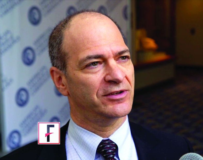
Dr. Bernard Zinman - EXSCEL: The results of this trial, also presented at ESC, showed that exenatide was noninferior to placebo with respect to cardiovascular safety but not superior with respect to efficacy. Presenter Robert J. Mentz, MD, of Duke University, Durham, N.C., will present more news from the EXSCEL (Exenatide Study of Cardiovascular Event Lowering) trial.
- EMPA-REG OUTCOME: The first trial to show a cardioprotective effect of an SGLT-2 inhibitor in type 2 diabetes patients, EMPA-REG OUTCOME, marked a new era in diabetes treatment. Bernard Zinman, MD, of the University of Toronto, will present an subset analysis of patients with type 2 diabetes and peripheral artery disease. The session title signals more good news: Empagliflozin Reduces Mortality and Hospitalization for Heart Failure in Patients With Type 2 Diabetes and Peripheral Artery Disease.
[email protected]
On Twitter @cardionews
Analyses of landmark, practice-changing trials will pepper the three late-breaking science sessions.
Late-Breaking Science 2
The Late-Breaking Science 2 session will focus on prevention and will include analyses from the two most illuminating and perhaps practice-changing trials presented this year, FOURIER and CANTOS. REVEAL, another large and important trial of 2017, also spawned a subanalysis in this deep-diving session at 9:00 a.m.-10:15 a.m.:
- REAL-CAD: This study evaluated the prevention of cardiovascular disease with pitavastatin 1 mg/day or 4 mg/day in patients with stable coronary artery disease followed for 3-6 years. In the REAL-CAD (Randomized Evaluation of Aggressive or Moderate Lipid Lowering Therapy With Pitavastatin in Coronary Artery Disease) trial, presented by Takeshi Kimura, MD, of Kyoto (Japan) University, the primary endpoint was the occurrence of cardiovascular death, nonfatal MI, nonfatal cerebral infarction, or unstable angina.
- REVEAL: Presented in August at the European Society of Cardiology Congress (ESC), the primary results of REVEAL (Randomized Evaluation of the Effects of Anacetrapib Through Lipid-Modification), a multicenter, pivotal trial with more than 30,000 patients treated for about 4 years, showed that patients treated with anacetrapib had a statistically significant 9% decrease in major coronary events, compared with placebo-treated controls. The analysis to be presented at AHA by Martin Landrey, MD of the University of Oxford, England, examined the effects of anacetrapib on the incidence of new-onset diabetes and on vascular events in people with diabetes.
Frontline Medical News
Dr. Paul M. Ridker - FOURIER: This blockbuster trial in more than 27,000 patients showed that the PCSK9 inhibitor evolocumab significantly improved cardiovascular outcomes in high-risk patients already on statin therapy and has already started spawning important subanalyses. Two will be presented in this session. First, Marc P. Bonaca, MD, of Brigham & Women’s Hospital in Boston, will present outcomes in patients with peripheral artery disease. Second, Marc S. Sabatine, MD, also of Brigham & Women’s, will present the clinical benefit of evolocumab in patients with a history of MI.
Dr. Marc S. Sabatine - CANTOS: The findings of CANTOS (Canakinumab Anti-Inflammatory Thrombosis Outcome Study) stunned the cardiovascular world and piqued interest in their oncology colleagues. “These data provide the first proof that inflammation inhibition in the absence of lipid lowering can improve atherogenic outcomes and potentially alter progression of some fatal cancers,” declared Paul M. Ridker, MD, of Brigham and Women’s Hospital and professor of medicine at Harvard Medical School, both in Boston, at ESC. In CANTOS, the fully human monoclonal antibody canakinumab, which targets IL-1B, reduced the risk of recurrent cardiovascular events by 15% in a very-high-risk population, compared with placebo, while cutting incident lung cancer by 67%.
Late-Breaking Science 3
This session promises insights into hypertension management and will be held at 10:45 a.m.-12:00 p.m.:
- Chinese BP trial: Presented by Mar Pujades-Rodriguez, PhD, of the University of Leeds, (England), this study examined time at blood pressure target and the risk of cardiovascular diseases and mortality in a Chinese population.
- SPRINT: Yes, the trial that upended hypertension guidelines is bearing fruit again. This analysis of SPRINT (Systolic Blood Pressure Intervention Trial) looked at blood pressure measurement. Karen C. Johnson, MD, of the University of Tennessee, Memphis, will present the results.
Dr. Robert J. Mentz - GATEWAY: As the benefits of bariatric surgery seem to pile up, the GATEWAY (Gastric Bypass to Treat Obese Patients With Steady Hypertension) trial focused on reducing the need for antihypertensive drugs and decreasing systemic arterial blood pressure and other risk factors for cardiovascular events in patients with arterial hypertension randomized to Roux-en-Y gastroplasty or to clinical treatments. Carlos A. Schiavon, MD, of Heart Hospital in São Paulo will present the results.
Late-Breaking Science 4
Billed as covering the “sweet spot in cardiometabolic care,” this session will highlight new findings from three of the biggest trials in type 2 diabetes management. The session will run at 3:45 p.m.-5:00 p.m.
- CANVAS: Presented at ESC, the results of this large, FDA-mandated cardiovascular outcomes trial showed that canagliflozin significantly reduced the risk of cardiovascular and renal events but doubled the risk of amputation, compared with placebo, in patients with type 2 diabetes. In this subanalysis of CANVAS (the Canagliflozin Cardiovascular Assessment Study), presenter Kenneth W. Mahaffey, MD, of Stanford (Calif.) University, and his coinvestigators looked at primary and secondary prevention of cardiovascular events in the same population.
Sara Freeman/Frontline Medical News
Dr. Bernard Zinman - EXSCEL: The results of this trial, also presented at ESC, showed that exenatide was noninferior to placebo with respect to cardiovascular safety but not superior with respect to efficacy. Presenter Robert J. Mentz, MD, of Duke University, Durham, N.C., will present more news from the EXSCEL (Exenatide Study of Cardiovascular Event Lowering) trial.
- EMPA-REG OUTCOME: The first trial to show a cardioprotective effect of an SGLT-2 inhibitor in type 2 diabetes patients, EMPA-REG OUTCOME, marked a new era in diabetes treatment. Bernard Zinman, MD, of the University of Toronto, will present an subset analysis of patients with type 2 diabetes and peripheral artery disease. The session title signals more good news: Empagliflozin Reduces Mortality and Hospitalization for Heart Failure in Patients With Type 2 Diabetes and Peripheral Artery Disease.
[email protected]
On Twitter @cardionews
Medical malpractice and the hospitalist: Reasons for optimism
Fear of malpractice litigation weighs on many physicians, including hospitalists. Specific concerns that physicians have about facing a malpractice claim include stigmatization, loss of confidence in one’s own clinical skills, and a possible personal financial toll if an award exceeds the limit of one’s malpractice insurance.
Physician worries about malpractice are increasingly being raised during discussions of burnout, with a recent National Academy of Medicine discussion paper listing malpractice concerns as a possible factor that could contribute to physician burnout.1

Malpractice fears also influence physician behavior generally, leading to defensive medicine, though the actual costs of defensive medicine are debated. A national survey of physicians by Bishop and colleagues found that 91% felt that physicians order more tests and procedures than patients require in order to try to avoid malpractice claims.3 A survey of 1,020 hospitalists asked what testing they would undertake when provided clinical vignettes involving preoperative evaluation and syncope.4 Overuse of testing was common among hospitalists, and most hospitalists who overused testing specified that a desire to reassure either themselves or the patient or patient’s family was the reason for ordering the unnecessary testing.
The extent to which this overuse was driven by liability fears specifically is not clear. Overuse of testing was less common among physicians associated with Veterans Affairs Hospitals, who generally are not subject to personal medical malpractice liability. But a history of a prior malpractice claim was not associated with significantly greater overuse in the survey.
Hospitalists’ concerns about medical liability notwithstanding, data on the absolute malpractice risk of hospitalists and current trends in medical liability are both encouraging. An important source of our understanding about the national medical malpractice landscape is CRICO Strategies National Comparative Benchmarking System (CBS), which includes the malpractice experience from multiple insurers and represents 400 hospitals and 165,000 physicians. A December 2014 analysis of cases involving hospitalists from the CBS database showed that the malpractice claims rate for hospitalists was lower than those for other comparable groups of physicians.5 Hospitalists (in internal medicine) had a claims rate of 0.52 claims per 100 physician coverage years, which was significantly lower than the claims rate for nonhospitalist internal medicine physicians (with a rate of 1.91 claims per 100 physician-coverage years) and for emergency medicine physicians (with a rate of 3.50 claims per 100 physician-coverage years).

A remarkable national trend in medical malpractice, based on an analysis of data supplied by the National Practitioner Data Bank, is that the overall rate of paid claims is decreasing. From 1992 to 2014, the overall rate of paid claims dropped 55.7%.7 To varying degrees, the drop in paid claims has occurred across all specialties, with internal medicine in particular dropping 46.1%. The reason for this decrease in paid claims is not clear. Improvements in patient safety are one possible explanation, with tort reforms also possibly contributing to this trend. An additional potential factor, which will likely become more important as it becomes more widespread, is the advent of communication and resolution programs (also known as disclosure, apology, and offer programs).
In communication and resolution programs, the response to a malpractice claim is to investigate the circumstances surrounding the adverse event underlying the claim to determine if it was the result of medical error. When the investigation finds no medical error, then the claim is defended. However, in cases in which there was a medical error leading to patient harm, then the error is disclosed to the patient and family, and an offer of compensation is made.
One of the most prominent communication and resolution programs exists at the University of Michigan, and published experience from this program shows that, after implementation of the program, significant drops were seen in the number of malpractice lawsuits, the time it took to resolve malpractice claims, the amount paid in patient compensation on malpractice claims, and the costs involved with litigating malpractice claims.8 One of the goals of communication and resolution programs is to utilize the information from the investigations of whether medical errors occurred to find areas where patient safety systems can be improved, thereby using the medical malpractice system to promote patient safety. Although the University of Michigan’s experience with its communication and resolution program is very encouraging, it remains to be seen how widely such programs will be adopted. Medical malpractice is primarily governed at the state level, and the liability laws of some states are more conducive than others to the implementation of these programs.
Hospitalist concerns about medical malpractice are likely to persist, as being named in a malpractice lawsuit is stressful, regardless of the outcome of the case. Contributing to the stress of facing a malpractice claim, cases typically take 3-5 years to be resolved. However, the risk for hospitalists of facing a medical malpractice claim is relatively low. Moreover, given national trends, hospitalists’ liability risk would be expected to remain low or decrease moving forward.
Dr. Schaffer is an attending physician in the Hospital Medicine Unit at Brigham and Women’s Hospital, an instructor at Harvard Medical School, and a senior clinical analytics specialist at CRICO/Risk Management Foundation of the Harvard Medical Institutions, all in Boston. Dr. Kachalia is an attending physician in the Hospital Medicine Unit at Brigham and Women’s Hospital, an associate professor at Harvard Medical School, and the chief quality officer at Brigham and Women’s Hospital.
References
1. Dyrbye LN et al. Burnout among health care professionals: A call to explore and address this underrecognized threat to safe, high-quality care. National Academy of Medicine Perspectives. 2017 Jul 5.
2. Helland E et al. Bargaining in the shadow of the website: Disclosure’s impact on medical malpractice litigation. American Law and Economics Review. 2010;12(2):423-61.
3. Bishop TF et al. Physicians’ views on defensive medicine: A national survey. Arch Intern Med. Jun 28 2010;170(12):1081-3.
4. Kachalia A et al. Overuse of testing in preoperative evaluation and syncope: A survey of hospitalists. Ann Intern Med. 2015 Jan 20;162(2):100-8.
5. Schaffer AC et al. Liability impact of the hospitalist model of care. J Hosp Med. Dec 2014;9(12):750-5.
6. CRICO Strategies. Medication-related malpractice risks: 2016 CBS Benchmarking Report. Boston. The Risk Management Foundation of Harvard Medical Institutions; 2016. Available at: www.rmf.harvard.edu/cbsreport (accessed Sept. 14, 2017).
7. Schaffer AC et al. Rates and characteristics of paid malpractice claims among US physicians by specialty, 1992-2014. JAMA Intern Med. May 2017;177(5):710-8.
8. Kachalia A et al. Liability claims and costs before and after implementation of a medical error disclosure program. Ann Intern Med. Aug 17 2010;153(4):213-21.
Fear of malpractice litigation weighs on many physicians, including hospitalists. Specific concerns that physicians have about facing a malpractice claim include stigmatization, loss of confidence in one’s own clinical skills, and a possible personal financial toll if an award exceeds the limit of one’s malpractice insurance.
Physician worries about malpractice are increasingly being raised during discussions of burnout, with a recent National Academy of Medicine discussion paper listing malpractice concerns as a possible factor that could contribute to physician burnout.1
Malpractice fears also influence physician behavior generally, leading to defensive medicine, though the actual costs of defensive medicine are debated. A national survey of physicians by Bishop and colleagues found that 91% felt that physicians order more tests and procedures than patients require in order to try to avoid malpractice claims.3 A survey of 1,020 hospitalists asked what testing they would undertake when provided clinical vignettes involving preoperative evaluation and syncope.4 Overuse of testing was common among hospitalists, and most hospitalists who overused testing specified that a desire to reassure either themselves or the patient or patient’s family was the reason for ordering the unnecessary testing.
The extent to which this overuse was driven by liability fears specifically is not clear. Overuse of testing was less common among physicians associated with Veterans Affairs Hospitals, who generally are not subject to personal medical malpractice liability. But a history of a prior malpractice claim was not associated with significantly greater overuse in the survey.
Hospitalists’ concerns about medical liability notwithstanding, data on the absolute malpractice risk of hospitalists and current trends in medical liability are both encouraging. An important source of our understanding about the national medical malpractice landscape is CRICO Strategies National Comparative Benchmarking System (CBS), which includes the malpractice experience from multiple insurers and represents 400 hospitals and 165,000 physicians. A December 2014 analysis of cases involving hospitalists from the CBS database showed that the malpractice claims rate for hospitalists was lower than those for other comparable groups of physicians.5 Hospitalists (in internal medicine) had a claims rate of 0.52 claims per 100 physician coverage years, which was significantly lower than the claims rate for nonhospitalist internal medicine physicians (with a rate of 1.91 claims per 100 physician-coverage years) and for emergency medicine physicians (with a rate of 3.50 claims per 100 physician-coverage years).
A remarkable national trend in medical malpractice, based on an analysis of data supplied by the National Practitioner Data Bank, is that the overall rate of paid claims is decreasing. From 1992 to 2014, the overall rate of paid claims dropped 55.7%.7 To varying degrees, the drop in paid claims has occurred across all specialties, with internal medicine in particular dropping 46.1%. The reason for this decrease in paid claims is not clear. Improvements in patient safety are one possible explanation, with tort reforms also possibly contributing to this trend. An additional potential factor, which will likely become more important as it becomes more widespread, is the advent of communication and resolution programs (also known as disclosure, apology, and offer programs).
In communication and resolution programs, the response to a malpractice claim is to investigate the circumstances surrounding the adverse event underlying the claim to determine if it was the result of medical error. When the investigation finds no medical error, then the claim is defended. However, in cases in which there was a medical error leading to patient harm, then the error is disclosed to the patient and family, and an offer of compensation is made.
One of the most prominent communication and resolution programs exists at the University of Michigan, and published experience from this program shows that, after implementation of the program, significant drops were seen in the number of malpractice lawsuits, the time it took to resolve malpractice claims, the amount paid in patient compensation on malpractice claims, and the costs involved with litigating malpractice claims.8 One of the goals of communication and resolution programs is to utilize the information from the investigations of whether medical errors occurred to find areas where patient safety systems can be improved, thereby using the medical malpractice system to promote patient safety. Although the University of Michigan’s experience with its communication and resolution program is very encouraging, it remains to be seen how widely such programs will be adopted. Medical malpractice is primarily governed at the state level, and the liability laws of some states are more conducive than others to the implementation of these programs.
Hospitalist concerns about medical malpractice are likely to persist, as being named in a malpractice lawsuit is stressful, regardless of the outcome of the case. Contributing to the stress of facing a malpractice claim, cases typically take 3-5 years to be resolved. However, the risk for hospitalists of facing a medical malpractice claim is relatively low. Moreover, given national trends, hospitalists’ liability risk would be expected to remain low or decrease moving forward.
Dr. Schaffer is an attending physician in the Hospital Medicine Unit at Brigham and Women’s Hospital, an instructor at Harvard Medical School, and a senior clinical analytics specialist at CRICO/Risk Management Foundation of the Harvard Medical Institutions, all in Boston. Dr. Kachalia is an attending physician in the Hospital Medicine Unit at Brigham and Women’s Hospital, an associate professor at Harvard Medical School, and the chief quality officer at Brigham and Women’s Hospital.
References
1. Dyrbye LN et al. Burnout among health care professionals: A call to explore and address this underrecognized threat to safe, high-quality care. National Academy of Medicine Perspectives. 2017 Jul 5.
2. Helland E et al. Bargaining in the shadow of the website: Disclosure’s impact on medical malpractice litigation. American Law and Economics Review. 2010;12(2):423-61.
3. Bishop TF et al. Physicians’ views on defensive medicine: A national survey. Arch Intern Med. Jun 28 2010;170(12):1081-3.
4. Kachalia A et al. Overuse of testing in preoperative evaluation and syncope: A survey of hospitalists. Ann Intern Med. 2015 Jan 20;162(2):100-8.
5. Schaffer AC et al. Liability impact of the hospitalist model of care. J Hosp Med. Dec 2014;9(12):750-5.
6. CRICO Strategies. Medication-related malpractice risks: 2016 CBS Benchmarking Report. Boston. The Risk Management Foundation of Harvard Medical Institutions; 2016. Available at: www.rmf.harvard.edu/cbsreport (accessed Sept. 14, 2017).
7. Schaffer AC et al. Rates and characteristics of paid malpractice claims among US physicians by specialty, 1992-2014. JAMA Intern Med. May 2017;177(5):710-8.
8. Kachalia A et al. Liability claims and costs before and after implementation of a medical error disclosure program. Ann Intern Med. Aug 17 2010;153(4):213-21.
Late-Breaking Science preview: Sunday, Nov. 12
The program committee of the American Heart Association deemed the following four studies the best for the first Late-Breaking Science 1 session at the AHA scientific sessions in Anaheim, exploring solutions to periprocedural dilemmas in coronary artery bypass surgery and electrophysiology.
The session is on Sunday, Nov. 12, 3:45-5:00 p.m. in Hall D.
- TRiCS III: Opening with a transfusion study, C. David Mazer, MD, of St. Michaels Hospital, University of Toronto, will present results of the Transfusion Requirements in Cardiac Surgery (TRiCS) III trial. The international, open-label, randomized noninferiority trial compared two commonly used transfusion strategies in high-risk patients having cardiac surgery. Specifically, it compared a restrictive transfusion strategy in which patients receive a red cell transfusion if their hemoglobin was below 75 g/L intraoperatively and/or postoperatively with a liberal transfusion strategy (where the cutoff for red cell transfusion was 95 g/L intraoperatively), or below 85 g/L postoperatively in the intensive care unit and on the ward. The primary outcome is a composite of any of the following events (up to hospital discharge or postoperative day 28, whichever comes first): all-cause mortality, myocardial infarction, new renal failure, or new focal neurological deficit.
- DACAB: Second in the session is the phase 4 trial to Compare the Efficacy of Different Antiplatelet Therapy Strategy After Coronary Artery Bypass Graft Surgery (DACAB), presented by Qiang Zhao, MD, of Shanghai Jiaotong University. This three-pronged study was designed to show the superiority of ticagrelor alone and ticagrelor plus aspirin over aspirin monotherapy for the 1-year primary efficacy endpoint of vein graft patency.
- PRESERVE: Seeking to reduce complications of angiography, presenter Steven Weisbord, MD, of the University of Pittsburgh, and his colleagues carried out the PRESERVE (Prevention of Serious Adverse Events Following Angiography) trial. It compared the effectiveness of intravenous isotonic sodium bicarbonate with intravenous isotonic sodium chloride and oral N-acetylcysteine with oral placebo for the prevention of serious adverse outcomes following angiographic procedures in high-risk patients. The primary outcome is a composite of serious, adverse, patient-centered events, including death, need for acute dialysis, or persistent decline in kidney function at 90 days.
- BRUISE CONTROL-2: The purpose of this study was to determine the best way to prevent hematoma by managing novel oral anticoagulants (NOACs) at the time of pacemaker or defibrillator surgery. Presenter David H. Birnie, MD, of the University of Ottawa Heart Institute, and his coinvestigators hypothesized that performing device surgery without interruption of the NOAC will result in a reduced rate of clinically significant hematoma. In the trial, called Strategy of Continued Versus Interrupted Novel Oral Anti-coagulant at Time of Device Surgery in Patients With Moderate to High Risk of Arterial Thromboembolic Events (BRUISE CONTROL-2), continued or interrupted anticoagulation with dabigatran, rivaroxaban, and apixaban were compared.
- ABRIDGE J: Presenter Akihiko Nogami, MD, of the University of Tsukuba (Japan), and his coinvestigators aimed to evaluate the efficacy and safety of perioperative anticoagulants during catheter ablation of atrial fibrillation. In the Clinical Benefit of Minimally-Interrupted Dabigatran versus Uninterrupted Warfarin for Catheter Ablation of Atrial Fibrillation (ABRIDGE-J) trial, patients with drug-resistant paroxysmal AF were randomized to receive dabigatran or warfarin perioperatively. The main outcome measures were incidence of embolism during the perioperative period and presence or absence of an intracardiac thrombus just before ablation.
[email protected]
On Twitter @cardionews
VIDEO: New PsA guideline expected in 2018
SAN DIEGO – For the first time, a forthcoming evidence-based guideline for the management of psoriatic arthritis recommends tumor necrosis factor inhibitor biologics as first-line therapy.
“Guidelines that have been around for the last several years have been skirting around the fact that there’s really no evidence that methotrexate works for PsA,” Dafna D. Gladman, MD, said during a press briefing at the annual meeting of the American College of Rheumatology. “So it’s refreshing and reassuring that when you do an appropriate, evidence-based approach, you finally find the truth in front of you, and you have TNF inhibitors as the first-line treatment. Obviously, they’re not for everybody. There are patients in whom we cannot use TNF inhibitors, either because they don’t like needles, or because they have contraindications to getting these particular needles, but at least we have a recommendation for the use of these drugs as a first-line treatment.”
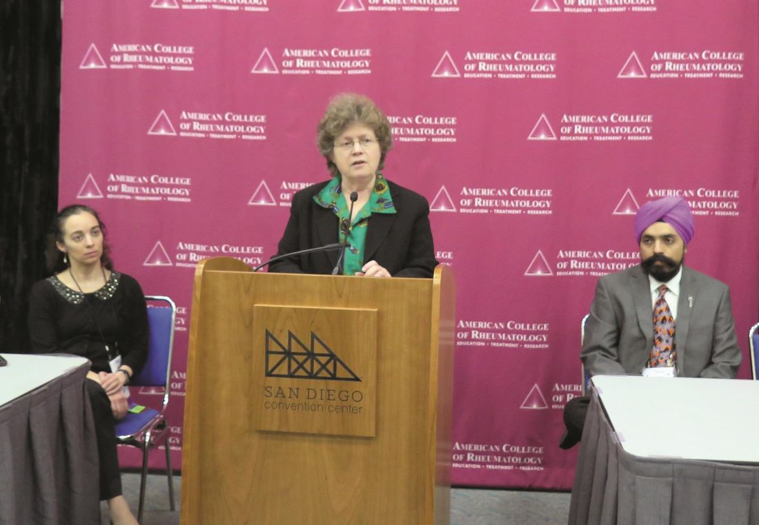
“At first, I wasn’t a big fan of the idea of the GRADE guidelines because the number of questions blows up so fast, [but] it really makes you focus on what the most common [clinical] settings are,” said another core oversight team member, Alexis Ogdie, MD, a rheumatologist at the University of Pennsylvania, Philadelphia. “These guidelines also reveal the major gap of no head-to-head studies. I think we’ve known that, but this really called that out as important. When we’re making a treatment decision between [drugs] A and B, we need those studies to be able to better understand how to treat our patients, rather than using the data from one trial to make a decision. ... For my patients, I’m excited that I can now use a TNF inhibitor as a first-line agent. When we have patients come in with very severe disease, occasionally they also have severe psoriasis, so we’ve been able to use TNF inhibitors as first-line treatment in some of our patients in Pennsylvania. This differs state by state. But the exciting thing is that they get better so fast and you don’t have to tell them to wait 12 weeks for methotrexate to work.”
The ACR/NPF guideline is currently under peer review and is expected to be published in Arthritis & Rheumatology, Arthritis Care & Research, and the Journal of Psoriasis and Psoriatic Arthritis in the spring or summer of 2018. It focuses on common PsA patients, not exceptional cases. It includes recommendations on the management of patients with active PsA that is defined by the patients’ self-report and judged by the examining clinician to be caused by PsA, based on the on the presence of at least one of the following: actively inflamed joints; dactylitis; enthesitis; axial disease; active skin and/or nail involvement; and/or extra-articular manifestations such as uveitis or inflammatory bowel disease. Authors of the guideline considered cost as one of many possible factors affecting the use of the recommendations, but explicit cost-effectiveness analyses were not conducted. Also, since the NPF and the American Academy of Dermatology are concurrently developing a psoriasis treatment guideline, the treatment of skin psoriasis was not included in the guideline.
According to the guideline’s principal investigator Jasvinder Singh, MD, professor of medicine and epidemiology at the University of Alabama at Birmingham, the guideline will include 80 recommendations, 75 (94%) that are rated as “conditional,” and 5 (6%) that are rated as “strong,” based on the quality of evidence in the existing medical literature. “Most of our treatment guidelines rely on very low-to-moderate quality evidence, which means that there needs to be an active discussion between the physician and the patient with regard to which treatment to choose,” said Dr. Singh, who is also a staff rheumatologist at the Birmingham Veterans Affairs Medical Center and who led development of the 2012 and 2015 ACR treatment guidelines for RA. “When you’re not choosing the preferred treatment, there are defined specific recommendations under which that second treatment may be preferred over the first treatment.”
During a separate session at the meeting, Dr. Singh unveiled a few of the draft recommendations. One calls for using a treat-to-target strategy over not using one. In the setting of immunizing patients who are receiving a biologic, another recommendation calls for clinicians to start the indicated biologic and administer killed vaccines (as indicated) in patients with active PsA rather than delaying the biologic to give the killed vaccines. In addition, delaying the start of the indicated biologic is recommended over not delaying in order to administer a live attenuated vaccine in patients with active PsA. When patients continue to have with active PsA despite being on a TNF inhibitor, the draft guideline recommends switching to a different TNF inhibitor rather than an IL-17 inhibitor, an IL-12/IL-23 inhibitor, abatacept (Orencia), tofacitinib (Xeljanz), or adding methotrexate. If PsA is still active, the guideline recommends switching to an IL-17 inhibitor instead of an IL-12/IL-23 inhibitor, abatacept, or tofacitinib. If PsA is still active, the guideline recommends switching to an IL-12/IL-23 inhibitor over abatacept or tofacitinib.
The guideline also includes suggestions for nonpharmacologic treatments, including recommending low-impact exercise over high-impact exercise, occupational therapy, physical therapy, and weight loss. It also includes a strong recommendation to provide smoking cessation advice to patients.
Dr. Singh acknowledged significant research gaps in the current PsA medical literature, including no head-to-head comparisons of treatments. He said that the field also could benefit from specific studies for enthesitis, axial disease, and arthritis mutilans; randomized trials of nonpharmacologic interventions; more trials of monotherapy vs. combination therapy; vaccination trials for live attenuated vaccines; trials and registry studies of patients with common comorbidities, and studies of NSAIDs and glucocorticoids, to define their role.
Possible topics for future PsA guidelines, he continued, include treatment options for patients for whom biologic medication is not an option; use of therapies in pregnancy and conception; incorporation of high-quality cost or cost-effectiveness analysis into recommendations; and the role of other comorbidities, such as fibromyalgia, hepatitis, depression/anxiety, malignancy, and cardiovascular disease.
“Evidence-based medicine needs to be practiced, even in situations where it’s difficult to get a drug,” Dr. Gladman said. “One of the things we hope will happen in the near future is that companies will start doing head-to-head studies, to help us support evidence-based recommendations in the future.”
None of the speakers reported having relevant financial disclosures.
SAN DIEGO – For the first time, a forthcoming evidence-based guideline for the management of psoriatic arthritis recommends tumor necrosis factor inhibitor biologics as first-line therapy.
“Guidelines that have been around for the last several years have been skirting around the fact that there’s really no evidence that methotrexate works for PsA,” Dafna D. Gladman, MD, said during a press briefing at the annual meeting of the American College of Rheumatology. “So it’s refreshing and reassuring that when you do an appropriate, evidence-based approach, you finally find the truth in front of you, and you have TNF inhibitors as the first-line treatment. Obviously, they’re not for everybody. There are patients in whom we cannot use TNF inhibitors, either because they don’t like needles, or because they have contraindications to getting these particular needles, but at least we have a recommendation for the use of these drugs as a first-line treatment.”
“At first, I wasn’t a big fan of the idea of the GRADE guidelines because the number of questions blows up so fast, [but] it really makes you focus on what the most common [clinical] settings are,” said another core oversight team member, Alexis Ogdie, MD, a rheumatologist at the University of Pennsylvania, Philadelphia. “These guidelines also reveal the major gap of no head-to-head studies. I think we’ve known that, but this really called that out as important. When we’re making a treatment decision between [drugs] A and B, we need those studies to be able to better understand how to treat our patients, rather than using the data from one trial to make a decision. ... For my patients, I’m excited that I can now use a TNF inhibitor as a first-line agent. When we have patients come in with very severe disease, occasionally they also have severe psoriasis, so we’ve been able to use TNF inhibitors as first-line treatment in some of our patients in Pennsylvania. This differs state by state. But the exciting thing is that they get better so fast and you don’t have to tell them to wait 12 weeks for methotrexate to work.”
The ACR/NPF guideline is currently under peer review and is expected to be published in Arthritis & Rheumatology, Arthritis Care & Research, and the Journal of Psoriasis and Psoriatic Arthritis in the spring or summer of 2018. It focuses on common PsA patients, not exceptional cases. It includes recommendations on the management of patients with active PsA that is defined by the patients’ self-report and judged by the examining clinician to be caused by PsA, based on the on the presence of at least one of the following: actively inflamed joints; dactylitis; enthesitis; axial disease; active skin and/or nail involvement; and/or extra-articular manifestations such as uveitis or inflammatory bowel disease. Authors of the guideline considered cost as one of many possible factors affecting the use of the recommendations, but explicit cost-effectiveness analyses were not conducted. Also, since the NPF and the American Academy of Dermatology are concurrently developing a psoriasis treatment guideline, the treatment of skin psoriasis was not included in the guideline.
According to the guideline’s principal investigator Jasvinder Singh, MD, professor of medicine and epidemiology at the University of Alabama at Birmingham, the guideline will include 80 recommendations, 75 (94%) that are rated as “conditional,” and 5 (6%) that are rated as “strong,” based on the quality of evidence in the existing medical literature. “Most of our treatment guidelines rely on very low-to-moderate quality evidence, which means that there needs to be an active discussion between the physician and the patient with regard to which treatment to choose,” said Dr. Singh, who is also a staff rheumatologist at the Birmingham Veterans Affairs Medical Center and who led development of the 2012 and 2015 ACR treatment guidelines for RA. “When you’re not choosing the preferred treatment, there are defined specific recommendations under which that second treatment may be preferred over the first treatment.”
During a separate session at the meeting, Dr. Singh unveiled a few of the draft recommendations. One calls for using a treat-to-target strategy over not using one. In the setting of immunizing patients who are receiving a biologic, another recommendation calls for clinicians to start the indicated biologic and administer killed vaccines (as indicated) in patients with active PsA rather than delaying the biologic to give the killed vaccines. In addition, delaying the start of the indicated biologic is recommended over not delaying in order to administer a live attenuated vaccine in patients with active PsA. When patients continue to have with active PsA despite being on a TNF inhibitor, the draft guideline recommends switching to a different TNF inhibitor rather than an IL-17 inhibitor, an IL-12/IL-23 inhibitor, abatacept (Orencia), tofacitinib (Xeljanz), or adding methotrexate. If PsA is still active, the guideline recommends switching to an IL-17 inhibitor instead of an IL-12/IL-23 inhibitor, abatacept, or tofacitinib. If PsA is still active, the guideline recommends switching to an IL-12/IL-23 inhibitor over abatacept or tofacitinib.
The guideline also includes suggestions for nonpharmacologic treatments, including recommending low-impact exercise over high-impact exercise, occupational therapy, physical therapy, and weight loss. It also includes a strong recommendation to provide smoking cessation advice to patients.
Dr. Singh acknowledged significant research gaps in the current PsA medical literature, including no head-to-head comparisons of treatments. He said that the field also could benefit from specific studies for enthesitis, axial disease, and arthritis mutilans; randomized trials of nonpharmacologic interventions; more trials of monotherapy vs. combination therapy; vaccination trials for live attenuated vaccines; trials and registry studies of patients with common comorbidities, and studies of NSAIDs and glucocorticoids, to define their role.
Possible topics for future PsA guidelines, he continued, include treatment options for patients for whom biologic medication is not an option; use of therapies in pregnancy and conception; incorporation of high-quality cost or cost-effectiveness analysis into recommendations; and the role of other comorbidities, such as fibromyalgia, hepatitis, depression/anxiety, malignancy, and cardiovascular disease.
“Evidence-based medicine needs to be practiced, even in situations where it’s difficult to get a drug,” Dr. Gladman said. “One of the things we hope will happen in the near future is that companies will start doing head-to-head studies, to help us support evidence-based recommendations in the future.”
None of the speakers reported having relevant financial disclosures.
AT ACR 2017
Chronicity of obesity provides rationale for physician-surgeon collaboration
NATIONAL HARBOR, MD. – When combined with bariatric surgery, adjunctive therapies for obesity should be individualized for specific drivers of weight gain, which can differ among obesity phenotypes, according to an expert view presented at an annual meeting presented by the American Society for Metabolic and Bariatric Surgery and The Obesity Society .
“Surgery is the most effective therapy we have, but it is only one of multiple treatments that should be considered in a treatment paradigm,” suggested Robert F. Kushner, MD, a professor of endocrinology and specialist in obesity at the Northwestern University, Chicago.
“It is very useful to take a narrative approach to understand the patients in front of us and to understand who they are and how they got to where they are now,” Dr. Kushner explained. Dr. Kushner often asks patients to graph weight history over time. This can connect social, biological, and psychological events with significant weight gains, and these connections can generate insight into the underlying obesity phenotype.
“It is a like a fingerprint. Everyone has his or her own story to tell,” Dr. Kushner explained. A clinical picture of patients’ phenotypes can be developed from observing large jumps in weight connected to such factors as a stressful life event, a period of sustained inactivity, or a pregnancy. Over a weight history, several events may be identified that provide insight into each patient’s “unique weight journey.”
This understanding provides the basis for a systematic approach to combining strategies that may include lifestyle changes, surgery, and pharmacologic management, all tailored for the specific triggers and needs of the patient. Dr. Kushner advised that, even for those who are candidates for surgery, bariatric procedures are just one component of the treatment and must be integrated in a team approach with other modalities.
This approach may include pharmacologic therapy both before and after surgery; Dr. Kushner noted that the availability of drug options has expanded in recent years with approval of such therapies as lorcaserin and liraglutide. According to Dr. Kushner, drug therapy can be used for preoperative weight loss and may be useful for preventing postoperative weight gain in certain patients.
“There are no randomized trials demonstrating efficacy for prevention of postoperative weight gain, but there is supportive evidence from a retrospective study,” said Dr. Kushner, referring to a recently published two-center evaluation (Surg Obes Relat Dis. 2017;13:491-500).
In that study, 258 patients underwent Roux-en-Y gastric bypass, and 61 patients underwent sleeve gastrectomy. Those who received adjunctive drug support, particularly in the RYGB group, had greater sustained weight loss than those who did not, leading to the conclusion that postoperative pharmacotherapy “is a useful adjunct.” The advantage for drug therapy was observed even though patients did not receive the most recently approved and potentially more effective drugs, according to Dr. Kushner. However, he cautioned that information about the optimal timing of treatment after surgery remains “limited.”
The variability in weight loss and weight regain after bariatric procedures is one reason to consider bariatric surgery as only one component in a continuum of care, according to Dr. Kushner. He emphasized that obesity is a chronic condition that requires ongoing and perhaps indefinite treatment. While surgeons may already work with a team that manages preoperative and postoperative lifestyle changes to improve immediate surgical outcomes, Dr. Kushner believes that surgeons and physicians should work more collaboratively toward long-term management plans. By also appreciating obesity phenotypes and the specific mix of treatments that are most likely to help individual patients achieve durable weight loss, surgeons and physicians working together are likely to improve outcomes beyond those that could be expected from either working alone.
Dr. Kushner reports he has financial relationships with Novo Nordisk, Retrofit, Takeda Pharmaceuticals, and Vivus.
AT OBESITY WEEK 2017
Key clinical point: For treatment of obesity as a chronic disease, bariatric surgery must be incorporated into a continuum of therapies.
Major finding: Obesity phenotypes differ, requiring individualized adjunctive therapies to surgery in order to ensure durable benefit.
Data source: Expert interpretation of published studies.
Disclosures: Dr. Kushner reports he has financial relationships with Novo Nordisk, Retrofit, Takeda Pharmaceuticals, and Vivus.
DACA: Time to act
President Trump recently announced his decision to officially end the Deferred Action for Childhood Arrivals program, also known as DACA. The program has been controversial since its inception, almost as controversial as the decision to end it. What impact has DACA had on the medical community, including hospitalists, and what are the implications of ending it?
DACA is a program started in 2012 by an executive action under the Obama administration. The program currently protects approximately 800,000 undocumented immigrants in the United States from being deported. All DACA recipients were brought to this country illegally as children. When the DACA program began, in order to enroll, recipients had to prove that they had arrived to here before age 16, and that they had been living in the United States continuously since 2007. Once enrolled, the protections they receive from the program include the ability to legally work and to go to school, as well as obtain a social security number and driver’s license. These protections are then afforded for renewable 2-year periods of time.1

When Trump was elected, the controversy over continuing the DACA program accelerated. Understandably, the volume of applications rose substantially, with some estimating ~8,000 renewal requests being filed each week since the election. As such, many estimate the number of illegal immigrants affected by DACA has reached almost 1 million.1
One of the reasons the Trump administration feels compelled to dismantle the program is they contend that DACA is unconstitutional, as it was established purely by executive order. In the meantime, Trump is urging Congress to replace DACA with some type of equivalent legislation. According to his staffers, the dismantling of DACA means:
- No new applications will be accepted.
- All existing permits will be honored until they expire.
- All applications in process will continue to be processed.
They contend that no current DACA recipients will be affected before March 2018. Unfortunately for the Trump administration, this has been a very unpopular move, as two-thirds of Americans support allowing the Dreamers to stay in the United States.1
Impact on health care
The concern for the medical industry is that a “dismantling” of DACA could exacerbate an already existing physician shortage in the United States. For example, the Association of American Medical Colleges estimates the physician shortage will rise as the population ages and medical access increases; they currently estimate a physician shortage of approximately 40,000-104,000 by 2030.

But objectively evaluating the impact of the DACA program on the medical industry is difficult. We do know that most of the DACA recipients arrived from Mexico, El Salvador, Guatemala, and Honduras, as well as from Asia (primarily South Korea and the Philippines). We also know they reside in every state, with the largest numbers in California (222,795), Texas (124,300), New York (41,970), Illinois (42,376), and Florida (32,795). Most appear to be using DACA to work and to go to school; in a recent survey, 91% were employed, and 45% were enrolled in school.1
Pertaining specifically to medical school, during the 2016-2017 school year, there were 113 DACA applicants to U.S. medical schools, 65 of which were accepted and enrolled. The AAMC expects the 2017-2018 enrollment to be even higher. Almost half of medical school enrollees attend Loyola University Chicago, Maywood, Ill.; this year alone, Loyola Stritch Medical School enrolled 32 DACA medical students. This is because, in 2013, Loyola was the first medical school nationwide to openly accept students with DACA status. They did this by creating a mechanism for DACA medical students to get student loans.
One of the biggest challenges for DACA students is paying for school, as they are not eligible for federal student loans. To remove this barrier, Loyola created a loan program through the Illinois Finance Authority, which offers interest-free loans to DACA students if they commit to paying back the principal and working after medical school for 4 years in an underserved area in Illinois. It is clear that no medical school in the country will feel the effects of the DACA dismantling more than will Loyola.3
Another unintended issue that the dismantling of DACA can have on the medical industry is the temptation for undocumented immigrants to avoid seeking medical care, for fear of being discovered and deported. Such delays in seeking care can result in these patients presenting with significant and expensive medical issues.
So what are the options for Congress and what is the likely fate of these DACA recipients whose lives have been placed in limbo? Proposals introduced to date include:
- The Bridge Act, which effectively extends the present DACA program by 3 years.
- Recognizing America’s Children Act, which would allow people who meet DACA eligibility criteria to apply for conditional permanent residence with a path toward citizenship.
- The American Hope Act and updated DREAM Act, both of which propose broader eligibility criteria and faster pathways to citizenship.4
There is great hope that some definitive action can be employed by Congress, as most legislators on both sides of the aisle have expressed some support for at least one of the proposed policies (although that certainly does not guarantee sufficient votes to pass). There also is support from many Americans, given that most DACA recipients have been productive members of society, and most Americans believe that DACA recipients should not be held accountable “for the sins of their parents.”4
It appears that the dismantling of DACA would be quite unsettling and certainly would affect some areas of the country more severely than others. Regardless of political stance, everyone can agree that Congress needs to do something, as the ambiguity and uncertainty caused by a million undocumented immigrants living and working in the United States cannot be ignored or indefinitely deferred. Any of the above options that offer a pathway to citizenship would be welcomed by most Americans. Having Dreamers in limbo is bad for everyone; the time to act is now.
Dr. Scheurer is a hospitalist and chief quality officer at the Medical University of South Carolina in Charleston. She is physician editor of The Hospitalist. Email her at [email protected].
References
1. http://www.npr.org/2017/09/05/548754723/5-things-you-should-know-about-daca
2. https://www.forbes.com/sites/brucejapsen/2017/09/05/how-trumps-move-to-end-daca-worsens-the-doctor-shortage/#5143320d5b06
3. http://www.chicagomag.com/city-life/September-2017/DACA-Stritch-Medical-School/
4. http://www.nejm.org/doi/full/10.1056/NEJMp1711416?query=TOC
President Trump recently announced his decision to officially end the Deferred Action for Childhood Arrivals program, also known as DACA. The program has been controversial since its inception, almost as controversial as the decision to end it. What impact has DACA had on the medical community, including hospitalists, and what are the implications of ending it?
DACA is a program started in 2012 by an executive action under the Obama administration. The program currently protects approximately 800,000 undocumented immigrants in the United States from being deported. All DACA recipients were brought to this country illegally as children. When the DACA program began, in order to enroll, recipients had to prove that they had arrived to here before age 16, and that they had been living in the United States continuously since 2007. Once enrolled, the protections they receive from the program include the ability to legally work and to go to school, as well as obtain a social security number and driver’s license. These protections are then afforded for renewable 2-year periods of time.1
When Trump was elected, the controversy over continuing the DACA program accelerated. Understandably, the volume of applications rose substantially, with some estimating ~8,000 renewal requests being filed each week since the election. As such, many estimate the number of illegal immigrants affected by DACA has reached almost 1 million.1
One of the reasons the Trump administration feels compelled to dismantle the program is they contend that DACA is unconstitutional, as it was established purely by executive order. In the meantime, Trump is urging Congress to replace DACA with some type of equivalent legislation. According to his staffers, the dismantling of DACA means:
- No new applications will be accepted.
- All existing permits will be honored until they expire.
- All applications in process will continue to be processed.
They contend that no current DACA recipients will be affected before March 2018. Unfortunately for the Trump administration, this has been a very unpopular move, as two-thirds of Americans support allowing the Dreamers to stay in the United States.1
Impact on health care
The concern for the medical industry is that a “dismantling” of DACA could exacerbate an already existing physician shortage in the United States. For example, the Association of American Medical Colleges estimates the physician shortage will rise as the population ages and medical access increases; they currently estimate a physician shortage of approximately 40,000-104,000 by 2030.
But objectively evaluating the impact of the DACA program on the medical industry is difficult. We do know that most of the DACA recipients arrived from Mexico, El Salvador, Guatemala, and Honduras, as well as from Asia (primarily South Korea and the Philippines). We also know they reside in every state, with the largest numbers in California (222,795), Texas (124,300), New York (41,970), Illinois (42,376), and Florida (32,795). Most appear to be using DACA to work and to go to school; in a recent survey, 91% were employed, and 45% were enrolled in school.1
Pertaining specifically to medical school, during the 2016-2017 school year, there were 113 DACA applicants to U.S. medical schools, 65 of which were accepted and enrolled. The AAMC expects the 2017-2018 enrollment to be even higher. Almost half of medical school enrollees attend Loyola University Chicago, Maywood, Ill.; this year alone, Loyola Stritch Medical School enrolled 32 DACA medical students. This is because, in 2013, Loyola was the first medical school nationwide to openly accept students with DACA status. They did this by creating a mechanism for DACA medical students to get student loans.
One of the biggest challenges for DACA students is paying for school, as they are not eligible for federal student loans. To remove this barrier, Loyola created a loan program through the Illinois Finance Authority, which offers interest-free loans to DACA students if they commit to paying back the principal and working after medical school for 4 years in an underserved area in Illinois. It is clear that no medical school in the country will feel the effects of the DACA dismantling more than will Loyola.3
Another unintended issue that the dismantling of DACA can have on the medical industry is the temptation for undocumented immigrants to avoid seeking medical care, for fear of being discovered and deported. Such delays in seeking care can result in these patients presenting with significant and expensive medical issues.
So what are the options for Congress and what is the likely fate of these DACA recipients whose lives have been placed in limbo? Proposals introduced to date include:
- The Bridge Act, which effectively extends the present DACA program by 3 years.
- Recognizing America’s Children Act, which would allow people who meet DACA eligibility criteria to apply for conditional permanent residence with a path toward citizenship.
- The American Hope Act and updated DREAM Act, both of which propose broader eligibility criteria and faster pathways to citizenship.4
There is great hope that some definitive action can be employed by Congress, as most legislators on both sides of the aisle have expressed some support for at least one of the proposed policies (although that certainly does not guarantee sufficient votes to pass). There also is support from many Americans, given that most DACA recipients have been productive members of society, and most Americans believe that DACA recipients should not be held accountable “for the sins of their parents.”4
It appears that the dismantling of DACA would be quite unsettling and certainly would affect some areas of the country more severely than others. Regardless of political stance, everyone can agree that Congress needs to do something, as the ambiguity and uncertainty caused by a million undocumented immigrants living and working in the United States cannot be ignored or indefinitely deferred. Any of the above options that offer a pathway to citizenship would be welcomed by most Americans. Having Dreamers in limbo is bad for everyone; the time to act is now.
Dr. Scheurer is a hospitalist and chief quality officer at the Medical University of South Carolina in Charleston. She is physician editor of The Hospitalist. Email her at [email protected].
References
1. http://www.npr.org/2017/09/05/548754723/5-things-you-should-know-about-daca
2. https://www.forbes.com/sites/brucejapsen/2017/09/05/how-trumps-move-to-end-daca-worsens-the-doctor-shortage/#5143320d5b06
3. http://www.chicagomag.com/city-life/September-2017/DACA-Stritch-Medical-School/
4. http://www.nejm.org/doi/full/10.1056/NEJMp1711416?query=TOC