User login
Omadacycline equivalent to linezolid for skin infections
SAN FRANCISCO – In early October, the Food and Drug Administration approved omadacycline (Nuzyra) for the treatment of community-acquired bacterial pneumonia and acute bacterial skin and skin structure infections in adults.
The drug is a synthetic tetracycline designed to overcome some of the resistance mechanisms that can undermine traditional tetracycline drugs. It gained approval on the strength of the Oasis-1 (NCT03482011) and Oasis-2 (NCT03535194) trials, which demonstrated the drug’s noninferiority to linezolid.
At IDWeek 2018, researchers combined the data from the two pivotal trials to gain more power in some of the secondary endpoints, such as adverse events.
Paul McGovern, MD, vice president of clinical and medical affairs at Paratek, which markets omadacycline, discussed the results of the analysis at an annual scientific meeting on infectious diseases.
Combined, the two studies included 691 patients who received omadacycline and 689 who received linezolid. The two drugs achieved similar results for early clinical response, defined as at least a 20% reduction in lesion size 48-72 hours after the first dose. The mean reduction in baseline lesion area at day 3 was 53.4% in the omadacycline group and 53.0% in the linezolid group. At the end of the treatment period, those values were 93.9% and 93.7%, respectively, Dr. McGovern reported.
A total of 28.5% of patients receiving omadacycline reported drug-related treatment-emergent adverse events, compared with 16.1% of the linezolid group. The omadacycline group experienced higher frequency of nausea (21.9% vs. 8.7%) and vomiting (11.4% vs. 3.9%), he said.
The Oasis 1 and Oasis 2 studies were funded by Paratek. Dr. McGovern is an employee of Paratek.
SOURCE: McGovern P et al. IDWeek 2018, poster abstract 1347.
REPORTING FROM IDWEEK 2018
Key clinical point:
Major finding: The mean reduction in lesion area at the end of treatment was 93.9% for omadacycline and 93.7% for linezolid.
Study details: Meta-analysis of two trials with 691 patients receiving omadacycline and 689 patients receiving linezolid.
Disclosures: The Oasis-1 and Oasis-2 studies were funded by Paratek. Dr. McGovern is an employee of Paratek.
Source: McGovern P et al. IDWeek 2018, poster abstract 1347.

Weighing the costs of CAR T-cell therapy

The cost-effectiveness of tisagenlecleucel (Kymriah) depends on long-term clinical outcomes, which are presently unknown, according to investigators.
If the long-term outcomes are more modest than clinical trials suggest, then payers may be unwilling to cover the costly therapy, reported John K. Lin, MD, of Stanford University, and his colleagues.
Lowering the price or setting up an outcomes-based pricing structure may be necessary to get insurers to cover the therapy.
Tisagenlecleucel is an anti-CD19 chimeric antigen receptor (CAR) T-cell therapy that was approved by the U.S. Food and Drug Administration in August 2017 for relapsed or refractory pediatric B-cell acute lymphoblastic leukemia (ALL).
In 2018, the FDA expanded the indication for tisagenlecleucel to include adults with relapsed or refractory large B-cell lymphoma, though outcomes from lymphoma trials are not analyzed in the current study.
At a wholesale acquisition cost of $475,000 per infusion, it is the most expensive existing oncology therapy to date, and can be accompanied by expensive, potentially fatal adverse effects.
However, clinical trials suggest that tisagenlecleucel can offer years of relapse-free remission, thereby allowing patients to forgo other expensive therapies such as hematopoietic stem cell transplantation (HSCT).
“Although tisagenlecleucel-induced remission rates are promising, compared with those of established therapies (greater than 80% vs. less than 50%), only short-term follow-up data currently exist,” the investigators wrote in the Journal of Clinical Oncology.
“Given the high cost and broad applicability in other malignancies of tisagenlecleucel, a pressing question for policy makers, payers, patients, and clinicians is whether the cost of therapy represents reasonable value.”
The study used a Markov model to assess various long-term clinical outcome rates and cost thresholds of tisagenlecleucel. The lifetime cost of therapy was assessed and compared with costs of existing therapies.
The results showed that a 5-year relapse free survival rate of 40% would make the present cost ($475,000) of tisagenlecleucel economically reasonable. In this scenario, the increased life expectancy would be 12.1 years and would result in an additional 5.07 quality-adjusted life years (QALY) gained at a cost of $61,000 per QALY, compared with blinatumomab.
But if long-term outcomes are less favorable, tisagenlecleucel becomes much less cost effective. A 5-year relapse-free survival rate of 20% would drop increased life expectancy to 3.8 years, resulting in 1.80 QALYs gained and raising the cost to $151,000 per QALY.
“Our results suggest that at tisagenlecleucel’s current price and payment structure, its economic value is uncertain,” the investigators wrote.
They suggested a price drop to $200,000 or $350,000, which would allow the drug to remain cost effective even in a worse-case scenario, in which patients relapse and tisagenlecleucel is a bridge to transplant.
Another option is to move to outcomes-based pricing. Making payment conditional on 7 months of remission would make the treatment cost effective, according to the analysis.
“Price reductions of tisagenlecleucel or payment only for longer-term remissions would favorably influence cost-effectiveness, even if long-term clinical outcomes are modest,” the investigators wrote.
The study was funded by a Veterans Affairs Office of Academic Affiliations advanced fellowship in health service and research development, and a National Center for Advancing Translational Science Clinical and Translational Science Award.
One of the study coauthors reported consulting and research funding from Novartis.
FDA expands approval of 9-valent HPV vaccine
The , men and women aged 27-45 years, the Food and Drug Administration announced on Oct. 5.
The vaccine (Gardasil 9) was previously approved for those aged 9-26 years.
The approval “represents an important opportunity to help prevent HPV-related diseases and cancers in a broader age range,” Peter Marks, M.D., Ph.D., director of the FDA’s Center for Biologics Evaluation and Research, said in the FDA statement announcing the approval.
“The Centers for Disease Control and Prevention has stated that HPV vaccination prior to becoming infected with the HPV types covered by the vaccine has the potential to prevent more than 90 percent of these cancers, or 31,200 cases every year, from ever developing,” he added.
Gardasil 9, approved in 2014, covers the four HPV types included in the original Gardasil vaccine approved in 2006, plus five additional HPV types.
The approval is based on the results of a study and follow-up of about 3,200 women aged 27-45 years, followed for an average of 3.5 years, which found that the vaccine was 88% percent effective “in the prevention of a combined endpoint of persistent infection, genital warts, vulvar and vaginal precancerous lesions, cervical precancerous lesions, and cervical cancer related to HPV types covered by the vaccine,” according to the FDA. The vaccine’s effectiveness in men in this age group is “inferred” from these results and from data on Gardasil in men aged 16-26 years, as well as “immunogenicity data from a clinical trial in which 150 men, 27 through 45 years of age, received a 3-dose regimen of Gardasil over 6 months,” the FDA statement noted.
Based on safety data in about 13,000 men and women, injection-site pain, swelling, redness, and headaches are the most common adverse reactions associated with Gardasil 9, the statement said. Gardasil 9 is manufactured by Merck.
Experimental drugs deployed in Ebola response
SAN FRANCISCO – For the first time, the World Health Organization’s Monitored Emergency Use of Unregistered and Investigational Interventions (MEURI) protocol is being field tested in the Democratic Republic of Congo (DRC), where four different therapeutics are being delivered to Ebola patients. MEURI was created after controversy surrounding the 2014-2016 Ebola outbreak in West Africa, which ultimately led to the decision to integrate research into outbreak responses.

MEURI is broadly designed for pathogens that have no proven intervention, on the premise that in a disease with high mortality, it can be ethically appropriate to provide experimental therapies during a response, as long as a set of criteria are met that ensure patient autonomy and give patients a reasonable opportunity to improve their condition.
The original plan was to employ experimental drugs under the MEURI umbrella during an Ebola outbreak in the DRC that began in May, but an effective response contained it so quickly that the program was canceled. However, when another Ebola outbreak occurred on Aug. 27, the material was still in the country and ready to be deployed. “That made it very easy to begin using them,” said Elizabeth Higgs, MD, global health science advisor for the division of clinical research at the National Institute for Allergy and Infectious Disease.
Dr. Higgs spoke about the developments during a late-breakers session at IDWeek 2018, an annual scientific meeting on infectious disease.
The therapies under consideration are Zmapp (Leaf Biopharmaceutical), which includes three chimeric monoclonal antibodies that target the main surface protein of the Ebola virus; mAb114 (NIAID), which was isolated in 1995 from a human survivor of Ebola and binds to the core of the Ebola surface protein; Remdesivir (Gilead), a small molecule that is believed to interfere with viral replication; and Regeneron’s Ebola triple monoclonal antibody cocktail, which also targets the surface protein.
Through Oct. 2, 43 Ebola patients have received one of the four drugs: 19 are cured and have been discharged, 12 have died, and 12 are still in recovery. Dr. Higgs cautioned against reading anything into those numbers, however, since no randomization was involved.
Although compassionate use is the primary aim of MEURI, it may be lead to some scientific benefit. The most that can be hoped for is to glean some insight into the safety of the interventions, although even that information is limited. “A colleague and I were talking about this the other day. After the first X number of patients, they were all still alive. What can you say from that? I think what you can say is that it didn’t kill them,” Dr. Higgs said.
Still, researchers are collecting laboratory data from patients to monitor their responses. “With Remdesivir we’re closely examining their transaminase, for example, so I think it will be helpful,” said Dr. Higgs.
SOURCE: Higgs E et al. IDWeek 2018.
SAN FRANCISCO – For the first time, the World Health Organization’s Monitored Emergency Use of Unregistered and Investigational Interventions (MEURI) protocol is being field tested in the Democratic Republic of Congo (DRC), where four different therapeutics are being delivered to Ebola patients. MEURI was created after controversy surrounding the 2014-2016 Ebola outbreak in West Africa, which ultimately led to the decision to integrate research into outbreak responses.
MEURI is broadly designed for pathogens that have no proven intervention, on the premise that in a disease with high mortality, it can be ethically appropriate to provide experimental therapies during a response, as long as a set of criteria are met that ensure patient autonomy and give patients a reasonable opportunity to improve their condition.
The original plan was to employ experimental drugs under the MEURI umbrella during an Ebola outbreak in the DRC that began in May, but an effective response contained it so quickly that the program was canceled. However, when another Ebola outbreak occurred on Aug. 27, the material was still in the country and ready to be deployed. “That made it very easy to begin using them,” said Elizabeth Higgs, MD, global health science advisor for the division of clinical research at the National Institute for Allergy and Infectious Disease.
Dr. Higgs spoke about the developments during a late-breakers session at IDWeek 2018, an annual scientific meeting on infectious disease.
The therapies under consideration are Zmapp (Leaf Biopharmaceutical), which includes three chimeric monoclonal antibodies that target the main surface protein of the Ebola virus; mAb114 (NIAID), which was isolated in 1995 from a human survivor of Ebola and binds to the core of the Ebola surface protein; Remdesivir (Gilead), a small molecule that is believed to interfere with viral replication; and Regeneron’s Ebola triple monoclonal antibody cocktail, which also targets the surface protein.
Through Oct. 2, 43 Ebola patients have received one of the four drugs: 19 are cured and have been discharged, 12 have died, and 12 are still in recovery. Dr. Higgs cautioned against reading anything into those numbers, however, since no randomization was involved.
Although compassionate use is the primary aim of MEURI, it may be lead to some scientific benefit. The most that can be hoped for is to glean some insight into the safety of the interventions, although even that information is limited. “A colleague and I were talking about this the other day. After the first X number of patients, they were all still alive. What can you say from that? I think what you can say is that it didn’t kill them,” Dr. Higgs said.
Still, researchers are collecting laboratory data from patients to monitor their responses. “With Remdesivir we’re closely examining their transaminase, for example, so I think it will be helpful,” said Dr. Higgs.
SOURCE: Higgs E et al. IDWeek 2018.
REPORTING FROM ID WEEK 2018
Key clinical point:
Major finding: Since Oct. 2, 43 Ebola patients have received one of the four drugs: 19 were cured; 12 died, and 12 are in recovery.
Study details: Therapies are Zmapp (Leaf Biopharmaceutical), mAb114 (NIAID), Remdesivir (Gilead), and Regeneron’s Ebola triple monoclonal antibody cocktail.
Disclosures: Dr. Higgs reported no conflicts of interest.
Source: Higgs E et al. ID Week 2018.
Stubborn derm condition? Don’t just rely on emails and data
MONTEREY, CALIF. – Looking for a better treatment for a stubborn dermatologic condition? Pick up the telephone, work closely with nondermatologists, and find a dermatology specialty pharmacy. Seek out clinical trials that fit your patient’s needs. And be aware that data may not provide the best dosage protocols.
These tips and more came from Neal Bhatia, MD, director of clinical dermatology at Therapeutics Clinical Research in San Diego, who spoke about best practices in drug therapy at the annual Coastal Dermatology Symposium. Dr. Bhatia urged colleagues not to give up on conditions such as vitiligo, alopecia areata, severe atopic dermatitis (AD), granulomatous disorders, recalcitrant urticaria, and itching, even when there is resistance from insurance companies. Instead, he said, rely on persistence and the power of a united front with other specialists.
Rheumatologists, oncologists, and allergists may be helpful allies in certain cases, he said, as can dermatologic specialty pharmacies. And if you’re dealing with a medical director of an insurance company, he said, make sure to call. Don’t write a letter or send a fax.
Because some treatments never get evaluated in clinical trials because of cost or lack of interest, he also recommended that dermatologists keep an eye on anecdotal protocols, which can provide helpful “real-world options,” he said in an interview. “Searching for conclusions from case reports and small independent studies can be just as informative and beneficial to patient care as pivotal data from large late-phase studies. Practical information, pearls on management, and other important tips can be found in anecdotes that were either too small or short in duration to be conducted as a validated trial.”
One option is to check ClinicalTrials.gov for a trial; another is to pursue an investigator-initiated study. “Some companies will offer the option to fund a small study for one or several patients to get them treatment and drugs without a high expense for the study or funding for the site’s costs,” Dr. Bhatia said.
In his presentation, Dr. Bhatia referred to a variety of dermatologic medications are showing promising results in trials, and some relatively new options.
Topical hypochlorous acid (Sebuderm gel) for dermatoses, such as seborrheic dermatitis. This prescription nonsteroidal gel is now available in the United States and was cleared by the Food and Drug Administration for seborrhea and seborrheic dermatitis in 2015. Dr. Bhatia referred to a study presented in a poster at a meeting in 2017 that showed improvement in 20 of 24 patients with mild to moderate seborrheic dermatitis treated with this product after 28 days. None of the patients worsened, and overall disease activity fell by more than half.
Loyon lotion (Cetiol oil and dimethicone), a nonmedicated descaling treatment for scaly patches in infants with cradle cap (seborrheic dermatitis). A 2014 pilot study found that it improved scaling in 80% of 20 infants and children aged 3-36 months over 8 days, and it reached “treatment success” in 50% (Dermatol Ther [Heidelb]. 2014 Dec;4[2]:221-32).
(As for cost, GoodRx.com states that various pharmacies sell one 8-ounce [227-gram] bottle of Sebuderm gel for about $300 with a coupon. Loyon is also expensive, with a GoodRx.com listing its price at about $300, with coupon, for one 50-ml spray bottle.)
Dr. Bhatia also reviewed drugs in the research pipeline. Several drugs for AD are in early phases of research, while some phase 3 trials involving Janus kinase (JAK) inhibitors for AD are starting – or are close to starting.
He pointed to other research that’s underway on treatments for acne and rosacea. Three acne drugs that he said are “almost here” are minocycline gel and cortexolone 17 alpha-propionate 1% cream. He also referred to sarecycline, a tetracycline-derived antibiotic taken orally, which was approved by the Food and Drug Administration on October 2 for moderate to severe acne vulgaris in patients aged 9 years and older.
This symposium was jointly presented by the University of Louisville (Ky.) and the Global Academy for Medical Education. This publication and the Global Academy for Medical Education are both owned by Frontline Medical Communications.
Dr. Bhatia reported affiliations with multiple drugmakers: Abbvie, Aclaris, Almirall, Bayer, Biofrontera, BioPharmX, Dermira, Encore, EPI Health, Ferndale, Foamix, Galderma, IntraDerm, ISDIN, La Roche-Posay, Leo, Mayne, Menlo, Novartis, Ortho, Pfizer, Pierre Fabre, Promius, Regeneron, Sanofi, Skinfix, Soligenix, Sun, and Vidac.
EXPERT ANALYSIS FROM THE COASTAL DERMATOLOGY SYMPOSIUM
In rosacea, a single treatment may not be enough
MONTEREY, CALIF. – , a dermatologist said at the annual Coastal Dermatology Symposium. Don’t assume you can just prescribe one drug like you might with acne, she advised.

“Treat everything that you see,” said dermatologist Julie C. Harper, MD, of Birmingham, Ala. “That may mean a laser or something you’re using off-label. Different lesions and signs of rosacea will require multiple modes of treatment.”
Dr. Harper offered these other pearls to consider when treating rosacea:
- Don’t get hung up on subtypes.
The four subtypes of rosacea should be used to classify lesions, not people, she said. That’s because patients can fall into more than one of the four categories – erythematotelangiectatic rosacea, papulopustular rosacea, phymatous rosacea, and ocular rosacea, she noted.
“Document the redness you see and ask them what’s bothering them the most,” she said. And ask yourself, she added, “Do I have them on everything that I should have them on?”
- Talk to patients about triggers.
For the first visit, “we have to talk to patients about skin care and triggers,” Dr. Harper noted. According to the American Academy of Dermatology, common rosacea triggers include sunlight, hairspray, heat, stress, alcohol, and spicy foods.
- Consider an ivermectin-brimonidine combination.
“Targeting inflammation in papules and pustules doesn’t necessarily translate to less background erythema,” Dr. Harper said. What to do? She pointed to a 2017 study that examined a combination treatment of ivermectin 1% topical cream (Soolantra) and brimonidine 0.33% topical gel (Mirvaso) for patients with rosacea with moderate to severe persistent erythema and inflammatory lesions. Ivermectin is indicated for inflammatory lesions, while brimonidine treats persistent erythema.
At week 12, the proportion of patients who achieved investigator global assessment of clear or almost clear was 55.8% in the combination group, versus 36.8% of those in the vehicle group (P = .007), according to the study (J Drugs Dermatol. 2017 Sep 1;16[9]:909-16). Dr. Harper highlighted the effect of brimonidine when added to ivermectin. “In a period of 3 hours,” she said, “we had twice as many people fall into clear or almost clear.”
- Consider adding botulinum toxin to your toolbox.
This “really does work,” Dr. Harper said. She pointed to a 2015 report of botulinum toxin use in two cases of refractory flushing and erythema and a 2012 report of 13 cases in patients with the same symptoms (Dermatology. 2015;230:299-301; J Drugs Dermatol. 2012 Dec;11[12]:e76-9). Dr. Harper said that she usually uses the full 50-unit dose of Botox.
- Consider a beta-blocker.
According to a 2018 report, the beta-blocker carvedilol (Coreg) showed benefit when added to other treatments in five patients with facial flushing and persistent erythema.
- Keep isotretinoin in mind.
A 2016 report suggested low-dose isotretinoin had value for difficult-to-treat papulopustular rosacea. As Dr. Harper noted, 57% of those who took isotretinoin reached the primary endpoint, versus 10% of those taking the placebo. However, relapses over 4 months were common, which is a sign that it may be wise to prescribe low doses over the long term, but not in females of child-bearing potential, she said.
The Coastal Dermatology Symposium is jointly presented by the University of Louisville and Global Academy for Medical Education. This publication and Global Academy for Medical Education are both owned by Frontline Medical Communications.
Dr. Harper disclosed speaker/advisor relationships with Allergan, Bayer, BioPharmX, Galderma, LaRoche Posay, and Ortho and has served as investigator for Bayer.
MONTEREY, CALIF. – , a dermatologist said at the annual Coastal Dermatology Symposium. Don’t assume you can just prescribe one drug like you might with acne, she advised.
“Treat everything that you see,” said dermatologist Julie C. Harper, MD, of Birmingham, Ala. “That may mean a laser or something you’re using off-label. Different lesions and signs of rosacea will require multiple modes of treatment.”
Dr. Harper offered these other pearls to consider when treating rosacea:
- Don’t get hung up on subtypes.
The four subtypes of rosacea should be used to classify lesions, not people, she said. That’s because patients can fall into more than one of the four categories – erythematotelangiectatic rosacea, papulopustular rosacea, phymatous rosacea, and ocular rosacea, she noted.
“Document the redness you see and ask them what’s bothering them the most,” she said. And ask yourself, she added, “Do I have them on everything that I should have them on?”
- Talk to patients about triggers.
For the first visit, “we have to talk to patients about skin care and triggers,” Dr. Harper noted. According to the American Academy of Dermatology, common rosacea triggers include sunlight, hairspray, heat, stress, alcohol, and spicy foods.
- Consider an ivermectin-brimonidine combination.
“Targeting inflammation in papules and pustules doesn’t necessarily translate to less background erythema,” Dr. Harper said. What to do? She pointed to a 2017 study that examined a combination treatment of ivermectin 1% topical cream (Soolantra) and brimonidine 0.33% topical gel (Mirvaso) for patients with rosacea with moderate to severe persistent erythema and inflammatory lesions. Ivermectin is indicated for inflammatory lesions, while brimonidine treats persistent erythema.
At week 12, the proportion of patients who achieved investigator global assessment of clear or almost clear was 55.8% in the combination group, versus 36.8% of those in the vehicle group (P = .007), according to the study (J Drugs Dermatol. 2017 Sep 1;16[9]:909-16). Dr. Harper highlighted the effect of brimonidine when added to ivermectin. “In a period of 3 hours,” she said, “we had twice as many people fall into clear or almost clear.”
- Consider adding botulinum toxin to your toolbox.
This “really does work,” Dr. Harper said. She pointed to a 2015 report of botulinum toxin use in two cases of refractory flushing and erythema and a 2012 report of 13 cases in patients with the same symptoms (Dermatology. 2015;230:299-301; J Drugs Dermatol. 2012 Dec;11[12]:e76-9). Dr. Harper said that she usually uses the full 50-unit dose of Botox.
- Consider a beta-blocker.
According to a 2018 report, the beta-blocker carvedilol (Coreg) showed benefit when added to other treatments in five patients with facial flushing and persistent erythema.
- Keep isotretinoin in mind.
A 2016 report suggested low-dose isotretinoin had value for difficult-to-treat papulopustular rosacea. As Dr. Harper noted, 57% of those who took isotretinoin reached the primary endpoint, versus 10% of those taking the placebo. However, relapses over 4 months were common, which is a sign that it may be wise to prescribe low doses over the long term, but not in females of child-bearing potential, she said.
The Coastal Dermatology Symposium is jointly presented by the University of Louisville and Global Academy for Medical Education. This publication and Global Academy for Medical Education are both owned by Frontline Medical Communications.
Dr. Harper disclosed speaker/advisor relationships with Allergan, Bayer, BioPharmX, Galderma, LaRoche Posay, and Ortho and has served as investigator for Bayer.
EXPERT ANALYSIS FROM THE COASTAL DERMATOLOGY SYMPOSIUM
CAR T therapy being explored in Hodgkin lymphoma

New York—Although the data set is small and not yet mature, chimeric antigen receptor (CAR) T-cell therapy appears to be a promising approach for Hodgkin lymphoma, according to Philippe Armand, MD, PhD, of Dana-Farber/Brigham and Women’s Cancer Center and the Massachusetts General Hospital Cancer Center.
While based on a handful of patients, the data do suggest this approach may play a role either by targeting CD30 or Epstein Barr virus (EBV), Dr. Armand said in a presentation at the NCCN 13th Annual Congress: Hematologic Malignancies.
“Most importantly perhaps, like it's experience outside of Hodgkin lymphoma, it may really have curative potential, based on the long CR rates that have been already exhibited,” he told attendees at the NCCN conference.
Much of the published clinical experience to date is with CD30-directed CAR Ts, Dr. Armand said, noting that in Hodgkin lymphoma, results so far show promise for this particular approach.
In a recent phase 1 dose escalation study, 9 patients with relapsed/refractory Hodgkin lymphoma or anaplastic large-cell lymphoma (ALCL) received infusions of autologous T cells modified to express CD30-specific CAR T cells encoding the CD28 costimulatory domain, with no conditioning regimen.
Out of 7 relapsed Hodgkin patients, one had a complete response (CR) lasting beyond 2.5 years following a second infusion. Another had a CR persisting almost 2 years and 3 had transient stable disease.
One of the 2 ALCL patients had a CR lasting 9 months after a fourth infusion. No toxicities attributable to the therapy were seen, according to investigators.
The CD30 CAR T cells are being evaluated with a conditioning regimen in the phase 1 RELY-30 trial. According to Dr. Armand, preliminary results presented at the EBMT 2018 meeting showed better expansion of CAR T cells and responses in 3 out of 5 patients, including 2 CRs.
A CD30-directed CAR T-cell therapy with a 4-1bb costimulatory domain has also been tested in a small group of Hodgkin patients with a response rate of 35%, including some CRs. Response rates were lower in patients with extranodal involvement, although that needs to be validated with further study, according to Dr. Armand.
A considerable amount of active research is ongoing in China, Dr. Armand said, while a phase 1 study of T cells expressing a fully human anti-CD30 CAR is being evaluated in the United States in CD30-expressing lymphomas, he added.
Among non-CD30-targeted products, a CD19 CAR-T approach has been tried in Hodgkin lymphoma, though preliminary results suggest only transient activity.
An interesting approach has been the targeting of EBV, Dr. Armand noted. Recently reported results showed that two doses of T cells with specificity for EBV-derived tumor antigens induced clinical responses in patients with EBV-positive Hodgkin lymphoma.
The cells were engineered to express dominant-negative TGF-β receptor type 2 (DNRII).
“We know that TGF-β provides a strong immunosuppressant signal in the tumor microenvironment,” Dr. Armand said, noting that some of the responses in the 7 evaluable patients lasted 4 years or more.
Pharmacist-led clinic boosted hypertension control after discharge
SAN DIEGO – Pharmacist-physician collaboration on hypertension management was effective for controlling blood pressure in patients discharged from an urban ED, results from a pilot study showed.
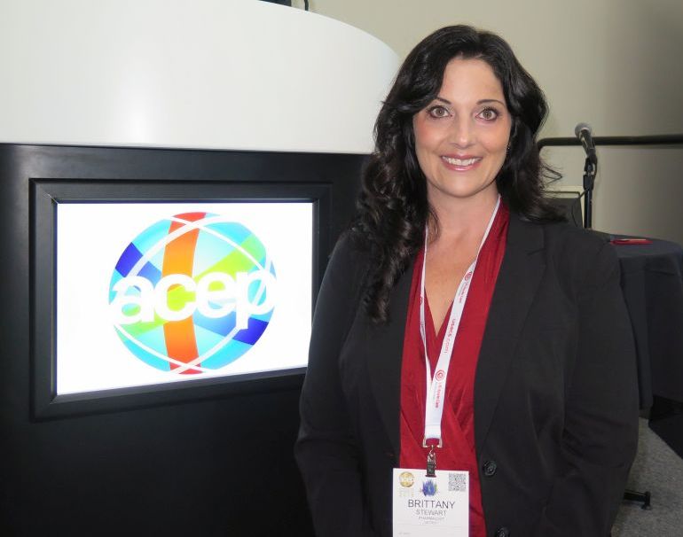
“Hypertension is the leading risk factor for cardiovascular events and stroke in this country,” Brittany Stewart, RD, PharmD, said at the annual meeting of the American College of Emergency Physicians. “The good news is that we’re not trying to figure out how to treat this disease, but the bad news is that we have 50% of people with uncontrolled high blood pressure.”
“Pharmacists are highly accessible in the community in outpatient settings,” Dr. Stewart of the department of pharmacy practice at Wayne State University, Detroit, said. “This is where ED providers can really integrate and work on a team to refer patients for their chronic diseases on an outpatient basis.”
In a prospective pilot trial, Dr. Stewart and her colleagues recruited 89 patients with uncontrolled hypertension who presented to the ED at Wayne State during May 24, 2017–May 18, 2018. Their average age was 43 years, 51% were male, 94% were black, 51% were current smokers, the mean body mass index was 34.5 kg/m2, and 18% had no health insurance.
“In Detroit, we have significant health disparities with our patient population,” she said. “They are high utilizers of the emergency department, not only for their acute illness but for chronic illness as well. There are several studies showing that team-based care has improved hypertension and blood pressure control. However, the adoption and sustainability of these models haven’t really taken off in our health care landscape yet.”
Over a period of 1.5 years, Dr. Stewart and two physicians developed a transitional care clinic, based on a collaborative practice agreement with emergency physicians. Per protocol, five follow-up visits were planned in an outpatient pharmacy clinic, where Dr. Stewart initiated and titrated antihypertensive medications and handled refills.
“The physician does not have to physically be at the clinic,” she said. “We work closely over the phone to make the best decisions, but it’s not typical ambulatory care where the physician has to be sitting right next to the pharmacist to make the best decisions for the patients.” The primary outcome was the transitional care clinic’s impact on blood pressure.
Dr. Stewart reported results from 47 medication interventions that were provided over 97 follow-up visits. The researchers found that the median blood pressure dropped from an initial reading of 160/102 mm Hg to 130/93 mm Hg by the fifth transitional care clinic visit. Across all patients, systolic blood pressure decreased by an average of 48 mm Hg. “I don’t think we were surprised by these results; we are getting people very much in need down to their blood pressure goals,” she said. “By the end of the study, patients were on an average of three antihypertensive medications.”
She and her colleagues plan to conduct a larger randomized, clinical trial of this care model. The research was supported by a faculty award from the Eugene Applebaum College of Pharmacy and Health Sciences at Wayne State University and by the National Association of Chain Drug Stores Foundation.
Source: Brody A et al. Ann Emerg Med. 2018 Oct;72;4:S36-7. doi. 10.1016/j.annemergmed.2018.08.088.
SAN DIEGO – Pharmacist-physician collaboration on hypertension management was effective for controlling blood pressure in patients discharged from an urban ED, results from a pilot study showed.
“Hypertension is the leading risk factor for cardiovascular events and stroke in this country,” Brittany Stewart, RD, PharmD, said at the annual meeting of the American College of Emergency Physicians. “The good news is that we’re not trying to figure out how to treat this disease, but the bad news is that we have 50% of people with uncontrolled high blood pressure.”
“Pharmacists are highly accessible in the community in outpatient settings,” Dr. Stewart of the department of pharmacy practice at Wayne State University, Detroit, said. “This is where ED providers can really integrate and work on a team to refer patients for their chronic diseases on an outpatient basis.”
In a prospective pilot trial, Dr. Stewart and her colleagues recruited 89 patients with uncontrolled hypertension who presented to the ED at Wayne State during May 24, 2017–May 18, 2018. Their average age was 43 years, 51% were male, 94% were black, 51% were current smokers, the mean body mass index was 34.5 kg/m2, and 18% had no health insurance.
“In Detroit, we have significant health disparities with our patient population,” she said. “They are high utilizers of the emergency department, not only for their acute illness but for chronic illness as well. There are several studies showing that team-based care has improved hypertension and blood pressure control. However, the adoption and sustainability of these models haven’t really taken off in our health care landscape yet.”
Over a period of 1.5 years, Dr. Stewart and two physicians developed a transitional care clinic, based on a collaborative practice agreement with emergency physicians. Per protocol, five follow-up visits were planned in an outpatient pharmacy clinic, where Dr. Stewart initiated and titrated antihypertensive medications and handled refills.
“The physician does not have to physically be at the clinic,” she said. “We work closely over the phone to make the best decisions, but it’s not typical ambulatory care where the physician has to be sitting right next to the pharmacist to make the best decisions for the patients.” The primary outcome was the transitional care clinic’s impact on blood pressure.
Dr. Stewart reported results from 47 medication interventions that were provided over 97 follow-up visits. The researchers found that the median blood pressure dropped from an initial reading of 160/102 mm Hg to 130/93 mm Hg by the fifth transitional care clinic visit. Across all patients, systolic blood pressure decreased by an average of 48 mm Hg. “I don’t think we were surprised by these results; we are getting people very much in need down to their blood pressure goals,” she said. “By the end of the study, patients were on an average of three antihypertensive medications.”
She and her colleagues plan to conduct a larger randomized, clinical trial of this care model. The research was supported by a faculty award from the Eugene Applebaum College of Pharmacy and Health Sciences at Wayne State University and by the National Association of Chain Drug Stores Foundation.
Source: Brody A et al. Ann Emerg Med. 2018 Oct;72;4:S36-7. doi. 10.1016/j.annemergmed.2018.08.088.
REPORTING FROM ACEP18
Key clinical point: Hypertensive patients discharged from an urban ED benefited from a pharmacist-led clinic.
Major finding: By the last follow-up visit, patients’ mean systolic blood pressure had decreased by 48 mm Hg.
Study details: A pilot study of 89 patients with uncontrolled blood pressure.
Disclosures: The research was supported by a faculty award from the Eugene Applebaum College of Pharmacy and Health Sciences at Wayne State University and by the National Association of Chain Drug Stores Foundation.
Source: Brody A et al. Ann Emerg Med. 2018 Oct;72;4:S36-7. doi. 10.1016/j.annemergmed.2018.08.088.
Half of outpatient antibiotics prescribed with no infectious disease code
based on a review of more than half a million outpatient prescriptions to more than a quarter million patients at 514 clinics around Chicago.
The researchers looked to see if prescriptions had an ICD-10 code that indicated an antibiotic; they were liberal in their approach, considering over 21,000 codes to at least possibly signal the need for an antibiotic.
Almost half the time, there was nothing in the codes related to bacterial infection: 29% of scripts were written in connection with codes for high blood pressure, annual visits, and other noninfectious disorders; 17% of prescriptions were written with no diagnosis code at all.
The study is likely the largest to date to look at outpatient antibiotic prescribing patterns in the United States, and the findings are worrisome. “Nearly half the time, clinicians have either a bad reason for prescribing antibiotics, or don’t provide a reason at all. When you consider about 80% of antibiotics are prescribed on an outpatient basis, that’s a concern,” lead investigator Jeffrey A. Linder, MD, MPH, chief of the division of general internal medicine and geriatrics at Northwestern University, Chicago, said in a written statement.
“At busy clinics, sadly, the most efficient thing to do is just call in an antibiotic prescription. We need to dig into the data more, but we believe there is a lot of antibiotic prescribing for colds, the flu, and non-specific symptoms such as just not feeling well,” he said.
With all the concern in recent years about overuse, it’s hard to imagine that prescribers are still being free and easy with antibiotics, and Dr. Linder’s study will certainly have its skeptics.
Sloppy record keeping could be one explanation for the findings. A patient could really have needed an antibiotic, but it just wasn’t captured in coding. There are also valid reasons for prescribing antibiotics over the phone, such as acne and recurrent UTIs.
Dr. Linder, however, thinks it’s more than that. He explained his study, its implications, and the next steps in an interview at ID Week, an annual scientific meeting on infectious diseases.
The 2,413 prescribers in the study included physicians, surgeons, residents, fellows, nurse practitioners, and physician assistants in general and specialty practices. Patients were a mean of 43 years old: 60% were women and 75% were white. The most common antibiotic classes were penicillins, macrolides, and cephalosporins. Prescriptions were written from November 2015 through October 2017.
The work was funded by the Agency for Healthcare Research and Quality. Dr. Linder did not have any disclosures.
SOURCE: Linder JA et al. ID Week 2018 abstract 1632.
REPORTING FROM ID WEEK 2018

Telehealth: States broaden options for locations, providers
More states are expanding their telehealth policies to reach patients, and pulling back on rigid in-person requirements.

Several state Medicaid programs now explicitly allow the home to serve as an originating site for telehealth, with 10 states – Delaware, Colorado, Maryland, Michigan, Minnesota, Montana, New York, Texas, Washington, and Wyoming – adding the home as an approved site since 2016.
In addition, 16 jurisdictions now allow schools to serve as originating sites for telehealth, although some have restrictions about when the sites are acceptable, said Mei Kwong, an attorney and executive director for the Center for Connected Health Policy and the author of the center’s Spring 2018 report on telehealth trends.
At the same time, nearly all states have now dropped Medicaid restrictions that limited reimbursable telehealth services to rural or underserved areas. Colorado, Idaho, Nebraska, New Hampshire, Nevada, and Missouri are the most recent states to remove such geographic restrictions.
“[The expanded locations are] extremely helpful in providing greater access for patients to needed services,” Ms. Kwong said in an interview. “For example, a person who has difficulty leaving his or her home for a physical or other reason, they can get care, [or] a child having a rough time in school, can seek out a mental health counselor while at school.”
More telehealth providers
In addition to expanding teleheath sites, states are increasing acceptance for telehealth providers beyond physicians. Most recently, New Jersey enacted a broad telemedicine law that includes doctors, nurses, psychologists, social workers, physician assistants, counselors, respiratory therapists, speech pathologists, and optometrists, among others. The New Jersey law addresses telemedicine practice standards, prescribing, patient consent, privacy, and other requirements for providers.
In addition, more states are carving out telehealth regulations. Since 2016, 11 states have revised or adopted new scope of practice restrictions for counselors providing telemedicine, according to a state telehealth analysis published in July 2018 by law firm Epstein, Becker, Green.
Arkansas, Idaho, Maine, New Jersey, and Rhode Island recently imposed regulations for the practice of telepsychology. In addition, seven states have new or revised scope of practice restrictions for advanced practice registered nurses (APRN) providing telehealth services, while eight states have new licensing requirements specific to telehealth practice by APRNs, according to the Epstein analysis.
A telehealth compact that would allow APRNs to practice nursing via telemedicine across state lines is also in the works. Similar to the physician Interstate Medical Licensure Compact, the APRN Compact would establish an interstate commission and guidelines for uniform licensing requirements and criminal background checks. The compact will become effective when 10 states enact the compact legislation. So far, three states – Wyoming, North Dakota, and Idaho – have enacted the model legislation.
Another telemedicine compact for psychologists is getting ready to launch. In August, Illinois became the seventh state to join the Psychology Interjurisdictional Compact (PSYPACT). The pact requires seven states to enact compact legislation to become effective, however Illinois law does not go into effect until 2020.

The developments highlight the rise in more mid- and lower-level providers practicing telemedicine, said Anjali B. Dooley, an attorney and chief legal and compliance officer for Forefront Telecare, a telehealth platform for behavioral health services. While the increase allows greater care access, the expansion also poses scope of practice challenges, she notes.
“Increasing scope of practice extensions also increases risk if physician extenders are not trained properly in telehealth technology use and protocols,” Ms. Dooley said in an interview. “Providers and provider extenders need to be educated and learned in human factors such as communication, empathy, and etiquette.”

A greater number of nonphysician telemedicine providers is beneficial as long as the providers are adhering to appropriate standards of care and consulting with supervising physicians when necessary, adds Jean R. Sumner, MD, dean of the School of Medicine at Mercer University in Macon, Georgia, and a telemedicine internist.
“The standard should always be equal to an in-person visit,” she said in an interview. “The patient has a right to know who is seeing them, too, to understand [their credentials]. They need to know the person on the telehealth unit is qualified to provide the care.”
Responding to the opioid crisis
The need for increased access to mental health care is a primary driver behind state efforts to expand the pool of telemedicine providers, adds Amy Lerman, an attorney at Epstein, Becker, Green and lead author of her firm’s report.
“The reason it is important for states to continue expanding the scope of health professionals, other than physicians, who can provide behavioral health telemedicine services, is not only to address an overall nationwide shortage of behavioral health providers, but also to expand access to behavioral health services because a wider range of providers are equipped to provide these services,” she said in an interview.
In the same vein, more states are using telehealth to address the opioid crisis, according to both the Epstein report and the Center for Connected Health Policy analysis.
In September, California enacted a law that would allow Medicaid reimbursement for certified substance use disorder counselors who provide treatment via telehealth. In August, Illinois approved a similar law that mandates reimbursement for behavioral and mental health experts who treat Medicaid patients through telehealth technologies.

The laws come after a June 2018 letter from the Centers for Medicare & Medicaid Services that encouraged states to utilize health technology efforts to address the opioid crisis, including through telemedicine and telepsychiatry, said Daniel Kim, an attorney with Epstein, Becker, Green and a coauthor of his firm’s report.
At the same time, a number of states have expanded their controlled substance laws to allow remote prescribing through telehealth for the treatment of psychiatric or substance use disorders. Connecticut’s law, for instance, allows providers to prescribe Schedule I-III controlled substances through telehealth platforms, while banning opioid prescribing. In Indiana, 2017 legislation expanded the types of controlled medications that providers can prescribe through telehealth platforms, primarily drugs used to treat or manage opioid dependence. The states join an increasing number that have enacted laws allowing the remote prescribing of controlled substances, including Delaware, Florida, Indiana, Michigan, New Hampshire, Ohio, and West Virginia.
The new laws will enhance the availability of behavioral health services, while allowing more treatment flexibility and privacy for patients, said Ms. Dooley.
“Treatment in one’s own environment where the addiction takes place is often more effective,” she said. People with addiction disorders “can also receive treatment without having to drive long distances.”
The disappearing in-person requirement
As states define their telehealth policies, they are fading out a once-prevalent requirement – the in-person visit. There is no longer a single state that requires physicians to meet with patients in-person before providing telemedicine services, according to the Epstein report.
States realized that requiring in-person visits before doctors can provide telemedicine creates a barrier to care, said Mr. Kim. A move to eliminate the requirement in Texas influenced other states in phasing out the common regulation. In the widely publicized Teladoc case, the national telemedicine company sued the Texas Medical Board in 2011 over its rule requiring Texas physicians to conduct a face-to-face evaluation before treating a patient via telemedicine. The legal battle continued for years, until Teladoc voluntarily dropped its lawsuit in 2017 after Texas adopted a new law that allowed doctors to treat first-time patients through telemedicine.
“The medical board [understood] that the in-person requirement wasn’t really a benefit to patients,” Mr. Kim said in an interview. “Once they changed it, a lot of other states have recognized the same and have moved toward getting rid of the requirement.”
In some states, midlevel providers still must see patients face-to-face before providing telehealth care. Arkansas for instance, requires that psychologists, counselors, and APRNs conduct an in-person exam before rendering telehealth. Professional boards in Colorado and Massachusetts recommend a face-to-face visit by midlevel providers as a best practice.
Reimbursement growing, but restrictions remain
Forty-nine states and the District of Columbia reimburse for some form of telehealth, mainly live video services. At least 20 states now pay providers for remote payment monitoring (RPM), according to the Center for Connected Health Policy report. The reimbursement is often restricted, however, to certain clinical conditions and/or rules that limit the type of monitoring device allowed. Colorado, for instance, only reimburses RPM for patients with congestive heart failure, chronic obstructive pulmonary disease, asthma, or diabetes and requires that the patient was hospitalized at least twice in the last 12 months for reasons associated with one of the conditions. Missouri has similar RPM criteria associated with hospitalizations, but allows for a greater number of conditions including pregnancy, stroke, and cancer.
Most states have yet to pay for store-and-forward services, technologies that enable the electronic transfer of photos, prerecorded videos, or documents. Only about 14 states reimburse for such technology, and many policies include limitations. California, for example, only reimburses for store-and-forward services in teledermatology, teledentistry, and teleophthalmology. Connecticut allows for store-and-forward payment between physicians through email. Missouri allows for store-and-forward services in orthopedics, dermatology, optometry, ophthalmology, and in cases of retinopathy, burn and wound care, dental services, and maternal-fetal ultrasounds.
Reimbursement for telehealth is still a challenge for many physicians, Dr. Sumner said. Part of the problem is the wide discrepancy in how telehealth is defined among states, she said. Some states only consider live or interactive two-way technology as telehealth, excluding services such as store-and-forward and RPM. Other states reimburse for certain technology-based services, but they do not consider them telehealth. Maryland’s Medicaid program for example, does not reimburse for store and forward under its telehealth policy. However, store and forward used in dermatology, radiology, and ophthalmology is reimbursed by Maryland under an alternate billing code, though not considered telehealth.
Establishing best practices in telehealth through careful evaluation and research would improve reimbursement, said S. David McSwain, MD, interim chief medical information officer at the Medical University of South Carolina, Charleston, and medical director for telehealth optimization.
“We can use that evidence to reduce the variation in telehealth payment policies across the states,” Dr. McSwain said in an interview. “By leveraging reimbursement models to promote best practices, we can encourage the spread of telehealth services that have the greatest impacts on patients, their families, and the health care system.”
More states are expanding their telehealth policies to reach patients, and pulling back on rigid in-person requirements.
Several state Medicaid programs now explicitly allow the home to serve as an originating site for telehealth, with 10 states – Delaware, Colorado, Maryland, Michigan, Minnesota, Montana, New York, Texas, Washington, and Wyoming – adding the home as an approved site since 2016.
In addition, 16 jurisdictions now allow schools to serve as originating sites for telehealth, although some have restrictions about when the sites are acceptable, said Mei Kwong, an attorney and executive director for the Center for Connected Health Policy and the author of the center’s Spring 2018 report on telehealth trends.
At the same time, nearly all states have now dropped Medicaid restrictions that limited reimbursable telehealth services to rural or underserved areas. Colorado, Idaho, Nebraska, New Hampshire, Nevada, and Missouri are the most recent states to remove such geographic restrictions.
“[The expanded locations are] extremely helpful in providing greater access for patients to needed services,” Ms. Kwong said in an interview. “For example, a person who has difficulty leaving his or her home for a physical or other reason, they can get care, [or] a child having a rough time in school, can seek out a mental health counselor while at school.”
More telehealth providers
In addition to expanding teleheath sites, states are increasing acceptance for telehealth providers beyond physicians. Most recently, New Jersey enacted a broad telemedicine law that includes doctors, nurses, psychologists, social workers, physician assistants, counselors, respiratory therapists, speech pathologists, and optometrists, among others. The New Jersey law addresses telemedicine practice standards, prescribing, patient consent, privacy, and other requirements for providers.
In addition, more states are carving out telehealth regulations. Since 2016, 11 states have revised or adopted new scope of practice restrictions for counselors providing telemedicine, according to a state telehealth analysis published in July 2018 by law firm Epstein, Becker, Green.
Arkansas, Idaho, Maine, New Jersey, and Rhode Island recently imposed regulations for the practice of telepsychology. In addition, seven states have new or revised scope of practice restrictions for advanced practice registered nurses (APRN) providing telehealth services, while eight states have new licensing requirements specific to telehealth practice by APRNs, according to the Epstein analysis.
A telehealth compact that would allow APRNs to practice nursing via telemedicine across state lines is also in the works. Similar to the physician Interstate Medical Licensure Compact, the APRN Compact would establish an interstate commission and guidelines for uniform licensing requirements and criminal background checks. The compact will become effective when 10 states enact the compact legislation. So far, three states – Wyoming, North Dakota, and Idaho – have enacted the model legislation.
Another telemedicine compact for psychologists is getting ready to launch. In August, Illinois became the seventh state to join the Psychology Interjurisdictional Compact (PSYPACT). The pact requires seven states to enact compact legislation to become effective, however Illinois law does not go into effect until 2020.
The developments highlight the rise in more mid- and lower-level providers practicing telemedicine, said Anjali B. Dooley, an attorney and chief legal and compliance officer for Forefront Telecare, a telehealth platform for behavioral health services. While the increase allows greater care access, the expansion also poses scope of practice challenges, she notes.
“Increasing scope of practice extensions also increases risk if physician extenders are not trained properly in telehealth technology use and protocols,” Ms. Dooley said in an interview. “Providers and provider extenders need to be educated and learned in human factors such as communication, empathy, and etiquette.”
A greater number of nonphysician telemedicine providers is beneficial as long as the providers are adhering to appropriate standards of care and consulting with supervising physicians when necessary, adds Jean R. Sumner, MD, dean of the School of Medicine at Mercer University in Macon, Georgia, and a telemedicine internist.
“The standard should always be equal to an in-person visit,” she said in an interview. “The patient has a right to know who is seeing them, too, to understand [their credentials]. They need to know the person on the telehealth unit is qualified to provide the care.”
Responding to the opioid crisis
The need for increased access to mental health care is a primary driver behind state efforts to expand the pool of telemedicine providers, adds Amy Lerman, an attorney at Epstein, Becker, Green and lead author of her firm’s report.
“The reason it is important for states to continue expanding the scope of health professionals, other than physicians, who can provide behavioral health telemedicine services, is not only to address an overall nationwide shortage of behavioral health providers, but also to expand access to behavioral health services because a wider range of providers are equipped to provide these services,” she said in an interview.
In the same vein, more states are using telehealth to address the opioid crisis, according to both the Epstein report and the Center for Connected Health Policy analysis.
In September, California enacted a law that would allow Medicaid reimbursement for certified substance use disorder counselors who provide treatment via telehealth. In August, Illinois approved a similar law that mandates reimbursement for behavioral and mental health experts who treat Medicaid patients through telehealth technologies.
The laws come after a June 2018 letter from the Centers for Medicare & Medicaid Services that encouraged states to utilize health technology efforts to address the opioid crisis, including through telemedicine and telepsychiatry, said Daniel Kim, an attorney with Epstein, Becker, Green and a coauthor of his firm’s report.
At the same time, a number of states have expanded their controlled substance laws to allow remote prescribing through telehealth for the treatment of psychiatric or substance use disorders. Connecticut’s law, for instance, allows providers to prescribe Schedule I-III controlled substances through telehealth platforms, while banning opioid prescribing. In Indiana, 2017 legislation expanded the types of controlled medications that providers can prescribe through telehealth platforms, primarily drugs used to treat or manage opioid dependence. The states join an increasing number that have enacted laws allowing the remote prescribing of controlled substances, including Delaware, Florida, Indiana, Michigan, New Hampshire, Ohio, and West Virginia.
The new laws will enhance the availability of behavioral health services, while allowing more treatment flexibility and privacy for patients, said Ms. Dooley.
“Treatment in one’s own environment where the addiction takes place is often more effective,” she said. People with addiction disorders “can also receive treatment without having to drive long distances.”
The disappearing in-person requirement
As states define their telehealth policies, they are fading out a once-prevalent requirement – the in-person visit. There is no longer a single state that requires physicians to meet with patients in-person before providing telemedicine services, according to the Epstein report.
States realized that requiring in-person visits before doctors can provide telemedicine creates a barrier to care, said Mr. Kim. A move to eliminate the requirement in Texas influenced other states in phasing out the common regulation. In the widely publicized Teladoc case, the national telemedicine company sued the Texas Medical Board in 2011 over its rule requiring Texas physicians to conduct a face-to-face evaluation before treating a patient via telemedicine. The legal battle continued for years, until Teladoc voluntarily dropped its lawsuit in 2017 after Texas adopted a new law that allowed doctors to treat first-time patients through telemedicine.
“The medical board [understood] that the in-person requirement wasn’t really a benefit to patients,” Mr. Kim said in an interview. “Once they changed it, a lot of other states have recognized the same and have moved toward getting rid of the requirement.”
In some states, midlevel providers still must see patients face-to-face before providing telehealth care. Arkansas for instance, requires that psychologists, counselors, and APRNs conduct an in-person exam before rendering telehealth. Professional boards in Colorado and Massachusetts recommend a face-to-face visit by midlevel providers as a best practice.
Reimbursement growing, but restrictions remain
Forty-nine states and the District of Columbia reimburse for some form of telehealth, mainly live video services. At least 20 states now pay providers for remote payment monitoring (RPM), according to the Center for Connected Health Policy report. The reimbursement is often restricted, however, to certain clinical conditions and/or rules that limit the type of monitoring device allowed. Colorado, for instance, only reimburses RPM for patients with congestive heart failure, chronic obstructive pulmonary disease, asthma, or diabetes and requires that the patient was hospitalized at least twice in the last 12 months for reasons associated with one of the conditions. Missouri has similar RPM criteria associated with hospitalizations, but allows for a greater number of conditions including pregnancy, stroke, and cancer.
Most states have yet to pay for store-and-forward services, technologies that enable the electronic transfer of photos, prerecorded videos, or documents. Only about 14 states reimburse for such technology, and many policies include limitations. California, for example, only reimburses for store-and-forward services in teledermatology, teledentistry, and teleophthalmology. Connecticut allows for store-and-forward payment between physicians through email. Missouri allows for store-and-forward services in orthopedics, dermatology, optometry, ophthalmology, and in cases of retinopathy, burn and wound care, dental services, and maternal-fetal ultrasounds.
Reimbursement for telehealth is still a challenge for many physicians, Dr. Sumner said. Part of the problem is the wide discrepancy in how telehealth is defined among states, she said. Some states only consider live or interactive two-way technology as telehealth, excluding services such as store-and-forward and RPM. Other states reimburse for certain technology-based services, but they do not consider them telehealth. Maryland’s Medicaid program for example, does not reimburse for store and forward under its telehealth policy. However, store and forward used in dermatology, radiology, and ophthalmology is reimbursed by Maryland under an alternate billing code, though not considered telehealth.
Establishing best practices in telehealth through careful evaluation and research would improve reimbursement, said S. David McSwain, MD, interim chief medical information officer at the Medical University of South Carolina, Charleston, and medical director for telehealth optimization.
“We can use that evidence to reduce the variation in telehealth payment policies across the states,” Dr. McSwain said in an interview. “By leveraging reimbursement models to promote best practices, we can encourage the spread of telehealth services that have the greatest impacts on patients, their families, and the health care system.”