User login
Young Surgeons: K08, K23 Grants See Changes
The National Heart, Lung and Blood institute has extended the combined number of years of K training support from six to eight years for the K08 and K23 grants. Clinician scientists who have received either award can stay on a K12 or KL2 program for up to three years and then request a five-year individual K award. This will help support the transition from training to independent investigators for those clinicians.
The National Heart, Lung and Blood institute has extended the combined number of years of K training support from six to eight years for the K08 and K23 grants. Clinician scientists who have received either award can stay on a K12 or KL2 program for up to three years and then request a five-year individual K award. This will help support the transition from training to independent investigators for those clinicians.
The National Heart, Lung and Blood institute has extended the combined number of years of K training support from six to eight years for the K08 and K23 grants. Clinician scientists who have received either award can stay on a K12 or KL2 program for up to three years and then request a five-year individual K award. This will help support the transition from training to independent investigators for those clinicians.
Promising novel antidepressant cruising in pipeline
BARCELONA – An investigational antidepressant known for now simply as MIN-117 shows the potential – at least, in phase 2 development – of offering significant advantages over currently available antidepressants in patients with major depressive disorder, Michael Davidson, MD, said at the annual congress of the European College of Neuropsychopharmacology.
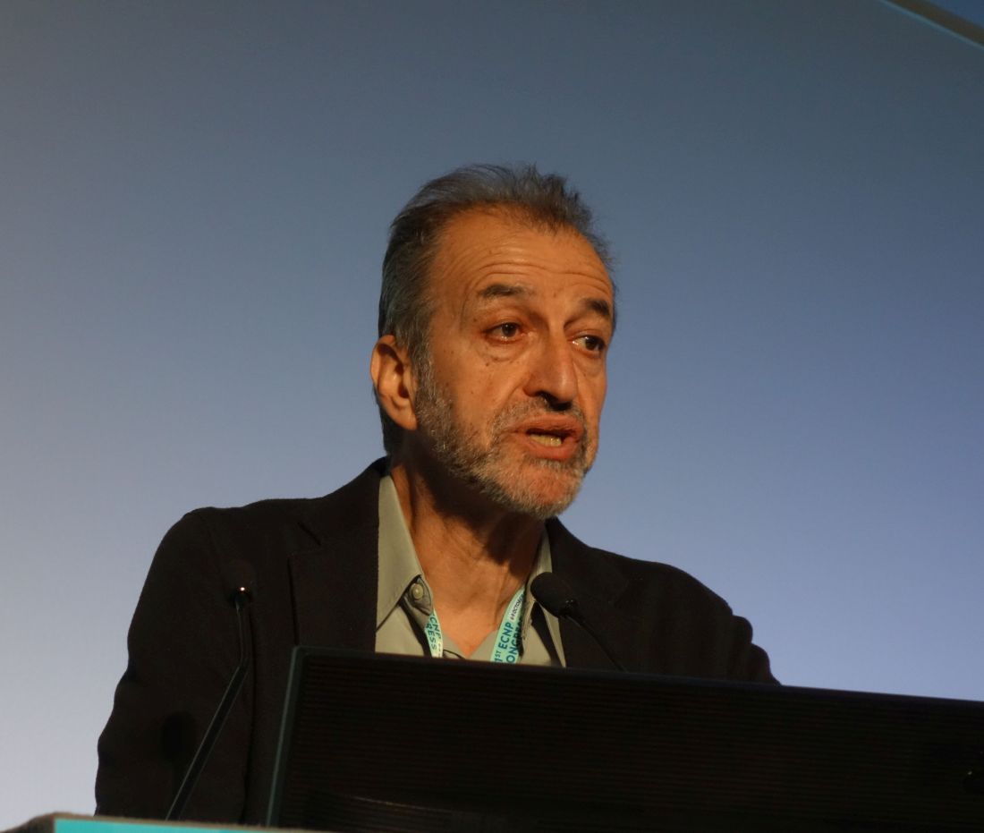
“This is a compound with a very, very rich pharmacology – so rich that we don’t know exactly which of the pharmacologic effects are really making the difference. This rich pharmacology is related to pathways that may confer to MIN-117 a unique positioning in the field of antidepressants and address unmet medical needs not well covered by existing therapies. For example, faster onset of action, complete restoration of euthymia, and beneficial effects on cognition and sexual functioning,” according to Michael Davidson, MD, chief medical officer at Minerva Neurosciences, which is developing the drug..
In the completed phase 2 study, the drug also displayed a strong anxiolytic effect and no prolongation of REM sleep latency in polysomnographic testing.
“So it may be that this is the first antidepressant which does not affect sleep architecture,” observed Dr. Davidson, professor of psychiatry at the Sackler School of Medicine in Tel Aviv.
Preclinical studies established that MIN-117 has high affinity for the 5HT serotonin transporter, serotonin 1A and 2A receptors, the dopamine transporter, and the alpha-1A and -1B adrenergic receptors. In animal models of depression, the drug results in sustained release of dopamine and serotonin. In human studies, the drug has a long half-life of roughly 60 hours.
“One possibility is that MIN-117 will be administered not once a day, but once or twice a week. It may be that here we have an antidepressant that doesn’t have to be administered every day,” the psychiatrist said.
In the completed phase 2 study, however, the drug was given once daily in what he described as a “classic design” for an antidepressant clinical trial: a 4-week washout period, then 6 weeks of double-blind treatment with MIN-117 at 2.5 or 0.5 mg/day, paroxetine at 20 mg/day, or placebo, then a 2-week posttreatment follow-up phase.
In describing the results of the study of 84 patients with major depressive disorder, Dr. Davidson painted a picture of MIN-117’s safety and efficacy with broad strokes because the trial wasn’t powered to demonstrate statistically significant differences. But the treatment effect sizes for the novel drug were impressive. A much more complete picture of the safety and efficacy of MIN-117 will be provided by an ongoing 324-patient phase 2b multicenter U.S. and European trial of the drug given at 2.5 or 5 mg/day or placebo.
The primary endpoint in the completed trial was change from baseline to 6 weeks in mean Montgomery-Åsberg Rating Depression Scale (MADRS) score. From a baseline score of about 33, MADRS scores improved by 9 points with placebo, 11 with MIN-117 at 0.5 mg, and by 12 points with 2.5 mg of the drug. MIN-117 was superior to placebo in this regard from the time of the earliest assessment, at 2 weeks.
One-quarter of patients on MIN-117 at 2.5 mg/day achieved remission, prospectively defined as a MADRS score below 12. Remission was 2.1-fold more likely in this group than with placebo at week 4 and 3.1-fold more likely at 6 weeks. The remission rate with MIN-117 also was better than with paroxetine.
“So , and that we’ll be able to produce full remission of the depressive symptoms,” Dr. Davidson said.
MIN-117 also showed a solid anxiolytic effect, with mean scores on the Hamilton Anxiety Rating Scale – a secondary endpoint – improving by 10 points in both the 0.5- and 2.5-mg groups from a baseline of 26 points. As a result of this impressive showing, the large ongoing phase 2b study is recruiting patients with an ongoing episode of major depressive disorder and prominent secondary anxiety.
Both doses of MIN-117 were well tolerated. Scores on the Arizona Sexual Experiences Scale showed that the drug did not result in any impairment of sexual function. Nor was MIN-117 associated with evidence of cognitive impairment; in fact, scores on the Digit-Symbol Substitution Test and Digit Span Backwards tool were better than in the placebo arm.
The study was sponsored by Minerva Neurosciences and presented by the company’s chief medical officer.
BARCELONA – An investigational antidepressant known for now simply as MIN-117 shows the potential – at least, in phase 2 development – of offering significant advantages over currently available antidepressants in patients with major depressive disorder, Michael Davidson, MD, said at the annual congress of the European College of Neuropsychopharmacology.
“This is a compound with a very, very rich pharmacology – so rich that we don’t know exactly which of the pharmacologic effects are really making the difference. This rich pharmacology is related to pathways that may confer to MIN-117 a unique positioning in the field of antidepressants and address unmet medical needs not well covered by existing therapies. For example, faster onset of action, complete restoration of euthymia, and beneficial effects on cognition and sexual functioning,” according to Michael Davidson, MD, chief medical officer at Minerva Neurosciences, which is developing the drug..
In the completed phase 2 study, the drug also displayed a strong anxiolytic effect and no prolongation of REM sleep latency in polysomnographic testing.
“So it may be that this is the first antidepressant which does not affect sleep architecture,” observed Dr. Davidson, professor of psychiatry at the Sackler School of Medicine in Tel Aviv.
Preclinical studies established that MIN-117 has high affinity for the 5HT serotonin transporter, serotonin 1A and 2A receptors, the dopamine transporter, and the alpha-1A and -1B adrenergic receptors. In animal models of depression, the drug results in sustained release of dopamine and serotonin. In human studies, the drug has a long half-life of roughly 60 hours.
“One possibility is that MIN-117 will be administered not once a day, but once or twice a week. It may be that here we have an antidepressant that doesn’t have to be administered every day,” the psychiatrist said.
In the completed phase 2 study, however, the drug was given once daily in what he described as a “classic design” for an antidepressant clinical trial: a 4-week washout period, then 6 weeks of double-blind treatment with MIN-117 at 2.5 or 0.5 mg/day, paroxetine at 20 mg/day, or placebo, then a 2-week posttreatment follow-up phase.
In describing the results of the study of 84 patients with major depressive disorder, Dr. Davidson painted a picture of MIN-117’s safety and efficacy with broad strokes because the trial wasn’t powered to demonstrate statistically significant differences. But the treatment effect sizes for the novel drug were impressive. A much more complete picture of the safety and efficacy of MIN-117 will be provided by an ongoing 324-patient phase 2b multicenter U.S. and European trial of the drug given at 2.5 or 5 mg/day or placebo.
The primary endpoint in the completed trial was change from baseline to 6 weeks in mean Montgomery-Åsberg Rating Depression Scale (MADRS) score. From a baseline score of about 33, MADRS scores improved by 9 points with placebo, 11 with MIN-117 at 0.5 mg, and by 12 points with 2.5 mg of the drug. MIN-117 was superior to placebo in this regard from the time of the earliest assessment, at 2 weeks.
One-quarter of patients on MIN-117 at 2.5 mg/day achieved remission, prospectively defined as a MADRS score below 12. Remission was 2.1-fold more likely in this group than with placebo at week 4 and 3.1-fold more likely at 6 weeks. The remission rate with MIN-117 also was better than with paroxetine.
“So , and that we’ll be able to produce full remission of the depressive symptoms,” Dr. Davidson said.
MIN-117 also showed a solid anxiolytic effect, with mean scores on the Hamilton Anxiety Rating Scale – a secondary endpoint – improving by 10 points in both the 0.5- and 2.5-mg groups from a baseline of 26 points. As a result of this impressive showing, the large ongoing phase 2b study is recruiting patients with an ongoing episode of major depressive disorder and prominent secondary anxiety.
Both doses of MIN-117 were well tolerated. Scores on the Arizona Sexual Experiences Scale showed that the drug did not result in any impairment of sexual function. Nor was MIN-117 associated with evidence of cognitive impairment; in fact, scores on the Digit-Symbol Substitution Test and Digit Span Backwards tool were better than in the placebo arm.
The study was sponsored by Minerva Neurosciences and presented by the company’s chief medical officer.
REPORTING FROM THE ECNP CONGRESS
Key clinical point: An investigational antidepressant might address important unmet needs in the treatment of major depression.
Major finding: Patients with major depressive disorder were 3.1-fold more likely to be in remission after 6 weeks on MIN-117 at 2.5 mg/day than with placebo.
Study details: This prospective, double-blind, randomized, double-blind, placebo- and active-controlled study included 84 patients with major depressive disorder.
Disclosures: The study was sponsored by Minerva Neurosciences and presented by the company’s chief medical officer.
Small Daily Steps Can Keep Heart Attacks at Bay
Despite being largely preventable, heart attacks, strokes, heart failure, and related conditions caused 2.2 million hospitalizations and 415,000 deaths in 2016, according to a Vital Signs report. Many of the events were in adults aged 35 to 64 years—middle-aged adults who would not normally be considered at risk.
But “many opportunities to find and treat risk factors are missed every day,” the CDC says. “Many of these [cardiovascular] events can be prevented through daily actions to help lower risk and better manage medical conditions,” said Dr. Anne Schuchat, principal deputy director of CDC. For instance, the report reveals that:
- 9 million American adults are not yet taking aspirin as recommended
- 40 million adults with high blood pressure are not yet under safe control
- 39 million adults can benefit from managing their cholesterol
- 54 million adults are smokers
- 71 million adults are not physically active
The CDC recommends that health care professionals can help by focusing on the ABCS (aspirin, blood pressure, cholesterol, smoking cessation), and using technology, customized processes, and the “skills of everyone in the health care system” to find and fill gaps in care.
Researchers say rethink ‘arbitrary categorization’ of VTE risk

Danish researchers conducted a large, 16-year study of patients with venous thromboembolism (VTE) and found a high recurrence risk in all types of VTE.
At 6 months of follow-up, patients with unprovoked and provoked VTE had similar risk of recurrence, lower than that for patients with cancer-related VTE.
But at a 10-year follow-up, patients with unprovoked VTE had a recurrence risk similar to cancer-related VTE.
Based on these findings, the investigators concluded that risk stratification for these patients needs to be optimized.
“Our findings indicate that we may need to rethink arbitrary categorization, considering the heterogeneity of patients with venous thromboembolism,” they wrote.
They published their findings in The American Journal of Medicine.
The investigators used data from 3 nationwide Danish registries to analyze the risk of recurrent VTE in 73,993 patients with incident VTE. They stratified the patients according to whether the VTE was unprovoked, provoked, or cancer-related.
Provoked VTE occurs in patients without cancer but with other contributing factors such as surgery or trauma, and unprovoked VTE occurs without well-known provoking risk factors. Investigators did not include non-melanoma skin cancer in the cancer-related VTE classification.
Median age of the study population was 62.3 years and 54.1% were women.
During a median follow-up of 3.7 years, 9,205 patients experienced a recurrent event.
At the 6-month follow-up, the recurrence rates per 100 person-years were 6.92 for provoked, 6.80 for unprovoked, and 9.06 for cancer-related VTE.
And at the 10-year follow-up, recurrence rates were 2.22 (provoked), 2.84 (unprovoked), and 3.70 (cancer-related). This corresponded to an 18% higher adjusted relative risk of recurrence for patients with unprovoked VTE than patients with provoked VTE.
The investigators observed that at 10 years, the recurrence risk following an unprovoked VTE resembled the risk of patients with cancer-related VTE.
They suggested the mechanism for this could be a “rebound thrombosis” caused by a discontinuation of oral anticoagulants after an unprovoked VTE.
The investigators noted that the findings persisted through various sensitivity analyses “conducted to challenge the robustness of our finding.”
Two areas of concern, they wrote, arise from these findings: how long to anticoagulate patients and which patients to treat.
"Optimal duration of anticoagulation is a pivotal and an ongoing scientific and clinical concern," explained lead investigator Ida Ehlers Albertsen, MD, of Aalborg University in Denmark.
"The emergence of the non-vitamin K antagonist oral anticoagulants has changed the landscape for prevention of thrombosis, and contemporary risk stratification approaches may need to be adjusted according to these effective and safer agents."
The Unsaid Dangers of NSAIDs
Q) Many total joint replacements and other orthopedic procedures are performed at the surgical center where I work. To decrease the use of narcotics, the anesthesiology department often uses IV push ketorolac postop. Our nephrology colleagues in the community are unhappy about this—but we think they’re overreacting, since these patients are often generally healthy. Is there any data on the use of ketorolac and orthopedic surgery?
All medications have associated risks. For example, while therapeutic dosages for a limited time are considered safe and effective, prolonged use of any NSAID can increase the risk for acute kidney injury (AKI) or chronic kidney disease (CKD) progression. We tend to associate these issues only with patients who are at higher risk for CKD: those who are older or who have diabetes or hypertension.
Thus, it was shocking to read a clinical report on four previously healthy young adults who were admitted for AKI three to four days after postoperative administration of ketorolac. None of these patients had risk factors that would predispose them to kidney disease. All had complained of gastrointestinal symptoms along with mild dehydration and flank pain; one young man even required a kidney biopsy and dialysis. All four did eventually recover kidney function. 1
Continue to: Ketorolac—like most NSAIDs...
Ketorolac—like most NSAIDs—can affect kidney function, decreasing renal plasma flow and causing a dysfunction in salt and water balance. Postoperative patients may have activity limitations (eg, the young healthy patient on crutches). Factor in kidney damage from presurgical/outpatient
With the opioid crisis at the forefront of national health news, nonnarcotic alternatives for pain control are much in demand. This puts a whole new population at risk for AKI. Educate patients and their families about preventive measures, such as controlling nausea, maintaining hydration, and monitoring urine output. Fever, flank pain, or any untoward symptoms should be reported. Remember, AKI may be more common in the older patient with diabetes—but it can occur in anyone. —EA
Ellen Apple
Dickson Schools Family Clinic, Tennessee
1. Mariano F, Cogno C, Giaretta F, et al. Urinary protein profiles in ketorolac-associated acute kidney injury in patients undergoing orthopedic day surgery. Int J Nephrol Renovasc Dis. 2017;10:269-274.
Clinician Reviews in partnership with
The National Kidney Foundation Council of Advanced Practitioners' (NKF-CAP) mission is to serve as an advisory resource for the NKF, nurse practitioners, physician assistants, clinical nurse specialists, and the community in advancing the care, treatment, and education of patients with kidney disease and their families. CAP is an advocate for professional development, research, and health policies that impact the delivery of patient care and professional practice. For more information on NKF-CAP, visit www.kidney.org/CAP. Renal Consult is edited by Jane S. Davis, CRNP, DNP, a member of the Clinician Reviews editorial board, who is a nurse practitioner in the Division of Nephrology at the University of Alabama at Birmingham and is the communications chairperson for the National Kidney Foundation's Council of Advanced Practitioners (NKF-CAP); and Kim Zuber, PA-C, MSPS, DFAAPA, a semi-retired PA who works with the American Academy of Nephrology PAs and is a past chair of the NKF-CAP. Clinician Reviews is the proud recipient of NKF-CAP’s Nostradamus Award, recognizing the journal’s forethought and vision in supporting the contributions of Advanced Practitioners in nephrology. This month's column was authored by Ellen Apple, MSN, APRN, FNP-C, who practices at the Dickson Schools Family Clinic in Tennessee.
Q) Many total joint replacements and other orthopedic procedures are performed at the surgical center where I work. To decrease the use of narcotics, the anesthesiology department often uses IV push ketorolac postop. Our nephrology colleagues in the community are unhappy about this—but we think they’re overreacting, since these patients are often generally healthy. Is there any data on the use of ketorolac and orthopedic surgery?
All medications have associated risks. For example, while therapeutic dosages for a limited time are considered safe and effective, prolonged use of any NSAID can increase the risk for acute kidney injury (AKI) or chronic kidney disease (CKD) progression. We tend to associate these issues only with patients who are at higher risk for CKD: those who are older or who have diabetes or hypertension.
Thus, it was shocking to read a clinical report on four previously healthy young adults who were admitted for AKI three to four days after postoperative administration of ketorolac. None of these patients had risk factors that would predispose them to kidney disease. All had complained of gastrointestinal symptoms along with mild dehydration and flank pain; one young man even required a kidney biopsy and dialysis. All four did eventually recover kidney function. 1
Continue to: Ketorolac—like most NSAIDs...
Ketorolac—like most NSAIDs—can affect kidney function, decreasing renal plasma flow and causing a dysfunction in salt and water balance. Postoperative patients may have activity limitations (eg, the young healthy patient on crutches). Factor in kidney damage from presurgical/outpatient
With the opioid crisis at the forefront of national health news, nonnarcotic alternatives for pain control are much in demand. This puts a whole new population at risk for AKI. Educate patients and their families about preventive measures, such as controlling nausea, maintaining hydration, and monitoring urine output. Fever, flank pain, or any untoward symptoms should be reported. Remember, AKI may be more common in the older patient with diabetes—but it can occur in anyone. —EA
Ellen Apple
Dickson Schools Family Clinic, Tennessee
1. Mariano F, Cogno C, Giaretta F, et al. Urinary protein profiles in ketorolac-associated acute kidney injury in patients undergoing orthopedic day surgery. Int J Nephrol Renovasc Dis. 2017;10:269-274.
1. Mariano F, Cogno C, Giaretta F, et al. Urinary protein profiles in ketorolac-associated acute kidney injury in patients undergoing orthopedic day surgery. Int J Nephrol Renovasc Dis. 2017;10:269-274.

FDA approves new treatment for hereditary transthyretin-mediated amyloidosis
, the second treatment approved in 2 months for the rare genetic disorder.

Inotersen is a transthyretin-directed antisense oligonucleotide that inhibits production of the transthyretin (TTR) protein (amyloid), according to the FDA statement announcing the approval on Oct. 5. It is administered in a subcutaneous injection once a week and will be marketed as Tegsedi. In August, the FDA approved the first treatment for hATTR, patisiran (Onpattro).
The FDA statement described hATTR as a “rare, debilitating, and often fatal genetic disease characterized by the buildup of abnormal amyloid protein in peripheral nerves, the heart, and other organs,” with signs and symptoms that include effects on “sensation (pain and temperature), autonomic function (blood pressure changes and bowel and digestive problems), and muscle strength (weakness and immobility in the arms, legs, hands, and feet),” caused by amyloid deposition in the peripheral nervous system.
The mechanism-of-action section of the prescribing information states that inotersen “causes degradation of mutant and wild-type TTR mRNA through binding to the TTR mRNA, which results in a reduction of serum TTR protein and TTR protein deposits in tissues.”
A statement issued by the manufacturer, Ionis Pharmaceuticals, said that approval was based on a phase 3 study of patients with hATTR amyloidosis and polyneuropathy symptoms, which found significantly greater benefits among those treated with inotersen, compared with placebo, using primary endpoints that measured quality of life related to neuropathy and a measure of neuropathic disease progression.
Contraindications to inotersen include a history of acute glomerulonephritis caused by inotersen, history of a hypersensitivity reaction to the agent, and a platelet count below 100 × 109/L; the label includes a boxed warning about thrombocytopenia and glomerulonephritis associated with treatment. The drug also is being marketed with a risk evaluation and mitigation strategy (REMS).
Patisiran, the treatment approved in August, results in “degradation of mutant and wild-type TTR mRNA through RNA interference, which results in a reduction of serum TTR protein and TTR protein deposits in tissues,” according to its prescribing information.
, the second treatment approved in 2 months for the rare genetic disorder.
Inotersen is a transthyretin-directed antisense oligonucleotide that inhibits production of the transthyretin (TTR) protein (amyloid), according to the FDA statement announcing the approval on Oct. 5. It is administered in a subcutaneous injection once a week and will be marketed as Tegsedi. In August, the FDA approved the first treatment for hATTR, patisiran (Onpattro).
The FDA statement described hATTR as a “rare, debilitating, and often fatal genetic disease characterized by the buildup of abnormal amyloid protein in peripheral nerves, the heart, and other organs,” with signs and symptoms that include effects on “sensation (pain and temperature), autonomic function (blood pressure changes and bowel and digestive problems), and muscle strength (weakness and immobility in the arms, legs, hands, and feet),” caused by amyloid deposition in the peripheral nervous system.
The mechanism-of-action section of the prescribing information states that inotersen “causes degradation of mutant and wild-type TTR mRNA through binding to the TTR mRNA, which results in a reduction of serum TTR protein and TTR protein deposits in tissues.”
A statement issued by the manufacturer, Ionis Pharmaceuticals, said that approval was based on a phase 3 study of patients with hATTR amyloidosis and polyneuropathy symptoms, which found significantly greater benefits among those treated with inotersen, compared with placebo, using primary endpoints that measured quality of life related to neuropathy and a measure of neuropathic disease progression.
Contraindications to inotersen include a history of acute glomerulonephritis caused by inotersen, history of a hypersensitivity reaction to the agent, and a platelet count below 100 × 109/L; the label includes a boxed warning about thrombocytopenia and glomerulonephritis associated with treatment. The drug also is being marketed with a risk evaluation and mitigation strategy (REMS).
Patisiran, the treatment approved in August, results in “degradation of mutant and wild-type TTR mRNA through RNA interference, which results in a reduction of serum TTR protein and TTR protein deposits in tissues,” according to its prescribing information.
Oral flu vaccine protects, evokes mucosal immunity
SAN FRANCISCO – In a phase II study, Vaxart’s oral flu vaccine was compared with a commercial injectable quadrivalent flu vaccine or placebo. The study found rates of illness were comparable between the oral vaccine and quadrivalent vaccinated groups.*

The recombinant adenovirus-based vaccine expresses hemagglutinin. It elicited a mucosal immune response, hinting that the mechanism of protection in flu vaccines may be dependent on the route of administration. It is also believed that a strong mucosal response is key to preventing future infections.
In an interview at IDWeek 2018, an annual scientific meeting on infectious diseases, Nikita Kolhatkar, PhD, a salaried employee of Vaxart, which makes the drug, describes the results of the study and explains the potential advantages of an oral flu vaccine versus a traditional injectable one. The oral formulation is cell-based and so is not vulnerable to the mutation and genetic drift that can occur in egg-based vaccines.
It is also more stable and, of course, less invasive than injectable vaccines, according to Dr. Kolhatkar.
*Correction, 10/9/2018: An earlier vs. of this article did not stress the comparability.
SOURCE: Kolhatkar N. IDWeek 2018. Poster abstract 1947.
SAN FRANCISCO – In a phase II study, Vaxart’s oral flu vaccine was compared with a commercial injectable quadrivalent flu vaccine or placebo. The study found rates of illness were comparable between the oral vaccine and quadrivalent vaccinated groups.*
The recombinant adenovirus-based vaccine expresses hemagglutinin. It elicited a mucosal immune response, hinting that the mechanism of protection in flu vaccines may be dependent on the route of administration. It is also believed that a strong mucosal response is key to preventing future infections.
In an interview at IDWeek 2018, an annual scientific meeting on infectious diseases, Nikita Kolhatkar, PhD, a salaried employee of Vaxart, which makes the drug, describes the results of the study and explains the potential advantages of an oral flu vaccine versus a traditional injectable one. The oral formulation is cell-based and so is not vulnerable to the mutation and genetic drift that can occur in egg-based vaccines.
It is also more stable and, of course, less invasive than injectable vaccines, according to Dr. Kolhatkar.
*Correction, 10/9/2018: An earlier vs. of this article did not stress the comparability.
SOURCE: Kolhatkar N. IDWeek 2018. Poster abstract 1947.
REPORTING FROM IDWEEK 2018

Acne more common in adults with hidradenitis suppurativa
, according to Sara Wertenteil and her colleagues in the department of dermatology at Hofstra University in Hempstead, N.Y.
Using data collected by IBM Watson Health, the study authors examined a total of 48,050 adults with HS and 16.9 million adults in the general U.S. population. In this study population, 15.2% of adults with HS had acne, compared with only 2.9% of adults in the general population (P less than .0001), the investigators wrote in the Journal of the American Academy of Dermatology.
After adjusting for age, sex, obesity, smoking status, and polycystic ovarian syndrome (PCOS) status, the odds ratio of adults with HS having acne was 4.51 over those without HS (95% confidence interval, 4.40-4.63). In all subgroups measured (male; female; adults aged 18-44 years, 45-64 years, and 65 years and older; white; nonwhite; obese; nonobese; smoker; nonsmoker; positive for PCOS; non-PCOS) adults with HS were significantly more likely to have acne. The strongest association was in patients who were aged 65 years and older (odds ratio, 10.14; 95% CI, 8.97-11.46).
“Patients with HS have an increased prevalence of [acne vulgaris]. Clinicians treating HS patients should be aware of this burden and its potential implications including a further impact on quality of life. Management strategies should include consideration of both conditions, either with treatments that have overlapping efficacy, or with concomitant therapies,” the authors concluded.
The study was sponsored in part by AbbVie. One coauthor reported having served as an advisor for AbbVie, Pfizer, Janssen, and Asana Biosciences.
SOURCE: Wertentiel S et al. J Am Acad Dermatol. 2018 Oct 1. doi: 10.1016/j.jaad.2018.09.040.
FROM THE JOURNAL OF THE AMERICAN ACADEMY OF DERMATOLOGY
How to vaccinate patients on biologics
SAN FRANCISCO – The new herpes zoster subunit vaccine (Shingrix) is on the short list of essential vaccines for immunocompromised adults, including those on biologics.
Ongoing research is demonstrating efficacy and safety in renal transplants patients, as well as those with hematologic cancer and stem cell transplants, according to Lorry Rubin, MD, director of pediatric infectious diseases at Cohen Children’s Medical Center, Queens, and professor of pediatrics at Hofstra University, Hempstead, N.Y.
Immunocompromised people, including those on biologics, should be immunized against a variety of diseases just like everyone else, but it’s tricky. There’s considerable variability in how biologics affect the immune system and subsequent vaccine potency. Timing is important, and although live vaccines are generally a no-go, there’s one class of biologics with which they’re safe, he said.
In a wide-ranging interview at IDWeek 2018, an annual scientific meeting on infectious diseases, Dr. Rubin shared his advice on immunizing the immunocompromised, including the other vaccines on the short list. He also tackled the common concern that vaccinations might trigger rejection in transplant patients.
He’s well qualified to address the issues: Dr. Rubin was lead author on the 2013 Infectious Diseases Society of America guidelines on vaccinating immunocompromised patients.
REPORTING FROM IDWEEK 2018

DAAs top PEG/RBV for reducing HCV cardiovascular risk
SAN FRANCISCO – , according to a review of over 30,000 patients in the ERCHIVES (Electronically Retrieved Cohort of HCV Infected Veterans) database of the Veterans Health Administration system.
In the study, 12,667 patients were treated with direct-acting antiretrovirals (DAAs) and 4,436 with pegylated interferon/ribavirin (PEG/RBV). Each subject was matched to an untreated control based on alcohol use, diabetes, and other confounders. Patients with HIV, hepatitis B, or previously diagnosed cardiovascular disease were excluded.
Over a follow-up of about 10 years, there were 2,361 strokes, heart attacks, or other cardiovascular (CV) events among untreated patients, which translated to an incidence of 30.9 events per 1,000 patient years. In the PEG/RBV group, there were 804 events, yielding an incidence of 23.5 per 1,000 patient years. The DAA group fared better, with 435 events and an incident rate of 16.3.
Sustained virologic response also was associated with lower CV risk, and the odds of attaining it were about 25% greater with DAAs vs. PEG/RBV. That might have played a role in the findings, since hepatitis C is known to be associated with CV disease and the virus has been found in atherosclerotic plaques.
Past investigations have been mixed on whether or not hepatitis C virus treatment reduces CV risk, but lead investigator Adeel Ajwad Butt, MD, professor of medicine at Cornell University, New York, said that his study was stronger than what has come before. He explained why, and also why the findings matter, in an interview at ID Week 2018, an annual scientific meeting on infectious diseases.
Most of the subjects were men, about a quarter were black, and the median age at baseline was 58 years.
Dr. Butt disclosed institutional research grants from Merck and Gilead.
SOURCE: Butt AA et al. ID Week 2018 abstract 930.
REPORTING FROM ID WEEK 2018