User login
Laurence Wellikson, MD, MHM, announces retirement as CEO of Society of Hospital Medicine
Society recognizes Dr. Wellikson’s leadership, retains Spencer Stuart for successor search
Philadelphia – After serving as the first and only chief executive officer of the Society of Hospital Medicine since January of 2000, Laurence Wellikson, MD, MHM, has announced his retirement effective on Dec. 31, 2020. In parallel, the SHM Board of Directors have commenced a search for his successor.

“When I began as CEO 20 years ago, SHM – then known as the National Association of Inpatient Physicians – was a young national organization with approximately 500 members, and there was minimal understanding as to the value that hospitalists could add to their health communities,” Dr. Wellikson said. “I am proud to say that, nearly 20 years later, SHM boasts a growing membership of more than 17,000, and hospitalists are on the front line of innovation as a driving force in improving patient care.”
SHM has not only grown its membership but also its diverse portfolio of offerings for hospital medicine professionals under Dr. Wellikson’s leadership. Its first annual conference welcomed approximately 300 attendees; the most recent conference, Hospital Medicine 2019, saw that number increase more than tenfold to nearly 4,000. Its conferences, publications, online education, chapter program, advocacy efforts, quality improvement programs, and more have evolved significantly to ensure hospitalists at all stages of their careers – and those who support them – have access to resources to keep them up to date and demonstrate their value in America’s health care system.
During Dr. Wellikson’s tenure, SHM launched its peer-reviewed Journal of Hospital Medicine, the premier, ISI-indexed publication for the specialty, successfully advocated for a Focused Practice in Hospital Medicine certification option and C6 hospitalist specialty code, and earned the John M. Eisenberg Patient Safety and Quality Award for its quality improvement programs. These are just a few of the noteworthy accomplishments that have elevated SHM as a key partner for hospitalists and their institutions.
To assist with the search for SHM’s next CEO, the society has retained Spencer Stuart, a leading global executive and leadership advisory firm. The search process is being overseen by a diverse search committee led by the president-elect of SHM’s Board of Directors, Danielle Scheurer, MD, MSCR, SFHM.
“On behalf of the society and its members, I want to extend a sincere thank you to Larry for his years of dedication and service to SHM, its staff, and the hospital medicine professionals we serve,” said Christopher Frost, MD, SFHM, president of SHM’s Board of Directors. “His legacy will allow SHM to continue its growth trajectory through key programs and services supporting members’ needs for years to come. Larry has taken the specialty of hospital medicine and created a movement in SHM, where the entire hospital medicine team can come for education, community, and betterment of the care we provide to our patients. We are indebted to him beyond words.”
Those who are interested in leading SHM into the future as its next CEO are encouraged to contact either Jennifer P. Heenan ([email protected]) or Mark Furman, MD ([email protected]).
Society recognizes Dr. Wellikson’s leadership, retains Spencer Stuart for successor search
Society recognizes Dr. Wellikson’s leadership, retains Spencer Stuart for successor search
Philadelphia – After serving as the first and only chief executive officer of the Society of Hospital Medicine since January of 2000, Laurence Wellikson, MD, MHM, has announced his retirement effective on Dec. 31, 2020. In parallel, the SHM Board of Directors have commenced a search for his successor.
“When I began as CEO 20 years ago, SHM – then known as the National Association of Inpatient Physicians – was a young national organization with approximately 500 members, and there was minimal understanding as to the value that hospitalists could add to their health communities,” Dr. Wellikson said. “I am proud to say that, nearly 20 years later, SHM boasts a growing membership of more than 17,000, and hospitalists are on the front line of innovation as a driving force in improving patient care.”
SHM has not only grown its membership but also its diverse portfolio of offerings for hospital medicine professionals under Dr. Wellikson’s leadership. Its first annual conference welcomed approximately 300 attendees; the most recent conference, Hospital Medicine 2019, saw that number increase more than tenfold to nearly 4,000. Its conferences, publications, online education, chapter program, advocacy efforts, quality improvement programs, and more have evolved significantly to ensure hospitalists at all stages of their careers – and those who support them – have access to resources to keep them up to date and demonstrate their value in America’s health care system.
During Dr. Wellikson’s tenure, SHM launched its peer-reviewed Journal of Hospital Medicine, the premier, ISI-indexed publication for the specialty, successfully advocated for a Focused Practice in Hospital Medicine certification option and C6 hospitalist specialty code, and earned the John M. Eisenberg Patient Safety and Quality Award for its quality improvement programs. These are just a few of the noteworthy accomplishments that have elevated SHM as a key partner for hospitalists and their institutions.
To assist with the search for SHM’s next CEO, the society has retained Spencer Stuart, a leading global executive and leadership advisory firm. The search process is being overseen by a diverse search committee led by the president-elect of SHM’s Board of Directors, Danielle Scheurer, MD, MSCR, SFHM.
“On behalf of the society and its members, I want to extend a sincere thank you to Larry for his years of dedication and service to SHM, its staff, and the hospital medicine professionals we serve,” said Christopher Frost, MD, SFHM, president of SHM’s Board of Directors. “His legacy will allow SHM to continue its growth trajectory through key programs and services supporting members’ needs for years to come. Larry has taken the specialty of hospital medicine and created a movement in SHM, where the entire hospital medicine team can come for education, community, and betterment of the care we provide to our patients. We are indebted to him beyond words.”
Those who are interested in leading SHM into the future as its next CEO are encouraged to contact either Jennifer P. Heenan ([email protected]) or Mark Furman, MD ([email protected]).

Changing the VA’s OC Pill Dispensing Could Save Money—and Avoid Unwanted Pregnancy
The VA currently stipulates a 3-month maximum dispensing limit for all medications, including oral contraceptive pills (OCPs). But a 12-month cycle for OCPs improves adherence, reduces coverage gaps, and reduces unintended pregnancy, say researchers from University of Pittsburgh and the VA Pittsburgh Health Care System, both in Pennsylvania. Not only that, they add, the VA could save > $2 million a year.
OCPs are among the most commonly used methods of contraception among women veterans. VA data indicate that 43% of women dispensed 3-month supplies experience ≤ 1 gap of ≤ 7 days between refills during a year of use. Citing research that has found women on 12-month dispensing cycles have fewer gaps, which leads to fewer unintended pregnancies and abortions. US guidelines now recommend routine initial dispensing of up to 1-year supplies of hormonal contraception.
However, the financial consequences for such a switch in the VA were unclear, the researchers say. To find out, they developed a decision analysis model from the VA perspective to compare incremental costs of a 12-month supply vs a 3-month supply dispensed quarterly. Basing their model on a cohort of 24,309 women, the researchers looked at the effects of each strategy on resulting coverage gaps, discontinuation of OCPs, pregnancy, birth, miscarriage, and abortion.
The model projected that the 12-month system would reduce unintended pregnancies by 14%, or 583 unintended pregnancies averted annually—a conservative estimate, the researchers say.
Overall, the model estimated total savings of > $2 million annually.
Their results suggest obvious financial benefits for the VA—for example, less money spent on intrapartum care, the researchers say. But they add, “it is vital that contraceptive policies serve first and foremost to augment women’s reproductive outcomes and autonomy.” They highlight the potential financial gains as a “secondary benefit to the more important and evidence-based goal of improving contraceptive access.”
2019 journal CHEST® impact factor update
June 27, 2019
The journal CHEST® has just been awarded an impact factor of 9.657, the highest in its history, which equates to a 26% increase over last year’s record-breaking score. CHEST is ranked 4th out of 33 journals in the Critical Care category and 5th out of 63 journals in the Respiratory System category.
Congratulations to all who contributed to this outstanding achievement.
Bronchoscopy coding and billing tips. HCV+ donors. Women and COPD. Treating penetrating trauma
Practice operations
Basic bronchoscopy coding and billing: Rules of the road
Although complex, reimbursement for bronchoscopy is based on appropriate billing, coding, and precise documentation. It is of utmost importance to have a detailed understanding of the various codes to optimize reimbursement. We understand this is a moving target and beyond the scope of this article to discuss all the specific details, so we will try to focus on “the road less travelled.”

Tip#1: When multiple techniques are performed during a bronchoscopy only one CPT® code is considered primary and fully paid while the rest are partially paid. However, there are certain CPT codes that are considered “add-ons” and, therefore, do not fall under the multiple bronchoscopy rules and are paid in full on top of the other codes.

Tip#2: When separate biopsies are performed on different sites or lesions during the same procedure, be sure to attach the Modifier 59 (distinct procedural service) code.
Tip#3: If the procedure performed was time consuming and/or difficult, attach the Modifier 22 (unusual procedural services) code as it increases the reimbursement by 20% to 25%.
Tip#4: The CPT codes for bronchoscopy with therapeutic aspiration are 31645 (initial) and 31646 (subsequent). These were revised in 2018. They are valued greater than 31622 (airway inspection).
Tip#5: Previously moderate sedation provided by the bronchoscopist was bundled in the CPT codes, but in 2017, CMS reduced the wRVUs of these codes by 0.25. This change was adapted due to the trend of billing for moderate sedation by separate providers and reflects the increased use of anesthetists in the endoscopy suite.
Different insurance companies have varying requirements regarding a lot of codes, particularly the modifiers. Therefore, physicians, hospitals, and the coders need to be aware of all the rules. Please do not hesitate to contact the Practice Operations NetWork for more information.
Salim Surani, MD, MPH, FCCP
Chair
Humayun Anjum, MD, FCCP
Vice-Chair
Additional reading:
Centers for Medicare & Medicaid Services (CMS). Fed Regist. 2017;82:52976.
Liu H, et al. JAMA. 2012;307:1178.
Nelson, ME. Chest. 2017;152:893.
Ninan N, et al. https://doi.org/10.1016/j.chest.2019.02.009
Transplant
Hepatitis C-positive donor organs and lung transplantation: Are we there yet?
The field of lung transplantation continues to be encumbered by the mismatch between organ supply and demand. Only approximately 15% of potential donor lungs are currently being used for transplantation, resulting in unacceptably high wait list mortality (17.2 deaths per 100 wait list years).

To counter this, the transplant community continues to invest in innovations such as ex vivo lung perfusion (EVLP) to increase the availability of suitable lungs for transplantation. At the same time, efforts to modify some of the existing practices are also underway. One area of interest has been the potential use of hepatitis C virus antibody positive (HCV +) donors in solid organ transplantation. Traditionally, the use of HCV + organs, especially when the donor is nucleic acid test (NAT)-positive, which indicates presence of HCV RNA, has been considered a contra-indication for solid organ transplantation. However, this has resulted in the exclusion of a significant number of potential HCV + donors (including young and otherwise healthy donor organs), the increased availability of which has been fueled by the opioid epidemic in the United States.

While kidney transplantation programs have been relatively more liberal with utilizing this subset of donors (due to requiring lesser degree of immunosuppression), heart and lung transplantation programs have shied away from this practice due to concerns for disease transmission and unfavorable outcomes, including reduced survival of the recipient (Englum BR, et al. J Heart Lung Transplant. 2016 Feb;35[2]:228).
Hepatitis C infection is one of the medical conditions for which the treatment of disease has changed substantially in the last decade. The advent of new classes of medications, direct acting antiviral agents (DAA), has ensured that a sustained virologic response (SVR), across all genotypes, is now possible in up to 98% of those who undergo treatment. Further, DAAs have a comparatively favorable pharmacokinetic profile and are well tolerated. Since the initial reports of success in the use of HCV + donor organs for lung transplantation, the results of a recently published trial lend further support to the continued use of these organs (Khan B, et al. Am J Transplant. 2017 Apr;17[4]:1129). One hundred percent of patients (n=35, 28 lung and 7 heart) who received organs from HCV + donors (NAT +) and were treated with DAA for 4 weeks (started immediately after transplantation) had an undetectable viral load and excellent graft function at 6 months posttransplantation (Woolley AE, et al. N Engl J Med. 2019 Apr 25;380[17]:1606). Similar studies with greater power and longer follow-up need to be conducted to instill greater confidence in the use of HCV + organs in potential lung recipients. In addition, ethical issues surrounding the use of HCV + organs should be carefully vetted, as the long-term outcomes regarding use of DAAs are not yet known. It is imperative that transplant centers ensure that patients who consent to receipt of HCV + organs fully comprehend the implications of doing so and have systematic posttransplant surveillance. It is also critical that ready access to the entire planned course of DAA is secured for recipients, since these agents could be cost-prohibitive in nonresearch settings. Willingness to comply with intense surveillance and therapy should also be assessed. While the notion of using HCV + donors has gained ground as a promising strategy, transplant centers have been rightfully cautious in its liberal use, until long-term outcomes are better characterized.
Anupam Kumar, MD
Fellow-in-Training Member
J. W. Awori Hayanga, MD, MPH, FCCP
Steering Committee
Women’s health
Women and COPD
While age-adjusted death rates from COPD declined for men in the US between 1999 and 2014, they did not change significantly for women. There have been increasing numbers of studies that have focused on differences in COPD risk factors and outcomes between men and women.

Health and disease are impacted by both sex and gender. Sex refers to biological differences, including chromosomal differences, sex organs, and endogenous hormone profiles. Gender refers to social and cultural differences and includes socially constructed roles and behaviors that vary across cultures and over time.
The prevalence of COPD is increasing more rapidly in women. Women are more likely to be misdiagnosed or have a delay in diagnosis (Chapman, et al. Chest. 2001;119[6]:1691). Evidence suggests that women with COPD have more exacerbations, worse health status, and greater dyspnea (Roche, et al. Respir Res. 2014;15:20; Celli, et al. Am J Respir Crit Care Med. 2011;183[3]:317). Women diagnosed with COPD are more likely to be nonsmokers, and those who smoke are more susceptible to the harmful effects of tobacco (Vestbo, et al. Am J Respir Crit Care Med. 2013;187[4]:347).
In examining differences in exacerbation risk/severity between men and women, 48% of patients with incident COPD were women. Women were 17% more likely to have a moderate/severe first disease exacerbation and shorter time from diagnosis to exacerbation. During three years of follow-up, women had higher annual rates of moderate to severe exacerbations, most pronounced in ages > 40 years to < 65 years (Stolz et al. Submitted for publication. Chest 2019).
NHLBI convened a workshop of experts to review the current understanding of sex and gender on lung disease. They concluded that sex-specific susceptibility to COPD is poorly understood, and gender-specific approaches to COPD are imperative (Han et al. Am J Respir Crit Care Med. 2018;198[7]:850).
Margaret Pisani, MD, MS, FCCP
Vice-Chair
Disaster response and global health
Treating penetrating trauma
The management of penetrating trauma is an unfortunate but all too common facet of critical care practice. A recent emphasis has been placed on the use of extremity tourniquets for hemorrhage control.
It has been embraced by organizations such as the Hartford Consensus Joint Committee, in which hemorrhage control is viewed as the critical step in eliminating preventable prehospital death, secondary only to neutralizing the threat posed by the shooter (Brinsfield et al. Bull Am Coll Surg. 2015;100(1 Suppl):24). Interestingly, a recent retrospective review of mass shootings incorporating 12 events and 139 fatalities indicated that only 20% of victims sustained an injury to an extremity, while 58% were shot in the head or chest.
Only 7% of deaths occurred in victims with potentially survivable wounds, while the vast majority of fatalities followed wounds to the chest (89%), and there were no reported events of potential survivors exsanguinating from extremity wounds (Smith et al. J Trauma Acute Care Surg. 2016; 81:86). This differs from recent military data, where the use of extremity tourniquets has been widely lauded for improving survival. The majority of military combat injuries has been due to blast injury (62%-74%), with a minority (22%-23%) due to gunshots (Eastridge et al. J Trauma Acute Care Surg. 2012;73:S431; Champion et al. J Trauma. 2003;54:S13). These data suggest that widespread use of pre-hospital extremity tourniquets for hemorrhage control in the treatment of gunshot wounds may not result in the anticipated survival improvement that has led to its widespread advocacy. Basic tenets of trauma care, such as rapid control of the airway and treatment of penetrating trauma to the thorax and abdomen, will continue to be of paramount importance.
Michael Powers, MD
Ryan Maves, MD, FCCP
Michael Tripp, MD, FCCP
Steering Committee Members
Dr. Powers is a United States military service member. This work was prepared as part of his official duties. Title 17 U.S.C. §105 provides that ‘Copyright protection under this title is not available for any work of the United States Government.’ Title 17 U.S.C. §101 defines a U.S. Government work as a work prepared by a military service member or employee of the U.S. Government as part of that person’s official duties. The views expressed in this article are those of the authors and do not necessarily reflect the official policy or position of the Departments of the Navy, the Department of Defense, nor the U.S. Government.
Practice operations
Basic bronchoscopy coding and billing: Rules of the road
Although complex, reimbursement for bronchoscopy is based on appropriate billing, coding, and precise documentation. It is of utmost importance to have a detailed understanding of the various codes to optimize reimbursement. We understand this is a moving target and beyond the scope of this article to discuss all the specific details, so we will try to focus on “the road less travelled.”
Tip#1: When multiple techniques are performed during a bronchoscopy only one CPT® code is considered primary and fully paid while the rest are partially paid. However, there are certain CPT codes that are considered “add-ons” and, therefore, do not fall under the multiple bronchoscopy rules and are paid in full on top of the other codes.
Tip#2: When separate biopsies are performed on different sites or lesions during the same procedure, be sure to attach the Modifier 59 (distinct procedural service) code.
Tip#3: If the procedure performed was time consuming and/or difficult, attach the Modifier 22 (unusual procedural services) code as it increases the reimbursement by 20% to 25%.
Tip#4: The CPT codes for bronchoscopy with therapeutic aspiration are 31645 (initial) and 31646 (subsequent). These were revised in 2018. They are valued greater than 31622 (airway inspection).
Tip#5: Previously moderate sedation provided by the bronchoscopist was bundled in the CPT codes, but in 2017, CMS reduced the wRVUs of these codes by 0.25. This change was adapted due to the trend of billing for moderate sedation by separate providers and reflects the increased use of anesthetists in the endoscopy suite.
Different insurance companies have varying requirements regarding a lot of codes, particularly the modifiers. Therefore, physicians, hospitals, and the coders need to be aware of all the rules. Please do not hesitate to contact the Practice Operations NetWork for more information.
Salim Surani, MD, MPH, FCCP
Chair
Humayun Anjum, MD, FCCP
Vice-Chair
Additional reading:
Centers for Medicare & Medicaid Services (CMS). Fed Regist. 2017;82:52976.
Liu H, et al. JAMA. 2012;307:1178.
Nelson, ME. Chest. 2017;152:893.
Ninan N, et al. https://doi.org/10.1016/j.chest.2019.02.009
Transplant
Hepatitis C-positive donor organs and lung transplantation: Are we there yet?
The field of lung transplantation continues to be encumbered by the mismatch between organ supply and demand. Only approximately 15% of potential donor lungs are currently being used for transplantation, resulting in unacceptably high wait list mortality (17.2 deaths per 100 wait list years).
To counter this, the transplant community continues to invest in innovations such as ex vivo lung perfusion (EVLP) to increase the availability of suitable lungs for transplantation. At the same time, efforts to modify some of the existing practices are also underway. One area of interest has been the potential use of hepatitis C virus antibody positive (HCV +) donors in solid organ transplantation. Traditionally, the use of HCV + organs, especially when the donor is nucleic acid test (NAT)-positive, which indicates presence of HCV RNA, has been considered a contra-indication for solid organ transplantation. However, this has resulted in the exclusion of a significant number of potential HCV + donors (including young and otherwise healthy donor organs), the increased availability of which has been fueled by the opioid epidemic in the United States.
While kidney transplantation programs have been relatively more liberal with utilizing this subset of donors (due to requiring lesser degree of immunosuppression), heart and lung transplantation programs have shied away from this practice due to concerns for disease transmission and unfavorable outcomes, including reduced survival of the recipient (Englum BR, et al. J Heart Lung Transplant. 2016 Feb;35[2]:228).
Hepatitis C infection is one of the medical conditions for which the treatment of disease has changed substantially in the last decade. The advent of new classes of medications, direct acting antiviral agents (DAA), has ensured that a sustained virologic response (SVR), across all genotypes, is now possible in up to 98% of those who undergo treatment. Further, DAAs have a comparatively favorable pharmacokinetic profile and are well tolerated. Since the initial reports of success in the use of HCV + donor organs for lung transplantation, the results of a recently published trial lend further support to the continued use of these organs (Khan B, et al. Am J Transplant. 2017 Apr;17[4]:1129). One hundred percent of patients (n=35, 28 lung and 7 heart) who received organs from HCV + donors (NAT +) and were treated with DAA for 4 weeks (started immediately after transplantation) had an undetectable viral load and excellent graft function at 6 months posttransplantation (Woolley AE, et al. N Engl J Med. 2019 Apr 25;380[17]:1606). Similar studies with greater power and longer follow-up need to be conducted to instill greater confidence in the use of HCV + organs in potential lung recipients. In addition, ethical issues surrounding the use of HCV + organs should be carefully vetted, as the long-term outcomes regarding use of DAAs are not yet known. It is imperative that transplant centers ensure that patients who consent to receipt of HCV + organs fully comprehend the implications of doing so and have systematic posttransplant surveillance. It is also critical that ready access to the entire planned course of DAA is secured for recipients, since these agents could be cost-prohibitive in nonresearch settings. Willingness to comply with intense surveillance and therapy should also be assessed. While the notion of using HCV + donors has gained ground as a promising strategy, transplant centers have been rightfully cautious in its liberal use, until long-term outcomes are better characterized.
Anupam Kumar, MD
Fellow-in-Training Member
J. W. Awori Hayanga, MD, MPH, FCCP
Steering Committee
Women’s health
Women and COPD
While age-adjusted death rates from COPD declined for men in the US between 1999 and 2014, they did not change significantly for women. There have been increasing numbers of studies that have focused on differences in COPD risk factors and outcomes between men and women.
Health and disease are impacted by both sex and gender. Sex refers to biological differences, including chromosomal differences, sex organs, and endogenous hormone profiles. Gender refers to social and cultural differences and includes socially constructed roles and behaviors that vary across cultures and over time.
The prevalence of COPD is increasing more rapidly in women. Women are more likely to be misdiagnosed or have a delay in diagnosis (Chapman, et al. Chest. 2001;119[6]:1691). Evidence suggests that women with COPD have more exacerbations, worse health status, and greater dyspnea (Roche, et al. Respir Res. 2014;15:20; Celli, et al. Am J Respir Crit Care Med. 2011;183[3]:317). Women diagnosed with COPD are more likely to be nonsmokers, and those who smoke are more susceptible to the harmful effects of tobacco (Vestbo, et al. Am J Respir Crit Care Med. 2013;187[4]:347).
In examining differences in exacerbation risk/severity between men and women, 48% of patients with incident COPD were women. Women were 17% more likely to have a moderate/severe first disease exacerbation and shorter time from diagnosis to exacerbation. During three years of follow-up, women had higher annual rates of moderate to severe exacerbations, most pronounced in ages > 40 years to < 65 years (Stolz et al. Submitted for publication. Chest 2019).
NHLBI convened a workshop of experts to review the current understanding of sex and gender on lung disease. They concluded that sex-specific susceptibility to COPD is poorly understood, and gender-specific approaches to COPD are imperative (Han et al. Am J Respir Crit Care Med. 2018;198[7]:850).
Margaret Pisani, MD, MS, FCCP
Vice-Chair
Disaster response and global health
Treating penetrating trauma
The management of penetrating trauma is an unfortunate but all too common facet of critical care practice. A recent emphasis has been placed on the use of extremity tourniquets for hemorrhage control.
It has been embraced by organizations such as the Hartford Consensus Joint Committee, in which hemorrhage control is viewed as the critical step in eliminating preventable prehospital death, secondary only to neutralizing the threat posed by the shooter (Brinsfield et al. Bull Am Coll Surg. 2015;100(1 Suppl):24). Interestingly, a recent retrospective review of mass shootings incorporating 12 events and 139 fatalities indicated that only 20% of victims sustained an injury to an extremity, while 58% were shot in the head or chest.
Only 7% of deaths occurred in victims with potentially survivable wounds, while the vast majority of fatalities followed wounds to the chest (89%), and there were no reported events of potential survivors exsanguinating from extremity wounds (Smith et al. J Trauma Acute Care Surg. 2016; 81:86). This differs from recent military data, where the use of extremity tourniquets has been widely lauded for improving survival. The majority of military combat injuries has been due to blast injury (62%-74%), with a minority (22%-23%) due to gunshots (Eastridge et al. J Trauma Acute Care Surg. 2012;73:S431; Champion et al. J Trauma. 2003;54:S13). These data suggest that widespread use of pre-hospital extremity tourniquets for hemorrhage control in the treatment of gunshot wounds may not result in the anticipated survival improvement that has led to its widespread advocacy. Basic tenets of trauma care, such as rapid control of the airway and treatment of penetrating trauma to the thorax and abdomen, will continue to be of paramount importance.
Michael Powers, MD
Ryan Maves, MD, FCCP
Michael Tripp, MD, FCCP
Steering Committee Members
Dr. Powers is a United States military service member. This work was prepared as part of his official duties. Title 17 U.S.C. §105 provides that ‘Copyright protection under this title is not available for any work of the United States Government.’ Title 17 U.S.C. §101 defines a U.S. Government work as a work prepared by a military service member or employee of the U.S. Government as part of that person’s official duties. The views expressed in this article are those of the authors and do not necessarily reflect the official policy or position of the Departments of the Navy, the Department of Defense, nor the U.S. Government.
From the President: IPF—Success or failure?
Last month, I attended the funeral of my uncle who died at age 88 from pulmonary fibrosis, a condition that he battled for more than a decade. Having the honor of delivering a tribute to his life during the ceremony, I included an explanation of the basics of the disease to the audience that had packed the church located in an upscale neighborhood in the Chicago suburbs.

Afterward, I wondered if medical science and we as medical providers are making any real progress in fighting this relentless fibrotic interstitial lung disease. After all, I had watched him gradually decline, first plagued by an endless nonproductive cough, then exertional dyspnea, exertional hypoxia, and followed by hypoxia at rest. Eventually, he was short of breath with moving from his bed to the bathroom – even on high flow supplemental oxygen. It was a slow, gradual process of asphyxiation.
He had every resource available to him. He was seen by the best subspecialty providers in his community and saw experts in major medical centers across the country. He had been given the seemingly usual trial of corticosteroids initially, and then for unclear reasons, several inhalers. Maybe because his doctors didn’t have much else to offer. He was then treated with pirfenidone, and later switched to nintedanib when the side effects were too much for him to handle. Lung transplant was considered and then ruled out. With more respiratory symptoms, and despite my caution against it, he even tried experimental therapy with stem cells – an unproven treatment with additional potential for untoward consequences. At that point, he considered my suggestion to hold off, shrugged, and indicated that if he had the wherewithal and means to try it, why not?
Besides watching the desperation of finding that elusive therapy that will somehow erase the progressive symptoms of shortness of breath and cough, what I realized from being on the “other side of the gurney” with a family member is that sometimes obtaining basic treatments like supplemental oxygen can be more challenging than obtaining the more expensive pharmacologic therapies. And, that having to use supplemental oxygen is like being tethered, especially if your portable oxygen delivery device is unreliable or battery power is questionable. I’ve thought about folks with lung disease who do not have a “best practice” resource that includes unlimited medical direction and access to care. What about the people who can’t afford experimental therapies, who cannot easily navigate the medical maze, or who do not have someone to phone and call to help them when their portable oxygen concentrator develops a fatal hardware error?
I pondered the reality of his illness. Yes, at some point all of us will die. But in the end, were his treatments a success? What exactly does success look like when it comes to treatment of idiopathic pulmonary fibrosis? Did we as a medical community slow down his inevitable decline in lung function? Did we make a meaningful difference? Ultimately, does it matter if someone has a better FEV1, or trek 30 feet farther on a 6-minute walk for a few more months? Maybe. Then, as I traveled with my uncle on his medical journey, I realized that, yes, even the small things really do matter, retrospectively.
I think in my uncle’s case, his eventual demise came, but only after great successes by the medical community. The inevitable was delayed for several years at a minimum. He remained comfortable. He was able to do things on his “bucket list” that he would not likely have been able to enjoy without treatment. It allowed him to take an Honor Flight to Washington, DC, to be remembered for his heroic service in the Korean War. It allowed him to attend the 150th annual convention of the American Legion last summer, as he completed his service as the Commander of his local post. On a personal and maybe more selfish level, it allowed me to be able to enjoy dinner with him on two or three occasions while visiting Chicago during CHEST business meetings at the headquarters location. How much value do we place on being able to hear a few more family stories, to watch someone who you know is going to die smile and laugh, and share memories together for what could be the final time?
Through this experience, I have been reminded to recognize that there are many small, silent victories for our medical community in the war against devastating disease. We need to celebrate even the tiny advances. The medical community did not let him down. It rallied to give him the best we could offer with the tools we had at hand. And, keeping in mind those yet to develop this condition, it’s time to keep working hard to make a difference. Whether doing basic research, creating the medications of the future to treat respiratory illness, diagnosing conditions earlier and more accurately, or providing compassionate, patient-centered care, let’s keep our focus on crushing lung disease -- a little bit at a time.
Last month, I attended the funeral of my uncle who died at age 88 from pulmonary fibrosis, a condition that he battled for more than a decade. Having the honor of delivering a tribute to his life during the ceremony, I included an explanation of the basics of the disease to the audience that had packed the church located in an upscale neighborhood in the Chicago suburbs.
Afterward, I wondered if medical science and we as medical providers are making any real progress in fighting this relentless fibrotic interstitial lung disease. After all, I had watched him gradually decline, first plagued by an endless nonproductive cough, then exertional dyspnea, exertional hypoxia, and followed by hypoxia at rest. Eventually, he was short of breath with moving from his bed to the bathroom – even on high flow supplemental oxygen. It was a slow, gradual process of asphyxiation.
He had every resource available to him. He was seen by the best subspecialty providers in his community and saw experts in major medical centers across the country. He had been given the seemingly usual trial of corticosteroids initially, and then for unclear reasons, several inhalers. Maybe because his doctors didn’t have much else to offer. He was then treated with pirfenidone, and later switched to nintedanib when the side effects were too much for him to handle. Lung transplant was considered and then ruled out. With more respiratory symptoms, and despite my caution against it, he even tried experimental therapy with stem cells – an unproven treatment with additional potential for untoward consequences. At that point, he considered my suggestion to hold off, shrugged, and indicated that if he had the wherewithal and means to try it, why not?
Besides watching the desperation of finding that elusive therapy that will somehow erase the progressive symptoms of shortness of breath and cough, what I realized from being on the “other side of the gurney” with a family member is that sometimes obtaining basic treatments like supplemental oxygen can be more challenging than obtaining the more expensive pharmacologic therapies. And, that having to use supplemental oxygen is like being tethered, especially if your portable oxygen delivery device is unreliable or battery power is questionable. I’ve thought about folks with lung disease who do not have a “best practice” resource that includes unlimited medical direction and access to care. What about the people who can’t afford experimental therapies, who cannot easily navigate the medical maze, or who do not have someone to phone and call to help them when their portable oxygen concentrator develops a fatal hardware error?
I pondered the reality of his illness. Yes, at some point all of us will die. But in the end, were his treatments a success? What exactly does success look like when it comes to treatment of idiopathic pulmonary fibrosis? Did we as a medical community slow down his inevitable decline in lung function? Did we make a meaningful difference? Ultimately, does it matter if someone has a better FEV1, or trek 30 feet farther on a 6-minute walk for a few more months? Maybe. Then, as I traveled with my uncle on his medical journey, I realized that, yes, even the small things really do matter, retrospectively.
I think in my uncle’s case, his eventual demise came, but only after great successes by the medical community. The inevitable was delayed for several years at a minimum. He remained comfortable. He was able to do things on his “bucket list” that he would not likely have been able to enjoy without treatment. It allowed him to take an Honor Flight to Washington, DC, to be remembered for his heroic service in the Korean War. It allowed him to attend the 150th annual convention of the American Legion last summer, as he completed his service as the Commander of his local post. On a personal and maybe more selfish level, it allowed me to be able to enjoy dinner with him on two or three occasions while visiting Chicago during CHEST business meetings at the headquarters location. How much value do we place on being able to hear a few more family stories, to watch someone who you know is going to die smile and laugh, and share memories together for what could be the final time?
Through this experience, I have been reminded to recognize that there are many small, silent victories for our medical community in the war against devastating disease. We need to celebrate even the tiny advances. The medical community did not let him down. It rallied to give him the best we could offer with the tools we had at hand. And, keeping in mind those yet to develop this condition, it’s time to keep working hard to make a difference. Whether doing basic research, creating the medications of the future to treat respiratory illness, diagnosing conditions earlier and more accurately, or providing compassionate, patient-centered care, let’s keep our focus on crushing lung disease -- a little bit at a time.
Update from AMA Annual Meeting 2019
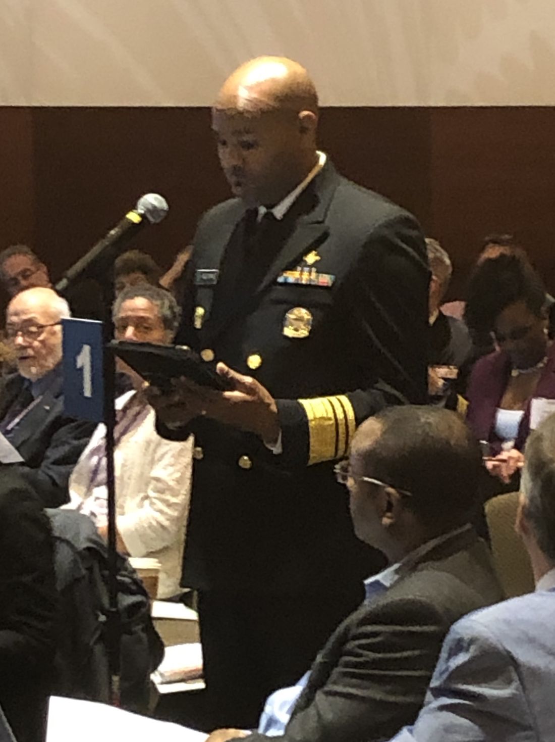
The American Medical Association (AMA) conducted the Annual Meeting of the AMA House of Delegates from June 8-12 in Chicago. The House of Delegates (HOD) is the principal policymaking body of the AMA, consisting of more than 600 delegates and accompanying alternate delegates who represent the medical specialty societies (including CHEST); the state and territorial medical associations; the uniformed services; and other stakeholder organizations. Leading policymakers including Centers for Medicare & Medicaid Services (CMS) Administrator Seema Verma and the Surgeon General of the United States, Vice Admiral Jerome M. Adams, MD, also participated in the meeting.
This year, the delegates (CHEST has three delegate positions) considered more than 200 policy proposals (resolutions and reports) in a multi-step process: caucuses, Reference Committees, and hearings before the full House of Delegates.
The caucuses are an important first step in the HOD process. The Chest/Allergy Section Council (participants at this meeting were from the AAAAI, AAOA, AASM, ACAAI, ATS, CHEST, and SCCM) met the day before the Reference Committee hearings to:
• Decide what resolutions and reports are most important to the chest diseases, critical care medicine, sleep medicine, and allergy communities;
• Determine (if possible) a unified position (support/oppose);
• Develop talking points; and
• Identify who will speak for the caucus (or as individuals if there were differing positions) at the various Reference Committee meetings.
Under the leadership of Tina Shah, MD, MPH, from the Society of Critical Care Medicine, the caucus decided to focus on 16 reports and resolutions that were slated for discussion at 7 different Reference Committees. The caucus used the GroupMe mobile, a group messaging app, to stay in touch during the meeting to ensure that someone from the caucus would be at all pertinent sessions and to communicate progress and results in real time.
The topics of the reports and resolutions selected by the Caucus for involvement included:
• Returning Liquid Oxygen to the Medicare Fee Schedule
• COPD National Action Plan
• Low Nicotine Product Standard
• Addressing the Vaping Crisis
• Regulating Liquid Nicotine and E-Cigarettes
• Put Over-the-Counter Inhaled Epinephrine Behind Pharmacy Counter
• Change in Marijuana Classification to Allow Research
• Promotion of Early Recognition and Treatment of Sepsis by Out-of-Hospital Healthcare Providers
• The Climate Change Lecture for US Medical Schools
• Physician-Assisted Suicide
• End-of-Life Care
The Reference Committees, where both AMA members and nonmembers (with permission) may testify, are organized by topic:
• Medical Service
• Legislation, Legal, and Regulatory Issues
• Medical Education
• Public Health
• Science and Technology
• AMA Governance and Finance
• Medical Practice
• Constitution and Bylaws
The Reference Committees hear testimony on each resolution, adjourn, and then meet privately (often into the wee hours) to develop recommendations to the full House. Their options include:
• Recommend Adoption
• Recommend Adoption With Amendment
• Recommend Referral (further study by one of several Councils)
• Recommend Referral for Decision (by the Board of Trustees after further study)
• Recommend for Non-Adoption
During the following 3 days, the full House of Delegates considers the Reference Committee recommendations. Any delegate may object to any recommendation and cause it to be debated and voted on by the full House of Delegates. Details about the outcomes of the 200+ resolutions are available at the AMA website (ama-assn.org).
The compendium of policies covers the entire range of topics impacting the practice of medicine – ethics, legislation, regulation, public health, individual health, and medical education among them. The full range of policies may be found in the AMA’s Policy Manual available on the AMA website (ama-assn.org).

CHEST members with an interest in the AMA policy-making process may observe any AMA-HOD meeting or participate in the AMA’s democratic processes. Attendees will also be able to increase their knowledge and skills with no cost at scores of educational sessions and will also be able to connect with more than 1,500 peers and other meeting attendees from across the country. CHEST members with the time (there are two 5-day meetings each year) and interest are invited to apply to be an official CHEST delegate to the AMA. Contact Jennifer Nemkovich at [email protected] for details.
Dr. Desai is with the Chicago Chest Center and Suburban Lung Associates; and the Division of Pulmonary, Critical Care, Sleep and Allergy, University of Illinois at Chicago. He is also the CHEST Delegate to the AMA House of Delegates. Mr. Newman is the Senior Director of Strategy, Product, & Global Development at CHEST.
The American Medical Association (AMA) conducted the Annual Meeting of the AMA House of Delegates from June 8-12 in Chicago. The House of Delegates (HOD) is the principal policymaking body of the AMA, consisting of more than 600 delegates and accompanying alternate delegates who represent the medical specialty societies (including CHEST); the state and territorial medical associations; the uniformed services; and other stakeholder organizations. Leading policymakers including Centers for Medicare & Medicaid Services (CMS) Administrator Seema Verma and the Surgeon General of the United States, Vice Admiral Jerome M. Adams, MD, also participated in the meeting.
This year, the delegates (CHEST has three delegate positions) considered more than 200 policy proposals (resolutions and reports) in a multi-step process: caucuses, Reference Committees, and hearings before the full House of Delegates.
The caucuses are an important first step in the HOD process. The Chest/Allergy Section Council (participants at this meeting were from the AAAAI, AAOA, AASM, ACAAI, ATS, CHEST, and SCCM) met the day before the Reference Committee hearings to:
• Decide what resolutions and reports are most important to the chest diseases, critical care medicine, sleep medicine, and allergy communities;
• Determine (if possible) a unified position (support/oppose);
• Develop talking points; and
• Identify who will speak for the caucus (or as individuals if there were differing positions) at the various Reference Committee meetings.
Under the leadership of Tina Shah, MD, MPH, from the Society of Critical Care Medicine, the caucus decided to focus on 16 reports and resolutions that were slated for discussion at 7 different Reference Committees. The caucus used the GroupMe mobile, a group messaging app, to stay in touch during the meeting to ensure that someone from the caucus would be at all pertinent sessions and to communicate progress and results in real time.
The topics of the reports and resolutions selected by the Caucus for involvement included:
• Returning Liquid Oxygen to the Medicare Fee Schedule
• COPD National Action Plan
• Low Nicotine Product Standard
• Addressing the Vaping Crisis
• Regulating Liquid Nicotine and E-Cigarettes
• Put Over-the-Counter Inhaled Epinephrine Behind Pharmacy Counter
• Change in Marijuana Classification to Allow Research
• Promotion of Early Recognition and Treatment of Sepsis by Out-of-Hospital Healthcare Providers
• The Climate Change Lecture for US Medical Schools
• Physician-Assisted Suicide
• End-of-Life Care
The Reference Committees, where both AMA members and nonmembers (with permission) may testify, are organized by topic:
• Medical Service
• Legislation, Legal, and Regulatory Issues
• Medical Education
• Public Health
• Science and Technology
• AMA Governance and Finance
• Medical Practice
• Constitution and Bylaws
The Reference Committees hear testimony on each resolution, adjourn, and then meet privately (often into the wee hours) to develop recommendations to the full House. Their options include:
• Recommend Adoption
• Recommend Adoption With Amendment
• Recommend Referral (further study by one of several Councils)
• Recommend Referral for Decision (by the Board of Trustees after further study)
• Recommend for Non-Adoption
During the following 3 days, the full House of Delegates considers the Reference Committee recommendations. Any delegate may object to any recommendation and cause it to be debated and voted on by the full House of Delegates. Details about the outcomes of the 200+ resolutions are available at the AMA website (ama-assn.org).
The compendium of policies covers the entire range of topics impacting the practice of medicine – ethics, legislation, regulation, public health, individual health, and medical education among them. The full range of policies may be found in the AMA’s Policy Manual available on the AMA website (ama-assn.org).
CHEST members with an interest in the AMA policy-making process may observe any AMA-HOD meeting or participate in the AMA’s democratic processes. Attendees will also be able to increase their knowledge and skills with no cost at scores of educational sessions and will also be able to connect with more than 1,500 peers and other meeting attendees from across the country. CHEST members with the time (there are two 5-day meetings each year) and interest are invited to apply to be an official CHEST delegate to the AMA. Contact Jennifer Nemkovich at [email protected] for details.
Dr. Desai is with the Chicago Chest Center and Suburban Lung Associates; and the Division of Pulmonary, Critical Care, Sleep and Allergy, University of Illinois at Chicago. He is also the CHEST Delegate to the AMA House of Delegates. Mr. Newman is the Senior Director of Strategy, Product, & Global Development at CHEST.
The robots are coming: How artificial intelligence will shape the future of chest medicine (and CHEST)*
The robots are coming – at least according to a report,1 which states that Google created an artificial intelligence model that was able to detect lung cancer and cut back on false-positives at a rate that beat experienced radiologists.
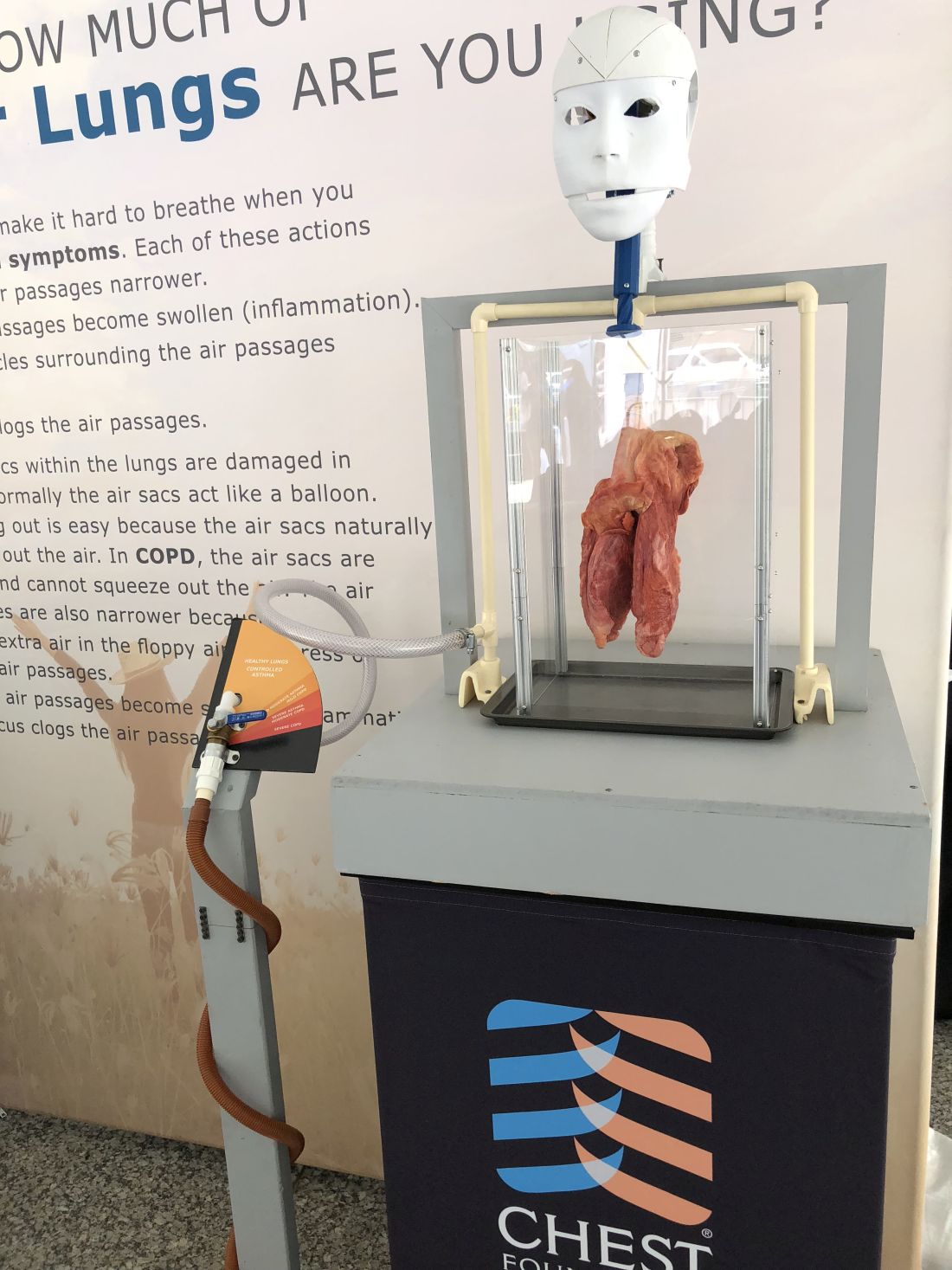
As a fan of Star Wars, sci-fi, and innovation, in general – I say, bring it on! It’s an exciting trend in medicine to see technology that has already changed the way we work and live enter into the world of health care. And, this is not about replacing humans either (like what occurs in a lot of sci-fi ); this technology will potentially provide clinicians with the tools they can use to improve outcomes for their patients.
The researchers for this study used a system called convolutional neural networks to study patterns in 3D CT scans. One advantage computers has over humans is that a computer can process the entire scan all at once while trained radiologists need to review individual slices of each scan to make their diagnosis.
While this technology will need more testing and large-scale trials before being used to diagnose patients’ disorders, the early results are encouraging. The researchers also picked a cancer that impacts so many of CHEST’s members and their patients. Lung cancer kills more Americans than any type of cancer while accounting for more than 25% of all cancer deaths annually.2 In a statistic that many people find shocking, lung cancer actually kills more women than breast, ovarian, and uterine cancers combined.3 Given the devastation of this disease, we can use all of the help we can get. Another positive from this study that might be overlooked is the rate of improvement for false-positive results. This could be a major benefit of both saving the time and preventing invasive treatments or attempts to confirm a diagnosis.
This news is exciting for everyone trying to (as we say at CHEST) crush lung disease, but seeing AI in medicine is not surprising, because we are already using it at CHEST. The AI projects at CHEST include analyzing the types of activities that are most beneficial to members and building predictive analytics models for a project. These were only initial forays into using this new technology. One of the even more mind-blowing developments has been rolling out natural language processing4 for our internal data reporting.
This is a new development, and much like the Google lung cancer study, we cannot tell you what the end result will be. What we can say is that it’s likely to change the way we work and provide new opportunities to create analytics solutions for our partners.
CHEST is not “just” a medical association: we are also an innovative group using the latest tools to create a better future for members, partners, and ,ultimately, patients and their families.
Mr. Jackson is CHEST Chief Innovation Officer & Vice President of Market Growth and Innovation.
References
1. https://www.statnews.com/2019/05/20/googles-ai-improves-accuracy-of-lung-cancer-diagnosis-study-shows/. Accessed July 17, 2019.
2. https://foundation.chestnet.org/patient-education-resources/lung-cancer/. Accessed July 17, 2019.
3. https://foundation.chestnet.org/wp-content/uploads/2017/01/15110313_10154723685773104_3710772562030977299_o-1.png. Accessed July 17, 2019.
4. https://www.tableau.com/products/new-features/ask-data. Accessed July 17, 2019.
*This article originally appeared as a blog July 1, 2019, on https://insights.chestnet.org/.
The robots are coming – at least according to a report,1 which states that Google created an artificial intelligence model that was able to detect lung cancer and cut back on false-positives at a rate that beat experienced radiologists.
As a fan of Star Wars, sci-fi, and innovation, in general – I say, bring it on! It’s an exciting trend in medicine to see technology that has already changed the way we work and live enter into the world of health care. And, this is not about replacing humans either (like what occurs in a lot of sci-fi ); this technology will potentially provide clinicians with the tools they can use to improve outcomes for their patients.
The researchers for this study used a system called convolutional neural networks to study patterns in 3D CT scans. One advantage computers has over humans is that a computer can process the entire scan all at once while trained radiologists need to review individual slices of each scan to make their diagnosis.
While this technology will need more testing and large-scale trials before being used to diagnose patients’ disorders, the early results are encouraging. The researchers also picked a cancer that impacts so many of CHEST’s members and their patients. Lung cancer kills more Americans than any type of cancer while accounting for more than 25% of all cancer deaths annually.2 In a statistic that many people find shocking, lung cancer actually kills more women than breast, ovarian, and uterine cancers combined.3 Given the devastation of this disease, we can use all of the help we can get. Another positive from this study that might be overlooked is the rate of improvement for false-positive results. This could be a major benefit of both saving the time and preventing invasive treatments or attempts to confirm a diagnosis.
This news is exciting for everyone trying to (as we say at CHEST) crush lung disease, but seeing AI in medicine is not surprising, because we are already using it at CHEST. The AI projects at CHEST include analyzing the types of activities that are most beneficial to members and building predictive analytics models for a project. These were only initial forays into using this new technology. One of the even more mind-blowing developments has been rolling out natural language processing4 for our internal data reporting.
This is a new development, and much like the Google lung cancer study, we cannot tell you what the end result will be. What we can say is that it’s likely to change the way we work and provide new opportunities to create analytics solutions for our partners.
CHEST is not “just” a medical association: we are also an innovative group using the latest tools to create a better future for members, partners, and ,ultimately, patients and their families.
Mr. Jackson is CHEST Chief Innovation Officer & Vice President of Market Growth and Innovation.
References
1. https://www.statnews.com/2019/05/20/googles-ai-improves-accuracy-of-lung-cancer-diagnosis-study-shows/. Accessed July 17, 2019.
2. https://foundation.chestnet.org/patient-education-resources/lung-cancer/. Accessed July 17, 2019.
3. https://foundation.chestnet.org/wp-content/uploads/2017/01/15110313_10154723685773104_3710772562030977299_o-1.png. Accessed July 17, 2019.
4. https://www.tableau.com/products/new-features/ask-data. Accessed July 17, 2019.
*This article originally appeared as a blog July 1, 2019, on https://insights.chestnet.org/.
Changing clinical practice to maximize success of ICU airway management
Airway management is a complex process that, if not performed in a proper and timely manner, may result in significant morbidity or mortality. The risk of intubation failure and associated adverse events is higher in critically ill patients due to differences in patient condition, environment, and practitioner experience. Even when controlling for provider experience, intubating conditions are worse and success rates are lower in the ICU compared with the controlled environment of the operating room (Taboada, et al. Anesthesiology. 2018;129[2]:321). Furthermore, the risk of injury and adverse events increases with the number of intubation attempts during an emergency (Sakles JC, et al. Acad Emerg Med. 2013;20[1]:71). Unfortunately, the paucity of high-grade evidence leads practitioners to rely on practice patterns developed during training and predicated on common sense airway management principles. The difficulty in evaluating airway management in the critically ill lies in the multi-step and complex nature of the process, including the pre-intubation, intubation, and post-intubation activities (Fig 1). Several recent publications have the potential to change airway management practice in the ICU. We will address the latest information on preoxygenation, use of neuromuscular blockade (NMB), and checklists in this setting.

Preoxygenation: Overrated?
Rapid-sequence intubation (RSI) is a technique intended to minimize the time from induction to intubation and reduce the risk of aspiration by primarily avoiding ventilation. The avoidance of bag-mask ventilation during this apneic period is common, due to concerns that positive pressure can produce gastric insufflation and regurgitation that may lead to aspiration. To attenuate the risk for critical desaturation, preoxygenation is classically provided prior to induction of anesthesia in the operative procedural areas. Although the benefit can be seen in patients undergoing elective intubation, critically ill patients often have difficulty in significantly raising the blood oxygen content despite preoxygenation with 100% oxygen delivered via face mask. As a result, the oxygen saturation can drop precipitously during the process of ICU intubation, especially if multiple or prolonged intubation attempts are required. These factors all contribute to the risk of hypoxemia and cardiac arrest during ICU intubations (De Jong A, et al. Crit Care Med. 2018;46[4]:532), which has led to the debate about the avoidance of ventilation during RSI in the critically ill. Recently, Casey and colleagues (Casey JD, et al. N Engl J Med. 2019;380[9]:811) evaluated the use of bag-mask ventilation (BMV) during RSI. In this ICU study, intubations were randomized to either include BMV or no ventilation after induction. The results suggested that the frequency of critical desaturation was lower in the patients receiving BMV after induction without a concomitant increase in frequency of aspiration. Although not powered to evaluate the difference in the incidence of aspiration, this study supports the use of BMV during the apneic phase of intubation, thereby decreasing the risk for critical desaturation.

Neuromuscular blockade: Yes or no?
Awake intubation, with or without sedation, is often employed for managing the airway in high-risk patients. This technique allows the patient to maintain spontaneous ventilation in the event of repeated intubation attempts and has a lower hypotension risk. However, many critically ill patients cannot be managed in this manner due to lack of patient cooperation, emergent airway management requirements, or practitioner inexperience with this technique. As a result, many of these patients will require an induction agent, and concomitant administration of a neuromuscular blocking agent (NMB) to optimize intubating conditions. However, the avoidance of NMBs in emergent airway scenarios was not uncommon among attending physicians and trainees (Schmidt UH, et al. Anesthesiology. 2008;109[6]:973). The American College of Chest Physicians (CHEST) Difficult Airway Course faculty also recommended to not use NMB because of the high risk of failure to ventilate/oxygenate. Without NMB, the patient might be allowed to recover to spontaneous ventilation. This approach is taken in the American Society of Anesthesiologists Practice Guidelines for the Management of the Difficult Airway but is not necessarily applicable to the critically ill patient (Apfelbaum JL, et al. Anesthesiology. 2013;118[2]:251-70). In the event of “can’t intubate, can’t oxygenate” (CICO), the critically ill patient in extremis may not tolerate an attempt to return to spontaneous ventilation because spontaneous ventilation may have been initially inadequate.
In 2010, Jaber and colleagues demonstrated a lower incidence of hypoxemia and severe hemodynamic collapse with the implementation of an intubation bundle that included the use of NMBs for all rapid-sequence inductions (Jaber S, et al. Int Care Med. 2010;36:248). The safety of using paralytics in critically ill patients was later investigated by Wilcox and colleagues in a prospective, observational study that suggested a decrease in the incidence of hypoxemia and complications when employing NMB (Wilcox SR, et al. Crit Care Med. 2012;40[6]:1808). Although Wilcox et al.’s study was hypothesis-generating by the nature of its design, it was consistent with both Jaber’s findings and a more recent observational study performed by Moser et al (Mosier JM, et al. Ann Am Thorac Soc. 2015;12[5]:734). Furthermore, there is no evidence that NMBs worsen bag mask ventilation in the critically ill patient. NMBs in addition to induction agents might be associated with optimal intubating conditions, reduced complications, and allow for placement of a supraglottic airway device or surgical airway in the event of a CICO (Higgs A, et al. Br J Anaesth. 2018;120[2]:323).
Checking the checklists
Checklists are another intervention with the potential to improve outcomes or reduce adverse events. Airway management is often a complex process with significant opportunities for failure. Therefore, having reminders or checklists available to the provider may encourage the use of best practices. Jaber demonstrated that a straightforward, 10-point intubation bundle reduced the incidence of severe complications associated with emergent intubation in the ICU. In the 4th National Audit Project of the Royal College of Anaesthetists and Difficult Airway Society, the use of checklists was recommended as a method to reduce adverse events and increase successful airway management (Cook TM, et al. Br J Anaesth. 2011;106[5]:632). In fact, several mnemonics have been developed to aid the practitioner, including the ‘7 Ps’ in the Manual of Emergency Airway Management (Walls RM, et al. Manual of Emergency Airway Management. 2012) and APPROACH from the CHEST Airway Management Training Team. More recently, Janz and colleagues developed and employed a checklist in a multicenter study and compared it with usual practice (Janz DR, et al. Chest. 2018;153[4]:816). Although the checklist was associated with improved provider compliance with airway assessment, preparation, and verbalization of a plan, it did not go far enough to include the known interventions for optimizing preoxygenation and hemodynamic stability. Two elements that might be included in a checklist include fluids and vasopressors administration during the pre-intubation and post-intubation period, and preoxygenation with noninvasive ventilation. The former is associated with a lower incidence of hypotension, while the latter may reduce the incidence of severe hypoxemia in ICU intubations (Baillard C, et al. Am J Respir Crit Care Med. 2006;174[2]:171).
Keeping apprised of evidence and adjusting practice are crucial to the competent clinician engaging in airway management, as they minimize the risk of harm while maximizing the benefit to the patient. However, the methods to achieve these goals are not always intuitive. Definitive high-level evidence is sparse. The use of neuromuscular blockade and BMV after induction has historically been controversial, but more recent evidence is favoring these approaches for RSI. The use of checklists or guidelines may ensure that the necessary safety steps are followed, especially at institutions that may not have experts in airway management. Over time, the hope is that many of our traditional practices are either supported by quality evidence or better techniques evolve.
Dr. Tokarczyk is with the Department of Anesthesia, NorthShore University HealthSystem; and Clinical Assistant Professor, University of Chicago, Pritzker School of Medicine. Dr. Greenberg is Editor-in-Chief, Anesthesia Patient Safety Foundation (APSF) Newsletter; Vice Chairperson, Education, Department of Anesthesiology; Director of Critical Care Services, Evanston Hospital; NorthShore University HealthSystem; and Clinical Professor, Department of Anesthesiology Critical Care, University of Chicago, Pritzker School of Medicine.
Airway management is a complex process that, if not performed in a proper and timely manner, may result in significant morbidity or mortality. The risk of intubation failure and associated adverse events is higher in critically ill patients due to differences in patient condition, environment, and practitioner experience. Even when controlling for provider experience, intubating conditions are worse and success rates are lower in the ICU compared with the controlled environment of the operating room (Taboada, et al. Anesthesiology. 2018;129[2]:321). Furthermore, the risk of injury and adverse events increases with the number of intubation attempts during an emergency (Sakles JC, et al. Acad Emerg Med. 2013;20[1]:71). Unfortunately, the paucity of high-grade evidence leads practitioners to rely on practice patterns developed during training and predicated on common sense airway management principles. The difficulty in evaluating airway management in the critically ill lies in the multi-step and complex nature of the process, including the pre-intubation, intubation, and post-intubation activities (Fig 1). Several recent publications have the potential to change airway management practice in the ICU. We will address the latest information on preoxygenation, use of neuromuscular blockade (NMB), and checklists in this setting.
Preoxygenation: Overrated?
Rapid-sequence intubation (RSI) is a technique intended to minimize the time from induction to intubation and reduce the risk of aspiration by primarily avoiding ventilation. The avoidance of bag-mask ventilation during this apneic period is common, due to concerns that positive pressure can produce gastric insufflation and regurgitation that may lead to aspiration. To attenuate the risk for critical desaturation, preoxygenation is classically provided prior to induction of anesthesia in the operative procedural areas. Although the benefit can be seen in patients undergoing elective intubation, critically ill patients often have difficulty in significantly raising the blood oxygen content despite preoxygenation with 100% oxygen delivered via face mask. As a result, the oxygen saturation can drop precipitously during the process of ICU intubation, especially if multiple or prolonged intubation attempts are required. These factors all contribute to the risk of hypoxemia and cardiac arrest during ICU intubations (De Jong A, et al. Crit Care Med. 2018;46[4]:532), which has led to the debate about the avoidance of ventilation during RSI in the critically ill. Recently, Casey and colleagues (Casey JD, et al. N Engl J Med. 2019;380[9]:811) evaluated the use of bag-mask ventilation (BMV) during RSI. In this ICU study, intubations were randomized to either include BMV or no ventilation after induction. The results suggested that the frequency of critical desaturation was lower in the patients receiving BMV after induction without a concomitant increase in frequency of aspiration. Although not powered to evaluate the difference in the incidence of aspiration, this study supports the use of BMV during the apneic phase of intubation, thereby decreasing the risk for critical desaturation.
Neuromuscular blockade: Yes or no?
Awake intubation, with or without sedation, is often employed for managing the airway in high-risk patients. This technique allows the patient to maintain spontaneous ventilation in the event of repeated intubation attempts and has a lower hypotension risk. However, many critically ill patients cannot be managed in this manner due to lack of patient cooperation, emergent airway management requirements, or practitioner inexperience with this technique. As a result, many of these patients will require an induction agent, and concomitant administration of a neuromuscular blocking agent (NMB) to optimize intubating conditions. However, the avoidance of NMBs in emergent airway scenarios was not uncommon among attending physicians and trainees (Schmidt UH, et al. Anesthesiology. 2008;109[6]:973). The American College of Chest Physicians (CHEST) Difficult Airway Course faculty also recommended to not use NMB because of the high risk of failure to ventilate/oxygenate. Without NMB, the patient might be allowed to recover to spontaneous ventilation. This approach is taken in the American Society of Anesthesiologists Practice Guidelines for the Management of the Difficult Airway but is not necessarily applicable to the critically ill patient (Apfelbaum JL, et al. Anesthesiology. 2013;118[2]:251-70). In the event of “can’t intubate, can’t oxygenate” (CICO), the critically ill patient in extremis may not tolerate an attempt to return to spontaneous ventilation because spontaneous ventilation may have been initially inadequate.
In 2010, Jaber and colleagues demonstrated a lower incidence of hypoxemia and severe hemodynamic collapse with the implementation of an intubation bundle that included the use of NMBs for all rapid-sequence inductions (Jaber S, et al. Int Care Med. 2010;36:248). The safety of using paralytics in critically ill patients was later investigated by Wilcox and colleagues in a prospective, observational study that suggested a decrease in the incidence of hypoxemia and complications when employing NMB (Wilcox SR, et al. Crit Care Med. 2012;40[6]:1808). Although Wilcox et al.’s study was hypothesis-generating by the nature of its design, it was consistent with both Jaber’s findings and a more recent observational study performed by Moser et al (Mosier JM, et al. Ann Am Thorac Soc. 2015;12[5]:734). Furthermore, there is no evidence that NMBs worsen bag mask ventilation in the critically ill patient. NMBs in addition to induction agents might be associated with optimal intubating conditions, reduced complications, and allow for placement of a supraglottic airway device or surgical airway in the event of a CICO (Higgs A, et al. Br J Anaesth. 2018;120[2]:323).
Checking the checklists
Checklists are another intervention with the potential to improve outcomes or reduce adverse events. Airway management is often a complex process with significant opportunities for failure. Therefore, having reminders or checklists available to the provider may encourage the use of best practices. Jaber demonstrated that a straightforward, 10-point intubation bundle reduced the incidence of severe complications associated with emergent intubation in the ICU. In the 4th National Audit Project of the Royal College of Anaesthetists and Difficult Airway Society, the use of checklists was recommended as a method to reduce adverse events and increase successful airway management (Cook TM, et al. Br J Anaesth. 2011;106[5]:632). In fact, several mnemonics have been developed to aid the practitioner, including the ‘7 Ps’ in the Manual of Emergency Airway Management (Walls RM, et al. Manual of Emergency Airway Management. 2012) and APPROACH from the CHEST Airway Management Training Team. More recently, Janz and colleagues developed and employed a checklist in a multicenter study and compared it with usual practice (Janz DR, et al. Chest. 2018;153[4]:816). Although the checklist was associated with improved provider compliance with airway assessment, preparation, and verbalization of a plan, it did not go far enough to include the known interventions for optimizing preoxygenation and hemodynamic stability. Two elements that might be included in a checklist include fluids and vasopressors administration during the pre-intubation and post-intubation period, and preoxygenation with noninvasive ventilation. The former is associated with a lower incidence of hypotension, while the latter may reduce the incidence of severe hypoxemia in ICU intubations (Baillard C, et al. Am J Respir Crit Care Med. 2006;174[2]:171).
Keeping apprised of evidence and adjusting practice are crucial to the competent clinician engaging in airway management, as they minimize the risk of harm while maximizing the benefit to the patient. However, the methods to achieve these goals are not always intuitive. Definitive high-level evidence is sparse. The use of neuromuscular blockade and BMV after induction has historically been controversial, but more recent evidence is favoring these approaches for RSI. The use of checklists or guidelines may ensure that the necessary safety steps are followed, especially at institutions that may not have experts in airway management. Over time, the hope is that many of our traditional practices are either supported by quality evidence or better techniques evolve.
Dr. Tokarczyk is with the Department of Anesthesia, NorthShore University HealthSystem; and Clinical Assistant Professor, University of Chicago, Pritzker School of Medicine. Dr. Greenberg is Editor-in-Chief, Anesthesia Patient Safety Foundation (APSF) Newsletter; Vice Chairperson, Education, Department of Anesthesiology; Director of Critical Care Services, Evanston Hospital; NorthShore University HealthSystem; and Clinical Professor, Department of Anesthesiology Critical Care, University of Chicago, Pritzker School of Medicine.
This month in the journal CHEST®
Editor’s Picks
COMMENTARY
Imaging of Pulmonary Hypertension: Pictorial Essay
By Dr. E. Altschul, et al.
ORIGINAL RESEARCH
Epidemiology of Quick Sequential Organ Failure Assessment Criteria in Undifferentiated Patients and Association With Suspected Infection and Sepsis
By Dr. V. Anand, et al.
Infectious Disease Hospitalizations: United States, 2001 to 2014
By Dr. J. L. Kennedy, et al.
Overdiagnosis of COPD in Subjects With Unobstructed Spirometry: A BOLD Analysis
By Dr. L. Sator, et al.
TRANSLATING BASIC RESEARCH INTO CLINICAL PRACTICE
Common Pathogenic Mechanisms Between Idiopathic Pulmonary Fibrosis and Lung Cancer
By Dr. A. Tzouvelekis, et al.
Editor’s Picks
Editor’s Picks
COMMENTARY
Imaging of Pulmonary Hypertension: Pictorial Essay
By Dr. E. Altschul, et al.
ORIGINAL RESEARCH
Epidemiology of Quick Sequential Organ Failure Assessment Criteria in Undifferentiated Patients and Association With Suspected Infection and Sepsis
By Dr. V. Anand, et al.
Infectious Disease Hospitalizations: United States, 2001 to 2014
By Dr. J. L. Kennedy, et al.
Overdiagnosis of COPD in Subjects With Unobstructed Spirometry: A BOLD Analysis
By Dr. L. Sator, et al.
TRANSLATING BASIC RESEARCH INTO CLINICAL PRACTICE
Common Pathogenic Mechanisms Between Idiopathic Pulmonary Fibrosis and Lung Cancer
By Dr. A. Tzouvelekis, et al.

Your CHEST board working for you
The CHEST Board of Regents (BOR) held their summer meeting for 3 days starting June 20 in Coeur D’Alene, Idaho. The key drivers of the BOR are to continuously assess and adjust our strategic plan to further CHEST’s mission and to be good fiducial stewards in allocating resources. At this meeting, the BOR continued its efforts in both of these areas.

CHEST’s mission is to champion the prevention, diagnosis, and treatment of chest diseases through education, communication, and research. The budget for fiscal year 2019-2020 facilitates the expansion of this mission by allocating more resources for e-learning and for the improving engagement and member experience.
Specifically, the Board is placing significant emphasis for more content to be digital, downloadable, digestible, and (hopefully) addictive. Aside from allocating capital for updates of existing equipment, Board Designated Funds were made available for an aggressive redesign of content delivery. Over the next 2 years, CHEST will achieve single sign on for all our learning platforms; enhance mobile access; and deploy mobile apps for gaming, personalized learning, and just-in-time education. The effort also aims to streamline journal and topic workflows and launch personalized content recommendations for our members.
Our formal strategic planning continued with an external review of our recent environmental scan and 5- year plan by a select group of entrepreneurs and innovators. Board members engaged in a rich debate about ways to better focus the organization. The group noted that the best time for strategic initiatives is now, while the association is doing well and highly functioning, rather than waiting for difficult times to initiate a potentially more painful course correction.
Education is the base for our entire enterprise. The BOR had a vigorous discussion about the development of a clinician educator track with certification and was introduced to CHEST’s new Chief Learning Officer. The CHEST staff plans a deeper dive into needs assessment and developing a business plan around this program.
Communication is also a core part of our mission. The BOR recognizes that more needs to be done to support the NetWorks. For this year’s annual meeting, plans have been made to move the NetWork meetings into two time slots, unopposed by other scientific content, and to rename these meetings “NetWork Featured Lecture and Open Forum.” The rationale for the name change is to make the meetings more inviting by referring to them as “open forums.” CHEST 2019 will feature improved signage in common areas to highlight the NetWork meetings, providing times and locations. The goal is to at least double attendance. Dr. Stephanie Levine, President-Elect of CHEST, is forming a task force to explore other ways of enhancing NetWork engagement.
In order to expand CHEST educational impact, the Board launched a new global events strategy. These global educational programs were another focus of the Board of Regents. Staff provided updates on the CHEST Congress Thailand 2019 in collaboration with the Thoracic Society of Thailand. There were more than 1,000 delegates representing 57 countries. The meeting was supported by our partner, Kenes, which will also be helping with smaller regional meetings, including the June meeting in Athens in collaboration with the Hellenic Thoracic Society. CHEST is in the advanced stages of planning a CHEST Congress in Bologna, Italy, in summer 2020 in conjunction with our colleagues in that country. CHEST is putting together a 5-year plan for regional meetings with a variety of local medical societies throughout the world. To support this, the Governance Committee recommended augmenting the Council of Global Governors with an Executive Committee. This group will serve as a small, strategic set of individuals, appointed by the Governance Committee, to assist in furthering the global strategy and efforts of the organization. Going forward, the Chair of the Executive Committee will serve as a member of the Board of Regents for a 2-year term, to better represent international concerns at a board level.
The BOR addressed additional items, including reviewing the process for selecting Master FCCPs and a very positive update on our CHEST 2019 meeting in New Orleans. They also enhanced coordination with the CHEST Foundation Board of Trustees (BOT) via joint meetings. The CHEST Foundation BOT celebrated success in matching funds for the one million dollar grant to establish the Erin Popovich Endowment. Additional fundraising plans include holding five separate events in the next year, including a repeat of last year’s Feldman Family Foundation Poker Night.
The CHEST Board of Regents (BOR) held their summer meeting for 3 days starting June 20 in Coeur D’Alene, Idaho. The key drivers of the BOR are to continuously assess and adjust our strategic plan to further CHEST’s mission and to be good fiducial stewards in allocating resources. At this meeting, the BOR continued its efforts in both of these areas.
CHEST’s mission is to champion the prevention, diagnosis, and treatment of chest diseases through education, communication, and research. The budget for fiscal year 2019-2020 facilitates the expansion of this mission by allocating more resources for e-learning and for the improving engagement and member experience.
Specifically, the Board is placing significant emphasis for more content to be digital, downloadable, digestible, and (hopefully) addictive. Aside from allocating capital for updates of existing equipment, Board Designated Funds were made available for an aggressive redesign of content delivery. Over the next 2 years, CHEST will achieve single sign on for all our learning platforms; enhance mobile access; and deploy mobile apps for gaming, personalized learning, and just-in-time education. The effort also aims to streamline journal and topic workflows and launch personalized content recommendations for our members.
Our formal strategic planning continued with an external review of our recent environmental scan and 5- year plan by a select group of entrepreneurs and innovators. Board members engaged in a rich debate about ways to better focus the organization. The group noted that the best time for strategic initiatives is now, while the association is doing well and highly functioning, rather than waiting for difficult times to initiate a potentially more painful course correction.
Education is the base for our entire enterprise. The BOR had a vigorous discussion about the development of a clinician educator track with certification and was introduced to CHEST’s new Chief Learning Officer. The CHEST staff plans a deeper dive into needs assessment and developing a business plan around this program.
Communication is also a core part of our mission. The BOR recognizes that more needs to be done to support the NetWorks. For this year’s annual meeting, plans have been made to move the NetWork meetings into two time slots, unopposed by other scientific content, and to rename these meetings “NetWork Featured Lecture and Open Forum.” The rationale for the name change is to make the meetings more inviting by referring to them as “open forums.” CHEST 2019 will feature improved signage in common areas to highlight the NetWork meetings, providing times and locations. The goal is to at least double attendance. Dr. Stephanie Levine, President-Elect of CHEST, is forming a task force to explore other ways of enhancing NetWork engagement.
In order to expand CHEST educational impact, the Board launched a new global events strategy. These global educational programs were another focus of the Board of Regents. Staff provided updates on the CHEST Congress Thailand 2019 in collaboration with the Thoracic Society of Thailand. There were more than 1,000 delegates representing 57 countries. The meeting was supported by our partner, Kenes, which will also be helping with smaller regional meetings, including the June meeting in Athens in collaboration with the Hellenic Thoracic Society. CHEST is in the advanced stages of planning a CHEST Congress in Bologna, Italy, in summer 2020 in conjunction with our colleagues in that country. CHEST is putting together a 5-year plan for regional meetings with a variety of local medical societies throughout the world. To support this, the Governance Committee recommended augmenting the Council of Global Governors with an Executive Committee. This group will serve as a small, strategic set of individuals, appointed by the Governance Committee, to assist in furthering the global strategy and efforts of the organization. Going forward, the Chair of the Executive Committee will serve as a member of the Board of Regents for a 2-year term, to better represent international concerns at a board level.
The BOR addressed additional items, including reviewing the process for selecting Master FCCPs and a very positive update on our CHEST 2019 meeting in New Orleans. They also enhanced coordination with the CHEST Foundation Board of Trustees (BOT) via joint meetings. The CHEST Foundation BOT celebrated success in matching funds for the one million dollar grant to establish the Erin Popovich Endowment. Additional fundraising plans include holding five separate events in the next year, including a repeat of last year’s Feldman Family Foundation Poker Night.