User login
The Health Legacies of Childhood Traumas
Adverse childhood experiences (ACEs), such as violence, abuse, and substance abuse in the household, have dramatic and lasting effects on adult health, according to a first-ever CDC analysis of data from 25 states. Toxic stress from ACEs can “derail optimal health and development by altering gene expression, brain connectivity and function, immune system function, and organ function,” as well as compromising health coping strategies, the researchers note.
In their study, ACEs were linked to at least 5 of the 10 leading causes of death. But they also found that preventing ACEs can have equally dramatic effects: for example, potentially reducing the number of cases of coronary artery disease (CAD) by 12.6%.
The researchers analyzed data from > 144,000 adults who responded to questions in the Behavior Risk Factor Surveillance System from 2015 through 2017. ACEs are common, the researchers say: 61% of adults had at least 1, and 1 in 6 adults experienced ≥ 4types of ACE. Women and several racial/ethnic minority groups had a greater risk. People who experienced ≥ 4types of ACE accounted for a “disproportionate share of the preventable fraction” of each of the 14 negative health and socioeconomic outcomes measured, including cancer, respiratory disease, diabetes, and suicide.
Extrapolating from the 25 states to national numbers, the researchers say preventing ACEs could result in 1.9 million fewer CADs, 2.5 million fewer overweight or obese adults, and 21 million fewer adults with depression.
The CDC has recommendations for rescuing potentially millions of adults from the lingering effects of childhood trauma: early intervention. The researchers cite, for instance, studies that have found preschool enrichment and early childhood home visitation programs reduce the rates of child abuse and neglect by 48% to 52%.
Moreover, health care providers can “anticipate and recognize” current risk for ACEs in children and history of ACEs in adults. They can refer patients to effective services and support, and link adults to family-centered treatment that includes substance abuse treatment and parenting interventions. The CDC also recommends that employers can adopt and support family-friendly policies, such as paid family leave and flexible work schedules. And states and communities can, among other initiatives, improve access to high-quality child care by expanding eligibility, activities offered, and family involvement.
FDA approves Oxbryta for sickle cell disease treatment
The Food and Drug Administration has approved voxelotor (Oxbryta) for adults and pediatric patients aged 12 years and older with sickle cell disease.

Approval was based on results from HOPE, a randomized, double-blind, placebo-controlled, multicenter trial of 274 patients with sickle cell disease (median age, 24 years) with a baseline hemoglobin level between 5.5 and 10.5 g/dL. Just over half of patients (51.1%) who received voxelotor at 1,500 mg had a hemoglobin increase of at least 1 g/dL over the 24-week study period, compared with 6.5% of patients who received placebo.
Patients in the 1,500-mg group also had reduced indirect bilirubin and percent reticulocyte count at –29.1% and –19.9%, respectively, compared with placebo, where the change was –3.2% and 4.5%, respectively.
The most common adverse events associated with voxelotor are headache, diarrhea, abdominal pain, nausea, rash, fatigue and pyrexia. The recommended voxelotor dose is 1,500 mg orally once daily with or without food, according to the FDA.
The Food and Drug Administration has approved voxelotor (Oxbryta) for adults and pediatric patients aged 12 years and older with sickle cell disease.
Approval was based on results from HOPE, a randomized, double-blind, placebo-controlled, multicenter trial of 274 patients with sickle cell disease (median age, 24 years) with a baseline hemoglobin level between 5.5 and 10.5 g/dL. Just over half of patients (51.1%) who received voxelotor at 1,500 mg had a hemoglobin increase of at least 1 g/dL over the 24-week study period, compared with 6.5% of patients who received placebo.
Patients in the 1,500-mg group also had reduced indirect bilirubin and percent reticulocyte count at –29.1% and –19.9%, respectively, compared with placebo, where the change was –3.2% and 4.5%, respectively.
The most common adverse events associated with voxelotor are headache, diarrhea, abdominal pain, nausea, rash, fatigue and pyrexia. The recommended voxelotor dose is 1,500 mg orally once daily with or without food, according to the FDA.
ASH preview: Key themes include tackling CAR T obstacles, sickle cell advances, VTE
Chimeric antigen receptor (CAR) T-cell therapies have garnered a great deal of attention given their “incredible efficacy” in treating B-cell malignancies, and new findings are taking aim at the drawbacks of therapy, such as the time, expense, and toxicity involved, according to Robert A. Brodsky, MD.

One example, from a study slated for presentation during a plenary session at the upcoming annual meeting of the American Society of Hematology involves the investigational T-cell bispecific antibody mosunetuzumab, which targets both CD20 on the surface of malignant B cells, and CD3 on cytotoxic T cells, engaging the T cells and directing their cytotoxicity against B cells.
In a study (Abstract 6) of 218 non-Hodgkin lymphoma patients, including 23 who had already received CAR T-cell therapy and had relapsed or were refractory to the treatment, 64% responded, 42% had a complete response, and the median duration of response is now out to 9 months, Dr. Brodsky, ASH secretary and director of the division of hematology at Johns Hopkins University, Baltimore, said during a premeeting press conference.
“It’s basically an antibody using the patient’s own T cell to do what a CAR-T cell would do – [a] very exciting study and large study,” he said. “It is an off-the-shelf product, it completely gets around the problem of the time to generate the CAR T-cell product, and because it’s going to be much simpler and faster to produce, it’s likely going to be much cheaper than CAR T cells.”
The preliminary results also suggest it is less toxic than CAR T-cell therapy, he added.
Two other CAR T-cell therapy–related studies highlighted during the press conference address its use for multiple myeloma. One, the phase 1b/2 CARTITUDE study (Abstract 577) uses CAR T cells against the B-cell maturation antigen (BCMA) in the relapsed/refractory setting.
Of 25 patients treated with chemotherapy followed by CAR T-cell infusion and followed for a median of 3 months, 91% responded, two achieved a complete remission, and “many other responses were very deep responses,” Dr. Brodsky said, noting that the second featured multiple myeloma trial (Abstract 930) looked at bispecific CAR T-cell therapy targeting BCMA and CD38 in an effort to reduce resistance to the therapy.
“Again, very interesting preliminary results,” he said, noting that of 16 patients followed for a median of 36 weeks, 87.5% responded, the treatment was well tolerated, and progression-free survival at 9 months was 75%.
In addition to the “key theme” of overcoming CAR T-cell therapy obstacles, three other themes have emerged from among the thousands of abstracts submitted for presentation at ASH. These, as presented during the press conference, include new venous thromboembolism (VTE) therapies and approaches to research; inclusive medicine, with abstracts focused on age- and race-related issues in clinical trials; and new advances in the treatment of sickle cell disease. All of these have potentially practice-changing implications, as do the six late-breaking abstracts selected from 93 abstracts submitted for consideration for oral presentation at ASH, Dr. Brodsky said.
One of the “truly practice-changing” late-breakers is a randomized phase 3 trial (Abstract LBA-1) comparing the bispecific antibody blinatumomab to chemotherapy for post-re-induction therapy in high- and intermediate-risk acute lymphoblastic leukemia (ALL) at first relapse in children, adolescents and young adults.
The study demonstrated the superiority of blinatumomab for efficacy and tolerability, which is particularly encouraging given the challenges in getting relapsed ALL patients back into remission so they can undergo bone marrow transplant, Dr. Brodsky said.
Of 208 patients randomized, 73% vs. 45% in the blinatumomab vs. chemotherapy arms were able to get to transplant – and therefore to potential cure, he said.
“Of note, the blinatumomab arm was less toxic and there was marked improvement in disease-free and overall survival, so this is clearly going to become a new standard of care for relapsed and refractory ALL,” he added.
Chimeric antigen receptor (CAR) T-cell therapies have garnered a great deal of attention given their “incredible efficacy” in treating B-cell malignancies, and new findings are taking aim at the drawbacks of therapy, such as the time, expense, and toxicity involved, according to Robert A. Brodsky, MD.
One example, from a study slated for presentation during a plenary session at the upcoming annual meeting of the American Society of Hematology involves the investigational T-cell bispecific antibody mosunetuzumab, which targets both CD20 on the surface of malignant B cells, and CD3 on cytotoxic T cells, engaging the T cells and directing their cytotoxicity against B cells.
In a study (Abstract 6) of 218 non-Hodgkin lymphoma patients, including 23 who had already received CAR T-cell therapy and had relapsed or were refractory to the treatment, 64% responded, 42% had a complete response, and the median duration of response is now out to 9 months, Dr. Brodsky, ASH secretary and director of the division of hematology at Johns Hopkins University, Baltimore, said during a premeeting press conference.
“It’s basically an antibody using the patient’s own T cell to do what a CAR-T cell would do – [a] very exciting study and large study,” he said. “It is an off-the-shelf product, it completely gets around the problem of the time to generate the CAR T-cell product, and because it’s going to be much simpler and faster to produce, it’s likely going to be much cheaper than CAR T cells.”
The preliminary results also suggest it is less toxic than CAR T-cell therapy, he added.
Two other CAR T-cell therapy–related studies highlighted during the press conference address its use for multiple myeloma. One, the phase 1b/2 CARTITUDE study (Abstract 577) uses CAR T cells against the B-cell maturation antigen (BCMA) in the relapsed/refractory setting.
Of 25 patients treated with chemotherapy followed by CAR T-cell infusion and followed for a median of 3 months, 91% responded, two achieved a complete remission, and “many other responses were very deep responses,” Dr. Brodsky said, noting that the second featured multiple myeloma trial (Abstract 930) looked at bispecific CAR T-cell therapy targeting BCMA and CD38 in an effort to reduce resistance to the therapy.
“Again, very interesting preliminary results,” he said, noting that of 16 patients followed for a median of 36 weeks, 87.5% responded, the treatment was well tolerated, and progression-free survival at 9 months was 75%.
In addition to the “key theme” of overcoming CAR T-cell therapy obstacles, three other themes have emerged from among the thousands of abstracts submitted for presentation at ASH. These, as presented during the press conference, include new venous thromboembolism (VTE) therapies and approaches to research; inclusive medicine, with abstracts focused on age- and race-related issues in clinical trials; and new advances in the treatment of sickle cell disease. All of these have potentially practice-changing implications, as do the six late-breaking abstracts selected from 93 abstracts submitted for consideration for oral presentation at ASH, Dr. Brodsky said.
One of the “truly practice-changing” late-breakers is a randomized phase 3 trial (Abstract LBA-1) comparing the bispecific antibody blinatumomab to chemotherapy for post-re-induction therapy in high- and intermediate-risk acute lymphoblastic leukemia (ALL) at first relapse in children, adolescents and young adults.
The study demonstrated the superiority of blinatumomab for efficacy and tolerability, which is particularly encouraging given the challenges in getting relapsed ALL patients back into remission so they can undergo bone marrow transplant, Dr. Brodsky said.
Of 208 patients randomized, 73% vs. 45% in the blinatumomab vs. chemotherapy arms were able to get to transplant – and therefore to potential cure, he said.
“Of note, the blinatumomab arm was less toxic and there was marked improvement in disease-free and overall survival, so this is clearly going to become a new standard of care for relapsed and refractory ALL,” he added.
Wide-area transepithelial sampling may be best method for detecting esophageal intestinal metaplasia
SAN ANTONIO – A wide-area transepithelial sampling (WATS) brush proved superior to standard forceps biopsies for detection of intestinal metaplasia (IM) in the esophagus and gastroesophageal junction in patients with no history of IM who had any visible columnar-lined esophagus on upper endoscopy in a large randomized trial.
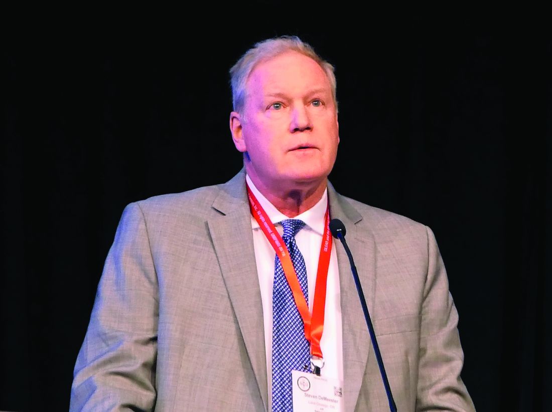
“This suggests that WATS should in fact be the preferred method of sampling these patients,” Steven R. DeMeester, MD, commented in presenting the study results at the annual meeting of the American College of Gastroenterology.
With that specific exception, however, the two sampling methods proved similarly effective at detecting IM and dysplasia, suggesting either technology can otherwise reliably be used to detect these conditions, added Dr. DeMeester, a general and thoracic surgeon at the Oregon Clinic, Portland.
This is important new information for clinicians. A prior multicenter, randomized trial by other investigators demonstrated that adding WATS to biopsy sampling improved the detection rate of dysplasia and esophageal adenocarcinoma, compared with biopsies alone (Gastrointest Endosc. 2018 Feb;87[2]:348-55). However, this dual-sampling strategy is too time-consuming for routine use in a busy practice. Given the new evidence that the two sampling methods can reliably be used interchangeably except in patients with no history of IM who have endoscopically visible columnar-lined esophagus, WATS offers a clear advantage in that it’s much quicker, especially in patients with long-segment Barrett’s esophagus, the surgeon said.
Unlike brush cytology, which merely collects surface epithelial cells, the WATS brush collects sheets of esophageal mucosa, which then undergo computer-assisted three-dimensional analysis.
“When done appropriately, a wide area of the esophageal mucosa can be sampled with the WATS brush,” Dr. DeMeester explained.
He reported on 1,002 patients who presented at nine U.S. centers for upper endoscopy for Barrett’s esophagus surveillance or evaluation of foregut symptoms. They were randomized to WATS or forceps biopsies using the Seattle protocol. In the entire group, WATS and forceps biopsies were similarly effective, each finding IM – a potentially premalignant mucosal change – in about 21% of patients, and dysplasia or cancer in 0.8%. Among the 185 patients who underwent endoscopy for follow-up of Barrett’s esophagus or post ablation, the two sampling strategies also performed similarly, detecting IM in roughly 36% of the 151 patients with columnar-lined esophagus shorter than 3 cm and in 56% of those with columnar-lined esophagus of greater length.
However, in the 196 patients with no history of IM who had any length of visible columnar-lined esophagus on endoscopy, the frequency of IM detection was 32.7% with WATS, compared with 15.3% with biopsy.
Dr. DeMeester and coinvestigators were also eager to identify factors associated with detection of IM in the esophagus or gastroesophageal junction in patients undergoing elective endoscopy with no history of IM. They found that whites were at greater risk than blacks, by a margin of 10.1% (17.1% vs. 7%). Moreover, the 23% IM detection rate in patients aged over age 70 years was significantly higher than the 15.5% rate in those under age 70. And to their surprise, there was no significant difference between the IM detection rate in men and women.
Nearly 15% of study participants with no measurable columnar-lined esophagus turned out to have IM. And two patients with low-grade dysplasia and one with adenocarcinoma were found among the group with no history of IM and no visible columnar-lined esophagus on upper endoscopy.
“This demonstrates that the absence of a measurable columnar-lined esophagus does not exclude patients from the risk of having intestinal metaplasia, dysplasia, or cancer at the gastroesophageal junction. Therefore evaluation of the gastroesophageal junction by biopsy or WATS should be considered during upper endoscopy, particularly in patients at increased risk for having intestinal metaplasia,” he concluded.
Dr. DeMeester reported serving as a consultant to BARD and receiving research funding from and serving as a paid speaker for CDx Diagnostics, which funded the trial.
SAN ANTONIO – A wide-area transepithelial sampling (WATS) brush proved superior to standard forceps biopsies for detection of intestinal metaplasia (IM) in the esophagus and gastroesophageal junction in patients with no history of IM who had any visible columnar-lined esophagus on upper endoscopy in a large randomized trial.
“This suggests that WATS should in fact be the preferred method of sampling these patients,” Steven R. DeMeester, MD, commented in presenting the study results at the annual meeting of the American College of Gastroenterology.
With that specific exception, however, the two sampling methods proved similarly effective at detecting IM and dysplasia, suggesting either technology can otherwise reliably be used to detect these conditions, added Dr. DeMeester, a general and thoracic surgeon at the Oregon Clinic, Portland.
This is important new information for clinicians. A prior multicenter, randomized trial by other investigators demonstrated that adding WATS to biopsy sampling improved the detection rate of dysplasia and esophageal adenocarcinoma, compared with biopsies alone (Gastrointest Endosc. 2018 Feb;87[2]:348-55). However, this dual-sampling strategy is too time-consuming for routine use in a busy practice. Given the new evidence that the two sampling methods can reliably be used interchangeably except in patients with no history of IM who have endoscopically visible columnar-lined esophagus, WATS offers a clear advantage in that it’s much quicker, especially in patients with long-segment Barrett’s esophagus, the surgeon said.
Unlike brush cytology, which merely collects surface epithelial cells, the WATS brush collects sheets of esophageal mucosa, which then undergo computer-assisted three-dimensional analysis.
“When done appropriately, a wide area of the esophageal mucosa can be sampled with the WATS brush,” Dr. DeMeester explained.
He reported on 1,002 patients who presented at nine U.S. centers for upper endoscopy for Barrett’s esophagus surveillance or evaluation of foregut symptoms. They were randomized to WATS or forceps biopsies using the Seattle protocol. In the entire group, WATS and forceps biopsies were similarly effective, each finding IM – a potentially premalignant mucosal change – in about 21% of patients, and dysplasia or cancer in 0.8%. Among the 185 patients who underwent endoscopy for follow-up of Barrett’s esophagus or post ablation, the two sampling strategies also performed similarly, detecting IM in roughly 36% of the 151 patients with columnar-lined esophagus shorter than 3 cm and in 56% of those with columnar-lined esophagus of greater length.
However, in the 196 patients with no history of IM who had any length of visible columnar-lined esophagus on endoscopy, the frequency of IM detection was 32.7% with WATS, compared with 15.3% with biopsy.
Dr. DeMeester and coinvestigators were also eager to identify factors associated with detection of IM in the esophagus or gastroesophageal junction in patients undergoing elective endoscopy with no history of IM. They found that whites were at greater risk than blacks, by a margin of 10.1% (17.1% vs. 7%). Moreover, the 23% IM detection rate in patients aged over age 70 years was significantly higher than the 15.5% rate in those under age 70. And to their surprise, there was no significant difference between the IM detection rate in men and women.
Nearly 15% of study participants with no measurable columnar-lined esophagus turned out to have IM. And two patients with low-grade dysplasia and one with adenocarcinoma were found among the group with no history of IM and no visible columnar-lined esophagus on upper endoscopy.
“This demonstrates that the absence of a measurable columnar-lined esophagus does not exclude patients from the risk of having intestinal metaplasia, dysplasia, or cancer at the gastroesophageal junction. Therefore evaluation of the gastroesophageal junction by biopsy or WATS should be considered during upper endoscopy, particularly in patients at increased risk for having intestinal metaplasia,” he concluded.
Dr. DeMeester reported serving as a consultant to BARD and receiving research funding from and serving as a paid speaker for CDx Diagnostics, which funded the trial.
REPORTING FROM ACG 2019
Not All Stool Discussions Are Unproductive
On August 22, 2014, a baby girl was born healthy and at full term. She was discharged on August 24 after a routine hospital stay. A day later, the infant’s mother called the pediatrician’s emergency after-hours hotline, in distress. She was concerned because the infant had not had a bowel movement since August 23. She was also concerned that each feeding was lasting an hour. A nurse told the mother that her concerns would be discussed in a routine follow-up appointment scheduled for the next morning.
At the appointment, the pediatrician noted the infant was having feeding problems and had lost 11% of her at-birth weight. The pediatrician also noted a high respiratory rate and abnormal skin coloring. However, the pediatrician concluded that the infant’s feeding problems had resolved, reassured the parents she was healthy, and discharged her, telling her parents to return in 2 weeks.
On August 29, the mother again called the pediatrician’s office, this time with concerns over a decrease in the infant’s feeding. A nurse told her to bring the infant to the emergency department (ED). At the ED, the infant was found to have lost 12% of her weight since birth and to be severely dehydrated. Due to hypovolemia, health care providers could not complete lab tests on the infant until aggressive resuscitation had been performed for 4 hours. The infant’s lab values showed she had hypernatremia, which put her at risk for brain injury through a decrease in cellular hydration, increased vascular permeability, or elevated intracranial pressure.
The infant did sustain a permanent brain injury from the condition: right-side paralysis. Her multi-organ failure from dehydration also caused significant damage to her left kidney, which stopped functioning altogether in 2017.
The infant sued her pediatrician and his office. Plaintiff’s counsel stated that the infant’s early weight loss was a red flag and that the standard of care required the pediatrician to order in-office follow-up within 24 hours to confirm if the loss had been corrected. Plaintiff’s counsel also alleged the baby’s respiratory rate and poor coloring were also red flags for dehydration. He further alleged that if the infant had returned to the office, her weight loss would have prompted supplementation before the dehydration caused her brain injury.
VERDICT
The case was settled for $1,375,000.
COMMENTARY
This case merits discussion because it involves a type of patient many clinicians see in practice. Complaints of newborn and infant feeding problems are common. Most cases require reassurance and troubleshooting about common feeding problems—but the clinician must be on the lookout for serious issues. Thus, it is helpful to revisit expected newborn feeding, stooling, and weight status.
Continue to: With regard to feeding...
With regard to feeding, breastfed infants should receive between 8-12 feeds per day during the newborn hospitalization.1 Bottlefed infants should be fed 20 kcal per 30 mL of iron-containing formula. Infants are fed on demand, and the duration of the feeding should not exceed 4 hours. The volume of the feed for the first few days of life should be 15-30 mL per feed.
For breastfed infants, clinicians should be attentive to issues that can impact nutrition, including latching difficulties, pain, mastitis, blocked duct, and engorgement. These can limit nutrition and be extremely upsetting to the mother.
Stooling frequency for newborns varies and depends on whether the infant is bottle fed or breast fed. During the first week of life, infants pass a mean of 4 stools per day.2 Breastfed infants may have as few as one stool per day, increasing as mother’s milk production increases.2
With regard to weight loss, term infants may lose up to 10% of their birth weight in the first few days of life, which is typically regained in 10 to 14 days.1 Infants born via cesarean section lose more weight, with 25% of these babies losing more than 10% by 72 hours. Of them, 76% return to birth weight by 14 days and 92% by 21 days. By contrast, vaginally born infants regain weight faster, with 86% returning to their birth weight by 14 days and 95% by the 21st day.1
Here, the infant was term and healthy, but we are not told some important details. Was she breast fed or bottle fed? Was she born by cesarean section or vaginally? We do know she was discharged on August 24th, her mother called the hotline on the 25th, and she was seen by the pediatrician on the 26th—at which point the infant had not stooled in 3 days. The mother called the physician’s office again on August 29th and was sent to the ED. We are told only that each feeding lasted an hour and the infant hadn’t stooled.
Continue to: Regarding stool...
Regarding stool—we have all had discussions with patients regarding hypervigilant concerns about stool color. We know there are some big things to look for with stool color (eg, black tarry reveals upper GI bleeding, while clay-colored or pale may reveal the absence of bile). Yet, patients’ expectations of the color-coded diagnostic abilities of their stool knows no bounds. Patients are convinced that we have some color wheel in our jacket pocket corresponding to stool color—and that the nuances between shades have important medical implications. If you ask, “Would you say it is more marigold, butterscotch yellow, or Tuscan sun?” … your patient will have an answer.
Patients reveal stool color hesitantly, reservedly, with nervous expectation. They wait for your response in quivering anticipation that the coming reply will include words to the effect that a boysenberry-purple stool is equivalent to a Death tarot card. A subconjunctival hemorrhage is the only thing that approaches the anxiety level of the oddly hued stool (Oddly Hued Stool Anxiety). Woe be the patient with both. For a patient bearing a subconjunctival hemorrhage who has also passed a jungle-green stool in the past 24 hours fully expects to explode within the next 60 minutes.
I’ve been rather facetious for a reason. My purpose is to acknowledge and to help you recognize that most discussions regarding stooling will not be (forgive me) productive. They are not productive because most stool coloration issues are nonentities—yet they produce patient anxiety that takes some time to address, leaving a busy clinician prone to curtly dismiss such discussions out of hand. Because many patient-initiated stool discussions aren’t productive, there is the risk for stool tune-out. As gross and unproductive as they can be, don’t tune out all stool discussions.
In this case, the appropriate frequency of stooling should have been at least once per day. For newborns, a stool color question is helpful; stool should not be white, pale, or clay-colored, which is suggestive of acholic stools from biliary atresia. Here, despite the absence of stool over a 3-day period, the defendant concluded the feeding problem was resolved and set a return visit for 2 weeks later. The plaintiff’s expert contended that the weight loss and absence of stooling was evidence of inadequate intake and warranted a return check the next day. Rather than risk the case going to trial, the defendant settled for $1,375,000.
IN SUM
Newborn nutrition is important. Understand that parents will be anxious about newborn feeding, although often there is no major medical concern. However, do not be dismissive of feeding concerns because of this expected anxiety. Listen to the parents fully, paying particular attention to quantifying feeding difficulty and stooling frequency matters. Don’t let patients’ rainbow parade of needless stool concerns blind you to considering important information. In other words, don’t be a stool fool.
1. McKee-Garrett TM. Overview of the routine management of the healthy newborn infant. UpToDate. Updated July 12, 2019. www.uptodate.com/contents/overview-of-the-routine-management-of-the-healthy-newborn-infant. Accessed November 18, 2019.
2. Sood MR. Constipation in infants and children: evaluation. UpToDate. Updated August 1, 2019. www.uptodate.com/contents/constipation-in-infants-and-children-evaluation. Accessed November 18, 2019.
On August 22, 2014, a baby girl was born healthy and at full term. She was discharged on August 24 after a routine hospital stay. A day later, the infant’s mother called the pediatrician’s emergency after-hours hotline, in distress. She was concerned because the infant had not had a bowel movement since August 23. She was also concerned that each feeding was lasting an hour. A nurse told the mother that her concerns would be discussed in a routine follow-up appointment scheduled for the next morning.
At the appointment, the pediatrician noted the infant was having feeding problems and had lost 11% of her at-birth weight. The pediatrician also noted a high respiratory rate and abnormal skin coloring. However, the pediatrician concluded that the infant’s feeding problems had resolved, reassured the parents she was healthy, and discharged her, telling her parents to return in 2 weeks.
On August 29, the mother again called the pediatrician’s office, this time with concerns over a decrease in the infant’s feeding. A nurse told her to bring the infant to the emergency department (ED). At the ED, the infant was found to have lost 12% of her weight since birth and to be severely dehydrated. Due to hypovolemia, health care providers could not complete lab tests on the infant until aggressive resuscitation had been performed for 4 hours. The infant’s lab values showed she had hypernatremia, which put her at risk for brain injury through a decrease in cellular hydration, increased vascular permeability, or elevated intracranial pressure.
The infant did sustain a permanent brain injury from the condition: right-side paralysis. Her multi-organ failure from dehydration also caused significant damage to her left kidney, which stopped functioning altogether in 2017.
The infant sued her pediatrician and his office. Plaintiff’s counsel stated that the infant’s early weight loss was a red flag and that the standard of care required the pediatrician to order in-office follow-up within 24 hours to confirm if the loss had been corrected. Plaintiff’s counsel also alleged the baby’s respiratory rate and poor coloring were also red flags for dehydration. He further alleged that if the infant had returned to the office, her weight loss would have prompted supplementation before the dehydration caused her brain injury.
VERDICT
The case was settled for $1,375,000.
COMMENTARY
This case merits discussion because it involves a type of patient many clinicians see in practice. Complaints of newborn and infant feeding problems are common. Most cases require reassurance and troubleshooting about common feeding problems—but the clinician must be on the lookout for serious issues. Thus, it is helpful to revisit expected newborn feeding, stooling, and weight status.
Continue to: With regard to feeding...
With regard to feeding, breastfed infants should receive between 8-12 feeds per day during the newborn hospitalization.1 Bottlefed infants should be fed 20 kcal per 30 mL of iron-containing formula. Infants are fed on demand, and the duration of the feeding should not exceed 4 hours. The volume of the feed for the first few days of life should be 15-30 mL per feed.
For breastfed infants, clinicians should be attentive to issues that can impact nutrition, including latching difficulties, pain, mastitis, blocked duct, and engorgement. These can limit nutrition and be extremely upsetting to the mother.
Stooling frequency for newborns varies and depends on whether the infant is bottle fed or breast fed. During the first week of life, infants pass a mean of 4 stools per day.2 Breastfed infants may have as few as one stool per day, increasing as mother’s milk production increases.2
With regard to weight loss, term infants may lose up to 10% of their birth weight in the first few days of life, which is typically regained in 10 to 14 days.1 Infants born via cesarean section lose more weight, with 25% of these babies losing more than 10% by 72 hours. Of them, 76% return to birth weight by 14 days and 92% by 21 days. By contrast, vaginally born infants regain weight faster, with 86% returning to their birth weight by 14 days and 95% by the 21st day.1
Here, the infant was term and healthy, but we are not told some important details. Was she breast fed or bottle fed? Was she born by cesarean section or vaginally? We do know she was discharged on August 24th, her mother called the hotline on the 25th, and she was seen by the pediatrician on the 26th—at which point the infant had not stooled in 3 days. The mother called the physician’s office again on August 29th and was sent to the ED. We are told only that each feeding lasted an hour and the infant hadn’t stooled.
Continue to: Regarding stool...
Regarding stool—we have all had discussions with patients regarding hypervigilant concerns about stool color. We know there are some big things to look for with stool color (eg, black tarry reveals upper GI bleeding, while clay-colored or pale may reveal the absence of bile). Yet, patients’ expectations of the color-coded diagnostic abilities of their stool knows no bounds. Patients are convinced that we have some color wheel in our jacket pocket corresponding to stool color—and that the nuances between shades have important medical implications. If you ask, “Would you say it is more marigold, butterscotch yellow, or Tuscan sun?” … your patient will have an answer.
Patients reveal stool color hesitantly, reservedly, with nervous expectation. They wait for your response in quivering anticipation that the coming reply will include words to the effect that a boysenberry-purple stool is equivalent to a Death tarot card. A subconjunctival hemorrhage is the only thing that approaches the anxiety level of the oddly hued stool (Oddly Hued Stool Anxiety). Woe be the patient with both. For a patient bearing a subconjunctival hemorrhage who has also passed a jungle-green stool in the past 24 hours fully expects to explode within the next 60 minutes.
I’ve been rather facetious for a reason. My purpose is to acknowledge and to help you recognize that most discussions regarding stooling will not be (forgive me) productive. They are not productive because most stool coloration issues are nonentities—yet they produce patient anxiety that takes some time to address, leaving a busy clinician prone to curtly dismiss such discussions out of hand. Because many patient-initiated stool discussions aren’t productive, there is the risk for stool tune-out. As gross and unproductive as they can be, don’t tune out all stool discussions.
In this case, the appropriate frequency of stooling should have been at least once per day. For newborns, a stool color question is helpful; stool should not be white, pale, or clay-colored, which is suggestive of acholic stools from biliary atresia. Here, despite the absence of stool over a 3-day period, the defendant concluded the feeding problem was resolved and set a return visit for 2 weeks later. The plaintiff’s expert contended that the weight loss and absence of stooling was evidence of inadequate intake and warranted a return check the next day. Rather than risk the case going to trial, the defendant settled for $1,375,000.
IN SUM
Newborn nutrition is important. Understand that parents will be anxious about newborn feeding, although often there is no major medical concern. However, do not be dismissive of feeding concerns because of this expected anxiety. Listen to the parents fully, paying particular attention to quantifying feeding difficulty and stooling frequency matters. Don’t let patients’ rainbow parade of needless stool concerns blind you to considering important information. In other words, don’t be a stool fool.
1. McKee-Garrett TM. Overview of the routine management of the healthy newborn infant. UpToDate. Updated July 12, 2019. www.uptodate.com/contents/overview-of-the-routine-management-of-the-healthy-newborn-infant. Accessed November 18, 2019.
2. Sood MR. Constipation in infants and children: evaluation. UpToDate. Updated August 1, 2019. www.uptodate.com/contents/constipation-in-infants-and-children-evaluation. Accessed November 18, 2019.

DAPA-HF: Dapagliflozin benefits regardless of age, HF severity
PHILADELPHIA – The substantial benefits from adding dapagliflozin to guideline-directed medical therapy for patients with heart failure with reduced ejection fraction enrolled in the DAPA-HF trial applied to patients regardless of their age or baseline health status, a pair of new post hoc analyses suggest.
These findings emerged a day after a report that more fully delineated dapagliflozin’s consistent safety and efficacy in patients with heart failure with reduced ejection fraction (HFrEF) regardless of whether they also had type 2 diabetes. One of the new, post hoc analyses reported at the American Heart Association scientific sessions suggested that even the most elderly enrolled patients, 75 years and older, had a similar cut in mortality and acute heart failure exacerbations, compared with younger patients. A second post hoc analysis indicated that patients with severe heart failure symptoms at entry into the trial received about as much benefit from the addition of dapagliflozin as did patients with mild baseline symptoms, measured by the Kansas City Cardiomyopathy Questionnaire (KCCQ).
The primary results from the DAPA-HF (Dapagliflozin and Prevention of Adverse Outcomes in Heart Failure) trial, first reported in August 2019, showed that among more than 4,700 patients with HFrEF randomized to receive the sodium-glucose cotransporter 2 (SGLT2) inhibitor dapagliflozin (Farxiga) on top of standard HFrEF medications or placebo, those who received dapagliflozin had a statistically significant, 26% decrease in their incidence of the primary study endpoint over a median 18 months, regardless of diabetes status (N Engl J Med. 2019 Nov 21;381[21]:1995-2008).
“These benefits were entirely consistent across the range of ages studied,” extending from patients younger than 55 years to those older than 75 years, John McMurray, MD, said at the meeting. “In many parts of the world, particularly North America and Western Europe, we have an increasingly elderly population. Many patients with heart failure are much older than in clinical trials,” he said.
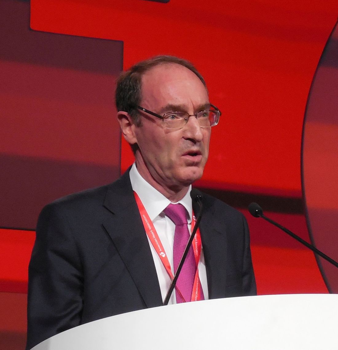
“The thing of concern is whether elderly patients get as much benefit and tolerate treatment as well as younger patients,” said Dr. McMurray, professor of medical cardiology at the University of Glasgow.
“Dapagliflozin worked across all ages, including some very elderly patients enrolled in the trial,” said Mary Norine Walsh, MD, medical director of the heart failure and transplant program at St. Vincent Heart Center of Indiana in Indianapolis. “Many trials have not looked at age like this. I hope this is a new way to analyze trials to produce more information that can help patients,” she said in an interview.
Quality-of-life outcomes
The other new, post hoc analysis showed that patients with severe HF symptoms at entry into the trial received about as much benefit from the addition of dapagliflozin as did patients with milder baseline symptoms and less impaired function, measured by the KCCQ. Dapagliflozin treatment “improved cardiovascular death and worsening heart failure to a similar extent across the entire range of KCCQ at baseline,” Mikhail N. Kosiborod, MD, said in a separate talk at the meeting. In addition, dapagliflozin treatment increased the rate of small, moderate, and large clinically meaningful improvements in patients’ KCCQ scores across all key domains of the metric, which scores symptom frequency and severity, physical and social limitations, and quality of life, said Dr. Kosiborod, a cardiologist and professor of medicine at the University of Missouri–Kansas City.
After the first 8 months of treatment in the DAPA-HF trial, 58% of the 2,373 patients who received dapagliflozin had a clinically meaningful improvement in their total KCCQ symptom score of at least 5 points, compared with a 51% rate in the 2,371 patients in the control arm, a statistically significant difference. This meant that the number needed to treat with dapagliflozin was 14 patients to produce one additional patient with at least a 5-point KCCQ improvement compared with controls, a “very small” number needed to treat, Dr. Kosiborod said in an interview.
Addition of the KCCQ to the panel of assessments that patients underwent during DAPA-HF reflected an evolved approach to measuring efficacy outcomes in clinical trials by including patient-reported outcomes. Earlier in 2019, the Food and Drug Administration released draft guidance for heart failure drug development that explicitly called for efficacy endpoints in pivotal studies that measure how patients feel and function, and stating that these endpoints can be the basis for new drug approvals.
“To many patients, how they feel matters as much if not more than how long they live,” Dr. Kosiborod noted. The goals of heart failure treatments are not only to extend survival and reduced hospitalizations, but also to improve symptoms, function, and quality of life, he said.
“There is a lot of interest now in having outcomes in heart failure trials that are more meaningful to patients, like feeling better and being able to do more,” noted Dr. Walsh.
The DAPA-HF results also showed that patients had similar rates of reduction in death, heart failure hospitalization, or urgent clinical visits, regardless of how severely they were affected by their heart failure when they began dapagliflozin treatment. The researchers ran an analysis that divided the entire trial population into tertiles based on their KCCQ score on entering the study. Patients in the most severely-affected tertile had a 30% cut in their rate of death or acute heart failure exacerbation on dapagliflozin compared with placebo, while patients in the tertile with the mildest symptoms at baseline had a 38% reduction in their primary outcome incidence compared with controls who received placebo. Concurrently with Dr. Kosiborod’s report, the results appeared in an article online (Circulation. 2019 Nov 17. doi: 10.1161/CIRCULATIONAHA.119.044138).
Outcomes by age
Not surprisingly in DAPA-HF, the older patients were, the sicker, Dr. McMurray observed. Of the study’s 1,149 patients (24% of the study cohort) who were at least 75 years old, 62% had chronic kidney disease, compared with a 14% prevalence among the 636 patients younger than age 55. The 75-and-older group showed a steeper, 32% decline in incidence of the primary endpoint – a composite of cardiovascular (CV) death, HF hospitalization, or urgent HF visit requiring intravenous therapy – than in the other studied age groups: a 24% decline in those 65-74 years old, a 29% cut in those 55-64 years old, and a 13% drop in patients younger than 55 years old.
In addition, patients aged 75 years or greater were just as likely as the overall group to show at least a 5-point improvement in their KCCQ Total Symptom Score on dapagliflozin, as well as about the same reduced rate of deterioration compared with placebo as tracked with the KCCQ.
Patients “got as much benefit in terms of symptoms as well as morbidity and mortality,” Dr. McMurray concluded. Concurrently with the meeting report the results appeared in an article online (Circulation. 2019 Nov 17. doi: 10.1161/CIRCULATIONAHA.119.044133).
“These data are of critical importance, as improving patient-reported outcomes in heart failure, especially in highly symptomatic patients, is an important goal in drug development,” G. Michael Felker, MD, wrote in an editorial accompanying the two published analyses (Circulation. 2019 Nov 17. doi: 10.1161/CIRCULATIONAHA.119.044578). These new analyses also highlight another attractive feature of dapagliflozin and, apparently, the entire class of SGLT2 inhibitors: They “ ‘play well with others’ when it comes to overlapping intolerances that often limit (either in reality or in perception) optimization of GDMT [guideline-directed medical therapy]. Although SGLT2 inhibitor therapy may lead to volume depletion and require adjustment of diuretics, the SGLT2 inhibitors generally lack some of the other dose-limiting adverse effects (such as renal dysfunction, hyperkalemia, and hypotension) that can make aggressive up-titration of GDMT problematic, particularly in older patients or those with more advanced disease,“ wrote Dr. Felker, professor of medicine at Duke University in Durham, N.C. “We stand at the beginning of a new era of ‘quadruple therapy’ for HFrEF with beta-blockers, an angiotensin receptor neprilysin inhibitor, mineralocorticoid receptor antagonists, and SGLT2 inhibitors,” he concluded.
A version of this article also appears on Medscape.com
In DAPA-HF, treatment with dapagliflozin met the three critical goals of heart failure management. When used on top of current guideline-directed medical therapy, the treatment reduced mortality, cut hospitalizations, and improved heart failure–related health status – all to a similar extent regardless of patients’ age or symptom severity at entry. These new, post hoc findings provide important, additional data supporting inhibition of sodium-glucose cotransporter (SGLT) 2 with dapagliflozin as the newest foundational pillar of treatment for heart failure with reduced ejection fraction (HFrEF).

The results of the analysis by baseline symptoms severity as measured by the Kansas City Cardiomyopathy Questionnaire (KCCQ) showed similar treatment effects from dapagliflozin regardless of a patient’s baseline KCCQ score, suggesting that the prior report of a blunted effect of dapagliflozin in patients classified at baseline as being in New York Heart Association functional class III or IV compared with class I and II patients was likely a chance finding.
Both the analyses by age and by KCCQ scores were limited by their post hoc status using data collected in a single study. No evidence addresses whether these are class effects for all drugs in the SGLT2-inhibitor class, whether these findings from DAPA-HF are generalizable to real world practice, or whether treatment with dapagliflozin would have similar effects on outcomes if it had been used more often in combination with sacubitril/valsartan. In DAPA-HF, 11% of patients also received sacubitril/valsartan even though existing management guidelines recommend sacubitril/valsartan as the preferred agent for inhibiting the renin-angiotensin system.
It’s also unclear whether patient-reported outcomes such as those measured by the KCCQ will help in sequencing the introduction of drugs for HFrEF patients, or drug selection by patients, providers, payers, and in guidelines.
Carolyn S.P. Lam, MD, is professor of medicine at Duke-National University of Singapore. She has been a consultant to and has received research funding from AstraZeneca and several other companies. She made these comments as designated discussant for the two reports.
In DAPA-HF, treatment with dapagliflozin met the three critical goals of heart failure management. When used on top of current guideline-directed medical therapy, the treatment reduced mortality, cut hospitalizations, and improved heart failure–related health status – all to a similar extent regardless of patients’ age or symptom severity at entry. These new, post hoc findings provide important, additional data supporting inhibition of sodium-glucose cotransporter (SGLT) 2 with dapagliflozin as the newest foundational pillar of treatment for heart failure with reduced ejection fraction (HFrEF).
The results of the analysis by baseline symptoms severity as measured by the Kansas City Cardiomyopathy Questionnaire (KCCQ) showed similar treatment effects from dapagliflozin regardless of a patient’s baseline KCCQ score, suggesting that the prior report of a blunted effect of dapagliflozin in patients classified at baseline as being in New York Heart Association functional class III or IV compared with class I and II patients was likely a chance finding.
Both the analyses by age and by KCCQ scores were limited by their post hoc status using data collected in a single study. No evidence addresses whether these are class effects for all drugs in the SGLT2-inhibitor class, whether these findings from DAPA-HF are generalizable to real world practice, or whether treatment with dapagliflozin would have similar effects on outcomes if it had been used more often in combination with sacubitril/valsartan. In DAPA-HF, 11% of patients also received sacubitril/valsartan even though existing management guidelines recommend sacubitril/valsartan as the preferred agent for inhibiting the renin-angiotensin system.
It’s also unclear whether patient-reported outcomes such as those measured by the KCCQ will help in sequencing the introduction of drugs for HFrEF patients, or drug selection by patients, providers, payers, and in guidelines.
Carolyn S.P. Lam, MD, is professor of medicine at Duke-National University of Singapore. She has been a consultant to and has received research funding from AstraZeneca and several other companies. She made these comments as designated discussant for the two reports.
PHILADELPHIA – The substantial benefits from adding dapagliflozin to guideline-directed medical therapy for patients with heart failure with reduced ejection fraction enrolled in the DAPA-HF trial applied to patients regardless of their age or baseline health status, a pair of new post hoc analyses suggest.
These findings emerged a day after a report that more fully delineated dapagliflozin’s consistent safety and efficacy in patients with heart failure with reduced ejection fraction (HFrEF) regardless of whether they also had type 2 diabetes. One of the new, post hoc analyses reported at the American Heart Association scientific sessions suggested that even the most elderly enrolled patients, 75 years and older, had a similar cut in mortality and acute heart failure exacerbations, compared with younger patients. A second post hoc analysis indicated that patients with severe heart failure symptoms at entry into the trial received about as much benefit from the addition of dapagliflozin as did patients with mild baseline symptoms, measured by the Kansas City Cardiomyopathy Questionnaire (KCCQ).
The primary results from the DAPA-HF (Dapagliflozin and Prevention of Adverse Outcomes in Heart Failure) trial, first reported in August 2019, showed that among more than 4,700 patients with HFrEF randomized to receive the sodium-glucose cotransporter 2 (SGLT2) inhibitor dapagliflozin (Farxiga) on top of standard HFrEF medications or placebo, those who received dapagliflozin had a statistically significant, 26% decrease in their incidence of the primary study endpoint over a median 18 months, regardless of diabetes status (N Engl J Med. 2019 Nov 21;381[21]:1995-2008).
“These benefits were entirely consistent across the range of ages studied,” extending from patients younger than 55 years to those older than 75 years, John McMurray, MD, said at the meeting. “In many parts of the world, particularly North America and Western Europe, we have an increasingly elderly population. Many patients with heart failure are much older than in clinical trials,” he said.
“The thing of concern is whether elderly patients get as much benefit and tolerate treatment as well as younger patients,” said Dr. McMurray, professor of medical cardiology at the University of Glasgow.
“Dapagliflozin worked across all ages, including some very elderly patients enrolled in the trial,” said Mary Norine Walsh, MD, medical director of the heart failure and transplant program at St. Vincent Heart Center of Indiana in Indianapolis. “Many trials have not looked at age like this. I hope this is a new way to analyze trials to produce more information that can help patients,” she said in an interview.
Quality-of-life outcomes
The other new, post hoc analysis showed that patients with severe HF symptoms at entry into the trial received about as much benefit from the addition of dapagliflozin as did patients with milder baseline symptoms and less impaired function, measured by the KCCQ. Dapagliflozin treatment “improved cardiovascular death and worsening heart failure to a similar extent across the entire range of KCCQ at baseline,” Mikhail N. Kosiborod, MD, said in a separate talk at the meeting. In addition, dapagliflozin treatment increased the rate of small, moderate, and large clinically meaningful improvements in patients’ KCCQ scores across all key domains of the metric, which scores symptom frequency and severity, physical and social limitations, and quality of life, said Dr. Kosiborod, a cardiologist and professor of medicine at the University of Missouri–Kansas City.
After the first 8 months of treatment in the DAPA-HF trial, 58% of the 2,373 patients who received dapagliflozin had a clinically meaningful improvement in their total KCCQ symptom score of at least 5 points, compared with a 51% rate in the 2,371 patients in the control arm, a statistically significant difference. This meant that the number needed to treat with dapagliflozin was 14 patients to produce one additional patient with at least a 5-point KCCQ improvement compared with controls, a “very small” number needed to treat, Dr. Kosiborod said in an interview.
Addition of the KCCQ to the panel of assessments that patients underwent during DAPA-HF reflected an evolved approach to measuring efficacy outcomes in clinical trials by including patient-reported outcomes. Earlier in 2019, the Food and Drug Administration released draft guidance for heart failure drug development that explicitly called for efficacy endpoints in pivotal studies that measure how patients feel and function, and stating that these endpoints can be the basis for new drug approvals.
“To many patients, how they feel matters as much if not more than how long they live,” Dr. Kosiborod noted. The goals of heart failure treatments are not only to extend survival and reduced hospitalizations, but also to improve symptoms, function, and quality of life, he said.
“There is a lot of interest now in having outcomes in heart failure trials that are more meaningful to patients, like feeling better and being able to do more,” noted Dr. Walsh.
The DAPA-HF results also showed that patients had similar rates of reduction in death, heart failure hospitalization, or urgent clinical visits, regardless of how severely they were affected by their heart failure when they began dapagliflozin treatment. The researchers ran an analysis that divided the entire trial population into tertiles based on their KCCQ score on entering the study. Patients in the most severely-affected tertile had a 30% cut in their rate of death or acute heart failure exacerbation on dapagliflozin compared with placebo, while patients in the tertile with the mildest symptoms at baseline had a 38% reduction in their primary outcome incidence compared with controls who received placebo. Concurrently with Dr. Kosiborod’s report, the results appeared in an article online (Circulation. 2019 Nov 17. doi: 10.1161/CIRCULATIONAHA.119.044138).
Outcomes by age
Not surprisingly in DAPA-HF, the older patients were, the sicker, Dr. McMurray observed. Of the study’s 1,149 patients (24% of the study cohort) who were at least 75 years old, 62% had chronic kidney disease, compared with a 14% prevalence among the 636 patients younger than age 55. The 75-and-older group showed a steeper, 32% decline in incidence of the primary endpoint – a composite of cardiovascular (CV) death, HF hospitalization, or urgent HF visit requiring intravenous therapy – than in the other studied age groups: a 24% decline in those 65-74 years old, a 29% cut in those 55-64 years old, and a 13% drop in patients younger than 55 years old.
In addition, patients aged 75 years or greater were just as likely as the overall group to show at least a 5-point improvement in their KCCQ Total Symptom Score on dapagliflozin, as well as about the same reduced rate of deterioration compared with placebo as tracked with the KCCQ.
Patients “got as much benefit in terms of symptoms as well as morbidity and mortality,” Dr. McMurray concluded. Concurrently with the meeting report the results appeared in an article online (Circulation. 2019 Nov 17. doi: 10.1161/CIRCULATIONAHA.119.044133).
“These data are of critical importance, as improving patient-reported outcomes in heart failure, especially in highly symptomatic patients, is an important goal in drug development,” G. Michael Felker, MD, wrote in an editorial accompanying the two published analyses (Circulation. 2019 Nov 17. doi: 10.1161/CIRCULATIONAHA.119.044578). These new analyses also highlight another attractive feature of dapagliflozin and, apparently, the entire class of SGLT2 inhibitors: They “ ‘play well with others’ when it comes to overlapping intolerances that often limit (either in reality or in perception) optimization of GDMT [guideline-directed medical therapy]. Although SGLT2 inhibitor therapy may lead to volume depletion and require adjustment of diuretics, the SGLT2 inhibitors generally lack some of the other dose-limiting adverse effects (such as renal dysfunction, hyperkalemia, and hypotension) that can make aggressive up-titration of GDMT problematic, particularly in older patients or those with more advanced disease,“ wrote Dr. Felker, professor of medicine at Duke University in Durham, N.C. “We stand at the beginning of a new era of ‘quadruple therapy’ for HFrEF with beta-blockers, an angiotensin receptor neprilysin inhibitor, mineralocorticoid receptor antagonists, and SGLT2 inhibitors,” he concluded.
A version of this article also appears on Medscape.com
REPORTING FROM THE AHA SCIENTIFIC SESSIONS
Combo elicits lasting responses in metastatic melanoma
NATIONAL HARBOR, MD. – The combination of bempegaldesleukin and nivolumab produced durable responses in a phase 1/2 trial of patients with previously untreated metastatic melanoma.
The overall response rate was 53%, and most responders were still in response at a median follow-up of about 19 months. The median progression-free survival was not reached, and the combination was considered well tolerated.
Adi Diab, MD, of the University of Texas MD Anderson Cancer Center, Houston, presented these results from the PIVOT-02 study at the annual meeting of the Society for Immunotherapy of Cancer.
Dr. Diab explained that bempegaldesleukin (bempeg) is a CD122-preferential interleukin-2 pathway agonist, and earlier results from the PIVOT-02 trial showed that adding bempeg to nivolumab can convert baseline tumors from programmed death–ligand 1 (PD-L1) negative to PD-L1 positive (SITC 2018, Abstract O4).
Dr. Diab presented updated results from PIVOT-02 (NCT02983045) in 41 patients with metastatic melanoma who received bempeg plus nivolumab as first-line treatment. The patients had a median age of 63 years (range, 22-80 years) at baseline, and 58.5% were male. Most patients (58.5%) were PD-L1 positive, although PD-L1 status was unknown in 7.3% of patients.
Patients received bempeg at 0.006 mg/kg and nivolumab at 360 mg every 3 weeks. They received a median of nine cycles (range, 1-34), and the median follow-up was 18.6 months.
Efficacy
In the 38 patients who were evaluable for efficacy, the overall response rate was 53% (n = 20), and the complete response rate was 34% (n = 13). The median time to response was 2.0 months, and the median time to complete response was 7.9 months.
Dr. Diab noted that responses were seen regardless of PD-L1 expression at baseline. The response rate was 39% among PD-L1-negative patients, 64% among PD-L1-positive patients, and 33% among patients whose PD-L1 status was unknown.
Dr. Diab also pointed out that responses were durable and deepened over time. The median duration of response was not reached, and 17 of the 20 responders had ongoing responses at last follow-up. The median progression-free survival has not been reached.
Safety
“This combination is safe and tolerable, there’s no overlapping immune-related adverse events, and the most common side effects are grade 1/2 flu-like symptoms,” Dr. Diab said.
The most common grade 1/2 treatment-related adverse events (AEs) were flu-like symptoms (80.5%), rash (70.7%), fatigue (65.9%), pruritus (48.8%), nausea (46.3%), arthralgia (43.9%), decreased appetite (36.6%), and myalgia (36.6%).
Dr. Diab noted that cytokine-related AEs (flu-like symptoms, rash, and pruritus) were easily managed with NSAIDs; decreased with subsequent cycles of treatment; and did not necessitate dose delays, reductions, or discontinuations.
Grade 3/4 treatment-related AEs included two cases of acute kidney injury, two cases of atrial fibrillation, one case of dizziness, one case of dyspnea, one case of hypoxia, one case of hyperglycemia, and one case of hypernatremia.
Five patients discontinued treatment because of related AEs, including cerebrovascular accident, peripheral edema, blood creatinine increase, malaise, and pharyngitis. There were no treatment-related deaths.
Dr. Diab said these results were used to support the recent breakthrough therapy designation granted to bempeg in combination with nivolumab. The results have also prompted a phase 3 trial in which researchers are comparing the combination with nivolumab alone (NCT03635983).
The phase 1/2 trial is sponsored by Nektar Therapeutics in collaboration with Bristol-Myers Squibb. Dr. Diab reported relationships with Nektar, Celgene, CureVac, Idera, and Pfizer.
SOURCE: Diab A et al. SITC 2019, Abstract O35.
REPORTING FROM SITC 2019
Hospitalist movers and shakers – November 2019
Amith Skandhan, MD, SFHM, has been announced as Southeast Health Statera Network’s (Dothan, Ala.) director of physician integration, and chairman of the network’s Physicians Participation Committee. Dr. Skandhan is senior lead hospitalist with Southeast Health, where he has worked for nearly a decade. He also champions the medical group’s clinical documentation improvement faction.

One of just 10 hospitalists in the nation to receive Top Hospitalist recognition by the American College of Physicians in 2018, Dr. Skandhan is also an assistant professor at Alabama College of Osteopathic Medicine and is one of Southeast Health’s Internal Medicine Residency Program’s core faculty members.
Ruby Sahoo, DO, has been promoted by Team Health (Knoxville, Tenn.) as regional performance director of its hospitalist services performance improvement team. Dr. Sahoo joined Team Health in 2016 and has most recently served as medical director and chief of staff at Grand Strand Medical Center (Myrtle Beach, S.C.).

Dr. Sahoo is a highly decorated internist and hospitalist. She was Team Health’s Medical Director of the Year for Hospital Medicine 2018, and a Frist Humanitarian Award winner in 2017. Additionally, Dr. Sahoo is a member of the Society of Hospital Medicine, the American College of Physicians, and the American Association of Physician Leadership.

David Vandenberg, MD, SFHM, recently was elevated to chief medical officer at St. Joseph Mercy Hospital (Ann Arbor and Livingston, Mich.). The hospitalist and senior fellow of hospital medicine previously has been St. Joseph’s vice chair of internal medicine and medical director of care management and documentation integrity. Dr. Vandenberg has spent 20 years as an employee at St. Joseph’s.
Cristian Andrade, MD, has been elevated to vice president of medical affairs with St. Joseph’s Health (Syracuse, N.Y.). A 16-year veteran with St. Joseph’s, Dr. Andrade has been a hospitalist with the system since 2006, and most recently has served as chief of hospitalist services.
Dr. Andrade now will provide guidance focusing on improving length of stay, as well as staff governance, utilization review, and the hospitalist program in general.
Paul DeJac, MD, has received a promotion to chief of hospitalist medicine at Roswell Park Comprehensive Cancer Center (Buffalo, N.Y.). Dr. DeJac was hired at Roswell Park in 2016, becoming lead hospitalist in 2017. He will look to boost professional development on the hospitalist team with a focus on improving patient care.
Independent Emergency Physicians (Farmington, Mich.), which provides hospitalist physicians, ED physicians, scribes, and more at a handful of hospitals in Michigan, has added urgent care facilities in Southfield, Mich., and Novi, Mich., to its portfolio. In addition, IEP has joined with Healthy Urgent Care to create a network of up to 15 urgent care centers in Southeast Michigan.
This is IEP’s first foray into urgent care. The company was founded in 1997 and practices at Ascension Health, Trinity Health, and Henry Ford Health System, covering four different hospitals.
Private hospitalist management provider Sound Physicians (Tacoma, Wash.) has grown once again, acquiring Indigo Health Partners (Traverse City, Mich.), one of Michigan’s largest private hospitalist groups. The new company will be known as Indigo, a division of Sound Inpatient Physicians.
Indigo’s approximately 150 providers are included in the transaction, which includes professionals in hospitals, skilled nursing facilities, and assisted living facilities. Indigo was previously known as Hospitalists of Northwest Michigan, based out of Munson Medical Center.
Amith Skandhan, MD, SFHM, has been announced as Southeast Health Statera Network’s (Dothan, Ala.) director of physician integration, and chairman of the network’s Physicians Participation Committee. Dr. Skandhan is senior lead hospitalist with Southeast Health, where he has worked for nearly a decade. He also champions the medical group’s clinical documentation improvement faction.
One of just 10 hospitalists in the nation to receive Top Hospitalist recognition by the American College of Physicians in 2018, Dr. Skandhan is also an assistant professor at Alabama College of Osteopathic Medicine and is one of Southeast Health’s Internal Medicine Residency Program’s core faculty members.
Ruby Sahoo, DO, has been promoted by Team Health (Knoxville, Tenn.) as regional performance director of its hospitalist services performance improvement team. Dr. Sahoo joined Team Health in 2016 and has most recently served as medical director and chief of staff at Grand Strand Medical Center (Myrtle Beach, S.C.).
Dr. Sahoo is a highly decorated internist and hospitalist. She was Team Health’s Medical Director of the Year for Hospital Medicine 2018, and a Frist Humanitarian Award winner in 2017. Additionally, Dr. Sahoo is a member of the Society of Hospital Medicine, the American College of Physicians, and the American Association of Physician Leadership.
David Vandenberg, MD, SFHM, recently was elevated to chief medical officer at St. Joseph Mercy Hospital (Ann Arbor and Livingston, Mich.). The hospitalist and senior fellow of hospital medicine previously has been St. Joseph’s vice chair of internal medicine and medical director of care management and documentation integrity. Dr. Vandenberg has spent 20 years as an employee at St. Joseph’s.
Cristian Andrade, MD, has been elevated to vice president of medical affairs with St. Joseph’s Health (Syracuse, N.Y.). A 16-year veteran with St. Joseph’s, Dr. Andrade has been a hospitalist with the system since 2006, and most recently has served as chief of hospitalist services.
Dr. Andrade now will provide guidance focusing on improving length of stay, as well as staff governance, utilization review, and the hospitalist program in general.
Paul DeJac, MD, has received a promotion to chief of hospitalist medicine at Roswell Park Comprehensive Cancer Center (Buffalo, N.Y.). Dr. DeJac was hired at Roswell Park in 2016, becoming lead hospitalist in 2017. He will look to boost professional development on the hospitalist team with a focus on improving patient care.
Independent Emergency Physicians (Farmington, Mich.), which provides hospitalist physicians, ED physicians, scribes, and more at a handful of hospitals in Michigan, has added urgent care facilities in Southfield, Mich., and Novi, Mich., to its portfolio. In addition, IEP has joined with Healthy Urgent Care to create a network of up to 15 urgent care centers in Southeast Michigan.
This is IEP’s first foray into urgent care. The company was founded in 1997 and practices at Ascension Health, Trinity Health, and Henry Ford Health System, covering four different hospitals.
Private hospitalist management provider Sound Physicians (Tacoma, Wash.) has grown once again, acquiring Indigo Health Partners (Traverse City, Mich.), one of Michigan’s largest private hospitalist groups. The new company will be known as Indigo, a division of Sound Inpatient Physicians.
Indigo’s approximately 150 providers are included in the transaction, which includes professionals in hospitals, skilled nursing facilities, and assisted living facilities. Indigo was previously known as Hospitalists of Northwest Michigan, based out of Munson Medical Center.
Have earlier diagnosis and treatment advances cut deaths from systemic sclerosis?
This abstract highlights a promising result for our younger patients diagnosed with systemic sclerosis. The fact that patients aged 44 years and younger with systemic sclerosis now have a death rate similar to that of the general population should be celebrated. Practicing clinicians can speculate why this trend has occurred. It is well known that these patients many times suffer from cardiopulmonary involvement.
Thus, the advances in the treatment of interstitial lung disease and pulmonary hypertension may account for the decreased mortality in this population.
Earlier detection has certainly helped this group. In addition, using immune modulators earlier at disease onset is likely another contributor to this positive trend. We are entering a decade and beyond filled with hope to change the course of this particular autoimmune disease.
Dr. Oberstein is a practicing rheumatologist at the University of Miami Health System and is senior medical director of musculoskeletal at Modernizing Medicine in Boca Raton, Fla. She has no relevant disclosures to report.
More studies like VERVE needed to test live vaccines in special populations
The VERVE study highlights a crucial topic for rheumatologists treating patients in clinical practice. The traditional thinking is to inform patients never to receive live vaccines when they are using TNF (tumor necrosis factor) inhibitors to treat their autoimmune disease. The VERVE study indicates that in the case of the Zostavax vaccine, patients on this form of biologic therapy for rheumatoid arthritis, psoriatic arthritis, and ankylosing spondylitis can safely receive this preventive measure. This study scratches the surface on an important topic, and other studies need to follow.
Many patients on biologic therapy want to travel. Many times, international travel requires vaccination that is only in the form of a live vaccine – for example, the yellow fever vaccine. It would be useful for us to better understand whether other live vaccines can safely be administered and better inform our patients who want to travel. In addition, many times mothers with young infants are nervous if they are on biologic therapy and their children need to receive a live vaccine. They are concerned that their children will shed the live virus and they will be in jeopardy. This study highlights that this may be more of an antiquated way of thinking. We need more studies of this kind to better understand and advise our patients properly without instilling unwarranted fear.
Dr. Oberstein is a practicing rheumatologist at the University of Miami Health System and is senior medical director of musculoskeletal at Modernizing Medicine in Boca Raton, Fla. She has no relevant disclosures to report.