User login
FDA gives thumbs up to tazemetostat for follicular lymphoma
The US Food and Drug Administration (FDA) has granted accelerated approval of the EZH2 inhibitor tazemetostat (Tazverik, Epizyme, Inc) for the treatment of relapsed or refractory follicular lymphoma in adult patients with tumors harboring an EZH2 mutation.
Eligible patients must have already received at least two prior systemic therapies and have tumors that are positive for an EZH2 mutation, as detected by an FDA-approved test. The FDA has also approved the cobas EZH2 Mutation Test (Roche Molecular Systems, Inc) as a companion diagnostic test for tazemetostat.
The new indication is also for adult patients with relapsed/refractory follicular lymphoma who have no other satisfactory alternative treatment options.
“In our view, there remains no clear standard of care in the relapsed and/or refractory [follicular lymphoma] population, as not all patients benefit from today’s available therapies,” said Shefali Agarwal, MD, chief medical officer of Epizyme, in a company press release. “Based on this label, physicians will have the ability to use their clinical discretion to prescribe tazemetostat for their relapsed or refractory patients regardless of EZH2 mutational status and without regard to a specific line of treatment where other options are not satisfactory.”
This accelerated approval is based on overall response rate and duration of response. Continued approval for these indications may be contingent upon verification and description of clinical benefit in confirmatory trials, the FDA notes.
Tazemetostat acts as an inhibitor of EZH2 methyltransferase. Earlier this year, the drug was approved for the treatment of metastatic or locally advanced epithelioid sarcoma in cases in which complete resection is not possible. It is the first drug with this mechanism of action and is the first to be indicated for epithelioid sarcoma.
Promising Efficacy in Phase 2 Trial
The new approval for use in follicular lymphoma was based on results from an open-label, single-arm, multicenter phase 2 clinical trial involving patients who had experienced disease progression after being treated with at least two prior systemic regimens. The cohort was divided into two treatment groups: One group consisted of 45 patients with EZH2-activating mutations, the other included 54 patients with wild-type EZH2.
All patients received tazemetostat at 800 mg administered orally twice a day. The primary efficacy outcome measures were overall response rate and duration of response, in accordance with International Working Group Non-Hodgkin Lymphoma criteria.
The median duration of follow-up was 22 months for patients with EZH2-activating mutations and 36 months for those with wild-type tumors.
Among the 45 patients with an EZH2-activating mutation, the median number of lines of prior systemic therapy was 2.0 (range, 1 – 11). In 49% of patients, disease was refractory to rituximab, and in 49%, it was refractory to the patient’s last therapy.
The overall response rate was 69%; 12% of patients achieved a complete response, and 57% achieved a partial response. The median duration of response was 10.9 months and ongoing.
In the cohort of 54 patients with wild-type EZH2, the median number previous therapies was 3.0 (range, 1 – 8); in 59% of patients, disease was refractory to rituximab, and in 41%, it was refractory to the patient’s last therapy.
The overall response rate to tazemetostat treatment was 34%; 4% of patients achieved a complete response, and 30% achieved a partial response. The median duration of response was 13 months.
Serious adverse reactions occurred in 30% of patients. The most common were fatigue, upper respiratory tract infection, musculoskeletal pain, nausea, and abdominal pain. Eight patients (8%) discontinued treatment during the trial because of adverse events. There were no reported deaths. No black box warnings have been published, and there are no contraindications.
“The durable responses observed with this drug are notable in the context of the safety profile and route of oral, at-home administration, and will offer an important new option for physicians as we care for patients with relapsed/refractory follicular lymphoma,” said John Leonard, MD, in a company press release. He is associate dean for clinical research and Richard T. Silver Distinguished Professor of Hematology and Medical Oncology, Meyer Cancer Center, Weill Cornell Medicine and New York–Presbyterian Hospital, New York, and an investigator in the ongoing phase 1b/3 confirmatory trial for tazemetostat.
“Follicular lymphoma remains an incurable disease, and even with the availability of new drugs in recent years, there have remained important unmet needs in the treatment of follicular lymphoma,” he commented.
This article first appeared on Medscape.com.
Cardiology care ups CV monitoring, BP control in HER2+ breast cancer
Specialty care from a cardiologist may confer clinical benefits for women with HER2-positive breast cancer treated with trastuzumab, a new study suggests.
Over 48 months of follow-up, results showed cardiology involvement prior to starting trastuzumab was associated with a higher rate of guideline-recommended cardiovascular (CV) monitoring and better systolic blood pressure (BP) control.
Trastuzumab is commonly used to treat HER2-positive breast cancer, which accounts for 20% of all breast cancers. But it carries a boxed warning for decreased left ventricular ejection fraction and heart failure (HF), and interval monitoring with echocardiography is recommended for all patients receiving the monoclonal antibody.
For the study, investigators analyzed electronic health records from 1,047 patients (mean age, 54 years) who received trastuzumab between January 2009 and July 2018 in the University of Pennsylvania health system, Philadelphia. Anthracyclines were used as part of treatment in 15% of patients.
Guideline-adherent cardiovascular monitoring was defined as echocardiography assessment in the 4 months before the initiation of trastuzumab and at least every 4 months during therapy.
Overall, 28% of patients visited a cardiology or cardio-oncology provider beginning 3 months before the baseline visit until the last contact date, the authors reported in JACC: CardioOncology.
Pre-existing HF, atrial fibrillation, and anthracycline treatment were independently associated with a cardiology visit either at baseline or during follow-up.
Patients who interacted with cardiologists, compared with those who did not, had more guideline-adherent cardiac monitoring (76.4% vs 60.1%; P = .007) and cardiac biomarker testing with troponin or N-terminal pro-B-type natriuretic peptide (27.8% vs 13.8%; P = .001).
The use of guideline-adherent cardiac monitoring was 36% to 46% in previous studies of patients with breast cancer treated with adjuvant trastuzumab-based therapy, the authors note.
Among the 5,815 echocardiographic procedures for which data on provider specialty were documented, most of the orders were authorized by oncologists (approximately 84% in those with no cardiology involvement and approximately 79% in those with cardiology involvement before trastuzumab initiation).
CV risk parameters
Cardiology involvement was associated with an average 1.5 mm Hg lower systolic BP, independent of baseline systolic BP and antihypertensive medication use (95% confidence interval, –2.9 to –0.1; P = .035).
The effect size was greater in patients with baseline hypertension, who had an average 2.7 mm Hg drop in systolic BP (95% CI, –4.6 to –0.7; P = .007) and were more likely to attain a target systolic BP below 140 mm Hg (odds ratio, 1.36; 95% CI, 1.06 to 1.74; P = .016).
Body mass index (BMI) did not budge significantly in the overall population when cardiologists were involved, but it dropped 0.5 kg/m2 in women who were overweight or obese at baseline.
“I think the results are encouraging,” senior author Bonnie Ky, MD, MSCE, University of Pennsylvania, told theheart.org | Medscape Cardiology. “These are modest changes but they are significant.”
These types of changes have been associated with significant reductions in cardiovascular disease risk over time in larger clinical trials, she noted. For example, a 2 mm Hg reduction in systolic BP has been linked to a 10% reduction in stroke mortality and a 7% reduction in ischemic heart disease mortality in middle-aged adults.
“We do think they are important and speak to more aggressive risk factor modification under the care of a specialist,” said Ky, who is also editor-in-chief of JACC: CardioOncology.
This broader role for cardiologists is particularly important given the burden of pre-existing CVD and CVD risk factors in patients with cancer and survivors. In the study, the baseline prevalence of hypertension was 40.6%, dyslipidemia 23.1%, HF 3.2%, atrial fibrillation 1.7%, and diabetes 5.9%.
“Ideally, collaboration between cardiology and oncology can improve the ability to cure a patient’s cancer while minimizing the risk of adverse cardiovascular occurrences,” Erica L. Mayer, MD, MPH, Dana-Farber Cancer Institute, Boston, told theheart.org | Medscape Cardiology. “Optimization of all cardiovascular parameters, including blood pressure, lipids, and weight, may allow a patient to protect her heart health while becoming a healthy cancer survivor.”
When asked about the 28% cardiology involvement at a U.S. cancer center with one of the most well-developed cardio-oncology programs, she said “the linkage with pre-existing cardiovascular conditions, as well as the likelihood of low incidence of cardiovascular disease, in the study population may have led to what appears to be a lower percentage of patients interacting with cardiology at baseline.”
In an accompanying editorial, Mayer says a case can be made from the findings that patients with pre-existing CV disease or at high risk for adverse CV events with cancer therapy should receive multidisciplinary care that involves a cardiologist. “However, for young, otherwise healthy patients with breast cancer with few or no cardiovascular risk factors, the benefits of [additional] subspecialty care may be less clear.”
Further, the rationale supporting the recommended frequency of cardiac monitoring may not be as “compelling” in this group, given the very low incidence of baseline cardiac dysfunction or cardiac events, particularly when treated with nonanthracycline regimens, she writes.
The findings are a call for further study and more personalized medicine, agreed Ky.
“I think there’s a need absolutely for established guidelines and/or expert consensus statements about who should be referred so patients can be referred more systematically,” she said. “Referral to cardiologists, however, is certainly a function of risk factors. Part of the challenge is identifying who will derive the most benefit from cardiovascular care.
“There are some obvious cases: Patients with heart failure and patients with pre-existing cardiovascular disease should be under the regular care of a cardiologist,” Ky added. “But there’s certainly a gray zone, especially as it relates, for example, to patients with hypertension and cardiovascular risk factors. It’s not a ‘one size fits all,’ and I believe it is a matter of defining who is at increased CV risk and who would derive the greatest clinical benefit.”
Researchers at the University of Pennsylvania have developed a clinical risk–prediction algorithm and are investigating both clinical- and biomarker-guided strategies to identify and treat patients at greatest risk of developing left ventricular declines and cardiac dysfunction with exposure to cancer therapies. “These studies are one step forward, but they will all need to be externally validated,” Ky said.
Ky and Mayer reported having no relevant conflicts of interest.
This article first appeared on Medscape.com.
Adding CGRP to Botox is safe and effective for migraine prevention
Investigators found the CGRP-mAbs significantly reduced the number of headache days and pain severity with adverse event rates similar to those reported in previous trials of these medications.
“The addition of a CGRP monoclonal antibody provided statistically significantly fewer monthly headache days,” said study investigator Fred Cohen, MD, an internal medicine resident physician at Montefiore Health System, New York. “However, this was a retrospective chart review, which is hindered by elements such as recall bias. Therefore, future prospective studies are warranted for higher quality data.”
The findings were presented at the virtual annual meeting of the American Headache Society.
Fewer headache days
Although Botox is associated with significant clinical improvement in chronic migraine, it often fails to adequately control headache frequency and additional medications are needed.
The CGRP-mAbs fremanezumab, galcanezumab, and erenumab, have recently been approved for migraine prevention, with results from clinical trials demonstrating they are effective for both chronic and episodic migraine. However, patients treated with Botox were excluded from these trials and to date there are no data on combination treatment with Botox and CGRP-mAbs.
To determine whether adjunctive treatment with CGRP-mAbs augments Botox therapy in chronic migraine the investigators conducted a retrospective chart review of patients receiving Botox and prescribed a CGRP-mAb.
Eligible patients met the International Classification of Headache Disorders, 3rd edition, criteria for chronic migraine; were age 18 years or older; and presented at a single headache center between May 2018 and May 2019. Patients who received another new therapy during the study or those taking CGRP-mAb treatment for less than 2 months were excluded.
The study’s primary outcome was change in the number of reported monthly headache days, and change in pain severity was the secondary outcome.
The final analysis included data on 153 patients. The population’s mean age was 47.1 years, and 139 patients (90.8%) were women. In all, 89 patients (58.0%) received erenumab (35 received 70 mg and 54 received 140 mg), 51 (33.0%) received galcanezumab, and 13 (9.0%) received fremanezumab.
Overall, 114 (74.5%) patients reported a decrease in monthly headache days or pain severity. In the group of 66 patients for whom quantitative data were available, the average number of monthly headache days before Botox treatment was 25.7. After Botox treatment, patients had an average decrease of 10.9 monthly headache days, a 42.4% reduction, so on average study participants continued to have an average of 14.8 monthly headache days.
After treatment with a CGRP-mAb the number decreased by 5.6 additional days (37.8%). Patients receiving combined therapy had an average of 9.1 monthly headache days. The total decrease from baseline was 16.6 fewer monthly headache days, a 64.6% reduction.
The number of headache days per month was reduced to 9.3 for erenumab and galcanezumab and 5.8 for fremanezumab. However, few patients in the study took fremanezumab so this result had less statistical power than the results for the other CGRP-mAbs.
A total of 13 patients (8.5%) reported side effects associated with the CGRP-mAbs, which included constipation, injection-site reaction, and fatigue.
More evidence is needed
Commenting on the findings, Peter McAllister, MD, medical director of the New England Institute for Neurology and Headache in Stamford, Conn., said the study’s main limitation is that it is a retrospective chart review, which yields lower level evidence than a prospective, double-blind, placebo-controlled study. Dr. McAllister, who was not involved in the research, also noted that the sample size was small, particularly with respect to fremanezumab.
“This study, despite its limitations, shows that addition of a monoclonal antibody to onabotulinumtoxinA is safe and well tolerated, and may confer additional reduction in migraine or headache days. The authors correctly state that more evidence via prospective study is warranted,” said Dr. McAllister, who is also chief medical officer of the New England Institute for Clinical Research and was not involved in the investigation.
Dr. Cohen has reported no relevant financial relationships. Dr. McAllister was an investigator in the PREEMPT trial of onabotulinumtoxinA, as well as in all of the phase 3 monoclonal antibody studies.
A version of this article originally appeared on Medscape.com.
FROM AHS 2020
Minority-serving hospitals had similar survival after liver cancer surgery
Overall survival after liver cancer surgery was similar regardless of whether patients were treated at minority-serving hospitals or at hospitals with proportionally fewer African American or Hispanic patients, investigators have found.
“[T]reatment of racial minorities is largely restricted to a subset of hospitals, often referred to as minority-serving hospitals. We sought to examine whether racial and ethnic minorities with hepatocellular carcinoma receive their surgical care at minority-serving hospitals, and whether treatment at minority-serving hospitals is associated with differences in overall survival,” explained Winta T. Mehtsun, MD, MPH, of Dana-Farber Cancer Institute in Boston and associates in an abstract released as part of the annual Digestive Disease Week.®
Hepatocellular carcinoma continues to have a low 5-year survival rate and exhibits marked racial and ethnic disparities in diagnosis, treatment, and outcomes. In a recent study of Surveillance Epidemiology and End Results (SEER) data, African American patients with hepatocellular carcinoma were significantly younger at diagnosis, were more likely to have metastatic disease, and were less likely to receive surgical treatment compared with whites (Am J Prevent Med 2018;55:S40-48). Among patients with early-stage liver cancer, Hispanic and African American patients are less likely to receive curative therapy and die sooner, on average, than do other patients (Clin Gastroenterol Hepatol. 2019;17:551-9).
Minority-serving hospitals also have improved significantly less over time on measures of critical care, length of stay, and mortality, but whether these issues extend to hepatocellular carcinoma remains unclear. Therefore, Dr. Mehtsun and her associates studied all 2,609 patients in the National Cancer Database who received surgical resection (not transplantation or local therapy) for nonmetastatic hepatocellular carcinoma between 2004 and 2014. They compared survival at minority-serving hospitals – those in the top 10% based on the proportion of patients who were African American or Hispanic – with survival at other hospitals.
“There was no association between minority-serving hospital and overall survival,” the researchers reported (multivariable hazard ratio for death, 0.89; 95% confidence interval, 0.72-1.11). In contrast, survival was significantly shorter among patients with more advanced disease (HR, 2.5; 95% CI, 2.1-2.8), patients who were treated at a community cancer program (HR, 1.7; 95% CI, 1.3-2.4), and patients whose Charlson Comorbidity Index was greater than 2 (HR, 1.2; 95% CI, 1.1-1.4).
Stage at diagnosis, comorbidities, and sex were not significantly related to hospital type, the investigators noted. A total of 298 patients (11%) were treated at minority-serving hospitals. Patients treated at minority-serving hospitals were significantly more likely to be uninsured (11% vs. 4% at other hospitals) and significantly less likely to be treated at an academic center (55% vs. 69%; both P less than .001).
Dr. Mehtsun reported having no relevant conflicts of interest.
SOURCE: Mehtsun WT et al. DDW 2020, Abstract Tu2043.
FROM DDW 2020
Commonalities challenge the threshold of high-frequency episodic and low-frequency chronic migraine
according to an analysis of almost 17,000 patients from the Chronic Migraine Epidemiology and Outcomes (CaMEO) study presented at the virtual annual meeting of the American Headache Society.

“The results showed substantial overlap in levels of burden, anxiety, depression and health utilization, including outpatient, inpatient and emergency department visits, among CaMEO respondents with high-frequency episodic migraine and those with low-frequency chronic migraine,” said Richard B. Lipton, MD, of the Albert Einstein College of Medicine, New York.
The study analyzed data on 16,789 respondents to CaMEO, the longitudinal, web-based study designed to characterize the course of episodic and chronic migraine. The study population consisted of four subgroups based on the number of self-reporting monthly headache days (MHDs):
- Low- and moderate-frequency episodic migraine (LFEM; zero to seven MHDs; n = 13,473).
- High-frequency episodic migraine (HFEM; 8-14 MHDs; n = 1,840).
- Low-frequency chronic migraine (LFCM; 15-23 MHDs; n = 1,035).
- High-frequency chronic migraine (HFCM; 24 or more MHDs; n = 441).
Dr. Lipton pointed out that the International Classification of Headache Disorders, 3rd edition, defines chronic migraine as 15 or more MHDs for 3 months or more with criteria for migraine with or without aura met on 8 days a month or more. It defines episodic migraine as less than 15 MHDs.
The study characterized migraine subgroups by various demographics. “The more frequent headache categories were associated with slightly older age of onset with a higher proportion of BMI [body mass index] in the obese range and overall with lower levels of household income and education,” Dr. Lipton said.
Similar headache characteristics
A comparison of headache characteristics and headache-related disabilities across subgroups revealed a number of commonalities between the HFEM and LFCM subgroups, Dr. Lipton said. Among them were presence of mild to severe allodynia, disability grade, interictal burden, and anxiety and depression scores. For example, 47.3% of the HFEM subgroup and 54.9% of the LFCM subgroup had Patient Health Questionnaire–9 depression test scores greater than 10.
The study also evaluated patterns of consultation, diagnosis, and health resource utilization and found similar rates between the HFEM and LCFM subgroups, Dr. Lipton said. Rates of overnight hospital stay in the past 6 months were almost identical between the two subgroups: 4.1% for the former and 4.2% for the latter. One striking difference between the two subgroups: the rate of medication overuse per ICHD-3 recommendations was 40.5% in HFEM and 63% in LFCM.
“These finding suggest that the treatment needs of people with HFEM may be similar to those of people with LFCM, suggesting that the 15-MHD threshold currently recommended by the ICHD-3 may merit reconsideration,” Dr. Lipton said.
An arbitrary cutoff?
The findings raise a valid point about reevaluating the thresholds for low- and high-frequency migraine, said Andrew Charles, MD, director of the Goldberg Migraine Program at the University of California, Los Angeles. “My own personal view is that they’re the same thing,” he said of HFEM and LFCM; The 15-day cutoff, he said, is “somewhat arbitrary.”
Dr. Charles suggested migraine categories address frequency and not characteristics – episodic versus chronic – and use a range rather than a threshold. “Define a range that’s more like 10-20 days per month rather than having that point at 15,” Dr. Charles said. “People sometimes make the mistake of thinking that that classification reflects some underlying pathophysiology, and that may not be necessarily true.”
Dr. Lipton disclosed financial relationships with Alder Biopharmaceuticals, Allergan (now AbbVie), Amgen, Biohaven Pharmaceuticals, Dr. Reddy’s/Promius, Electrocore, Eli Lilly, eNeura Therapeutics, GlaxoSmithKline, Lundbeck (Alder), Merck, Pernix Therapeutics, Pfizer, Supernus, Teva, Trigemina, Axsome Therapeutics, Vector, and Vedanta. Dr. Charles disclosed he is a consultant to Amgen, Biohaven Pharmaceuticals, Eli Lilly, Lundbeck, and Novartis.
according to an analysis of almost 17,000 patients from the Chronic Migraine Epidemiology and Outcomes (CaMEO) study presented at the virtual annual meeting of the American Headache Society.
“The results showed substantial overlap in levels of burden, anxiety, depression and health utilization, including outpatient, inpatient and emergency department visits, among CaMEO respondents with high-frequency episodic migraine and those with low-frequency chronic migraine,” said Richard B. Lipton, MD, of the Albert Einstein College of Medicine, New York.
The study analyzed data on 16,789 respondents to CaMEO, the longitudinal, web-based study designed to characterize the course of episodic and chronic migraine. The study population consisted of four subgroups based on the number of self-reporting monthly headache days (MHDs):
- Low- and moderate-frequency episodic migraine (LFEM; zero to seven MHDs; n = 13,473).
- High-frequency episodic migraine (HFEM; 8-14 MHDs; n = 1,840).
- Low-frequency chronic migraine (LFCM; 15-23 MHDs; n = 1,035).
- High-frequency chronic migraine (HFCM; 24 or more MHDs; n = 441).
Dr. Lipton pointed out that the International Classification of Headache Disorders, 3rd edition, defines chronic migraine as 15 or more MHDs for 3 months or more with criteria for migraine with or without aura met on 8 days a month or more. It defines episodic migraine as less than 15 MHDs.
The study characterized migraine subgroups by various demographics. “The more frequent headache categories were associated with slightly older age of onset with a higher proportion of BMI [body mass index] in the obese range and overall with lower levels of household income and education,” Dr. Lipton said.
Similar headache characteristics
A comparison of headache characteristics and headache-related disabilities across subgroups revealed a number of commonalities between the HFEM and LFCM subgroups, Dr. Lipton said. Among them were presence of mild to severe allodynia, disability grade, interictal burden, and anxiety and depression scores. For example, 47.3% of the HFEM subgroup and 54.9% of the LFCM subgroup had Patient Health Questionnaire–9 depression test scores greater than 10.
The study also evaluated patterns of consultation, diagnosis, and health resource utilization and found similar rates between the HFEM and LCFM subgroups, Dr. Lipton said. Rates of overnight hospital stay in the past 6 months were almost identical between the two subgroups: 4.1% for the former and 4.2% for the latter. One striking difference between the two subgroups: the rate of medication overuse per ICHD-3 recommendations was 40.5% in HFEM and 63% in LFCM.
“These finding suggest that the treatment needs of people with HFEM may be similar to those of people with LFCM, suggesting that the 15-MHD threshold currently recommended by the ICHD-3 may merit reconsideration,” Dr. Lipton said.
An arbitrary cutoff?
The findings raise a valid point about reevaluating the thresholds for low- and high-frequency migraine, said Andrew Charles, MD, director of the Goldberg Migraine Program at the University of California, Los Angeles. “My own personal view is that they’re the same thing,” he said of HFEM and LFCM; The 15-day cutoff, he said, is “somewhat arbitrary.”
Dr. Charles suggested migraine categories address frequency and not characteristics – episodic versus chronic – and use a range rather than a threshold. “Define a range that’s more like 10-20 days per month rather than having that point at 15,” Dr. Charles said. “People sometimes make the mistake of thinking that that classification reflects some underlying pathophysiology, and that may not be necessarily true.”
Dr. Lipton disclosed financial relationships with Alder Biopharmaceuticals, Allergan (now AbbVie), Amgen, Biohaven Pharmaceuticals, Dr. Reddy’s/Promius, Electrocore, Eli Lilly, eNeura Therapeutics, GlaxoSmithKline, Lundbeck (Alder), Merck, Pernix Therapeutics, Pfizer, Supernus, Teva, Trigemina, Axsome Therapeutics, Vector, and Vedanta. Dr. Charles disclosed he is a consultant to Amgen, Biohaven Pharmaceuticals, Eli Lilly, Lundbeck, and Novartis.
FROM AHS 2020
The evolution of “COVIDists”
Adapting to the demands placed on hospital resources by COVID-19
The challenges posed by COVID-19 have crippled health care systems around the globe. By February 2020, the first outbreak in the United States had been set off in Washington State. We quickly became the world’s epicenter of the epidemic, with over 1.8 million patients and over 110,000 deaths.1 The rapidity of spread and the severity of the disease created a tremendous strain on resources. It blindsided policymakers and hospital administrators, which left little time to react to the challenges placed on hospital operations all over the country.
The necessity of a new care model

Although health systems in the United States are adept in managing complications of common seasonal viral respiratory illnesses, COVID-19 presented an entirely different challenge with its significantly higher mortality rate. A respiratory disease turning into a multiorgan disease that causes debilitating cardiac, renal, neurological, hematological, and psychosocial complications2 was not something we had experience managing effectively. Additional challenges included a massive surge of COVID-19 patients, a limited supply of personal protective equipment (PPE), an inadequate number of intensivists for managing the anticipated ventilated patients, and most importantly, the potential of losing some of our workforce if they became infected.
Based on the experiences in China and Italy, and various predictive models, the division of hospital medicine at Baystate Health quickly realized the necessity of a new model of care for COVID-19 patients. We came up with an elaborate plan to manage the disease burden and the strain on resources effectively. The measures we put in place could be broadly divided into three categories following the timeline of the disease: the preparatory phase, the execution phase, and the maintenance phase.
The preparatory phase: From “Hospitalists” to “COVIDists”

As in most hospitals around the country, hospitalists are the backbone of inpatient clinical operations at our health system. A focused group of 10 hospitalists who volunteered to take care of COVID-19 patients with a particular interest in the pandemic and experience in critical care were selected, and the term “COVIDists” was coined to refer to them.

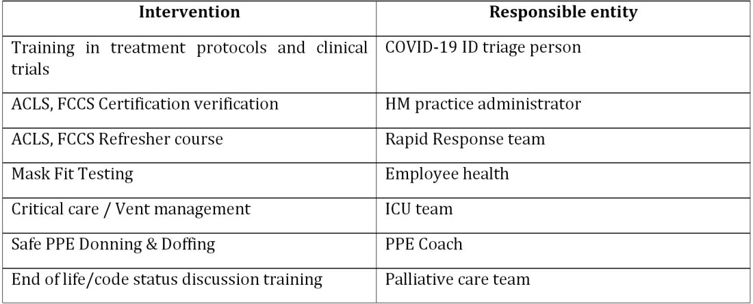
COVIDists were trained in various treatment protocols and ongoing clinical trials. They were given refresher training in Advanced Cardiac Life Support (ACLS) and Fundamental Critical Care Support (FCCS) courses and were taught in critical care/ventilator management by the intensivists through rapid indoctrination in the ICU. All of them had their N-95 mask fitting updated and were trained in the safe donning and doffing of all kinds of PPE by PPE coaches. The palliative care team trained them in conducting end-of-life/code status discussions with a focus on being unable to speak with family members at the bedside. COVIDists were also assigned as Code Blue leaders for any “COVID code blue” in the hospital.
In addition to the rapid training course, COVID-related updates were disseminated daily using three different modalities: brief huddles at the start of the day with the COVIDists; a COVID-19 newsletter summarizing daily updates, new treatments, strategies, and policies; and a WhatsApp group for instantly broadcasting information to the COVIDists (Table 1).
The execution phase
All the hospitalized COVID-19 patients were grouped together to COVID units, and the COVIDists were deployed to those units geographically. COVIDists were given lighter than usual patient loads to deal with the extra time needed for donning and doffing of PPE and for coordination with specialists. COVIDists were almost the only clinicians physically visiting the patients in most cases, and they became the “eyes and ears” of specialists since the specialists were advised to minimize exposure and pursue telemedicine consults. The COVIDists were also undertaking the most challenging part of the care – talking to families about end-of-life issues and the futility of aggressive care in certain patients with preexisting conditions.
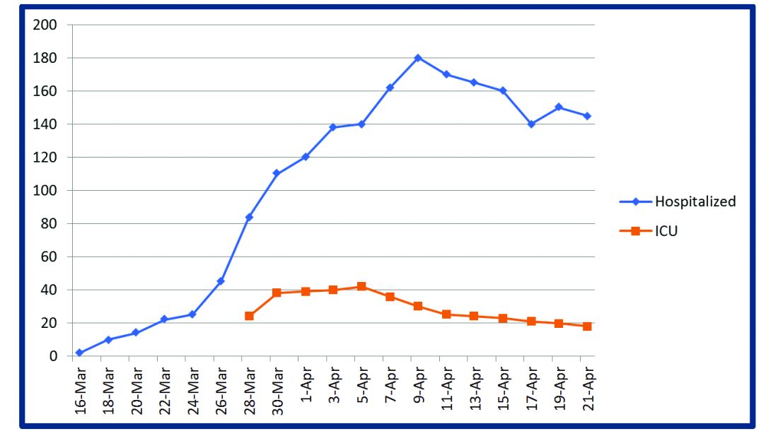
Some COVIDists were deployed to the ICU to work alongside the intensivists and became an invaluable resource in ICU management when the ICU census skyrocketed during the initial phase of the outbreak. This helped in tiding the health system over during the initial crisis. Within a short time, we shifted away from an early intubation strategy, and most of the ICU patients were managed in the intermediate care units on high flow oxygen along with the awake-proning protocol. The COVIDists exclusively managed these units. They led multidisciplinary rounds two times a day with the ICU, rapid response team (RRT), the palliative care team, and the nursing team. This step drastically decreased the number of intubations, RRT activations, reduced ICU census,3 and helped with hospital capacity and patient flow (Tables 2 and 3).
This strategy also helped build solidarity and camaraderie between all these groups, making the COVIDists feel that they were never alone and that the whole hospital supported them. We are currently evaluating clinical outcomes and attempting to identify effects on mortality, length of stay, days on the ventilator, and days in ICU.

The maintenance phase
It is already 2 months since the first devising COVIDists. There is no difference in sick callouts between COVIDists and non-COVIDists. One COVIDist and one non-COVIDist contracted the disease, but none of them required hospitalization. Although we initially thought that COVIDists would be needed for only a short period of time, the evolution of the disease is showing signs that it might be prolonged over the next several months. Hence, we are planning to continue COVIDist service for at least the next 6 months and reevaluate the need.
Hospital medicine leadership checked on COVIDists daily in regard to their physical health and, more importantly, their mental well-being. They were offered the chance to be taken off the schedule if they felt burned out, but no one wanted to come off their scheduled service before finishing their shifts. BlueCross MA recognized one of the COVIDists, Raghuveer Rakasi, MD, as a “hero on the front line.”4 In Dr. Rakasi’s words, “We took a nosedive into something without knowing its depth, and aware that we could have fatalities among ourselves. We took up new roles, faced new challenges, learned new things every day, evolving every step of the way. We had to change the way we practice medicine, finding new ways to treat patients, and protecting the workforce by limiting patient exposure, prioritizing investigations.” He added that “we have to adapt to a new normal; we should be prepared for this to come in waves. Putting aside our political views, we should stand united 6 feet apart, with a mask covering our brave faces, frequently washing our helping hands to overcome these uncertain times.”
Conclusion

The creation of a focused group of hospitalists called COVIDists and providing them with structured and rapid training (in various aspects of clinical care of COVID-19 patients, critical care/ventilator management, efficient and safe use of PPE) and daily information dissemination allowed our health system to prepare for the large volume of COVID-19 patients. It also helped in preserving the larger hospital workforce for a possible future surge.
The rapid development and implementation of the COVIDist strategy succeeded because of the intrinsic motivation of the providers to improve the outcomes of this high-risk patient population and the close collaboration of the stakeholders. Our institution remains successful in managing the pandemic in Western Massachusetts, with reserve capacity remaining even during the peak of the epidemic. A large part of this was because of creating and training a pool of COVIDists.
Dr. Medarametla is medical director, clinical operations, in the division of hospital medicine at Baystate Health, and assistant professor at University of Massachusetts, Worcester. Readers can contact him at [email protected]. Dr. Prabhakaran is unit medical director, geriatrics unit, in the division of hospital medicine at Baystate Health and assistant professor at University of Massachusetts. Dr. Bryson is associate program director of the Internal Medicine Residency at Baystate Health and assistant professor at University of Massachusetts. Dr. Umar is medical director, clinical operations, in the division of hospital medicine at Baystate Health. Dr. Natanasabapathy is division chief of hospital medicine at Baystate Health and assistant professor at University of Massachusetts.
References
1. Centers for Disease Control and Prevention. Coronavirus Disease 2019 (COVID-19). Updated Jun 10, 2020. https://www.cdc.gov/coronavirus/2019-ncov/cases-updates/cases-in-us.html.
2. Zhou F et al. Clinical course and risk factors for mortality of adult inpatients with COVID-19 in Wuhan, China: A retrospective cohort study. Lancet. 2020 Mar 28;395(10229):1054-62.
3. Westafer LM et al. A transdisciplinary COVID-19 early respiratory intervention protocol: An implementation story. J Hosp Med. 2020 May 21;15(6):372-374.
4. Miller J. “Heroes on the front line: Dr. Raghuveer Rakasi.” Coverage. May 18, 2020. https://coverage.bluecrossma.com/article/heroes-front-line-dr-raghuveer-rakasi
Adapting to the demands placed on hospital resources by COVID-19
Adapting to the demands placed on hospital resources by COVID-19
The challenges posed by COVID-19 have crippled health care systems around the globe. By February 2020, the first outbreak in the United States had been set off in Washington State. We quickly became the world’s epicenter of the epidemic, with over 1.8 million patients and over 110,000 deaths.1 The rapidity of spread and the severity of the disease created a tremendous strain on resources. It blindsided policymakers and hospital administrators, which left little time to react to the challenges placed on hospital operations all over the country.
The necessity of a new care model
Although health systems in the United States are adept in managing complications of common seasonal viral respiratory illnesses, COVID-19 presented an entirely different challenge with its significantly higher mortality rate. A respiratory disease turning into a multiorgan disease that causes debilitating cardiac, renal, neurological, hematological, and psychosocial complications2 was not something we had experience managing effectively. Additional challenges included a massive surge of COVID-19 patients, a limited supply of personal protective equipment (PPE), an inadequate number of intensivists for managing the anticipated ventilated patients, and most importantly, the potential of losing some of our workforce if they became infected.
Based on the experiences in China and Italy, and various predictive models, the division of hospital medicine at Baystate Health quickly realized the necessity of a new model of care for COVID-19 patients. We came up with an elaborate plan to manage the disease burden and the strain on resources effectively. The measures we put in place could be broadly divided into three categories following the timeline of the disease: the preparatory phase, the execution phase, and the maintenance phase.
The preparatory phase: From “Hospitalists” to “COVIDists”
As in most hospitals around the country, hospitalists are the backbone of inpatient clinical operations at our health system. A focused group of 10 hospitalists who volunteered to take care of COVID-19 patients with a particular interest in the pandemic and experience in critical care were selected, and the term “COVIDists” was coined to refer to them.
COVIDists were trained in various treatment protocols and ongoing clinical trials. They were given refresher training in Advanced Cardiac Life Support (ACLS) and Fundamental Critical Care Support (FCCS) courses and were taught in critical care/ventilator management by the intensivists through rapid indoctrination in the ICU. All of them had their N-95 mask fitting updated and were trained in the safe donning and doffing of all kinds of PPE by PPE coaches. The palliative care team trained them in conducting end-of-life/code status discussions with a focus on being unable to speak with family members at the bedside. COVIDists were also assigned as Code Blue leaders for any “COVID code blue” in the hospital.
In addition to the rapid training course, COVID-related updates were disseminated daily using three different modalities: brief huddles at the start of the day with the COVIDists; a COVID-19 newsletter summarizing daily updates, new treatments, strategies, and policies; and a WhatsApp group for instantly broadcasting information to the COVIDists (Table 1).
The execution phase
All the hospitalized COVID-19 patients were grouped together to COVID units, and the COVIDists were deployed to those units geographically. COVIDists were given lighter than usual patient loads to deal with the extra time needed for donning and doffing of PPE and for coordination with specialists. COVIDists were almost the only clinicians physically visiting the patients in most cases, and they became the “eyes and ears” of specialists since the specialists were advised to minimize exposure and pursue telemedicine consults. The COVIDists were also undertaking the most challenging part of the care – talking to families about end-of-life issues and the futility of aggressive care in certain patients with preexisting conditions.
Some COVIDists were deployed to the ICU to work alongside the intensivists and became an invaluable resource in ICU management when the ICU census skyrocketed during the initial phase of the outbreak. This helped in tiding the health system over during the initial crisis. Within a short time, we shifted away from an early intubation strategy, and most of the ICU patients were managed in the intermediate care units on high flow oxygen along with the awake-proning protocol. The COVIDists exclusively managed these units. They led multidisciplinary rounds two times a day with the ICU, rapid response team (RRT), the palliative care team, and the nursing team. This step drastically decreased the number of intubations, RRT activations, reduced ICU census,3 and helped with hospital capacity and patient flow (Tables 2 and 3).
This strategy also helped build solidarity and camaraderie between all these groups, making the COVIDists feel that they were never alone and that the whole hospital supported them. We are currently evaluating clinical outcomes and attempting to identify effects on mortality, length of stay, days on the ventilator, and days in ICU.
The maintenance phase
It is already 2 months since the first devising COVIDists. There is no difference in sick callouts between COVIDists and non-COVIDists. One COVIDist and one non-COVIDist contracted the disease, but none of them required hospitalization. Although we initially thought that COVIDists would be needed for only a short period of time, the evolution of the disease is showing signs that it might be prolonged over the next several months. Hence, we are planning to continue COVIDist service for at least the next 6 months and reevaluate the need.
Hospital medicine leadership checked on COVIDists daily in regard to their physical health and, more importantly, their mental well-being. They were offered the chance to be taken off the schedule if they felt burned out, but no one wanted to come off their scheduled service before finishing their shifts. BlueCross MA recognized one of the COVIDists, Raghuveer Rakasi, MD, as a “hero on the front line.”4 In Dr. Rakasi’s words, “We took a nosedive into something without knowing its depth, and aware that we could have fatalities among ourselves. We took up new roles, faced new challenges, learned new things every day, evolving every step of the way. We had to change the way we practice medicine, finding new ways to treat patients, and protecting the workforce by limiting patient exposure, prioritizing investigations.” He added that “we have to adapt to a new normal; we should be prepared for this to come in waves. Putting aside our political views, we should stand united 6 feet apart, with a mask covering our brave faces, frequently washing our helping hands to overcome these uncertain times.”
Conclusion
The creation of a focused group of hospitalists called COVIDists and providing them with structured and rapid training (in various aspects of clinical care of COVID-19 patients, critical care/ventilator management, efficient and safe use of PPE) and daily information dissemination allowed our health system to prepare for the large volume of COVID-19 patients. It also helped in preserving the larger hospital workforce for a possible future surge.
The rapid development and implementation of the COVIDist strategy succeeded because of the intrinsic motivation of the providers to improve the outcomes of this high-risk patient population and the close collaboration of the stakeholders. Our institution remains successful in managing the pandemic in Western Massachusetts, with reserve capacity remaining even during the peak of the epidemic. A large part of this was because of creating and training a pool of COVIDists.
Dr. Medarametla is medical director, clinical operations, in the division of hospital medicine at Baystate Health, and assistant professor at University of Massachusetts, Worcester. Readers can contact him at [email protected]. Dr. Prabhakaran is unit medical director, geriatrics unit, in the division of hospital medicine at Baystate Health and assistant professor at University of Massachusetts. Dr. Bryson is associate program director of the Internal Medicine Residency at Baystate Health and assistant professor at University of Massachusetts. Dr. Umar is medical director, clinical operations, in the division of hospital medicine at Baystate Health. Dr. Natanasabapathy is division chief of hospital medicine at Baystate Health and assistant professor at University of Massachusetts.
References
1. Centers for Disease Control and Prevention. Coronavirus Disease 2019 (COVID-19). Updated Jun 10, 2020. https://www.cdc.gov/coronavirus/2019-ncov/cases-updates/cases-in-us.html.
2. Zhou F et al. Clinical course and risk factors for mortality of adult inpatients with COVID-19 in Wuhan, China: A retrospective cohort study. Lancet. 2020 Mar 28;395(10229):1054-62.
3. Westafer LM et al. A transdisciplinary COVID-19 early respiratory intervention protocol: An implementation story. J Hosp Med. 2020 May 21;15(6):372-374.
4. Miller J. “Heroes on the front line: Dr. Raghuveer Rakasi.” Coverage. May 18, 2020. https://coverage.bluecrossma.com/article/heroes-front-line-dr-raghuveer-rakasi
First-in-kind anti-CD47 antibody shows promise for MDS and AML treatment
Magrolimab plus azacitidine (AZA) improved outcomes in acute myeloid leukemia (AML) and myelodysplastic syndromes (MDS) patients according to the results of a phase 1b study (NCT03248479) presented at the virtual ASCO meeting. The combo especially was promising for the underserved patient population that have the TP53 (p53) mutation.
Magrolimab is a first-in-kind IgG anti-CD47 monoclonal antibody that promotes the elimination of tumor cells through macrophage phagocytosis. CD47 is a “do not eat me” signal on cancer cells that allows the cells to evade macrophages. Its increased expression is predictive of a worse outcome in AML patients, according to David A. Sallman, MD, of the Moffitt Cancer Center, Tampa, Fla., and colleagues.
Dr. Sallman presented the results of a study examining whether magrolimab would provide a synergistic benefit when combined with AZA (which induces other prophagocytic “eat me” signals such as calreticulin on cancer cells). The primary objectives of the study were to examine the safety of magrolimab alone or with AZA, and to assess the efficacy of the magrolimab/AZA combo in 29 untreated AML patients and 39 untreated MDS patients. The majority of both the MDS and AML patients were poor cytogenetic risk at 64% and 72%, respectively. Mutant p53 was present in 13% of the MDS patients and 45% of the AML patients.
No deaths occurred in the first 60 days of the study among either the MDS or AML patients and discontinuation of treatment because of drug-related adverse events was seen in only one of the patients (1.5%) treated with magrolimab/AZA. There was no significant neutropenia or thrombocytopenia caused by the therapy seen, and the majority of the patients improved their neutrophil and platelet counts while on therapy.
Anemia from CD47 blockade was mitigated by the use of a priming dose of magrolimab coupled to a maintenance-dose regimen, resulting in a mild hemoglobin drop on the first dose, which returned to baseline with a majority of patients experiencing significant hemoglobin improvement and a decrease in transfusion frequency over time, according to Dr. Sallman and his colleagues.
The results showed that magrolimab/AZA induced a 91% overall response rate (ORR), with a 42% complete remission (CR) that increased to 56% at 6 months, in the MDS patients. AML patients experienced a 64% ORR (56% CR/CRi [CR with incomplete hematological remission]). These results compare favorably with the CR rate of 6%-17% rate seen for AZA monotherapy, according to Dr. Sallman.
Red blood cell transfusion independence was achieved in 58% of the MDS patients and 64% of the AML patients, and a complete cytogenetic response was seen in 35% and 50% of the MDS and AML patients, respectively.
The combined treatment was especially effective in the patients with p53 mutations, with an overall response rate of 75% for both MDS and AML, and a complete response of 42% and 50%, respectively. During the reported time of the study, the median survival was not reached, which compares favorably with current therapies, according to Dr. Sallman.
“Specifically looking at a very-high-risk p53-mutant subset, complete remissions have been observed in the majority of patients. And again, these have been durable. Based on all of these data, expansion cohorts both in MDS and p53 and AML continue to accrue with registrational studies in progress for MDS and planned for p53-mutant AML,” Dr. Sallman concluded.
The trial was sponsored by Gilead Sciences, and funding was obtained from the California Institute for Regenerative Medicine. Dr. Sallman disclosed that he received research funding from Celgene and has acted in a consulting or advisory role for Agios, argenx, and Celyad. He was also on the speaker’s bureau for a variety of pharmaceutical/biotech companies.
SOURCE: Sallman DA et al. ASCO 2020, Abstract 7507.
FROM ASCO 2020
Daily Recap: Lung ultrasound helps diagnose COVID-19 in kids, first treatment approved for adult-onset Still’s disease
Here are the stories our MDedge editors across specialties think you need to know about today:
Lung ultrasound works well in children with COVID-19
Lung ultrasound has “high concordance” with radiologic findings in children with COVID-19 and offers benefits over other imaging techniques, such as CT. “First, it may reduce the number of radiologic examinations, lowering the radiation exposure of the patients,” wrote Marco Denina, MD, and colleagues from the pediatric infectious diseases unit at Regina Margherita Children’s Hospital in Turin, Italy. “Secondly, when performed at the bedside, [lung ultrasound] allows for the reduction of the patient’s movement within the hospital; thus, it lowers the number of health care workers and medical devices exposed to [SARS-CoV-2].” The findings of the small, observational study were published in Pediatrics. Read more.
New hypertension definitions reveal preclampsia risk
Using the new clinical definitions of hypertension, pregnant women with even modest elevations in blood pressure are at increased risk for preeclampsia, according to results from a large retrospective cohort study. Elizabeth F. Sutton, PhD, of the University of Pittsburgh and colleagues looked at records from 18,162 women who had given birth to a single baby. The authors found preeclampsia risk increased with increasing blood pressure elevation. Among women with normal blood pressure before 20 weeks’ gestation, 5% had preeclampsia, while 7% of those with elevated blood pressure did, as did 12% of women with stage 1 hypertension and 30% of women with stage 2 hypertension. The increase in risk of preeclampsia was because of preterm preeclampsia in the women with elevated blood pressure. Preeclampsia researcher Mark Santillan, MD, PhD, of the University of Iowa in Iowa City, said in an interview that the results “open the door to considering these new blood pressure categories as a prognosticator” for preeclampsia. “This paper furthers the field by applying these new categories to hypertensive diseases in pregnancy, which are not well studied” in comparison to nonpregnant hypertensive states. Read more.
Face mask type matters when sterilizing
When sterilizing face masks, the type of mask and the method of sterilization have a bearing on subsequent filtration efficiency, according to new research published in JAMA Network Open. The greatest reduction in filtration efficiency after sterilization occurred with surgical face masks. With plasma vapor hydrogen peroxide (H2O2) sterilization, filtration efficiency of N95 and KN95 masks was maintained at more than 95%, but for surgical face masks, filtration efficiency was reduced to less than 95%. With chlorine dioxide (ClO2) sterilization, on the other hand, filtration efficiency was maintained at above 95% for N95 masks, but for KN95 and surgical face masks, filtration efficiency was reduced to less than 80%. Read more.
FDA approves first treatment for adult-onset Still’s disease
The Food and Drug Administration has expanded the indications for canakinumab (Ilaris) to include all patients with active Still’s disease older than 2 years, adding adult-onset Still’s disease (AOSD) to a previous approval for juvenile-onset Still’s disease, also known as systemic juvenile idiopathic arthritis (sJIA). That makes Ilaris the first approved treatment for AOSD. The results from a randomized, double-blind, placebo-controlled study of 36 patients with AOSD aged 22-70 years showed that the efficacy and safety data in AOSD were generally consistent with the results of a pooled analysis of sJIA patients, according to Novartis, which markets canakinumab. Read more.
Intranasal DHE shows promise in migraine
An intranasal form of dihydroergotamine (DHE) targeting the upper nasal region is safe and effective for the treatment of migraine, according to results from a phase 3 clinical trial. The new formulation could offer patients an at-home alternative to intramuscular infusions or intravenous injections currently used to deliver DHE. The STOP 301 phase 3 open-label safety and tolerability trial treated over 5,650 migraine attacks in 354 patients who self-administered INP104 for up to 52 weeks. They were provided up to three doses per week (1.45 mg in a dose of two puffs, one per nostril). A total of 66.3% of participants reported pain relief by 2 hours following a dose, and 38% had freedom from pain. Read more.
For more on COVID-19, visit our Resource Center. All of our latest news is available on MDedge.com.
New health policy and advocacy committee (HPAC)
What a privilege it has been over the last several months to participate as staff support along with Jenny Nemkovich and Michelle Kosobucki to CHEST’s new Health Policy and Advocacy Committee (HPAC). The opportunity to serve on a committee of CHEST from the perspective of staff rather than in a volunteer/leadership role has been very enlightening and clearly a learning experience.
Background
As most know, CHEST in the summer of 2019 made the decision to proactively strengthen our position in the areas of public policy, both advocacy and the regulatory space.

This decision will provide CHEST with the mechanism to have greater control over determining and influencing the pulmonary, critical care, and sleep agenda that directly impacts our members and our patients. Adding this piece to the CHEST portfolio is particularly fortuitous in light of the increased advocacy needs in this COVID-19 environment. Having recently completed the acquisition of NAMDRC, CHEST has jump-started our return to this space. While this acquisition does not represent a single source solution, it does represent a key component to a comprehensive approach to policy and advocacy. The rich experience of our new colleagues from NAMDRC brings incredible value and insights to our efforts.
Health policy and advocacy committee
The initial composition of the HPAC is made up of equal numbers of members drawn from the NAMDRC leadership pool, as well as members of both the CHEST Foundation Board of Trustees and the Board of Regents of the College. This group represents a very energetic, talented, and diverse group. Experience in the space of policy and advocacy in areas such as home ventilation, oxygen issues, telemedicine, and pulmonary rehab reimbursement is blended with presidential leadership of both the CHEST Foundation and CHEST, as well as talent in areas such as coding and reimbursement, social media applications, and also leadership representing our NetWorks.
Policy priorities
Having had three virtual meetings, the HPAC has initially been focusing on developing and discussing an initial group of policy priorities. These topics are being vetted and held to a rigorous discussion, including what success looks like in these areas, potential barriers or obstacles to making an impact, and who could represent important collaborative partners in these areas. These priorities will be coupled with an effort to define short-term and longer term performance indicators to help try to assess meaningful impact. Once these are better defined, we plan to reach out to our CHEST NetWorks, partners in Industry, sister societies, and friends in patient advocacy groups to get their input and, when appropriate, their collaboration. The BOR will be kept informed and eventually comment and hopefully endorse these policy priorities.
Member engagement
In my opinion, our approach in this area of policy and advocacy is somewhat unique in the associational arena. Rather than policy staff driving the agenda, we are following the example of other committees at CHEST in having volunteers and leadership developing the “what” and staff creating the “how.” At that point, a team of leadership/staff will deliver the product. I feel that this somewhat “bottom up” approach will lead to much more productive and effective member engagement and a growing group of advocacy aware and committed members.
Washington watchline
To complement the work of HPAC and better communicate important issues related to policy and advocacy, our Publications team, led by Nicki Augustyn, has taken over the production of what was NAMDRC’s valuable periodical, the Washington Watchline. Under the editorship for many years of past CHEST President, Jim Mathers, MD, FCCP, this resource has been a valuable and respected source of information for NAMDRC membership. The June edition has recently been published.
Spring meeting, 2021
The HPAC’s Chair and Vice-Chair, Drs. Neil Freedman and Jim Lamberti, are serving as the Program Directors for our first meeting that will blend the NAMDRC perspective and experience in a program around policy and advocacy with the traditional expertise in education delivery of CHEST. This meeting will be in conjunction with our Spring Leadership meetings in Sonoma, California. Save the date, as this promises to be a great meeting, with unique educational opportunities and policy and advocacy insights.
Thanks again to the members of HPAC and to Bob Musacchio for giving me an opportunity to provide staff assistance in this exciting new endeavor for CHEST.
What a privilege it has been over the last several months to participate as staff support along with Jenny Nemkovich and Michelle Kosobucki to CHEST’s new Health Policy and Advocacy Committee (HPAC). The opportunity to serve on a committee of CHEST from the perspective of staff rather than in a volunteer/leadership role has been very enlightening and clearly a learning experience.
Background
As most know, CHEST in the summer of 2019 made the decision to proactively strengthen our position in the areas of public policy, both advocacy and the regulatory space.
This decision will provide CHEST with the mechanism to have greater control over determining and influencing the pulmonary, critical care, and sleep agenda that directly impacts our members and our patients. Adding this piece to the CHEST portfolio is particularly fortuitous in light of the increased advocacy needs in this COVID-19 environment. Having recently completed the acquisition of NAMDRC, CHEST has jump-started our return to this space. While this acquisition does not represent a single source solution, it does represent a key component to a comprehensive approach to policy and advocacy. The rich experience of our new colleagues from NAMDRC brings incredible value and insights to our efforts.
Health policy and advocacy committee
The initial composition of the HPAC is made up of equal numbers of members drawn from the NAMDRC leadership pool, as well as members of both the CHEST Foundation Board of Trustees and the Board of Regents of the College. This group represents a very energetic, talented, and diverse group. Experience in the space of policy and advocacy in areas such as home ventilation, oxygen issues, telemedicine, and pulmonary rehab reimbursement is blended with presidential leadership of both the CHEST Foundation and CHEST, as well as talent in areas such as coding and reimbursement, social media applications, and also leadership representing our NetWorks.
Policy priorities
Having had three virtual meetings, the HPAC has initially been focusing on developing and discussing an initial group of policy priorities. These topics are being vetted and held to a rigorous discussion, including what success looks like in these areas, potential barriers or obstacles to making an impact, and who could represent important collaborative partners in these areas. These priorities will be coupled with an effort to define short-term and longer term performance indicators to help try to assess meaningful impact. Once these are better defined, we plan to reach out to our CHEST NetWorks, partners in Industry, sister societies, and friends in patient advocacy groups to get their input and, when appropriate, their collaboration. The BOR will be kept informed and eventually comment and hopefully endorse these policy priorities.
Member engagement
In my opinion, our approach in this area of policy and advocacy is somewhat unique in the associational arena. Rather than policy staff driving the agenda, we are following the example of other committees at CHEST in having volunteers and leadership developing the “what” and staff creating the “how.” At that point, a team of leadership/staff will deliver the product. I feel that this somewhat “bottom up” approach will lead to much more productive and effective member engagement and a growing group of advocacy aware and committed members.
Washington watchline
To complement the work of HPAC and better communicate important issues related to policy and advocacy, our Publications team, led by Nicki Augustyn, has taken over the production of what was NAMDRC’s valuable periodical, the Washington Watchline. Under the editorship for many years of past CHEST President, Jim Mathers, MD, FCCP, this resource has been a valuable and respected source of information for NAMDRC membership. The June edition has recently been published.
Spring meeting, 2021
The HPAC’s Chair and Vice-Chair, Drs. Neil Freedman and Jim Lamberti, are serving as the Program Directors for our first meeting that will blend the NAMDRC perspective and experience in a program around policy and advocacy with the traditional expertise in education delivery of CHEST. This meeting will be in conjunction with our Spring Leadership meetings in Sonoma, California. Save the date, as this promises to be a great meeting, with unique educational opportunities and policy and advocacy insights.
Thanks again to the members of HPAC and to Bob Musacchio for giving me an opportunity to provide staff assistance in this exciting new endeavor for CHEST.
Virtual visits for patients with neuromuscular respiratory failure in the time of COVID-19: A potential bright spot from the pandemic
On March 17, 2020, I entered my patients electronic medical record and hit the “Connect with Zoom” button in her Epic (Epic Systems Corporation) chart. About 20 seconds later, the face of my 28-year-old patient with advanced spinal muscular atrophy type 2 (SMA-2) appeared virtually and not live for the first time since I had met her some 10 years previously. She appeared well and her history supported that. We spent most of the time reviewing recent events and surveying her home ventilation equipment. She felt well and sleep was of good quality. She was performing her normal activities without dyspnea. Her mechanical insufflator-exsufflator was working fine, although she used it only as needed, and she was performing lung volume recruitment maneuvers with a resuscitator bag three times a day with assistance. Her mask for nocturnal NPPV was getting old, and she showed me where the straps were fraying. We noted that her bilevel device was now 8 years old and that she needed a new one. We concluded our conversation in 20 minutes and she blurted out: “Wow, that was easy. Thanks, Dr. Benditt.” I got off the phone and put in the order for a new mask and bilevel device with our clinic respiratory therapist. She received the equipment 48 hours later and sent an electronic message through her chart to let me know it had arrived. A total of five in-person visits including me and other providers had been cancelled and replaced by virtual visits. She has made one visit to the hospital in the last 3 months for an intrathecal nusinersen (Spinraza) injection that was done with a COVID-19 prescreening and full PPE.

One week prior to our virtual visit, my university hospital had reduced in-person clinic visits to those deemed absolutely necessary due to the COVID-19 pandemic. Visits considered absolutely necessary included such patients as postoperative transplant visits and preoperative evaluations for urgent surgeries. All other patient visits were canceled with plans to reschedule them once the COVID-19 pandemic was controlled. As the breadth and depth of the pandemic became apparent, a very rapid ramp-up of “virtual visits” via telemedicine capacity was rolled out. I had not previously used telemedicine, and the learning curve was steep, although once in place, the technology was straightforward from the provider perspective. The telemedicine visits for our hospital for the entire year of 2019 totaled about 800. In the month of April of 2020 we engaged in 40,000 telemedicine visits. This explosive growth of telemedicine implementation has occurred around the country and world during the COVID-19 pandemic (Olayiwola JN, et al. JMIR Public Health Surveill. 2020, May 29. doi: 10.2196/19045). This recent growth of telemedicine in the US has been fueled by the need for social distancing and quarantine, the lack of universal testing and COVID-19 case tracking, and the realization by CMS that coverage of telemedicine services had to be expanded rapidly to allow for continued patient care in the setting of stay-at-home orders. A rapid role out of application technology support and online training classes for health-care providers was undertaken. Privileges for telemedicine virtual visits were approved when providers completed the informational online modules and set up their HIPPA compliant Zoom accounts (Zoom Video Communications, San Jose, CA). All of us had minor stumbles initially with the equipment, software, and getting the patients connected online. After four or five visits, the process started to click and has become rather routine. Many providers and patients found this quite a positive development in terms of patient-provider visits but a question arose almost immediately: “Will this continue to be supported by insurers and allow us to integrate this practice into our outpatient clinic setting once the pandemic was controlled?” Time with tell, but an opportunity has presented itself.
For patients with neuromuscular disease and respiratory failure, telemedicine is a technology that may be particularly attractive for a number of reasons. First, patients with neuromuscular respiratory failure are likely at a particularly high risk of death if they develop full-blown COVID-19 infection. Development of acute respiratory distress syndrome (ARDS) on top of underlying neuromuscular respiratory failure is likely to be particularly deadly, although, very fortunately, there are no published reports of widespread infections in patients with neuromuscular respiratory disease. We have known for many decades that pneumonia is the leading cause of death for these patients. Second, patients with neuromuscular respiratory failure often find it quite difficult to come to the hospital for clinic visits. Mobilizing equipment, caregivers, and transportation can take days to arrange. For this reason, many neuromuscular clinics provide a multidisciplinary/multi-provider half-day visit to reduce the need to come into the hospital for multiple separate visits. Lastly, there are relatively few respiratory health-care providers in the United States and around the world who focus on patients with neuromuscular respiratory disease. Many neuromuscular clinics and providers will, therefore, have a very wide patient catchment area. For instance, my practice, based in Seattle, Washington, includes patients from Alaska, Montana, Idaho, and Wyoming. In-person hospital visits more than once per year may be virtually impossible.
Telemedicine is a methodology that has long been considered helpful in the arena of home ventilation and, in fact, we have been using some telemedicine technologies for some time (Casavant DW, et al. J Telemed Telecare. 2014;20[8]:441). Telemedicine (telehealth) includes the use of electronic information and communications technologies to provide and support health care when distance separates the participants. For instance, monitoring of nocturnal ventilation via downloads from Internet-connected noninvasive or invasive ventilation devices, overnight oximetry, and even phone calls from durable medical equipment providers during a home visit would be considered telemedicine. Many of us have been using these methods for many years. It is really the face-to-face “virtual visit” frequency that the COVID-19 pandemic has accelerated. This is a crucial advance in the process of telehealth because we may be able to reduce visits to our clinics from once every 3 to 6 months to perhaps once per year if support for virtual visits by insurers continues and if home monitoring can expand to include accurate home measurement of patient CO2 levels by either end-tidal CO2, transcutaneous CO2, or point of care arterial or capillary blood gases, as well as home pulmonary function monitoring. Measurement of CO2 levels and pulmonary function has generally been done at the hospital or in the clinic although there is no reason that with home visit support from appropriate services (that might even include durable medical equipment companies) that this could not be accomplished. This is not to say that there are not hurdles to the application of telehealth in the neuromuscular disease and home ventilation population. Not all patients have the equipment or technology savvy to participate in virtual visits, and not all insurers cover these visits even now during COVID-19. However, I imagine a future where a significant number of visits for patients with neuromuscular respiratory disease and home ventilation needs could be performed virtually. I envision that this would reduce patient and home caregiver travel burdens, make more efficient use of health-care provider time, expand the number of patients that a neuromuscular respiratory disease practitioner could serve, and perhaps reduce health-care expenditures per patient. This may be a real health-care bright spot in the huge difficulties of COVID-19. Fingers crossed.
Dr. Benditt is Medical Director of Respiratory Care Services and Professor of Medicine, University of Washington Medical Center, Seattle, Washington.
On March 17, 2020, I entered my patients electronic medical record and hit the “Connect with Zoom” button in her Epic (Epic Systems Corporation) chart. About 20 seconds later, the face of my 28-year-old patient with advanced spinal muscular atrophy type 2 (SMA-2) appeared virtually and not live for the first time since I had met her some 10 years previously. She appeared well and her history supported that. We spent most of the time reviewing recent events and surveying her home ventilation equipment. She felt well and sleep was of good quality. She was performing her normal activities without dyspnea. Her mechanical insufflator-exsufflator was working fine, although she used it only as needed, and she was performing lung volume recruitment maneuvers with a resuscitator bag three times a day with assistance. Her mask for nocturnal NPPV was getting old, and she showed me where the straps were fraying. We noted that her bilevel device was now 8 years old and that she needed a new one. We concluded our conversation in 20 minutes and she blurted out: “Wow, that was easy. Thanks, Dr. Benditt.” I got off the phone and put in the order for a new mask and bilevel device with our clinic respiratory therapist. She received the equipment 48 hours later and sent an electronic message through her chart to let me know it had arrived. A total of five in-person visits including me and other providers had been cancelled and replaced by virtual visits. She has made one visit to the hospital in the last 3 months for an intrathecal nusinersen (Spinraza) injection that was done with a COVID-19 prescreening and full PPE.
One week prior to our virtual visit, my university hospital had reduced in-person clinic visits to those deemed absolutely necessary due to the COVID-19 pandemic. Visits considered absolutely necessary included such patients as postoperative transplant visits and preoperative evaluations for urgent surgeries. All other patient visits were canceled with plans to reschedule them once the COVID-19 pandemic was controlled. As the breadth and depth of the pandemic became apparent, a very rapid ramp-up of “virtual visits” via telemedicine capacity was rolled out. I had not previously used telemedicine, and the learning curve was steep, although once in place, the technology was straightforward from the provider perspective. The telemedicine visits for our hospital for the entire year of 2019 totaled about 800. In the month of April of 2020 we engaged in 40,000 telemedicine visits. This explosive growth of telemedicine implementation has occurred around the country and world during the COVID-19 pandemic (Olayiwola JN, et al. JMIR Public Health Surveill. 2020, May 29. doi: 10.2196/19045). This recent growth of telemedicine in the US has been fueled by the need for social distancing and quarantine, the lack of universal testing and COVID-19 case tracking, and the realization by CMS that coverage of telemedicine services had to be expanded rapidly to allow for continued patient care in the setting of stay-at-home orders. A rapid role out of application technology support and online training classes for health-care providers was undertaken. Privileges for telemedicine virtual visits were approved when providers completed the informational online modules and set up their HIPPA compliant Zoom accounts (Zoom Video Communications, San Jose, CA). All of us had minor stumbles initially with the equipment, software, and getting the patients connected online. After four or five visits, the process started to click and has become rather routine. Many providers and patients found this quite a positive development in terms of patient-provider visits but a question arose almost immediately: “Will this continue to be supported by insurers and allow us to integrate this practice into our outpatient clinic setting once the pandemic was controlled?” Time with tell, but an opportunity has presented itself.
For patients with neuromuscular disease and respiratory failure, telemedicine is a technology that may be particularly attractive for a number of reasons. First, patients with neuromuscular respiratory failure are likely at a particularly high risk of death if they develop full-blown COVID-19 infection. Development of acute respiratory distress syndrome (ARDS) on top of underlying neuromuscular respiratory failure is likely to be particularly deadly, although, very fortunately, there are no published reports of widespread infections in patients with neuromuscular respiratory disease. We have known for many decades that pneumonia is the leading cause of death for these patients. Second, patients with neuromuscular respiratory failure often find it quite difficult to come to the hospital for clinic visits. Mobilizing equipment, caregivers, and transportation can take days to arrange. For this reason, many neuromuscular clinics provide a multidisciplinary/multi-provider half-day visit to reduce the need to come into the hospital for multiple separate visits. Lastly, there are relatively few respiratory health-care providers in the United States and around the world who focus on patients with neuromuscular respiratory disease. Many neuromuscular clinics and providers will, therefore, have a very wide patient catchment area. For instance, my practice, based in Seattle, Washington, includes patients from Alaska, Montana, Idaho, and Wyoming. In-person hospital visits more than once per year may be virtually impossible.
Telemedicine is a methodology that has long been considered helpful in the arena of home ventilation and, in fact, we have been using some telemedicine technologies for some time (Casavant DW, et al. J Telemed Telecare. 2014;20[8]:441). Telemedicine (telehealth) includes the use of electronic information and communications technologies to provide and support health care when distance separates the participants. For instance, monitoring of nocturnal ventilation via downloads from Internet-connected noninvasive or invasive ventilation devices, overnight oximetry, and even phone calls from durable medical equipment providers during a home visit would be considered telemedicine. Many of us have been using these methods for many years. It is really the face-to-face “virtual visit” frequency that the COVID-19 pandemic has accelerated. This is a crucial advance in the process of telehealth because we may be able to reduce visits to our clinics from once every 3 to 6 months to perhaps once per year if support for virtual visits by insurers continues and if home monitoring can expand to include accurate home measurement of patient CO2 levels by either end-tidal CO2, transcutaneous CO2, or point of care arterial or capillary blood gases, as well as home pulmonary function monitoring. Measurement of CO2 levels and pulmonary function has generally been done at the hospital or in the clinic although there is no reason that with home visit support from appropriate services (that might even include durable medical equipment companies) that this could not be accomplished. This is not to say that there are not hurdles to the application of telehealth in the neuromuscular disease and home ventilation population. Not all patients have the equipment or technology savvy to participate in virtual visits, and not all insurers cover these visits even now during COVID-19. However, I imagine a future where a significant number of visits for patients with neuromuscular respiratory disease and home ventilation needs could be performed virtually. I envision that this would reduce patient and home caregiver travel burdens, make more efficient use of health-care provider time, expand the number of patients that a neuromuscular respiratory disease practitioner could serve, and perhaps reduce health-care expenditures per patient. This may be a real health-care bright spot in the huge difficulties of COVID-19. Fingers crossed.
Dr. Benditt is Medical Director of Respiratory Care Services and Professor of Medicine, University of Washington Medical Center, Seattle, Washington.