User login
Daily Recap: From hospitalist to ‘COVIDist’; Systolic BP -- How low should you go?
Here are the stories our MDedge editors across specialties think you need to know about today:
A ‘Fraternity of People Who Are Struggling’
Kathleen Ronan spent a week in a New Jersey hospital, including 5 days in the ICU, battling the novel coronavirus.
Her years of working as a home health nurse told her that the return home wouldn’t be easy, but nothing prepared her for just how much she would struggle. The once-active Ronan, 51, now needed a walker to traverse the few steps from her bed to the toilet, an effort that left her gasping for air. Her brain couldn’t even focus on an audiobook, let alone a short magazine article. “It just completely knocked the stuffing out of me,” Ronan said.
Ronan’s lingering symptoms aren’t unique to COVID-19 patients. In as many as 80% of patients leaving the ICU, researchers have documented what they call post–intensive care syndrome (PICS) — a constellation of physical, cognitive, and psychiatric symptoms that result from an ICU stay. Although underlying illness plays a role in these symptoms, the amount of time spent in critical care is a major factor.
The good news is that over the past decade, researchers have made important strides in understanding what makes PICS symptoms worse and how critical care physicians can tweak ICU protocols to reduce PICS severity. Practitioners will need to draw on this knowledge to help Ronan and the thousands of COVID-19 ICU patients like her. Read more.
The evolution of ‘COVIDists’
At the start of the pandemic earlier this year hospitalists at Baystate Health in Western Massachusetts realized the necessity of a new model of care for COVID-19 patients. Challenges included a massive surge of COVID-19 patients, a limited supply of PPE, an inadequate number of intensivists for managing the anticipated ventilated patients, and the potential of losing some of our workforce if they became infected. Hospitalists there came up with an elaborate plan to manage the disease burden and the strain on resources effectively.
A focused group of 10 hospitalists who volunteered to take care of COVID-19 patients with a particular interest in the pandemic and experience in critical care were selected, and the term “COVIDists” was coined to refer to them. The group underwent rapid training in various treatment protocols and ongoing clinical trials.
All the hospitalized COVID-19 patients were grouped together to COVID units, and the COVIDists were deployed to those units geographically. COVIDists were given lighter than usual patient loads to deal with the extra time needed for donning and doffing of PPE and for coordination with specialists. COVIDists were almost the only clinicians physically visiting the patients in most cases, and they became the “eyes and ears” of specialists since the specialists were advised to minimize exposure and pursue telemedicine consults. Read more.
How low should you go?
Cardiovascular risk continues to reduce as systolic blood pressure decreases right down to levels as low as 90 mm Hg, according to a new study.
Researchers analyzed data from a cohort of 1,457 participants (mean age, 58 years) who did not have any traditional cardiovascular risk factors and had a systolic blood pressure level between 90 and 129 mm Hg at baseline. Results showed that, during a mean follow-up of 14.5 years, there was an increase in traditional cardiovascular risk factors, coronary artery calcium, and incident cardiovascular events with increasing systolic blood pressure levels.
“We modeled systolic blood pressure on a continuous scale and saw the risk increasing in a linear fashion as blood pressure increased and this occurred right down to 90 mm Hg. We didn’t see any nadir or J-point where there may be an increased risk at lower pressures,” said lead author Seamus Whelton, MD, assistant professor of medicine at the division of cardiology at Johns Hopkins Medicine, Baltimore.
“From an individual level we can now say that in healthy individuals, a systolic pressure in the 90s is not too low. It is a positive thing. And it is recommended to try and keep systolic pressure at these levels if possible by maintaining a healthy lifestyle,” Dr. Whelton said in an interview. Read more.
Asthma tops spending on avoidable pediatric inpatient stays
Asthma costs nearly equaled potentially avoidable hospital bills for diabetes, gastroenteritis, and UTIs combined in a study of in-patient stays among children aged 3 months to 17 years.
Indeed, hospital charges for the treatment of children with asthma made up nearly half of all potentially avoidable pediatric inpatient costs in 2017, according to the Agency for Healthcare Research and Quality.
The cost of potentially avoidable visits for asthma that year was $278 million, versus $284 million combined for the other three conditions, Kimberly W. McDermott, PhD, and H. Joanna Jiang, PhD, reported in an AHRQ statistical brief.
The state inpatient databases of the AHRQ’s Healthcare Cost and Utilization Project included 1.4 million inpatient stays among children aged 3 months to 17 years in 2017, of which 8% (108,300) were deemed potentially preventable.
Rates of potentially avoidable stays for asthma (159 per 100,000 population), gastroenteritis (90 per 100,000), and UTIs (41 per 100,000) were highest for children aged 0-4 years and generally decreased with age, but diabetes stays increased with age, rising from 12 per 100,000 in children aged 5-9 years to 38 per 100,000 for those 15-17 years old, the researchers said. Read more.
Adding monoclonal antibodies to Botox for migraine prevention
Adjunctive preventive therapy with a calcitonin gene–related peptide monoclonal antibody (CGRP-mAb) medication is safe and effective in patients with chronic migraine who have only achieved a partial response to onabotulinumtoxinA (Botox) treatment.
Investigators found the CGRP-mAbs significantly reduced the number of headache days and pain severity with adverse event rates similar to those reported in previous trials of these medications.
Although Botox is associated with significant clinical improvement in chronic migraine, it often fails to adequately control headache frequency and additional medications are needed. Three CGRP-mAbs have recently been approved for migraine prevention, with results from clinical trials demonstrating they are effective for both chronic and episodic migraine. Patients treated with Botox had been excluded from these earlier trials, however. Read more.
For more on COVID-19, visit our Resource Center. All of our latest news is available on MDedge.com.
After the ICU: A ‘fraternity of people who are struggling’
By the time she was discharged from a suburban New Jersey hospital on April 10, Kathleen Ronan thought the worst was behind her. For a week before her husband rushed her to the emergency department (ED), incoherent and struggling to breathe, the novel coronavirus had ravaged her body. She tried to treat her fevers with acetaminophen and ice packs. Despite taking enough Tylenol to risk liver damage and packing herself on ice like the catch of the day, Ronan’s fever continued to rise. By the time her temperature reached 104.5° F, Ronan knew the time had come for more drastic measures.
A team of masked and gowned nurses greeted her at a triage tent outside the ED, and from there, everything becomes hazy for Ronan. She was immediately rushed to the hospital’s special COVID-19 intensive care unit (ICU), where she spent 5 days. But she has few distinct memories from this time. What she does remember is the exhaustion, the pain, the loneliness, and the fear. Her family couldn’t visit, and though Ronan works as a home health nurse, her brain was so addled with fever that she couldn’t make sense of what was happening. After a week in the hospital, 5 days of which were spent in the ICU, 51-year-old Ronan was discharged.
Her years of working as a home health nurse told her that the return home wouldn’t be easy, but nothing prepared her for just how much she would struggle. The once-active Ronan, who had supplemented long days on her feet caring for others as a nurse with regular trips to the gym, now needed a walker to traverse the few steps from her bed to the toilet, an effort that left her gasping for air. Her brain couldn’t even focus on an audiobook, let alone a short magazine article.
“It just completely knocked the stuffing out of me,” Ronan said.
Ronan’s lingering symptoms aren’t unique to COVID-19 patients. In as many as 80% of patients leaving the ICU, . Although underlying illness plays a role in these symptoms, the amount of time spent in critical care is a major factor.
Nor is PICS simply a set of side effects that will go away on their own. It includes ongoing cognitive difficulties and physical weakness, both of which can lead to employment problems. Beyond that, depression and anxiety can exacerbate – and be exacerbated by – these challenges. Psychologist Jim Jackson, PsyD, assistant director of the ICU Recovery Center at Vanderbilt University Medical Center, Nashville, Tennessee, recently spoke with a former ICU patient who has struggled since her discharge 30 years ago.
“Her life essentially stopped with her critical care stay. She hasn’t been able to move forward,” he said. “She’s part of a whole fraternity of people who are struggling.”
The good news is that over the past decade, researchers have made important strides in understanding what makes PICS symptoms worse and how critical care physicians can tweak ICU protocols to reduce PICS severity. Practitioners will need to draw on this knowledge to help Ronan and the thousands of COVID-19 ICU patients like her.
Surviving the ICU
Although the new coronavirus has pushed the world’s critical care system to its limits, it was an outbreak in 1952 that inspired the creation of intensive care units. That summer, a wave of paralytic polio swept over Copenhagen, Denmark, and anesthesiologist Bjørn Ibsen, MD, PhD, used mechanical ventilation — physically operated by medical and dental students – to help 316 children breathe for weeks at a time while their small bodies worked to fight off the virus. The effort halved the mortality rate from polio that affected breathing, from 80% to 40%.
In these wards, dedicated to the very sickest, each patient was assigned his or her own nurse. Over the next decade, hospitals in the United Kingdom and the United States established their own ICUs to treat patients with a variety of conditions. Although it helped improve survival, mortality rates in critical care units remained stubbornly high, owing to the patients’ severe underlying illnesses.
“We thought we were doing a good job if the patient survived, but we had no idea what happened after discharge,” said Carla Sevin, MD, medical director of Vanderbilt’s ICU Recovery Center. Nor did their efforts to find out always bring answers. “We struggled to get people to come in for support — they were debilitated, physically burdened, and weak.”
Through further advances in life support, by the early 2000s, the average mortality rates in American ICUs had dropped to 8% to 19%. As the number of critical care survivors began to climb, clinical researchers noticed that the lives of these patients and their families were profoundly altered by their severe illness.
As Dale Needham, MD, PhD, began his pulmonology and critical care residency in Toronto, Canada, in 2005, a group of physicians there began a 5-year longitudinal study to assess long-term outcomes of patients who developed acute respiratory distress syndrome (ARDS). Although ARDS is an acute condition, the investigators found that patients felt effects for years. Younger patients recovered better than older ones, but none of the patients› physical functioning was equivalent to that of age-matched control persons. Even 5 years later, former ICU patients only reached 76% of expected physical functioning, according to results published in the New England Journal of Medicine. The study was a wake-up call.
At a meeting in Chicago in 2010, Needham, now an intensivist at Johns Hopkins Hospital in Baltimore, Maryland, gathered an interdisciplinary group of colleagues, including patients and caregivers, to clarify the phenomena they were seeing. What emerged from that meeting, published in 2012 in Critical Care Medicine, were the diagnostic criteria for PICS: According to the new definition, PICS is characterized by new or worsening physical and neuropsychiatric deficits that range from forgetfulness and loss of motivation to physical weakness and insomnia.
The issue, Needham says, is that although the trouble starts in the ICU, it only becomes clear once patients leave. “ICU doctors aren’t the ones dealing with this,” Needham said. “We need to build stronger bridges between critical care and other professions.” That’s where PICS comes in, a definition that exists explicitly to alert healthcare providers about the constellation of challenges many of these individuals face as they try to reenter “normal” life.
Defining the problem
As an ICU nurse at the Mayo Clinic in Rochester, Minnesota, Annie Johnson, ACNP-BC, knew lots about helping hospitalized patients, but she says she didn’t know anything about what to do after discharge – at least not until her own mother became a patient.
On the first day of retirement in October 2014, Johnson’s mother flatlined. Quick-thinking paramedics resuscitated her, and after several days in critical care, she was discharged. Since then, her heart has remained healthy. Johnson’s sister, who spent time worrying over her mother at the hospital, also had lingering effects. Both have since struggled, plagued by nightmares, flashbacks, and insomnia.
Johnson initially believed her mom’s and sister’s neuropsychiatric, post-ICU struggles were unique to her family. It was only a year later, at a seminar she was attending, that she first heard the words “post–intensive care syndrome.” Suddenly, Johnson had a name for her family’s experiences, and she began to create support groups and resources to help other families like hers.
“I thought of all the patients I had treated over the years who had been on ventilators for days and days and days. And if this happened to my mom after 48 hours, what must they be going through?” she asked.
Once physicians formally defined PICS, the Society for Critical Care Medicine helped create programs to educate ICU staff, patients, and families about potential post-discharge challenges. Researchers also began to investigate factors affecting post-ICU functioning. Follow-up studies of patients with delirium (ranging from general confusion about time and place to extreme agitation and violence) showed they had striking cognitive deficits. Problems with short-term memory, flexible thinking, and motivation plagued patients for years after their critical illness, similar to the physical deficiencies seen after ARDS. Delirium was one of the strongest risk factors for neuropsychiatric problems.
“Delirium is basically a stress test for the brain,” said Babar Khan, MD, a critical care specialist at Indiana University’s Regenstrief Institute, in Bloomington. But whether delirium accentuates preexisting cognitive difficulties or creates them afresh isn’t yet clear.
Sophia Wang, MD, a geriatric psychiatrist at Indiana University who works with many critical care patients, says patients who had experienced delirium in the ICU showed significant defects in memory and executive functioning long after their hospital stay. She points to a 2015 study that followed 47 ICU patients for a year post discharge. Among those who experienced delirium, brain volumes, as measured by MRI, were smaller at 3 months, something associated with cognitive problems at 1 year. Many struggled at work, and unemployment was common. Depression and posttraumatic stress compounded these difficulties. Among those with acute respiratory distress, ICU patients who are young, female, and unemployed are most likely to suffer from posttraumatic stress disorder after they are discharge.
Critical care medicine may have given these patients a second chance at life, Wang says, but the life they return to often looks nothing like the one they had before their illness.
Prolonged mechanical ventilation and the heavy sedation that often accompanies it are predictors of PICS severity. Some of these links could be explained by the gravity of the illness that landed someone in critical care, but others are more likely to be iatrogenic, says Gerald Weinhouse, MD, a pulmonology and critical care physician and co-director of the Critical Illness Recovery Program at the Brigham and Women’s Hospital in Boston. The involvement of loved ones at the patient’s bedside, however, improved the entire family’s outcome.
When Weinhouse saw those data, he and his colleagues founded a peer support program for ICU survivors. In a study published in 2019 in Critical Care Medicine, they identified six different models for peer support for those with PICS and their families, including both online and in-person approaches. An ongoing challenge for physicians, Weinhouse says, is getting patients to engage with these programs, given that their calendars are crowded with medical appointments and that they suffer from increased physical and mental disability.
Studies such as these led critical care physicians to form the ICU Liberation Collaborative to rethink critical care medicine. At Vanderbilt, Sevin and Jackson headed up one of the world’s first post-ICU clinics, which uses an interdisciplinary team to help patients maximize their functioning. They redesigned their critical care unit in a way that allows families to spend the night and that encourages patient mobility. Both Needham and Weinhouse continue tracking patient outcomes.
Even before the novel coronavirus struck, the United States — and the world — had begun to realize that graduating from the ICU was only the start of what was often an extensive recovery.
The long road back
When COVID-19 patients began flooding intensive care wards around the world, physicians scrambled to meet their complex and desperate acute medical needs. Over the past few months, physicians have focused on keeping these patients alive. “We’ve never seen anything like it ― not even during polio — with the sheer number of patients, all with respiratory distress,” Needham said.
But he and his colleagues know this is only the beginning.
“We’re aware that survivorship issues are coming. There’s going to be a wave of sick people who survived the coronavirus but are going to need more help,” Weinhouse said.
Intensivists have been drawing on PICS research in their fight to help COVID-19 patients. Work from the past few years has shown that although sedation is required during intubation itself, not everyone needs it while on a ventilator. Titrating down sedating medication helps reduce delirium, Wang says. Such medication has been shown to contribute to later cognitive problems. Needham’s studies showing that prolonged bedrest by ICU patients causes muscular atrophy has led him to encourage patients to move as much as possible. With the help of physical therapists, many patients on ventilators can be awake, alert, and moving around the ward.
One of the biggest challenges critical-care coronavirus patients face is prolonged isolation. The constant presence of a familiar face helps orient confused and delirious patients and provides emotional support during a frightening time. But because the immediate need for infection control outweighs these benefits, few hospitals allow visitors, especially for COVID-19 patients.
To address this, some units have been using video technology to allow loved ones to call in. At Johns Hopkins, physicians have also been relying on the expertise of occupational therapists (OTs). Needham says that one OT found that rubbing the hand and back of an agitated, delirious patient helped soothe and calm him better than many medications.
Ronan, who spent 5 days in intensive care, echoes that problem. She says she found the relative lack of human contact to be one of the most challenging parts of being in a bed on a COVID-19 ward. Separated from her husband and daughter, suffering from high fever and severe illness, she lost all track of time.
Her return home was difficult, too. Although her job as a home health nurse had prepared her on some level for the challenges she would face after discharge, Ronan says the hospital provided little practical help.
“Everything is so much harder at home, even little things like going to the bathroom,” she said. “I feel like I’m trying to bail out a sinking ship with a teacup.”
Khan and other physicians, aware of the challenges Ronan and others face once home, aim to create post-ICU clinics specifically for COVID-19 patients. They want to build what Khan calls a “one-stop shop” for all the support patients need to recover. Some of that can be provided via telehealth, which may also help ease the physical burden.
Because there’s so much physicians don’t know about the coronavirus, Johnson says, such clinics are not only a chance to help the sickest COVID-19 patients, they will also help researchers learn more about the virus and improve critical care for other illnesses.
Today, nearly 2 months after discharge, Ronan is back on the job but struggles with a persistent cough — likely due to the lung damage she sustained while ill. She has constant fatigue, as well as ongoing upset stomach from all the medications she took to reduce fever and body aches. When she dons a mask for work, the tangible reminder of her hospital stay sends her into a panic attack. Physically, she’s weaker than before.
Researchers are still trying to understand everything that Ronan and other COVID-19 patients need to move on with their lives after being in the ICU. Mysteries abound, but the ground laid by Sevin, Needham, Weinhouse, and others has provided a solid foundation on which to build.
This article first appeared on Medscape.com.
CVD risk continues to fall down to systolic BP of 90 mm HG
The study analyzed data from a cohort of 1,457 participants (mean age, 58 years) who did not have any traditional cardiovascular risk factors and had a systolic blood pressure level between 90 and 129 mm Hg at baseline. Results showed that, during a mean follow-up of 14.5 years, there was an increase in traditional cardiovascular risk factors, coronary artery calcium, and incident cardiovascular events with increasing systolic blood pressure levels.
“We modeled systolic blood pressure on a continuous scale and saw the risk increasing in a linear fashion as blood pressure increased and this occurred right down to 90 mm Hg. We didn’t see any nadir or J-point where there may be an increased risk at lower pressures,” said lead author Seamus Whelton, MD.
Dr. Whelton is assistant professor of medicine at the division of cardiology at Johns Hopkins Medicine, Baltimore. He is the son of Paul Whelton, MD, chair of the 2017 American College of Cardiology/American Heart Association hypertension guideline writing committee.
“From an individual level we can now say that in healthy individuals, a systolic pressure in the 90s is not too low. It is a positive thing. And it is recommended to try and keep systolic pressure at these levels if possible by maintaining a healthy lifestyle,” Dr. Whelton said in an interview. “At a population level this finding could lead to stronger recommendations on interventions to prevent increasing blood pressure such as healthier diets, reducing sodium intake, and increasing exercise. Small changes in blood pressure on a population level will lead to large changes in cardiovascular risk on a population a level.”
The study was published online in JAMA Cardiology on June 10.
The researchers noted that populations in nonindustrialized countries have little to no increase in systolic blood pressure levels with age, while systolic blood pressure levels typically increase with age in countries with industrialized diets and lifestyles. This has important implications, because atherosclerosis is a slowly progressive disease and the lower an individual’s lifetime exposure to cardiovascular risk factors, such as increased systolic blood pressure, the lower their probable risk for a future cardiovascular event, they wrote.
While the association between systolic blood pressure level, coronary artery calcium, and atherosclerotic cardiovascular disease is well established at higher blood pressure levels, optimal systolic pressure levels for a healthy adult and whether there is a J-shaped relationship or lower limit of systolic pressure necessary to maintain adequate organ perfusion has been uncertain, they explained.
In addition, prior studies have typically used a reference systolic pressure of less than 115-120 mm Hg to define a normal level, and it is uncertain whether there is a lower level at which the risk for incident cardiovascular disease plateaus or increases.
To investigate this, they analyzed data from the Multi-Ethnic Study of Atherosclerosis, a community-based, multiethnic cohort free from known cardiovascular disease at enrollment. The current analysis included individuals with a systolic blood pressure between 90 and 129 mm Hg without other traditional cardiovascular risk factors including dyslipidemia (LDL cholesterol >160 mg/dL or HDL cholesterol <40 mg/dL), diabetes, or current tobacco use.
Results showed an adjusted hazard ratio for atherosclerotic cardiovascular disease was 1.53 for every 10 mm Hg increase in systolic blood pressure levels.
Compared with people with systolic pressures of 90-99 mm Hg, the adjusted hazard ratio for atherosclerotic cardiovascular disease risk was 3.00 for those with 100-109 mm Hg, 3.10 for those with 110-119 mm Hg, and 4.58 for those with 120-129 mm Hg.
There was also a graded increase in the prevalence of coronary artery calcium starting from systolic blood pressure levels as low as 90 mm Hg.
“Previous research on the J-shaped curve for blood pressure has primarily focused on diastolic pressure. We did control for diastolic pressure in this analysis but that was not the focus,” Dr. Whelton said. “Obviously, there will be a minimum optimum value for both diastolic and systolic pressure. But from this study we can say that for systolic pressure, that minimum recommended value is below 90 mm Hg.”
In terms of implications, the researchers wrote: “Among individuals at low or intermediate atherosclerotic cardiovascular risk, it may be more efficacious to focus on a life-course approach for preventing an increase in systolic blood pressure levels rather than treatment of established hypertension to lower systolic blood pressure levels.”
What is a normal blood pressure?
In an accompanying commentary, Daniel Jones, MD, of the University of Mississippi Medical Center, Jackson, said these new findings support the position that risk imposed by blood pressure level begins well below the current 130/80 mm Hg definition of hypertension and guideline-recommended goal.
The study is “a reminder that even a good execution of treatment of hypertension is far from an ideal way to prevent atherosclerotic cardiovascular disease,” he said.
“A systolic of 130 is not the number we should focus on for patients who are not yet hypertensive, as 130 is not a normal blood pressure,” Dr. Jones added in an audio interview on the JAMA website.
“The findings also suggest that the disease process for atherosclerotic cardiovascular disease begins early in life and support the importance of primordial prevention through a healthy lifestyle, including a healthy diet and levels of physical activity. In addition, the findings highlight the need for a population-based strategy focusing on primordial prevention to reduce the age-related increase in BP reported in all industrialized societies,” Dr. Jones wrote.
He recommended that clinicians encourage a healthy lifestyle in patients and families of patients with cardiovascular disease. “This intervention requires no sophisticated genetic testing or clinical trials to credibly inform a family that the children and grandchildren of a patient with atherosclerotic cardiovascular disease or risk factors will benefit from a healthy lifestyle beginning at the earliest age.
“Clinicians often lose sight of the big picture with regard to blood pressure because they have the patient in front of them. But that patient has children and grandchildren who may share the risk and may be in a better position with regard to prevention of future [coronary artery disease], stroke, and kidney disease,” he said.
Conducting the JAMA audio interview, Clyde Yancy, MD, chief of cardiology at Northwestern University, Chicago, said that “this is very stimulating research. It is not asking the question of what is the target blood pressure for patients with hypertension, but rather: What is the goal blood pressure if you actually want to avoid atherosclerotic cardiovascular disease risk altogether?
“These data have made us understand that there is a difference between the goal blood pressure reduction and treatment thresholds that we respect, the normative blood pressure values we see in a clinical setting, and what is truly normal blood pressure,” Dr. Yancy concluded. “That is a very important nuance, especially when we’re talking about population health. Families and communities need to understand what the true normal is.”
A version of this article originally appeared on Medscape.com.
CMSC MRI guidelines evolve into international consensus protocol
, with the hope that internationally accepted consensus guidelines will improve lagging conformity with the recommendations.
“We’ve always envisioned the guidelines as being international, but now we have harmony with the groups, so this is truly a global protocol,” Anthony Traboulsee, MD, a professor of neurology and director of the MS clinic and neuromyelitis optica clinic at the University of British Columbia in Vancouver, said in presenting the proposed updates during the virtual meeting of the CMSC.
The updates reflect the input of an international expert panel convened by the CMSC in October 2019, made up of neurologists, radiologists, magnetic resonance technologists, and imaging scientists with expertise in MS. Attendees represented groups including the European-based Magnetic Resonance Imaging in MS (MAGNIMS), North American Imaging in Multiple Sclerosis Cooperative, National MS Society, Multiple Sclerosis Association of America, MRI manufacturers, and commercial image analysis.
Standardizing scans
While the mission was to review and update the current guidelines, an important overriding objective was to boost universal acceptance and improve the utilization of the protocol, which research shows is surprisingly low. According to one poster presented at the meeting, a real-world MRI dataset of 1,233 sessions showed only 8% satisfied criteria for the T1 sequence outlined in the 2018 guidelines, and only 7% satisfied criteria for the T2 sequence. “In a real-world MRI dataset of patients with MS, the conformance to the CMSC brain MRI guidelines was extremely low,” concluded the authors, who were with Icometrix, in Chicago and Belgium.
David Li, MD, also of the University of British Columbia and cochair of the MRI guideline committee, said the nonconformity has important implications. “Nonstandardized scans, with inconsistent slice thickness and gaps, nonstandardized slice acquisition (not in the subcallosal plane), and incomplete brain coverage, all contribute to scans that are difficult to compare,” he said. Those factors, “allow for assessment of new lesions and lesion activity that are invaluable for diagnosis as well as determining the effectiveness of therapy or the need for initiating/changing therapy.”
Dr. Traboulsee said the lack of adherence to guidelines may simply have to do with a mistaken perception of complexity. “Part of the challenge is MRI centers don’t realize how easy it is to implement these guidelines,” he said in presenting the proposed updates.
Dr. Traboulsee noted that the CMSC has been working with manufacturers to try to incorporate the protocol into the scanners “so that it’s just a button to press” for the MRI. “I think that will get us over a major hurdle of adaptation,” Dr. Traboulsee said. “Most radiologists said once they started using it they were really happy with it. They found they were using it beyond MS for other basic neurologic imaging, so just raising awareness and making things more of a one-step process for individuals to use will be helpful,” he said.
Repositioning consistency is key
Among key suggestions that the expert panel proposed for guideline updates include the use of the subcallosal plane for consistent repositioning, which should allow for more accuracy and consistency in the identification of lesions in MS, Dr. Traboulsee said. “A major change reflecting improvements in MRI technology is the ability to acquire high-resolution 3-D images and that’s particularly helpful with fluid attenuation inversion recovery (FLAIR) sequences, which is what we do to identify lesions,” he explained. “The repositioning along the subcallosal line is important because it allows us to easily compare studies over time. It takes very little time but allows us to prepare studies over time much more easily,” he said.
Central vein sign
Another update is the establishment of a new category of optimum plus sequences allowing for the monitoring of brain atrophy and identifying lesions with a central vein sign, which has gained high interest as a marker on 3T MRI of demyelinating plaques in MS. As described in recent research, the central vein sign shows high accuracy in differentiating between MS and non-MS lesions.
“Many people have a few white spots on neuroimaging, but with MRI so much more available around the world, many of them are being misdiagnosed with MS,” Dr. Traboulsee said. “But the central vein sign, using a very simple MRI technique, can identify lesions with a vein in the center that (distinguishes them as) MS lesions.”
Though the process is still several years from routine clinical use, the proposed update would better implement susceptibility weighted imaging, which has traditionally been used for functional MRI.
PML Surveillance
The updates also include recommendations to help in the detection of the rare but potentially serious complication of some disease-modifying therapies of progressive multifocal leukoencephalopathy (PML). “We need a very quick and comprehensive way to monitor patients for PML before symptoms develop,” Dr. Traboulsee said. “The sequences we recommended were based on expert opinion of people who have worked quite a bit with PML in MS, and if one wants to survey for PML it’s only about a 10-minute scan.”
International protocol
Corey Ford, MD, a professor of neurology and director of the MS Specialty Clinic at the University of New Mexico Health Sciences Center in Albuquerque, commented that, with imaging playing such an important role in MS, the lack of adherence to the protocol can be a significant hindrance. “MRI is the most important imaging tool we have in the diagnosing and management of MS, but ... it’s quite amazing how different the sequences that are used can be when imaging centers are asked to image someone with a diagnosis of MS, so it’s a problem,” he said.
Dr. Ford speculated that part of the problem is simply inertia at some imaging centers. “Practices will have been programmed into their protocol for a long time, so when a patient comes in for imaging regarding MS, they may [turn to] their typical sequence,” he said. “There is an inertial barrier to upgrading that sequence, which can involve testing it out on the machine, training the techs to do it that way, and interpreting it for the physician clients who requested the imaging.”
In addition, there is a lack of exposure of MS imaging guidelines in the radiology literature, Dr. Ford added. “Maybe it’s a matter of giving more presentations at meetings that include radiologists, or getting the information out through the manufacturers. I think at the end of the day it could be a combination of all of those things,” he said.
However, the CMSC collaboration could make a big difference, Dr. Ford noted. “This is where the international protocol could be important in terms of making all of this happen,” he said. “What we’re seeing is the confluence of representatives of the U.S. and European centers hash out a consensus, and if it’s international, I think that adds a lot of weight to an eventual implementation on a wider basis.”
“I think the group has done a stellar job, and we should not try to be too focused on adding everyone’s little tweak,” he noted. “If we can get a good baseline foundational imaging sequence that can be implemented worldwide, we would be much better off.”
The CMSC updated imaging guidelines are expected to be published in coming months. The most recent previous updates are available online.
Dr. Traboulsee disclosed relationships with Biogen, Chugai, Roche, Sanofi, and Teva. Dr. Ford and Dr. Li have disclosed no relevant financial relationships.
A version of this article originally appeared on Medscape.com.
From CMSC 2020
For urban-based African Americans, proximity to a dermatologist varies by ZIP code
Urban ZIP codes with higher percentages of African American people tend to have fewer dermatologists. In these areas, dermatologists are not able meet the populations’ needs, based on the suggested ratio of patients per dermatologist.

The findings come from an analysis which used U.S. Census data to compare dermatologists’ distribution in urban ZIP codes with high and low representation of African Americans.
“It has been demonstrated that there is a non-uniform geographic distribution of dermatologists, in which they tend to practice in urban settings,” the study’s first author, Nathan Vengalil, MD, said in an interview following the virtual annual meeting of the American Academy of Dermatology. “Furthermore, it is also an unfortunate reality that African Americans suffer from inferior access to care compared to whites across health care. The same is true within dermatology; for example, African Americans face higher morbidity and mortality from melanoma, compared to their white counterparts.”
For the current study, Dr. Vengalil, a recent graduate of the University of Michigan, Ann Arbor, and associates in the university’s department of dermatology drew from 2010 U.S. Census data to identify ZIP codes with populations of 25,000 people and greater, because these should have at least one dermatologist to care for a community of that size (JAMA Dermatology 2017;153[5]:472-3).
Next, they ordered these ZIP codes from low to high concentrations of African American people. Those that fell in the 15th percentile or fewer were categorized as “low” (a total of 370 ZIP codes), while those that fell in the 85th percentile or higher were categorized as “high” (a total of 443 ZIP codes). Following this, the Definitive Healthcare provider database was used to identify the number of dermatologists practicing within each ZIP code and to calculate the average number of people per dermatologist in the “low” and “high” categories.
The researchers found that ZIP codes with high percentage of African American people have an average of 1.02 dermatologists (1 per 39,367 people), which is below the recommended limit of 1 per 25,000 people. Meanwhile, ZIP codes with a low percentage of African American people averaged 2.84 dermatologists (1 per 14,000 people), which is above the adequate limit. “ZIP codes with a low percentage of African Americans had, on average, almost three times more dermatologists than ZIP codes with a high percentage of American Americans,” Dr. Vengalil said. “This means that predominantly African American urban communities may face consequences of low provider availability including longer waits times, decreased diagnosis of skin cancer, and worse health care outcomes.”
He acknowledged certain limitations of the study, including the fact that it focused only on the distribution of dermatologists to assess communities’ access to dermatologic care. “It would be interesting to measure how much mid-level providers such as nurse practitioners and physician assistants are able to compensate for the low number of dermatologists in urban ZIP codes with high numbers of African Americans,” Dr. Vengalil said.
The study’s findings suggest that the distribution of dermatologists is not uniform even within the urban environment, especially when comparing areas with different representations of African American persons. “This reveals that lack of provider proximity may contribute to barriers that urban African Americans face in accessing dermatologic care,” he concluded. “Looking toward the future, it will be important to incentivize the development of practice locations in urban African American communities to combat the health disparities of our most underserved patients.”
The study’s other authors were Mio Nakamura, MD, MS, and Yolanda Helfrich, MD. The researchers reported having no financial disclosures.
SOURCE: Vengalil N et al. AAD 20, abstract 16772.
Urban ZIP codes with higher percentages of African American people tend to have fewer dermatologists. In these areas, dermatologists are not able meet the populations’ needs, based on the suggested ratio of patients per dermatologist.
The findings come from an analysis which used U.S. Census data to compare dermatologists’ distribution in urban ZIP codes with high and low representation of African Americans.
“It has been demonstrated that there is a non-uniform geographic distribution of dermatologists, in which they tend to practice in urban settings,” the study’s first author, Nathan Vengalil, MD, said in an interview following the virtual annual meeting of the American Academy of Dermatology. “Furthermore, it is also an unfortunate reality that African Americans suffer from inferior access to care compared to whites across health care. The same is true within dermatology; for example, African Americans face higher morbidity and mortality from melanoma, compared to their white counterparts.”
For the current study, Dr. Vengalil, a recent graduate of the University of Michigan, Ann Arbor, and associates in the university’s department of dermatology drew from 2010 U.S. Census data to identify ZIP codes with populations of 25,000 people and greater, because these should have at least one dermatologist to care for a community of that size (JAMA Dermatology 2017;153[5]:472-3).
Next, they ordered these ZIP codes from low to high concentrations of African American people. Those that fell in the 15th percentile or fewer were categorized as “low” (a total of 370 ZIP codes), while those that fell in the 85th percentile or higher were categorized as “high” (a total of 443 ZIP codes). Following this, the Definitive Healthcare provider database was used to identify the number of dermatologists practicing within each ZIP code and to calculate the average number of people per dermatologist in the “low” and “high” categories.
The researchers found that ZIP codes with high percentage of African American people have an average of 1.02 dermatologists (1 per 39,367 people), which is below the recommended limit of 1 per 25,000 people. Meanwhile, ZIP codes with a low percentage of African American people averaged 2.84 dermatologists (1 per 14,000 people), which is above the adequate limit. “ZIP codes with a low percentage of African Americans had, on average, almost three times more dermatologists than ZIP codes with a high percentage of American Americans,” Dr. Vengalil said. “This means that predominantly African American urban communities may face consequences of low provider availability including longer waits times, decreased diagnosis of skin cancer, and worse health care outcomes.”
He acknowledged certain limitations of the study, including the fact that it focused only on the distribution of dermatologists to assess communities’ access to dermatologic care. “It would be interesting to measure how much mid-level providers such as nurse practitioners and physician assistants are able to compensate for the low number of dermatologists in urban ZIP codes with high numbers of African Americans,” Dr. Vengalil said.
The study’s findings suggest that the distribution of dermatologists is not uniform even within the urban environment, especially when comparing areas with different representations of African American persons. “This reveals that lack of provider proximity may contribute to barriers that urban African Americans face in accessing dermatologic care,” he concluded. “Looking toward the future, it will be important to incentivize the development of practice locations in urban African American communities to combat the health disparities of our most underserved patients.”
The study’s other authors were Mio Nakamura, MD, MS, and Yolanda Helfrich, MD. The researchers reported having no financial disclosures.
SOURCE: Vengalil N et al. AAD 20, abstract 16772.
FROM AAD 20
Increased hypothyroidism risk seen in young men with HS
Anna Figueiredo, MD, declared at the virtual annual meeting of the American Academy of Dermatology.
The surprise about this finding from a large retrospective case-control study stems from the fact that the elevated risk for hypothyroidism didn’t also extend to younger women with hidradenitis suppurativa (HS) nor to patients older than 40 years of either gender, explained Dr. Figueiredo of the department of dermatology at Northwestern University, Chicago.
She presented a retrospective case-control study based on information extracted from a medical records database of more than 8 million Midwestern adults. Among nearly 141,000 dermatology patients with follow-up in the database for at least 1 year, there were 405 HS patients aged 18-40 years and 327 aged 41-89.
In an age-matched comparison with the dermatology patients without HS, the younger HS cohort was at a significant 1.52-fold increased risk for comorbid hypothyroidism. Upon further stratification by sex, only the younger men with HS were at increased risk. Those patients were at 3.95-fold greater risk for having a diagnosis of hypothyroidism than were age-matched younger male dermatology patients.
Both younger and older HS patients were at numerically increased risk for being diagnosed with hyperthyroidism; however, this difference didn’t approach statistical significance because there were so few cases: a total of just eight in the HS population across the full age spectrum.
Hidradenitis suppurativa is a chronic inflammatory disease with an estimated prevalence of up to 4% in the United States. Growing evidence suggests it is an immune-mediated disorder because the tumor necrosis factor inhibitor adalimumab (Humira) has been approved for treatment of HS.
Thyroid disease is also often autoimmune-mediated, but its relationship with HS hasn’t been extensively examined. A recent meta-analysis of five case-control studies concluded that HS was associated with a 1.36-fold increased risk of thyroid disease; however, the Nepalese investigators didn’t distinguish between hypo- and hyperthyroidism (J Am Acad Dermatol. 2020 Feb;82[2]:491-3).
Dr. Figueiredo reported having no financial conflicts regarding her study, which was without commercial support.
FROM AAD 20
Race and race relations: Be curious, not furious
Racism has been around for a very long time, and we still have a long way to go to eradicate it, in all of its forms. Racism can be subtle, such as not offering employment to a fully qualified candidate or lowering your level of care because of the color of a person’s skin. Also, you never know if the future will place you in the same position as that of the person you are discriminating against or excluding. Diversity through the mixture of cultures and races is what provides a richness to our communities and our country.

No matter what race we may be, we all are human and deserve to be treated and respected as such. The patient you misunderstood, feared, or dismissed could be the same person who helps you become a better physician. For instance, one of my teenage patients of Chinese descent confessed one day that she was feeling depressed, sometimes to the point of suicidal ideation. However, she was adamant that I not report this condition to her parents. From her, I learned that mental illness, such as depression, are taboo subjects in Asian cultures. This information enabled me to be more sensitive with handling this patient’s condition and treatment.
In many cities across America, people have been protesting the recent tragic death of Mr. George Floyd, an African American man killed by a white police officer. In the past few months, unfortunately, we have seen similar cases of racist acts against African Americans. Sadly, this is nothing new.
There are examples of racist acts against other racial groups as well. Since the coronavirus pandemic became global news, Asian Americans have faced a wave of intense xenophobia in the United States. Be mindful that one race suffering injustice in one country could themselves be racist against another group given the opportunity. An example of this was reported in an April 16, 2020, article in the Los Angeles Times. The events took place in Guangzhou, China. The article reported that Africans living there were harassed, targeted, and evicted from their homes in the port city following the positive COVID-19 tests of five Nigerians. Instead of imposing quarantine based on contact history, China’s response has been based on race amid the coronavirus crisis. Stories like this remind us that racism is not just black and white, but can occur by any dominant culture against the minority. To be clear, not everyone is a racist.
Fear of the unknown causes misunderstanding and weakens the relationship between a pediatrician and the patient. Instead, let us “be curious and not furious.”1 We may look different on the outside, but inside we are all human, with feelings, desires, and dreams. An example of being misunderstood is commonly observed as others stereotype African American populations. For example, an African American mother may be described as rude, loud, and disrespectful by those in your office. Such labeling fails to take the time necessary to understand the other’s perspective, and it dismisses her. Why might she be acting this way? What false assumptions are you making? How would you react if you were frequently disrespected or dismissed? How would you react if you had to worry about being physically harmed? Your visage could appear to be angry or guarded – not exactly welcoming or pleasant. It is much easier to quickly dismiss such a patient and not be sincerely interested in what she or her child’s medical needs may be. Such a disposition only results in frustrating outcomes and the destruction of trust between a patient and the provider.

Although I encounter racism daily in my work, I strive to put aside those violations as I treat my patients and interact with their parents. The decision to be inquisitive and empathetic is a conscious one, which can disarm strangers, allowing for trust to be built. It can engender a smile as well.
Teachers frequently refer parents to us when their children are having learning or behavioral difficulties in school. One challenging case for me involved a Latino boy with learning difficulties. The mother, who does not speak English, had been struggling with getting help for her son. I decided to attend a meeting for the patient’s Individualized Education Plan (IEP) at his school (an IEP is a requirement of the Individuals with Disabilities Education Act, or IDEA). My presence at the meeting, given that I am also fluent in Spanish, provided a bridge in communication between the parent and the teachers. Moreover, my presence persuaded the patient’s teachers to be more aggressive in designing an individualized plan to truly help my patient. Latino and African American students commonly suffer from disparities in health and education. In my own practice, I also work toward improving disparities within Latino and African American communities through medical education initiatives. There is so much we, as pediatricians, can do to advocate for these communities.
The absence of empathy leading to the killing of Mr. Floyd admittedly is not the same as what generates an inadequate IEP or the desire to avoid a “loud” parent. Even so, any lack of empathy lowers the quality of patient care. It takes conscious effort to be open to helping someone you do not innately understand. Quality pediatric care cannot happen where racism and misunderstanding exist between a patient and provider. Until we truly stop being selfish, the issue of racism will continue to resurface. One impactful way the majority population can help people of color is by not being a bystander to injustice. Inaction makes you an accomplice to the racist act. We must be brave – “be curious, not furious.” Remember that an injustice to one culture eventually becomes an injustice against us all. Being open to what is different, new, or not well known is how a culture becomes richer and even better.
Dr. Mba Wright is a primary care pediatrician practicing in Sacramento, Calif., for more than 14 years. She has no relevant financial disclosures. Email her at [email protected].
Reference
1. “Going the Distance: Finding and Keeping Lifelong Love” (New York, N.Y.: Doubleday, 1991).
Racism has been around for a very long time, and we still have a long way to go to eradicate it, in all of its forms. Racism can be subtle, such as not offering employment to a fully qualified candidate or lowering your level of care because of the color of a person’s skin. Also, you never know if the future will place you in the same position as that of the person you are discriminating against or excluding. Diversity through the mixture of cultures and races is what provides a richness to our communities and our country.
No matter what race we may be, we all are human and deserve to be treated and respected as such. The patient you misunderstood, feared, or dismissed could be the same person who helps you become a better physician. For instance, one of my teenage patients of Chinese descent confessed one day that she was feeling depressed, sometimes to the point of suicidal ideation. However, she was adamant that I not report this condition to her parents. From her, I learned that mental illness, such as depression, are taboo subjects in Asian cultures. This information enabled me to be more sensitive with handling this patient’s condition and treatment.
In many cities across America, people have been protesting the recent tragic death of Mr. George Floyd, an African American man killed by a white police officer. In the past few months, unfortunately, we have seen similar cases of racist acts against African Americans. Sadly, this is nothing new.
There are examples of racist acts against other racial groups as well. Since the coronavirus pandemic became global news, Asian Americans have faced a wave of intense xenophobia in the United States. Be mindful that one race suffering injustice in one country could themselves be racist against another group given the opportunity. An example of this was reported in an April 16, 2020, article in the Los Angeles Times. The events took place in Guangzhou, China. The article reported that Africans living there were harassed, targeted, and evicted from their homes in the port city following the positive COVID-19 tests of five Nigerians. Instead of imposing quarantine based on contact history, China’s response has been based on race amid the coronavirus crisis. Stories like this remind us that racism is not just black and white, but can occur by any dominant culture against the minority. To be clear, not everyone is a racist.
Fear of the unknown causes misunderstanding and weakens the relationship between a pediatrician and the patient. Instead, let us “be curious and not furious.”1 We may look different on the outside, but inside we are all human, with feelings, desires, and dreams. An example of being misunderstood is commonly observed as others stereotype African American populations. For example, an African American mother may be described as rude, loud, and disrespectful by those in your office. Such labeling fails to take the time necessary to understand the other’s perspective, and it dismisses her. Why might she be acting this way? What false assumptions are you making? How would you react if you were frequently disrespected or dismissed? How would you react if you had to worry about being physically harmed? Your visage could appear to be angry or guarded – not exactly welcoming or pleasant. It is much easier to quickly dismiss such a patient and not be sincerely interested in what she or her child’s medical needs may be. Such a disposition only results in frustrating outcomes and the destruction of trust between a patient and the provider.
Although I encounter racism daily in my work, I strive to put aside those violations as I treat my patients and interact with their parents. The decision to be inquisitive and empathetic is a conscious one, which can disarm strangers, allowing for trust to be built. It can engender a smile as well.
Teachers frequently refer parents to us when their children are having learning or behavioral difficulties in school. One challenging case for me involved a Latino boy with learning difficulties. The mother, who does not speak English, had been struggling with getting help for her son. I decided to attend a meeting for the patient’s Individualized Education Plan (IEP) at his school (an IEP is a requirement of the Individuals with Disabilities Education Act, or IDEA). My presence at the meeting, given that I am also fluent in Spanish, provided a bridge in communication between the parent and the teachers. Moreover, my presence persuaded the patient’s teachers to be more aggressive in designing an individualized plan to truly help my patient. Latino and African American students commonly suffer from disparities in health and education. In my own practice, I also work toward improving disparities within Latino and African American communities through medical education initiatives. There is so much we, as pediatricians, can do to advocate for these communities.
The absence of empathy leading to the killing of Mr. Floyd admittedly is not the same as what generates an inadequate IEP or the desire to avoid a “loud” parent. Even so, any lack of empathy lowers the quality of patient care. It takes conscious effort to be open to helping someone you do not innately understand. Quality pediatric care cannot happen where racism and misunderstanding exist between a patient and provider. Until we truly stop being selfish, the issue of racism will continue to resurface. One impactful way the majority population can help people of color is by not being a bystander to injustice. Inaction makes you an accomplice to the racist act. We must be brave – “be curious, not furious.” Remember that an injustice to one culture eventually becomes an injustice against us all. Being open to what is different, new, or not well known is how a culture becomes richer and even better.
Dr. Mba Wright is a primary care pediatrician practicing in Sacramento, Calif., for more than 14 years. She has no relevant financial disclosures. Email her at [email protected].
Reference
1. “Going the Distance: Finding and Keeping Lifelong Love” (New York, N.Y.: Doubleday, 1991).

Belimumab safely improved renal function in lupus nephritis patients
compared with control patients who only received standard therapy, in a randomized, multicenter trial with 446 evaluable patients, a finding that may help extend this treatment to a new group of lupus patients.

“The largest” treatment study of lupus nephritis reported to date showed that belimumab, approved by the Food and Drug Administration in 2011 for treating patients with systemic lupus erythematosus (SLE), administered at a standard dosage of 10 mg intravenously every 4 weeks, “significantly improved multiple lupus nephritis renal responses versus standard therapy alone while maintaining an acceptable safety profile,” Richard A. Furie, MD, said at the annual European Congress of Rheumatology, held online this year due to COVID-19.
The study’s primary endpoint was a composite measure that Dr. Furie and associates called the Primary Endpoint Renal Response, which required patients to have achieved a urinary protein-to-creatinine ratio of 0.7 or less (compared with an enrollment level of 1.0 or greater), an estimated glomerular filtration rate (eGFR) of at least 60 mL/min/1.73 kg/m2 and no more than 20% below its preflare level, and continuation on the assigned treatment regimen. After 104 weeks on this treatment, which followed a 60-day induction phase that included treatment with a high-dose glucocorticoid, the percentages of patients who met the Primary Endpoint Renal Response criteria were 32% in the control arm who received standard treatment at the discretion of their treating clinicians plus placebo infusions and 43% in patients who received belimumab infusions in addition to their standard care. This calculated out to a 55% relative increase in this response with belimumab, a statistically significant result, reported Dr. Furie, professor of medicine at Hofstra University, Hempstead, N.Y., and chief of rheumatology at Northwell Health in Manhasset, N.Y.
Patients who received belimumab also had similar and statistically significant levels of improvement for several secondary endpoints, including one called Complete Renal Response, which required a protein-to-creatinine ratio of no greater than 0.5, an eGFR of at least 90 mL/min per 1.73 kg/m2 and no more than 10% below its preflare level, and maintaining the assigned treatment. The Complete Renal Response after 104 weeks was 20% among control patients and 30% among those maintained on belimumab, a 74% relative improvement that was statistically significant. The total percentage of patients with any renal-related event after 104 weeks was 28% among the control patients and 16% among those who received belimumab, a statistically significant difference.
“The fact that the primary and all key secondary endpoints were successfully attained is a major accomplishment in lupus nephritis as well as in any SLE study,” Dr. Furie said in an interview. The study’s 2-year design “provided insight into the durability of the response,” and the steady divergence of the endpoint events in the two study arms beginning after about 24 weeks into the randomized phase “provided data regarding the rapidity of onset of action.” Collectively, the endpoints “mimic our real-life treatment goals: reduce disease activity, prevent flares, preserve renal function, lower steroid treatment, and do it all safely,” he concluded.
Results confirm benefit to subset of patients
“Belimumab is a safe and effective treatment for a significant subset of patients with lupus. We already knew that. Now we have even more confirmation,” commented Joan T. Merrill, MD, a professor of medicine at the University of Oklahoma Health Sciences Center and a rheumatologist who specializes in SLE at the Oklahoma Medical Research Foundation, both in Oklahoma City. “There have already been at least four international trials demonstrating belimumab’s efficacy in general lupus. Some patients in these earlier trials had nephritis, so it should not be surprising to see similar results in a trial restricted to patients with active nephritis, given the drug’s mechanism of action. Belimumab has repeatedly shown early and sustained benefits above what background treatments achieve, and belimumab has also proven to be safe to add to standard-of-care treatments,” she said in an interview.

The BLISS-LN (Efficacy and Safety of Belimumab in Patients With Active Lupus Nephritis) study enrolled patients at any of 118 centers in 20 countries, including the United States. All patients enrolled in the trial were adults with biopsy-confirmed, clinically active lupus nephritis and a urinary protein-to-creatinine ratio of at least 1.0, and need for induction therapy. The 60-day induction run-in phase began with high-dose glucocorticoids plus either cyclophosphamide or mycophenolate mofetil (CellCept), followed by maintenance on low-dose glucocorticoids and either azathioprine or mycophenolate mofetil. Nearly three-quarters of patients received mycophenolate mofetil–based induction. Once treatment with either belimumab or placebo began in the study’s main phase, the glucocorticoid dosage had to drop with tapering to no more than 10 mg/day within 24 weeks or the patient was considered a treatment failure.
Thoughts on current and future use of belimumab
The current labeled indication for belimumab is for “treatment of patients aged 5 years and older with active, autoantibody-positive systemic lupus erythematosus who are receiving standard therapy,” an inclusive SLE population, but the label also adds this caveat: “Limitations of use: The efficacy of Benlysta has not been evaluated in patients with severe active lupus nephritis or severe active central nervous system lupus.” According to Dr. Furie’s report, GlaxoSmithKline, the company that markets belimumab, plans to seek a labeled indication for lupus nephritis for the drug during 2020.
“I doubt the drug is widely used as yet in clinical practice for lupus nephritis,” although it is being prescribed to selected SLE patients in current, routine practice, said Dr. Merrill, a coinvestigator on some belimumab studies. What also remains unknown is the efficacy of belimumab monotherapy. “We don’t know which subset of patients might benefit from belimumab alone,” she noted. Nor is it known whether belimumab treatment of patients with SLE but without lupus nephritis will forestall later development of lupus nephritis.
“With the introduction of the subcutaneous formulation a few years ago, there has been greater belimumab use” overall in patients with SLE, said Dr. Furie, and with a safety and efficacy record now established in five separate, reported studies in addition to the new BLISS-LN study: BLISS-52, BLISS-76, BLISS-SC, BLISS-NE ASIA, and PLUTO. “The pivotal studies [BLISS-52 and BLISS-76] were done in patients with SLE but without nephritis in need of aggressive induction therapy. About 15% of the trial cohorts had low-level renal involvement,” and post hoc analyses suggested that the benefit in those patients was similar to patients without renal involvement, which led to the BLISS-LN study. “In theory, no SLE patients with high-level nephritis should be on belimumab at this time,” based on its labeling, although some SLE patients with low-level renal disease may now receive the drug because they also have other affected organs, such as skin and joints, Dr. Furie said.

“These are encouraging results,” commented George K. Bertsias, MD, a rheumatologist and SLE specialist at the University of Crete in Heraklion, Greece. He particularly cited the “significant effect from add-on belimumab” on top of treatment with mycophenolate mofetil, an “established and effective treatment for lupus nephritis. The data provide additional evidence for the efficacy of belimumab in SLE, and also in lupus nephritis,” he said in an interview, and “having an official labeled indication for active nephritis will enhance use of the drug” in such patients. “Considering the favorable effects of the drug on SLE, especially preventing major flares, and on lupus nephritis it is possible that the drug will be particularly suitable for SLE patients who are at high risk for developing lupus nephritis, although such an effect remains to be determined.” Until now, belimumab has generally been prescribed to SLE patients who have disease manifestations in organs outside of the kidneys, he noted.
BLISS-LN was sponsored by GlaxoSmithKline. Dr. Furie is a consultant to and has received research funding from GlaxoSmithKline, and several of the study’s coauthors are employees of the company. Dr. Merrill has been a consultant to GlaxoSmithKline as well as to several other companies and has been a coinvestigator on belimumab studies. Dr. Bertsias has been a consultant to Novartis and has received research funding from GlaxoSmithKline.
SOURCE: Furie RA et al. Ann Rheum Dis. 2020 Jun;79[suppl 1]:103, Abstract OP0164.
compared with control patients who only received standard therapy, in a randomized, multicenter trial with 446 evaluable patients, a finding that may help extend this treatment to a new group of lupus patients.
“The largest” treatment study of lupus nephritis reported to date showed that belimumab, approved by the Food and Drug Administration in 2011 for treating patients with systemic lupus erythematosus (SLE), administered at a standard dosage of 10 mg intravenously every 4 weeks, “significantly improved multiple lupus nephritis renal responses versus standard therapy alone while maintaining an acceptable safety profile,” Richard A. Furie, MD, said at the annual European Congress of Rheumatology, held online this year due to COVID-19.
The study’s primary endpoint was a composite measure that Dr. Furie and associates called the Primary Endpoint Renal Response, which required patients to have achieved a urinary protein-to-creatinine ratio of 0.7 or less (compared with an enrollment level of 1.0 or greater), an estimated glomerular filtration rate (eGFR) of at least 60 mL/min/1.73 kg/m2 and no more than 20% below its preflare level, and continuation on the assigned treatment regimen. After 104 weeks on this treatment, which followed a 60-day induction phase that included treatment with a high-dose glucocorticoid, the percentages of patients who met the Primary Endpoint Renal Response criteria were 32% in the control arm who received standard treatment at the discretion of their treating clinicians plus placebo infusions and 43% in patients who received belimumab infusions in addition to their standard care. This calculated out to a 55% relative increase in this response with belimumab, a statistically significant result, reported Dr. Furie, professor of medicine at Hofstra University, Hempstead, N.Y., and chief of rheumatology at Northwell Health in Manhasset, N.Y.
Patients who received belimumab also had similar and statistically significant levels of improvement for several secondary endpoints, including one called Complete Renal Response, which required a protein-to-creatinine ratio of no greater than 0.5, an eGFR of at least 90 mL/min per 1.73 kg/m2 and no more than 10% below its preflare level, and maintaining the assigned treatment. The Complete Renal Response after 104 weeks was 20% among control patients and 30% among those maintained on belimumab, a 74% relative improvement that was statistically significant. The total percentage of patients with any renal-related event after 104 weeks was 28% among the control patients and 16% among those who received belimumab, a statistically significant difference.
“The fact that the primary and all key secondary endpoints were successfully attained is a major accomplishment in lupus nephritis as well as in any SLE study,” Dr. Furie said in an interview. The study’s 2-year design “provided insight into the durability of the response,” and the steady divergence of the endpoint events in the two study arms beginning after about 24 weeks into the randomized phase “provided data regarding the rapidity of onset of action.” Collectively, the endpoints “mimic our real-life treatment goals: reduce disease activity, prevent flares, preserve renal function, lower steroid treatment, and do it all safely,” he concluded.
Results confirm benefit to subset of patients
“Belimumab is a safe and effective treatment for a significant subset of patients with lupus. We already knew that. Now we have even more confirmation,” commented Joan T. Merrill, MD, a professor of medicine at the University of Oklahoma Health Sciences Center and a rheumatologist who specializes in SLE at the Oklahoma Medical Research Foundation, both in Oklahoma City. “There have already been at least four international trials demonstrating belimumab’s efficacy in general lupus. Some patients in these earlier trials had nephritis, so it should not be surprising to see similar results in a trial restricted to patients with active nephritis, given the drug’s mechanism of action. Belimumab has repeatedly shown early and sustained benefits above what background treatments achieve, and belimumab has also proven to be safe to add to standard-of-care treatments,” she said in an interview.
The BLISS-LN (Efficacy and Safety of Belimumab in Patients With Active Lupus Nephritis) study enrolled patients at any of 118 centers in 20 countries, including the United States. All patients enrolled in the trial were adults with biopsy-confirmed, clinically active lupus nephritis and a urinary protein-to-creatinine ratio of at least 1.0, and need for induction therapy. The 60-day induction run-in phase began with high-dose glucocorticoids plus either cyclophosphamide or mycophenolate mofetil (CellCept), followed by maintenance on low-dose glucocorticoids and either azathioprine or mycophenolate mofetil. Nearly three-quarters of patients received mycophenolate mofetil–based induction. Once treatment with either belimumab or placebo began in the study’s main phase, the glucocorticoid dosage had to drop with tapering to no more than 10 mg/day within 24 weeks or the patient was considered a treatment failure.
Thoughts on current and future use of belimumab
The current labeled indication for belimumab is for “treatment of patients aged 5 years and older with active, autoantibody-positive systemic lupus erythematosus who are receiving standard therapy,” an inclusive SLE population, but the label also adds this caveat: “Limitations of use: The efficacy of Benlysta has not been evaluated in patients with severe active lupus nephritis or severe active central nervous system lupus.” According to Dr. Furie’s report, GlaxoSmithKline, the company that markets belimumab, plans to seek a labeled indication for lupus nephritis for the drug during 2020.
“I doubt the drug is widely used as yet in clinical practice for lupus nephritis,” although it is being prescribed to selected SLE patients in current, routine practice, said Dr. Merrill, a coinvestigator on some belimumab studies. What also remains unknown is the efficacy of belimumab monotherapy. “We don’t know which subset of patients might benefit from belimumab alone,” she noted. Nor is it known whether belimumab treatment of patients with SLE but without lupus nephritis will forestall later development of lupus nephritis.
“With the introduction of the subcutaneous formulation a few years ago, there has been greater belimumab use” overall in patients with SLE, said Dr. Furie, and with a safety and efficacy record now established in five separate, reported studies in addition to the new BLISS-LN study: BLISS-52, BLISS-76, BLISS-SC, BLISS-NE ASIA, and PLUTO. “The pivotal studies [BLISS-52 and BLISS-76] were done in patients with SLE but without nephritis in need of aggressive induction therapy. About 15% of the trial cohorts had low-level renal involvement,” and post hoc analyses suggested that the benefit in those patients was similar to patients without renal involvement, which led to the BLISS-LN study. “In theory, no SLE patients with high-level nephritis should be on belimumab at this time,” based on its labeling, although some SLE patients with low-level renal disease may now receive the drug because they also have other affected organs, such as skin and joints, Dr. Furie said.
“These are encouraging results,” commented George K. Bertsias, MD, a rheumatologist and SLE specialist at the University of Crete in Heraklion, Greece. He particularly cited the “significant effect from add-on belimumab” on top of treatment with mycophenolate mofetil, an “established and effective treatment for lupus nephritis. The data provide additional evidence for the efficacy of belimumab in SLE, and also in lupus nephritis,” he said in an interview, and “having an official labeled indication for active nephritis will enhance use of the drug” in such patients. “Considering the favorable effects of the drug on SLE, especially preventing major flares, and on lupus nephritis it is possible that the drug will be particularly suitable for SLE patients who are at high risk for developing lupus nephritis, although such an effect remains to be determined.” Until now, belimumab has generally been prescribed to SLE patients who have disease manifestations in organs outside of the kidneys, he noted.
BLISS-LN was sponsored by GlaxoSmithKline. Dr. Furie is a consultant to and has received research funding from GlaxoSmithKline, and several of the study’s coauthors are employees of the company. Dr. Merrill has been a consultant to GlaxoSmithKline as well as to several other companies and has been a coinvestigator on belimumab studies. Dr. Bertsias has been a consultant to Novartis and has received research funding from GlaxoSmithKline.
SOURCE: Furie RA et al. Ann Rheum Dis. 2020 Jun;79[suppl 1]:103, Abstract OP0164.
FROM THE EULAR 2020 E-CONGRESS
More phase 3 data reported for abrocitinib for atopic dermatitis
Melinda Gooderham, MD, reported at the virtual annual meeting of the American Academy of Dermatology.
The positive results of this 391-patient, international, randomized, double-blind, placebo-controlled clinical trial mirror those previously reported in the identically designed JADE-MONO-1 pivotal phase 3 trial, noted Dr. Gooderham, medical director of the SKiN Centre for Dermatology and a dermatologist at Queen’s University in Kingston, Ont.
Participants in JADE-MONO-2 were randomized 2:2:1 to abrocitinib at 200 mg once daily, 100 mg once daily, or placebo for 12 weeks. The coprimary endpoint of skin clearance as reflected in an Investigator’s Global Assessment (IGA) score of 0 or 1 (clear or almost clear) with an improvement of at least two grades at week 12 was achieved in 38.1% and 28.4% of patients on 200 and 100 mg of the JAK-1 inhibitor, respectively, compared with 9.1% of placebo-treated controls. The other coprimary endpoint – significant improvement in disease extent as defined by at least a 75% reduction from baseline in Eczema Area and Severity Index (EASI-75 response) at 12 weeks – was reached in 61% of patients on abrocitinib at 200 mg/day, 44.5% on 100 mg/day, and 10.4% of controls.
A key secondary endpoint was improvement in itch based on at least a 4-point improvement at week 12 on the Peak Pruritus Numerical Rating Scale from a mean baseline score of 7. This outcome was reached by 55.3% of patients on abrocitinib at 200 mg, 45.2% on 100 mg, and 11.5% on placebo. Of note, the reduction in itch was impressively fast, with significant separation from placebo occurring within the first 24 hours of the study, after just a single dose of abrocitinib. By week 2, roughly one-third of patients on high-dose and one-quarter of those on low-dose abrocitinib had already reached the itch endpoint, the dermatologist continued.
The improvement in pruritus scores in abrocitinib-treated patients was accompanied by significantly greater gains on validated measures of quality of life, another secondary endpoint. The EASI-90 response rate, yet another key secondary outcome, was 37.7% with abrocitinib at 200 mg, 23.9% with 100 mg, and 3.9% with placebo.
The safety profile of abrocitinib was essentially the same as for placebo with the exception of a 3.2% incidence of thrombocytopenia in patients on abrocitinib at 200 mg/day; no cases occurred in controls or patients on abrocitinib at 100 mg/day. Although venous thromboembolism has arisen as a potential concern in clinical trials of oral JAK inhibitors for rheumatoid arthritis, there were no cases in JADE-MONO-2. A long-term safety extension study in JADE-MONO participants is underway.
In an interview, Dr. Gooderham said that, based on the phase 2 study data that’s available for upadacitinib, another JAK-1-selective oral agent, abrocitinib and upadacitinib appear to be in the same ballpark with respect to efficacy as defined by IGA response, EASI improvement, and itch relief.
“The JAK-1 selectivity does seem to offer some advantage in levels of response over more broad JAK inhibition, such as with baritinib,” she added.
Asked how she foresees abrocitinib fitting into clinical practice, should it win regulatory approval for treatment of atopic dermatitis, Dr. Gooderham said it might be considered on a par with the injectable interleukin-4 and -13 inhibitor dupilumab (Dupixent) as next-line therapy after failure on topical therapy or as an option in patients who haven’t responded to or could not tolerate dupilumab. Abrocitinib will be an attractive option for patients who prefer oral therapy and will be an especially appealing medication in patients with a strong itch component to their atopic dermatitis, she added.
The results of JADE COMPARE, a phase 3, head-to-head randomized comparison of abrocitinib and dupilumab, are expected to be presented later this year at the virtual annual congress of the European Academy of Dermatology and Venereology. Pfizer has announced the key results, reporting that the JAK inhibitor at 200 mg/day achieved significantly greater improvements than dupilumab in the coprimary IGA and EASI-75 endpoints at 12 weeks.
JADE-MONO-2 was sponsored by Pfizer. Dr. Gooderham reported receiving research grants from that company and close to two dozen others.
The JADE-MONO-2 results have been published online (JAMA Dermatol. 2020 Jun 3;e201406. doi: 10.1001/jamadermatol.2020.1406).
FROM AAD 20
Asthma leads spending on avoidable pediatric inpatient stays
according to the Agency for Healthcare Research and Quality.
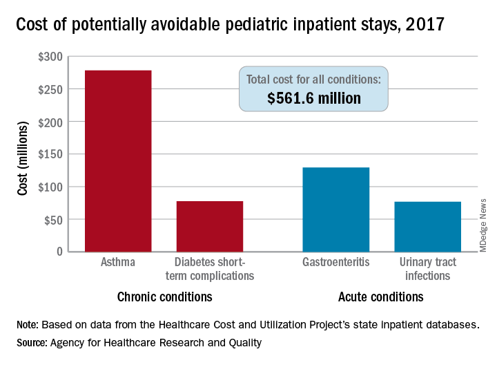
The cost of potentially avoidable visits for asthma that year was $278 million, versus $284 million combined for the other three conditions “that evidence suggests may be avoidable, in part, through timely and quality primary and preventive care,” Kimberly W. McDermott, PhD, and H. Joanna Jiang, PhD, said in an AHRQ statistical brief.
Those three other conditions are diabetes short-term complications, gastroenteritis, and urinary tract infections (UTIs). Neonatal stays were excluded from the analysis, Dr. McDermott of IBM Watson Health and Dr. Jiang of the AHRQ noted.
The state inpatient databases of the AHRQ’s Healthcare Cost and Utilization Project included 1.4 million inpatient stays among children aged 3 months to 17 years in 2017, of which 8% (108,300) were deemed potentially preventable. Hospital charges for the preventable stays came to $561.6 million, or 3% of the $20 billion in total costs for all nonneonatal stays, they said.
Rates of potentially avoidable stays for asthma (159 per 100,000 population), gastroenteritis (90 per 100,000), and UTIs (41 per 100,000) were highest for children aged 0-4 years and generally decreased with age, but diabetes stays increased with age, rising from 12 per 100,000 in children aged 5-9 years to 38 per 100,000 for those 15-17 years old, the researchers said.
Black children had a much higher rate of potentially avoidable stays for asthma (218 per 100,000) than did Hispanic children (74), Asian/Pacific Islander children (46), or white children (43), but children classified as other race/ethnicity were higher still: 380 per 100,000. Rates for children classified as other race/ethnicity were highest for the other three conditions as well, they reported.
Comparisons by sex for the four conditions ended up in a 2-2 tie: Girls had higher rates for diabetes (28 vs. 23) and UTIs (35 vs. 8), and boys had higher rates for asthma (96 vs. 67) and gastroenteritis (38 vs. 35), Dr. McDermott and Dr. Jiang reported.
SOURCE: McDermott KW, Jiang HJ. HCUP Statistical Brief #259. June 2020.
according to the Agency for Healthcare Research and Quality.
The cost of potentially avoidable visits for asthma that year was $278 million, versus $284 million combined for the other three conditions “that evidence suggests may be avoidable, in part, through timely and quality primary and preventive care,” Kimberly W. McDermott, PhD, and H. Joanna Jiang, PhD, said in an AHRQ statistical brief.
Those three other conditions are diabetes short-term complications, gastroenteritis, and urinary tract infections (UTIs). Neonatal stays were excluded from the analysis, Dr. McDermott of IBM Watson Health and Dr. Jiang of the AHRQ noted.
The state inpatient databases of the AHRQ’s Healthcare Cost and Utilization Project included 1.4 million inpatient stays among children aged 3 months to 17 years in 2017, of which 8% (108,300) were deemed potentially preventable. Hospital charges for the preventable stays came to $561.6 million, or 3% of the $20 billion in total costs for all nonneonatal stays, they said.
Rates of potentially avoidable stays for asthma (159 per 100,000 population), gastroenteritis (90 per 100,000), and UTIs (41 per 100,000) were highest for children aged 0-4 years and generally decreased with age, but diabetes stays increased with age, rising from 12 per 100,000 in children aged 5-9 years to 38 per 100,000 for those 15-17 years old, the researchers said.
Black children had a much higher rate of potentially avoidable stays for asthma (218 per 100,000) than did Hispanic children (74), Asian/Pacific Islander children (46), or white children (43), but children classified as other race/ethnicity were higher still: 380 per 100,000. Rates for children classified as other race/ethnicity were highest for the other three conditions as well, they reported.
Comparisons by sex for the four conditions ended up in a 2-2 tie: Girls had higher rates for diabetes (28 vs. 23) and UTIs (35 vs. 8), and boys had higher rates for asthma (96 vs. 67) and gastroenteritis (38 vs. 35), Dr. McDermott and Dr. Jiang reported.
SOURCE: McDermott KW, Jiang HJ. HCUP Statistical Brief #259. June 2020.