User login
GALACTIC-HF: New ‘myotropic’ drug class shows modest HFrEF benefit
Omecamtiv mecarbil, a member of the novel myotropic drug class that improves cardiac performance, safely produced a significant but modest improvement in heart failure events or cardiovascular death in a pivotal trial with HFrEF patients, leaving experts unsure about the role this drug could have on top of an already crowded list of four first-line drug classes for this condition.
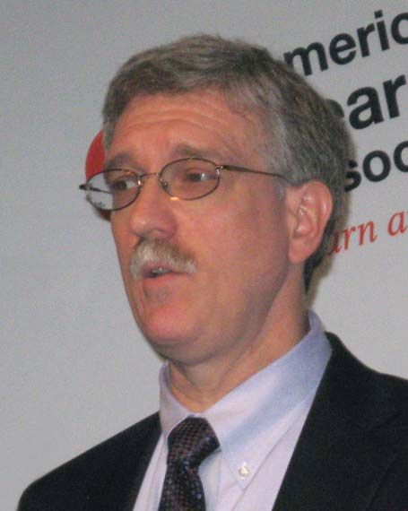
“It remains to be investigated and discussed where omecamtiv mecarbil fits in” the overall approach to treating patients with heart failure with reduced ejection fraction (HFrEF), commented Paul Heidenreich, MD, designated discussant for the report at the virtual scientific sessions of the American Heart Association.
Omecamtiv mecarbil (OM) treatment produced a positive result for the study’s primary endpoint, with a 2.1% absolute cut in the combined rate of cardiovascular death, first heart failure hospitalization, or first urgent visit for heart failure compared with placebo during a median follow-up of about 22 months This represented an 8% relative risk reduction, reported John R. Teerlink, MD, at the meeting, and broke down as a 0.6% absolute drop in cardiovascular death compared with the placebo arm, a 0.7% cut in heart failure hospitalization, and a 0.8% drop in urgent outpatient visits for heart failure. Dr. Teerlink and his associates called this benefit “modest” in their simultaneous publication in the New England Journal of Medicine.
Room for a fifth HFrEF drug?
In addition to the limited benefit, another question raised by the trial is how OM would perform when used on top of what is now considered standard, quadruple therapy for most HFrEF patients: a beta-blocker, a mineralocorticoid receptor antagonist, sacubitril-valsartan (Entresto), and an agent from the sodium glucose co-transporter 2 (SGLT2) inhibitor class, specifically dapagliflozin (Farxiga) or empagliflozin (Jardiance). During the period when the new OM trial was run, 2017-2019, the SGLT2 inhibitors had not yet been established as a key part of standard HFrEF treatment, and hence fewer than 3% of enrolled patients were on one of these drugs.
Because of this evidence gap, OM “can’t be across the board a fifth drug on top of standard treatment,” based on the new results, cautioned Dr. Heidenreich, a cardiologist and professor of medicine at Stanford (Calif.) University School of Medicine.

The new evidence for OM’s efficacy is “not compelling” when compared with what dapagliflozin and empagliflozin each showed in recent trials, with the SGLT2 inhibitors producing about a 25% cut compared with placebo in a primary outcome that was similar to the one used in the OM trial, commented Douglas L. Mann, MD, a heart failure physician and professor of medicine at Washington University School of Medicine in St. Louis. “Would OM still show a benefit with an SGLT2 inhibitor? That’s not known” on the basis of the available data, he said in an interview.
A related factor that could influence potential use of OM in routine practice is that with four established, foundational drug classes, adding a fifth drug that will only be available in a branded formulation raises issues of incremental cost and compliance issues, Dr. Mann noted.
The positives of omecamtiv mercarbil
But in addition to its positive result in the GALACTIC-HF trial, treatment with OM showed other attractive characteristics in a study that treated a wide spectrum of 4,120 patients with HFrEF as well as including 4,112 patients randomized to placebo. Most notably, OM had a very clean safety profile, with adverse event rates similar to placebo patients across all adverse event subtypes, as well as causing no drop in blood pressure and actually an average 2.0–mm Hg increase in systolic blood pressure, no increase in potassium, no apparent impact on renal function, and a small but significant decline in N-terminal pro-B-type natriuretic peptide (NT-proBNP) compared with placebo.
This coupled with the novel mechanism of action of OM – direct augmentation of cardiac sarcomere function by increasing myosin attachment to actin – suggests that OM can be safely added on top of existing HFrEF treatment to provide an unique and incremental benefit.
“Other heart failure drugs [like beta-blockers and sacubitril-valsartan] lower blood pressure, so what can happen is that clinicians run out of room to add full dosages” when patients’ pressures fall too low, commented Gregory D. Lewis, MD, head of Heart Failure at Massachusetts General Hospital in Boston. He is principle investigator for another OM trial, METEORIC-HF, which is examining the possible impact of the drug on exercise capacity in a randomized study with about 270 HFrEF patients.
If the METEORIC-HF results can could confirm some of the GALACTIC-HF results that suggested improvements in patient function, the combined data could potentially lead to regulatory approval for U.S. marketing of the drug, Dr. Lewis suggested. Results from that study are expected in 2021, he said in an interview.
The GALACTIC-HF results hinted at possible functional improvement after 24 weeks on treatment among patients who required hospitalization as measured by the Kansas City Cardiomyopathy Questionnaire, which measures quality life. However, this difference failed to meet the study’s prespecified definition of a significant effect.
Another intriguing suggestion of focused benefit was in patients with a left ventricular ejection fraction at or below the median in GALACTIC-HF of 28%. In that subgroup, OM treatment was linked with a significant 16% relative reduction in the primary endpoint compared with placebo, while it had no significant effect in the other 50% of patients with higher ejection fractions. (The maximum left ventricular ejection fraction for enrollment was 35%.) This apparent subgroup interaction was statistically significant, reported Dr. Teerlink, a professor of medicine at the University of California, San Francisco, and director of Heart Failure at the San Francisco V.A. Medical Center.
Further analysis of the study data “will provide greater insight into subgroups who may demonstrate greater benefit, such as patients with lower ejection fraction in whom improving cardiac function may have a greater role,” he said. The idea that a drug that improves myocyte function at the molecular level could especially benefit patients with the lowest ejection fractions is “biologically plausible,” Dr. Teerlink said.
This scenario looks reasonable, and could make OM something of a niche drug for at least the near term, said Dr. Mann.
The world’s first myotropic drug
Possibly the most notable aspect of GALACTIC-HF is that it proved the efficacy, modest though it was, of a novel drug mechanism that fulfills a decades-long quest of heart failure researchers: a safe way to improve the heart’s pumping action.
“For years, the heart failure community struggled with treatment to improve cardiac performance, but invariably it ended in disaster by worsening cardiac deaths,” problems that led to abandonment of early inotropic drugs more than a generation ago, noted Dr. Mann.
But a more nuanced approach to inotropic agents recently has emerged from Dr. Teerlink and his associates, built on the premise that the dangers seen years ago related to the calcium modulations they caused. Their new paradigm is that the dangers of these “calcitropic” agents can be sidestepped with different agents that either mediate their effects via myosin, the myotropes like OM, or mitochondrial effects from mitotropic drugs.
The inotrope debacle from the 1990s made that drug-class name “a dirty word that causes fear and loathing in the heart failure community,” observed Dr. Mann. While the term myotrope has not yet really caught on, “If omecamtiv mecarbil starts getting used in routine practice, then I think you’ll start seeing uptake of the term myotrope,” he predicted.
GALACTIC-HF was sponsored by Amgen, Cytokinetics, and Servier, the companies developing omecamtiv mecarbil. Dr. Teerlink has received research support from and been a consultant to Amgen, Cytokinetics, and Servier, as well as Abbott, AstraZeneca, Bayer, Boehringer Ingelheim, Bristol-Myers Squibb, Medtronic, Merck, and Novartis. Dr. Heidenreich had no disclosures. Dr. Mann is on a steering committee for a trial sponsored by Novartis and has no other commercial disclosures. Dr. Lewis is principal investigator for a trial of omecamtiv mecarbil and has no other commercial disclosures.
[email protected]
On Twitter @mitchelzoler
Omecamtiv mecarbil, a member of the novel myotropic drug class that improves cardiac performance, safely produced a significant but modest improvement in heart failure events or cardiovascular death in a pivotal trial with HFrEF patients, leaving experts unsure about the role this drug could have on top of an already crowded list of four first-line drug classes for this condition.
“It remains to be investigated and discussed where omecamtiv mecarbil fits in” the overall approach to treating patients with heart failure with reduced ejection fraction (HFrEF), commented Paul Heidenreich, MD, designated discussant for the report at the virtual scientific sessions of the American Heart Association.
Omecamtiv mecarbil (OM) treatment produced a positive result for the study’s primary endpoint, with a 2.1% absolute cut in the combined rate of cardiovascular death, first heart failure hospitalization, or first urgent visit for heart failure compared with placebo during a median follow-up of about 22 months This represented an 8% relative risk reduction, reported John R. Teerlink, MD, at the meeting, and broke down as a 0.6% absolute drop in cardiovascular death compared with the placebo arm, a 0.7% cut in heart failure hospitalization, and a 0.8% drop in urgent outpatient visits for heart failure. Dr. Teerlink and his associates called this benefit “modest” in their simultaneous publication in the New England Journal of Medicine.
Room for a fifth HFrEF drug?
In addition to the limited benefit, another question raised by the trial is how OM would perform when used on top of what is now considered standard, quadruple therapy for most HFrEF patients: a beta-blocker, a mineralocorticoid receptor antagonist, sacubitril-valsartan (Entresto), and an agent from the sodium glucose co-transporter 2 (SGLT2) inhibitor class, specifically dapagliflozin (Farxiga) or empagliflozin (Jardiance). During the period when the new OM trial was run, 2017-2019, the SGLT2 inhibitors had not yet been established as a key part of standard HFrEF treatment, and hence fewer than 3% of enrolled patients were on one of these drugs.
Because of this evidence gap, OM “can’t be across the board a fifth drug on top of standard treatment,” based on the new results, cautioned Dr. Heidenreich, a cardiologist and professor of medicine at Stanford (Calif.) University School of Medicine.
The new evidence for OM’s efficacy is “not compelling” when compared with what dapagliflozin and empagliflozin each showed in recent trials, with the SGLT2 inhibitors producing about a 25% cut compared with placebo in a primary outcome that was similar to the one used in the OM trial, commented Douglas L. Mann, MD, a heart failure physician and professor of medicine at Washington University School of Medicine in St. Louis. “Would OM still show a benefit with an SGLT2 inhibitor? That’s not known” on the basis of the available data, he said in an interview.
A related factor that could influence potential use of OM in routine practice is that with four established, foundational drug classes, adding a fifth drug that will only be available in a branded formulation raises issues of incremental cost and compliance issues, Dr. Mann noted.
The positives of omecamtiv mercarbil
But in addition to its positive result in the GALACTIC-HF trial, treatment with OM showed other attractive characteristics in a study that treated a wide spectrum of 4,120 patients with HFrEF as well as including 4,112 patients randomized to placebo. Most notably, OM had a very clean safety profile, with adverse event rates similar to placebo patients across all adverse event subtypes, as well as causing no drop in blood pressure and actually an average 2.0–mm Hg increase in systolic blood pressure, no increase in potassium, no apparent impact on renal function, and a small but significant decline in N-terminal pro-B-type natriuretic peptide (NT-proBNP) compared with placebo.
This coupled with the novel mechanism of action of OM – direct augmentation of cardiac sarcomere function by increasing myosin attachment to actin – suggests that OM can be safely added on top of existing HFrEF treatment to provide an unique and incremental benefit.
“Other heart failure drugs [like beta-blockers and sacubitril-valsartan] lower blood pressure, so what can happen is that clinicians run out of room to add full dosages” when patients’ pressures fall too low, commented Gregory D. Lewis, MD, head of Heart Failure at Massachusetts General Hospital in Boston. He is principle investigator for another OM trial, METEORIC-HF, which is examining the possible impact of the drug on exercise capacity in a randomized study with about 270 HFrEF patients.
If the METEORIC-HF results can could confirm some of the GALACTIC-HF results that suggested improvements in patient function, the combined data could potentially lead to regulatory approval for U.S. marketing of the drug, Dr. Lewis suggested. Results from that study are expected in 2021, he said in an interview.
The GALACTIC-HF results hinted at possible functional improvement after 24 weeks on treatment among patients who required hospitalization as measured by the Kansas City Cardiomyopathy Questionnaire, which measures quality life. However, this difference failed to meet the study’s prespecified definition of a significant effect.
Another intriguing suggestion of focused benefit was in patients with a left ventricular ejection fraction at or below the median in GALACTIC-HF of 28%. In that subgroup, OM treatment was linked with a significant 16% relative reduction in the primary endpoint compared with placebo, while it had no significant effect in the other 50% of patients with higher ejection fractions. (The maximum left ventricular ejection fraction for enrollment was 35%.) This apparent subgroup interaction was statistically significant, reported Dr. Teerlink, a professor of medicine at the University of California, San Francisco, and director of Heart Failure at the San Francisco V.A. Medical Center.
Further analysis of the study data “will provide greater insight into subgroups who may demonstrate greater benefit, such as patients with lower ejection fraction in whom improving cardiac function may have a greater role,” he said. The idea that a drug that improves myocyte function at the molecular level could especially benefit patients with the lowest ejection fractions is “biologically plausible,” Dr. Teerlink said.
This scenario looks reasonable, and could make OM something of a niche drug for at least the near term, said Dr. Mann.
The world’s first myotropic drug
Possibly the most notable aspect of GALACTIC-HF is that it proved the efficacy, modest though it was, of a novel drug mechanism that fulfills a decades-long quest of heart failure researchers: a safe way to improve the heart’s pumping action.
“For years, the heart failure community struggled with treatment to improve cardiac performance, but invariably it ended in disaster by worsening cardiac deaths,” problems that led to abandonment of early inotropic drugs more than a generation ago, noted Dr. Mann.
But a more nuanced approach to inotropic agents recently has emerged from Dr. Teerlink and his associates, built on the premise that the dangers seen years ago related to the calcium modulations they caused. Their new paradigm is that the dangers of these “calcitropic” agents can be sidestepped with different agents that either mediate their effects via myosin, the myotropes like OM, or mitochondrial effects from mitotropic drugs.
The inotrope debacle from the 1990s made that drug-class name “a dirty word that causes fear and loathing in the heart failure community,” observed Dr. Mann. While the term myotrope has not yet really caught on, “If omecamtiv mecarbil starts getting used in routine practice, then I think you’ll start seeing uptake of the term myotrope,” he predicted.
GALACTIC-HF was sponsored by Amgen, Cytokinetics, and Servier, the companies developing omecamtiv mecarbil. Dr. Teerlink has received research support from and been a consultant to Amgen, Cytokinetics, and Servier, as well as Abbott, AstraZeneca, Bayer, Boehringer Ingelheim, Bristol-Myers Squibb, Medtronic, Merck, and Novartis. Dr. Heidenreich had no disclosures. Dr. Mann is on a steering committee for a trial sponsored by Novartis and has no other commercial disclosures. Dr. Lewis is principal investigator for a trial of omecamtiv mecarbil and has no other commercial disclosures.
[email protected]
On Twitter @mitchelzoler
FROM AHA 2020
Lancet panel calls for urgent global action to combat diabetes
The article was published online Nov. 12, just ahead of World Diabetes Day.
Of the 463 million people with diabetes worldwide in 2019, 80% live in low- and middle-income countries. The condition reduces life expectancy in middle-aged adults by 4-10 years, including increasing the risk of death from cardiovascular disease, kidney disease, and cancer by up to threefold. It is also a leading cause of nontraumatic amputation and blindness.
Use of evidence-based interventions, if implemented and managed properly, could prevent thousands of deaths globally every day, stressed the commission.
“There is an enormous amount of knowledge that we have amassed over the years. We need good preventive care and we need to ensure that diabetes patients, once diagnosed, have good continuous care. There is an urgent need for decision-makers, policymakers, and payers to make things happen,” the leader of the multidisciplinary commission, Juliana C.N. Chan, MBChB, MD, said in an interview.
And now diabetes has emerged as a major risk factor for death from COVID-19, particularly in the setting of inadequate glycemic control.
“COVID-19 has exposed the vulnerability of individuals with diabetes,” said Dr. Chan, of the Hong Kong Institute of Diabetes and Obesity. “We should use the pandemic as an opportunity to implement solutions.”
Physician education key, trickling down to field workers and patients
First on the agenda, she says, should be “physician education. There are many primary care providers and internal medicine physicians whose knowledge needs to be updated.”
“Then doctors need to transfer this information to other people, such as nurses and community field workers. We cannot just rely on doctors; we need to train nonmedics” so that knowledge about how to prevent, treat, and manage diabetes long term is communicated right down the health care chain, she explained.
“They need to know how to look at people’s eyes and feet, how to do blood and urine tests, and how to collect data. Then they need to educate patients on what they should be doing, on how to practice self-care,” she added.
“We need to change our way of thinking, redesign clinic flow and how you build a team. And those care teams need to know how to collect data, and then use that data to monitor patients and to stratify individual risk, to ensure that what has been said has been done, as well as to inform practice and policies” through, for example, the establishment of diabetes registers.
The focus needs to be on “lifelong integrated care, the right treatment at the right time,” she emphasized. History-taking, clinical and laboratory assessments, as well as monitoring of macrovascular and microvascular complications, comorbidities, and medications, are all key.
Just a few simple things, if properly implemented, could make a big difference, Dr. Chan stressed.
For example, implementing a structured lifestyle intervention and use of metformin can each prevent or delay type 2 diabetes in individuals with impaired glucose tolerance by 30%-50%, and sustained weight reduction in patients with obesity by 15 kg (33 lb) or more can induce remission of type 2 diabetes for up to 2 years.
And there are plenty of medications that are “very affordable even in low- and middle-income countries” to treat diabetes and associated risk factors, including metformin, “statins, and RAS inhibitors,” she noted.
For instance, the 10 low- and middle-income countries with the greatest burden of diabetes (China, India, Brazil, Mexico, Indonesia, Egypt, Pakistan, Bangladesh, Turkey, Thailand) account for 217 million cases of type 2 diabetes, representing nearly 50% of all diabetes cases.
The commission estimated that 3.2 million of these individuals would die in 3 years if not treated, with 1.3 million of these deaths due to cardiovascular disease.
By reducing hemoglobin A1c, blood pressure, and LDL-cholesterol through achieving a diagnosis rate of 50%, ensuring access to essential medicines in at least 70% of patients, and with a support system to sustain reductions in these risk factors over 3 years, up to 800,000 premature deaths could be avoided.
People with type 1 diabetes dying; WHO launches initiative
In an accompanying commentary (2020 Nov 12. doi: 10.1016/S0140-6736[20]32378-3), Katie Dain, chief executive officer of the Noncommunicable Diseases (NCD) Alliance, points out that only half of people living with diabetes around the world – and just one in seven in Africa – have reliable access to insulin.
“Lots of people with type 1 diabetes are still dying due to lack of insulin,” Dr. Chan said in an interview. “We need to elevate basic care to intermediate and ensure that basal-bolus insulin and glucose-monitoring tools are available and that patients are trained in self-care. In that way, 80% of type 1 diabetes deaths could be prevented.”
Ms. 3Dain agrees, stressing, “Political rhetoric and commitments have yet to translate into sufficient and sustainable action for people living with diabetes worldwide, and particularly for those in [low- and middle-income countries].”
The Lancet Commission document also emphasizes the importance of support for pregnant women with diabetes and attention to the psychosocial needs of people with diabetes.
And it stresses society-, population-, and community-based strategies for type 2 diabetes prevention including health awareness programs, food policies, and broad use of nonphysician personnel to deliver diabetes prevention efforts.
In tandem with World Diabetes Day, the World Health Organization will announce the development of the WHO Global Diabetes Compact, which will be launched in April 2021.
This will aim to implement the commission’s recommendations through partnerships with governments, care providers, patient advocates, and nongovernmental organizations.
Together, they will “support countries to mobilize resources and accelerate structural transformations, which will enable the scale-up of access to essential diabetes medicines and technologies, inclusion of diagnosis and treatment of diabetes in primary health care and universal health coverage packages, and reduction of major population-level diabetes risk factors such as obesity,” according to another Lancet editorial accompanying the report.
“The evidence-base for improving diabetes prevention and care is strong. The question now for diabetes advocates is how to achieve the comprehensive, systems-level change needed to translate this evidence into action.”
Dr. Chan has reported receiving grants from AstraZeneca, Lilly, Lee Powder, Hua Medicine, and Qualigenics, as well as grants and personal fees from Bayer, Boehringer Ingelheim, Sanofi, Novartis, Merck, and MSD outside the submitted work. She has reported being the chief executive officer (pro bono) of the Asia Diabetes Foundation and a cofounder of GemVCare. She also holds a patent for genetic markers for diabetes and its complications. Ms. Dain has reported no relevant financial relationships.
A version of this article originally appeared on Medscape.com.
Pediatric News board welcomes back Dr. Breach Washington
She currently is employed as a medical director at WellCare of North Carolina/Centene. Her career spans 32 years in Charlotte, N.C., where she has practiced pediatrics and held medical director positions in a large health care system and a managed care organization, as well as worked in private practice and in a public health clinic.

A native of North Babylon, N.Y., she received her undergraduate degree at Hofstra University, Hempstead, N.Y., and her medical degree at George Washington University, Washington. Dr. Breach Washington has been an advocate for children, access to health care, and diversity and inclusion.
An active member of the American Academy of Pediatrics, Dr. Breach Washington served as an elected representative to the AAP National Nominating Committee, following her term as president of the North Carolina chapter of the AAP.
She is a past pediatric section chair of the National Medical Association and past president of the Charlotte Medical, Dental, and Pharmaceutical Society. She has served on the boards of the Simmons Branch YMCA, the Mecklenburg County Medical Society, and the Teen Health Connection (a facility of Carolinas Medical Center), plus many associated committees.
Dr. Breach Washington serves the community as an active life member of Alpha Kappa Alpha Sorority, Jack and Jill of America, and a member of The Links. She is the recipient of several awards for her commitment to diversity and inclusion, philanthropy, and community service.
Dr. Breach Washington is married and has an adult daughter. She is an avid fan of all sports, a dancer, and enjoys theater, fine dining, and travel.
She currently is employed as a medical director at WellCare of North Carolina/Centene. Her career spans 32 years in Charlotte, N.C., where she has practiced pediatrics and held medical director positions in a large health care system and a managed care organization, as well as worked in private practice and in a public health clinic.
A native of North Babylon, N.Y., she received her undergraduate degree at Hofstra University, Hempstead, N.Y., and her medical degree at George Washington University, Washington. Dr. Breach Washington has been an advocate for children, access to health care, and diversity and inclusion.
An active member of the American Academy of Pediatrics, Dr. Breach Washington served as an elected representative to the AAP National Nominating Committee, following her term as president of the North Carolina chapter of the AAP.
She is a past pediatric section chair of the National Medical Association and past president of the Charlotte Medical, Dental, and Pharmaceutical Society. She has served on the boards of the Simmons Branch YMCA, the Mecklenburg County Medical Society, and the Teen Health Connection (a facility of Carolinas Medical Center), plus many associated committees.
Dr. Breach Washington serves the community as an active life member of Alpha Kappa Alpha Sorority, Jack and Jill of America, and a member of The Links. She is the recipient of several awards for her commitment to diversity and inclusion, philanthropy, and community service.
Dr. Breach Washington is married and has an adult daughter. She is an avid fan of all sports, a dancer, and enjoys theater, fine dining, and travel.
Escalate HIV adherence strategies amid COVID-19
"The writing is on the wall” that virtual care is not meeting the needs of people with HIV who struggled with viral suppression even before the COVID-19 pandemic, said Jason Farley, PhD, ANP-BC, AACRN, associate professor of nursing at Johns Hopkins University, Baltimore. So it’s time for HIV care teams, especially clinics in the Ryan White HIV/AIDS Program, to get creative in bringing wraparound services to patients.
That may mean reallocating the workforce so that one person serves as a community health worker. Or it could mean increasing texts and video calls; helping patients find online support groups to address problems with alcohol or drug use; and conducting an overall assessment of patients’ needs as the pandemic continues.
“The virtual patient-centered medical home may be the new normal after COVID-19, and we have to be thinking about how we use this model with patients for whom it works, but supplement this model in patients that it does not,” Farley said at the virtual Association of Nurses in AIDS Care (ANAC) 2020 Annual Meeting. That work “is essential to our being able to facilitate the best patient outcomes possible.”
Early data, tiered interventions
Farley referred to an article published in September in the Journal AIDS that confirmed unpublished data mentioned at the International AIDS Conference 2020. The article reported that viral suppression rates among people with HIV who attended San Francisco’s Ward 86 HIV clinic dropped by 31% from pre-COVID levels.
Of the 1766 people who attended the clinic, about 1 in 5 had detectable HIV viral loads at any point in 2019. But that rate was 31% higher after shelter-in-place orders were issued. And although patients participated in telemedicine visits at more or less the same rate before and after the pandemic (31% vs. 30% no-shows), viral suppression rates dropped. The impact was especially acute for homeless individuals.
“This destabilization occurred despite our population attending telemedicine visits at a higher rate than expected, given the 60% drop in ambulatory care visit volume nationwide,” the authors stated in their article. “Telehealth visits, while offering greater patient convenience, may lead to less access to clinic-based social support services essential to achieving viral suppression among vulnerable groups.”
That’s the challenge HIV clinics now face, Farley said at the ANAC meeting.
He suggested a differentiated care approach in which there are four tiers of care, starting with the standard level of outreach, which may include email, electronic health record blasts, and robo-calls to remind people of their appointments and to refill their medications. Those with sustained viral suppression may only need 90-day automatic refills of their medications. Those who are vulnerable to nonadherence may need to be contacted weekly or more often by the clinic. Such contact could be made by a social worker, a community health worker, or through some form of virtual support.
Patients at tier 4, who have labile viral suppression, need far more than that. These are the 15% of patients with HIV who struggled with viral suppression before the pandemic. They are the patients that Farley’s team focuses on at Baltimore’s John G. Bartlett Specialty Clinic for Infectious Disease.
“We’ve completely deconstructed the patient-centered medical home,” he said of the early move to virtual care. He suggested that clinicians assess their services and ask themselves some questions:
- Has someone on the team reached out to every patient and checked in to see what their biggest needs are, medical or not, during the pandemic? Have they assessed the patient’s ability to receive video calls or text messages?
- How have group-support programs that address stigma or the social determinants of health fared in the transition to virtual medicine?
- Are patients who are in recovery being supported in order that they may engage with recovery programs online?
- How well have counseling services done in engaging people in virtual care? Currently, given the overall increase in mental health challenges during the pandemic, one would expect that the use of mental health counseling is increasing. “If they’re stagnant or going down, someone needs to be reflecting on that issue internally in the clinic,” he said.
- Are patients being contacted regarding the effects that isolation is having on their lives? “The things that would normally allow us to self-mitigate and self-manage these conditions, like going to the gym, meeting with friends, religious services – all of those are being cut,” he said.
- Is there an early alert from an in-person pharmacy to trigger outreach via a community health worker for patients who haven’t picked up their medications in a week or more?
Farley pointed to a 2015 model for an enhanced e-health approach to chronic care management that called for e-support from the community and that was enhanced through virtual communities.
These are some of the approaches Farley has taken at his clinic. He leads a team that focuses specifically on patients who struggled with engagement before the pandemic. Through a grant from the US Department of Health & Human Services’ Health Resources and Services Administration – even before the pandemic – that team has been funding community health workers who have multiple contacts with patients online and virtually and are able to offer what he calls “unapologetically enabling” support for patients so that they are able to focus on their health.
He gave the following example. Before the pandemic, a community health worker on the team had been working with a patient who showed up at every scheduled visit and swore that she was taking her medications, although clearly she was not. A community health worker, who was made available through the grant, was able to recognize that the patient’s biggest challenge in her life was providing childcare for her special-needs child. The community health worker worked with the patient for months to find stable childcare for the child, paid 2 months of rent for the patient so that she would not become homeless, and helped her find transitional housing. When the pandemic hit, the community health worker was already texting and conducting video calls with the patient regularly.
For the past 9 months, that patient has had an undetectable viral load, Farley said.
“Nine months during a pandemic,” Farley reiterated, “and the community health worker keeps working with her, keeps meeting with her.”
Stigma on stigma
The need for this level of support from the clinic may be even more important for people with HIV who acquire COVID-19, said Orlando Harris, PhD, assistant professor of community health systems at the University of California, San Francisco, (UCSF) School of Nursing. HIV-related stigma is a well-known deterrent to care for people living with the virus. During the presentation, Harris asked Farley about the impact of COVID-19 stigma on people with both HIV and COVID-19.
Farley said that patients at his clinic have told him that they have “ostracized” friends who have tested positive for COVID-19. Harris remembered a person with HIV who participated in one of his trials telling the researchers that despite all his precautions – wearing a mask, staying socially distant – he still acquired COVID-19. There was nothing he could have done, Harris said, other than just not go to the grocery store.
The fear of contracting another disease that is associated with stigma, as well as the need to disclose it, can inflame memories of the trauma of being diagnosed with HIV, Harris said. And with patient-centered medical homes struggling to reconstitute their wraparound services via telehealth, he said he wonders whether clinicians should be doing more.
“I worry about people who have survived being diagnosed with HIV in the ‘80s and the ‘90s before antiretroviral therapy showed up on the scene,” he told Medscape Medical News. “I worry that the folks that survived one pandemic [may] be feeling fearful or living in that fear that this new pandemic might take them out. That’s why I’m stressing the need for us to really consider, as clinicians and also as researchers the support systems, the coping mechanisms, the counseling, or what have you to support those living with HIV and vulnerable to COVID-19.”
During telehealth visits, that can be achieved simply by asking people how they are really doing and what their coping mechanisms are.
For their part, the clinicians at San Francisco’s Ward 86 are not trying to provide that support through telehealth on the same level as they were at the beginning of the pandemic, said Matthew Spinelli, MD, assistant professor of medicine, and Monica Gandhi, MD, associate chief of the Division of HIV, Infectious Diseases and Global Medicine, who are both at UCSF and are coauthors of the study.
They still offer telemedicine appointments to patients who request them, said Spinelli. He said about one-third of his patients still prefer to receive their care virtually. The rest have gone back to face-to-face support.
“The analysis led us to promptly open up care as much as possible to our patients, with the idea that telehealth is not cutting it for vulnerable patients with HIV,” Gandhi told Medscape Medical News via email. “We don’t think it’s right for a population who relies on social support from the clinic.”
This article first appeared on Medscape.com.
Testicular cancer link to below-the-waist scans, x-rays?
Over the last 40 years, there has been a doubling in the incidence of testicular cancer. Over the same period, there has been a 20-fold increase in the use of diagnostic radiation, and the doses of radiation used have increased sevenfold. Perhaps the two are related?
The suggestion comes from researchers reporting a new case-control study in which they show that repeated diagnostic radiation below the waist was associated with an increase in the risk of testicular germ cell tumor (TGCT).
The study involved 315 men with TGCT and 931 controls from hospital and population-based settings. Researchers analyzed medical records and individuals’ recall of diagnostic radiation, such as CT scans, x-rays, and barium enema.
Compared with men who did not undergo any diagnostic radiation, the risk of testicular cancer was nearly twofold higher among men who had undergone three or more diagnostic x-rays and CT scans, and more than fourfold higher among men who had undergone three or more lower gastrointestinal series or barium enema.
The study was published online Nov. 11 in PLoS ONE.
Testicular cancer, the most common cancer in white men aged 15-44 years, has steadily increased in incidence in the United States over the past 3 or 4 decades. The rate has jumped from 3 out of 100,000 men in 1975 to 6 out of 100,000 men today, said senior author Katherine Nathanson, MD, of the University of Pennsylvania, Philadelphia, and colleagues.
“Our data suggests that the increased use of diagnostic radiation below the waist in men over that same time may contribute to the increase in incidence,” Dr. Nathanson said in a press statement.
Reducing diagnostic radiation doses to the testes “should be prioritized,” said the investigators, if these results are validated by additional research.
It’s “undoubtedly prudent” to reduce those doses “as much as possible,” said David Brenner, PhD, director of the Center for Radiological Research, Columbia University Medical Center, New York.
However, Dr. Brenner also had reservations about the new study. “I do find the results quite surprising,” given what is known historically about testicular cancer and radiation exposure.
Higher radiation doses (than in the current study) in two other settings (among post–atomic bomb survivors and in the treatment of ankylosing spondylitis) are not associated with an increased risk of testicular cancer, Dr. Brenner said in an interview.
“Underlying conditions for which the individual received radiation imaging,” may be one potential explanation for the increased testicular cancer risk seen in this study, he added.
Dr. Nathanson responded to Dr. Brenner’s critique by focusing on atomic bomb survivors and pointing out that the baseline rate of testicular cancer in Asia, including Japan, is “very, very low” and therefore that population, including bomb survivors, is not a highly applicable comparison.
Take-home message: Testicular shielding
Dr. Nathanson noted that diagnostic imaging below the waist is most commonly administered as a result of GI tract complaints such as constipation and abdominal pain. For clinicians, the study’s take-home message supports testicular shielding.
Testicular shielding during diagnostic imaging can be protective, but audits show that correct use and positioning occurs in just 25% of pediatric diagnostic scans, the authors commented.
Recent epidemiologic studies support a tie between diagnostic radiation and other cancers, the authors also pointed out.
For example, women undergoing frequent x-rays for scoliosis may have an increased risk of breast cancer, and likewise for individuals undergoing repeat routine dental x-rays and thyroid cancer. Additionally, a National Health Service study among 175,000 children revealed an excess risk of subsequent brain tumors and leukemia tied to CT scans, said Dr. Nathanson and coauthors.
Total dose and number of exposures were associated with an increasing cancer risk in each of the aforementioned studies.
Dr. Nathanson pointed out that Penn researchers have collected testicular cancer data since 2001 for the sake of research on related, inherited risk. The new study is an outgrowth of those efforts and is a rare effort in this subject area, in part, because testicular cancer is a very treatable rare cancer, with a 95% survival rate at 5 years.
A larger prospective study of the association between diagnostic radiation and testicular cancer would be better for determining causality, but is impractical given the needed time and associated costs. “A case-control design is a methodology that is ideally suited for studies investigating rare disease outcomes such as TGCT,” the authors comment.
The study was supported with grants from the National Cancer Institute and the National Institutes of Health. The study authors and Dr. Brenner disclosed no relevant financial relationships.
This article first appeared on Medscape.com.
New guidelines address diabetes management in kidney disease
A new guideline from the Kidney Disease: Improving Global Outcomes group addressing issues around diabetes management in patients with chronic kidney disease (CKD) has just been published in synopsis form in Annals of Internal Medicine.
The full guideline, including 12 recommendations and 48 practice points for clinicians caring for patients with diabetes and CKD, was published last month in Kidney International and on the KDIGO website.
More than 40% of people with diabetes develop CKD, and a significant number develop kidney failure requiring dialysis or transplant. This is the first guidance from KDIGO to address the comorbidity.
The new synopsis is aimed at primary care and nonnephrology specialist clinicians who manage patients with diabetes and CKD, in addition to nephrologists, first author Sankar D. Navaneethan, MD, said in an interview.
“Most of these patients are in the hands of primary care, endocrinology, and cardiology. We want to emphasize when they see patients with different severities of kidney disease [is] what are some of the things they have to be cognizant of,” said Dr. Navaneethan, professor of medicine and director of clinical research in the section of nephrology at Baylor College of Medicine, Houston.
The synopsis summarizes key recommendations from the larger guidance regarding comprehensive care needs, glycemic monitoring and targets, lifestyle interventions, glucose-lowering therapies, and educational/integrated care approaches.
It does not depart from prior diabetes guidelines, but it does provide advice for specific situations relevant to CKD, such as the limitations of hemoglobin A1c when estimated glomerular filtration rate (eGFR) drops below 30 mL/min per 1.73m2, and dietary protein consumption. It is based on published evidence up until February 2020.
For the nephrologist audience in particular, Dr. Navaneethan said, “we wanted to highlight team-based care, interacting with other specialists and working with them.”
“We [nephrologists] are more used to team-based care in dialysis patients. ... So we wanted to highlight that self-management programs and team-based care are important for empowering patients.”
“As nephrologists, we might not be comfortable starting patients on an SGLT2 [sodium-glucose cotransporter 2] inhibitor. We may need to reach out to our endocrinology or primary care colleagues and learn from them,” he explained.
RAS inhibitor use, smoking cessation, glycemic targets
Under “comprehensive care,” the guideline panel recommends treatment with an ACE inhibitor or an angiotensin II receptor blocker – renin-angiotensin system (RAS) blockade – for patients with diabetes, hypertension, and albuminuria (albumin-creatinine ratio >30 mg/g).
These medications should be titrated to the highest approved tolerated dose, with close monitoring of serum potassium and serum creatinine levels within 2-4 weeks of initiation or change in dose.
The document guides clinicians on that monitoring, as well as on RAS blockade use in patient subgroups, use of alternative agents, and mitigation of adverse effects.
Patients with diabetes and CKD who use tobacco should be advised to quit.
The group recommended A1c to monitor glycemic control in patients with diabetes and CKD not receiving dialysis.
However, when eGFR is below 30 mL/min per 1.73m2, A1c levels tend to be lower because of shortened erythrocyte lifespan, which interpretation should take into account. Continuous glucose monitoring can be used as an alternative because it is not affected by CKD.
Glycemic targets should be individualized depending on hypoglycemia risk, ranging from 6.5% to 8.0% for A1c or time in range of 70-180 mg/dL for continuous glucose monitoring readings.
SGLT2 inhibitors, metformin, and GLP-1 agonists
The panel also recommends treatment with both metformin and an SGLT2 inhibitor for patients with type 2 diabetes, CKD, and an eGFR ≥30 mL/min per 1.73m2.
For those who do not achieve glycemic targets or who cannot take those medications, a long-acting glucagonlike peptide–1 receptor agonist can be used instead.
Clinical trial data are summarized for the SGLT2 inhibitor canagliflozin supporting its use in patients with CKD specifically, along with mitigation of adverse events. Last year, the Food and Drug Administration approved this agent to slow the progression of diabetic nephropathy based on the CREDENCE study.
Results from the DAPA-CKD trial showing CKD reduction with another SGLT2 inhibitor, dapagliflozin, were not available at the time the new document was written, nor was the recent study showing diabetic CKD benefit for the novel mineralocorticoid receptor antagonist finerenone, Dr. Navaneethan noted.
The panel determined that there is insufficient evidence for adding other glucose-lowering agents to insulin in patients with type 1 diabetes and CKD.
Lifestyle interventions: Dietary protein, sodium, and physical activity
Most of the dietary guidance for patients with diabetes and CKD is the same as for the general population, including a recommendation to eat a diet high in vegetables, fruits, whole grains, fiber, legumes, plant-based proteins, unsaturated fats, and nuts, and lower in processed meats, refined carbohydrates, and sweetened beverages.
However, the guideline details two key areas that differ, one with regard to protein intake and the other on sodium.
Although lower protein intake had been advised in the past for patients with CKD, clinical trial evidence has not shown protein restriction to reduce glomerular hyperfiltration or slow kidney disease progression.
Therefore, the same level recommended for the general population – 0.8 g/kg per day – is also advised for those with diabetes and CKD who are not on dialysis.
Those who are on dialysis can increase daily protein intake to 1.0-1.2 g/kg per day to offset catabolism and negative nitrogen imbalance.
Because kidney function decline is associated with sodium retention that can raise cardiovascular risk, sodium should be limited to less than 2 g/day (or less than 90 mmol or 5 g of sodium chloride per day).
The panel also recommended moderate-intensity physical activity for at least 150 minutes per week or to tolerance.
“We wanted to emphasize how important lifestyle is. It’s the foundation you want to build on. You can take medications without all these other things – exercise, diet, weight loss – but they won’t be nearly as effective,” Dr. Navaneethan commented.
Self-management education, team-based care
The final section of the synopsis advises that people with diabetes and CKD receive structured self-management educational programs, and that “policy makers and institutional decision-makers implement team-based, integrated care focused on risk evaluation and patient empowerment to provide comprehensive care in patients with diabetes and CKD.”
Despite limited data for those measures specifically in patients with diabetes and CKD, “the working group believed that well-informed patients would choose self-management as the cornerstone of any chronic care model; therefore, a high value was placed on the potential benefits of self-management education programs in persons with diabetes and CKD.”
And regarding team-based care, “despite a paucity of direct evidence, the working group judged that multidisciplinary integrated care for patients with diabetes and CKD would represent a good investment.”
The guidelines will likely be updated in the next 1-2 years, Dr. Navaneethan said in an interview.
Dr. Navaneethan has reported receiving consultancy fees from Bayer, Boehringer Ingelheim, Reata, and Tricida, and research support from Keryx.
A version of this article originally appeared on Medscape.com.
Don’t miss cardiovascular risk factors in transgender patients
Cardiovascular disease risk is elevated among transgender individuals seeking gender-affirming hormone therapy, according to a retrospective study in 427 patients.

The transgender population often experiences socioeconomic and health disparities, including reduced access to care, Kara J. Denby, MD, said in an interview.
Previous research suggests that the use of gender-affirming hormone therapy (GAHT) may place transgender persons at increased cardiovascular risk, she said.
To identify the potential risk for transgender individuals, the researchers identified baseline cardiovascular risk in patients who had not yet undergone GAHT. Study participants were enrolled in a multidisciplinary transgender program, and the researchers collected data on demographics, medical history, vitals, medications, and laboratory results. The average age of the participants was 26 years, 172 identified as men, 236 as women, and 20 as nonbinary.
Overall, 55% of the participants had a chronic medical condition at baseline. Of these, 74 patients had hypertension, 41 had hyperlipidemia, 2 had a history of stroke, 7 had coronary artery disease, and 4 had chronic obstructive pulmonary disease.
For all patients who did not have documented atherosclerotic cardiovascular disease, their American College of Cardiology/American Heart Association ASCVD and QRISK3 risk scores were calculated. “The incidence of undiagnosed hypertension and hyperlipidemia was 6.8% and 11.3% respectively, and of these cases, only 64% and 24% were on appropriate therapies,” noted Dr. Denby of the Cleveland (Ohio) Clinic.
She reported the results Nov. 13 in a presentation at the at the virtual American Heart Association scientific sessions.
The findings were limited by the observational nature of the study.
However, the results suggest that transgender patients “appear to be at higher risk than their age-matched historical cohorts regardless of gender,” said Dr. Denby. More research is needed, but cardiovascular disease–prevention efforts may be inadequate in the transgender population given the elevated risk observed in this study, she concluded.
Growing transgender population is medically underserved
The transgender population is growing in the United States and internationally, said Dr. Denby. “This group has a history of being marginalized as a result of their transgender status with socioeconomic and health repercussions,” she said. “It is well known that transgender patients are less likely to have access to health care or utilize health care for a variety of reasons, including stigma and fear of mistreatment. This often leads transgender individuals to present to care late in disease processes which makes their disease harder to treat and often leads to emergent medical conditions,” she added.
“Transgender men and women are at high risk for cardiovascular disease and often aren’t screened at recommended intervals because of decreased health care use compared to their cisgender counterparts,” she said. “This may lead to untreated diseases that make them even more likely to suffer poor health outcomes.”
The current study is important because there are “almost no prior data regarding the cardiovascular health status of this population prior to gender-affirming care,” Dr. Denby emphasized. “There are data that gay, lesbian, and bisexual individuals are at higher risk for poor cardiovascular outcomes, but the same data are lacking in the transgender group,” she said.
“As transgender individuals have frequent physician visits while on hormonal therapy, this seems like the opportune time to screen for cardiovascular risk factors and treat previously undiagnosed diseases that can lead to poor health outcomes in the future,” Dr. Denby explained. “If we are able to intervene at an earlier age, perhaps we can help prevent poor health outcomes down the road,” she said.
Additional research can inform practice
Dr. Denby said she was not surprised by the findings. “This is a very high-risk population that often doesn’t follow closely in the health care system,” she said. “These data are very important in thinking holistically about transgender patients.” Clinicians can “use the opportunities we have when they present for gender-affirming care to optimize their overall health status, promote long-term health, and reduce the risks associated with hormonal therapy and gender-affirming surgeries,” she noted. “We hope to use this information to change our practice at the Cleveland Clinic and nationally as well. Transgender patients should be screened and aggressively treated for cardiovascular disease and risk factors,” she said.
Key barriers to overcome include determining the best way to reach out to transgender individuals and then making them feel comfortable in the clinical setting, Dr. Denby said. “This means that we must set up clinics that are approachable and safe for all comers. The lack of laws in many states that protect this vulnerable population also contributes to lack of access to care,” she added.
“We hope to continue research in this arena about how to effectively screen and treat transgender patients as they present to care, not only in the transgender clinic, but also to primary care providers (ob.gyn., internal medicine, family medicine, pediatrics) who also care for this population” since no specific guidelines currently exist to direct the screening for cardiovascular patients in particular, she said.
Findings offer foundation for LGBTQ cardiovascular studies
“This [study] provides us with a good rationale for why we should be considering cardiovascular health in transgender adults,” Billy A. Caceres, PhD, RN, of Columbia University School of Nursing, New York, said in an interview. “It is largely descriptive, but I think that that’s a good step in terms of at least understanding the magnitude of this problem. In addition, I think that what this abstract might do is help lead to future research that examines potentially the associations between not only gender-affirming hormone therapies but other potential social determinants like discrimination or poverty on the cardiovascular health of transgender people,” he noted.
Dr. Caceres served as chair of the writing group for the recent American Heart Association Scientific Statement: LGBTQ Heart Health published in Circulation. He had no financial conflicts to disclose.
The study received no outside funding. Dr. Denby had no financial conflicts to disclose.
SOURCE: Denby KJ et al. AHA 2020, Presentation P2274.
Cardiovascular disease risk is elevated among transgender individuals seeking gender-affirming hormone therapy, according to a retrospective study in 427 patients.
The transgender population often experiences socioeconomic and health disparities, including reduced access to care, Kara J. Denby, MD, said in an interview.
Previous research suggests that the use of gender-affirming hormone therapy (GAHT) may place transgender persons at increased cardiovascular risk, she said.
To identify the potential risk for transgender individuals, the researchers identified baseline cardiovascular risk in patients who had not yet undergone GAHT. Study participants were enrolled in a multidisciplinary transgender program, and the researchers collected data on demographics, medical history, vitals, medications, and laboratory results. The average age of the participants was 26 years, 172 identified as men, 236 as women, and 20 as nonbinary.
Overall, 55% of the participants had a chronic medical condition at baseline. Of these, 74 patients had hypertension, 41 had hyperlipidemia, 2 had a history of stroke, 7 had coronary artery disease, and 4 had chronic obstructive pulmonary disease.
For all patients who did not have documented atherosclerotic cardiovascular disease, their American College of Cardiology/American Heart Association ASCVD and QRISK3 risk scores were calculated. “The incidence of undiagnosed hypertension and hyperlipidemia was 6.8% and 11.3% respectively, and of these cases, only 64% and 24% were on appropriate therapies,” noted Dr. Denby of the Cleveland (Ohio) Clinic.
She reported the results Nov. 13 in a presentation at the at the virtual American Heart Association scientific sessions.
The findings were limited by the observational nature of the study.
However, the results suggest that transgender patients “appear to be at higher risk than their age-matched historical cohorts regardless of gender,” said Dr. Denby. More research is needed, but cardiovascular disease–prevention efforts may be inadequate in the transgender population given the elevated risk observed in this study, she concluded.
Growing transgender population is medically underserved
The transgender population is growing in the United States and internationally, said Dr. Denby. “This group has a history of being marginalized as a result of their transgender status with socioeconomic and health repercussions,” she said. “It is well known that transgender patients are less likely to have access to health care or utilize health care for a variety of reasons, including stigma and fear of mistreatment. This often leads transgender individuals to present to care late in disease processes which makes their disease harder to treat and often leads to emergent medical conditions,” she added.
“Transgender men and women are at high risk for cardiovascular disease and often aren’t screened at recommended intervals because of decreased health care use compared to their cisgender counterparts,” she said. “This may lead to untreated diseases that make them even more likely to suffer poor health outcomes.”
The current study is important because there are “almost no prior data regarding the cardiovascular health status of this population prior to gender-affirming care,” Dr. Denby emphasized. “There are data that gay, lesbian, and bisexual individuals are at higher risk for poor cardiovascular outcomes, but the same data are lacking in the transgender group,” she said.
“As transgender individuals have frequent physician visits while on hormonal therapy, this seems like the opportune time to screen for cardiovascular risk factors and treat previously undiagnosed diseases that can lead to poor health outcomes in the future,” Dr. Denby explained. “If we are able to intervene at an earlier age, perhaps we can help prevent poor health outcomes down the road,” she said.
Additional research can inform practice
Dr. Denby said she was not surprised by the findings. “This is a very high-risk population that often doesn’t follow closely in the health care system,” she said. “These data are very important in thinking holistically about transgender patients.” Clinicians can “use the opportunities we have when they present for gender-affirming care to optimize their overall health status, promote long-term health, and reduce the risks associated with hormonal therapy and gender-affirming surgeries,” she noted. “We hope to use this information to change our practice at the Cleveland Clinic and nationally as well. Transgender patients should be screened and aggressively treated for cardiovascular disease and risk factors,” she said.
Key barriers to overcome include determining the best way to reach out to transgender individuals and then making them feel comfortable in the clinical setting, Dr. Denby said. “This means that we must set up clinics that are approachable and safe for all comers. The lack of laws in many states that protect this vulnerable population also contributes to lack of access to care,” she added.
“We hope to continue research in this arena about how to effectively screen and treat transgender patients as they present to care, not only in the transgender clinic, but also to primary care providers (ob.gyn., internal medicine, family medicine, pediatrics) who also care for this population” since no specific guidelines currently exist to direct the screening for cardiovascular patients in particular, she said.
Findings offer foundation for LGBTQ cardiovascular studies
“This [study] provides us with a good rationale for why we should be considering cardiovascular health in transgender adults,” Billy A. Caceres, PhD, RN, of Columbia University School of Nursing, New York, said in an interview. “It is largely descriptive, but I think that that’s a good step in terms of at least understanding the magnitude of this problem. In addition, I think that what this abstract might do is help lead to future research that examines potentially the associations between not only gender-affirming hormone therapies but other potential social determinants like discrimination or poverty on the cardiovascular health of transgender people,” he noted.
Dr. Caceres served as chair of the writing group for the recent American Heart Association Scientific Statement: LGBTQ Heart Health published in Circulation. He had no financial conflicts to disclose.
The study received no outside funding. Dr. Denby had no financial conflicts to disclose.
SOURCE: Denby KJ et al. AHA 2020, Presentation P2274.
FROM AHA 2020
An 11-year-old female with a 3-year history of alopecia
Given the longstanding scarring alopecia, with negative fungal cultures and with perifollicular erythema and scaling, this diagnosis is most consistent with lichen planopilaris.
Lichen planopilaris (LPP) is considered one of the primary scarring alopecias, a group of diseases characterized by inflammation and subsequent irreversible hair loss.1 LPP specifically is believed to be caused by dysfunction of cell-mediated immunity, resulting in T lymphocytes attacking follicular hair stem cells.2 It typically presents with hair loss, pruritus, scaling, burning pain, and tenderness of the scalp when active,1,3 with exam showing perifollicular scale and erythema on the borders of the patches of alopecia.4,5 Over time, scarring of the scalp develops with loss of follicular ostia.1 Definitive diagnosis typically requires punch biopsy of the affected scalp, as such can determine the presence or absence of inflammation in affected areas of the scalp.1
What’s the treatment plan?
Given that LPP is an autoimmune inflammatory disease process, the goal of treatment is to calm down the inflammation of the scalp to prevent further progression of a patient’s hair loss. This is typically achieved with superpotent topical corticosteroids, such as clobetasol applied directly to the scalp, and/or intralesional corticosteroids, such as triamcinolone acetonide suspension injected directly to the affected scalp.3,6,7 Other treatment options include systemic agents, such as hydroxychloroquine, methotrexate, mycophenolate mofetil, pioglitazone, and doxycycline.3,6 Hair loss is not reversible as loss of follicular ostia and hair stem cells results in permanent scarring.1 Management often requires a referral to dermatology for aggressive treatment to prevent further hair loss.
What’s the differential diagnosis?
The differential diagnosis of lichen planopilaris includes other scarring alopecias, including central centrifugal cicatricial alopecia, discoid lupus erythematosus, folliculitis decalvans. While nonscarring, alopecia areata, trichotillomania, and telogen effluvium are discussed below as well.

Central centrifugal cicatricial alopecia is very rare in pediatrics, and is a type of asymptomatic scarring alopecia that begins at the vertex of the scalp, spreading centrifugally and resulting in shiny plaque development. Treatment involves reduction of hair grooming as well as topical and intralesional steroids.
Discoid lupus erythematosus presents as scaling erythematous plaques on the face and scalp that result in skin pigment changes and atrophy over time. Scalp involvement results in scarring alopecia. Treatment includes the use of high-potency topical corticosteroids, topical calcineurin inhibitors, and hydroxychloroquine.
Folliculitis decalvans is another form of scarring alopecia believed to be caused by an inflammatory response to Staphylococcus aureus in the scalp, resulting in the formation of scarring of the scalp and perifollicular pustules. Treatment is topical antibiotics and intralesional steroids.
Alopecia areata is a form of nonscarring alopecia resulting in small round patches of partially reversible hair loss characterized by the pathognomonic finding of so-called exclamation point hairs that are broader distally and taper toward the scalp on physical exam. Considered an autoimmune disorder, it varies greatly in extent and course. While focal hair loss is the hallmark of this disease, usually hair follicles are present.

Trichotillosis, also known as trichotillomania (hair pulling), results in alopecia with irregular borders and broken hairs of different lengths secondary to the urge to remove or pull one’s own hair, resulting in nonscarring alopecia. It may be associated with stress or anxiety, obsessive-compulsive disorders, or other repetitive body-altering behaviors. Treatments include reassurance and education as it can be self-limited in some, behavior modification, or systemic therapy including tricyclic antidepressants or SSRIs.
Our patient underwent scalp punch biopsy to confirm the diagnosis and was started on potent topical corticosteroids with good disease control.
Dr. Haft is a pediatric dermatology research associate in the division of pediatric and adolescent dermatology, University of California, San Diego, and Rady Children’s Hospital, San Diego. Dr. Eichenfield is the vice chair of the department of dermatology and a professor of dermatology and pediatrics at the university, and he is chief of pediatric and adolescent dermatology at the hospital. Neither of the doctors had any relevant financial disclosures. Email them at [email protected].
References
1. J Am Acad Dermatol. 2005 Jul. doi: 10.1016/j.jaad.2004.06.015.
2. J Pathol. 2013 Oct. doi: 10.1002/path.4233.
3. Pediatr Dermatol. 2015 Sep-Oct. doi: 10.1111/pde.12624.
4. J Am Acad Dermatol. 2004 Jan. doi: 10.1016/j.jaad.2003.04.001.
5. J Am Acad Dermatol. 1992 Dec. doi: 10.1016/0190-9622(92)70290-v.
6. Clin Cosmet Investig Dermatol. 2018 Feb 27. doi: 10.2147/CCID.S137870.
7. Semin Cutan Med Surg. 2009 Mar. doi: 10.1016/j.sder.2008.12.006.
Given the longstanding scarring alopecia, with negative fungal cultures and with perifollicular erythema and scaling, this diagnosis is most consistent with lichen planopilaris.
Lichen planopilaris (LPP) is considered one of the primary scarring alopecias, a group of diseases characterized by inflammation and subsequent irreversible hair loss.1 LPP specifically is believed to be caused by dysfunction of cell-mediated immunity, resulting in T lymphocytes attacking follicular hair stem cells.2 It typically presents with hair loss, pruritus, scaling, burning pain, and tenderness of the scalp when active,1,3 with exam showing perifollicular scale and erythema on the borders of the patches of alopecia.4,5 Over time, scarring of the scalp develops with loss of follicular ostia.1 Definitive diagnosis typically requires punch biopsy of the affected scalp, as such can determine the presence or absence of inflammation in affected areas of the scalp.1
What’s the treatment plan?
Given that LPP is an autoimmune inflammatory disease process, the goal of treatment is to calm down the inflammation of the scalp to prevent further progression of a patient’s hair loss. This is typically achieved with superpotent topical corticosteroids, such as clobetasol applied directly to the scalp, and/or intralesional corticosteroids, such as triamcinolone acetonide suspension injected directly to the affected scalp.3,6,7 Other treatment options include systemic agents, such as hydroxychloroquine, methotrexate, mycophenolate mofetil, pioglitazone, and doxycycline.3,6 Hair loss is not reversible as loss of follicular ostia and hair stem cells results in permanent scarring.1 Management often requires a referral to dermatology for aggressive treatment to prevent further hair loss.
What’s the differential diagnosis?
The differential diagnosis of lichen planopilaris includes other scarring alopecias, including central centrifugal cicatricial alopecia, discoid lupus erythematosus, folliculitis decalvans. While nonscarring, alopecia areata, trichotillomania, and telogen effluvium are discussed below as well.
Central centrifugal cicatricial alopecia is very rare in pediatrics, and is a type of asymptomatic scarring alopecia that begins at the vertex of the scalp, spreading centrifugally and resulting in shiny plaque development. Treatment involves reduction of hair grooming as well as topical and intralesional steroids.
Discoid lupus erythematosus presents as scaling erythematous plaques on the face and scalp that result in skin pigment changes and atrophy over time. Scalp involvement results in scarring alopecia. Treatment includes the use of high-potency topical corticosteroids, topical calcineurin inhibitors, and hydroxychloroquine.
Folliculitis decalvans is another form of scarring alopecia believed to be caused by an inflammatory response to Staphylococcus aureus in the scalp, resulting in the formation of scarring of the scalp and perifollicular pustules. Treatment is topical antibiotics and intralesional steroids.
Alopecia areata is a form of nonscarring alopecia resulting in small round patches of partially reversible hair loss characterized by the pathognomonic finding of so-called exclamation point hairs that are broader distally and taper toward the scalp on physical exam. Considered an autoimmune disorder, it varies greatly in extent and course. While focal hair loss is the hallmark of this disease, usually hair follicles are present.
Trichotillosis, also known as trichotillomania (hair pulling), results in alopecia with irregular borders and broken hairs of different lengths secondary to the urge to remove or pull one’s own hair, resulting in nonscarring alopecia. It may be associated with stress or anxiety, obsessive-compulsive disorders, or other repetitive body-altering behaviors. Treatments include reassurance and education as it can be self-limited in some, behavior modification, or systemic therapy including tricyclic antidepressants or SSRIs.
Our patient underwent scalp punch biopsy to confirm the diagnosis and was started on potent topical corticosteroids with good disease control.
Dr. Haft is a pediatric dermatology research associate in the division of pediatric and adolescent dermatology, University of California, San Diego, and Rady Children’s Hospital, San Diego. Dr. Eichenfield is the vice chair of the department of dermatology and a professor of dermatology and pediatrics at the university, and he is chief of pediatric and adolescent dermatology at the hospital. Neither of the doctors had any relevant financial disclosures. Email them at [email protected].
References
1. J Am Acad Dermatol. 2005 Jul. doi: 10.1016/j.jaad.2004.06.015.
2. J Pathol. 2013 Oct. doi: 10.1002/path.4233.
3. Pediatr Dermatol. 2015 Sep-Oct. doi: 10.1111/pde.12624.
4. J Am Acad Dermatol. 2004 Jan. doi: 10.1016/j.jaad.2003.04.001.
5. J Am Acad Dermatol. 1992 Dec. doi: 10.1016/0190-9622(92)70290-v.
6. Clin Cosmet Investig Dermatol. 2018 Feb 27. doi: 10.2147/CCID.S137870.
7. Semin Cutan Med Surg. 2009 Mar. doi: 10.1016/j.sder.2008.12.006.
An 11-year-old female is seen in clinic with a 3-year history of alopecia. The patient recently immigrated to the United States from Afghanistan. Prior to immigrating, she was evaluated for "scarring alopecia" and had been treated with oral and topical steroids as well as oral and topical antifungals. When active, she had itching and tenderness. She is not actively losing any hair at this time, but she has not regrown any of her hair. The patient has no family members with alopecia. She reports some burning pain and itching of her scalp, and denies any muscle pain or weakness or sun sensitivity.

On physical exam, you see 50% loss of hair on the superior scalp with preservation of the anterior hair line. Patches of hair can be seen throughout, with segments of smooth-skinned alopecia, without pustules. There is a loss of the follicle pattern in scarred areas, and magnification or "dermoscopy" shows perifollicular erythema and scaling at the border of the affected scalp. Labs are all within normal limits. Bacterial and fungal cultures of the scalp do not grow organisms.
Major breakthrough? Average 10% weight loss with semaglutide
In a phase 3 trial where all participants received intensive behavior therapy, investigational 2.4-mg once-weekly subcutaneous semaglutide (Novo Nordisk) resulted in a 10.3% greater average weight loss than placebo over a period of 68 weeks.
If approved, this medication could be a “potential major breakthrough” in obesity management, the investigators suggested. But other experts urged caution, as cost and uptake are important considerations.
‘Potential weight loss that patients would be happy with’
Thomas A. Wadden, PhD, presented results from the study of 611 adults with overweight or obesity but no diabetes at the virtual ObesityWeek® Interactive 2020 meeting.
“Perhaps even more impressive was the finding that 75% of patients lost 10% or more of baseline body weight,” said Dr. Wadden, of the department of psychiatry at the University of Pennsylvania, Philadelphia.
Moreover, in this trial of semaglutide, a glucagonlike peptide–1 (GLP-1) receptor agonist that is approved for treating type 2 diabetes at a weekly subcutaneous dose of 1 mg, but is being investigated at the higher dose for weight loss – 55% of patients lost ≥15% of their initial weight, and 36% lost ≥20% of their initial weight.
“These large categorical weight losses – particularly of 15% and 20% of initial weight – are potentially a major breakthrough in the management of obesity,” Dr. Wadden said in an interview.
Weight losses of this size, he added, “should confer greater improvements in cardiometabolic risk factors (such as hypertension, sleep apnea, and type 2 diabetes) as compared with losses of 5%-10% achieved with current behavioral or pharmacological approaches.” And patients are generally not satisfied with losses of less than 10% of initial weight when participating in intensive behavior programs or taking weight-loss medications.
Now, “the larger categorical weight losses will mean that a greater number of patients with obesity will be able to achieve a weight loss with which they are ... happy,” Dr. Wadden said in an interview.
According to Louis J. Aronne, MD, Weill Professor of Metabolic Research, Weill Cornell Medicine, New York, who is an investigator for another trial of semaglutide: “Even though it has the same mechanism of action [as liraglutide], the weight loss is two or more times greater [with semaglutide]. In my opinion, it’s really going to be a major advance in the treatment of obesity.”
In the discussion that followed the virtual presentation, one attendee asked about potential weight regain if a patient stopped taking the drug. Based on experience with another subcutaneous injectable GLP-1 receptor agonist, liraglutide (Saxenda), already approved for obesity, it may be that taking medicine for chronic overweight may become like taking a statin for elevated cholesterol, said Dr. Wadden.
Novo Nordisk has now completed the four trials in the STEP (Semaglutide Treatment Effect in People With Obesity) global phase 3 clinical development program, and plans to file applications with the Food and Drug Administration later this year and with the European Medicines Agency in early 2021 for review of semaglutide 2.4 mg for weight management.
“Fundamental issues need to be figured out”
Invited to comment, Scott Kahan, MD, said: “This is impressive data, confirming that semaglutide, particularly when used in concert with evidence-based counseling, is a highly effective agent for obesity management.”
However, “the real question, though, is what comes next,” stressed Dr. Kahan, director of the National Center for Weight and Wellness, Washington, DC.
“Will it be approved by the U.S. FDA? I believe so,” he said in an interview. “Yet we already have several effective obesity medications approved over the past decade – all of which are rarely used and therefore make little impact for patients in the real world.”
“Will there be insurance coverage, and therefore practical access for those who could most benefit?” he continued. “Will prescribers counsel their patients about obesity management, including the use of effective medications? Will patients utilize available options?”
“These and other fundamental issues must be figured out before we anoint any treatment option as a meaningful step forward, let alone a transformative development,” according to Dr. Kahan.
Similarly, Irl B. Hirsch, MD, stressed that, should this medication be approved for weight loss, cost would be a major factor in its uptake.
“I’m old enough to recall when we started using lovastatin in the late 1980s,” Dr. Hirsch, professor of medicine, University of Washington Medicine Diabetes Institute, Seattle, said in an interview.
“We used it without the type of evidence of statin use we have today. A pill, but in those days the statins were expensive. But over time, the evidence for statins grew and over the next 15 years it was quite clear that for both primary prevention (for those with diabetes) and secondary intervention these drugs needed to be used by millions of people. These recommendations became easier once the drugs became generic.
“Will the same thing happen for GLP-1 agonists? The problem is we need both ‘hard-outcome data’ [such as 3-point major adverse cardiovascular events] and more reasonable cost before we see this expanding to an entire population.
“In the future perhaps we could have a biosimilar GLP-1 agonist that would be more affordable than what we pay now, but even before that we need agreement from our reimbursement thought leaders that our society should reimburse these agents.
“My thinking now is the cost-benefit could be favorable, but this is all dependent on what happens to the cost of the drugs over time,” he said.
Additive effect of intensive behavior therapy plus medication
Dr. Wadden explained that intensive behavioral therapy “provides 14 or more counseling sessions in 6 months to modify diet and physical activity, through the patients’ use of behavioral strategies (such as keeping daily food and activity diaries).”
Such programs typically produce mean weight loss of 5%-8% of initial weight; less frequent (e.g., monthly) programs typically produce weight loss of only 1%-3%.
Prior studies suggest that intensive behavioral therapy and medication have additive effects. To investigate this, Dr. Wadden and colleagues randomized 611 adults (81% women) who were a mean age of 46 years and had a mean body mass index of 38 kg/m2.
All participants received 30 intensive behavior therapy sessions provided by a registered dietitian (or other qualified provider), which typically lasted 20-30 minutes and were given weekly for 12 weeks, every other week for the next 12 weeks, and then monthly.
The dietitian gave participants behavioral strategies to help them adhere to diet and physical activity goals.
During the first 8 weeks, participants were provided with a 1,000-1,200 kcal/day meal replacement diet that included liquid shakes, meal bars, and prepared entrees designed to facilitate a large initial weight loss.
They then transitioned to a diet of conventional foods (of their choosing), with a goal of 1,200-1,800 kcal/day based on body weight.
The physical activity goal was 100 minutes/week of walking or other aerobic activity in the first month, building up to 200 minutes/week by month 6.
‘More effective than current FDA-approved weight-loss medications’
At week 68, mean body weight decreased from baseline by 16.0% in the semaglutide group versus 5.7% in the placebo group (P < .0001).
In this trial, where all participants received extensive intensive behavior therapy, more participants had weight loss ≥5%, ≥10%, ≥15%, and ≥20% of their initial weight with semaglutide versus placebo (87% vs. 48%; 75% vs. 27%; 56% vs. 13%; 36% vs. 4%, respectively; all P < .0001).
From baseline to week 68, the proportion of participants with prediabetes decreased from 48% to 7% in the semaglutide group and from 53% to 26% in the placebo group.
Patients who received semaglutide had greater improvements in lipids, too.
Although the weight loss was 10.3% (10.6 kg) greater with semaglutide, Dr. Wadden noted, “additional studies have shown this net benefit to be as great as 11%-12%, which would make semaglutide 2.4 mg more effective than current [FDA-approved] weight-loss medications.”
“Naltrexone-bupropion (Contrave) with lifestyle counseling, for example,” he continued, “produces a loss that is 5 kg greater than lifestyle counseling plus placebo, liraglutide 3.0 mg (Saxenda) a loss 5.3 kg greater than placebo, and phentermine-topiramate (Qsymia) a loss that is 8.8 kg greater than placebo.”
Semaglutide was well tolerated. Gastrointestinal adverse events, the most common type, occurred in 83% of patients in the semaglutide group and 63% of patients in the placebo group.
Nausea, as well as constipation and diarrhea, are common in medications that increase GLP-1 levels, Dr. Wadden noted. Side effects can be managed by slowly increasing the medication dose over 4 months.
Dr. Wadden expects that, if approved, semaglutide 2.4 mg subcutaneous once-weekly will be recommended as an adjunct to a reduced calorie diet and increased physical activity. Additional studies suggest that monthly counseling should be sufficient to obtain similar weight losses as those seen in the current trial, which had more intensive counseling.
As well as being approved as a weekly subcutaneous injectable treatment for type 2 diabetes, semaglutide is also approved as an once-daily oral agent for the same indication (Rybelsus, Novo Nordisk) in doses of 7 mg and 14 mg to improve glycemic control along with diet and exercise. It is the first GLP-1 agonist available in tablet form.
Dr. Wadden serves on scientific advisory boards for Novo Nordisk and WW (formerly Weight Watchers), and has received grant support, on behalf of the University of Pennsylvania, from Novo Nordisk. Dr. Aronne is an investigator in a long-term trial of semaglutide and has served on scientific advisory boards for Novo Nordisk in the past. He also has other industry relationships that are not related to semaglutide.
A version of this article originally appeared on Medscape.com.
Lung cancer: Proton beam radiotherapy likely reduces cardiovascular events
The findings were reported at the American Society for Radiation Oncology Annual Meeting 2020.
Patients with lung cancer often have underlying cardiac risk factors, noted lead investigator Timothy Kegelman, MD, PhD, of University of Pennsylvania in Philadelphia.
“The dose to the heart correlates with adverse cardiovascular events following radiation therapy. One strategy to minimize dose to the heart is proton beam radiation,” Dr. Kegelman said.
He and his colleagues retrospectively studied consecutive patients with locally advanced non–small cell lung cancer (NSCLC) treated with chemotherapy plus either proton beam radiotherapy or conventional photon radiotherapy.
The team used electronic health records to ascertain incidence of six cardiovascular events: MI, atrial fibrillation, coronary artery disease, heart failure, stroke, and transient ischemic attack. Patients who had previously experienced an event were not considered as part of the at-risk population for that specific event after radiotherapy.
Analyses were based on 98 patients who received proton beam radiotherapy and 104 patients who received conventional photon radiotherapy.
At baseline, the proton cohort was older, had a heavier smoking history, and had a higher prevalence of previous cardiovascular events (46.9% vs. 31.7%; P = .03).
The total median radiation dose was identical for the proton and photon groups (66.6 Gy), but the former group had significantly lower measures of cardiac radiation dose, including roughly half the mean dose to the heart (6.9 vs. 13.3 Gy).
Outcomes and next steps
At a median follow-up of 29 months, the proton beam radiotherapy group had a significantly lower incidence of transient ischemic attack, compared with the photon radiotherapy group (1.1% vs. 8.2%; P = .04).
The proton group also had numerically lower incidences of MI (2.3% vs. 9.0%; P = .06) and stroke (3.2% vs. 6.1%; P = .50).
The proton and photon groups were similar as far as the incidence of total cardiovascular events (53.1% vs. 47.1%; P = .48) and the 3-year overall survival rate (38.8% vs. 42.1%; P = .99).
“Our future studies aim to examine the potential relationships between grade of cardiac event and type of radiotherapy and dose to cardiac substructures,” Dr. Kegelman commented.
In addition, his institution is participating in RTOG 1308, a phase 3 trial comparing photon and proton beam radiotherapy in patients with inoperable lung cancer that will better assess cardiac-related morbidity and mortality. The trial is expected to be completed by the end of 2025.
Accumulating evidence
“This study adds to a growing body of evidence about the potential importance of heart dose in any radiation modality,” said Daniel Gomez, MD, MBA, of Memorial Sloan Kettering Cancer Center in New York, who was not involved in the study.

The RTOG 0617 trial and the Lung ART trial previously showed correlations between lower radiation dose to the heart and better survival in patients with lung cancer, Dr. Gomez noted.
“It’s been well established that protons can improve heart dose, and therefore it’s been inferred that they may improve outcomes, but the exact mechanisms remain unclear,” Dr. Gomez said.
Proton beam radiotherapy performed well in a single-arm, phase 2 trial among patients with unresectable NSCLC.
“The ongoing phase 3 trial is using a more modern proton technique and has a larger population, with a randomized study design. It will be much more informative,” Dr. Gomez predicted.
The current study did not receive specific funding. Dr. Kegelman disclosed no relevant conflicts of interest. Dr. Gomez disclosed honoraria from Varian.
SOURCE: Kegelman TP et al. ASTRO 2020, Abstract 1046.
The findings were reported at the American Society for Radiation Oncology Annual Meeting 2020.
Patients with lung cancer often have underlying cardiac risk factors, noted lead investigator Timothy Kegelman, MD, PhD, of University of Pennsylvania in Philadelphia.
“The dose to the heart correlates with adverse cardiovascular events following radiation therapy. One strategy to minimize dose to the heart is proton beam radiation,” Dr. Kegelman said.
He and his colleagues retrospectively studied consecutive patients with locally advanced non–small cell lung cancer (NSCLC) treated with chemotherapy plus either proton beam radiotherapy or conventional photon radiotherapy.
The team used electronic health records to ascertain incidence of six cardiovascular events: MI, atrial fibrillation, coronary artery disease, heart failure, stroke, and transient ischemic attack. Patients who had previously experienced an event were not considered as part of the at-risk population for that specific event after radiotherapy.
Analyses were based on 98 patients who received proton beam radiotherapy and 104 patients who received conventional photon radiotherapy.
At baseline, the proton cohort was older, had a heavier smoking history, and had a higher prevalence of previous cardiovascular events (46.9% vs. 31.7%; P = .03).
The total median radiation dose was identical for the proton and photon groups (66.6 Gy), but the former group had significantly lower measures of cardiac radiation dose, including roughly half the mean dose to the heart (6.9 vs. 13.3 Gy).
Outcomes and next steps
At a median follow-up of 29 months, the proton beam radiotherapy group had a significantly lower incidence of transient ischemic attack, compared with the photon radiotherapy group (1.1% vs. 8.2%; P = .04).
The proton group also had numerically lower incidences of MI (2.3% vs. 9.0%; P = .06) and stroke (3.2% vs. 6.1%; P = .50).
The proton and photon groups were similar as far as the incidence of total cardiovascular events (53.1% vs. 47.1%; P = .48) and the 3-year overall survival rate (38.8% vs. 42.1%; P = .99).
“Our future studies aim to examine the potential relationships between grade of cardiac event and type of radiotherapy and dose to cardiac substructures,” Dr. Kegelman commented.
In addition, his institution is participating in RTOG 1308, a phase 3 trial comparing photon and proton beam radiotherapy in patients with inoperable lung cancer that will better assess cardiac-related morbidity and mortality. The trial is expected to be completed by the end of 2025.
Accumulating evidence
“This study adds to a growing body of evidence about the potential importance of heart dose in any radiation modality,” said Daniel Gomez, MD, MBA, of Memorial Sloan Kettering Cancer Center in New York, who was not involved in the study.
The RTOG 0617 trial and the Lung ART trial previously showed correlations between lower radiation dose to the heart and better survival in patients with lung cancer, Dr. Gomez noted.
“It’s been well established that protons can improve heart dose, and therefore it’s been inferred that they may improve outcomes, but the exact mechanisms remain unclear,” Dr. Gomez said.
Proton beam radiotherapy performed well in a single-arm, phase 2 trial among patients with unresectable NSCLC.
“The ongoing phase 3 trial is using a more modern proton technique and has a larger population, with a randomized study design. It will be much more informative,” Dr. Gomez predicted.
The current study did not receive specific funding. Dr. Kegelman disclosed no relevant conflicts of interest. Dr. Gomez disclosed honoraria from Varian.
SOURCE: Kegelman TP et al. ASTRO 2020, Abstract 1046.
FROM ASTRO 2020