User login
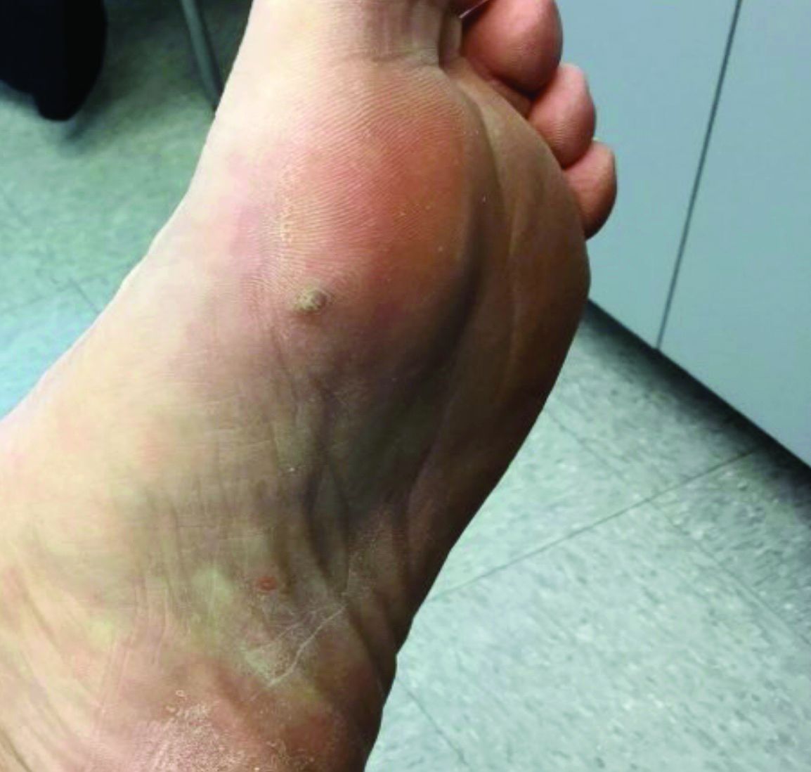
A 70-year-old presented with a 3-week history of asymptomatic violaceous papules on his feet
and named the condition multiple benign pigmented hemorrhagic sarcoma. The disease emerged again at the onset of the AIDS epidemic among homosexual men. There are five variants: HIV/AIDS–related KS, classic KS, African cutaneous KS, African lymphadenopathic KS, and immunosuppression-associated KS (from immunosuppressive therapy or malignancies such as lymphoma).

KS is caused by human herpes virus type 8 (HHV-8). Patients with KS have an increased risk of developing other malignancies such as lymphomas, leukemia, and myeloma. This patient exhibited classic KS.
The various forms of KS may appear different clinically. The lesions may appear as erythematous macules, small violaceous papules, large plaques, or ulcerated nodules. In classic KS, violaceous to bluish-black macules evolve to papules or plaques. Lesions are generally asymptomatic. The most common locations are the toes and soles, although other areas may be affected. Any mucocutaneous surface can be involved. The most common areas of internal involvement are the gastrointestinal system and lymphatics.
Histology reveals angular vessels lined by atypical cells. An associated inflammatory infiltrate containing plasma cells may be present in the upper dermis and perivascular areas. Nodules and plaques reveal a spindle cell neoplasm pattern. Lesions will stain positive for HHV-8.
In patients with HIV/AIDS–related KS, highly active antiretroviral therapy is the most important and beneficial treatment. Since the introduction of HAART, the incidence of KS has greatly decreased. However, there are a proportion of HIV/AIDS–associated Kaposi’s sarcoma patients with well-controlled HIV and undetectable viral loads who require further treatment.
Lesions may spontaneously resolve on their own. Other treatment methods include: cryotherapy, topical alitretinoin (9-cis-retinoic acid), intralesional interferon-alpha or vinblastine, superficial radiotherapy, liposomal doxorubicin, daunorubicin or paclitaxel. Small lesions that are asymptomatic may be monitored.
This patient had no internal involvement and responded well to cryotherapy.
This case and photo were provided by Dr. Bilu Martin.

Dr. Bilu Martin is a board-certified dermatologist in private practice at Premier Dermatology, MD, in Aventura, Fla. More diagnostic cases are available at mdedge.com/dermatology. To submit a case for possible publication, send an email to [email protected].
and named the condition multiple benign pigmented hemorrhagic sarcoma. The disease emerged again at the onset of the AIDS epidemic among homosexual men. There are five variants: HIV/AIDS–related KS, classic KS, African cutaneous KS, African lymphadenopathic KS, and immunosuppression-associated KS (from immunosuppressive therapy or malignancies such as lymphoma).
KS is caused by human herpes virus type 8 (HHV-8). Patients with KS have an increased risk of developing other malignancies such as lymphomas, leukemia, and myeloma. This patient exhibited classic KS.
The various forms of KS may appear different clinically. The lesions may appear as erythematous macules, small violaceous papules, large plaques, or ulcerated nodules. In classic KS, violaceous to bluish-black macules evolve to papules or plaques. Lesions are generally asymptomatic. The most common locations are the toes and soles, although other areas may be affected. Any mucocutaneous surface can be involved. The most common areas of internal involvement are the gastrointestinal system and lymphatics.
Histology reveals angular vessels lined by atypical cells. An associated inflammatory infiltrate containing plasma cells may be present in the upper dermis and perivascular areas. Nodules and plaques reveal a spindle cell neoplasm pattern. Lesions will stain positive for HHV-8.
In patients with HIV/AIDS–related KS, highly active antiretroviral therapy is the most important and beneficial treatment. Since the introduction of HAART, the incidence of KS has greatly decreased. However, there are a proportion of HIV/AIDS–associated Kaposi’s sarcoma patients with well-controlled HIV and undetectable viral loads who require further treatment.
Lesions may spontaneously resolve on their own. Other treatment methods include: cryotherapy, topical alitretinoin (9-cis-retinoic acid), intralesional interferon-alpha or vinblastine, superficial radiotherapy, liposomal doxorubicin, daunorubicin or paclitaxel. Small lesions that are asymptomatic may be monitored.
This patient had no internal involvement and responded well to cryotherapy.
This case and photo were provided by Dr. Bilu Martin.
Dr. Bilu Martin is a board-certified dermatologist in private practice at Premier Dermatology, MD, in Aventura, Fla. More diagnostic cases are available at mdedge.com/dermatology. To submit a case for possible publication, send an email to [email protected].
Peripheral neuropathy tied to mortality in adults without diabetes
researchers reported in Annals of Internal Medicine.

The findings do not necessarily mean that doctors should implement broader screening for peripheral neuropathy at this time, however, the investigators said.
“Doctors don’t typically screen for peripheral neuropathy in persons without diabetes,” senior author Elizabeth Selvin, PhD, MPH, professor of epidemiology at the Johns Hopkins Bloomberg School of Public Health, Baltimore, said in an interview.
“Our study shows that peripheral neuropathy – as assessed by decreased sensation in the feet – is common, even in people without diabetes,” Dr. Selvin explained. “It is not yet clear whether we should be screening people without diabetes since we don’t have clear treatments, but our study does suggest that this condition is an underrecognized condition that is associated with poor outcomes.”
Patients with diabetes typically undergo annual foot examinations that include screening for peripheral neuropathy, but that’s not the case for most adults in the absence of diabetes.
“I don’t know if we can make the jump that we should be screening people without diabetes,” said first author Caitlin W. Hicks, MD, assistant professor of surgery, division of vascular surgery and endovascular therapy, Johns Hopkins University, Baltimore. “Right now, we do not exactly know what it means in the people without diabetes, and we definitely do not know how to treat it. So, screening for it will tell us that this person has this and is at higher risk of mortality than someone who doesn’t, but we do not know what to do with that information yet.”
Nevertheless, the study raises the question of whether physicians should pay more attention to peripheral neuropathy in people without diabetes, said Dr. Hicks, director of research at the university’s diabetic foot and wound service.
Heightened risk
To examine associations between peripheral neuropathy and all-cause and cardiovascular mortality in U.S. adults, Dr. Hicks and colleagues analyzed data from 7,116 adults aged 40 years or older who participated in the National Health and Nutrition Examination Survey (NHANES) between 1999 and 2004.
The study included participants who underwent monofilament testing for peripheral neuropathy. During testing, technicians used a standard 5.07 Semmes-Weinstein nylon monofilament to apply slight pressure to the bottom of each foot at three sites. If participants could not correctly identify where pressure was applied, the test was repeated. After participants gave two incorrect or undeterminable responses for a site, the site was defined as insensate. The researchers defined peripheral neuropathy as at least one insensate site on either foot.
The researchers determined deaths and causes of death using death certificate records from the National Death Index through 2015.
In all, 13.5% of the participants had peripheral neuropathy, including 27% of adults with diabetes and 11.6% of adults without diabetes. Those with peripheral neuropathy were older, were more likely to be male, and had lower levels of education, compared with participants without peripheral neuropathy. They also had higher body mass index, were more often former or current smokers, and had a higher prevalence of hypertension, hypercholesterolemia, and cardiovascular disease.
During a median follow-up of 13 years, 2,128 participants died, including 488 who died of cardiovascular causes.
The incidence rate of all-cause mortality per 1,000 person-years was 57.6 in adults with diabetes and peripheral neuropathy, 34.3 in adults with peripheral neuropathy but no diabetes, 27.1 in adults with diabetes but no peripheral neuropathy, and 13.0 in adults without diabetes or peripheral neuropathy.
Among participants with diabetes, the leading cause of death was cardiovascular disease (31% of deaths), whereas among participants without diabetes, the leading cause of death was malignant neoplasms (27% of deaths).
After adjustment for age, sex, race, or ethnicity, and risk factors such as cardiovascular disease, peripheral neuropathy was significantly associated with all-cause mortality (hazard ratio [HR], 1.49) and cardiovascular mortality (HR, 1.66) in participants with diabetes. In participants without diabetes, peripheral neuropathy was significantly associated with all-cause mortality (HR, 1.31), but its association with cardiovascular mortality was not statistically significant.
The association between peripheral neuropathy and all-cause mortality persisted in a sensitivity analysis that focused on adults with normoglycemia.
Related conditions
The study confirms findings from prior studies that examined the prevalence of loss of peripheral sensation in populations of older adults with and without diabetes, said Elsa S. Strotmeyer, PhD, MPH, associate professor of epidemiology at the University of Pittsburgh. “The clinical significance of the loss of peripheral sensation in older adults without diabetes is not fully appreciated,” she said.
A limitation of the study is that peripheral neuropathy was not a clinical diagnosis. “Monofilament testing at the foot is a quick clinical screen for decreased lower-extremity sensation that likely is a result of sensory peripheral nerve decline,” Dr. Strotmeyer said.
Another limitation is that death certificates are less accurate than medical records for determining cause of death.
“Past studies have indicated that peripheral nerve decline is related to common conditions in aging such as the metabolic syndrome and cardiovascular disease, cancer treatment, and physical function loss,” Dr. Strotmeyer said. “Therefore it is not surprising that is related to mortality as these conditions in aging are associated with increased mortality. Loss of peripheral sensation at the foot may also be related to fall injuries, and mortality from fall injuries has increased dramatically in older adults over the past several decades.”
Prior research has suggested that monofilament testing may play a role in screening for fall risk in older adults without diabetes, Dr. Strotmeyer added.
“For older adults both with and without diabetes, past studies have recommended monofilament testing be incorporated in geriatric screening for fall risk. Therefore, this article expands implications of clinical importance to understanding the pathology and consequences of loss of sensation at the foot in older patients,” she said.
The study was funded by the National Institute of Diabetes and Digestive and Kidney Diseases and the National Heart, Lung, and Blood Institute. Dr. Hicks, Dr. Selvin, and a coauthor, Kunihiro Matsushita, MD, PhD, disclosed NIH grants. In addition, Dr. Selvin disclosed personal fees from Novo Nordisk and grants from the Foundation for the National Institutes of Health outside the submitted work, and Dr. Matsushita disclosed grants and personal fees from Fukuda Denshi outside the submitted work. Dr. Strotmeyer receives funding from the National Institute on Aging and the National Institute of Arthritis and Musculoskeletal and Skin Diseases and is chair of the health sciences section of the Gerontological Society of America.
A version of this article originally appeared on Medscape.com.
researchers reported in Annals of Internal Medicine.
The findings do not necessarily mean that doctors should implement broader screening for peripheral neuropathy at this time, however, the investigators said.
“Doctors don’t typically screen for peripheral neuropathy in persons without diabetes,” senior author Elizabeth Selvin, PhD, MPH, professor of epidemiology at the Johns Hopkins Bloomberg School of Public Health, Baltimore, said in an interview.
“Our study shows that peripheral neuropathy – as assessed by decreased sensation in the feet – is common, even in people without diabetes,” Dr. Selvin explained. “It is not yet clear whether we should be screening people without diabetes since we don’t have clear treatments, but our study does suggest that this condition is an underrecognized condition that is associated with poor outcomes.”
Patients with diabetes typically undergo annual foot examinations that include screening for peripheral neuropathy, but that’s not the case for most adults in the absence of diabetes.
“I don’t know if we can make the jump that we should be screening people without diabetes,” said first author Caitlin W. Hicks, MD, assistant professor of surgery, division of vascular surgery and endovascular therapy, Johns Hopkins University, Baltimore. “Right now, we do not exactly know what it means in the people without diabetes, and we definitely do not know how to treat it. So, screening for it will tell us that this person has this and is at higher risk of mortality than someone who doesn’t, but we do not know what to do with that information yet.”
Nevertheless, the study raises the question of whether physicians should pay more attention to peripheral neuropathy in people without diabetes, said Dr. Hicks, director of research at the university’s diabetic foot and wound service.
Heightened risk
To examine associations between peripheral neuropathy and all-cause and cardiovascular mortality in U.S. adults, Dr. Hicks and colleagues analyzed data from 7,116 adults aged 40 years or older who participated in the National Health and Nutrition Examination Survey (NHANES) between 1999 and 2004.
The study included participants who underwent monofilament testing for peripheral neuropathy. During testing, technicians used a standard 5.07 Semmes-Weinstein nylon monofilament to apply slight pressure to the bottom of each foot at three sites. If participants could not correctly identify where pressure was applied, the test was repeated. After participants gave two incorrect or undeterminable responses for a site, the site was defined as insensate. The researchers defined peripheral neuropathy as at least one insensate site on either foot.
The researchers determined deaths and causes of death using death certificate records from the National Death Index through 2015.
In all, 13.5% of the participants had peripheral neuropathy, including 27% of adults with diabetes and 11.6% of adults without diabetes. Those with peripheral neuropathy were older, were more likely to be male, and had lower levels of education, compared with participants without peripheral neuropathy. They also had higher body mass index, were more often former or current smokers, and had a higher prevalence of hypertension, hypercholesterolemia, and cardiovascular disease.
During a median follow-up of 13 years, 2,128 participants died, including 488 who died of cardiovascular causes.
The incidence rate of all-cause mortality per 1,000 person-years was 57.6 in adults with diabetes and peripheral neuropathy, 34.3 in adults with peripheral neuropathy but no diabetes, 27.1 in adults with diabetes but no peripheral neuropathy, and 13.0 in adults without diabetes or peripheral neuropathy.
Among participants with diabetes, the leading cause of death was cardiovascular disease (31% of deaths), whereas among participants without diabetes, the leading cause of death was malignant neoplasms (27% of deaths).
After adjustment for age, sex, race, or ethnicity, and risk factors such as cardiovascular disease, peripheral neuropathy was significantly associated with all-cause mortality (hazard ratio [HR], 1.49) and cardiovascular mortality (HR, 1.66) in participants with diabetes. In participants without diabetes, peripheral neuropathy was significantly associated with all-cause mortality (HR, 1.31), but its association with cardiovascular mortality was not statistically significant.
The association between peripheral neuropathy and all-cause mortality persisted in a sensitivity analysis that focused on adults with normoglycemia.
Related conditions
The study confirms findings from prior studies that examined the prevalence of loss of peripheral sensation in populations of older adults with and without diabetes, said Elsa S. Strotmeyer, PhD, MPH, associate professor of epidemiology at the University of Pittsburgh. “The clinical significance of the loss of peripheral sensation in older adults without diabetes is not fully appreciated,” she said.
A limitation of the study is that peripheral neuropathy was not a clinical diagnosis. “Monofilament testing at the foot is a quick clinical screen for decreased lower-extremity sensation that likely is a result of sensory peripheral nerve decline,” Dr. Strotmeyer said.
Another limitation is that death certificates are less accurate than medical records for determining cause of death.
“Past studies have indicated that peripheral nerve decline is related to common conditions in aging such as the metabolic syndrome and cardiovascular disease, cancer treatment, and physical function loss,” Dr. Strotmeyer said. “Therefore it is not surprising that is related to mortality as these conditions in aging are associated with increased mortality. Loss of peripheral sensation at the foot may also be related to fall injuries, and mortality from fall injuries has increased dramatically in older adults over the past several decades.”
Prior research has suggested that monofilament testing may play a role in screening for fall risk in older adults without diabetes, Dr. Strotmeyer added.
“For older adults both with and without diabetes, past studies have recommended monofilament testing be incorporated in geriatric screening for fall risk. Therefore, this article expands implications of clinical importance to understanding the pathology and consequences of loss of sensation at the foot in older patients,” she said.
The study was funded by the National Institute of Diabetes and Digestive and Kidney Diseases and the National Heart, Lung, and Blood Institute. Dr. Hicks, Dr. Selvin, and a coauthor, Kunihiro Matsushita, MD, PhD, disclosed NIH grants. In addition, Dr. Selvin disclosed personal fees from Novo Nordisk and grants from the Foundation for the National Institutes of Health outside the submitted work, and Dr. Matsushita disclosed grants and personal fees from Fukuda Denshi outside the submitted work. Dr. Strotmeyer receives funding from the National Institute on Aging and the National Institute of Arthritis and Musculoskeletal and Skin Diseases and is chair of the health sciences section of the Gerontological Society of America.
A version of this article originally appeared on Medscape.com.
monarchE: Abemaciclib reigns on in high-risk breast cancer
The addition of the CDK4/6 inhibitor abemaciclib (Verzenio) to endocrine therapy continues to offer improved event-free survival in women with high-risk hormone receptor-positive (HR+), HER2-negative breast cancer, indicate updated results, which now extend to about a year and a half, from the landmark monarchE trial.
However, experts warned that longer follow-up – at least to 5 years – will be required to understand the impact of the combination treatment on survival, particularly as HR+ breast cancer is associated with a high rate of late recurrences.
The research was presented Dec. 9 at the 2020 San Antonio Breast Cancer Symposium, being held online this year because of the pandemic.
An earlier preplanned interim analysis the phase 3 trial of over 5600 patients was presented at the ESMO Virtual Congress 2020, and simultaneously published in the Journal of Clinical Oncology.
As previously reported by Medscape Medical News, this showed that, after a median follow-up of 15.5 months, abemaciclib plus endocrine therapy was associated with a 25% relative risk reduction in the primary endpoint of invasive disease-free survival (IDFS) vs endocrine therapy alone.
At the time, the findings were hailed as practice changing and, once approved for high-risk HR+ HER2-negative early breast cancer, as the “new standard of care” by one expert.
Now, with median follow-up extended to 19.1 months, Priya Rastogi, MD, associate professor at the University of Pittsburgh Department of Medicine, Pittsburgh, Pennsylvania, presented new study data, including additional results on patients with a Ki-67 index ≥20%, which is indicative of fast tumor growth.
Abemaciclib plus endocrine therapy was associated with a significant 28.7% reduction in the relative risk of developing an IDFS event vs endocrine therapy alone across the whole patient population, and with a 30.9% risk reduction in those with a Ki-67 index ≥20%.
Moreover, patients taking the drug combination had a significant 31.3% reduction in the relative risk of a distant relapse-free survival (DRFS) event.
Crucially, these improvements, which were deemed clinically meaningful, were not gained at the expense of additional safety concerns, although high rates of any grade diarrhea, fatigue, and neutropenia were noted.
Rastogi said the findings underline that abemaciclib combined with standard endocrine therapy “is the first CDK4/6 inhibitor to demonstrate efficacy and tolerability for patients with HR+ HER2-negative, node-positive, high-risk early breast cancer.”
Longer follow-up is ‘reassuring’ but still ‘quite short’
George W. Sledge Jr, MD, professor of medicine at Stanford University Medical Center, Palo Alto, California, was the study discussant for the earlier interim analysis presented at ESMO 2020.
At the time, he said that the study had “very, very short follow-up,” and it was consequently unclear whether the improvements will “lead to what we really care about: improved overall survival.”
Approached to comment on the current analysis, Sledge told Medscape Medical News the data “appear quite consistent” with those presented earlier this year, “which is certainly reassuring.”
Referring to the analysis in patients with a Ki-67 index ≥20%, he added the results “show a higher absolute benefit in patients with more rapidly proliferating tumors, as might be expected for a drug affecting cell-cycle division.”
However, Sledge underlined that the median follow-up time “is still quite short for a study of ER-positive adjuvant therapy, where the majority of recurrences and deaths occur after 5 years in many studies”.
Consequently, “we still have a long way to go to understand the ultimate effects of CDK4/6 inhibition on early-stage, ER-positive breast cancer, particularly on late recurrences.”
Agreed, said C. Kent Osborne, MD, codirector of SABCS and founding director of the Duncan Cancer Center at Baylor College of Medicine, Houston, Texas.
Commenting in a press conference, he said the results are “very encouraging, especially in the subgroup of tumors with high proliferation.”
However, Osborne also urged “caution” in the interpretation of the results “given the still rather short follow-up [and] given that ER+ disease is known for its persistent recurrence rate, even past 10 years.”
He also noted “this class of inhibitors is likely cytostatic, rather than cytocidal, meaning that it blocks cell proliferation rather than killing the cells.” Questions therefore remain over whether the survival curves for combination therapy will come together with those for endocrine therapy alone once the drug is stopped.
Osborne nevertheless said that, “with these caveats in mind, this is still an extremely important trial that could be practice changing in this very high-risk patient population…if the results continue to be positive and show improved overall survival with longer follow-up.”
During the press conference, Rastogi confirmed that the study will indeed to continue out to 10 years until the final assessment of overall survival.
More study details
The 5637 women who were enrolled in monarchE were divided into two cohorts:
Cohort 1, which included patients with four or more positive nodes, or those with up to three positive nodes and a tumor size ≥5 cm or grade 3 disease
Cohort 2, which included women with up to three positive nodes and a Ki-67 index ≥20% based on a standard assay
“Cohort 2 opened one year after Cohort 1 and enrolled 517 patients,” Rastogi said.
Regardless of cohort, the patients were randomly assigned in a 1:1 fashion to abemaciclib for 2 years plus endocrine therapy for 5 to 10 years, as clinically indicated, or endocrine therapy alone.
At the primary efficacy outcome analysis, 395 IDFS events had occurred in the intention-to-treat analysis. The median follow-up was 19.1 months, and 25.5% of patients had completed the 2-year treatment period. A further 58.2% were still on treatment.
The results showed 163 IDFS events had occurred with abemaciclib plus endocrine therapy vs 232 with endocrine therapy alone, to give a 2-year IDFS rate of 92.3% vs 89.3%, at a hazard ratio of 0.713 (P = .0009).
Moving on to the subgroup analysis, Rastogi added that there were “no statistically significant interactions observed, indicating a consistent treatment benefit across all groups.”
The researchers also looked at IDFS rates in patients from both cohorts with Ki-67 index ≥20%, again finding that abemaciclib plus endocrine therapy was associated with significantly fewer events than endocrine therapy alone.
There were 82 events with the combination treatment vs 115 with endocrine therapy, at a 2-year IDFS rate of 91.6% vs 87.1% and a hazard ratio of 0.691 (P = .0111).
DRFS also significantly improved with abemaciclib plus endocrine therapy vs endocrine therapy alone, at a 2-year DRFS rate of 93.8% vs 90% or a hazard ratio of 0.687 (P = .0009).
“Safety remained consistent with the known profile of abemaciclib,” Rastogi said, “and what was observed at the second interim analysis, [with] minimal increases in any grade and grade ≥3 treatment-related adverse events.”
The most common adverse events were diarrhea, fatigue, and neutropenia, which were largely grades 1 and 2.
Notably, 2.4% of combination therapy patients experienced a venous thromboembolic event of any grade vs 0.6% of endocrine therapy patients, while 2.9% and 1.2%, respectively, had any grade interstitial lung disease.
At least one dose hold because of an adverse event was reported by 59.5% of patients on abemaciclib plus endocrine therapy, while 42.5% had at least one dose reduction because of an adverse event. In both cases, the primary reason was diarrhea.
Finally, Rastogi said that “over half of the discontinuations of abemaciclib due to adverse events occurred during the first 5 months of treatment, with the highest number…in the first month.”
Ki-67 issues
During the press conference, Virginia Kaklamani, MD, codirector of SABCS and leader of the Breast Cancer Program at the UT Health San Antonio Cancer Center, Texas, asked about the practicalities of the Ki-67 index.
She said that testing is “great when it’s centrally done, but what do we expect physicians to do when some institutions do it, some don’t, and obviously it’s not really validated in most institutions around the world?”
Rastogi replied that is a “great question,” adding “this is something that is going to have to be sorted as we continue to have discussions and get more granularity of what to do when abemaciclib is administered in that patient population.”
Carlos L. Arteaga, MD, codirector of SABCS and director of the Simmons Comprehensive Cancer Center at UT Southwestern Medical Center, Dallas, Texas, added that “most of us think of 10% as a cutoff between high and low” for the Ki-67 index, rather than ≥20%.
He said that he knows it is “arbitrary” but he thinks of 20% as “extremely high” and asked whether the researchers would be able to conduct a retrospective analysis to look at Ki-67 as a gradient to determine “at what point it stops predicting.”
Rastogi replied that, at the time monarchE was developed, some of the international guidelines used a Ki-67 index ≥20% as the cutoff.
She also noted that, although Ki-67 index ≥20% was an entry criterion for cohort 2, cohort 1 patients also provided tissue after randomization, “and so we’ll be able to look at these types of questions with our translational research committee.”
This study was sponsored by Eli Lilly. Rastogi has financial ties to AstraZeneca, Genentech/Roche, and the study sponsor, Eli Lilly. Regan has ties to Lilly and multiple other pharmaceutical companies. Sledge has ties to Lilly and other companies. Osborne has ties to Lilly and other companies.
This article first appeared on Medscape.com.
FROM SABCS 2020
PENELOPE-B: Palbociclib disappoints in HR+, HER2– breast cancer
The CDK4/6 inhibitor palbociclib provides no significant benefit in patients with hormone receptor–positive (HR+), HER2-negative primary breast cancer, according to first results from the PENELOPE-B trial.
In this phase 3 trial, adding palbociclib to standard endocrine therapy did not improve invasive disease-free survival (iDFS) in patients who were at high risk of relapse following neoadjuvant chemotherapy (NACT).
There was no significant difference in iDFS rates with palbociclib or placebo at 2 years (88.3% vs. 84%), 3 years (81.2% vs. 77.7%), or even 4 years (73% vs. 72.4%), with a stratified hazard ratio of 0.93 (P = .525).
“This, unfortunately, was a negative trial,” said Ruth M. O’Regan, MD, of the University of Wisconsin–Madison, who independently commented after the trial’s virtual presentation at the 2020 San Antonio Breast Cancer Symposium.
Discussing the iDFS curves for palbociclib and placebo over time, Dr. O’Regan observed that there was a slight benefit for palbociclib in the range of 4.3%, compared with placebo at 2 years and 3.5% at 3 years. The benefit was small but “still meaningful,” she added. However, “at 4 years, you can see the curves have completely come together.”
“This begs the question of whether this trial was just treating patients with occult metastatic disease because I think these curves kind of mirror what we might see in the first-line metastatic setting,” Dr. O’Regan suggested.
Study details
The PENELOPE-B trial was designed to see if palbociclib could improve iDFS in women with HR+, HER2-negative primary breast cancer who were at high risk of relapse following NACT.
The clinical-pathologic stage plus estrogen receptor and grade (CPS-EG) staging system was used to identify patients at high risk for relapse. A CPS-EG score of 3 or higher or 2 or higher with nodal involvement was used as an entry criterion. This has been associated with a 3-year disease-free survival rate of 77%, noted Sibylle Loibl, MD, PhD, head of the German Breast Group in Neu-Isenburg, who presented PENELOPE-B data at the meeting.
The trial enrolled 1,250 women who did not have a pathological complete response after NACT. After surgery, with or without radiotherapy, patients were randomized to receive 13 cycles of either palbociclib or placebo (125 mg once daily; 21 days on and 7 days off treatment). All patients received concomitant endocrine therapy according to local standards.
“Although the compliance was lower in the palbociclib arm than the placebo arm, it was still satisfactory, and the relative dose intensity was over 80%,” Dr. Loibl noted.
The primary endpoint was iDFS rate. As noted previously, there were no significant between-arm differences in 2-, 3-, or 4-year iDFS rates.
Likewise, there were no significant differences between palbociclib and placebo in terms of the secondary endpoints, which included the type of iDFS event (distant recurrences, invasive locoregional recurrences, contralateral breast cancer, second primary invasive nonbreast cancer, and death without previous event).
Subgroup iDFS analyses showed no differences between the study arms. “No group could be identified with a higher benefit from palbociclib,” Dr. Loibl reported.
An interim overall survival (OS) analysis showed no significant differences between palbociclib and placebo. The 2-year OS rate was 96.3% with palbociclib and 94.5% with placebo. The 3-year OS rates were 93.6% and 90.5%, respectively. The 4-year OS rates were 90.4% and 87.3%, respectively.
“Today, the results of the PENELOPE-B study do not support the addition of 1 year of palbociclib to endocrine therapy. Long-term follow up from all neoadjuvant CDK4/6 inhibitor studies should continue and must be awaited,” Dr. Loibl concluded.
Findings in context
PENELOPE-B is the second phase 3 trial to show no benefit for palbociclib in the treatment of early high-risk breast cancer. The first was the PALLAS trial, which was reported only a few months ago at the European Society for Medical Oncology Virtual Congress 2020.
In PALLAS, palbociclib plus endocrine therapy was compared with endocrine therapy alone in the treatment of women with HR+, HER2-negative early breast cancer, but there was no additional benefit seen.
The results of both PENELOPE-B and PALLAS contrast those recently seen with another CDK4/6 inhibitor, abemaciclib, in HR+/HER2-negative early breast cancer.
Results of the phase 3 monarchE trial showed that, when abemaciclib was added to endocrine treatment, there was a significant (P = .0096) 25% relative risk reduction (HR, 0.75; 95% confidence interval, 0.60-0.93) in iDFS, compared with endocrine therapy alone. These results were also presented at the ESMO Virtual Congress 2020 and published in the Journal of Clinical Oncology.
While it’s not clear why one CDK4/6 inhibitor may be of benefit in HR+/HER2-negative early breast cancer while the other is not, “PENELOPE-B is clearly quite a different trial to the other two adjuvant CDK inhibitor trials,” Dr. O’Regan observed.
For one thing, PENELOPE-B participants were only eligible for the trial if they did not have a pathological complete response after NACT. In addition, PENELOPE-B is a smaller trial, enrolling well under a third of the number of patients who participated in the PALLAS and monarchE trials. Furthermore, the CPS-EG score, rather than anatomic staging, was used to determine eligibility.
“Abemaciclib could be a more effective CDK inhibitor; that’s most certainly a possibility,” Dr. O’Regan suggested. “However, it’s not supported by the metastatic first-line trials, which have remarkably similar hazard ratios and favor CDK inhibitors.”
Perhaps the shorter duration of palbociclib treatment in the PENELOPE-B trial (12 months vs. 24 months) had an effect.
“Clearly, we need to await the results of NATALEE that uses 3 years of ribociclib,” Dr. O’Regan said. It also remains to be seen if the results of the monarchE trial hold up with longer follow-up.
The PENELOPE-B trial was sponsored by the German Breast Group in collaboration with Pfizer, the AGO Study Group, NSABP Foundation, and the Breast International Group. Dr. Loibl disclosed grant and other support from Pfizer during the conduct of the study and relationships with other companies outside the submitted work. She also disclosed intellectual property rights and being a pending patent holder (EP14153692.0) for a method to predict response to anti-HER2–containing therapy and/or chemotherapy in patients with breast cancer, and she disclosed a relationship with Medscape, which is owned by the same company as MDedge. Dr. O’Regan disclosed relationships with Novartis, Eli Lilly, Genentech, PUMA, Macrogenics, Immunomedics, Biotheranostics, and Eisai.
SOURCE: Loibl S et al. SABCS 2020, Abstract GS1-02.
FROM SABCS 2020
New laser therapy shows promise in children with treatment-resistant epilepsy
A new type of laser therapy is safe and effective for children with drug-resistant epilepsy, new research suggests.
Results show that this “is a new and promising therapy” for children for whom drug therapy has failed, said study investigator Elysa Widjaja, MD, a pediatric neuroradiologist at the Hospital for Sick Children and professor in the department of medical imaging, University of Toronto.
In addition, the procedure is less invasive and requires a shorter hospital stay than does open epilepsy surgery, Dr. Widjaja said.
The findings were presented at the annual meeting of the American Epilepsy Society, which was held online this year because of the COVID-19 pandemic.
Registry study
To date, most published studies on the laser procedure have had a small number of participants from only a few centers, Dr. Widjaja noted. “The aim of our registry is to collect data from multiple centers in both Canada and the U.S. to try to get a better understanding of the outcomes following laser therapy and the complications associated with this treatment,” she said.
In the procedure, a surgeon drills a tiny hole through the skull and, guided by MRI, inserts a very thin laser fiber into the center of the lesion. Heat then ablates the affected brain region.
From the dedicated registry, researchers recruited 182 children who were treated with MRgLITT at seven pediatric centers in the United States and two centers in Canada. The youngest patient was aged 14 months, and the oldest was aged 21 years (mean age, 11.2 years). Some pediatric hospitals treat patients up to age 21, Dr. Widjaja noted.
All of the study participants had focal epilepsy, “so the seizures are coming from a defined area of the brain,” she added. In addition, study participants’ conditions were drug-resistant, defined as conditions in which two antiseizure medications had previously failed.
The mean age at seizure onset was 5.4 years, and the mean number of antiepileptic drugs that were taken was 2.3.
Before receiving the therapy, children typically undergo extensive analyses, including MRI and video electroencephalography, to pinpoint where in the brain the seizures originate. Dr. Widjaja noted that the specific area of the brain that is affected varies widely from child to child.
The investigators collected baseline clinical characteristic and procedural data, including ablation site, type of lesion, length of stay, complications, number of MRgLITT procedures, and seizure outcome. To gather this information, they used a secure electronic platform designed to collect and store research data.
Seizure freedom
Among 137 patients for whom 1-year seizure outcomes were available, seizure freedom was reported for 74 patients (54%). In a recent meta-analysis conducted by the investigators, the rate of seizure-free outcomes following epilepsy surgery was about 65%. Although this rate is higher than with the laser therapy, Dr. Widjaja pointed out that the laser intervention is less invasive and the hospital stay of a mean of 3.3 days is shorter than the week or so needed after surgery. This, she said, makes the procedure cost-effective.
Unlike surgery, laser therapy is also “particularly good” at reaching lesions deep in the brain without damaging surrounding tissue, Dr. Widjaja said.
Although the researchers have not evaluated seizure outcomes with respect to age, Dr. Widjaja believes age is not a major factor in outcomes. “I suspect it’s the type of lesion and how big the lesion is that mainly influences the outcome, rather than actual age,” she said.
Complications related to the laser therapy, including infections and bleeding, occurred in 15% of patients. Neurologic deficits affected about 8% of patients; however, these tended to be transient, Dr. Widjaja noted. There were two cases (1%) of permanent neurologic deficits, both of which involved weakness of arms or legs. This, said Dr. Widjaja, is less than the 5% rate of permanent neurologic deficits that occur following surgery, as reported in the literature.
There were no cases of major intracranial hemorrhage among the participants. At 30 days, there was one reported death.
Laser therapy is limited to relatively small lesions of no more than about 2 cm on average, Dr. Widjaja said. “We normally can’t treat huge lesions using laser therapy; they would need surgery.” However, it is possible to treat the same area twice. In the current study, 20 patients (11%) underwent laser therapy on one region on two occasions. Of these participants, 12 (60%) achieved freedom from seizures.
Dr. Widjaja noted that two additional epilepsy centers will soon be providing laser therapy and will expand the registry. In addition, the investigators are building a surgery registry that will enable them to compare outcomes of laser treatment with surgery.
Currently, laser therapy is available only at specialized epilepsy centers that perform surgery.
‘Very important’ research
Commenting on the study, Daniel Goldenholz, MD, PhD, division of epilepsy, department of neurology, Beth Israel Deaconess Medical Center, Boston, called this is “a very important study.”
Laser therapy “offers the opportunity for very rapid recovery from a minimally invasive, targeted technique while simultaneously offering promising outcomes,” said Dr. Goldenholz, who was not involved with the research.
He noted the importance of the investigators’ choosing freedom from seizures as the outcome of interest. In addition, the 54% seizure-freedom rate in the study is “substantially better” than rates from other interventions, he said.
“To put the results into perspective, other work has found that these same patients would have a less than 10% chance of seizure freedom if many different drug combinations were tried,” said Dr. Goldenholz.
He noted that the 1-year outcomes “are a good first time point” but that it is very important to assess longer-term outcomes. “Often, postsurgical outcomes are worse when looking at 2 or 5 years postoperatively,” he added. These longer-term data will be important “to fully inform our patients about long-term prognosis,” Dr. Goldenholz said.
Still, given the overall favorable results so far, “I think more centers will be likely to explore this newer technology,” he said.
The study was funded by the Pediatric Epilepsy Research Foundation. The study authors and Dr. Goldenholz report no relevant financial relationships.
A version of this article originally appeared on Medscape.com.
From AES 2020
SUDEP may explain 3% of all sudden deaths in children
– new research shows.
Just a few years ago, the message regarding SUDEP was that “it’s very rare in children so you don’t need to worry about it,” said study investigator Vicky Whittemore, PhD, program director at the National Institute of Neurological Disorders and Stroke.
These new study results should refocus the message that “the condition is rare, but not as rare as we thought it was,” she said.
The findings were presented at the American Epilepsy Society’s 74th Annual Meeting, which was held online this year because of the COVID-19 pandemic.
Population-based study
Most of the research examining the pediatric SUDEP rate in the United States is based on convenience samples, with few population-based studies.
The investigators used data from the National Institutes of Health/Centers for Disease Control and Prevention Sudden Death in the Young Case Registry. The CDC set up the registry several years ago to record cases of sudden infant death syndrome and sudden deaths in children resulting from violence, trauma, and abuse. Its mandate has since expanded, and the registry now includes data on sudden cardiac death and SUDEP in children.
The current study included children with SUDEP or cardiac/SUDEP who were aged 0-17 years from several states or jurisdictions from 2015 to 2017. Cases were deemed to be SUDEP if the patient had a history of epilepsy, with or without evidence of seizure at the time of death, but excluding status epilepticus.
Criteria for cardiac/SUDEP cases included having a family history of a heritable cardiac condition or sudden death before age 50 years, a personal history of cardiac disease, or a clinical history suggestive of a cardiac disorder, such as death during exertion.
This second category, said Dr. Whittemore, might capture children with Dravet syndrome, a type of epilepsy caused by a genetic mutation that affects both the heart and the brain. “In these cases, it’s sometimes difficult to tell if the child died due to a heart complication or due to epilepsy,” she said.
The analysis included 1,776 cases. Of these, 3% were categorized as SUDEP, and 1% were categorized as cardiac/SUDEP.
The relatively high prevalence of SUDEP was somewhat unexpected, inasmuch as previous reports estimated the rate to be 0.5%-1%, said Dr. Whittemore.
She noted that the current study is population based and included all cases of child death, whereas past reports relied on death certificates. “That probably missed a lot of deaths because they weren’t recorded accurately on the death certificate or weren’t reported in a way that anyone could ascertain that it was a death in someone that had epilepsy.”
Racial differences
Autopsy rates were lower for SUDEP (70%), compared with other categories of death in the registry (81%-100%).
In most jurisdictions, parents must give consent for an autopsy to be performed for a child, and many parents who have suffered such a sudden loss don’t want further investigation, said Dr. Whittemore. “If you know your child had epilepsy, doing an autopsy really isn’t going to tell you very much. You already know they had epilepsy; you may not know the cause of the epilepsy, but an autopsy isn’t going to reveal as much as it would in children with sudden cardiac death.”
SUDEP was equally common in boys and girls. However, the SUDEP mortality rate was higher in Black children (0.32/100,000) than in White children (0.22/100,000). It’s unclear from this study why this is so, but another study that examined SUDEP rates by ZIP code suggested that the higher rate may be caused by socioeconomic factors, said Dr. Whittemore. “Black children from a lower-income family who don’t have access to care may not be getting as good treatment and so have more uncontrolled seizures, which may lead to higher incidence of SUDEP.”
SUDEP occurred at all ages, but mortality rates were highest among patients aged 0-1 year (0.53/100,000) and in those aged 14-17 years (0.31/100,000). Dr. Whittemore speculated that SUDEP rates were higher among the youngest patients because their seizures have just started, and it may be more difficult to bring them under control. In the past, some of these cases may have been classified as sudden infant death syndrome but are now recognized as SUDEP.
As for the older group, research shows that puberty can result in poorer seizure control, which may put teens at elevated risk for SUDEP, said Dr. Whittemore. She added that, as teens continue to age, SUDEP risk may continue to increase. Dr. Whittemore suggested that young adults who head off to college may stop taking their antiseizure medications or consume alcohol while taking these drugs.
Failure of arousal
The study results revealed that most SUDEP cases occurred during sleep without a witness. Dr. Whittemore believes that sleeping with one’s face in a pillow may prevent the reflex required to turn the head to breathe. “It’s sort of a failure of arousal that is potentially the underlying mechanism.”
In some cases, there are signs children had a seizure just prior to death, said Dr. Whittemore.
The researchers have now collected information for 2018 and 2019 and plan to add these data to the current 3-year results. “We will now expand our analysis to include these new numbers to make sure the trends we saw in those 3 years are continuing,” said Dr. Whittemore. The new results should help raise awareness that SUDEP is not as rare as previously believed.
Parents of children with epilepsy can take steps to help reduce the risk for SUDEP, she added. For example, they can use night monitors, and for the children at highest risk (e.g., those with Dravet syndrome), they can use an “alarm blanket” that alerts them when the child moves.
Much is still unknown
Commenting on the study, Daniel Goldenholz, MD, PhD, division of epilepsy, department of neurology, Beth Israel Deaconess Medical Center, New York, who has participated in SUDEP research, said it “raises important questions about SUDEP in children and about racial disparities in SUDEP.”
The understanding of SUDEP so far “leaves much to be desired,” said Dr. Goldenholz. “We don’t yet know why it happens, and we don’t yet know how to prevent it.” The current study “brings a couple of new data points to the table which need further validation, confirmation, and explanation.”
The Sudden Death in Young Case Registry is supported by the National Heart, Lung, and Blood Institute; the National Institute of Neurological Disorders and Stroke; and the CDC. The investigators and Dr. Goldenholz disclosed no relevant financial relationships.
A version of this article originally appeared on Medscape.com.
FROM AES 2020
Rechallenge ‘reasonable’ for some with small-cell lung cancer
In a phase 3 trial, carboplatin plus etoposide significantly prolonged progression-free survival (PFS), when compared with topotecan, in patients with advanced or relapsed, sensitive SCLC.
All patients had responded to first-line platinum plus etoposide, but they experienced relapse or progression 90 days or more after completing that treatment, according to study author Nathalie Baize, MD, of Angers University Hospital in France, and colleagues.
For this trial, Dr. Baize and colleagues enrolled 164 patients with advanced or relapsed SCLC. The median age of the 162 evaluable patients was 64 years, about two-thirds were men, and about 60% had an Eastern Cooperative Oncology Group performance status of 1.
The patients were randomized 1:1 to intravenous carboplatin (area under the curve 5 mg/mL per min on day 1) plus intravenous etoposide (100 mg/m² from day 1 to day 3) or to oral topotecan (2.3 mg/m² from day 1 to day 5 for six cycles). Primary prophylactic filgrastim was recommended for all patients in both treatment groups.
Results: Survival and adverse events
The median follow-up was 22.7 months. The median PFS was significantly longer in the combination therapy arm, at 4.7 months versus 2.7 months in the topotecan arm (stratified hazard ratio 0.57, P = .0041).
The median overall survival was similar in both arms, at 7.5 months in the carboplatin-etoposide arm and 7.4 months in the topotecan arm.
Patients in the carboplatin-etoposide arm had a significantly higher objective response rate, at 49% versus 25% in the topotecan arm (P = .0024).
The most common grade 3-4 adverse events (in the topotecan and combination arms, respectively) were neutropenia (22% vs. 14%), thrombocytopenia (36% vs. 31%), and anemia (21% vs. 25%).
Serious adverse events with hospitalization were reported in 37% of patients in the carboplatin-etoposide arm 43% in the topotecan arm. Febrile neutropenia with sepsis led to two treatment-related deaths in the topotecan group but none in the carboplatin-etoposide group.
Reasonable option for some
Based on the results of this trial, Dr. Baize and colleagues concluded that carboplatin-etoposide rechallenge “can be considered a reasonable second-line chemotherapy option for patients with sensitive relapsed small-cell lung cancer.”
However, while this trial was enrolling patients, immunotherapy and chemotherapy combinations became the standard of care in SCLC, Oscar Arrieta, MD, of Instituto Nacional de Cancerología in Mexico City, and colleagues noted in a related editorial.
Therefore, “reasonable doubts emerge regarding the application of this strategy in patients receiving immunotherapy,” Dr. Arrieta and colleagues wrote.
The editorialists urged conduct of a randomized trial to evaluate rechallenge with carboplatin plus etoposide versus lurbinectedin, which was approved earlier this year by the Food and Drug Administration for the treatment of sensitive and resistant relapsed SCLC.
Commenting on the choice between a platinum-etoposide combination and lurbinectedin, Sarah Goldberg, MD, of Yale University, New Haven, Conn., noted that she and her colleagues have been using the chemotherapy combination for several years.
“This trial confirms that practice and that it’s still a reasonable option for some patients,” Dr. Goldberg said in an interview.
For patients who had a very good first-line response to platinum-etoposide, longer than 180 days (even longer than the 90-day standard in the current trial), she said, “it seems like a rechallenge with platinum-etoposide would potentially be even more effective, and I’d save lurbinectedin for a later line.
“With refractory disease, less than 90 days, I would consider lurbinectedin,” Dr. Goldberg said.
This study was funded by Amgen and the French Lung Cancer Group (Groupe Français de Pneumo-Cancérologie). The researchers disclosed relationships with Pfizer, Roche, AbbVie, and many other companies. Dr. Arrieta disclosed relationships with AstraZeneca, Boehringer Ingelheim, Roche, Lilly, Merck, Pfizer, and Bristol-Myers Squibb. The other editorialists declared no competing interests. Dr. Goldberg disclosed relationships with AstraZeneca, Boehringer Ingelheim, Eli Lilly, Bristol-Myers Squibb, Genentech, Amgen, Spectrum, Blueprint Medicine, Sanofi Genzyme, Daiichi Sankyo, and Regeneron.
SOURCE: Baize N et al. Lancet Oncol. 2020;21:1224-33.
FROM LANCET ONCOLOGY
Fracking sites tied to increased heart failure hospitalizations
Living near hydraulic fracturing is associated with increased risk of hospitalization in people with heart failure (HF), a new study from Pennsylvania suggests.
The link was strongest among those with more severe heart failure but patients with either HF phenotype showed this association of increased risk with exposure to fracking activities, according to the investigators, led by Tara P. McAlexander, PhD, MPH, Drexel University Dornsife School of Public Health in Philadelphia.
“Our understanding has expanded well beyond the famous Harvard Six Cities study to know that it’s not just a short-term uptick in air pollution that›s going to send someone to the hospital a couple days later,” said Dr. McAlexander in an interview, referring to the study conducted from the mid-1970s through 1991. “We know that people who live in these environments and are exposed for long periods of time may have long-term detrimental effects.”
Although questions remain about specific mechanisms and how best to assess exposure, the evidence is mounting in a way that is consistent with the biologic hypotheses of how fracking would adversely affect health, Dr. McAlexander said. “We have many studies now on adverse pregnancy and birth outcomes, and that’s just the tip of the iceberg.”
Pennsylvania is a hot spot for fracking, also known as unconventional natural gas development (UNGD), with more than 12,000 wells drilled in the Marcellus shale since 2004. The shale extends from upstate New York in the north to northeastern Kentucky and Tennessee in the south and covers about 72,000 square miles. Last year, Pennsylvania pledged $3 million to study clusters of rare pediatric cancers and asthma near fracking operations. A recent grand jury report concluded government officials failed to protect residents from the health effects of fracking.
Fracking involves a cascade of activities that can trigger neural circuitry, sympathetic activation, and inflammation – all well-known pathways that potentiate heart failure, said Sanjay Rajagopalan, MD, who has researched the health effects of air pollution for two decades and was not involved with the study.
“If you think about it, it’s like environmental perturbation on steroids in some ways where they are pulling the trigger from a variety of different ways: noise, air pollution, social displacement, psychosocial impacts, economic disparities. So it’s not at all surprising that they saw an association,” said Dr. Rajagopalan, chief of cardiovascular medicine at University Hospitals Harrington Heart & Vascular Institute and director of the Case Western Cardiovascular Research Institute, both in Cleveland, Ohio.
As reported in the Journal of the American College of Cardiology, Dr. McAlexander and colleagues at Johns Hopkins University, Baltimore, used electronic health data from the Geisinger Health System to identify 9,054 patients with heart failure seen between 2008 and 2015. Of these, 5,839 patients had an incident HF hospitalization and 3,215 served as controls. Geisinger operates 13 hospitals and two research centers in 45 of Pennsylvania’s 67 counties, serving more than 3 million of the state’s residents.
Patients’ residential addresses were used to identify latitude and longitude coordinates that were matched with 9,669 UNGD wells in Pennsylvania and the location of major and minor roadways. The researchers also calculated a measure of community socioeconomic deprivation.
The adjusted odds of hospitalization were higher for patients in the highest quartile of exposure for three of the four UNGD phases: pad preparation (odds ratio, 1.70; 95% confidence interval, 1.35-2.13), stimulation or the actual fracking (OR, 1.80; 95% CI, 1.35-2.40), and production (OR, 1.62; 95% CI, 1.07-2.45).
Dr. McAlexander said she initially thought the lack of association with drilling (OR, 0.97; 95% CI, 0.75-1.27) was a mistake but noted that the drilling metric reflects a shorter time period than, for example, 30 days needed to clear the well pad and bring in the necessary equipment.
Stronger associations between pad preparation, fracking, and production are also consistent with the known increases in air pollution, traffic, and noise associated with these phases.
Individuals with more severe HF had greater odds of hospitalization, but the effect sizes were generally comparable between HF with preserved versus reduced ejection fraction. For those with the highest exposure to fracking, the odds ratios for hospitalization reached 2.25 (95% CI, 1.56-3.25) and 2.09 (95% CI, 1.44-3.03), respectively.
Notably, patients who could be phenotyped versus those who could not were more likely to die, to be hospitalized for HF, and to have a higher Charlson Comorbidity Index and other relevant diagnoses like myocardial infarction.
“Clinicians need to be increasingly aware that the environments their patients are in are a huge factor in their disease progression and outlook,” McAlexander said. “We know that UNGD, specifically now, is something that could be impacting a heart failure patient’s survival.”
She also suggested that the findings may also spur more advocacy work and “across-silo” collaboration between clinicians and environmental researchers.
Dr. Rajagopalan said there is increasing recognition that physicians need to be aware of environmental health links as extreme events like the California and Oregon wildfires and coastal flooding become increasingly common. “Unfortunately, unconventional is becoming the new convention.”
The problem for many physicians, however, is just having enough bandwidth to get through the day and get enough learning to keep above water, he said. Artificial intelligence could be used to seed electronic medical records with other personalized information from a bevy of sources including smartphones and the internet of things, but fundamental changes are also needed in the educational process to emphasize the environment.
“It’s going to take a huge societal shift in the way we view commodities, what we consider healthy, etc, but it can happen very quickly because all it takes is a crisis like COVID-19 to bring people to their knees and make them understand how this is going to take over our lives over the next decade,” Dr. Rajagopalan said.
The scientific community has been calling for “good” epidemiologic studies on the health effects of fracking since the early 2010s, Barrak Alahmad, MBChB, MPH, Harvard T.H. Chan School of Public Health, and Haitham Khraishah, MD, Beth Israel Deaconess Medical Center, Harvard Medical School, Boston, point out in an accompanying editorial.
The current study applied “extensive and rigorous methods” involving both the design and statistical approach, including use of a negative control analysis to assess for sources of spurious causal inference, several sensitivity analyses, and controlled for a wide range of covariates.
“Their results were consistent and robust across all these measures,” the editorialists wrote. “Most importantly, the effect size is probably too large to be explained away by an unmeasured confounder.”
Dr. Alahmad and Dr. Khraishah call for advancements in exposure assessment, citing a recent study reporting that ambient particle radioactivity near unconventional oil and gas sites could induce adverse health effects. Other unmet needs include a better understanding of racial disparities in the impacts of fracking and a fine-tuning of cause-specific cardiovascular morbidity and mortality.
The study was supported by training grants from the National Institute of Environmental Health Sciences to Dr. McAlexander and principal investigator Brian Schwartz, MD. The authors, Dr. Rajagopalan, Dr. Alahmad, and Dr. Khraishah have disclosed no relevant financial relationships.
A version of this article originally appeared on Medscape.com.
Calcium burden drives CV risk whether coronary disease is obstructive or not
Coronary artery calcium (CAC) score as a measure of plaque burden more reliably predicts future cardiovascular (CV) risk in patients with suspected coronary disease (CAD) than whether or not the disease is obstructive, a large retrospective study suggests.
Indeed, CV risk went up in tandem with growing plaque burden regardless of whether there was obstructive disease in any coronary artery, defined as a 50% or greater stenosis by computed tomographic angiography (CTA).
The findings argue for plaque burden as measured by CAC score, rather than percent-stenosis severity, for guiding further treatment decisions in such patients, researchers say.
The research was based on more than 20,000 symptomatic patients referred to diagnostic CTA in the Western Denmark Heart Registry who were then followed for about 4 years for major CV events, including death, myocardial infarction, or stroke.
“What we show is that CAC is important for prognosis, and that patients with no stenosis have similar high risk as patients with stenosis when CAC burden is similar,” Martin Bødtker Mortensen, MD, PhD, Aarhus (Denmark) University Hospital, said in an interview.
The guidelines “distinguish between primary and secondary prevention patients” based on the presence or absence of obstructive CAD, he said, but “our results challenge this long-held approach. We show that patients with nonobstructive CAD carry similar risk as patients with obstructive CAD.”
In practice, risk tends to be greater in patients with obstructive compared with nonobstructive CAD. But the reason “is simply that they normally have higher atherosclerosis burden,” Dr. Mortensen said. “When you stratify based on atherosclerosis burden, then patients with obstructive and nonobstructive CAD have similar risk.”
The analysis was published online Dec. 7 in the Journal of the American College of Cardiology with Mortensen as lead author.
Until recently, it had long been believed that CV-event risk was driven by ischemia – but “ischemia is just a surrogate for the extent of atherosclerotic disease,” Armin Arbab Zadeh, MD, PhD, MPH, who is not connected with the current study, said in an interview.
The finding that CV risk climbs with growing coronary plaque burden “essentially confirms” other recent studies, but with “added value in showing how well the calcium scores, compared to obstructive disease, track with risk. So it’s definitely a nice extension of the evidence,” said Dr. Zadeh, director of cardiac CT at Johns Hopkins University, Baltimore.
“This study clearly shows that there is no ischemia ‘threshold,’ that the risk starts from mild and goes up with the burden of atherosclerotic disease. We were essentially taught wrong for decades.”
Dr. Mortensen said that the new results “are in line with previous studies showing that atherosclerosis burden is very important for risk.” They also help explain why revascularization of patients with stable angina failed to cut the risk of MI or death in trials like COURAGE, FAME-2, and ISCHEMIA. It’s because “stenosis per se explains little of the risk compared to atherosclerosis burden.”
In the current analysis, for example, about 65% of events were in patients who did not show obstructive CAD at CTA. Its 23,759 patients with symptoms suggestive of CAD were referred for CTA from 2008 through 2017; 5,043 (21.2%) were found to have obstructive disease and 18,716 (78.8%) either had no CAD or nonobstructive disease.
About 4.4% of patients experienced a first major CV event over a median follow-up of 4.3 years. Only events occurring later than 90 days after CTA were counted in an effort to exclude any directly related to revascularization, Dr. Mortensen noted.
The risk of events went up proportionally with both CAC score and the number of coronaries with obstructive disease.
The number of major CV events per 1,000 person-years was 6.2 for patients with a CAC score of 0, of whom 87% had no CAD by CTA, 7% had nonobstructive CAD, and 6% had obstructive CAD.
The corresponding rate was 17.5 among patients with a CAC score >100-399 for a hazard ratio (HR) of 1.7 (95% confidence interval [CI] 1.4-2.1) vs. a CAC score of 0.
And it was 42.3 per 1,000 patient-years among patients with CAC score >1000, HR 3.4 (95% CI, 2.5-4.6) vs. a CAC score of 0. Among those with the highest-tier CAC score, none were without CAD by CTA, 17% had nonobstructive disease, and 83% had obstructive CAD.
The major CV event rate rose similarly by number of coronaries with obstructive disease. It was 6.1 per 1,000 person-years in patients with no CAD. But it was 12.3 in those with nonobstructive disease, HR 1.3 (95% CI 1.1-1.6), up to 34.7 in those with triple-vessel obstructive disease, HR 2.9 (95% CI 2.2-3.9), vs. no CAD.
However, in an analysis with stratification by CAC score tier (0, 1-99, 100-399, 400-1,000, and >1,000), obstructive CAD was not associated with increased major CV-event risk in any stratum. The findings were similar in each subgroup with 1-vessel, 2-vessel, or 3-vessel CAD when stratified by CAC score.
Nor did major CV event risk track with obstructive CAD in analyses by age or after excluding all patients who underwent coronary revascularization within 90 days of CTA, the group reported.
“I believe these results support the use of CTA as a first-line test in patients with symptoms suggestive of CAD, as it provides valuable information for both diagnosis and prognosis in symptomatic patients,” Dr. Mortensen said. Those found to have a higher burden of atherosclerosis, he added, should receive aggressive preventive therapy regardless of whether or not they have obstructive disease.
The evidence from this study and others “supports a CTA-based approach” in such patients, Dr. Zadeh said. “And I would go further to say that a stress test is really inadequate,” in that it “detects the disease at such a late stage, you’re missing the opportunity to identify these patients who have atherosclerotic disease while you can do something about it.”
Its continued use as a first-line test, Dr. Zadeh said, “is essentially, in my mind, dismissing the evidence.”
An accompanying editorial Todd C. Villines, MD, and Patricia Rodriguez Lozano, MD, of the University of Virginia, Charlottesville agreed that “it is time that the traditional definitions of primary and secondary prevention evolve to incorporate CAC and CTA measures of patient risk based on coronary artery plaque burden.”
But they pointed out some limitations of the current study.
“The authors compared CAC with ≥50% stenosis, not CAC to comprehensive, contemporary coronary CTA,” and so “did not assess numerous other well-validated measures of coronary plaque burden that are routinely obtained from coronary CTA that typically improve the prognostic accuracy of coronary CTA beyond stenosis alone.” Also not performed was “plaque quantification on coronary CTA, an emerging field of study.”
The editorialists noted that noncontrast CT as used in the study for CAC scoring “is generally not recommended as a standalone test in symptomatic patients. Most studies have shown that coronary CTA, a test that accurately detects stenosis and identifies all types of coronary atherosclerosis (calcified and noncalcified), has significantly higher diagnostic and prognostic accuracy than CAC when performed in symptomatic patients without known coronary artery disease.”
Dr. Mortensen has disclosed no relevant financial relationships. Disclosures for the other authors are in the report. Dr. Villines and Dr. Rodriguez Lozano have disclosed no relevant financial relationships. Dr. Zadeh disclosed receiving grant support from Canon Medical Systems.
A version of this article originally appeared on Medscape.com.
Study results support screening rosacea patients for cardiometabolic disease
according to the results of a meta-analysis of more than 50,000 patients.
To date, “mounting comorbidities of rosacea have been identified, suggesting that rosacea is not simply a skin disease but has links to multiple systemic illnesses,” wrote Qi Chen, MD, of Central South University, Changsha, China, and colleagues. The association with rosacea and cardiometabolic disease has been controversial, they added.
In a study published in the Journal of the American Academy of Dermatology, they identified 13 studies including 50,442 rosacea patients and 1,525,864 controls. Approximately 71% of the rosacea patients were women.
Overall, patients with rosacea showed a statistically significant association for hypertension (risk ratio, 1.20; 95% confidence interval, 1.08-1.34; P = .001) and dyslipidemia (RR, 1.32; 95% CI, 1.10-1.58; P = .002). Specifically, rosacea patients averaged higher standard mean differences of systolic and diastolic blood pressure, total cholesterol, HDL cholesterol and LDL cholesterol, and triglycerides, compared with controls.
Rosacea was not significantly associated with an increased risk for ischemic heart disease, stroke, or diabetes, although the rosacea patients showed significantly increased risk of higher fasting blood glucose, compared with controls.
Findings don’t show causality
The study findings were limited by several factors, including the observational nature of some of the studies and the inability to perform subgroup analyses based on subtype and disease severity, the researchers noted. In addition, most of the rosacea patients were outpatients. “Further investigations are warranted to identify the relationship between rosacea and [cardiometabolic disease] in general populations to further validate the significance of our findings.”
However, the results support the value of screening for cardiometabolic disease in rosacea patients to facilitate diagnosis and treatment of disease at an early stage, they concluded.
“Rosacea has been linked statistically to many comorbidities including depression, anxiety, hypertension, and diabetes mellitus,” Julie Harper, MD, of the Dermatology and Skin Care Center of Birmingham (Alabama), said in an interview.
“This study looked more specifically at cardiometabolic disease and found a statistically significant correlation between rosacea and hypertension, higher total cholesterol, higher triglycerides and higher fasting blood glucose,” she said. However, “while there is an association present in this meta-analysis, we cannot assume a cause-and-effect relationship.”
Although the analysis does not prove causality, the key message for clinicians is that cardiometabolic disease is quite common in rosacea patients, and risk factors should be identified and treated early, said Dr. Harper. “Our patients with and without rosacea will benefit from age-appropriate screening, physical examination, and laboratory evaluation with a primary care physician. For rosacea patients in particular, we can advise them that early research suggests that individuals with rosacea might have an increased risk of hypertension and/or high cholesterol and triglycerides. It never hurts to make an appointment with primary care and to be checked.”
“We need more confirmatory studies that minimize the influence of confounding,” Dr. Harper added. Rosacea also has also been linked to obesity, which is another risk factor for cardiometabolic disease.
The study was supported by multiple grants from the National Natural Science Foundation of China. The researchers had no financial conflicts to disclose. Dr. Harper had no relevant financial conflicts to disclose.
SOURCE: Chen Q et al. J Am Acad Dermatol. 2020 Nov;83(5):1331-40.
FROM THE JOURNAL OF THE AMERICAN ACADEMY OF DERMATOLOGY