User login
Antihistamine prescribing for AD varies by specialty
(AD), according to an analysis of a national database.
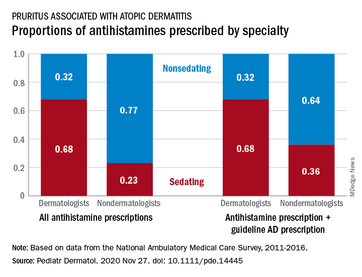
Dermatologists were more likely to prescribe sedating than nonsedating antihistamines (0.68 vs. 0.32) for patients with AD, but the reverse applied to nondermatologists, whose antihistamine distribution was 0.23 sedating and 0.77 nonsedating, based on 2011-2016 data from the National Ambulatory Medical Care Survey.
The numbers were similar for new antihistamine prescriptions, with sedating/nonsedating proportions of 0.60/0.40 for dermatologists and 0.24/0.76 for nondermatologists. Addition of guideline-recommended drugs such as topical corticosteroids and calcineurin inhibitors to the AD equation did not change the result, as dermatologists again showed a preference for sedating antihistamines, compared with nondermatologists, the investigators said.
The data also showed that Black patients with AD were more likely than were White patients to receive prescriptions for first-generation antihistamines and for therapies recommended by the AAD guidelines, and that patients under 21 years received more sedating antihistamines than did patients over age 21, they reported.
The age disparity “may be due to patient preference, as sedation effects may be less desirable to adult patients,” the investigators noted.
SOURCE: Garg S et al. Pediatr Dermatol. 2020 Nov 27. doi: 10.1111/pde.14445.
(AD), according to an analysis of a national database.
Dermatologists were more likely to prescribe sedating than nonsedating antihistamines (0.68 vs. 0.32) for patients with AD, but the reverse applied to nondermatologists, whose antihistamine distribution was 0.23 sedating and 0.77 nonsedating, based on 2011-2016 data from the National Ambulatory Medical Care Survey.
The numbers were similar for new antihistamine prescriptions, with sedating/nonsedating proportions of 0.60/0.40 for dermatologists and 0.24/0.76 for nondermatologists. Addition of guideline-recommended drugs such as topical corticosteroids and calcineurin inhibitors to the AD equation did not change the result, as dermatologists again showed a preference for sedating antihistamines, compared with nondermatologists, the investigators said.
The data also showed that Black patients with AD were more likely than were White patients to receive prescriptions for first-generation antihistamines and for therapies recommended by the AAD guidelines, and that patients under 21 years received more sedating antihistamines than did patients over age 21, they reported.
The age disparity “may be due to patient preference, as sedation effects may be less desirable to adult patients,” the investigators noted.
SOURCE: Garg S et al. Pediatr Dermatol. 2020 Nov 27. doi: 10.1111/pde.14445.
FROM PEDIATRIC DERMATOLOGY
Advocate for legislation to improve, protect LGBTQ lives
In January in many states, the start of a new year also means the start of a new legislative session. For LGBTQ youth and their families, these sessions can create a significant amount of anxiety, as legislators in several states introduce legislation to curtail the rights of this population. In some cases, legislators have attempted to criminalize the provision of gender-affirming medical care to the trans and gender-diverse adolescents that many of us provide care to on a daily basis. As pediatricians,
2020 started on a positive note for LGBTQ children and adolescents, with Virginia becoming the 20th state to ban conversion therapy for minors. Legislation was introduced in several other states to prohibit this practice, including Kentucky, Missouri, and Ohio, and but they ultimately died in committee or were never referred. While there is not yet a nationwide ban on conversion therapy, legislation was introduced in the last three U.S. Congress sessions to ban this harmful practice. In June 2020, the Supreme Court decision in Bostock vs. Clayton County stated that employers could not fire an employee solely because of that person’s sexual orientation and/or gender identity.
However, 19 separate bills were introduced in 2020 alone in states across the United States that would prohibit gender-affirming care for adolescents under age 18.1 Many of these bills also would make the provision of gender-affirming medical care codified as felony child abuse, with loss of licensure, fines and/or jail time a possibility for physicians who prescribe hormones or puberty blockers for gender-affirming care to minors. Fortunately, these bills either died in committee or never had a hearing. However, legislation has been prefiled in several states for their 2021 session to again attempt to prohibit minors from obtaining gender-affirming medical care and/or criminalizing the provision of this care by physicians. Other bills were filed or have been prefiled again to allow various medical and mental health providers to refuse to treat LGBTQ patients because of their personal religious beliefs and/or forcing these same providers to tell a parent if a minor reveals to that provider that they are LGBTQ.
Even if this legislation does not pass or get a hearing, the fact that the bills were introduced can have a profound impact on LGBTQ patients and their families. After a bill was introduced in Texas in their 2017 legislative session that would require trans and gender-diverse (TGD) people to use the bathroom based on their sex assigned at birth, the Trevor Project reported that it had an increase of 34% in crisis calls from trans youth who were in distress.2 This was similar, but slightly less, than was reported by the Trevor Project in September 2015 when in the run-up to a vote on Houston’s Equal Rights Ordinance, advertising was run equating trans women as predators who could be lying in wait in bathrooms. On the converse, when LGBTQ youth feel supported in the media, courts, and legislatures, this can have a positive impact on their mental health. A 2017 study found that, in states who enacted same-sex marriage laws prior to the 2015 Supreme Court decision in Obergefell, compared with those who did not, there was a 7% relative reduction in the proportion of high school students who attempted suicide.3
The American Academy of Pediatrics published its policy statement in September 2018 outlining suggestions for pediatricians to provide support to TGD youth.4 In this position statement, recommendation No. 7 states “that pediatricians have a role in advocating for policies and laws that protect youth who identify as TGD from discrimination and violence.” Therefore, it is incumbent upon us to use our voices to support our LGBTQ youth. In 2020, several pediatricians from the South Dakota chapter of the AAP provided testimony – and organized public rallies – against legislation in that state which would have made gender-affirming care to minors under age 16 punishable by a fine and/or up to 10 years in prison.5

So what can you do? First, get to know your local and state legislators. While it was difficult to meet them in person for much of 2020, you can always call their district and/or Capitol offices, email them, or fill out their constituent contact form typically found on their website. Let them know that you oppose bills which introduce discrimination against your LGBTQ patients or threaten to criminalize the care that you provide to these patients.
Second, work with your state medical association or state AAP chapter to encourage them to oppose these harmful laws and support laws that improve the lives of LGBTQ patients. Third, you can write op-eds to your local newspaper, expressing your support for your patients and outlining the detrimental effects that anti-LGBTQ laws have on your patients. Lastly, you can be active on Twitter, Facebook, or other social media platforms sharing stories of how harmful or helpful certain pieces of legislation can be for your patients.
Dr. Cooper is assistant professor of pediatrics at the University of Texas, Dallas, and an adolescent medicine specialist at Children’s Medical Center Dallas. He has no relevant financial disclosures. Email Dr. Cooper at [email protected].
References
1. “Leglislation affecting LGBT rights across country.” www.aclu.org.
2. “Bathroom Bills Fuel Spike In Calls From Trans Youth To Suicide Hotline.” www.outsmartmagazine.com. 2017 Aug.
3. JAMA Pediatr. 2017 Apr 1. doi: 10.1001/jamapediatrics.2016.4529.
4. Pediatrics. 2018 Oct. doi: 10.1542/peds.2018-2162.
5. Wyckoff AS. “State bills seek to place limits on transgender care, ‘punish’ physicians.” AAP News. 2020 Feb 18.
In January in many states, the start of a new year also means the start of a new legislative session. For LGBTQ youth and their families, these sessions can create a significant amount of anxiety, as legislators in several states introduce legislation to curtail the rights of this population. In some cases, legislators have attempted to criminalize the provision of gender-affirming medical care to the trans and gender-diverse adolescents that many of us provide care to on a daily basis. As pediatricians,
2020 started on a positive note for LGBTQ children and adolescents, with Virginia becoming the 20th state to ban conversion therapy for minors. Legislation was introduced in several other states to prohibit this practice, including Kentucky, Missouri, and Ohio, and but they ultimately died in committee or were never referred. While there is not yet a nationwide ban on conversion therapy, legislation was introduced in the last three U.S. Congress sessions to ban this harmful practice. In June 2020, the Supreme Court decision in Bostock vs. Clayton County stated that employers could not fire an employee solely because of that person’s sexual orientation and/or gender identity.
However, 19 separate bills were introduced in 2020 alone in states across the United States that would prohibit gender-affirming care for adolescents under age 18.1 Many of these bills also would make the provision of gender-affirming medical care codified as felony child abuse, with loss of licensure, fines and/or jail time a possibility for physicians who prescribe hormones or puberty blockers for gender-affirming care to minors. Fortunately, these bills either died in committee or never had a hearing. However, legislation has been prefiled in several states for their 2021 session to again attempt to prohibit minors from obtaining gender-affirming medical care and/or criminalizing the provision of this care by physicians. Other bills were filed or have been prefiled again to allow various medical and mental health providers to refuse to treat LGBTQ patients because of their personal religious beliefs and/or forcing these same providers to tell a parent if a minor reveals to that provider that they are LGBTQ.
Even if this legislation does not pass or get a hearing, the fact that the bills were introduced can have a profound impact on LGBTQ patients and their families. After a bill was introduced in Texas in their 2017 legislative session that would require trans and gender-diverse (TGD) people to use the bathroom based on their sex assigned at birth, the Trevor Project reported that it had an increase of 34% in crisis calls from trans youth who were in distress.2 This was similar, but slightly less, than was reported by the Trevor Project in September 2015 when in the run-up to a vote on Houston’s Equal Rights Ordinance, advertising was run equating trans women as predators who could be lying in wait in bathrooms. On the converse, when LGBTQ youth feel supported in the media, courts, and legislatures, this can have a positive impact on their mental health. A 2017 study found that, in states who enacted same-sex marriage laws prior to the 2015 Supreme Court decision in Obergefell, compared with those who did not, there was a 7% relative reduction in the proportion of high school students who attempted suicide.3
The American Academy of Pediatrics published its policy statement in September 2018 outlining suggestions for pediatricians to provide support to TGD youth.4 In this position statement, recommendation No. 7 states “that pediatricians have a role in advocating for policies and laws that protect youth who identify as TGD from discrimination and violence.” Therefore, it is incumbent upon us to use our voices to support our LGBTQ youth. In 2020, several pediatricians from the South Dakota chapter of the AAP provided testimony – and organized public rallies – against legislation in that state which would have made gender-affirming care to minors under age 16 punishable by a fine and/or up to 10 years in prison.5
So what can you do? First, get to know your local and state legislators. While it was difficult to meet them in person for much of 2020, you can always call their district and/or Capitol offices, email them, or fill out their constituent contact form typically found on their website. Let them know that you oppose bills which introduce discrimination against your LGBTQ patients or threaten to criminalize the care that you provide to these patients.
Second, work with your state medical association or state AAP chapter to encourage them to oppose these harmful laws and support laws that improve the lives of LGBTQ patients. Third, you can write op-eds to your local newspaper, expressing your support for your patients and outlining the detrimental effects that anti-LGBTQ laws have on your patients. Lastly, you can be active on Twitter, Facebook, or other social media platforms sharing stories of how harmful or helpful certain pieces of legislation can be for your patients.
Dr. Cooper is assistant professor of pediatrics at the University of Texas, Dallas, and an adolescent medicine specialist at Children’s Medical Center Dallas. He has no relevant financial disclosures. Email Dr. Cooper at [email protected].
References
1. “Leglislation affecting LGBT rights across country.” www.aclu.org.
2. “Bathroom Bills Fuel Spike In Calls From Trans Youth To Suicide Hotline.” www.outsmartmagazine.com. 2017 Aug.
3. JAMA Pediatr. 2017 Apr 1. doi: 10.1001/jamapediatrics.2016.4529.
4. Pediatrics. 2018 Oct. doi: 10.1542/peds.2018-2162.
5. Wyckoff AS. “State bills seek to place limits on transgender care, ‘punish’ physicians.” AAP News. 2020 Feb 18.

High-need, high-cost lupus patients described for first time
according to a retrospective analysis of hospitalization data from a tertiary care center.

“The identification of the HNHC [high-need, high-cost] cohort and the risk factors for hospitalizations for this cohort will help pave the way to develop programs that improve the quality of care for high-risk lupus patients and [at the same time] lower the cost of care for all lupus patients,” first author Allen Anandarajah, MBBS, and colleagues at the University of Rochester (N.Y.) wrote in Arthritis Care & Research.
Hospitalizations and readmissions are known to be common in patients with SLE, the authors said, and they “account for a large proportion of the direct costs associated with the care of this disease.”
“While HNHC cohorts have been described with other chronic diseases, this report is the first to describe the existence of such a cohort in the SLE population,” the researchers said.
To see if a small group of SLE patients would constitute the majority of hospitalizations and consequently the costs of such care, Dr. Anandarajah and associates analyzed data from 202 SLE patients and their 467 hospitalizations at the University of Rochester–affiliated Strong Memorial Hospital during July 1, 2013, to June 30, 2016. The patients had a mean age of 46 years and included 183 females. A total of 46.5% were White, 43.1% were African American, 6.9% were Hispanic, and 3.5% were of Asian descent. These patients had median lengths of stay of 7 days per SLE patient and 4 days per admission, with median costs of $19,271 per patient and $14,375 per admission.
The researchers identified 44 patients (22%) who accounted for 275 admissions (59%) during the 3-year period. This group’s median of 4 admissions per patient was significantly higher than the median of 1 recorded in all the other hospitalized SLE patients, as was its number of readmissions within 30 days (105 total and median of 1 vs. 11 total and median of 0). The high-risk SLE patients spent a significantly greater amount of time in the hospital than did other patients (median of 30 days vs. 5 days), and their median cost was more than six times as great ($95,262 vs. $14,360). High-risk patients’ median cost per admission also was significantly greater ($19,376 vs. $12,833).
Infections were the most common cause of hospitalization among both high-risk patients and others (28% vs. 23%, respectively) and the rate of involvement of different organ systems as a cause for hospitalization were similar between the groups, except that patients at lower risk significantly more often had gynecologic/obstetric concerns (10% vs. 2%) or nervous system involvement (16% vs. 5%), and high-risk patients were significantly more likely to have gastrointestinal complaints (20% vs. 8%).
Clinically, high-risk patients had significantly higher median scores on the Systemic Lupus International Collaborating Clinics Damage Index and the Comorbidity Index, as well as a significantly higher median level of double-stranded DNA. However, they had no differences in complement factor levels or body mass index.
The high-risk patients also were younger (mean of 42 vs. 46 years) and were diagnosed at a younger mean age (26 vs. 31 years). More high-risk patients were African American (55% vs. 40%) and were more likely to live in areas identified with poverty (50% vs. 29%).
A multivariate analysis that controlled for relevant confounders showed that high-risk patients had a 10 percentage point lower medication possession ratio, which is an indicator of whether a patient had adequate medication supply in a given time frame. High-risk patients overall had a higher average number of medications to treat lupus.
“Our findings underscore the importance of identifying HNHC SLE patients when designing and implementing interventions to lower hospitalizations and improve the quality of care for lupus patients. Furthermore, it is imperative that we develop programs to address the modifiable social and behavioral factors in addition to providing high-quality clinical care targeted for this group,” the researchers wrote.
Some of the limitations in the generalizability of the results include the use of data from a large tertiary medical center serving a large catchment area, with a consequently sicker group of patients, and the potential to miss readmissions to other nearby hospitals. However, “as one of the few centers [in the region] that provides in-patient rheumatology care ... it is less likely that patients would have sought care elsewhere,” they noted.
The study involved no outside source of funding, and the authors had no relevant conflicts of interest.
SOURCE: Anandarajah A et al. Arthritis Care Res. 2020 Nov 17. doi: 10.1002/acr.24510.
according to a retrospective analysis of hospitalization data from a tertiary care center.
“The identification of the HNHC [high-need, high-cost] cohort and the risk factors for hospitalizations for this cohort will help pave the way to develop programs that improve the quality of care for high-risk lupus patients and [at the same time] lower the cost of care for all lupus patients,” first author Allen Anandarajah, MBBS, and colleagues at the University of Rochester (N.Y.) wrote in Arthritis Care & Research.
Hospitalizations and readmissions are known to be common in patients with SLE, the authors said, and they “account for a large proportion of the direct costs associated with the care of this disease.”
“While HNHC cohorts have been described with other chronic diseases, this report is the first to describe the existence of such a cohort in the SLE population,” the researchers said.
To see if a small group of SLE patients would constitute the majority of hospitalizations and consequently the costs of such care, Dr. Anandarajah and associates analyzed data from 202 SLE patients and their 467 hospitalizations at the University of Rochester–affiliated Strong Memorial Hospital during July 1, 2013, to June 30, 2016. The patients had a mean age of 46 years and included 183 females. A total of 46.5% were White, 43.1% were African American, 6.9% were Hispanic, and 3.5% were of Asian descent. These patients had median lengths of stay of 7 days per SLE patient and 4 days per admission, with median costs of $19,271 per patient and $14,375 per admission.
The researchers identified 44 patients (22%) who accounted for 275 admissions (59%) during the 3-year period. This group’s median of 4 admissions per patient was significantly higher than the median of 1 recorded in all the other hospitalized SLE patients, as was its number of readmissions within 30 days (105 total and median of 1 vs. 11 total and median of 0). The high-risk SLE patients spent a significantly greater amount of time in the hospital than did other patients (median of 30 days vs. 5 days), and their median cost was more than six times as great ($95,262 vs. $14,360). High-risk patients’ median cost per admission also was significantly greater ($19,376 vs. $12,833).
Infections were the most common cause of hospitalization among both high-risk patients and others (28% vs. 23%, respectively) and the rate of involvement of different organ systems as a cause for hospitalization were similar between the groups, except that patients at lower risk significantly more often had gynecologic/obstetric concerns (10% vs. 2%) or nervous system involvement (16% vs. 5%), and high-risk patients were significantly more likely to have gastrointestinal complaints (20% vs. 8%).
Clinically, high-risk patients had significantly higher median scores on the Systemic Lupus International Collaborating Clinics Damage Index and the Comorbidity Index, as well as a significantly higher median level of double-stranded DNA. However, they had no differences in complement factor levels or body mass index.
The high-risk patients also were younger (mean of 42 vs. 46 years) and were diagnosed at a younger mean age (26 vs. 31 years). More high-risk patients were African American (55% vs. 40%) and were more likely to live in areas identified with poverty (50% vs. 29%).
A multivariate analysis that controlled for relevant confounders showed that high-risk patients had a 10 percentage point lower medication possession ratio, which is an indicator of whether a patient had adequate medication supply in a given time frame. High-risk patients overall had a higher average number of medications to treat lupus.
“Our findings underscore the importance of identifying HNHC SLE patients when designing and implementing interventions to lower hospitalizations and improve the quality of care for lupus patients. Furthermore, it is imperative that we develop programs to address the modifiable social and behavioral factors in addition to providing high-quality clinical care targeted for this group,” the researchers wrote.
Some of the limitations in the generalizability of the results include the use of data from a large tertiary medical center serving a large catchment area, with a consequently sicker group of patients, and the potential to miss readmissions to other nearby hospitals. However, “as one of the few centers [in the region] that provides in-patient rheumatology care ... it is less likely that patients would have sought care elsewhere,” they noted.
The study involved no outside source of funding, and the authors had no relevant conflicts of interest.
SOURCE: Anandarajah A et al. Arthritis Care Res. 2020 Nov 17. doi: 10.1002/acr.24510.
FROM ARTHRITIS CARE & RESEARCH
FDA panel overwhelmingly backs emergency authorization for Pfizer COVID vaccine
Federal advisers on Thursday told US regulators that the benefits of Pfizer's COVID vaccine outweigh its risks for people aged 16 years and older, moving this product closer to a special emergency clearance.
The US Food and Drug Administration (FDA) put Pfizer's application before its Vaccines and Related Biological Products Advisory Committee (VRBPAC), seeking expert feedback on what is likely to be the first COVID-19 vaccine cleared for use in the United States.
New York-based Pfizer is seeking an emergency use authorization (EUA) for its vaccine, known as BNT162b2, which it developed with Germany's BioNTech. The FDA asked its advisers to vote on a single question regarding this product: "Based on the totality of scientific evidence available, do the benefits of the Pfizer-BioNTech COVID-19 Vaccine outweigh its risks for use in individuals 16 years of age and older?"
The members of VRBPAC voted 17-4 in favor of the Pfizer vaccine, with one panelist abstaining. The FDA considers the recommendations of its panels, but is not bound by them. The agency is expected to quickly grant the special clearance to Pfizer's vaccine, with the company then expected to complete work needed for a more complete biologics license application (BLA).
The FDA often allows members of its advisory committees to explain the reasons for their decisions to vote for or against an application after the tallies are publicly counted.
But the FDA did not give VRBPAC members this opportunity on Thursday, leaving the public without detailed insight into their support or objections.
Before the vote, several panelists had asked if the FDA could rephrase the voting question, raising the age for the approved group to perhaps 18 years of age. During the day, panelists also had questioned whether Pfizer's studies give enough information to judge whether the vaccine works against severe cases of COVID. And there was a discussion about how Pfizer could address concerns about the potential for allergic reactions to the vaccine, given the news of two healthcare workers who experienced allergic reactions after having the vaccine but who have since recovered.
In closing the meeting, VRBPAC chairman, Arnold Monto, MD, noted that the panel will on Dec. 17 meet again to offer recommendations on Moderna Inc.'s COVID vaccine.
"I believe most of us are going to be revisiting some of these issues in about a week," he said.
The panelist who abstained was H. Cody Meissner, MD, an expert in pediatric infectious disease from Tufts University. He earlier was among the several panelists who raised questions about the limited data available about the benefit to those ages 16 and 17. Those voting against the application were Michael Kurilla, MD, PhD; Archana Chatterjee, MD, PhD; A. Oveta Fuller, PhD, and David Kim, MD, MA, according to a tally read by the FDA staff after the vote.
Meanwhile, Sheldon Toubman, JD, voted in favor of the application according to the FDA staff's tally. Toubman had been a chief critic among VRBPAC members in reviewing Pfizer's application at the meeting. He'd suggested limiting the EUA to healthcare workers and residents of nursing homes. Members of these two groups are expected to be the first in the US to get Pfizer's vaccine, for which there will be only a limited initial supply. That idea gained no traction.
Toubman also pressed for more evidence that Pfizer's vaccine will work against severe cases of COVID.
The FDA staff on December 8 released a largely positive agency review of Pfizer vaccine. The efficacy of a two-dose administration of the vaccine has been pegged at 95.0%, with eight COVID-19 cases in the vaccine group and 162 COVID-19 cases in the placebo group. The FDA staff said that the 95% credible interval for the vaccine efficacy was 90.3% to 97.6%.
In that review, the FDA staff said there may be a hint from the results observed to date that the Pfizer vaccine may help ward off severe cases of COVID-19. There were 10 study participants that had severe COVID-19 disease after the first dose: one who received the vaccine and nine who received placebo.
"The total number of severe cases is small, which limits the overall conclusions that can be drawn; however, the case split does suggest protection from severe COVID-19 disease," the FDA staff said.
At the meeting today, Doron Fink, MD, PhD, a lead FDA official on the COVID vaccine review, responded directly to Toubman's concerns. There are many examples of vaccines that protect as well if not better against severe disease as they do against mild to moderate disease, Fink said.
"Protecting against disease of any severity is actually a pretty good predictor of protection against severe disease," Fink said, adding that there's already been a "strong result" shown in terms of the efficacy of Pfizer's vaccine.
Rolling out
Canadian health regulators on December 9 announced their nation's conditional approval of Pfizer's vaccine for people ages 16 and older. In the United Kingdom, a widely publicized rollout of Pfizer's vaccine began on Dec. 8. News quickly spread about two workers in the National Health Service having allergic reactions following vaccination. Both of these workers carry adrenaline autoinjectors, suggesting they have suffered reactions in the past, the Guardian reported. These kinds of autoinjectors are well known in the United States under the brand name EpiPen.
A noted vaccine expert serving on VRBPAC, Paul Offit, MD, of Children's Hospital of Philadelphia, Philadelphia, Pennsylvania, urged the FDA and Pfizer to investigate any connection between reaction to the vaccine and known allergies. If not fully addressed, reports of the reactions seen in initial vaccinations in the UK could prove to unnecessarily frighten people who have allergies away from getting the COVID shot, he said.
Offit suggested running tests where people with egg and peanut allergies would get the Pfizer vaccine under close medical observation "to prove that this is not going to be a problem."
"This is a practical solution because this issue is not going to die until we have better data," Offit said.
More than a dozen COVID-19 vaccines have reached advanced stages of testing, including ones developed in Russia and China, according to the World Health Organization (WHO). The two leading candidates for the US market are the Pfizer/BioNTech vaccine and a similar vaccine developed by Moderna and the National Institute of Allergy and Infectious Diseases. Johnson & Johnson and AstraZeneca are among the other companies with COVID-19 vaccines in testing.
The rapid development of COVID vaccines will create challenges in testing these products. A key issue will be how and whether to continue with placebo-controlled trials, even though such research would be helpful, FDA advisers said.
The FDA tasked Steven Goodman, MD, MHS, PhD, of Stanford University with presenting an overview of considerations for continuing a placebo-controlled trial as COVID vaccines become available. Once a COVID-19 vaccine becomes available to the public, people who have received placebo in the Pfizer trial should not be allowed to immediately receive the vaccine, Goodman said.
There isn't a strong medically-based argument against placebo-controlled research in COVID-19, as many people can take steps to reduce their risk for the infection, Goodman said.
"So as long as there are still important things to learn about the vaccine, placebo-controlled trials should not be regarded as unethical," Goodman said. " I think, however, they might be infeasible. And that is a big issue, because people may not be willing to either remain in the study or to enroll."
During the public comment session, a former FDA official spoke of a need for careful consideration of study volunteers' needs in designing trials of COVID-19 vaccines.
"Reasonable people can disagree over whether study subjects should have priority access to a product whose efficacy they helped demonstrate," said Peter Lurie, MD, president of the nonprofit Center for Science in the Public Interest. "But we ought to be able to agree on this: No subject who has put their body on the line in a vaccine study should be at a disadvantage in terms of vaccine access as a result of their participation."
Lurie argued against extended periods of blinded follow-up after authorization of a COVID-19 vaccine. Such a requirement would be "hard to justify ethically, if it is inconsistent with public health recommendations, particularly with rapidly rising case rates and the reported levels of effectiveness" of the Pfizer vaccine, said Lurie, who served as an associate commissioner at FDA from 2014 to 2017.
Lurie also noted the FDA staff's identification of what he called "disproportionate numbers of Bell's Palsy cases (4 in the vaccine groups vs. 0 in the placebo group)" as a matter that should continue to be monitored, including in the postmarketing phase. He raised no objections to the EUA.
Sidney Wolfe, MD, founder and senior adviser to Public Citizen's Health Research Group, also spoke at the public comment session, citing no objection to an EUA for the Pfizer vaccine. Like Lurie, he urged special consideration of people who have or will receive placebo in COVID-19 vaccine trials.
The Thursday advisory committee on the Pfizer vaccine differed from those held for many other products. The discussion focused more on how to monitor and evaluate the vaccine once approved, while advisory committees sometimes include a detailed look at whether a company has proven that its product works. One of the special advisers serving temporarily on VRBPAC, Eric J. Rubin, MD, PhD, also today published an editorial in The New England Journal of Medicine, titled "SARS-CoV-2 Vaccination — An Ounce (Actually, Much Less) of Prevention."
In the editorial, Rubin and coauthor, Dan L. Longo, MD, called the Pfizer vaccine results seen so far "impressive."
"In the primary analysis, only 8 cases of Covid-19 were seen in the vaccine group, as compared with 162 in the placebo group, for an overall efficacy of 95% (with a 95% credible interval of 90.3 to 97.6%)," they write. "Although the trial does not have the statistical power to assess subgroups, efficacy appeared to be similar in low-risk and high-risk persons, including some from communities that have been disproportionately affected by disease, and in participants older than 55 years of age and those younger than 55."
Intense Scrutiny
The FDA has come under intense scrutiny this year in part because of the aggressive — and ultimately unrealistic — timelines for COVID-19 treatments promoted by the Trump administration. President Donald Trump several times suggested a COVID-19 vaccine could be approved before the November election. Many concerned physicians and scientists including Medscape Editor-in-Chief Eric Topol, MD, called on FDA staff to fight back against any bid to inappropriately speed the approval process for political reasons.
"Any shortcuts will not only jeopardize the vaccine programs but betray the public trust, which is already fragile about vaccines, and has been made more so by your lack of autonomy from the Trump administration and its overt politicization of the FDA," Topol wrote in an August open letter to FDA Commissioner Stephen Hahn, MD.
In an October interview with Topol, Hahn noted that there has been some pushback against the idea of an EUA for a COVID-19 vaccine, with some people preferring to wait for a more complete biological license application.
"When you're talking about a pandemic where people are dying, you want to expedite it as much as possible," Hahn told Topol in the interview.
On Thursday, Hahn issued a public statement about the VRBPAC meeting. Hahn said the FDA's "career staff — made up of physicians, biologists, chemists, epidemiologists, statisticians, and other professionals — have been working around the clock to thoroughly evaluate the data and information in the EUA request."
"I can assure you that no vaccine will be authorized for use in the United States until FDA career officials feel confident in allowing their own families to receive it," Hahn said.
Many clinicians offered their views on the FDA meeting during the day on Twitter.
Robert Wachter, MD, chair of the Department of Medicine at the University of California, San Francisco, who has been a vocal opponent of some of Trump's public statements on COVID-19, urged state officials to stick with the FDA's call on the Pfizer vaccine. In a tweet, he noted that officials in California and several other states have called for independent reviews of COVID-19 vaccines.
If such reviews were to delay distribution of vaccines, this would "lead to more harm than good," Wachter tweeted. "Once FDA says 'go', we should go."
This article was updated 12/10/20.
This article originally appeared on Medscape.com.

Planning—and Parsing—Priorities for COVID-19 Vaccinations
When a COVID-19 vaccine becomes available, who will be first in line? According to an “Interim Playbook for Jurisdiction Operations” published by the Centers for Disease Control and Prevention (CDC) in October, the Departments of Defense (DoD) and State, the Veterans Health Administration (VHA), Indian Health Services, and Bureau of Prisons will all receive a direct allocation, but the distribution of those allocations will depend on the outcomes of an elaborately branched decision tree.
Although the DoD will distribute the vaccine directly to all of its personnel, including retirees and dependents, the plan for members of the Reserve and National Guard is not yet clear. Employees at VHA facilities will receive the vaccine from their departments. The US Department of Veterans Affairs (VA) has announced a “limited-supply phase” for distribution followed by a “general implementation phase.”
The CDC will provide the vaccine to tribal nations that have selected the IHS for distribution (as opposed to the state). According to Government Executive, a separate internal IHS plan suggests the agency would distribute immunizations to tens of thousands of health care and other essential workers, but it isn’t known whether that number includes all of the agency’s workers.
The VA is likely to receive the largest distribution of vaccine doses, according to internal documents obtained by Government Executive. The agency has said it will only finalize its vaccine strategy after a candidate is approved for use. In a Nov. 17 press release, the VA said, “The plan will be a phased approach based on scientific and historical evidence, lessons learned from past pandemic vaccine plans and input from scientific experts both within and outside VA.”
However, the VA has been criticized for not publicizing a detailed vaccine distribution plan. Several Democrats on the Senate Committee on Veterans’ Affairs, for instance, sent a cautionary letter to VA leaders on Nov. 23, saying, “For COVID-19 vaccine distribution to succeed there must be a well-organized plan to meet the needs of all veterans and their providers…. If the states and other federal agencies have already publicly released their plans, why is VA lagging behind?”
As with most federal and non-federal entities, the VA’s allotment of vaccine will be distributed in phases, based on availability and the number of those in the highest-risk populations, such as frontline health care workers and the elderly, within the organization. But the distribution strategy resembles a set of matryoshka dolls, with priorities nested within priorities.
Staff will be 4 of the first 5 populations to receive the vaccine. Employees at VA nursing homes and the 25 Spinal Cord Injuries and Disorders Centers also are due to receive the first batch, followed by veterans at those facilities. Next would come staff in emergency departments, COVID-19 ICUs, and COVID-19 non-ICUs. After that, employees in “other congregate living settings” and veterans aged > 85 years would be vaccinated. Other employees critical in the COVID-19 response, such as police, would be next, followed by inpatient staff in other units and staff performing high-risk procedures. Finally, the priorities will be other groups of patients, veterans in other descending-age groups, and homeless veterans and homeless outreach workers.
The general thinking is that it makes more sense to vaccinate the health care workers first. However, even within that calculation there are further decisions to make. The VA has said it’s focusing on the highest-risk individuals for the first inoculations, but it has emphasized that it considers the risk of transmitting the virus to others above the personal risk of severe illness to the employees themselves.
The VA will not require employees to receive an inoculation, although it plans to encourage staff to do so to protect themselves and veterans they serve.
IHS Publishes COVID-19 Vaccination Plan
COVID-19 infection rates have been nearly 4 times higher among American Indians/Alaska Natives (AI/ANs) when compared with those of non-Hispanic Whites, and AI/ANs are more than 4 times more likely to be hospitalized with the virus. Some mitigation measures have been harder to maintain in Native American communities. Frequent handwashing is difficult when water is at a premium, and social distancing is not always possible when extended families—including elderly—may be living in a single residence.
So vaccination “remains the most promising intervention,” the Indian Health Service Vaccine Task Force wrote in its COVID-19 Pandemic Vaccine Plan, released in November. The plan details how the IHS health care system will prepare for and distribute a vaccine when one becomes available in the US.
The Vaccine Task Force was established by the IHS Headquarters Incident Command Structure, which was activated in early March to respond to COVID-19. In September, the US Department of Health and Human Services (HHS) began a series of consultations with tribes and urban Indian organizations for input on the plan, which aligns as well with recommendations from the Centers for Disease Control and Prevention (CDC).
To “ensure that vaccines are effectively delivered throughout Indian Country in ways that make sense for tribal communities,” HHS Secretary Alex Azar says the Trump Administration has given all tribal health programs and urban Indian organizations two ways to receive the vaccine: through the IHS or through the state.
The CDC, along with IHS, states, and tribes, are coordinating the distribution of a vaccine for federal sites, tribal health programs, and Urban Indian Organizations (UIOs). CDC has issued data requirements that all health care facilities must meet for COVID-19 vaccine administration, inventory, and monitoring.
“There are system-wide planning efforts in place to make sure we’re ready to implement vaccination activities as soon as a US Food and Drug Administration authorized or approved vaccine is available,” said IHS Director RADM Michael Weahkee in a press release. The program’s success, he said, depends on “the strong partnership between the federal government, tribes, and urban leaders.”
The list of IHS, tribal health programs, and UIOs facilities that will receive the COVID-19 vaccine from the IHS, broken down by IHS area, is available on the IHS coronavirus website.
HCPs and COVID-19 Risk: Safer at Home or at Work?
The Centers for Disease Control and Prevention (CDC) and Michigan Department of Health and Human Services surveyed health care personnel in 27 hospitals and 7 medical control agencies that coordinate emergency medical services in the Detroit metropolitan area. Of 16,397 participants, 6.9% had COVID-19 antibodies (although only 2.7% reported a history of a positive real-time transcription polymerase chain reaction test); however, participants had about 6 times the odds of exposure to the virus at home when compared with the workplace. Of those who reported close contact (within 6 feet) of a person with confirmed COVID-19 for ≥ 10 minutes, seroprevalence was highest among those with exposure to a household member (34.3%).
The survey revealed a pattern that suggested community acquisition was a common underlying factor of infection risk, the researchers say. Workers were only more vulnerable at home and when they were closer to the metropolitan center. Seropositivity was more common within 9 miles of Detroit’s center, regardless of occupation and health care setting. The farther away from the center, the lower the seroprevalence.
By work location, seroprevalence was highest among participants who worked in hospital wards (8.8%) and lowest among those in police departments (3.9%). In hospitals, participants working in wards and EDs had higher seropositivity (8.8% and 8.1%, respectively) than did those in ICUs and ORs (6.1% and 4.5%, respectively). Nurses and nurse assistants were more likely to be seropositive than physicians. Nurse assistants had the highest incidence, regardless of where they worked.
Reducing community spread through population-based measures may directly protect healthcare workers on 2 fronts, the researchers say: reduced occupational exposure as a result of fewer infected patients in the less controlled workplace setting such as the ED, and reduced exposure in their homes and communities.
FDA approves liraglutide for adolescents with obesity
The Food and Drug Administration’s new indication for liraglutide (Saxenda) for weight loss in adolescents with obesity, announced on Dec. 4, received welcome as a milestone for advancing a field that’s seen no new drug options since 2003 and boosted by 50% the list of agents indicated for weight loss in this age group.

But liraglutide’s track record in adolescents in the key study published earlier in 2020 left some experts unconvinced that liraglutide’s modest effects would have much impact on blunting the expanding cohort of teens who are obese.
“Until now, we’ve had phentermine and orlistat with FDA approval” for adolescents with obesity, and phentermine’s label specifies only patients older than 16 years. “It’s important that the FDA deemed liraglutide’s benefits greater than its risks for adolescents,” said Aaron S. Kelly, PhD, leader of the 82-week, multicenter, randomized study of liraglutide in 251 adolescents with obesity that directly led to the FDA’s action.
“We have results from a strong, published randomized trial, and the green light from the FDA, and that should give clinicians reassurance and confidence to use liraglutide clinically,” said Dr. Kelly, professor of pediatrics and codirector of the Center for Pediatric Obesity Medicine at the University of Minnesota in Minneapolis.
An ‘unimpressive’ drop in BMI
Sonia Caprio, MD, had a more skeptical take on liraglutide’s role with its new indication: “Approval of higher-dose liraglutide is an improvement that reflects a willingness to accept adolescent obesity as a disease that needs treatment with pharmacological agents. However, the study, published in New England Journal of Medicine, was not impressive in terms of weight loss, and more importantly liraglutide was not associated with any significant changes in metabolic markers” such as insulin resistance, high-sensitivity C-reactive protein, lipoproteins and triglycerides, and hemoglobin A1c.
The observed average 5% drop in body mass index seen after a year on liraglutide treatment, compared with baseline and relative to no average change from baseline in the placebo arm, was “totally insufficient, and will not diminish any of the metabolic complications in youth with obesity,” commented Dr. Caprio, an endocrinologist and professor of pediatrics at Yale University in New Haven, Conn.
Results from the study led by Dr. Kelly also showed that liraglutide for 56 weeks cut BMI by 5% in 43% of patients, and by 10% in 26%, compared with respective rates of 19% and 8% among those in the placebo-control arm. He took a more expansive view of the potential benefits from weight loss of the caliber demonstrated by liraglutide in the study.
“In general, we wait too long with obesity in children; the earlier the intervention the better. A 3% or 4% reduction in BMI at 12 or 13 years old can pay big dividends down the road” when a typical adolescent trajectory of steadily rising weight can be flattened, he said in an interview.
Bariatric and metabolic surgery, although highly effective and usually safe, is seen by many clinicians, patients, and families as an “intervention of last resort,” and its very low level of uptake in adolescents bears witness to that reputation. It also creates an important niche for safe and effective drugs to fill as an adjunct to lifestyle changes, which are often ineffective when used by themselves. Liraglutide’s main mechanism for weight loss is depressing hunger, Dr. Kelly noted.
Existing meds have limitations
The existing medical treatments, orlistat and phentermine, both have significant drawbacks that limit their use. Orlistat (Xenical, Alli), FDA approved for adolescents 12-16 years old since 2003, limits intestinal fat absorption and as a result often produces unwanted GI effects. Phentermine’s approval for older adolescents dates from 1959 and has a weak evidence base, its label limits it to “short-term” use that’s generally taken to mean a maximum of 12 weeks. And, as a stimulant, phentermine has often been regarded as potentially dangerous, although Dr. Kelly noted that stimulants are well-accepted treatments for other disorders in children and adolescents.
“The earlier we treat obesity in youth, the better, given that it tends to track into adulthood,” agreed Dr. Caprio. “However, it remains to be seen whether weight reduction with a pharmacological agent is going to help prevent the intractable trajectories of weight and its complications. So far, it looks like surgery may be more efficacious,” she said in an interview.
Another drawback for the near future with liraglutide will likely be its cost for many patients, more than $10,000/year at full retail prices for the weight-loss formulation, given that insurers have had a poor record of covering the drug for this indication in adults, both Dr. Caprio and Dr. Kelly noted.
Compliance with liraglutide is also important. Dr. Kelly’s study followed patients for their first 26 weeks off treatment after 56 weeks on the drug, and showed that on average weights rebounded to virtually baseline levels by 6 months after treatment stopped.
Obesity treatment lasts a lifetime
“Obesity is a chronic disease, that requires chronic treatment, just like hypertension,” Dr. Kelly stressed, and cited the rebound seen in his study when liraglutide stopped as further proof of that concept. “All obesity treatment is lifelong,” he maintained.
He highlighted the importance of clinicians discussing with adolescent patients and their families the prospect of potentially remaining on liraglutide treatment for years to maintain weight loss. His experience with the randomized study convinced him that many adolescents with obesity are amenable to daily subcutaneous injection using the pen device that liraglutide comes in, but he acknowledged that some teens find this off-putting.
For the near term, Dr. Kelly foresaw liraglutide treatment of adolescents as something that will mostly be administered to patients who seek care at centers that specialize in obesity management. “I’ll think we’ll eventually see it move to more primary care settings, but that will be down the road.”
The study of liraglutide in adolescents was sponsored by Novo Nordisk, the company that markets liraglutide (Saxenda). Dr. Kelly has been a consultant to Novo Nordisk and also to Orexigen Therapeutics, Vivus, and WW, and he has received research funding from AstraZeneca. Dr. Caprio had no disclosures.
The Food and Drug Administration’s new indication for liraglutide (Saxenda) for weight loss in adolescents with obesity, announced on Dec. 4, received welcome as a milestone for advancing a field that’s seen no new drug options since 2003 and boosted by 50% the list of agents indicated for weight loss in this age group.
But liraglutide’s track record in adolescents in the key study published earlier in 2020 left some experts unconvinced that liraglutide’s modest effects would have much impact on blunting the expanding cohort of teens who are obese.
“Until now, we’ve had phentermine and orlistat with FDA approval” for adolescents with obesity, and phentermine’s label specifies only patients older than 16 years. “It’s important that the FDA deemed liraglutide’s benefits greater than its risks for adolescents,” said Aaron S. Kelly, PhD, leader of the 82-week, multicenter, randomized study of liraglutide in 251 adolescents with obesity that directly led to the FDA’s action.
“We have results from a strong, published randomized trial, and the green light from the FDA, and that should give clinicians reassurance and confidence to use liraglutide clinically,” said Dr. Kelly, professor of pediatrics and codirector of the Center for Pediatric Obesity Medicine at the University of Minnesota in Minneapolis.
An ‘unimpressive’ drop in BMI
Sonia Caprio, MD, had a more skeptical take on liraglutide’s role with its new indication: “Approval of higher-dose liraglutide is an improvement that reflects a willingness to accept adolescent obesity as a disease that needs treatment with pharmacological agents. However, the study, published in New England Journal of Medicine, was not impressive in terms of weight loss, and more importantly liraglutide was not associated with any significant changes in metabolic markers” such as insulin resistance, high-sensitivity C-reactive protein, lipoproteins and triglycerides, and hemoglobin A1c.
The observed average 5% drop in body mass index seen after a year on liraglutide treatment, compared with baseline and relative to no average change from baseline in the placebo arm, was “totally insufficient, and will not diminish any of the metabolic complications in youth with obesity,” commented Dr. Caprio, an endocrinologist and professor of pediatrics at Yale University in New Haven, Conn.
Results from the study led by Dr. Kelly also showed that liraglutide for 56 weeks cut BMI by 5% in 43% of patients, and by 10% in 26%, compared with respective rates of 19% and 8% among those in the placebo-control arm. He took a more expansive view of the potential benefits from weight loss of the caliber demonstrated by liraglutide in the study.
“In general, we wait too long with obesity in children; the earlier the intervention the better. A 3% or 4% reduction in BMI at 12 or 13 years old can pay big dividends down the road” when a typical adolescent trajectory of steadily rising weight can be flattened, he said in an interview.
Bariatric and metabolic surgery, although highly effective and usually safe, is seen by many clinicians, patients, and families as an “intervention of last resort,” and its very low level of uptake in adolescents bears witness to that reputation. It also creates an important niche for safe and effective drugs to fill as an adjunct to lifestyle changes, which are often ineffective when used by themselves. Liraglutide’s main mechanism for weight loss is depressing hunger, Dr. Kelly noted.
Existing meds have limitations
The existing medical treatments, orlistat and phentermine, both have significant drawbacks that limit their use. Orlistat (Xenical, Alli), FDA approved for adolescents 12-16 years old since 2003, limits intestinal fat absorption and as a result often produces unwanted GI effects. Phentermine’s approval for older adolescents dates from 1959 and has a weak evidence base, its label limits it to “short-term” use that’s generally taken to mean a maximum of 12 weeks. And, as a stimulant, phentermine has often been regarded as potentially dangerous, although Dr. Kelly noted that stimulants are well-accepted treatments for other disorders in children and adolescents.
“The earlier we treat obesity in youth, the better, given that it tends to track into adulthood,” agreed Dr. Caprio. “However, it remains to be seen whether weight reduction with a pharmacological agent is going to help prevent the intractable trajectories of weight and its complications. So far, it looks like surgery may be more efficacious,” she said in an interview.
Another drawback for the near future with liraglutide will likely be its cost for many patients, more than $10,000/year at full retail prices for the weight-loss formulation, given that insurers have had a poor record of covering the drug for this indication in adults, both Dr. Caprio and Dr. Kelly noted.
Compliance with liraglutide is also important. Dr. Kelly’s study followed patients for their first 26 weeks off treatment after 56 weeks on the drug, and showed that on average weights rebounded to virtually baseline levels by 6 months after treatment stopped.
Obesity treatment lasts a lifetime
“Obesity is a chronic disease, that requires chronic treatment, just like hypertension,” Dr. Kelly stressed, and cited the rebound seen in his study when liraglutide stopped as further proof of that concept. “All obesity treatment is lifelong,” he maintained.
He highlighted the importance of clinicians discussing with adolescent patients and their families the prospect of potentially remaining on liraglutide treatment for years to maintain weight loss. His experience with the randomized study convinced him that many adolescents with obesity are amenable to daily subcutaneous injection using the pen device that liraglutide comes in, but he acknowledged that some teens find this off-putting.
For the near term, Dr. Kelly foresaw liraglutide treatment of adolescents as something that will mostly be administered to patients who seek care at centers that specialize in obesity management. “I’ll think we’ll eventually see it move to more primary care settings, but that will be down the road.”
The study of liraglutide in adolescents was sponsored by Novo Nordisk, the company that markets liraglutide (Saxenda). Dr. Kelly has been a consultant to Novo Nordisk and also to Orexigen Therapeutics, Vivus, and WW, and he has received research funding from AstraZeneca. Dr. Caprio had no disclosures.

Disabling stroke reduced with ticagrelor after minor stroke, TIA
Additional results from the THALES trial have shown that 1 month’s dual antiplatelet therapy with ticagrelor (Brilinta; Astra Zeneca) plus aspirin is associated with a reduction in disabling stroke, compared with aspirin alone in patients with minor stroke or high-risk transient ischemic attack (TIA).

Primary results of the THALES trial, published earlier this year in the New England Journal of Medicine, showed a reduction in the primary endpoint of stroke or death within 30 days with the combination of ticagrelor plus aspirin versus aspirin alone, although this was accompanied by an increase in bleeding. In terms of risk/benefit, the main results showed that for every 1,000 patients treatment with ticagrelor on top of aspirin would prevent 11 strokes or deaths at the cost of four severe hemorrhages.
The current exploratory analysis, which focuses on the severity of the strokes occurring in the trial, was published online Nov. 7 in JAMA Neurology to coincide with its presentation at the European Stroke Organisation-World Stroke Organization Conference 2020.
Results showed that, compared with aspirin alone, ticagrelor plus aspirin significantly reduced the 30-day risk for disabling stroke or death (4.0% versus 4.7%), and the total disability burden (the shift analysis of the distribution of modified Rankin scale) following subsequent ischemic stroke was reduced by a significant 23%.
“This new information on disabling stroke underlines the importance of getting patients on dual antiplatelet therapy quickly after a TIA or mild stroke,” said principal investigator of the THALES trial, S. Claiborne Johnston, MD, PhD.
Dr. Johnston, who is dean of Dell Medical School at the University of Texas at Austin, added: “It’s reassuring that ticagrelor has this effect, which was pretty robust. An accompanying editorial to the THALES publication in the NEJM incorrectly stated that ticagrelor did not reduce risk of disabling stroke, so it is good to be able to correct that misconception with this new data.”
Lead author of the exploratory analysis, Pierre Amarenco, MD, professor of neurology at Bichat University Hospital, Paris, added: “The main results showed that ticagrelor on top of aspirin reduced stroke but now we have new information showing reduction in disabling stroke. Obviously, these are the most important types of stroke to prevent. These are the strokes that will impact patients functionally.”
The THALES trial included 11,016 patients with a noncardioembolic, nonsevere ischemic stroke (National Institutes of Health Stroke Scale [NIHSS] score ≤ 5) or high-risk TIA, of whom 10,803 had modified Rankin Scale (mRS) functional score recorded at 30 days.
They were randomized within 24 hours of symptom onset to ticagrelor (180-mg loading dose on day 1 followed by 90 mg twice daily for 1 month) or placebo. All patients received aspirin (300-325 mg on day 1 followed by 75-100 mg daily for 1 month).
In the new analysis, time to occurrence of disabling stroke (mRS greater than 1) or death within 30 days occurred in 221 of 5,511 patients (4.0%) randomized to ticagrelor and in 260 of 5,478 patients (4.7%) randomized to placebo (hazard ratio, 0.83; P = .04).
The ordinal analysis of mRS in patients with recurrent stroke showed a shift of the disability burden following a recurrent ischemic stroke in favor of ticagrelor (odds ratio, 0.77; P = .002).
Factors associated with disability were baseline NIHSS score of 4-5, ipsilateral stenosis of at least 30%, Asian race/ethnicity, older age, and higher systolic blood pressure.
Asked how the current results compared with observations reported in the main NEJM paper of similar incidences of disability (mRS > 1) in the two groups, Dr. Johnston explained that the result in the original paper looked at disability in the overall population, not just those who went on to have a stroke during follow-up.
“The problem with looking at overall disability is that most of it is actually from the index stroke (the one that led to the patient being enrolled in the trial). That creates a lot of noise that overwhelms the benefit in reducing disability due to new stroke, the thing we really care about and the subject of the new paper,” he commented.
Ticagrelor or clopidogrel?
Ticagrelor now becomes the second antiplatelet agent to have shown benefits on top of aspirin in the minor stroke and high-risk TIA population. Clopidogrel also showed a reduction in major ischemic events in the POINT trial as well as in the Chinese CHANCE trial in similar populations.
Dr. Amarenco pointed out, however, that until now the only treatment that has been shown to reduce disabling stroke in the minor stroke/high risk TIA population in a single trial is aspirin. “The CHANCE and POINT trials of clopidogrel did not show a reduction in disabling stroke individually but this was observed when the trials were combined,” he noted.
“Clinicians will now have to choose between ticagrelor and clopidogrel. We don’t have a head-to-head comparison yet but ticagrelor is effective in all patients whereas clopidogrel may not be as effective in the large subgroup of patients who carry the loss of function gene which make up about 20% of the western population and about 40% of the Asian population,” he said.
“It is very important in the acute phase of stroke to know that the antiplatelet drug is immediately effective as the risk of a recurrent event is highest in the first few hours and days.”
Dr. Amarenco acknowledged that some hospitals may favor clopidogrel because of cost, as it is available generically so is much cheaper than ticagrelor. “But we are only talking about 30 days of treatment, so cost is not too much of an issue,” he pointed out.
The Food and Drug Administration recently approved use of ticagrelor in this indication on the basis of the THALES study.
“It is great news that vascular neurologists now have a new player for reducing future stroke in these patients,” Dr. Amarenco said. Clopidogrel is not approved for this indication but is recommended in American Heart Association/American Stroke Association guidelines, he added.
Dr. Johnston, who was also the lead investigator of the POINT trial with clopidogrel, suggested that it is more important to get patients on dual-antiplatelet therapy rather than worrying too much about which agent to use. “I think we can use aspirin plus either ticagrelor or clopidogrel. The effect on disabling stroke was not significant in POINT but it did reach significance in a meta-analysis combining POINT and CHANCE,” he noted.
He said that choosing between ticagrelor and clopidogrel is tricky without head-to-head data. “Differences in the studied populations makes direct comparison of the trials unwise,” he stressed.
Dr. Johnston pointed out that neither of the clopidogrel trials included moderate strokes (NIHSS scores of 4 and 5) in their study population. “We only have data on ticagrelor for this important group, which accounted for 30% of the THALES study population,” he noted.
“Some people are concerned about the limited efficacy of clopidogrel in large subgroups of patients who do not metabolize it to its active form, but on the flip side, clopidogrel is cheaper – though a 21- to 30-day course [of ticagrelor] probably isn’t that costly – and has more data in combination with aspirin,” he added.
Dr. Johnston said that the approval of ticagrelor for this new indication was “reassuring,” and “provides some air cover for practitioners given the risks of hemorrhage.” He added: “We didn’t bother with an FDA submission after POINT because it was an NIH-sponsored trial. The drug company normally prioritizes regulatory approvals for marketing purposes but their interests were limited because clopidogrel has exceeded its patent life.”
Cost-utility analyses are not yet available, but Dr. Johnston noted: “I suspect both drugs will have substantial benefits and be cost saving. Stroke is expensive, particularly disabling stroke.”
Dr. Johnston said that the more important message is: “Get these people on dual-antiplatelet therapy as soon as possible. Too many patients are not getting the right treatment immediately after symptom onset. We have lots of work to do here.”
Reassuring information
Commenting on the research, J. David Spence, MD, professor of neurology at the Robarts Research Institute, London, Ont., who was not involved in the THALES trial, said this new analysis provided useful and important information that should reassure and encourage clinicians to use dual-antiplatelet therapy in this patient population.
He pointed out that the shift analysis gives the most clinically relevant results. “While the number of patients with a disabling stroke defined as an mRS greater than 1 is lower in the ticagrelor group, I am much more interested in the effect on more severe disability levels – those with an mRS score of 3 or more. Those are the disabilities that we really want to prevent. And from examining the shift analysis distribution, we can see that these more severe disabilities are being reduced with ticagrelor.”
Dr. Spence believes the benefit/risk ratio of dual-antiplatelet therapy could be further improved by better control of blood pressure. “The absolute risk of severe hemorrhage was low in this study, but in my view, most of this could have been prevented by better control of hypertension, as 20 of the 28 severe hemorrhages in the ticagrelor group were intracranial bleeds which can be significantly reduced by good blood pressure control.
“In my view, the increased risk of hemorrhage with dual-antiplatelet therapy should not be regarded as inevitable; it can be virtually eliminated with better medical care,” he stated.
Another outside commentator, Peter Rothwell, MD, PhD, professor of neurology, University of Oxford (England), also believes this is an important paper. “The main NEJM report presented the data on overall disability, but did not present a clear analysis of the effect of ticagrelor plus aspirin on disabling recurrent stroke, but disability in all patients is mainly determined by nonvascular premorbid disability and by the effects of the initial prerandomization stroke. It was highly unlikely that ticagrelor plus aspirin would change these pretrial factors. The only thing that treatment could change was the severity of any posttreatment recurrent stroke, which it did,” he said.
“There is evidence that aspirin plus clopidogrel has the same effect on disabling recurrent stroke. So we now know that ticagrelor plus aspirin also has this effect, which informs consideration of the relative merits of the two treatment strategies,” Dr. Rothwell added.
The THALES trial was sponsored by Astra Zeneca. Dr. Johnston reports support from Sanofi and AstraZeneca outside the submitted work. Dr. Amarenco reports grants and personal fees from AstraZeneca and Bristol-Myers Squibb during the conduct of the study.
A version of this article originally appeared on Medscape.com.
Additional results from the THALES trial have shown that 1 month’s dual antiplatelet therapy with ticagrelor (Brilinta; Astra Zeneca) plus aspirin is associated with a reduction in disabling stroke, compared with aspirin alone in patients with minor stroke or high-risk transient ischemic attack (TIA).
Primary results of the THALES trial, published earlier this year in the New England Journal of Medicine, showed a reduction in the primary endpoint of stroke or death within 30 days with the combination of ticagrelor plus aspirin versus aspirin alone, although this was accompanied by an increase in bleeding. In terms of risk/benefit, the main results showed that for every 1,000 patients treatment with ticagrelor on top of aspirin would prevent 11 strokes or deaths at the cost of four severe hemorrhages.
The current exploratory analysis, which focuses on the severity of the strokes occurring in the trial, was published online Nov. 7 in JAMA Neurology to coincide with its presentation at the European Stroke Organisation-World Stroke Organization Conference 2020.
Results showed that, compared with aspirin alone, ticagrelor plus aspirin significantly reduced the 30-day risk for disabling stroke or death (4.0% versus 4.7%), and the total disability burden (the shift analysis of the distribution of modified Rankin scale) following subsequent ischemic stroke was reduced by a significant 23%.
“This new information on disabling stroke underlines the importance of getting patients on dual antiplatelet therapy quickly after a TIA or mild stroke,” said principal investigator of the THALES trial, S. Claiborne Johnston, MD, PhD.
Dr. Johnston, who is dean of Dell Medical School at the University of Texas at Austin, added: “It’s reassuring that ticagrelor has this effect, which was pretty robust. An accompanying editorial to the THALES publication in the NEJM incorrectly stated that ticagrelor did not reduce risk of disabling stroke, so it is good to be able to correct that misconception with this new data.”
Lead author of the exploratory analysis, Pierre Amarenco, MD, professor of neurology at Bichat University Hospital, Paris, added: “The main results showed that ticagrelor on top of aspirin reduced stroke but now we have new information showing reduction in disabling stroke. Obviously, these are the most important types of stroke to prevent. These are the strokes that will impact patients functionally.”
The THALES trial included 11,016 patients with a noncardioembolic, nonsevere ischemic stroke (National Institutes of Health Stroke Scale [NIHSS] score ≤ 5) or high-risk TIA, of whom 10,803 had modified Rankin Scale (mRS) functional score recorded at 30 days.
They were randomized within 24 hours of symptom onset to ticagrelor (180-mg loading dose on day 1 followed by 90 mg twice daily for 1 month) or placebo. All patients received aspirin (300-325 mg on day 1 followed by 75-100 mg daily for 1 month).
In the new analysis, time to occurrence of disabling stroke (mRS greater than 1) or death within 30 days occurred in 221 of 5,511 patients (4.0%) randomized to ticagrelor and in 260 of 5,478 patients (4.7%) randomized to placebo (hazard ratio, 0.83; P = .04).
The ordinal analysis of mRS in patients with recurrent stroke showed a shift of the disability burden following a recurrent ischemic stroke in favor of ticagrelor (odds ratio, 0.77; P = .002).
Factors associated with disability were baseline NIHSS score of 4-5, ipsilateral stenosis of at least 30%, Asian race/ethnicity, older age, and higher systolic blood pressure.
Asked how the current results compared with observations reported in the main NEJM paper of similar incidences of disability (mRS > 1) in the two groups, Dr. Johnston explained that the result in the original paper looked at disability in the overall population, not just those who went on to have a stroke during follow-up.
“The problem with looking at overall disability is that most of it is actually from the index stroke (the one that led to the patient being enrolled in the trial). That creates a lot of noise that overwhelms the benefit in reducing disability due to new stroke, the thing we really care about and the subject of the new paper,” he commented.
Ticagrelor or clopidogrel?
Ticagrelor now becomes the second antiplatelet agent to have shown benefits on top of aspirin in the minor stroke and high-risk TIA population. Clopidogrel also showed a reduction in major ischemic events in the POINT trial as well as in the Chinese CHANCE trial in similar populations.
Dr. Amarenco pointed out, however, that until now the only treatment that has been shown to reduce disabling stroke in the minor stroke/high risk TIA population in a single trial is aspirin. “The CHANCE and POINT trials of clopidogrel did not show a reduction in disabling stroke individually but this was observed when the trials were combined,” he noted.
“Clinicians will now have to choose between ticagrelor and clopidogrel. We don’t have a head-to-head comparison yet but ticagrelor is effective in all patients whereas clopidogrel may not be as effective in the large subgroup of patients who carry the loss of function gene which make up about 20% of the western population and about 40% of the Asian population,” he said.
“It is very important in the acute phase of stroke to know that the antiplatelet drug is immediately effective as the risk of a recurrent event is highest in the first few hours and days.”
Dr. Amarenco acknowledged that some hospitals may favor clopidogrel because of cost, as it is available generically so is much cheaper than ticagrelor. “But we are only talking about 30 days of treatment, so cost is not too much of an issue,” he pointed out.
The Food and Drug Administration recently approved use of ticagrelor in this indication on the basis of the THALES study.
“It is great news that vascular neurologists now have a new player for reducing future stroke in these patients,” Dr. Amarenco said. Clopidogrel is not approved for this indication but is recommended in American Heart Association/American Stroke Association guidelines, he added.
Dr. Johnston, who was also the lead investigator of the POINT trial with clopidogrel, suggested that it is more important to get patients on dual-antiplatelet therapy rather than worrying too much about which agent to use. “I think we can use aspirin plus either ticagrelor or clopidogrel. The effect on disabling stroke was not significant in POINT but it did reach significance in a meta-analysis combining POINT and CHANCE,” he noted.
He said that choosing between ticagrelor and clopidogrel is tricky without head-to-head data. “Differences in the studied populations makes direct comparison of the trials unwise,” he stressed.
Dr. Johnston pointed out that neither of the clopidogrel trials included moderate strokes (NIHSS scores of 4 and 5) in their study population. “We only have data on ticagrelor for this important group, which accounted for 30% of the THALES study population,” he noted.
“Some people are concerned about the limited efficacy of clopidogrel in large subgroups of patients who do not metabolize it to its active form, but on the flip side, clopidogrel is cheaper – though a 21- to 30-day course [of ticagrelor] probably isn’t that costly – and has more data in combination with aspirin,” he added.
Dr. Johnston said that the approval of ticagrelor for this new indication was “reassuring,” and “provides some air cover for practitioners given the risks of hemorrhage.” He added: “We didn’t bother with an FDA submission after POINT because it was an NIH-sponsored trial. The drug company normally prioritizes regulatory approvals for marketing purposes but their interests were limited because clopidogrel has exceeded its patent life.”
Cost-utility analyses are not yet available, but Dr. Johnston noted: “I suspect both drugs will have substantial benefits and be cost saving. Stroke is expensive, particularly disabling stroke.”
Dr. Johnston said that the more important message is: “Get these people on dual-antiplatelet therapy as soon as possible. Too many patients are not getting the right treatment immediately after symptom onset. We have lots of work to do here.”
Reassuring information
Commenting on the research, J. David Spence, MD, professor of neurology at the Robarts Research Institute, London, Ont., who was not involved in the THALES trial, said this new analysis provided useful and important information that should reassure and encourage clinicians to use dual-antiplatelet therapy in this patient population.
He pointed out that the shift analysis gives the most clinically relevant results. “While the number of patients with a disabling stroke defined as an mRS greater than 1 is lower in the ticagrelor group, I am much more interested in the effect on more severe disability levels – those with an mRS score of 3 or more. Those are the disabilities that we really want to prevent. And from examining the shift analysis distribution, we can see that these more severe disabilities are being reduced with ticagrelor.”
Dr. Spence believes the benefit/risk ratio of dual-antiplatelet therapy could be further improved by better control of blood pressure. “The absolute risk of severe hemorrhage was low in this study, but in my view, most of this could have been prevented by better control of hypertension, as 20 of the 28 severe hemorrhages in the ticagrelor group were intracranial bleeds which can be significantly reduced by good blood pressure control.
“In my view, the increased risk of hemorrhage with dual-antiplatelet therapy should not be regarded as inevitable; it can be virtually eliminated with better medical care,” he stated.
Another outside commentator, Peter Rothwell, MD, PhD, professor of neurology, University of Oxford (England), also believes this is an important paper. “The main NEJM report presented the data on overall disability, but did not present a clear analysis of the effect of ticagrelor plus aspirin on disabling recurrent stroke, but disability in all patients is mainly determined by nonvascular premorbid disability and by the effects of the initial prerandomization stroke. It was highly unlikely that ticagrelor plus aspirin would change these pretrial factors. The only thing that treatment could change was the severity of any posttreatment recurrent stroke, which it did,” he said.
“There is evidence that aspirin plus clopidogrel has the same effect on disabling recurrent stroke. So we now know that ticagrelor plus aspirin also has this effect, which informs consideration of the relative merits of the two treatment strategies,” Dr. Rothwell added.
The THALES trial was sponsored by Astra Zeneca. Dr. Johnston reports support from Sanofi and AstraZeneca outside the submitted work. Dr. Amarenco reports grants and personal fees from AstraZeneca and Bristol-Myers Squibb during the conduct of the study.
A version of this article originally appeared on Medscape.com.
Pediatric regimens better for adolescents/young adults with aggressive B-cell NHL
Adolescents and young adults with aggressive mature B-cell non-Hodgkin lymphomas appear to have better outcomes when they’re treated under pediatric protocols rather than adult regimens, Canadian investigators say.
Results of a study of patients from the ages of 15 to 21 years with either diffuse large B-cell lymphoma (DLBCL) or Burkitt’s lymphoma treated at regional or community cancer centers in the province of Ontario indicated that adolescents and young adult (AYA) patients treated at adult centers had a more than fourfold risk for disease relapse or progression, compared with their counterparts who were treated at pediatric centers, reported Sumit Gupta, MD, PhD, from the Hospital for Sick Children in Toronto and colleagues.
“Our data suggest that pediatric approaches are associated with improved event-free survival and overall survival, primarily due to a decrease in the risk of relapse or progression, while still using lower cumulative doses of chemotherapy,” he said in an oral abstract presented at the American Society of Hematology annual meeting, held virtually.
The findings echo those seen in the treatment of patients with acute lymphoblastic leukemia (ALL). As previously reported, a study from Nordic and Baltic countries showed that young adults with ALL who were treated with a pediatric regimen had a 4-year event-free survival rate of 73%, compared with 42% for historical controls.
Similarly, a prospective U.S. study reported in 2014 showed that AYA with ALL treated with a pediatric regimen had better overall and event-free survival rates, compared with historical controls.
As with ALL, pediatric and adult regimens for treatment of patients with aggressive mature B-cell NHL differ substantially, with pediatric patients receiving more intensive short-term therapy with lower cumulative doses.
In addition, while pediatric regimens for DLBCL and Burkitt’s lymphoma are identical, adult regimens differ substantially between the two histologies, Dr. Gupta pointed out.
Adult regimens for DLBCL most often incorporate CHOP (cyclophosphamide, doxorubicin, vincristine and prednisone) or CHOP plus rituximab (R-CHOP), whereas Burkitt’s lymphoma in adults is generally treated with more aggressive multidrug regimens, in combination with rituximab.
Rituximab was incorporated into adults’ regimens far earlier than in pediatric regimens, with Food and Drug Administration approval of rituximab in frontline therapy of adults with DLBCL in 2006, “whereas the first pediatric large-scale randomized controlled trial of rituximab in pediatric mature B-cell lymphoma was only published earlier this year,” he noted.
Population-based study
To see how treatment patterns for AYA patients with aggressive mature B-cell non-Hodgkin lymphomas differ between pediatric and adult centers, Dr. Gupta and colleagues conducted a population-based study of all AYA in Ontario diagnosed with Burkitt’s or DLBCL from the ages of 15 to 21 years from 1992 through 2012.
AYA from the ages of 15 to 18 years who were treated at pediatric centers were identified through the Provincial Pediatric Oncology Registry, which includes data on demographics, disease treatment, and outcomes from each of Ontario’s five childhood cancer treatments centers.
Adolescents and young adults from 15 to 21 years who were treated at adult centers with adult regimens were identified through the Ontario Cancer Registry using chart abstraction by trained personnel at all treatment centers, with all data validated by clinician reviewers.
A total of 176 patients were identified, 129 with DLBCL and 47 with Burkitt’s lymphoma. In all, 62 of the 176 patients (35.2%) were treated in pediatric centers. Not surprisingly, multivariable analysis showed that AYA treated in adult centers were older, and more likely to have been treated earlier in the study period.
Comparing treatment patterns by locus of care, the investigators found that patients with DLBCL in pediatric centers received half of the cumulative anthracycline doses as those in adult centers (150 mg/m2 vs. 300 mg/m2; P < .001) and about 75% of cumulative alkylating agent doses (3,300 mg/m2 vs. 4,465 mg/m2; P = .009).
Patients with Burkitt’s lymphoma had identical exposures to anthracyclines in pediatric vs. adult centers (120 mg/m2), but those treated in pediatric centers had half the exposure to alkylators as those treated in adult centers (3,300 mg/m2 vs. 6,600 mg/m2; P = .03).
Among patients with DLBCL, none of those treated at pediatric centers received rituximab, compared with 32.3% of those treated at adult centers (P < .001), whereas only a handful of patients with Burkitt’s lymphoma received rituximab in both pediatric and adult centers (nonsignificant).
Among all patients. 5-year event-free survival was 82.3% for those treated in pediatric centers, compared with 66.7% for those treated in adult centers (P = .02). Respective 5-year overall survival rates were 85.5% and 71.1% (P = .03).
Looking at survival by histology, the investigators saw that 5-year event-free survival for patients with DLBCL was 83.3% when they were treated like children vs. 66.7% when they were treated like adults (P = .04). Respective 5-year overall survival rates were 88.9% and 72% (P = .04).
Both event-free survival (80.8% vs. 66.7%) and overall survival (80.8% vs. 66.7%) were numerically but not statistically higher among patients with Burkitt’s treated at pediatric vs. adult centers.
An analysis adjusting for disease histology, stage, and time period of diagnosis showed that treatment at an adult center was associated with higher risk for death, with a hazard ratio of 2.4 (P = .03).
Additionally, an analysis adjusted for age, disease stage, and histology showed that patients treated in adult centers had a significantly increased risk of relapse or progression, compared with a HR of 4.4 (95% confidence interval; P = .008).
There were no significant differences in the risk of treatment-related mortality between the center types, however.
“It is important to note, however, that pediatric approaches to mature B-cell NHL [non-Hodgkin lymphoma] are associated with increased inpatient needs as compared to adult approaches, and with greater supportive care requirements. Thus the safety of such approaches in adults centers need to be established,” Dr. Gupta said.
Lower doses, better outcomes
In the question and answer session following the presentation, Jennifer Teichman, MD, MSc, a fellow in hematology at the University of Toronto who was not involved in the study asked why patients treated at adult centers would have higher relapse rates despite receiving higher doses of chemotherapy, noting that the poorer outcomes in those patients were not attributable to treatment-related mortality.
“I think one of the distinctions is that higher cumulative doses versus higher intensity of treatment over a shorter period of time are two different things, perhaps, and so giving lower cumulative doses but over a short period of time, and so giving higher intensity within that short period of time, may be what explains the higher success rate in pediatric trials,” Dr. Gupta said.
R. Michael Crump, MD, from the Princess Margaret Cancer Center, also in Toronto, asked whether the study results could have been influenced by differences between the pediatric center and adult center datasets in regard to pathology review, staging information, and International Prognostic Index.
Dr. Gupta acknowledged that, while the pediatric data were captured prospectively at each center by pediatric cancer registry staff and adult data were extracted retrospectively by trained chart reviewers, “the information that we were collecting was relatively basic – basic stage, basic histology, and that is a limitation.”
He also noted that clinicians reviewed the submitted retrospective data for completeness and had the ability to request chart extractors to return to a particular record for additional information or to correct potential errors.
The study was supported by the Canadian Institutes of Health Research, the C17 Council on Children’s Cancer & Blood Disorders, and the Pediatric Oncology Group of Ontario. Dr. Gupta, Dr. Teichman, and Dr. Crump all reported no relevant conflicts of interest.
SOURCE: Gupta S et al. ASH 2020, Abstract 708.
FROM ASH 2020
Key clinical point: Pediatric cancer regimens may offer better outcomes for adolescents/young adults with aggressive mature B-cell lymphomas.
Major finding: The hazard ratio for relapse or progression for patients treated in adults centers was 4.4 (P = .008)
Study details: Retrospective study of 176 adolescents/young adults with diffuse large B-cell lymphoma or Burkitt’s lymphoma.
Disclosures: The study was supported the Canadian Institutes of Health Research, the C17 Council on Children’s Cancer & Blood Disorders, and the Pediatric Oncology Group of Ontario. Dr. Gupta, Dr. Teichman, and Dr. Crump all reported no relevant conflicts of interest.
Source: Gupta S. et al. ASH 2020, Abstract 708.