User login
Reducing admissions for alcohol withdrawal syndrome
Hospitalists can drive major changes with a QI project
Hospitalists in the VA system see patients with symptoms of alcohol withdrawal frequently – there are about 33,000 hospital admissions each year for alcohol withdrawal syndrome (AWS), says Robert Patrick, MD, of the Louis Stokes Cleveland VA Medical Center.
“By contrast, the number of admissions for the largest ambulatory care sensitive condition (heart failure) is only about 28,000,” he said. “If alcohol detox were an ambulatory care sensitive condition, it would be the largest in the VA by a substantial margin.”The purpose of the project he and his co-author, Laura Brown, MD, created to address the problem was to increase the number of patients treated for AWS as outpatients and decrease hospital admissions – without increasing readmissions or clinical deterioration.
They introduced four core operational changes for their study:
1. Standardized risk stratification in the Emergency Department (ED) to identify low risk patients for outpatient treatment.
2. Benzodiazepine sparing symptom triggered medication regimen.
3. Daily clinical dashboard surveillance and risk stratification for continued hospital stay.
4. Telephone follow-up for patients discharged from the ED or hospital.
With these changes in place, eight months of data showed a 50% reduction in AWS admissions and a 40% reduction in length of stays.
Their conclusion? “A well designed and executed QI project can dramatically reduce hospitalist workload, while at the same time improving patient safety,” Dr. Patrick said. “Hospitalists just have to be willing to think outside the box, work with nursing and coordinate care outside of the hospital to make it happen.”
He added a caveat for hospital medicine groups still in a fee-for-service environment. “This saves money for the payer, not the hospital,” he said. “In our case they are one and the same, so the ROI is huge. If you are part of an ACO this is probably true for you, but I would check with your ACO first.”
Reference
1. Patrick RM, Brown LZ. Decreasing Admissions, Readmissions and Length of Stay While Improving Patent Safety for Alcohol Withdrawal Syndrome. Abstract published at Hospital Medicine 2019, March 24-27, National Harbor, Md. Abstract Plenary. https://www.shmabstracts.com/abstract/decreasing-admissions-readmissions-and-length-of-stay-while-improving-patient-safety-for-alcohol-withdrawal-syndrome/.
Hospitalists can drive major changes with a QI project
Hospitalists can drive major changes with a QI project
Hospitalists in the VA system see patients with symptoms of alcohol withdrawal frequently – there are about 33,000 hospital admissions each year for alcohol withdrawal syndrome (AWS), says Robert Patrick, MD, of the Louis Stokes Cleveland VA Medical Center.
“By contrast, the number of admissions for the largest ambulatory care sensitive condition (heart failure) is only about 28,000,” he said. “If alcohol detox were an ambulatory care sensitive condition, it would be the largest in the VA by a substantial margin.”The purpose of the project he and his co-author, Laura Brown, MD, created to address the problem was to increase the number of patients treated for AWS as outpatients and decrease hospital admissions – without increasing readmissions or clinical deterioration.
They introduced four core operational changes for their study:
1. Standardized risk stratification in the Emergency Department (ED) to identify low risk patients for outpatient treatment.
2. Benzodiazepine sparing symptom triggered medication regimen.
3. Daily clinical dashboard surveillance and risk stratification for continued hospital stay.
4. Telephone follow-up for patients discharged from the ED or hospital.
With these changes in place, eight months of data showed a 50% reduction in AWS admissions and a 40% reduction in length of stays.
Their conclusion? “A well designed and executed QI project can dramatically reduce hospitalist workload, while at the same time improving patient safety,” Dr. Patrick said. “Hospitalists just have to be willing to think outside the box, work with nursing and coordinate care outside of the hospital to make it happen.”
He added a caveat for hospital medicine groups still in a fee-for-service environment. “This saves money for the payer, not the hospital,” he said. “In our case they are one and the same, so the ROI is huge. If you are part of an ACO this is probably true for you, but I would check with your ACO first.”
Reference
1. Patrick RM, Brown LZ. Decreasing Admissions, Readmissions and Length of Stay While Improving Patent Safety for Alcohol Withdrawal Syndrome. Abstract published at Hospital Medicine 2019, March 24-27, National Harbor, Md. Abstract Plenary. https://www.shmabstracts.com/abstract/decreasing-admissions-readmissions-and-length-of-stay-while-improving-patient-safety-for-alcohol-withdrawal-syndrome/.
Patients with lung and blood cancers most vulnerable to COVID-19
Patients with cancer are at significantly increased risk for COVID-19 and worse outcomes, a new review confirms. It also found that patients with leukemia, non-Hodgkin lymphoma, and lung cancer are at greatest risk.
Blacks with cancer are at even higher risk, and for patients with colorectal cancer and non-Hodgkin lymphoma, the risk is higher for women than for men. (This contrasts with findings in noncancer populations, where men are more at risk from COVID-19 and severe outcomes than women.)
These findings come from a huge review of electronic health records of 73.4 million patients in the United States. They “highlight the need to protect and monitor patients with cancer as part of the strategy to control the pandemic,” the authors wrote.
The review was published online Dec. 10 in JAMA Oncology.
The greater risk for COVID-19 among patients with cancer is well known, but breaking the risk down by cancer type is novel, wrote the investigators, led by Quanqiu Wang, MS, Center for Artificial Intelligence in Drug Discovery, Case Western Reserve University, Cleveland.
Cancer patients are immunocompromised and have more contact with the health care system, which increases their risk for COVID-19. But which bodily systems are affected by cancer seems to matter. In patients with blood cancer, for example, COVID-19 is probably more dangerous, because blood cancer weakens the immune system directly, the authors suggested.
The increased risk for infection and hospitalization with SARS-CoV-2 among Black patients with cancer might be because of biology, but it is more likely because of factors that weren’t captured in the database review. Such factors include social adversity, economic status, access to health care, and lifestyle, the researchers noted.
For this study, the investigators analyzed electronic health records held in the IBM Watson Health Explorys system, which captures about 15% of new cancer diagnoses in the United States.
The analysis found that, as of Aug. 14, 2020, 16,570 patients (0.02%) had been diagnosed with COVID-19; about 1,200 also had been diagnosed with cancer. Of those, 690 were diagnosed with cancer in the previous year, which counted as a recent cancer diagnosis in the analysis. The study included 13 common cancers, including endometrial, kidney, liver, lung, gastrointestinal, prostate, skin, and thyroid cancers, among others.
Patients with any cancer diagnosis (adjusted odds ratio, 1.46) as well as those with a recent cancer diagnosis (aOR, 7.14) had a significantly higher risk for COVID-19 than those without cancer, after adjusting for asthma, cardiovascular diseases, nursing home stays, and other risk factors.
The risk for COVID-19 was highest among patients recently diagnosed with leukemia (aOR, 12.16), non-Hodgkin lymphoma (aOR, 8.54), and lung cancer (aOR 7.66). The risk for COVID-19 was lower for patients with cancers associated with worse prognoses, including pancreatic (aOR, 6.26) and liver (aOR, 6.49) cancer. It was weakest for patients with thyroid cancer (aOR, 3.10; P for all < .001).
Hospitalization was more common in recent cancer patients with COVID-19 than in COVID-19 patients without cancer (47.46% vs. 24.6%), as was COVID-19–related death (14.93% vs. 5.26%). Among cancer patients who did not have COVID-19, 12.39% were hospitalized, and 4.03% died. The findings suggest a synergistic effect between the COVID-19 and cancer, the team noted.
Among patients recently diagnosed with cancer, Black patients – 10.3% of the overall study population – had a significantly higher risk for COVID-19 than White patients. The racial disparity was largest for patients with breast cancer (aOR, 5.44), followed by patients with prostate cancer (aOR, 5.10), colorectal cancer (aOR, 3.30), and lung cancer (aOR, 2.53; P for all < .001).
Hospitalizations were more common among Black patients with cancer and COVID-19 than White patients. There was also a trend toward higher mortality among Black patients (18.52% vs. 13.51%; P = .11)
However, these differences may not be related to race, oncologist Aakash Desai, MBBS, of the Mayo Clinic, Rochester, Minn., and colleagues noted in an accompanying commentary. “Interestingly, a previous study of hospitalized patients with COVID-19 without cancer demonstrated that mortality rates for Black patients were comparable to those for White patients after adjustment for both comorbidities and deprivation index, suggesting that observed differences are mainly owing to societal disparities rather than biology.”
The editorialists also noted that the finding that Black patients with cancer are at greater risk for COVID-19 (aOR, 1.58-5.44, depending on cancer) echoes the findings in the general population. The Centers for Disease Control and Prevention estimates a severalfold increased risk among Black patients. These higher rates may largely be explained by social determinants, they suggested. Such factors include increased burden of comorbidities, crowded living conditions (inner cities, multigenerational homes, etc.), dependence on public transportation or child care, and higher work-related exposures. “Until such societal disparities are accounted for, we cannot presume these findings are caused by any inherent differences among racial groups,” the editorialists wrote.
“Clearly, the haunting spotlight of COVID-19 has dramatically illuminated known U.S. health care and societal disparities,” Dr. Desai and colleagues wrote. “This situation should be a wake-up call that brings much-needed improvements in U.S. equity policies, including but not limited to better health care access. Nothing appears more critical for alleviating these disparate clinical outcomes in this time of crisis and beyond,” they declared.
The study was funded by the National Institutes of Health, the American Cancer Society, and other organizations. The investigators disclosed having no relevant financial relationships.
A version of this article originally appeared on Medscape.com.
Focal radiation boost nets better outcomes in prostate cancer
The results were reported at the European Society for Radiology and Oncology 2020 Online Congress.

“We know that local recurrences most often occur at the location of the primary tumor or the dominant intraprostatic lesion, and we also know that biochemical disease–free survival improves with increasing dose to the whole prostate gland,” said principal investigator Linda G.W. Kerkmeijer, MD, PhD, of Radboud University Medical Center Nijmegen, the Netherlands.
However, “with whole-gland dose escalation, increased toxicity has been observed in both external-beam and brachytherapy trials,” she added.
FLAME trial details
A total of 571 patients took part in the FLAME trial, which is a collaboration of UMC Utrecht, the Netherlands Cancer Institute, University Hospitals Leuven, and Radboudumc.
The patients were randomized evenly to standard radiation therapy alone (77 Gy to the whole prostate in 35 fractions of 2.2 Gy) or with an integrated boost to the macroscopically visible tumor on multiparametric MRI (to reach a total dose of up to 95 Gy in 35 fractions of 2.7 Gy).
In treatment planning, the organs-at-risk constraints were prioritized over the focal boost dose, Dr. Kerkmeijer pointed out.
A majority of patients (84%) had high-risk disease, and two-thirds received hormonal therapy (usually in the adjuvant setting) with equal distribution across study arms, she reported.
With a median follow-up of 72 months, the 5-year rate of biochemical disease–free survival, the trial’s primary endpoint, was superior with the addition of the focal boost as compared with standard radiation therapy alone (92% vs. 85%; P < .001).
The boost also netted significantly better disease-free survival (P < .001).
The arms were similar on distant metastasis–free survival (P = .26), prostate cancer–specific survival (P = .49), and overall survival (P = .50), although longer follow-up is needed to fully assess these outcomes, Dr. Kerkmeijer noted.
The boost and standard therapy arms had much the same late grade 3 or higher genitourinary toxicity (5.6% vs. 3.5%; P = .22) and late grade 3 or higher gastrointestinal toxicity (1.4% vs. 1.4%; P = .99).
The arms were essentially identical on long-term patient-reported urinary symptoms, bowel symptoms, sexual activity, and sexual function, as measured with the EORTC QLQ-PR25 tool and detailed in a companion presentation (abstract OC-0315).
‘A standard option’ and next steps
“FLAME is the first phase 3 randomized controlled trial to show that focal boosting works and that biochemical recurrence was reduced at 5 years,” Dr. Kerkmeijer said. “We propose that the FLAME scheme can be considered as a standard option for patients with intermediate- but especially high-risk prostate cancer.”
“For patients, biochemical recurrence may have impact, as this causes additional follow-up and diagnostic exams, potential anxiety, and potential side effects of subsequent treatments,” she added. “Biochemical recurrences were reduced by almost half and at no additional cost and no additional toxicity by this FLAME isotoxic approach and by using conventional radiotherapy techniques.”
The next step is pairing the boost with ultra-hypofractionation, which requires highly accurate targeting, Dr. Kerkmeijer said. In fact, favorable early toxicity results of the subsequent Hypo-FLAME trial, which tested this strategy, were also reported at the congress (abstract OC-0209), and a trial taking the strategy even further, Hypo-FLAME 2.0, is ongoing.
“The FLAME trial’s results are probably true but may have been impacted by the use of hormonal therapy,” Anthony V. D’Amico, MD, PhD, of the Dana Farber Cancer Institute and Harvard Medical School, Boston, said in an interview.

Any imbalance in the use and duration of hormonal therapy, given that it can delay the time to prostate-specific antigen failure, could lead to overestimation or underestimation of the benefit of the focal boost, with respect to the primary endpoint of the study, he elaborated. Typical durations of this therapy range from 4 to 6 months for patients with intermediate-risk disease and from 18 to 36 months for patients with high-risk disease.
“So it’s important to know and to adjust not just for hormonal therapy use, but for duration between the two arms, stratified by risk group, in a multivariable regression analysis,” Dr. D’Amico concluded.
The FLAME trial was funded by the Dutch Cancer Society and Stand Up Against Cancer Belgium. Dr. Kerkmeijer and Dr. D’Amico disclosed having no conflicts of interest.
SOURCE: De Boer HCJ et al. ESTRO 2020. Abstract OC-0612.
The results were reported at the European Society for Radiology and Oncology 2020 Online Congress.
“We know that local recurrences most often occur at the location of the primary tumor or the dominant intraprostatic lesion, and we also know that biochemical disease–free survival improves with increasing dose to the whole prostate gland,” said principal investigator Linda G.W. Kerkmeijer, MD, PhD, of Radboud University Medical Center Nijmegen, the Netherlands.
However, “with whole-gland dose escalation, increased toxicity has been observed in both external-beam and brachytherapy trials,” she added.
FLAME trial details
A total of 571 patients took part in the FLAME trial, which is a collaboration of UMC Utrecht, the Netherlands Cancer Institute, University Hospitals Leuven, and Radboudumc.
The patients were randomized evenly to standard radiation therapy alone (77 Gy to the whole prostate in 35 fractions of 2.2 Gy) or with an integrated boost to the macroscopically visible tumor on multiparametric MRI (to reach a total dose of up to 95 Gy in 35 fractions of 2.7 Gy).
In treatment planning, the organs-at-risk constraints were prioritized over the focal boost dose, Dr. Kerkmeijer pointed out.
A majority of patients (84%) had high-risk disease, and two-thirds received hormonal therapy (usually in the adjuvant setting) with equal distribution across study arms, she reported.
With a median follow-up of 72 months, the 5-year rate of biochemical disease–free survival, the trial’s primary endpoint, was superior with the addition of the focal boost as compared with standard radiation therapy alone (92% vs. 85%; P < .001).
The boost also netted significantly better disease-free survival (P < .001).
The arms were similar on distant metastasis–free survival (P = .26), prostate cancer–specific survival (P = .49), and overall survival (P = .50), although longer follow-up is needed to fully assess these outcomes, Dr. Kerkmeijer noted.
The boost and standard therapy arms had much the same late grade 3 or higher genitourinary toxicity (5.6% vs. 3.5%; P = .22) and late grade 3 or higher gastrointestinal toxicity (1.4% vs. 1.4%; P = .99).
The arms were essentially identical on long-term patient-reported urinary symptoms, bowel symptoms, sexual activity, and sexual function, as measured with the EORTC QLQ-PR25 tool and detailed in a companion presentation (abstract OC-0315).
‘A standard option’ and next steps
“FLAME is the first phase 3 randomized controlled trial to show that focal boosting works and that biochemical recurrence was reduced at 5 years,” Dr. Kerkmeijer said. “We propose that the FLAME scheme can be considered as a standard option for patients with intermediate- but especially high-risk prostate cancer.”
“For patients, biochemical recurrence may have impact, as this causes additional follow-up and diagnostic exams, potential anxiety, and potential side effects of subsequent treatments,” she added. “Biochemical recurrences were reduced by almost half and at no additional cost and no additional toxicity by this FLAME isotoxic approach and by using conventional radiotherapy techniques.”
The next step is pairing the boost with ultra-hypofractionation, which requires highly accurate targeting, Dr. Kerkmeijer said. In fact, favorable early toxicity results of the subsequent Hypo-FLAME trial, which tested this strategy, were also reported at the congress (abstract OC-0209), and a trial taking the strategy even further, Hypo-FLAME 2.0, is ongoing.
“The FLAME trial’s results are probably true but may have been impacted by the use of hormonal therapy,” Anthony V. D’Amico, MD, PhD, of the Dana Farber Cancer Institute and Harvard Medical School, Boston, said in an interview.
Any imbalance in the use and duration of hormonal therapy, given that it can delay the time to prostate-specific antigen failure, could lead to overestimation or underestimation of the benefit of the focal boost, with respect to the primary endpoint of the study, he elaborated. Typical durations of this therapy range from 4 to 6 months for patients with intermediate-risk disease and from 18 to 36 months for patients with high-risk disease.
“So it’s important to know and to adjust not just for hormonal therapy use, but for duration between the two arms, stratified by risk group, in a multivariable regression analysis,” Dr. D’Amico concluded.
The FLAME trial was funded by the Dutch Cancer Society and Stand Up Against Cancer Belgium. Dr. Kerkmeijer and Dr. D’Amico disclosed having no conflicts of interest.
SOURCE: De Boer HCJ et al. ESTRO 2020. Abstract OC-0612.
FROM ESTRO 2020
Bispecific antibody odronextamab demonstrates durable complete responses in refractory NHL
The novel bispecific antibody odronextamab (REGN1979) is demonstrating encouraging activity, durable responses, and acceptable safety in a phase 1 study of patients with highly refractory B-cell non-Hodgkin lymphoma, according to an investigator.
Durable complete responses (CRs) to odronextamab are being observed in more than 80% of heavily pretreated patients with follicular lymphoma (FL) in the ongoing study, said Rajat Bannerji, MD, PhD, of Rutgers Cancer Institute of New Jersey, New Brunswick.
Likewise, durable CRs were seen in greater than 80% of patients diffuse large B-cell lymphoma (DLBCL) not previously exposed to chimeric antigen receptor (CAR) T-cell therapy, and also in about 20% of patients who were treated with CAR T cells, Dr. Bannerji reported at the annual meeting of the American Society of Hematology, held virtually this year.
For these patients with FL or DLBCL in the phase 1 study, cytokine release syndrome (CRS) and neurotoxicity events did not exceed grade 3 in severity, and no cases of tumor lysis syndrome (TLS) were observed, Dr. Bannerji added in his presentation.
Those findings suggest odronextamab, which binds to CD3 on T cells and CD20 on malignant B cells, may offer an “off-the-shelf, primarily outpatient treatment option” for patients with relapsed or refractory B-cell NHL, he said in concluding remarks on the study.
This first-in-human study took a conservative approach, according to Dr. Bannerji, by mandating hospital admission during an initial step-up dosing schedule used along with dexamethasone to mitigate risk of CRS.
“With our step-up dosing and steroid premedication, we really have not seen too many cytokine release issues, and I do think that in the future it would be safe even to do step-up in the majority of patients as an outpatient,” he said in a discussion following his presentation.
Durability with further follow-up
Phase 1 data for odronextamab reported by Dr. Bannerji at the 2019 ASH meeting showed encouraging safety, tolerability, and preliminary efficacy in patients with relapsed or refractory B-cell NHL at doses up to 320 mg weekly.
In the presentation at this year’s ASH meeting, Dr. Bannerji provided updated safety and efficacy results, including longer follow-up for duration of response.
In patients with relapsed/refractory FL, the overall response rate (ORR) was 90% (27 of 30 patients), including a CR rate of 70% (21 of 30 patients), it was reported at ASH 2020. The median duration of complete response (DoCR) was not reached, with 81% of CRs durable and ongoing for up to 41 months, according to Dr. Bannerji.
In patients with relapsed/refractory DLBCL who had not received prior CAR T-cell therapy, the ORR was 55% (6 of 11 patients), all of which were complete responses, data show. The median DoCR was again not reached, with 83% of CRs durable and ongoing for up to 21 months as of this report.
In a larger group of patients with relapsed/refractory DLBCL who had received CAR T-cell therapy, the ORR was 33% (8 of 24 patients) including a 21% CR rate (5 of 24 patients). Median DoCR was not reached, the study data show, with 100% of these CRs ongoing for up to 20 months.
Odronextamab was given up to 320 mg weekly with no dose-limiting toxicities and the maximum tolerated dose not reached, according to Dr. Bannerji, who noted that no patients had discontinued treatment because of CRS or neurotoxicity.
Cytokine release syndrome was seen in about 35% of patients with DLBCL, FL, or other B-cell NHLs (48 of 136 patients), and most cases were grade 1 or 2 in severity. No FL or DLBCL patients experienced CRS higher than grade 3, according to the investigator, who reported one case of grade 3 CRS occurring out of 38 FL patients (about 3%) and four cases of grade 3 CRS out of 78 total DLBCL patients (about 5%).
No patients with FL experienced immune effector cell-associated neurotoxicity syndrome (ICANS)-like events of grade 3 or greater, the investigator said. Three cases of grade 3 ICANS-like events were reported among DLBCL patients: two cases that occurred during the step-up dosing phase and one that occurred at full dose.
No TLS events of grade 3 or greater were observed in any FL or DLBCL patients, he added.
More research needed
Although efficacy and safety results from this phase 1 study of odronextamab are encouraging, the durability, combinability, and potential for sequencing of bispecific antibodies deserves further investigation, said Catherine M. Diefenbach, MD, director of the clinical lymphoma program at NYU Langone’s Perlmutter Cancer Center in New York.
“Bispecifics in lymphoma as a class are extremely promising,” Dr. Diefenbach said in an interview. “They’re highly active and they activate an immune response against the tumor without inducing, for the most part, the same degree of neurotoxicity and CRS most CAR T cells do.
“I think the challenge is going to be to figure out how to give them in combination with other therapies to maximize durability, and how to sequence bispecifics and CAR T cells,” she added.
A global phase 2 trial of odronextamab in patients with relapsed or refractory B-cell NHL is currently recruiting. According to Dr. Bannerji, further studies are planned to evaluate odronextamab with chemotherapy and in chemotherapy-free combinations in earlier lines of treatment.
The study is sponsored by Regeneron Pharmaceuticals. Dr. Bannerji reported research funding from Regeneron, AbbVie, F. Hoffmann La Roche Ltd/Genentech Inc., and Pharmacyclics LLC, an AbbVie company. Dr. Bannerji’s spouse is an employee of Sanofi Pasteur.
SOURCE: Bannerji R et al. ASH 2020, Abstract 400.
FROM ASH 2020
Palpation key when evaluating the skin for suspected MCC

“The lack of a pathognomonic appearance is often what precludes an early diagnosis of this cancer,” Dr. Thakuria, a dermatologist at Brigham and Women’s Hospital, Boston, said during a virtual forum on cutaneous malignancies jointly presented by Postgraduate Institute for Medicine and the Global Academy for Medical Education. “MCCs can vary in appearance in their color, from pink to red to purple, or sometimes they have no color at all. They can be exophytic and obvious, or subtle, deeper tumors. These tumors are generally firm and nontender and are characterized by rapid growth, which is usually but not exclusively the feature that prompts biopsy.”
The typical patient with MCC is elderly, with an average age of 75 years. It affects males more than females by an approximately 2:1 ratio and tends to occur in fair-skinned individuals, although MCC does develop in skin of color. “While the majority of patients with this disease are immunocompetent, immunosuppressed patients are overrepresented in this disease, compared with the general population,” she said.
The clinical differential diagnosis is broad and includes both malignant and benign tumors, which requires a high index of suspicion. Most primary lesions are located on the head and neck, lower limb, and upper limb, but they may appear in non–sun exposed areas, such as the buttocks, as well.
One prospective study of 195 MCC patients found that 56% of clinicians presumed that these tumors were benign at the time of biopsy, and 32% were thought to have a cyst or acneiform lesion. The study authors summarized key clinical features of MCC with the acronym AEIOU: A stands for asymptomatic or nontender; E stands for expanding rapidly, usually over a duration less than 3 months; I stands for immunosuppression; O stands for patient older than age 50 years; and U stands for UV exposed skin location. The researchers found that 89% of the patients studied met three or more of the AEIOU criteria.
Dr. Thakuria, codirector of the Merkel Cell Carcinoma Center of Excellence at the Dana-Farber/Brigham and Women’s Cancer Center and assistant professor of dermatology at Harvard University, both in Boston, shared the following tips for dermatologic evaluation when MCC is suspected:
- Measure and record the clinical diameter of the lesions. “This helps you determine the T staging later, and from there can help you decide on proper treatment,” she said.
- Inspect and palpate the surrounding skin to look for in-transit metastases. “This may actually upstage the patient.”
- For a subcutaneous nodule, hub your punch biopsy. “These tumors can be centered in the deep dermis or fat,” Dr. Thakuria said. “If you really suspect MCC and you don’t get a result on your first biopsy, you may want to consider doing a second deeper biopsy, perhaps even a telescoping biopsy. This is especially true if your first biopsy was via shave technique and showed normal skin.”
- Refer to surgical oncology and radiation oncology ASAP. “You want to call them to ensure speedy consultation, within 1 week if possible,” she said. “Remember that all clinically node-negative MCCs warrant consideration of sentinel lymph node biopsy, regardless of tumor size. Upstaging will occur in 25%-32% of patients.”
Staging workup includes a full skin and lymph node exam to identify in-transit metastases and regional lymphadenopathy. “Palpation is key,” Dr. Thakuria said. “Next, you want to do some form of radiographic examination, so either a scalp to toes PET/CT or CT scan of the chest, abdomen, and pelvis. Finally, sentinel lymph node biopsy is going to be important if you have a clinically node-negative patient but you want to pathologically stage the person appropriately.” Although not formally part of the staging workup, she recommends ordering an AMERK test at diagnosis. AMERK detects antibodies to a Merkel cell polyomavirus oncoprotein, which is a marker of disease status present in about half of MCC patients. It falls with the treatment of cancer and rises with recurrence.
Discussing prognosis with MCC patients “can be challenging and uncomfortable, but even more so if you’re unfamiliar with some of the nuances of the terminology that is used,” Dr. Thakuria said. “Patients who go to Google are often going to encounter overall survival numbers, which are going to be worse than disease-specific numbers in any disease because they take into account death from any cause. This effect is heightened in MCC because this is cancer of predominately older adults, so there are other competing causes of death in this population, which drags down the overall survival estimates.”
Another point to remember when discussing survival with patients is that advances in immunotherapy are not necessarily reflected in national databases. “This is important, because usually in any cancer there’s a 5- to 10-year lag in survival information,” she said. “The last 5 years have brought an incredible change to MCC because of the advent of immunotherapy. Now we’re seeing incredible responses [in the clinic], but we’re not yet seeing those reflected in our survival tables.”
According to an analysis of prognostic factors from 9,387 MCC cases, nodal status is one of most important predictors of lower survival at 5 years, compared with having local disease: 35% versus 51%, respectively. Among patients with macroscopic lymph nodes, having known primary disease is associated with a lower survival at 5 years, compared with having unknown primary disease (27% vs. 42% at five years).
Dr. Thakuria concluded her presentation by recommending a three-step plan for surveillance, starting with a full skin and lymph node exam every 3-6 months for the first 3 years and every 6-12 months thereafter. Second, she advised routine imaging for high risk patients (American Joint Committee on Cancer stage 2 and above) and symptom-directed imaging for low-risk patients. Finally, she recommended the AMERK test every 3 months for the first 2-3 years in patients who were seropositive at diagnosis. A rising titer may be an early indicator of recurrence.
Global Academy for Medical Education and this news organization are owned by the same parent company.
Dr. Thakuria reported having no financial disclosures.
FROM THE CUTANEOUS MALIGNANCIES FORUM
Pfizer can’t supply additional vaccines to U.S. until June
Pfizer won’t be able to provide more COVID-19 vaccine doses to the United States until late June or July because other countries have bought up the available supply, according to The Washington Post.
The U.S. government signed a deal with the giant pharmaceutical company earlier this year to provide 100 million doses for $1.95 billion – enough for 50 million Americans to receive the two-dose vaccine. At that time, Pfizer officials encouraged Operation Warp Speed officials to purchase an additional 100 million doses, The New York Times first reported Dec. 7, but the federal officials declined.
Since then, other countries have signed vaccine deals with Pfizer, so the U.S. may not be able to receive a second major allotment until the summer of 2021, The Washington Post reported. Without a substantial number of additional doses, the U.S. may not be able to follow its schedule of vaccinating the majority of Americans against COVID-19 by April or May.
However, Trump administration officials told the newspaper that there won’t be issues, citing other vaccine companies such as Moderna.
“I’m not concerned about our ability to buy vaccines to offer to all of the American public,” Gen. Paul Ostrowski, who oversees logistics for Operation Warp Speed, told The Washington Post.
“It’s clear that Pfizer made plans with other countries. Many have been announced. We understand those pieces,” he said.
With Pfizer’s COVID-19 vaccine on the verge of FDA approval, federal officials contacted the company last weekend to buy another 100 million doses, but the company said its current supply is already committed, the newspaper reported.
The vaccine from Pfizer and BioNTech is expected to win emergency approval within days and has been shown to be effective against COVID-19.
Pfizer added that it may be able to provide 50 million doses at the end of the second quarter and another 50 million doses during the third quarter. However, the company can’t offer anything “substantial” until next summer.
Beyond the initial 100 million doses that the U.S. has already secured, Pfizer and federal officials would need to negotiate a new, “separate and mutually acceptable agreement,” Amy Rose, a spokeswoman for Pfizer, told the newspaper.
On Dec. 8, President Donald Trump was expected to sign an executive order prioritizing vaccination for Americans first before providing doses to other countries, according to Fox News.
The order will provide guidelines to the Department of Health and Human Services, the U.S. Agency for International Development and the U.S. International Development Finance Corporation for foreign assistance with vaccines, the news outlet reported.
It’s unclear whether the executive order is related to the Pfizer issue, whether the president can prevent a private company from fulfilling contracts with other countries, and whether President-elect Joe Biden will create his own policy, according to CNBC. The order may prove to be mostly symbolic.
The FDA could issue an emergency use authorization for Pfizer’s coronavirus vaccine this week and will likely approve Moderna’s vaccine next week. The U.S. has signed a contract with Moderna for 100 million doses.
During a call with reporters on Dec. 7, a spokeswoman for the Department of Health and Human Services said, “We are confident that we will have 100 million doses of Pfizer’s vaccine as agreed to in our contract, and beyond that, we have five other vaccine candidates, including 100 million doses on the way from Moderna.”
Federal officials are counting on vaccine candidates from AstraZeneca and Johnson & Johnson to seek FDA approval in January and be ready for shipment in February.
“We could have all of them,” Moncef Slaoui, the chief science adviser for Operation Warp Speed, told The Washington Post on Dec. 7.
“And for this reason, we feel confident we could cover the needs without a specific cliff,” he said. “We have planned things in such a way as we would indeed avoid a cliff.”
This article first appeared on WebMD.com.
Tattoo removal techniques continue to be refined
According to a 2016 Harris Poll, 29% of Americans have at least one tattoo, up from 21% in 2012. At the same time, 23% of Americans polled in 2016 regret having their tattoo, which means big business for dermatologists who practice laser tattoo removal.

Prior to the theory of selective photothermolysis, tattoo removal mostly consisted of chemical or mechanical abrasion, surgical removal, or using some sort of caustic chemical or thermal destruction of the tattoo, Omar A. Ibrahimi, MD, PhD, said during a virtual course on laser and aesthetic skin therapy. “The earliest lasers prior to refinement by the theory of selective photothermolysis also fell into these categories: just basically crudely removing the skin and trying to get under to where the tattoo is,” said Dr. Ibrahimi, a dermatologist with the Connecticut Skin Institute in Stamford. “These would often heal with horrible scarring.”
Today, clinicians use Q-switched nanosecond and picosecond lasers for tattoo removal, though appropriate wavelengths need to be selected based on the tattoo ink color. Tattoo ink particles average about 0.1 mcm in size, and the thermal relaxation size works out to be about 10 nanoseconds. Black is the most common color dermatologists will treat. “For that, you can typically use a 1064, which has the highest absorption, but you can also use many of the other wavelengths,” he said. “The other colors are less common, followed by red, for which you would use a 532-nm wavelength.”
The clinical endpoint to strive for during tattoo removal is a whitening of the ink. That typically fades after about 20 minutes. “This whitening corresponds to cavitation [the production of gas vacuoles in the cells that were holding the ink],” Dr. Ibrahimi explained during the meeting, which was sponsored by Harvard Medical School, Massachusetts General Hospital, and the Wellman Center for Photomedicine. “These vacuoles are what lead to the whitening when using a high-gigawatt laser in a very short pulse. This causes highly localized heating, cavitation, and cell rupture. We don’t fully understand how tattoos are removed today, but the working models include some of the residual ink coming out through transepidermal elimination, some of it being removed via lymphatics, and some of it being removed by rephagocytosis.”
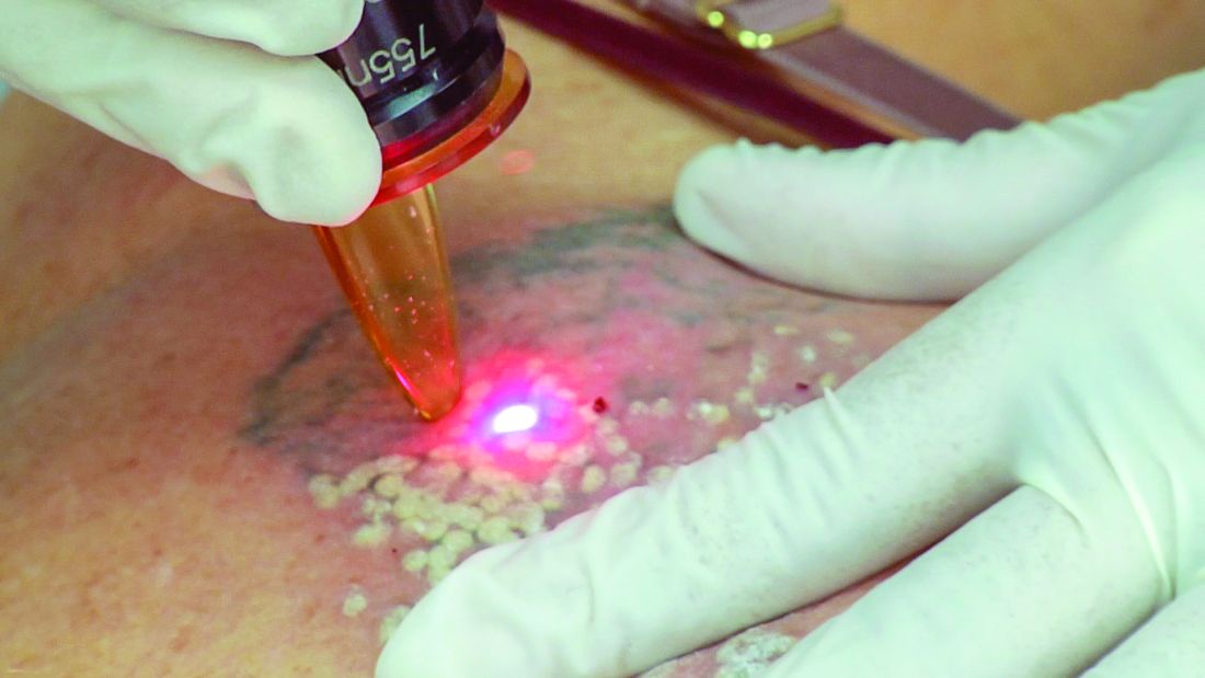
For optimal results, determine if the tattoo is professional, amateur, traumatic, or cosmetic. “That’s going to give you some insight as to what kind of expectations to set for the patient,” he said. “Black ink is often the easiest to remove, while certain colors like white are more challenging. Certain colors are more prone to paradoxical ink darkening, like red or orange, or pink. These can undergo a chemical reaction where they darken. This is something important to discuss with patients in advance.”
Older tattoos “tend to be less hearty” and usually respond better to laser, he continued. Location of the tattoo also plays a role. “I find that tattoos below the knee are very slow to respond. Smaller tattoos will respond faster.”
During the focused medical exam, ask patients about any history of keloid scarring, vitiligo or any dermatologic conditions with a Koebner phenomenon, and rule out a history of parental gold salt administration for arthritis. “During your informed consent you want to make sure you address the expected healing time and the risks such as hyper- and hypopigmentation, blistering, and scarring,” Dr. Ibrahimi said. “You also want to set the expectation that this is not going to be a one and done procedure. Laser tattoo removal takes a series of treatments, often more than what we think – sometimes in the range of 15-20. And you may not get complete clearance. I liken it to breaking it up enough so that if somebody sees it, they won’t be able to recognize what the tattoo is. But you won’t be able to erase it 100%.”
Black, dark blue, and red tattoo colors respond best to laser light. Light blue, green, and purple colors are slower to respond, while yellow and orange colors respond poorly. “Now that we have picosecond lasers, we’re a little better at treating these tougher colors, but I think we still have a lot of room for improvement,” Dr. Ibrahimi said.
Melanin is a competing chromophore, which complicates treatment of tanned individuals and those with darker skin types. “The Q-switched 1064-nm laser is the safest device to use for these patients but it’s not effective for many ink colors,” he said.
Options to keep patients comfortable during the procedure include application of ice or forced chilled air. “You can also use topical anesthetics such as EMLA or liposomal lidocaine cream under occlusion,” he said. “You can also use injectable lidocaine. If you go that route, I recommend a ring block. If you inject right into the tattoo sometimes the ink can get leeched out after treatment. As for spot size, a larger spot size will penetrate deeper, so I try to treat tattoos with the biggest spot size. It also results in less bleeding, less splatter, less side effects, and you get better results.”
Common adverse events from tattoo removal include prolonged erythema, blistering, hyperpigmentation, hypopigmentation, and scarring. Less frequent complications include ink darkening, chrysiasis, and transient immunoreactivity. “We don’t really know what’s in a lot of these ink residues,” Dr. Ibrahimi said. “We know they’re getting mobilized and some of it’s going into the lymphatics. What’s happening with these ink particles? We don’t fully know.”
He also warned against using hair-removal devices to treat a tattoo. “It is the wrong pulse duration,” he said. “You need a picosecond or nanosecond device. You cannot use any other pulse durations, or you will horribly scar your patient.”
In 2012, R. Rox Anderson, MD, director of the Wellman Center for Photomedicine at Massachusetts General Hospital, and colleagues published results of a study that compared a single Q-switched laser treatment pass with four treatment passes separated by 20 minutes. After treating 18 tattoos in 12 adults, they found that the technique, known as the R20 method, was more effective than a single-pass treatment (P < .01). “Subsequent papers have shown that this result isn’t as impressive as initially reported, but I think it’s a method that persists,” Dr. Ibrahimi said.
Another recent advance is use of a topical square silicone patch infused with perfluorodecalin patch during tattoo removal, which has been shown to reduce epidermal whitening. “So, instead of waiting 20 minutes you wait 0 minutes,” he said. “This is called the R0 method,” he added, noting that there are also some secondary benefits to using this patch, including possibly helping as an optical clearing agent for deeper penetration of the laser. “Often after treatment you can see ink on the underside of the patch, which speaks to the transdermal elimination mechanism of action for removal of tattoos.”
As for future directions, Dr. Ibrahimi predicted that there will be better picosecond lasers coming down the pike. He also anticipates that Soliton’s Rapid Acoustic Pulse (RAP) device will make a significant impact in the field. The device was cleared for tattoo removal in 2019 and is being investigated as an option to improve the appearance of cellulite. The manufacturer anticipates that an upgraded RAP device will be cleared for use by the end of the first quarter of 2021.
Dr. Ibrahimi disclosed that he has received research funding and speaker honorarium from Cutera, Lumenis, Lutronic, and Syneron-Candela. He also holds stock in Soliton.
According to a 2016 Harris Poll, 29% of Americans have at least one tattoo, up from 21% in 2012. At the same time, 23% of Americans polled in 2016 regret having their tattoo, which means big business for dermatologists who practice laser tattoo removal.
Prior to the theory of selective photothermolysis, tattoo removal mostly consisted of chemical or mechanical abrasion, surgical removal, or using some sort of caustic chemical or thermal destruction of the tattoo, Omar A. Ibrahimi, MD, PhD, said during a virtual course on laser and aesthetic skin therapy. “The earliest lasers prior to refinement by the theory of selective photothermolysis also fell into these categories: just basically crudely removing the skin and trying to get under to where the tattoo is,” said Dr. Ibrahimi, a dermatologist with the Connecticut Skin Institute in Stamford. “These would often heal with horrible scarring.”
Today, clinicians use Q-switched nanosecond and picosecond lasers for tattoo removal, though appropriate wavelengths need to be selected based on the tattoo ink color. Tattoo ink particles average about 0.1 mcm in size, and the thermal relaxation size works out to be about 10 nanoseconds. Black is the most common color dermatologists will treat. “For that, you can typically use a 1064, which has the highest absorption, but you can also use many of the other wavelengths,” he said. “The other colors are less common, followed by red, for which you would use a 532-nm wavelength.”
The clinical endpoint to strive for during tattoo removal is a whitening of the ink. That typically fades after about 20 minutes. “This whitening corresponds to cavitation [the production of gas vacuoles in the cells that were holding the ink],” Dr. Ibrahimi explained during the meeting, which was sponsored by Harvard Medical School, Massachusetts General Hospital, and the Wellman Center for Photomedicine. “These vacuoles are what lead to the whitening when using a high-gigawatt laser in a very short pulse. This causes highly localized heating, cavitation, and cell rupture. We don’t fully understand how tattoos are removed today, but the working models include some of the residual ink coming out through transepidermal elimination, some of it being removed via lymphatics, and some of it being removed by rephagocytosis.”
For optimal results, determine if the tattoo is professional, amateur, traumatic, or cosmetic. “That’s going to give you some insight as to what kind of expectations to set for the patient,” he said. “Black ink is often the easiest to remove, while certain colors like white are more challenging. Certain colors are more prone to paradoxical ink darkening, like red or orange, or pink. These can undergo a chemical reaction where they darken. This is something important to discuss with patients in advance.”
Older tattoos “tend to be less hearty” and usually respond better to laser, he continued. Location of the tattoo also plays a role. “I find that tattoos below the knee are very slow to respond. Smaller tattoos will respond faster.”
During the focused medical exam, ask patients about any history of keloid scarring, vitiligo or any dermatologic conditions with a Koebner phenomenon, and rule out a history of parental gold salt administration for arthritis. “During your informed consent you want to make sure you address the expected healing time and the risks such as hyper- and hypopigmentation, blistering, and scarring,” Dr. Ibrahimi said. “You also want to set the expectation that this is not going to be a one and done procedure. Laser tattoo removal takes a series of treatments, often more than what we think – sometimes in the range of 15-20. And you may not get complete clearance. I liken it to breaking it up enough so that if somebody sees it, they won’t be able to recognize what the tattoo is. But you won’t be able to erase it 100%.”
Black, dark blue, and red tattoo colors respond best to laser light. Light blue, green, and purple colors are slower to respond, while yellow and orange colors respond poorly. “Now that we have picosecond lasers, we’re a little better at treating these tougher colors, but I think we still have a lot of room for improvement,” Dr. Ibrahimi said.
Melanin is a competing chromophore, which complicates treatment of tanned individuals and those with darker skin types. “The Q-switched 1064-nm laser is the safest device to use for these patients but it’s not effective for many ink colors,” he said.
Options to keep patients comfortable during the procedure include application of ice or forced chilled air. “You can also use topical anesthetics such as EMLA or liposomal lidocaine cream under occlusion,” he said. “You can also use injectable lidocaine. If you go that route, I recommend a ring block. If you inject right into the tattoo sometimes the ink can get leeched out after treatment. As for spot size, a larger spot size will penetrate deeper, so I try to treat tattoos with the biggest spot size. It also results in less bleeding, less splatter, less side effects, and you get better results.”
Common adverse events from tattoo removal include prolonged erythema, blistering, hyperpigmentation, hypopigmentation, and scarring. Less frequent complications include ink darkening, chrysiasis, and transient immunoreactivity. “We don’t really know what’s in a lot of these ink residues,” Dr. Ibrahimi said. “We know they’re getting mobilized and some of it’s going into the lymphatics. What’s happening with these ink particles? We don’t fully know.”
He also warned against using hair-removal devices to treat a tattoo. “It is the wrong pulse duration,” he said. “You need a picosecond or nanosecond device. You cannot use any other pulse durations, or you will horribly scar your patient.”
In 2012, R. Rox Anderson, MD, director of the Wellman Center for Photomedicine at Massachusetts General Hospital, and colleagues published results of a study that compared a single Q-switched laser treatment pass with four treatment passes separated by 20 minutes. After treating 18 tattoos in 12 adults, they found that the technique, known as the R20 method, was more effective than a single-pass treatment (P < .01). “Subsequent papers have shown that this result isn’t as impressive as initially reported, but I think it’s a method that persists,” Dr. Ibrahimi said.
Another recent advance is use of a topical square silicone patch infused with perfluorodecalin patch during tattoo removal, which has been shown to reduce epidermal whitening. “So, instead of waiting 20 minutes you wait 0 minutes,” he said. “This is called the R0 method,” he added, noting that there are also some secondary benefits to using this patch, including possibly helping as an optical clearing agent for deeper penetration of the laser. “Often after treatment you can see ink on the underside of the patch, which speaks to the transdermal elimination mechanism of action for removal of tattoos.”
As for future directions, Dr. Ibrahimi predicted that there will be better picosecond lasers coming down the pike. He also anticipates that Soliton’s Rapid Acoustic Pulse (RAP) device will make a significant impact in the field. The device was cleared for tattoo removal in 2019 and is being investigated as an option to improve the appearance of cellulite. The manufacturer anticipates that an upgraded RAP device will be cleared for use by the end of the first quarter of 2021.
Dr. Ibrahimi disclosed that he has received research funding and speaker honorarium from Cutera, Lumenis, Lutronic, and Syneron-Candela. He also holds stock in Soliton.
FROM A LASER & AESTHETIC SKIN THERAPY COURSE
Diabetes prevention diet may lower mortality risk in breast cancer
Women who more closely followed a diabetes risk-reduction diet both before and after a diagnosis of breast cancer had lower risks for breast cancer–specific and all-cause mortality when compared with women with less healthy diets or those who did not substantially modify what they ate following diagnosis, according to pooled data from two prospective cohort studies.
Among more than 8,000 participants in the Nurses’ Health Study and NHS II, those who most closely adhered to a dietary pattern associated with lower risk for type 2 diabetes had a 13% lower risk for breast cancer–specific mortality and a 31% lower risk for death from any cause, compared with those at the bottom of the diabetes risk-reduction diet chart, reported Tengteng Wang, PhD, of the Harvard School of Public Health, Boston, and colleagues.
“Promoting dietary changes consistent with prevention of type 2 diabetes may be very important for breast cancer survivors,” Dr. Wang said in an oral abstract presentation at the 2020 San Antonio Breast Cancer Symposium.
Poor outcomes
Type 2 diabetes has been shown to be associated with poor outcomes for women with breast cancer, prompting the investigators to see whether diet modification could play a role in improving prognosis.
They looked at self-reported dietary data from 8,320 women diagnosed with stage I-III breast cancer who were participants in NHS, with data from 1980 to 2014, and NHS II, with data from 1991 to 2015.
Every 2-4 years, participants filled out validated follow-up questionnaires, including information on diet.
The investigators calculated a diabetes risk-reduction diet (DRRD) adherence score based on nine components, including higher intakes of cereal fiber, coffee, nuts, and whole fruits, as well as a higher polyunsatured to saturated fat ratio, and lower glycemic index, plus lower intakes of trans fats, sugar-sweetened beverages and/or fruit juices, and red meat.
The investigators calculated cumulative average DRRD scores based on repeated measures of diet after breast cancer diagnosis. They obtained data on deaths from family reports or the National Death Index, and they determined causes of death from either death certificates or medical records.
At a median follow-up of 13 years, 2,146 participants had died, with 948 of the deaths attributed to breast cancer.
After adjusting for socioeconomic factors, postdiagnosis time-varying covariates, and key breast cancer clinical factors, there was a nonsignificant trend toward a lower risk for breast cancer–specific deaths in the women in the highest versus lowest quintiles of DRRD score (hazard ratio, 0.87; P = .13), but significantly lower risk for all-cause mortality risk (HR, 0.69; P < .0001).
Looking at participants who changed their diet following breast cancer diagnosis, those who went from a low DRRD score prediagnosis to a high score post diagnosis had a 20% reduction in risk for breast cancer–specific mortality and a 14% reduction in risk for all-cause mortality, the investigators found (P values for this analysis were not shown).
There were no differences in results by either tumor estrogen receptor status or stage.
Dr. Wang acknowledged that the study was limited by the population (which was predominantly composed of educated, non-Hispanic White women), errors in dietary measurement, and limited power for estrogen receptor–negative tumor analysis.
Will patients do what’s good for them?
While this study adds to the body of evidence linking diet and cancer, putting the information into action is another story, according to Halle Moore, MD, of the Cleveland Clinic, who was not involved in this study.
“We have had supportive data for the role of diet in general health outcomes, including cancer-related outcomes, for a long time. But getting the public to implement these dietary changes is a challenge, so certainly the more convincing data that we have and the more specific we can be with specific types of dietary interventions, it does make it more helpful to counsel patients,” Dr. Moore said in an interview.
She said the finding that dietary change post diagnosis can have a significant effect on lowering both all-cause and breast cancer–specific mortality is compelling evidence for a role of diet in breast cancer outcomes.
In the question-and-answer session following Dr. Wang’s presentation, Hans-Christian Kolberg, MD, from Marienhospital Bottrop at the University of Duisburg-Essen (Germany), echoed the sentiment when he commented, “you have an important result that you did not mention in the conclusion: It is not too late to change diet after breast cancer diagnosis!”
This study was supported, in part, by grants from the National Cancer Institute, Breast Cancer Research Foundations, and Susan G. Komen Breast Cancer Foundations. Dr. Wang, Dr. Moore, and Dr. Kolberg reported no relevant conflicts of interest.
SOURCE: Wang T et al. SABCS 2020, Abstract GS2-09.
FROM SABCS 2020
Can a health care worker refuse the COVID-19 vaccine?
As hospitals across the country develop their plans to vaccinate their health care employees against COVID-19, a key question has come to the fore: What if an employee – whether nurse, physician, or other health care worker – refuses to receive the vaccine? Can hospitals require their employees to be vaccinated against COVID-19? And what consequences could an employee face for refusing the vaccine?
My answer needs to be based, in part, on the law related to previous vaccines – influenza, for example – because at the time of this writing (early December 2020), no vaccine for COVID-19 has been approved, although approval of at least one vaccine is expected within a week. So there have been no offers of vaccine and refusals yet, nor are there any cases to date involving an employee who refused a COVID-19 vaccine. As of December 2020, there are no state or federal laws that either require an employee to be vaccinated against COVID-19 or that protect an employee who refuses vaccination against COVID-19. It will take a while after the vaccine is approved and distributed before refusals, reactions, policies, cases, and laws begin to emerge.
If we look at the law related to health care workers refusing to be vaccinated against the closest relative to COVID-19 – influenza – then the answer would be yes, employers can require employees to be vaccinated.
An employer can fire an employee who refuses influenza vaccination. If an employee who refused and was fired sues the employer for wrongful termination, the employee has more or less chance of success depending on the reason for refusal. Some courts and the Equal Employment Opportunity Commission have held that a refusal on religious grounds is protected by the U.S. Constitution, as in this recent case. The Constitution protects freedom to practice one’s religion. Specific religions may have a range of tenets that support refusal to be vaccinated.
A refusal on medical grounds has been successful if the medical grounds fall under the protections of the Americans with Disabilities Act but may fail when the medical grounds for the claim are not covered by the ADA.
Refusal for secular, nonmedical reasons, such as a health care worker’s policy of treating their body as their temple, has not gone over well with employers or courts. However, in at least one case, a nurse who refused vaccination on secular, nonmedical grounds won her case against her employer, on appeal. The appeals court found that the hospital violated her First Amendment rights.
Employees who refuse vaccination for religious or medical reasons still will need to take measures to protect patients and other employees from infection. An employer such as a hospital can, rather than fire the employee, offer the employee an accommodation, such as requiring that the employee wear a mask or quarantine. There are no cases that have upheld an employee’s right to refuse to wear a mask or quarantine.
The situation with the COVID-19 vaccine is different from the situation surrounding influenza vaccines. There are plenty of data on effectiveness and side effects of influenza vaccines, but there is very little evidence of short- or long-term effects of the COVID-19 vaccines currently being tested and/or considered for approval. One could argue that the process of vaccine development is the same for all virus vaccines. However, public confidence in the vaccine vetting process is not what it once was. It has been widely publicized that the COVID-19 vaccine trials have been rushed. As of December 2020, only 60% of the general population say they would take the vaccine, although researchers say confidence is increasing.
The Centers for Disease Control and Prevention has designated health care workers as first in line to get the vaccine, but some health care workers may not want to be the first to try it. A CDC survey found that 63% of health care workers polled in recent months said they would get a COVID-19 vaccine.
Unions have entered the conversation. A coalition of unions that represent health care workers said, “we need a transparent, evidence-based federal vaccine strategy based on principles of equity, safety, and priority, as well as robust efforts to address a high degree of skepticism about safety of an authorized vaccine.” The organization declined to promote a vaccine until more is known.
As of publication date, the EEOC guidance for employers responding to COVID-19 does not address vaccines.
The CDC’s Interim Guidance for Businesses and Employers Responding to Coronavirus Disease 2019, May 2020, updated Dec. 4, 2020, does not address vaccines. The CDC’s page on COVID-19 vaccination for health care workers does not address a health care worker’s refusal. The site does assure health care workers that the vaccine development process is sound: “The current vaccine safety system is strong and robust, with the capacity to effectively monitor COVID-19 vaccine safety. Existing data systems have validated analytic methods that can rapidly detect statistical signals for possible vaccine safety problems. These systems are being scaled up to fully meet the needs of the nation. Additional systems and data sources are also being developed to further enhance safety monitoring capabilities. CDC is committed to ensuring that COVID-19 vaccines are safe.”
In the coming months, government officials and vaccine manufacturers will be working to reassure the public of the safety of the vaccine and the rigor of the vaccine development process. In November 2020, National Institute of Allergy and Infectious Diseases Director Anthony Fauci, MD, told Kaiser Health News: “The company looks at the data. I look at the data. Then the company puts the data to the FDA. The FDA will make the decision to do an emergency-use authorization or a license application approval. And they have career scientists who are really independent. They’re not beholden to anybody. Then there’s another independent group, the Vaccines and Related Biological Products Advisory Committee. The FDA commissioner has vowed publicly that he will go according to the opinion of the career scientists and the advisory board.” President-elect Joe Biden said he would get a vaccine when Dr. Fauci thinks it is safe.
An employee who, after researching the vaccine and the process, still wants to refuse when offered the vaccine is not likely to be fired for that reason right away, as long as the employee takes other precautions, such as wearing a mask. If the employer does fire the employee and the employee sues the employer, it is impossible to predict how a court would decide the case.
Related legal questions may arise in the coming months. For example:
- Is an employer exempt from paying workers’ compensation to an employee who refuses to be vaccinated and then contracts the virus while on the job?
- Can a prospective employer require COVID-19 vaccination as a precondition of employment?
- Is it within a patient’s rights to receive an answer to the question: Has my health care worker been vaccinated against COVID-19?
- If a hospital allows employees to refuse vaccination and keep working, and an outbreak occurs, and it is suggested through contact tracing that unvaccinated workers infected patients, will a court hold the hospital liable for patients’ damages?
Answers to these questions are yet to be determined.
Carolyn Buppert (www.buppert.com) is an attorney and former nurse practitioner who focuses on the legal issues affecting nurse practitioners.
A version of this article originally appeared on Medscape.com.
Natural history of adrenal incidentalomas with and without mild autonomous cortisol excess
Background: Studies have suggested that adrenal incidentalomas may increase risk of cardiometabolic disease in patients. Guidelines for repeat imaging and hormonal assessment of adrenal incidentalomas are inconsistent because of inadequate studies.

Study design: Systematic review and meta-analysis.
Setting: MEDLINE, EMBASE, Cochrane Central Register of Controlled Trials, Cochrane Database of Systematic Reviews, and Scopus were searched.
Synopsis: Of 1,139 studies screened; 32 met inclusion criteria: adult patients with adrenal adenoma who had 12 or more months of follow-up and outcomes of interest. Larger adrenal adenomas were less likely to have significant change in size on repeat imaging than did smaller tumors. There was no malignant transformation observed. Development of Cushing syndrome was seen in 6 of 2,745 patients. Cardiometabolic comorbid conditions were common in both MACE and NFAT patients with hypertension being the most frequently reported (64% and 58.2% respectively). Worsening of dyslipidemia was observed in both groups. Weight gain and the development of type 2 diabetes occurred more frequently in MACE than in NFAT patients (21.0% vs. 8.7%). In 1,356 patients, all-cause mortality was 11.2% (95% confidence interval, 9.5%-13.0%) for both groups over a mean follow-up of 56.3 months. Cardiovascular events accounted for 43.2% deaths. Limitations included the small number of patients in the studies assessed and the inconsistent definition of outcomes.
Bottom line: Patients with adrenal adenomas should be counseled on modifying cardiovascular risk factors whereas tumor growth, change in hormone production, and malignant transformation are less concerning based on the studies included.
Citation: Elhassan YS et al. Natural history of adrenal incidentalomas with and without mild autonomous cortisol excess: A systematic review and meta-analysis. Ann Intern Med. 2019 Jun 25;121:107-16.
Dr. Thompson is a hospitalist and assistant professor of medicine in the division of general internal medicine at Saint Louis University School of Medicine.
Background: Studies have suggested that adrenal incidentalomas may increase risk of cardiometabolic disease in patients. Guidelines for repeat imaging and hormonal assessment of adrenal incidentalomas are inconsistent because of inadequate studies.
Study design: Systematic review and meta-analysis.
Setting: MEDLINE, EMBASE, Cochrane Central Register of Controlled Trials, Cochrane Database of Systematic Reviews, and Scopus were searched.
Synopsis: Of 1,139 studies screened; 32 met inclusion criteria: adult patients with adrenal adenoma who had 12 or more months of follow-up and outcomes of interest. Larger adrenal adenomas were less likely to have significant change in size on repeat imaging than did smaller tumors. There was no malignant transformation observed. Development of Cushing syndrome was seen in 6 of 2,745 patients. Cardiometabolic comorbid conditions were common in both MACE and NFAT patients with hypertension being the most frequently reported (64% and 58.2% respectively). Worsening of dyslipidemia was observed in both groups. Weight gain and the development of type 2 diabetes occurred more frequently in MACE than in NFAT patients (21.0% vs. 8.7%). In 1,356 patients, all-cause mortality was 11.2% (95% confidence interval, 9.5%-13.0%) for both groups over a mean follow-up of 56.3 months. Cardiovascular events accounted for 43.2% deaths. Limitations included the small number of patients in the studies assessed and the inconsistent definition of outcomes.
Bottom line: Patients with adrenal adenomas should be counseled on modifying cardiovascular risk factors whereas tumor growth, change in hormone production, and malignant transformation are less concerning based on the studies included.
Citation: Elhassan YS et al. Natural history of adrenal incidentalomas with and without mild autonomous cortisol excess: A systematic review and meta-analysis. Ann Intern Med. 2019 Jun 25;121:107-16.
Dr. Thompson is a hospitalist and assistant professor of medicine in the division of general internal medicine at Saint Louis University School of Medicine.