User login
Vonoprazan promising for erosive esophagitis
, according to results of the phase 3 PHALCON-EE trial.
Vonoprazan achieved higher rates of healing and maintenance of healing than lansoprazole, with the benefit seen primarily in patients with more severe esophagitis.
The differences in healing rates were evident after 2 weeks of therapy and were maintained throughout the 24-week study, report Loren Laine, MD, Yale University, New Haven, Conn., and colleagues.
The study was published online in Gastroenterology.
More potent acid suppression
Gastroesophageal reflux disease is one of the most common disorders of the gastrointestinal tract, and erosive esophagitis is its most common complication.
Although standard PPI therapy is effective for healing erosive esophagitis, some patients do not achieve success with this conventional treatment.
Studies suggest that lack of healing of erosive esophagitis with 8 weeks of PPI therapy can be expected in roughly 5%-20% of patients, with rates up to 30% reported in patients with more severe esophagitis.
The PCAB vonoprazan provides more potent inhibition of gastric acid than PPIs and is seen as a potential alternative. However, data on its efficacy for erosive esophagitis are limited, the authors note.
The PHALCON-EE trial enrolled 1,024 adults from the United States and Europe with erosive esophagitis without Helicobacter pylori infection or Barrett esophagus.
Participants were randomized to receive once-daily vonoprazan 20 mg or lansoprazole 30 mg for up to 8 weeks in the healing phase.
The 878 patients with healing were then rerandomized to receive once-daily vonoprazan 10 mg, vonoprazan 20 mg, or lansoprazole 15 mg for 24 weeks in the maintenance phase.
For healing by week 8, vonoprazan was noninferior to lansoprazole in the primary analysis and superior to lansoprazole in a predefined exploratory analysis (92.9% vs. 84.6%; P < .0001).
Secondary analyses showed that vonoprazan was noninferior to lansoprazole in mean 24-hour heartburn-free days and superior in healing at week 2 for grade C/D esophagitis (70.2% vs. 52.6%; P = .0008).
For maintenance of healing at week 24, vonoprazan was noninferior to lansoprazole in the primary analysis and superior on secondary analysis of healing (80.7% for vonoprazan 20 mg and 79.2% for vonoprazan 10 mg vs. 72.0% for lansoprazole; P < .0001 for both comparisons).
The most common adverse event reported in the healing phase was diarrhea and in the maintenance phase was COVID-19. Two deaths occurred, both from COVID-19, during the maintenance phase in the vonoprazan 20-mg group.
As expected, serum gastrin increased to a greater extent with vonoprazan than lansoprazole, with levels > 500 pg/mL in 16% of those taking 20 mg at the end of maintenance therapy, the authors report. After stopping vonoprazan, gastrin levels dropped by roughly 60%-65% within 4 weeks.
Promising new option
“PCABs are a promising new option,” Avin Aggarwal, MD, who was not involved in the study, told this news organization.
They have a “more potent acid inhibitory effect” and have shown “superior healing of erosive esophagitis,” said Dr. Aggarwal, a gastroenterologist and medical director of Banner Health’s South Campus endoscopy services and clinical assistant professor at the University of Arizona in Tucson.
The results of the PHALCON-EE trial “validate noninferiority of PCABs compared to standard PPI therapy in the Western population after being proven in multiple Asian studies,” he said.
Dr. Aggarwal noted that PCABs work the same way as PPIs, by blocking the proton pumps, but “the longer half-life of PCABs and action on both active and inactive proton channels result in greater acid inhibition.”
Long-term effects of PCAB therapy from stronger acid inhibition and resulting hypergastrinemia still remain to be determined, he said.
Earlier this year, the U.S. Food and Drug Administration accepted Phathom Pharmaceuticals’ new drug application for vonoprazan for the treatment of erosive esophagitis.
Last May, the FDA approved two vonoprazan-based therapies for the treatment of H. pylori infection.
The study was funded by Phathom Pharmaceuticals. Dr. Laine and several coauthors have disclosed financial relationships with the company. Dr. Aggarwal reports no relevant financial relationships.
A version of this article first appeared on Medscape.com.
FROM GASTROENTEROLOGY
EHR alerts flag acute kidney injury and avert progression
ORLANDO – Automated alerts sent to clinicians via patients’ electronic health records identified patients with diagnosable acute kidney injury (AKI) who were taking one or more medications that could potentially further worsen their renal function. This led to a significant increase in discontinuations of the problematic drugs and better clinical outcomes in a subgroup of patients in a new multicenter, randomized study with more than 5,000 participants.
“Automated alerts for AKI can increase the rate of cessation of potentially nephrotoxic medications without endangering patients,” said F. Perry Wilson, MD, at Kidney Week 2022, organized by the American Society of Nephrology.
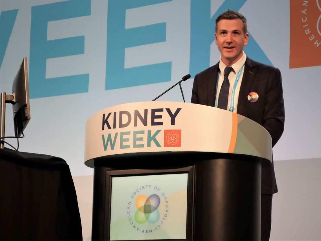
In addition, the study provides “limited evidence that these alerts change clinical practice,” said Dr. Wilson, a nephrologist and director of the clinical and translational research accelerator at Yale School of Medicine in New Haven, Conn.
“It was encouraging to get providers to change their behavior” by quickly stopping treatment with potentially nephrotoxic medications in patients with incident AKI. But the results also confirmed that “patient decision-support systems tend to not be panaceas,” Dr. Wilson explained in an interview. Instead, “they tend to marginally improve” patients’ clinical status.
“Our hope is that widespread use may make some difference on a population scale, but rarely are these game changers,” he admitted.
“This was a very nice study showing how we can leverage the EHR to look not only at drugs but also contrast agents to direct educational efforts aimed at clinicians about when to discontinue” these treatments, commented Karen A. Griffin, MD, who was not involved with the study.
A danger for alert fatigue
But the results also showed that more research is needed to better refine this approach, added Dr. Griffin, a professor at Loyola University Chicago, Maywood, Ill., and chief of the renal section at the Edward Hines Jr. VA Medical Center in Hines, Ill. And she expressed caution about expanding the alerts that clinicians receive “because of the potential for alert fatigue.”
Dr. Wilson also acknowledged the danger for alert fatigue. “We’re doing these studies to try to reduce the number of alerts,” he said. “Most clinicians say that if we could show an alert improves patient outcomes, they would embrace it.”
Dr. Wilson and associates designed their current study to evaluate an enhanced type of alert that not only warned clinicians that a patient had developed AKI but also gave them an option to potentially intervene by stopping treatment with a medication that could possibly exacerbate worsening renal function. This enhancement followed their experience in a 2021 study that tested a purely informational alert that gave physicians no guidance about what actions to take to more quickly resolve the AKI.
These findings plus results from other studies suggested that “purely informational alerts may not be enough. They need to be linked” to suggested changes in patient management, Dr. Wilson explained.
Targeting NSAIDS, RAAS inhibitors, and PPIs
The new study used automated EHR analysis to not only identify patients with incident AKI, but also to flag medications these patients were receiving from any of three classes suspected of worsening renal function: nonsteroidal anti-inflammatory drugs, renin-angiotensin-aldosterone system (RAAS) inhibitors (which include angiotensin-converting enzyme inhibitors and angiotensin receptor blockers), and proton-pump inhibitors (PPIs).
“Our hypothesis was that giving clinicians actionable advice could significantly improve patient outcomes,” Dr. Wilson said. “NSAIDs are frequently discontinued” in patients who develop AKI. “RAAS inhibitors are sometimes discontinued,” although the benefit from doing this remains unproven and controversial. “PPIs are rarely discontinued,” and may be an underappreciated contributor to AKI by causing interstitial nephritis in some patients.
The prospective study included 5,060 adults admitted with a diagnosis of stage 1 AKI at any of four Yale-affiliated teaching hospitals who were also taking agents from at least one of the three targeted drug classes at the time of admission. Clinicians caring for 2,532 of these patients received an alert about the AKI diagnosis and use of the questionable medications, while those caring for the 2,528 control patients received no alert and delivered usual care.
The study excluded patients with higher-risk profiles, including those with extremely elevated serum creatinine levels at admission (4.0 mg/dL or higher), those recently treated with dialysis, and patients with end-stage kidney disease.
The study had two primary outcomes. One measured the impact of the intervention on stopping the targeted drugs. The second assessed the clinical effect of the intervention on progression of AKI to a higher stage, need for dialysis, or death during either the duration of hospitalization or during the first 14 days following randomization.
Overall, a 9% relative increase in discontinuations
In general, the intervention had a modest but significant effect on cessation of the targeted drug classes within 24 hours of sending the alert.
Overall, there was about a 58% discontinuation rate among controls and about a 62% discontinuation rate among patients managed using the alerts, a significant 9% relative increase in any drug discontinuation, Dr. Wilson reported.
Discontinuations of NSAIDs occurred at the highest rate, in about 80% of patients in both groups, and the intervention showed no significant effect on stopping agents from this class. Discontinuations of RAAS inhibitors showed the largest absolute difference in between-group effect, about a 10–percentage point increase that represented a significant 14% relative increase in stopping agents from this class. Discontinuations of PPIs occurred at the lowest rate, in roughly 20% of patients, but the alert intervention had the greatest impact by raising the relative rate of stopping by a significant 26% compared with controls.
Analysis of the effect of the intervention on the combined clinical outcome showed a less robust impact. The alerts produced no significant change in the clinical outcome overall, or in the use of NSAIDs or RAAS inhibitors. However, the change in use of PPIs following the alerts significantly linked with a 12% relative drop in the incidence of the combined clinical endpoint of progression of AKI to a higher stage, need for dialysis, or death.
The results were consistent across several prespecified subgroups based on parameters such as age, sex, and race, but these analyses showed a signal that the alerts were most helpful for patients who had serum creatinine levels at admission of less than 0.5 mg/dL.
Dr. Wilson speculated that the alerts might have been especially effective for these patients because their low creatinine levels might otherwise mask AKI onset.
A safety analysis showed no evidence that the alert interventions and drug cessations increased the incidence of any complications.
PPIs may distinguish ‘sicker’ patients
Dr. Wilson cited two potential explanations for why the tested alerts appeared most effective for patients taking a PPI at the time of admission. One is that PPIs are underappreciated as a contributor to AKI, a possibility supported by the low rates of discontinuation in both the control and intervention groups.
In addition, treatment with a PPI may be a marker of “sicker” patients who may have more to gain from quicker identification of their AKI. For example, 28% of the patients who were taking a PPI at admission were in the ICU when they entered the study compared with a 14% rate of ICU care among everyone else.
PPIs were also the most-used targeted drug class among enrolled patients, used by 65% at baseline, compared with 53% who were taking a RAAS inhibitor and about 31% who were taking an NSAID. About 6% of enrolled patients were taking agents from all three classes at baseline, and 36% were on treatment with agents from two of the classes.
The next step is to assess adding more refinement to the alert process, Dr. Wilson said. He and his associates are now running a study in which an AKI alert goes to a “kidney action team” that includes a trained clinician and a pharmacist. The team would review the patient who triggered the alert and quickly make an individualized assessment of the best intervention for resolving the AKI.
The study received no commercial funding. Dr. Wilson has received research funding from AstraZeneca, Boehringer Ingelheim, Vifor, and Whoop. Dr. Griffin has reported no relevant financial relationships.
A version of this article first appeared on Medscape.com.
ORLANDO – Automated alerts sent to clinicians via patients’ electronic health records identified patients with diagnosable acute kidney injury (AKI) who were taking one or more medications that could potentially further worsen their renal function. This led to a significant increase in discontinuations of the problematic drugs and better clinical outcomes in a subgroup of patients in a new multicenter, randomized study with more than 5,000 participants.
“Automated alerts for AKI can increase the rate of cessation of potentially nephrotoxic medications without endangering patients,” said F. Perry Wilson, MD, at Kidney Week 2022, organized by the American Society of Nephrology.
In addition, the study provides “limited evidence that these alerts change clinical practice,” said Dr. Wilson, a nephrologist and director of the clinical and translational research accelerator at Yale School of Medicine in New Haven, Conn.
“It was encouraging to get providers to change their behavior” by quickly stopping treatment with potentially nephrotoxic medications in patients with incident AKI. But the results also confirmed that “patient decision-support systems tend to not be panaceas,” Dr. Wilson explained in an interview. Instead, “they tend to marginally improve” patients’ clinical status.
“Our hope is that widespread use may make some difference on a population scale, but rarely are these game changers,” he admitted.
“This was a very nice study showing how we can leverage the EHR to look not only at drugs but also contrast agents to direct educational efforts aimed at clinicians about when to discontinue” these treatments, commented Karen A. Griffin, MD, who was not involved with the study.
A danger for alert fatigue
But the results also showed that more research is needed to better refine this approach, added Dr. Griffin, a professor at Loyola University Chicago, Maywood, Ill., and chief of the renal section at the Edward Hines Jr. VA Medical Center in Hines, Ill. And she expressed caution about expanding the alerts that clinicians receive “because of the potential for alert fatigue.”
Dr. Wilson also acknowledged the danger for alert fatigue. “We’re doing these studies to try to reduce the number of alerts,” he said. “Most clinicians say that if we could show an alert improves patient outcomes, they would embrace it.”
Dr. Wilson and associates designed their current study to evaluate an enhanced type of alert that not only warned clinicians that a patient had developed AKI but also gave them an option to potentially intervene by stopping treatment with a medication that could possibly exacerbate worsening renal function. This enhancement followed their experience in a 2021 study that tested a purely informational alert that gave physicians no guidance about what actions to take to more quickly resolve the AKI.
These findings plus results from other studies suggested that “purely informational alerts may not be enough. They need to be linked” to suggested changes in patient management, Dr. Wilson explained.
Targeting NSAIDS, RAAS inhibitors, and PPIs
The new study used automated EHR analysis to not only identify patients with incident AKI, but also to flag medications these patients were receiving from any of three classes suspected of worsening renal function: nonsteroidal anti-inflammatory drugs, renin-angiotensin-aldosterone system (RAAS) inhibitors (which include angiotensin-converting enzyme inhibitors and angiotensin receptor blockers), and proton-pump inhibitors (PPIs).
“Our hypothesis was that giving clinicians actionable advice could significantly improve patient outcomes,” Dr. Wilson said. “NSAIDs are frequently discontinued” in patients who develop AKI. “RAAS inhibitors are sometimes discontinued,” although the benefit from doing this remains unproven and controversial. “PPIs are rarely discontinued,” and may be an underappreciated contributor to AKI by causing interstitial nephritis in some patients.
The prospective study included 5,060 adults admitted with a diagnosis of stage 1 AKI at any of four Yale-affiliated teaching hospitals who were also taking agents from at least one of the three targeted drug classes at the time of admission. Clinicians caring for 2,532 of these patients received an alert about the AKI diagnosis and use of the questionable medications, while those caring for the 2,528 control patients received no alert and delivered usual care.
The study excluded patients with higher-risk profiles, including those with extremely elevated serum creatinine levels at admission (4.0 mg/dL or higher), those recently treated with dialysis, and patients with end-stage kidney disease.
The study had two primary outcomes. One measured the impact of the intervention on stopping the targeted drugs. The second assessed the clinical effect of the intervention on progression of AKI to a higher stage, need for dialysis, or death during either the duration of hospitalization or during the first 14 days following randomization.
Overall, a 9% relative increase in discontinuations
In general, the intervention had a modest but significant effect on cessation of the targeted drug classes within 24 hours of sending the alert.
Overall, there was about a 58% discontinuation rate among controls and about a 62% discontinuation rate among patients managed using the alerts, a significant 9% relative increase in any drug discontinuation, Dr. Wilson reported.
Discontinuations of NSAIDs occurred at the highest rate, in about 80% of patients in both groups, and the intervention showed no significant effect on stopping agents from this class. Discontinuations of RAAS inhibitors showed the largest absolute difference in between-group effect, about a 10–percentage point increase that represented a significant 14% relative increase in stopping agents from this class. Discontinuations of PPIs occurred at the lowest rate, in roughly 20% of patients, but the alert intervention had the greatest impact by raising the relative rate of stopping by a significant 26% compared with controls.
Analysis of the effect of the intervention on the combined clinical outcome showed a less robust impact. The alerts produced no significant change in the clinical outcome overall, or in the use of NSAIDs or RAAS inhibitors. However, the change in use of PPIs following the alerts significantly linked with a 12% relative drop in the incidence of the combined clinical endpoint of progression of AKI to a higher stage, need for dialysis, or death.
The results were consistent across several prespecified subgroups based on parameters such as age, sex, and race, but these analyses showed a signal that the alerts were most helpful for patients who had serum creatinine levels at admission of less than 0.5 mg/dL.
Dr. Wilson speculated that the alerts might have been especially effective for these patients because their low creatinine levels might otherwise mask AKI onset.
A safety analysis showed no evidence that the alert interventions and drug cessations increased the incidence of any complications.
PPIs may distinguish ‘sicker’ patients
Dr. Wilson cited two potential explanations for why the tested alerts appeared most effective for patients taking a PPI at the time of admission. One is that PPIs are underappreciated as a contributor to AKI, a possibility supported by the low rates of discontinuation in both the control and intervention groups.
In addition, treatment with a PPI may be a marker of “sicker” patients who may have more to gain from quicker identification of their AKI. For example, 28% of the patients who were taking a PPI at admission were in the ICU when they entered the study compared with a 14% rate of ICU care among everyone else.
PPIs were also the most-used targeted drug class among enrolled patients, used by 65% at baseline, compared with 53% who were taking a RAAS inhibitor and about 31% who were taking an NSAID. About 6% of enrolled patients were taking agents from all three classes at baseline, and 36% were on treatment with agents from two of the classes.
The next step is to assess adding more refinement to the alert process, Dr. Wilson said. He and his associates are now running a study in which an AKI alert goes to a “kidney action team” that includes a trained clinician and a pharmacist. The team would review the patient who triggered the alert and quickly make an individualized assessment of the best intervention for resolving the AKI.
The study received no commercial funding. Dr. Wilson has received research funding from AstraZeneca, Boehringer Ingelheim, Vifor, and Whoop. Dr. Griffin has reported no relevant financial relationships.
A version of this article first appeared on Medscape.com.
AT KIDNEY WEEK 2022
Denosumab may halt erosive hand OA progression
But pain outcomes questionable
PHILADELPHIA – A double dose of the antiosteoporosis biologic denosumab (Prolia) slowed progression and repaired joints in erosive hand osteoarthritis (OA) but showed no impact on pain levels until 2 years after patients received the first dose, the lead investigator of a Belgium-based randomized clinical trial reported at the annual meeting of the American College of Rheumatology.
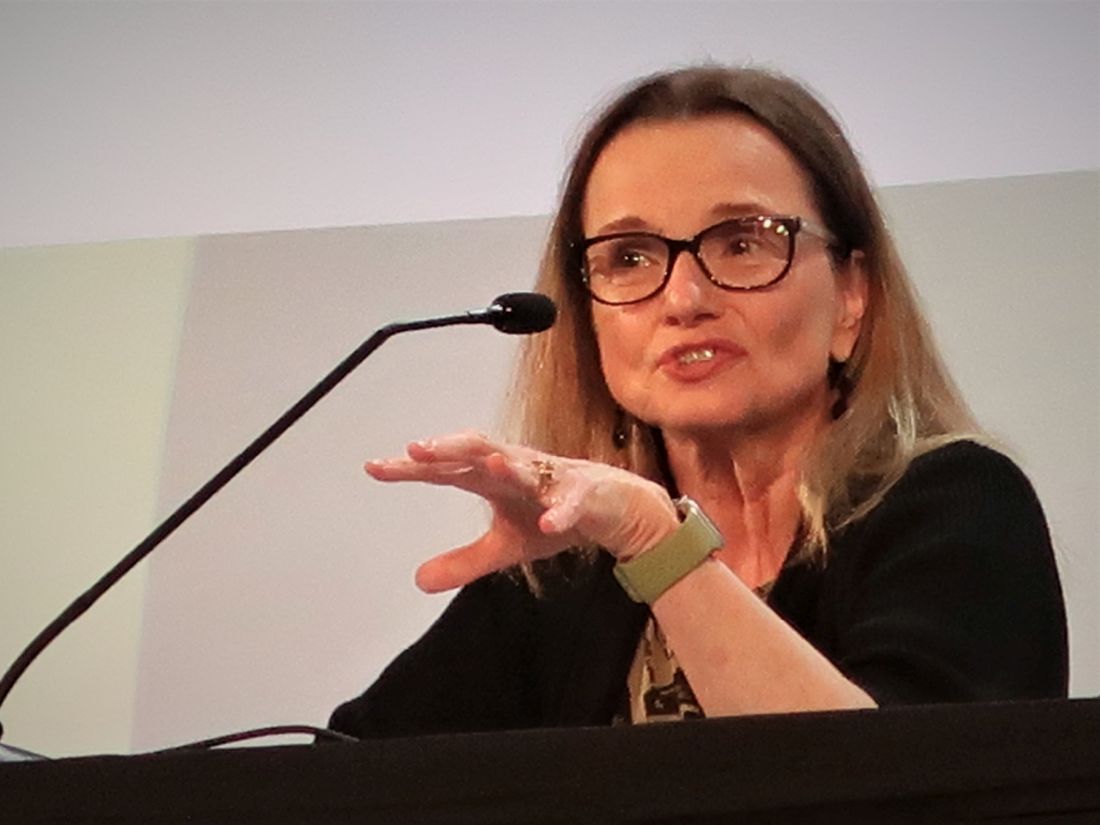
“This is the first placebo-controlled, randomized clinical trial showing the efficacy of denosumab double-dosing regimen in structural modification of erosive hand osteoarthritis,” Ruth Wittoek, MD, PhD, a rheumatologist at Ghent (Belgium) University, said in presenting the results.

“Our primary endpoint was confirmed by a more robust secondary endpoint, both showing that denosumab stopped erosive progression and induced remodeling in patients with erosive hand OA,” she added. “Moreover, the double-dosing regimen was well-tolerated.”
However, during the question-and-answer period after her presentation, Dr. Wittoek acknowledged the study didn’t evaluate the impact denosumab had on cartilage and didn’t detect a signal for pain resolution until 96 weeks during the open-label extension phase. “I’m not quite sure if denosumab is sufficient to treat symptoms in osteoarthritis,” she said. “There were positive signals but, of course, having to wait 2 years for an effect is kind of hard for our patients.”
The trial randomized 100 adult patients 1:1 to denosumab 60 mg every 12 weeks – double the normal dose for osteoporosis – or placebo. The primary endpoint was changes in erosive progression and signs of repair based on x-ray at 48 weeks, after which all patients were switched to denosumab for the open-label study. To quantify changes, the investigators used the Ghent University Scoring System (GUSS), which uses a scale of 0-300 to quantify radiographic changes in erosive hand OA.
Dr. Wittoek said that the average change in GUSS at week 24 was +6 vs. –2.8 (P = .024) in the treatment and placebo groups, respectively, widening at week 48 to +10.1 and –7.9 (P = .003). By week 96, the variation was +18.8 for denosumab and +17 for placebo with switch to denosumab (P = .03).
“During the open-label extension the denosumab treatment group continued to increase to show remodeling while the former placebo treatment group, now also receiving denosumab, also showed signs of remodeling,” she said. “So, there was no more erosive progression.”
The secondary endpoint was the percentage of new erosive joint development at week 48: 1.8% in the denosumab group and 7% in placebo group (odds ratio, 0.23; 95% confidence interval, 0.10-0.50; P < .001). “Meaning the odds of erosive progression is 77% lower in the denosumab treatment group,” Dr. Wittoek said.
By week 96, those percentages were 0% and 0.7% in the respective treatment groups. “During the open-label extension, it was clear that denosumab blocked all new development of erosive joints,” she said.
Pain was one of the study’s exploratory endpoints, and the mean numeric rating scale showed no difference between treatment arms until the 96-week results, with a reduction by almost half in the denosumab group (from 4.2 at week 48 to 2.4) and a lesser reduction in the placebo-switched-to-denosumab arm (from 4.2 to 3.5; P = .028) between arms.
The placebo group was more susceptible to adverse events, namely musculoskeletal complaints and nervous system disorders, Dr. Wittoek noted. Infection rates, the most common adverse event, were similar between the two groups: 41 and 39 in the respective arms. Despite the double dose of denosumab, safety and tolerability in this trial was comparable to other trials, she said.
In comments submitted by e-mail, Dr. Wittoek noted that the extension study results will go out to 144 weeks. She also addressed the issues surrounding pain as an outcome.
“Besides disability, pain is also important from the patient’s perspective,” Dr. Wittoek said in the e-mailed comments. “However, pain and radiographic progression are undeniably coupled, but it’s unclear how.”
In erosive hand OA, structural progression and pain may not be related on a molecular level, she said. “Therefore, we don’t deny that pain levels should also be covered by treatment, but they should not be confused with structural modification; it is just another domain, not more nor less important.
The second year of the open-label extension study should clarify the pain outcomes, she said.
In an interview, David T. Felson, MD, MPH, professor and director of clinical epidemiology research at Boston University, questioned the delayed pain effect the study suggested. “It didn’t make any sense to me that there would be because both groups at that point got denosumab, so if there was going to be a pain effect that would’ve happened,” he said.
The pain effect is “really important,” he said. “We don’t use denosumab in rheumatoid arthritis to treat erosions because it doesn’t necessarily affect the pain and dysfunction of rheumatoid arthritis, and I’m not sure that isn’t going to be true in erosive hand osteoarthritis, but it’s possible.”
To clarify the pain outcomes, he said, “They’re going to have to work on the data.”
Amgen sponsored the trial but had no role in the design. Dr. Wittoek and Dr. Felson reported no relevant disclosures.
But pain outcomes questionable
But pain outcomes questionable
PHILADELPHIA – A double dose of the antiosteoporosis biologic denosumab (Prolia) slowed progression and repaired joints in erosive hand osteoarthritis (OA) but showed no impact on pain levels until 2 years after patients received the first dose, the lead investigator of a Belgium-based randomized clinical trial reported at the annual meeting of the American College of Rheumatology.
“This is the first placebo-controlled, randomized clinical trial showing the efficacy of denosumab double-dosing regimen in structural modification of erosive hand osteoarthritis,” Ruth Wittoek, MD, PhD, a rheumatologist at Ghent (Belgium) University, said in presenting the results.
“Our primary endpoint was confirmed by a more robust secondary endpoint, both showing that denosumab stopped erosive progression and induced remodeling in patients with erosive hand OA,” she added. “Moreover, the double-dosing regimen was well-tolerated.”
However, during the question-and-answer period after her presentation, Dr. Wittoek acknowledged the study didn’t evaluate the impact denosumab had on cartilage and didn’t detect a signal for pain resolution until 96 weeks during the open-label extension phase. “I’m not quite sure if denosumab is sufficient to treat symptoms in osteoarthritis,” she said. “There were positive signals but, of course, having to wait 2 years for an effect is kind of hard for our patients.”
The trial randomized 100 adult patients 1:1 to denosumab 60 mg every 12 weeks – double the normal dose for osteoporosis – or placebo. The primary endpoint was changes in erosive progression and signs of repair based on x-ray at 48 weeks, after which all patients were switched to denosumab for the open-label study. To quantify changes, the investigators used the Ghent University Scoring System (GUSS), which uses a scale of 0-300 to quantify radiographic changes in erosive hand OA.
Dr. Wittoek said that the average change in GUSS at week 24 was +6 vs. –2.8 (P = .024) in the treatment and placebo groups, respectively, widening at week 48 to +10.1 and –7.9 (P = .003). By week 96, the variation was +18.8 for denosumab and +17 for placebo with switch to denosumab (P = .03).
“During the open-label extension the denosumab treatment group continued to increase to show remodeling while the former placebo treatment group, now also receiving denosumab, also showed signs of remodeling,” she said. “So, there was no more erosive progression.”
The secondary endpoint was the percentage of new erosive joint development at week 48: 1.8% in the denosumab group and 7% in placebo group (odds ratio, 0.23; 95% confidence interval, 0.10-0.50; P < .001). “Meaning the odds of erosive progression is 77% lower in the denosumab treatment group,” Dr. Wittoek said.
By week 96, those percentages were 0% and 0.7% in the respective treatment groups. “During the open-label extension, it was clear that denosumab blocked all new development of erosive joints,” she said.
Pain was one of the study’s exploratory endpoints, and the mean numeric rating scale showed no difference between treatment arms until the 96-week results, with a reduction by almost half in the denosumab group (from 4.2 at week 48 to 2.4) and a lesser reduction in the placebo-switched-to-denosumab arm (from 4.2 to 3.5; P = .028) between arms.
The placebo group was more susceptible to adverse events, namely musculoskeletal complaints and nervous system disorders, Dr. Wittoek noted. Infection rates, the most common adverse event, were similar between the two groups: 41 and 39 in the respective arms. Despite the double dose of denosumab, safety and tolerability in this trial was comparable to other trials, she said.
In comments submitted by e-mail, Dr. Wittoek noted that the extension study results will go out to 144 weeks. She also addressed the issues surrounding pain as an outcome.
“Besides disability, pain is also important from the patient’s perspective,” Dr. Wittoek said in the e-mailed comments. “However, pain and radiographic progression are undeniably coupled, but it’s unclear how.”
In erosive hand OA, structural progression and pain may not be related on a molecular level, she said. “Therefore, we don’t deny that pain levels should also be covered by treatment, but they should not be confused with structural modification; it is just another domain, not more nor less important.
The second year of the open-label extension study should clarify the pain outcomes, she said.
In an interview, David T. Felson, MD, MPH, professor and director of clinical epidemiology research at Boston University, questioned the delayed pain effect the study suggested. “It didn’t make any sense to me that there would be because both groups at that point got denosumab, so if there was going to be a pain effect that would’ve happened,” he said.
The pain effect is “really important,” he said. “We don’t use denosumab in rheumatoid arthritis to treat erosions because it doesn’t necessarily affect the pain and dysfunction of rheumatoid arthritis, and I’m not sure that isn’t going to be true in erosive hand osteoarthritis, but it’s possible.”
To clarify the pain outcomes, he said, “They’re going to have to work on the data.”
Amgen sponsored the trial but had no role in the design. Dr. Wittoek and Dr. Felson reported no relevant disclosures.
AT ACR 2022
A plane crash interrupts a doctor’s vacation
Emergencies happen anywhere, anytime – and sometimes physicians find themselves in situations where they are the only ones who can help. “Is There a Doctor in the House?” is a new series telling these stories.
When the plane crashed, I was asleep. I had arrived the evening before with my wife and three sons at a house on Kezar Lake on the Maine–New Hampshire border. I jumped out of bed and ran downstairs. My kids had been watching a float plane circling and gliding along the lake. It had crashed into the water and flipped upside down. My oldest brother-in-law jumped into his ski boat and we sped out to the scene.
All we can see are the plane’s pontoons. The rest is underwater. A woman has already surfaced, screaming. I dive in.
I find the woman’s husband and 3-year-old son struggling to get free from the plane through the smashed windshield. They manage to get to the surface. The pilot is dead, impaled through the chest by the left wing strut.
The big problem: A little girl, whom I would learn later is named Lauren, remained trapped. The water is murky but I can see her, a 5- or 6-year-old girl with this long hair, strapped in upside down and unconscious.
The mom and I dive down over and over, pulling and ripping at the door. We cannot get it open. Finally, I’m able to bend the door open enough where I can reach in, but I can’t undo the seatbelt. In my mind, I’m debating, should I try and go through the front windshield? I’m getting really tired, I can tell there’s fuel in the water, and I don’t want to drown in the plane. So I pop up to the surface and yell, “Does anyone have a knife?”
My brother-in-law shoots back to shore in the boat, screaming, “Get a knife!” My niece gets in the boat with one. I’m standing on the pontoon, and my niece is in the front of the boat calling, “Uncle Todd! Uncle Todd!” and she throws the knife. It goes way over my head. I can’t even jump for it, it’s so high.
I have to get the knife. So, I dive into the water to try and find it. Somehow, the black knife has landed on the white wing, 4 or 5 feet under the water. Pure luck. It could have sunk down a hundred feet into the lake. I grab the knife and hand it to the mom, Beth. She’s able to cut the seatbelt, and we both pull Lauren to the surface.
I lay her out on the pontoon. She has no pulse and her pupils are fixed and dilated. Her mom is yelling, “She’s dead, isn’t she?” I start CPR. My skin and eyes are burning from the airplane fuel in the water. I get her breathing, and her heart comes back very quickly. Lauren starts to vomit and I’m trying to keep her airway clear. She’s breathing spontaneously and she has a pulse, so I decide it’s time to move her to shore.
We pull the boat up to the dock and Lauren’s now having anoxic seizures. Her brain has been without oxygen, and now she’s getting perfused again. We get her to shore and lay her on the lawn. I’m still doing mouth-to-mouth, but she’s seizing like crazy, and I don’t have any way to control that. Beth is crying and wants to hold her daughter gently while I’m working.
Someone had called 911, and finally this dude shows up with an ambulance, and it’s like something out of World War II. All he has is an oxygen tank, but the mask is old and cracked. It’s too big for Lauren, but it sort of fits me, so I’m sucking in oxygen and blowing it into the girl’s mouth. I’m doing whatever I can, but I don’t have an IV to start. I have no fluids. I got nothing.
As it happens, I’d done my emergency medicine training at Maine Medical Center, so I tell someone to call them and get a Life Flight chopper. We have to drive somewhere where the chopper can land, so we take the ambulance to the parking lot of the closest store called the Wicked Good Store. That’s a common thing in Maine. Everything is “wicked good.”
The whole town is there by that point. The chopper arrives. The ambulance doors pop open and a woman says, “Todd?” And I say, “Heather?”
Heather is an emergency flight nurse whom I’d trained with many years ago. There’s immediate trust. She has all the right equipment. We put in breathing tubes and IVs. We stop Lauren from seizing. The kid is soon stable.
There is only one extra seat in the chopper, so I tell Beth to go. They take off.
Suddenly, I begin to doubt my decision. Lauren had been underwater for 15 minutes at minimum. I know how long that is. Did I do the right thing? Did I resuscitate a brain-dead child? I didn’t think about it at the time, but if that patient had come to me in the emergency department, I’m honestly not sure what I would have done.
So, I go home. And I don’t get a call. The FAA and sheriff arrive to take statements from us. I don’t hear from anyone.
The next day I start calling. No one will tell me anything, so I finally get to one of the pediatric ICU attendings who had trained me. He says Lauren literally woke up and said, “I have to go pee.” And that was it. She was 100% normal. I couldn’t believe it.
Here’s a theory: In kids, there’s something called the glottic reflex. I think her glottic reflex went off as soon as she hit the water, which basically closed her airway. So when she passed out, she could never get enough water in her lungs and still had enough air in there to keep her alive. Later, I got a call from her uncle. He could barely get the words out because he was in tears. He said Lauren was doing beautifully.
Three days later, I drove to Lauren’s house with my wife and kids. I had her read to me. I watched her play on the jungle gym for motor function. All sorts of stuff. She was totally normal.
Beth told us that the night before the accident, her mother had given the women in her family what she called a “miracle bracelet,” a bracelet that is supposed to give you one miracle in your life. Beth said she had the bracelet on her wrist the day of the accident, and now it’s gone. “Saving Lauren’s life was my miracle,” she said.
Funny thing: For 20 years, I ran all the EMS, police, fire, ambulance, in Boulder, Colo., where I live. I wrote all the protocols, and I would never advise any of my paramedics to dive into jet fuel to save someone. That was risky. But at the time, it was totally automatic. I think it taught me not to give up in certain situations, because you really don’t know.
Dr. Dorfman is an emergency medicine physician in Boulder, Colo., and medical director at Cedalion Health.
A version of this article first appeared on Medscape.com.
The tale of two scenarios of gender dysphoria
In a recent column, I cautiously discussed what has been called gender-affirming or transgender care.
In the days following the appearance of that Letters From Maine column on this topic, I received an unusual number of responses from readers suggesting I had touched on a topic that was on the minds of many pediatricians.

Since then, the Florida Board of Medicine and Osteopathic Medicine voted to forbid physicians from prescribing puberty blockers and hormones and/or performing surgeries in patients under age 18 who were seeking transgender care. Children already receiving treatments were exempt from the ruling. The osteopathic board added a second exception in cases where the child was a participant in a research protocol. The board of medicine inexplicably did not include this exception.
Regardless of how one feels about the ethics and the appropriateness of transgender care, it is not an issue to be decided by a politically appointed entity.
As I look back over what I have learned by watching this tragic drama play out, I am struck by a distinction that has yet to receive enough attention. When we are discussing gender dysphoria we are really talking about two different pediatric populations and scenarios. There is the child who from a very young age has consistently preferred to dress and behave in a manner that is different from the gender he or she was assigned at birth. The management of this child is a challenge that requires a careful balance of support and protection from the harsh realities of the gender-regimented world.
The second scenario stars the adolescent who has no prior history of gender dysphoria, or at least no outward manifestations. Then, faced by the challenges of puberty and adolescence, something or things happen that erupt into a full-blown gender-dysphoric storm. We currently have very little understanding of what those “things” are.
Each population can probably be further divided into subgroups – and that’s just the point. Every gender-dysphoric child, whether their dysphoria began at age 2 or 12, is an individual with a unique family dynamic and socioeconomic background. They may share some as yet unknown genetic signature, but in our current state of ignorance they deserve, as do all of our patients, to be treated as individuals by their primary care physicians and consultants who must at first do no harm. One size does not fit all and certainly their care should not be dictated by a politically influenced entity.
Dr. Wilkoff practiced primary care pediatrics in Brunswick, Maine, for nearly 40 years. He has authored several books on behavioral pediatrics, including “How to Say No to Your Toddler.” Other than a Littman stethoscope he accepted as a first-year medical student in 1966, Dr. Wilkoff reports having nothing to disclose. Email him at [email protected].
In a recent column, I cautiously discussed what has been called gender-affirming or transgender care.
In the days following the appearance of that Letters From Maine column on this topic, I received an unusual number of responses from readers suggesting I had touched on a topic that was on the minds of many pediatricians.
Since then, the Florida Board of Medicine and Osteopathic Medicine voted to forbid physicians from prescribing puberty blockers and hormones and/or performing surgeries in patients under age 18 who were seeking transgender care. Children already receiving treatments were exempt from the ruling. The osteopathic board added a second exception in cases where the child was a participant in a research protocol. The board of medicine inexplicably did not include this exception.
Regardless of how one feels about the ethics and the appropriateness of transgender care, it is not an issue to be decided by a politically appointed entity.
As I look back over what I have learned by watching this tragic drama play out, I am struck by a distinction that has yet to receive enough attention. When we are discussing gender dysphoria we are really talking about two different pediatric populations and scenarios. There is the child who from a very young age has consistently preferred to dress and behave in a manner that is different from the gender he or she was assigned at birth. The management of this child is a challenge that requires a careful balance of support and protection from the harsh realities of the gender-regimented world.
The second scenario stars the adolescent who has no prior history of gender dysphoria, or at least no outward manifestations. Then, faced by the challenges of puberty and adolescence, something or things happen that erupt into a full-blown gender-dysphoric storm. We currently have very little understanding of what those “things” are.
Each population can probably be further divided into subgroups – and that’s just the point. Every gender-dysphoric child, whether their dysphoria began at age 2 or 12, is an individual with a unique family dynamic and socioeconomic background. They may share some as yet unknown genetic signature, but in our current state of ignorance they deserve, as do all of our patients, to be treated as individuals by their primary care physicians and consultants who must at first do no harm. One size does not fit all and certainly their care should not be dictated by a politically influenced entity.
Dr. Wilkoff practiced primary care pediatrics in Brunswick, Maine, for nearly 40 years. He has authored several books on behavioral pediatrics, including “How to Say No to Your Toddler.” Other than a Littman stethoscope he accepted as a first-year medical student in 1966, Dr. Wilkoff reports having nothing to disclose. Email him at [email protected].
Randomized trial finds community-based weight-loss programs ease knee OA pain
PHILADELPHIA – What works in the clinic can also work in community settings: Patients who are overweight or obese with knee osteoarthritis can find relief from pain through diet and exercise programs conducted in recreation centers, local gyms, fitness centers, and other places close to home, according to investigators in a pragmatic randomized trial.
The Weight Loss and Exercise for Communities With Arthritis in North Carolina (WE-CAN) study was modeled after the successful Intensive Diet and Exercise for Arthritis trial, which showed that adults randomized to 18 months of either a diet and exercise program or diet alone had more weight loss and larger reductions in levels of the inflammatory cytokine interleukin-6 than patients randomized to exercise alone, and that diet alone was associated with greater reductions in knee compressive force than exercise alone.

That study was conducted by Stephen P. Messier, PhD, and colleagues at Wake Forest University, Winston-Salem, N.C.. As previously reported, the investigators also saw continued benefits for participants years after the original trial.
With the WE-CAN trial, results of which were reported at the annual meeting of the American College of Rheumatology, Dr. Messier and colleagues took the intervention one step further, randomizing 823 community-dwelling adults who were overweight or obese (body mass index [BMI], ≥ 27 kg/m2) with knee OA to either an 18-month diet and exercise intervention or attention control group consisting of five 1-hour face-to-face meetings over 18 months, plus information packets and phone sessions during alternate months.
“Compared to the control group, diet plus exercise had a statistically significant but modest reduction in pain. Diet plus exercise was 20% more likely to attain a clinically important 2-point improvement in pain,” Dr. Messier said in an oral abstract session at ACR.
Real-world setting
The primary goal of WE-CAN was to “determine whether adaptation of a diet and exercise academic center–based efficacy trial to community settings results in a statistically significant reduction in pain relative to an attention control.”

A total of 3,751 potential candidates were screened, and 823 were randomized and assigned to either a diet and exercise arm (414) or attention control arm (409). Of the patients randomized, 336 in the diet/exercise arm and 322 in the control arm attended the final 18-month follow-up visit.
The exercise component consisted of a 15-minute walking period, followed by a 20-minute weight-training period, and ending with a second 15-minute walking period. The diet goal was 10% or greater weight loss, aided by a distribution of low-calorie recipes to produce a reduced-calorie diet of the patient’s choice, with the option to include nutritional powder to make low-calories shakes as meal replacements, one or two per day for the first 6 months, with the option of one per day for the remaining months.
The pragmatic components included the use of established community facilities in both urban and rural counties in North Carolina, broad inclusion criteria, patient-centered outcomes, use of community-based staff to deliver the treatment, nonphysicians trained by study physicians to perform knee exams, and various means of communication, Dr. Messier said.
Participants in each arm were closely matched by demographic and clinical characteristics, with a mean age of 64.5 years in the diet/exercise group and 64.7 years in the attention control group, respective mean weight of 100.7 kg and 101.1 kg, and respective BMI of 36.7 and 36.9. Women comprised about 77% of participants in each group.
Endpoints met
The trial met its primary endpoint of a significantly greater reduction in pain at 18 months in the diet and exercise group as measured by the Western Ontario and McMaster Universities Osteoarthritis Index (WOMAC) and scored on a scale of 0 (no pain) to 20 (worst pain).
In an analysis adjusted for sex, BMI, and baseline values, there was a 32% reduction in pain scores from baseline in the active intervention arm versus 24% in the control arm (P = .02).
In all, 60.2% of participants assigned to diet and exercise had a minimum reduction in pain scores of at least 2 points at 18 months, compared with 49.7% of participants assigned to the attention control group. This translated into a relative risk for achieving at least a 2-point improvement with diet and exercise was 1.20 (P = .01).
Among participants who remained in the study for the entire 18 months, there were significant improvements in the diet and exercise group compared with controls in the prespecified secondary endpoints of weight change (–8 kg vs. –2 kg), waist circumference, WOMAC function, 6-minute walk distance, and mean Short Form–36 health-related quality of life subscale (P < .001 for all comparisons).
Dr. Messier acknowledged that the diagnosis of knee OA was based only on ACR clinical criteria and was not confirmed with imaging. In addition, offering patients the option of free meal replacement limited the pragmatic nature of the intervention.
He also noted that the 24% reduction in pain seen in the control group suggests that interacting with patients can improve clinical outcomes.
‘Tour de force’
In the question-and-answer session following Dr. Messier’s presentation, David T. Felson, MD, a rheumatologist at Boston Medical Center, called in and said the study was “a tour de force” and congratulated Dr. Messier and colleagues on “a lovely study.”
Dr. Felson asked whether the investigators had conducted a mediation analysis to determine what proportion of the improvement was attributable to weight loss, and whether patients assigned to exercise were sticking with it throughout the study.
Dr. Messier replied that they had not yet done a mediation analysis but were continuing to examine the data. Regarding the exercise question, he noted that “the adherence was over 80% for 6 months and over 70% for the whole 18 months, so they did a really nice job.”
In an interview, session moderator Anne Davidson, MBBS, director of the rheumatology program at Northwell Health in Manhasset, N.Y., commented that the investigators managed to accomplish a very challenging task.
“In terms of recruitment of patients with engagement of community facilities and quality of data, I would say that, as far as an osteoarthritis study goes, this was really a tremendous effort on the part of all people involved,” she said.
She noted that, while the WE-CAN program may work in North Carolina, there may be barriers to implementing it elsewhere, such as large suburban areas where some patients experience food insecurity and others have difficulty with transportation and access to treatment facilities.
“The question here that remains is, as Dr. Felson asked, what is the contribution of weight loss and what is the contribution of exercise? Because if it’s just weight loss, we have a whole lot of new things coming to help with that,” she said.
The WE-CAN study was supported by the National Institute of Arthritis and Musculoskeletal and Skin Diseases. Dr. Messier disclosed that GNC, a health food and nutrition chain, donated the meal replacements used by patients. Dr. Davidson reported no relevant conflicts of interest.
PHILADELPHIA – What works in the clinic can also work in community settings: Patients who are overweight or obese with knee osteoarthritis can find relief from pain through diet and exercise programs conducted in recreation centers, local gyms, fitness centers, and other places close to home, according to investigators in a pragmatic randomized trial.
The Weight Loss and Exercise for Communities With Arthritis in North Carolina (WE-CAN) study was modeled after the successful Intensive Diet and Exercise for Arthritis trial, which showed that adults randomized to 18 months of either a diet and exercise program or diet alone had more weight loss and larger reductions in levels of the inflammatory cytokine interleukin-6 than patients randomized to exercise alone, and that diet alone was associated with greater reductions in knee compressive force than exercise alone.
That study was conducted by Stephen P. Messier, PhD, and colleagues at Wake Forest University, Winston-Salem, N.C.. As previously reported, the investigators also saw continued benefits for participants years after the original trial.
With the WE-CAN trial, results of which were reported at the annual meeting of the American College of Rheumatology, Dr. Messier and colleagues took the intervention one step further, randomizing 823 community-dwelling adults who were overweight or obese (body mass index [BMI], ≥ 27 kg/m2) with knee OA to either an 18-month diet and exercise intervention or attention control group consisting of five 1-hour face-to-face meetings over 18 months, plus information packets and phone sessions during alternate months.
“Compared to the control group, diet plus exercise had a statistically significant but modest reduction in pain. Diet plus exercise was 20% more likely to attain a clinically important 2-point improvement in pain,” Dr. Messier said in an oral abstract session at ACR.
Real-world setting
The primary goal of WE-CAN was to “determine whether adaptation of a diet and exercise academic center–based efficacy trial to community settings results in a statistically significant reduction in pain relative to an attention control.”
A total of 3,751 potential candidates were screened, and 823 were randomized and assigned to either a diet and exercise arm (414) or attention control arm (409). Of the patients randomized, 336 in the diet/exercise arm and 322 in the control arm attended the final 18-month follow-up visit.
The exercise component consisted of a 15-minute walking period, followed by a 20-minute weight-training period, and ending with a second 15-minute walking period. The diet goal was 10% or greater weight loss, aided by a distribution of low-calorie recipes to produce a reduced-calorie diet of the patient’s choice, with the option to include nutritional powder to make low-calories shakes as meal replacements, one or two per day for the first 6 months, with the option of one per day for the remaining months.
The pragmatic components included the use of established community facilities in both urban and rural counties in North Carolina, broad inclusion criteria, patient-centered outcomes, use of community-based staff to deliver the treatment, nonphysicians trained by study physicians to perform knee exams, and various means of communication, Dr. Messier said.
Participants in each arm were closely matched by demographic and clinical characteristics, with a mean age of 64.5 years in the diet/exercise group and 64.7 years in the attention control group, respective mean weight of 100.7 kg and 101.1 kg, and respective BMI of 36.7 and 36.9. Women comprised about 77% of participants in each group.
Endpoints met
The trial met its primary endpoint of a significantly greater reduction in pain at 18 months in the diet and exercise group as measured by the Western Ontario and McMaster Universities Osteoarthritis Index (WOMAC) and scored on a scale of 0 (no pain) to 20 (worst pain).
In an analysis adjusted for sex, BMI, and baseline values, there was a 32% reduction in pain scores from baseline in the active intervention arm versus 24% in the control arm (P = .02).
In all, 60.2% of participants assigned to diet and exercise had a minimum reduction in pain scores of at least 2 points at 18 months, compared with 49.7% of participants assigned to the attention control group. This translated into a relative risk for achieving at least a 2-point improvement with diet and exercise was 1.20 (P = .01).
Among participants who remained in the study for the entire 18 months, there were significant improvements in the diet and exercise group compared with controls in the prespecified secondary endpoints of weight change (–8 kg vs. –2 kg), waist circumference, WOMAC function, 6-minute walk distance, and mean Short Form–36 health-related quality of life subscale (P < .001 for all comparisons).
Dr. Messier acknowledged that the diagnosis of knee OA was based only on ACR clinical criteria and was not confirmed with imaging. In addition, offering patients the option of free meal replacement limited the pragmatic nature of the intervention.
He also noted that the 24% reduction in pain seen in the control group suggests that interacting with patients can improve clinical outcomes.
‘Tour de force’
In the question-and-answer session following Dr. Messier’s presentation, David T. Felson, MD, a rheumatologist at Boston Medical Center, called in and said the study was “a tour de force” and congratulated Dr. Messier and colleagues on “a lovely study.”
Dr. Felson asked whether the investigators had conducted a mediation analysis to determine what proportion of the improvement was attributable to weight loss, and whether patients assigned to exercise were sticking with it throughout the study.
Dr. Messier replied that they had not yet done a mediation analysis but were continuing to examine the data. Regarding the exercise question, he noted that “the adherence was over 80% for 6 months and over 70% for the whole 18 months, so they did a really nice job.”
In an interview, session moderator Anne Davidson, MBBS, director of the rheumatology program at Northwell Health in Manhasset, N.Y., commented that the investigators managed to accomplish a very challenging task.
“In terms of recruitment of patients with engagement of community facilities and quality of data, I would say that, as far as an osteoarthritis study goes, this was really a tremendous effort on the part of all people involved,” she said.
She noted that, while the WE-CAN program may work in North Carolina, there may be barriers to implementing it elsewhere, such as large suburban areas where some patients experience food insecurity and others have difficulty with transportation and access to treatment facilities.
“The question here that remains is, as Dr. Felson asked, what is the contribution of weight loss and what is the contribution of exercise? Because if it’s just weight loss, we have a whole lot of new things coming to help with that,” she said.
The WE-CAN study was supported by the National Institute of Arthritis and Musculoskeletal and Skin Diseases. Dr. Messier disclosed that GNC, a health food and nutrition chain, donated the meal replacements used by patients. Dr. Davidson reported no relevant conflicts of interest.
AT ACR 2022
Safety and Efficacy of GLP-1 Receptor Agonists and SGLT2 Inhibitors Among Veterans With Type 2 Diabetes
Selecting the best medication regimen for a patient with type 2 diabetes mellitus (T2DM) depends on many factors, such as glycemic control, adherence, adverse effect (AE) profile, and comorbid conditions.1 Selected agents from 2 newer medication classes, glucagon-like peptide 1 receptor agonists (GLP-1 RA) and sodium-glucose cotransporter 2 inhibitors (SGLT2i), have demonstrated cardiovascular and renal protective properties, creating a new paradigm in management.
The American Diabetes Association recommends medications with proven benefit in cardiovascular disease (CVD), such as the GLP-1 RAs liraglutide, injectable semaglutide, or dulaglutide, or the SGLT2i empagliflozin or canagliflozin, as second-line after metformin in patients with established atherosclerotic CVD or indicators of high risk to reduce the risk of major adverse cardiovascular events (MACE).1 SGLT2i are preferred in patients with diabetic kidney disease, and GLP-1 RAs are next in line for selection of agents with proven nephroprotection (liraglutide, injectable semaglutide, dulaglutide). The mechanisms of these benefits are not fully understood but may be due to their extraglycemic effects. The classes likely induce these benefits by different mechanisms: SGLT2i by hemodynamic effects and GLP-1 RAs by anti-inflammatory mechanisms.2 Although there is much interest, evidence is limited regarding the cardiovascular and renal protection benefits of these agents used in combination.
The combined use of GLP-1 RA and SGLT2i agents demonstrated greater benefit than separate use in trials with nonveteran populations.3-7 These studies evaluated effects on hemoglobin A1c (HbA1c) levels, weight loss, blood pressure (BP), and estimated glomerular filtration rate (eGFR). A meta-analysis of 7 trials found that the combination of GLP-1 RA and SGLT2i reduced HbA1c levels, body weight, and systolic blood pressure (SBP).8 All of the changes were statistically significant except for body weight with combination vs SGLT2i alone. Combination therapy was not associated with increased risk of severe hypoglycemia compared with either therapy separately.
The purpose of our study was to evaluate the safety and efficacy of the combined use of GLP-1 RA and SGLT2i in a real-world, US Department of Veterans Affairs (VA) population with T2DM.
Methods
This study was a pre-post, retrospective, single-center chart review. Subjects served as their own control. The project was reviewed and approved by the VA Ann Arbor Healthcare System Institutional Review Board. Subjects prescribed both a GLP-1 RA (semaglutide or liraglutide) and SGLT2i (empagliflozin) between January 1, 2014, and November 10, 2019, were extracted from the Corporate Data Warehouse (CDW) for possible inclusion in the study.
Patients were excluded if they received < 12 weeks of combination GLP-1 RA and SGLT2i therapy or did not have a corresponding 12-week HbA1c level. Patients also were excluded if they had < 12 weeks of monotherapy before starting combination therapy or did not have a baseline HbA1c level, or if the start date of combination therapy was not recorded in the VA electronic health record (EHR). We reviewed data for each patient from 6 months before to 1 year after the second agent was started. Start of the first agent (GLP-1 RA or SGLT2i) was recorded as the date the prescription was picked up in-person or 7 days after release date if mailed to the patient. Start of the second agent (GLP-1 RA or SGLT2i) was defined as baseline and was the date the prescription was picked up in person or 7 days after the release date if mailed.
Baseline measures were taken anytime from 8 weeks after the start of the first agent through 2 weeks after the start of the second agent. Data collected included age, sex, race, height, weight, BP, HbA1c levels, serum creatinine (SCr), eGFR, classes of medications for the treatment of T2DM, and the number of prescribed antihypertensive medications. HbA1c levels, SCr, eGFR, weight, and BP also were collected at 12 weeks (within 8-21 weeks); 26 weeks (within 22-35 weeks); and 52 weeks (within 36-57 weeks) of combination therapy. We reviewed progress notes and laboratory results to determine AEs within 26 weeks before initiating second agent (baseline) and 0 to 26 weeks and 26 to 52 weeks after initiating combination therapy.
The primary objective was to determine the effect on HbA1c levels at 12 weeks when using a GLP-1 RA and SGLT2i in combination vs separately. Secondary objectives were to determine change from baseline in mean body weight, BP, SCr, and eGFR at 12, 26, and 52 weeks; change in HbA1c levels at 26 and 52 weeks; and incidence of prespecified adverse drug reactions during combination therapy vs separately.
Statistical Analysis
Assuming a SD of 1, 80% power, significance level of P < .05, 2-sided test, and a correlation between baseline and follow-up of 0.5, we determined that a sample size of 34 subjects was required to detect a 0.5% change in baseline HbA1c level at 12 weeks. A t test (or Wilcoxon signed rank test if outcome not normally distributed) was conducted to examine whether the expected change from baseline was different from 0 for continuous outcomes. Median change from baseline was reported for SCr as a nonparametric t test (Wilcoxon signed rank test) was used.
Results
We identified 110 patients for possible study inclusion and 39 met eligibility criteria. After record review, 30 patients were excluded for receiving < 12 weeks of combination therapy or no 12 week HbA1c level; 26 patients were excluded for receiving < 12 weeks of monotherapy before starting combination therapy or no baseline HbA1c level; and 15 patients were excluded for lack of documentation in the VA EHR. Of the 39 patients included, 24 (62%) were prescribed empagliflozin first and then 8 started liraglutide and 16 started semaglutide. 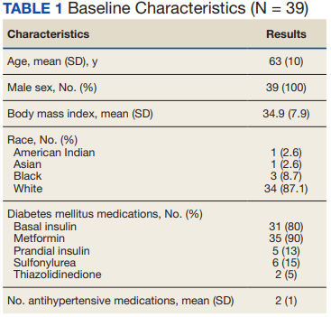
HbA1c levels decreased by 1% after 12 weeks of combination therapy compared with baseline (P < .001), and this reduction was sustained through the duration of the study period (Table 2). 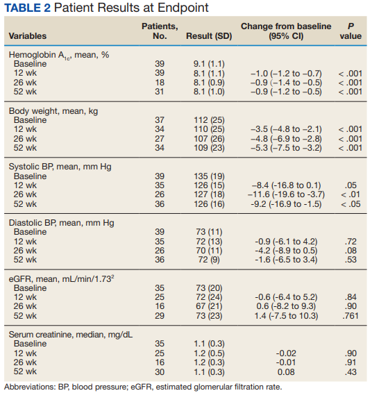
The most common AE during the trial was hypoglycemia, which was mostly mild (level 1) (Table 3). 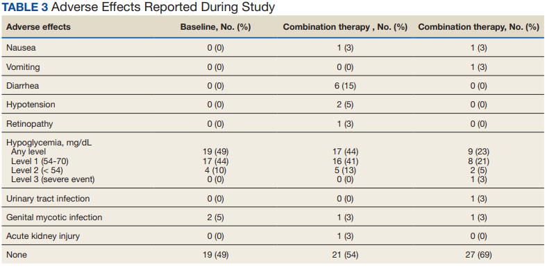
Discussion
This study evaluated the safety and efficacy of combined use of semaglutide or liraglutide and empagliflozin in a veteran population with T2DM. The retrospective chart review captured real-world practice and outcomes. Combination therapy was associated with a significant reduction in HbA1c levels, body weight, and SBP compared with either agent alone. No significant change was seen in DBP, SCr, or eGFR. Overall, the combination of GLP-1 RA and SGLT2i medications demonstrated a good safety profile with most patients reporting no AEs.
Several other studies have assessed the safety and efficacy of using GLP-1 RA and SGLT2i in combination. The DURATION 8 trial is the only double-blind trial to randomize subjects to receive either exenatide once weekly, dapagliflozin, or the combination of both for up to 52 weeks.3 Other controlled trials required stable background therapy with either SGLT2i or GLP-1 RA before randomization to receive the other class or placebo and had durations between 18 and 30 weeks.4-7 The AWARD 10 trial studied the combination of canagliflozin and dulaglutide, which both have proven CVD benefit.4 Other studies did not restrict SGLT2i or GLP-1 RA background therapy to agents with proven CVD benefit.5-7 The present study evaluated the combination of empagliflozin plus liraglutide or semaglutide, agents that all have proven CVD benefit.
A meta-analysis of 7 trials, including those previously mentioned, was conducted to evaluate the combination of GLP-1 RA and SGLT2i.8 The combination significantly reduced HbA1c levels by 0.61% and 0.85% compared with GLP-1 RA or SGLT2i, respectively. Our trial showed greater HbA1c level reduction of 1% with combination therapy compared with either agent separately. This may have been due in part to a higher baseline HbA1c level in our real-world veteran population. The meta-analysis found the combination decreased body weight 2.6 kg and 1.5 kg compared with GLP-1 RA or SGLT2i, respectively.8 This only reached significance with comparison vs GLP-1 RA alone. Our study demonstrated impressive weight loss of up to about 5 kg after 26 and 52 weeks of combination therapy. This is equivalent to about 5% weight loss from baseline, which is clinically significant.9 Liraglutide and semaglutide are the GLP-1 RAs associated with the greatest weight loss, which may contribute to greater weight loss efficacy seen in the present trial.1
In our trial SBP fell lower compared with the meta-analysis. Combination therapy significantly reduced SBP by 4.1 mm Hg and 2.7 mm Hg compared with GLP-1 RA or SGLT2i, respectively, in the meta-analysis.8 We observed a significant 9 to 12 mm Hg reduction in SBP after 26 to 52 weeks of combination therapy compared with baseline. This reduction occurred despite relatively controlled SBP at baseline (135 mm Hg). Each reduction of 10 mm Hg in SBP significantly reduces the risk of MACE, stroke, and heart failure, making our results clinically significant.10 Neither the meta-analysis nor present study found a significant difference in DBP or eGFR with combination therapy.
AEs were similar in this trial compared with the meta-analysis. Combination treatment with GLP-1 RA and SGLT2i did not increase the incidence of severe hypoglycemia in either study.8 Hypoglycemia was the most common AE in this study, but frequency was similar with combination and separate therapy. Both medication classes are associated with low or no risk of hypoglycemia on their own.1 Baseline medications likely contributed to episodes of hypoglycemia seen in this study: About 80% of patients were prescribed basal insulin, 15% were prescribed a sulfonylurea, and 13% were prescribed prandial insulin. There is limited overlap between the known AEs of GLP-1 RA and SGLT2i, making combination therapy a safe option for use in patients with T2DM.
Our study confirms greater reduction in HbA1c levels, weight, and SBP in veterans taking GLP-1 RA and SGLT2i medications in combination compared with separate use in a real-world setting in a veteran population. The magnitude of change seen in this population appears greater compared with previous studies.
Limitations
There were several limitations to our study. Given the retrospective nature, many patients included in the study did not have bloodwork drawn during the specified time frames. Because of this, many patients were excluded and missing data on renal outcomes limited the power to detect differences. Data regarding AEs were limited to what was recorded in the EHR, which may underrepresent the AEs that patients experienced. Finally, our study size was small, consisting primarily of a White and male population, which may limit generalizability.
Further research is needed to validate these findings in this population and should include a larger study population. The impact of combining GLP-1 RA with SGLT2i on cardiorenal outcomes is an important area of ongoing research.
ConclusionS
The combined use of GLP-1 RA and SGLT2i resulted in significant improvement in HbA1c levels, weight, and SBP compared with separate use in this real-world study of a VA population with T2DM. The combination was well tolerated overall. Awareness of these results can facilitate optimal care and outcomes in the VA population.
Acknowledgments
Serena Kelley, PharmD, and Michael Brenner, PharmD, assisted with study design and initial data collection. Julie Strominger, MS, provided statistical support.
1. American Diabetes Association. 9. Pharmacologic approaches to glycemic treatment: standards of medical care in diabetes-2021. Diabetes Care. 2021;44(suppl 1):S111-S124. doi.10.2337/dc21-S009
2. DeFronzo RA. Combination therapy with GLP-1 receptor agonist and SGLT2 inhibitor. Diabetes Obes Metab. 2017;19(10):1353-1362. doi.10.1111/dom.12982
3. Jabbour S, Frias J, Guja C, Hardy E, Ahmed A, Ohman P. Effects of exenatide once weekly plus dapagliflozin, exenatide once weekly, or dapagliflozin, added to metformin monotherapy, on body weight, systolic blood pressure, and triglycerides in patients with type 2 diabetes in the DURATION-8 study. Diabetes Obes Metab. 2018;20(6):1515-1519. doi:10.1111/dom.13206
4. Ludvik B, Frias J, Tinahones F, et al. Dulaglutide as add-on therapy to SGLT2 inhibitors in patients with inadequately controlled type 2 diabetes (AWARD-10): a 24-week, randomised, double-blind, placebo-controlled trial. Lancet Diabetes Endocrinol. 2018;6(5):370-381. doi:10.1016/S2213-8587(18)30023-8
5. Blonde L, Belousova L, Fainberg U, et al. Liraglutide as add-on to sodium-glucose co-transporter-2 inhibitors in patients with inadequately controlled type 2 diabetes: LIRA-ADD2SGLT2i, a 26-week, randomized, double-blind, placebo-controlled trial. Diabetes Obes Metab. 2020;22(6):929-937. doi:10.1111/dom.13978
6. Fulcher G, Matthews D, Perkovic V, et al; CANVAS trial collaborative group. Efficacy and safety of canagliflozin when used in conjunction with incretin-mimetic therapy in patients with type 2 diabetes. Diabetes Obes Metab. 2016;18(1):82-91. doi:10.1111/dom.12589
7. Zinman B, Bhosekar V, Busch R, et al. Semaglutide once weekly as add-on to SGLT-2 inhibitor therapy in type 2 diabetes (SUSTAIN 9): a randomised, placebo-controlled trial. Lancet Diabetes Endocrinol. 2019;7(5):356-367. doi:10.1016/S2213-8587(19)30066-X
8. Mantsiou C, Karagiannis T, Kakotrichi P, et al. Glucagon-like peptide-1 receptor agonists and sodium-glucose co-transporter-2 inhibitors as combination therapy for type 2 diabetes: a systematic review and meta-analysis. Diabetes Obes Metab. 2020;22(10):1857-1868. doi:10.1111/dom.14108
9. US Department of Veterans Affairs, Department of Defense. VA/DoD clinical practice guideline for the management of adult overweight and obesity. Version 3.0. Accessed August 18, 2022. www.healthquality.va.gov/guidelines/CD/obesity/VADoDObesityCPGFinal5087242020.pdf
10. Ettehad D, Emdin CA, Kiran A, et al. Blood pressure lowering for prevention of cardiovascular disease and death: a systematic review and meta-analysis. Lancet. 2015;387(10022):957-967. doi.10.1016/S0140-6736(15)01225-8
Selecting the best medication regimen for a patient with type 2 diabetes mellitus (T2DM) depends on many factors, such as glycemic control, adherence, adverse effect (AE) profile, and comorbid conditions.1 Selected agents from 2 newer medication classes, glucagon-like peptide 1 receptor agonists (GLP-1 RA) and sodium-glucose cotransporter 2 inhibitors (SGLT2i), have demonstrated cardiovascular and renal protective properties, creating a new paradigm in management.
The American Diabetes Association recommends medications with proven benefit in cardiovascular disease (CVD), such as the GLP-1 RAs liraglutide, injectable semaglutide, or dulaglutide, or the SGLT2i empagliflozin or canagliflozin, as second-line after metformin in patients with established atherosclerotic CVD or indicators of high risk to reduce the risk of major adverse cardiovascular events (MACE).1 SGLT2i are preferred in patients with diabetic kidney disease, and GLP-1 RAs are next in line for selection of agents with proven nephroprotection (liraglutide, injectable semaglutide, dulaglutide). The mechanisms of these benefits are not fully understood but may be due to their extraglycemic effects. The classes likely induce these benefits by different mechanisms: SGLT2i by hemodynamic effects and GLP-1 RAs by anti-inflammatory mechanisms.2 Although there is much interest, evidence is limited regarding the cardiovascular and renal protection benefits of these agents used in combination.
The combined use of GLP-1 RA and SGLT2i agents demonstrated greater benefit than separate use in trials with nonveteran populations.3-7 These studies evaluated effects on hemoglobin A1c (HbA1c) levels, weight loss, blood pressure (BP), and estimated glomerular filtration rate (eGFR). A meta-analysis of 7 trials found that the combination of GLP-1 RA and SGLT2i reduced HbA1c levels, body weight, and systolic blood pressure (SBP).8 All of the changes were statistically significant except for body weight with combination vs SGLT2i alone. Combination therapy was not associated with increased risk of severe hypoglycemia compared with either therapy separately.
The purpose of our study was to evaluate the safety and efficacy of the combined use of GLP-1 RA and SGLT2i in a real-world, US Department of Veterans Affairs (VA) population with T2DM.
Methods
This study was a pre-post, retrospective, single-center chart review. Subjects served as their own control. The project was reviewed and approved by the VA Ann Arbor Healthcare System Institutional Review Board. Subjects prescribed both a GLP-1 RA (semaglutide or liraglutide) and SGLT2i (empagliflozin) between January 1, 2014, and November 10, 2019, were extracted from the Corporate Data Warehouse (CDW) for possible inclusion in the study.
Patients were excluded if they received < 12 weeks of combination GLP-1 RA and SGLT2i therapy or did not have a corresponding 12-week HbA1c level. Patients also were excluded if they had < 12 weeks of monotherapy before starting combination therapy or did not have a baseline HbA1c level, or if the start date of combination therapy was not recorded in the VA electronic health record (EHR). We reviewed data for each patient from 6 months before to 1 year after the second agent was started. Start of the first agent (GLP-1 RA or SGLT2i) was recorded as the date the prescription was picked up in-person or 7 days after release date if mailed to the patient. Start of the second agent (GLP-1 RA or SGLT2i) was defined as baseline and was the date the prescription was picked up in person or 7 days after the release date if mailed.
Baseline measures were taken anytime from 8 weeks after the start of the first agent through 2 weeks after the start of the second agent. Data collected included age, sex, race, height, weight, BP, HbA1c levels, serum creatinine (SCr), eGFR, classes of medications for the treatment of T2DM, and the number of prescribed antihypertensive medications. HbA1c levels, SCr, eGFR, weight, and BP also were collected at 12 weeks (within 8-21 weeks); 26 weeks (within 22-35 weeks); and 52 weeks (within 36-57 weeks) of combination therapy. We reviewed progress notes and laboratory results to determine AEs within 26 weeks before initiating second agent (baseline) and 0 to 26 weeks and 26 to 52 weeks after initiating combination therapy.
The primary objective was to determine the effect on HbA1c levels at 12 weeks when using a GLP-1 RA and SGLT2i in combination vs separately. Secondary objectives were to determine change from baseline in mean body weight, BP, SCr, and eGFR at 12, 26, and 52 weeks; change in HbA1c levels at 26 and 52 weeks; and incidence of prespecified adverse drug reactions during combination therapy vs separately.
Statistical Analysis
Assuming a SD of 1, 80% power, significance level of P < .05, 2-sided test, and a correlation between baseline and follow-up of 0.5, we determined that a sample size of 34 subjects was required to detect a 0.5% change in baseline HbA1c level at 12 weeks. A t test (or Wilcoxon signed rank test if outcome not normally distributed) was conducted to examine whether the expected change from baseline was different from 0 for continuous outcomes. Median change from baseline was reported for SCr as a nonparametric t test (Wilcoxon signed rank test) was used.
Results
We identified 110 patients for possible study inclusion and 39 met eligibility criteria. After record review, 30 patients were excluded for receiving < 12 weeks of combination therapy or no 12 week HbA1c level; 26 patients were excluded for receiving < 12 weeks of monotherapy before starting combination therapy or no baseline HbA1c level; and 15 patients were excluded for lack of documentation in the VA EHR. Of the 39 patients included, 24 (62%) were prescribed empagliflozin first and then 8 started liraglutide and 16 started semaglutide.
HbA1c levels decreased by 1% after 12 weeks of combination therapy compared with baseline (P < .001), and this reduction was sustained through the duration of the study period (Table 2).
The most common AE during the trial was hypoglycemia, which was mostly mild (level 1) (Table 3).
Discussion
This study evaluated the safety and efficacy of combined use of semaglutide or liraglutide and empagliflozin in a veteran population with T2DM. The retrospective chart review captured real-world practice and outcomes. Combination therapy was associated with a significant reduction in HbA1c levels, body weight, and SBP compared with either agent alone. No significant change was seen in DBP, SCr, or eGFR. Overall, the combination of GLP-1 RA and SGLT2i medications demonstrated a good safety profile with most patients reporting no AEs.
Several other studies have assessed the safety and efficacy of using GLP-1 RA and SGLT2i in combination. The DURATION 8 trial is the only double-blind trial to randomize subjects to receive either exenatide once weekly, dapagliflozin, or the combination of both for up to 52 weeks.3 Other controlled trials required stable background therapy with either SGLT2i or GLP-1 RA before randomization to receive the other class or placebo and had durations between 18 and 30 weeks.4-7 The AWARD 10 trial studied the combination of canagliflozin and dulaglutide, which both have proven CVD benefit.4 Other studies did not restrict SGLT2i or GLP-1 RA background therapy to agents with proven CVD benefit.5-7 The present study evaluated the combination of empagliflozin plus liraglutide or semaglutide, agents that all have proven CVD benefit.
A meta-analysis of 7 trials, including those previously mentioned, was conducted to evaluate the combination of GLP-1 RA and SGLT2i.8 The combination significantly reduced HbA1c levels by 0.61% and 0.85% compared with GLP-1 RA or SGLT2i, respectively. Our trial showed greater HbA1c level reduction of 1% with combination therapy compared with either agent separately. This may have been due in part to a higher baseline HbA1c level in our real-world veteran population. The meta-analysis found the combination decreased body weight 2.6 kg and 1.5 kg compared with GLP-1 RA or SGLT2i, respectively.8 This only reached significance with comparison vs GLP-1 RA alone. Our study demonstrated impressive weight loss of up to about 5 kg after 26 and 52 weeks of combination therapy. This is equivalent to about 5% weight loss from baseline, which is clinically significant.9 Liraglutide and semaglutide are the GLP-1 RAs associated with the greatest weight loss, which may contribute to greater weight loss efficacy seen in the present trial.1
In our trial SBP fell lower compared with the meta-analysis. Combination therapy significantly reduced SBP by 4.1 mm Hg and 2.7 mm Hg compared with GLP-1 RA or SGLT2i, respectively, in the meta-analysis.8 We observed a significant 9 to 12 mm Hg reduction in SBP after 26 to 52 weeks of combination therapy compared with baseline. This reduction occurred despite relatively controlled SBP at baseline (135 mm Hg). Each reduction of 10 mm Hg in SBP significantly reduces the risk of MACE, stroke, and heart failure, making our results clinically significant.10 Neither the meta-analysis nor present study found a significant difference in DBP or eGFR with combination therapy.
AEs were similar in this trial compared with the meta-analysis. Combination treatment with GLP-1 RA and SGLT2i did not increase the incidence of severe hypoglycemia in either study.8 Hypoglycemia was the most common AE in this study, but frequency was similar with combination and separate therapy. Both medication classes are associated with low or no risk of hypoglycemia on their own.1 Baseline medications likely contributed to episodes of hypoglycemia seen in this study: About 80% of patients were prescribed basal insulin, 15% were prescribed a sulfonylurea, and 13% were prescribed prandial insulin. There is limited overlap between the known AEs of GLP-1 RA and SGLT2i, making combination therapy a safe option for use in patients with T2DM.
Our study confirms greater reduction in HbA1c levels, weight, and SBP in veterans taking GLP-1 RA and SGLT2i medications in combination compared with separate use in a real-world setting in a veteran population. The magnitude of change seen in this population appears greater compared with previous studies.
Limitations
There were several limitations to our study. Given the retrospective nature, many patients included in the study did not have bloodwork drawn during the specified time frames. Because of this, many patients were excluded and missing data on renal outcomes limited the power to detect differences. Data regarding AEs were limited to what was recorded in the EHR, which may underrepresent the AEs that patients experienced. Finally, our study size was small, consisting primarily of a White and male population, which may limit generalizability.
Further research is needed to validate these findings in this population and should include a larger study population. The impact of combining GLP-1 RA with SGLT2i on cardiorenal outcomes is an important area of ongoing research.
ConclusionS
The combined use of GLP-1 RA and SGLT2i resulted in significant improvement in HbA1c levels, weight, and SBP compared with separate use in this real-world study of a VA population with T2DM. The combination was well tolerated overall. Awareness of these results can facilitate optimal care and outcomes in the VA population.
Acknowledgments
Serena Kelley, PharmD, and Michael Brenner, PharmD, assisted with study design and initial data collection. Julie Strominger, MS, provided statistical support.
1. American Diabetes Association. 9. Pharmacologic approaches to glycemic treatment: standards of medical care in diabetes-2021. Diabetes Care. 2021;44(suppl 1):S111-S124. doi.10.2337/dc21-S009
2. DeFronzo RA. Combination therapy with GLP-1 receptor agonist and SGLT2 inhibitor. Diabetes Obes Metab. 2017;19(10):1353-1362. doi.10.1111/dom.12982
3. Jabbour S, Frias J, Guja C, Hardy E, Ahmed A, Ohman P. Effects of exenatide once weekly plus dapagliflozin, exenatide once weekly, or dapagliflozin, added to metformin monotherapy, on body weight, systolic blood pressure, and triglycerides in patients with type 2 diabetes in the DURATION-8 study. Diabetes Obes Metab. 2018;20(6):1515-1519. doi:10.1111/dom.13206
4. Ludvik B, Frias J, Tinahones F, et al. Dulaglutide as add-on therapy to SGLT2 inhibitors in patients with inadequately controlled type 2 diabetes (AWARD-10): a 24-week, randomised, double-blind, placebo-controlled trial. Lancet Diabetes Endocrinol. 2018;6(5):370-381. doi:10.1016/S2213-8587(18)30023-8
5. Blonde L, Belousova L, Fainberg U, et al. Liraglutide as add-on to sodium-glucose co-transporter-2 inhibitors in patients with inadequately controlled type 2 diabetes: LIRA-ADD2SGLT2i, a 26-week, randomized, double-blind, placebo-controlled trial. Diabetes Obes Metab. 2020;22(6):929-937. doi:10.1111/dom.13978
6. Fulcher G, Matthews D, Perkovic V, et al; CANVAS trial collaborative group. Efficacy and safety of canagliflozin when used in conjunction with incretin-mimetic therapy in patients with type 2 diabetes. Diabetes Obes Metab. 2016;18(1):82-91. doi:10.1111/dom.12589
7. Zinman B, Bhosekar V, Busch R, et al. Semaglutide once weekly as add-on to SGLT-2 inhibitor therapy in type 2 diabetes (SUSTAIN 9): a randomised, placebo-controlled trial. Lancet Diabetes Endocrinol. 2019;7(5):356-367. doi:10.1016/S2213-8587(19)30066-X
8. Mantsiou C, Karagiannis T, Kakotrichi P, et al. Glucagon-like peptide-1 receptor agonists and sodium-glucose co-transporter-2 inhibitors as combination therapy for type 2 diabetes: a systematic review and meta-analysis. Diabetes Obes Metab. 2020;22(10):1857-1868. doi:10.1111/dom.14108
9. US Department of Veterans Affairs, Department of Defense. VA/DoD clinical practice guideline for the management of adult overweight and obesity. Version 3.0. Accessed August 18, 2022. www.healthquality.va.gov/guidelines/CD/obesity/VADoDObesityCPGFinal5087242020.pdf
10. Ettehad D, Emdin CA, Kiran A, et al. Blood pressure lowering for prevention of cardiovascular disease and death: a systematic review and meta-analysis. Lancet. 2015;387(10022):957-967. doi.10.1016/S0140-6736(15)01225-8
Celiac disease linked to higher risk for rheumatoid arthritis, juvenile idiopathic arthritis
Celiac disease is linked to juvenile idiopathic arthritis (JIA) in children and rheumatoid arthritis (RA) in adults, according to an analysis of nationwide data in Sweden.
“I hope that our study can ultimately change clinical practice by lowering the threshold to evaluate celiac disease patients for inflammatory joint diseases,” John B. Doyle, MD, a gastroenterology fellow at Columbia University Irving Medical Center in New York, told this news organization.
“Inflammatory joint diseases, such as JIA and RA, are notoriously difficult to diagnose given their variable presentations,” he said. “But if JIA or RA can be identified sooner by physicians, patients will ultimately benefit by starting disease-modifying therapy earlier in their disease course.”
The study was published online in The American Journal of Gastroenterology.
Analyzing associations
Celiac disease has been linked to numerous autoimmune diseases, including type 1 diabetes, autoimmune thyroid disease, lupus, and inflammatory bowel disease (IBD), Dr. Doyle noted. However, a definitive epidemiologic association between celiac disease and inflammatory joint diseases such as JIA or RA hasn›t been established.
Dr. Doyle and colleagues conducted a nationwide population-based, retrospective matched cohort study using the Epidemiology Strengthened by Histopathology Reports in Sweden. They identified 24,014 patients diagnosed with biopsy-proven celiac disease between 2004 and 2017.
With these data, each patient was matched to five reference individuals in the general population by age, sex, calendar year, and geographic region, for a total of 117,397 people without a previous diagnosis of celiac disease. The researchers calculated the incidence and estimated the relative risk for JIA in patients younger than 18 years and RA in patients aged 18 years or older.
For those younger than 18 years, the incidence rate of JIA was 5.9 per 10,000 person-years among the 9,415 patients with celiac disease versus 2.2 per 10,000 person-years in the general population, over a follow-up of 7 years. Those with celiac disease were 2.7 times as likely to develop JIA.
The association between celiac disease and JIA remained similar after adjustment for education, Nordic country of birth, type 1 diabetes, autoimmune thyroid disease, lupus, and IBD. The incidence rate of JIA among patients with celiac disease was higher in both females and males, and across all age groups studied.
When 6,703 children with celiac disease were compared with their 9,089 siblings without celiac disease, the higher risk for JIA in patients with celiac disease fell slightly short of statistical significance.
For those aged 18 years or older, the incidence rate of RA was 8.4 per 10,000 person-years among the 14,599 patients with celiac disease versus 5.1 per 10,000 person-years in the general population, over a follow-up of 8.8 years. Those with celiac disease were 1.7 times as likely to develop RA.
As with the younger cohort, the association between celiac disease and RA in the adult group remained similar after adjustment for education, Nordic country of birth, type 1 diabetes, autoimmune thyroid disease, lupus, and IBD. Although both men and women with celiac disease had higher rates of RA, the risk was higher among those in whom disease was diagnosed at age 18-59 years compared with those who received a diagnosis at age 60 years or older.
When 9,578 adults with celiac disease were compared with their 17,067 siblings without celiac disease, the risk for RA remained higher in patients with celiac disease.
This suggests “that the association between celiac disease and RA is unlikely to be explained by environmental factors alone,” Dr. Doyle said.
Additional findings
Notably, the primary analysis excluded patients diagnosed with JIA or RA before their celiac disease diagnosis. In additional analyses, however, significant associations emerged.
Among children with celiac disease, 0.5% had a previous diagnosis of JIA, compared with 0.1% of matched comparators. Those with celiac disease were 3.5 times more likely to have a JIA diagnosis.
Among adults with celiac disease, 0.9% had a previous diagnosis of RA, compared with 0.6% of matched comparators. Those with celiac disease were 1.4 times more likely to have a RA diagnosis.
“We found that diagnoses of these types of arthritis were more common before a diagnosis of celiac disease compared to the general population,” Benjamin Lebwohl, MD, director of clinical research at the Celiac Disease Center at Columbia University, New York, told this news organization.
“This suggests that undiagnosed and untreated celiac disease might be contributing to these others autoimmune conditions,” he said.
Dr. Doyle and Dr. Lebwohl emphasized the practical implications for clinicians caring for patients with celiac disease. Among patients with celiac disease and inflammatory joint symptoms, clinicians should have a low threshold to evaluate for JIA or RA, they said.
“Particularly in pediatrics, we are trained to screen patients with JIA for celiac disease, but this study points to the possible bidirectional association and the importance of maintaining a clinical suspicion for JIA and RA among established celiac disease patients,” Marisa Stahl, MD, assistant professor of pediatrics and associate program director of the pediatric gastroenterology, hepatology, and nutrition fellowship training program at the University of Colorado at Denver, Aurora, said in an interview.
Dr. Stahl, who wasn’t involved with this study, conducts research at the Colorado Center for Celiac Disease. She and colleagues are focused on understanding the genetic and environmental factors that lead to the development of celiac disease and other autoimmune diseases.
Given the clear association between celiac disease and other autoimmune diseases, Dr. Stahl agreed that clinicians should have a low threshold for screening, with “additional workup for other autoimmune diseases once an autoimmune diagnosis is established.”
The study was supported by Karolinska Institutet and the Swedish Research Council. Dr. Lebwohl coordinates a study on behalf of the Swedish IBD quality register, which has received funding from Janssen. The other authors declared no conflicts of interest. Dr. Stahl reported no relevant disclosures.
A version of this article first appeared on Medscape.com.
FROM THE AMERICAN JOURNAL OF GASTROENTEROLOGY
Sick call
They call me and I go.
– William Carlos Williams
I never get sick. I’ve never had the flu. When everyone’s got a cold, I’m somehow immune. The last time I threw up was June 29th, 1980. You see, I work out almost daily, eat vegan, and sleep plenty. I drink gallons of pressed juice and throw down a few high-quality supplements. Yes, I’m that guy: The one who never gets sick. Well, I was anyway.
I am no longer that guy since our little girl became a supersocial little toddler. My undefeated welterweight “never-sick” title has been obliterated by multiple knockouts. One was a wicked adenovirus that broke the no-vomit streak. At one point, I lay on the luxury gray tile bathroom floor hoping to go unconscious to make the nausea stop. I actually called out sick that day. Then with a nasty COVID-despite-vaccine infection. I called out again. Later with a hacking lower respiratory – RSV?! – bug. Called out. All of which our 2-year-old blonde, curly-haired vector transmitted to me with remarkable efficiency.
In fact, That’s saying a lot. Our docs, like most, don’t call out sick.
We physicians have legendary stamina. Compared with other professionals, we are no less likely to become ill but a whopping 80% less likely to call out sick.
Presenteeism is our physician version of Omerta, a code of honor to never give in even at the expense of our, or our family’s, health and well-being. Every medical student is regaled with stories of physicians getting an IV before rounds or finishing clinic after their water broke. Why? In part it’s an indoctrination into this thing of ours we call Medicine: An elitist club that admits only those able to pass O-chem and hold diarrhea. But it is also because our medical system is so brittle that the slightest bend causes it to shatter. When I cancel a clinic, patients who have waited weeks for their spot have to be sent home. And for critical cases or those patients who don’t get the message, my already slammed colleagues have to cram the unlucky ones in between already-scheduled appointments. The guilt induced by inconveniencing our colleagues and our patients is more potent than dry heaves. And so we go. Suck it up. Sip ginger ale. Load up on acetaminophen. Carry on. This harms not only us, but also patients whom we put in the path of transmission. We become terrible 2-year-olds.

Of course, it’s not always easy to tell if you’re sick enough to stay home. But the stigma of calling out is so great that we often show up no matter what symptoms. A recent Medscape survey of physicians found that 85% said they had come to work sick in 2022.
We can do better. Perhaps creating sick-leave protocols could help? For example, if you have a fever above 100.4, have contact with someone positive for influenza, are unable to take POs, etc. then stay home. So might building rolling slack into schedules to accommodate the inevitable physician illness, parenting emergency, or death of an beloved uncle. And if there is one thing artificial intelligence could help us with, it would be smart scheduling. Can’t we build algorithms for anticipating and absorbing these predictable events? I’d take that over an AI skin cancer detector any day. Yet this year we’ll struggle through the cold and flu (and COVID) season again and nothing will have changed.
Our daughter hasn’t had hand, foot, and mouth disease yet. It’s not a question of if, but rather when she, and her mom and I, will get it. I hope it happens on a Friday so that my Monday clinic will be bearable when I show up.
Dr. Benabio is director of Healthcare Transformation and chief of dermatology at Kaiser Permanente San Diego. The opinions expressed in this column are his own and do not represent those of Kaiser Permanente. Dr. Benabio is @Dermdoc on Twitter. Write to him at [email protected]
They call me and I go.
– William Carlos Williams
I never get sick. I’ve never had the flu. When everyone’s got a cold, I’m somehow immune. The last time I threw up was June 29th, 1980. You see, I work out almost daily, eat vegan, and sleep plenty. I drink gallons of pressed juice and throw down a few high-quality supplements. Yes, I’m that guy: The one who never gets sick. Well, I was anyway.
I am no longer that guy since our little girl became a supersocial little toddler. My undefeated welterweight “never-sick” title has been obliterated by multiple knockouts. One was a wicked adenovirus that broke the no-vomit streak. At one point, I lay on the luxury gray tile bathroom floor hoping to go unconscious to make the nausea stop. I actually called out sick that day. Then with a nasty COVID-despite-vaccine infection. I called out again. Later with a hacking lower respiratory – RSV?! – bug. Called out. All of which our 2-year-old blonde, curly-haired vector transmitted to me with remarkable efficiency.
In fact, That’s saying a lot. Our docs, like most, don’t call out sick.
We physicians have legendary stamina. Compared with other professionals, we are no less likely to become ill but a whopping 80% less likely to call out sick.
Presenteeism is our physician version of Omerta, a code of honor to never give in even at the expense of our, or our family’s, health and well-being. Every medical student is regaled with stories of physicians getting an IV before rounds or finishing clinic after their water broke. Why? In part it’s an indoctrination into this thing of ours we call Medicine: An elitist club that admits only those able to pass O-chem and hold diarrhea. But it is also because our medical system is so brittle that the slightest bend causes it to shatter. When I cancel a clinic, patients who have waited weeks for their spot have to be sent home. And for critical cases or those patients who don’t get the message, my already slammed colleagues have to cram the unlucky ones in between already-scheduled appointments. The guilt induced by inconveniencing our colleagues and our patients is more potent than dry heaves. And so we go. Suck it up. Sip ginger ale. Load up on acetaminophen. Carry on. This harms not only us, but also patients whom we put in the path of transmission. We become terrible 2-year-olds.
Of course, it’s not always easy to tell if you’re sick enough to stay home. But the stigma of calling out is so great that we often show up no matter what symptoms. A recent Medscape survey of physicians found that 85% said they had come to work sick in 2022.
We can do better. Perhaps creating sick-leave protocols could help? For example, if you have a fever above 100.4, have contact with someone positive for influenza, are unable to take POs, etc. then stay home. So might building rolling slack into schedules to accommodate the inevitable physician illness, parenting emergency, or death of an beloved uncle. And if there is one thing artificial intelligence could help us with, it would be smart scheduling. Can’t we build algorithms for anticipating and absorbing these predictable events? I’d take that over an AI skin cancer detector any day. Yet this year we’ll struggle through the cold and flu (and COVID) season again and nothing will have changed.
Our daughter hasn’t had hand, foot, and mouth disease yet. It’s not a question of if, but rather when she, and her mom and I, will get it. I hope it happens on a Friday so that my Monday clinic will be bearable when I show up.
Dr. Benabio is director of Healthcare Transformation and chief of dermatology at Kaiser Permanente San Diego. The opinions expressed in this column are his own and do not represent those of Kaiser Permanente. Dr. Benabio is @Dermdoc on Twitter. Write to him at [email protected]
Higher metal contact allergy rates found in metalworkers
a systematic review and meta-analysis reports.
“Metal allergy to all three metals was significantly more common in European metalworkers with dermatitis attending patch test clinics as compared to ESSCA [European Surveillance System on Contact Allergies] data, indicating a relationship to occupational exposures,” senior study author Jeanne D. Johansen, MD, professor, department of dermatology and allergy, Copenhagen University Hospital, Hellerup, Denmark, and colleagues at the University of Copenhagen wrote in Contact Dermatitis. “However, confounders could not be accounted for.”
How common is metal allergy in metalworkers?
Occupational hand eczema is known to be common in metalworkers. Touching oils, greases, metals, leather gloves, rubber materials, and metalworking fluids as they repeatedly cut, shape, and process raw metals and minerals derived from ore mining exposes metalworkers to allergens and skin irritants, the authors wrote. But the prevalence of allergy to certain metals has not been well characterized.
So they searched PubMed for full-text studies in English that reported metal allergy prevalence in metalworkers, from the database’s inception through April 2022.
They included studies with absolute numbers or proportions of metal allergy to cobalt, chromium, or nickel, in all metalworkers with suspected allergic contact dermatitis who attended outpatient clinics or who worked at metalworking plants participating in workplace studies.
The researchers performed a random-effects meta-analysis to calculate the pooled prevalence of metal allergy. Because 85%-90% of metalworkers in Denmark are male, they compared the estimates they found with ESSCA data on 13,382 consecutively patch-tested males with dermatitis between 2015 and 2018.
Of the 1,667 records they screened, they analyzed data from 29 that met their inclusion criteria: 22 patient studies and 7 workplace studies involving 5,691 patients overall from 22 studies from Europe, 5 studies from Asia, and 1 from Africa. Regarding European metalworkers, the authors found:
- Pooled proportions of allergy in European metalworkers with dermatitis referred to patch test clinics were 8.2% to cobalt (95% confidence interval, 5.3%-11.7%), 8.0% to chromium (95% CI, 5.1%-11.4%), and 11.0% to nickel (95% CI, 7.3%-15.4%).
- In workplace studies, the pooled proportions of allergy in unselected European metalworkers were 4.9% to cobalt, (95% CI, 2.4%-8.1%), 5.2% to chromium (95% CI, 1.0% - 12.6%), and 7.6% to nickel (95% CI, 3.8%-12.6%).
- By comparison, ESSCA data on metal allergy prevalence showed 3.9% allergic to cobalt (95% CI, 3.6%-4.2%), 4.4% allergic to chromium (95% CI, 4.1%-4.8%), and 6.7% allergic to nickel (95% CI, 6.3%-7.0%).
- Data on sex, age, body piercings, and atopic dermatitis were scant.
Thorough histories, protective regulations and equipment
Providers need to ask their dermatitis patients about current and past occupations and hobbies, and employers need to provide employees with equipment that protects them from exposure, Kelly Tyler, MD, associate professor of dermatology, Ohio State University Wexner Medical Center, Columbus, said in an interview.
“Repeated exposure to an allergen is required for sensitization to develop,” said Dr. Tyler, who was not involved in the study. “Metalworkers, who are continually exposed to metals and metalworking fluids, have a higher risk of allergic contact dermatitis to cobalt, chromium, and nickel.”
“The primary treatment for allergic contact dermatitis is preventing continued exposure to the allergen,” she added. “This study highlights the importance of asking about metal or metalworking fluid in the workplace and of elucidating whether the employer is providing appropriate protective gear.”
To prevent occupational dermatitis, workplaces need to apply regulatory measures and provide their employees with protective equipment, Dr. Tyler advised.
“Body piercings are a common sensitizer in patients with metal allergy, and the prevalence of body piercings among metalworkers was not included in the study,” she noted.
The results of the study may not be generalizable to patients in the United States, she added, because regulations and requirements to provide protective gear here may differ.
“Taking a thorough patient history is crucial when investigating potential causes of dermatitis, especially in patients with suspected allergic contact dermatitis,” Dr. Tyler urged.
Funding and conflict-of-interest details were not provided. Dr. Tyler reported no relevant financial relationships.
A version of this article first appeared on Medscape.com.
FROM CONTACT DERMATITIS