User login
Primary care’s eroding earnings: Is Congress concerned?
- Write your senator and congressional representative about the need for Medicare payment reform that addresses the primary care/specialist payment gap. Let them know, too, if you are no longer able to accept Medicare patients due to reduced payments.
Purpose: Despite increasing data demonstrating the positive impact primary care has on quality of care and costs, our specialty faces uncertainty. Its popularity among medical students is declining, and the income gap is growing between primary care and other specialties. Congress has the power to intervene in this impending crisis. If we want to influence lawmakers’ actions, we need to know how they are thinking about these issues.
Methods: Using a set of questions covering several physician payment topics, we interviewed 14 congressional staff aides (5 aides on Medicare-oversight committees, 9 general staff aides) and one representative from each of 3 governmental agencies: the Medicare Payment Advisory Commission, Congressional Budget Office, and Government Accountability Office.
Results: Interviewees revealed that issues in primary care are not high on the congressional agenda, and that Medicare’s Sustainable Growth Rate (SGR) is the physician-payment issue on the minds of congressional staff members.
Conclusion: Attempts to solve primary care’s reimbursement difficulties should be tied to SGR reform.
The viability of primary care in the United States is in question, attributable in large part to declining provider payments in the face of rising medical school debt and fee-for-service pressures to increase patient volume.1-3 Congress—which has authority over Medicare and its price-setting function for provider reimbursement overall—is seemingly unaware of the problems facing primary care, including barriers to payment reform. The future of our specialty may hinge on our ability to persuade Congress that these problems are dire. A growing body of evidence supports the essential and integrative function primary care plays in health systems, and its positive impact on quality of care and costs.4-6
The confused order of things now
Advantages of primary care are proven. Regions with higher ratios of primary care physicians relative to specialists have lower rates of hospitalizations, lower Medicare costs, and higher quality of care.7,8 People with a primary care physician are more satisfied with their care and more likely to receive preventive services and better chronic disease management.9-11 Most countries that have built their health care systems on a strong foundation of primary care demonstrate better health outcomes, fewer health care disparities, and lower costs.4,6 Thus the waning of primary care presents risks to both personal and population health.
Still, society undervalues primary care. Despite evidence of the benefits just cited, the income disparity between primary care physicians and specialists continues to grow, discouraging medical students from entering primary care careers.12 The Medical Group Management Association shows that between 2000 and 2004, the median income for a family physician increased 7.5% to $156,000; for invasive cardiologists, 16.9% to $428,000; and for diagnostic radiologists, 36.2% to $407,000. Adjusted for inflation, primary care income fell 10% from 1995 to 2004.13
No wonder students shy away from primary care. Though there is little public sympathy for the financial woes of primary care doctors, lower incomes are contributing to a drying of the primary care pipeline.14,15 The number of US medical school graduates choosing family medicine residencies dropped by 50% between 1997 and 2005.16 From 1998 to 2004, the number of internal medicine residents choosing careers in primary care plummeted from 54% to 25%.17,18 This waning interest in primary care coincides, unfortunately, with the aging of the US baby boomers and an increasing prevalence of chronic disease.
How Congress could help fix the disparity
Medicare reimbursement has 2 components that Congress could amend to narrow the payment gap and help open the primary care pipeline: the Sustainable Growth Rate (SGR) and the Resource-Based Relative Value Scale (RBRVS) process.
The SGR formula sets a target for Medicare physician expenditures each year. Recently, physician expenditure growth has exceeded the target and, by law, the difference is subtracted from the fees paid to all physicians. According to the Medicare Payment Advisory Commission (MedPAC), much of the excess spending has come from rapidly increasing volumes of procedures used by specialists.19 The SGR system therefore disproportionately penalizes primary care physicians because payments to all physicians are cut regardless of which specialties are responsible for excess spending.
RBRVS is the system of relative values applied to every procedure and office visit. The Relative Value Units (RVUs) for each procedure or office visit are multiplied by a conversion factor determined by the SGR formula. RVUs are largely governed by the Relative Value Scale Update Committee (RUC), which advises the Centers for Medicare and Medicaid Services (CMS) on revisions to physician reimbursement.
The RUC reviews the relative value scale at least every 5 years. Though primary care physicians provide about half of Medicare physician visits, they represent just 15% of the RUC’s voting members.12
The committee’s reevaluation process tends to raise some RVUs without sufficiently deflating others.20 The resulting overall inflation of fees forces CMS to reduce payments equally to all physicians, meaning primary care is again disproportionately penalized. Moreover, both Medicare and private insurance companies follow the RUC’s recommendations.
Influencing Congress: Where to begin? As Congress escalates its deliberations on Medicare physician spending, we investigated how key legislators perceive issues in primary care and physician payment.
Methods
To better understand perspectives of congressional committees with jurisdiction over health care spending, we conducted semistructured key informant interviews in March 2007 with 14 health staff aides to members of Congress who have jurisdiction over Medicare. Interviews were done face to face and lasted 30 to 60 minutes.
The congressional committees with jurisdiction over Medicare physician payment are Senate Finance, House Ways and Means, and House Energy and Commerce. Each committee has 1 majority and 1 minority staffer specializing in Medicare part B, which includes physician payment. Of these 6 specialized staffers, 5 agreed to participate in semistructured interviews. Other staffers were contacted by using a purposeful sampling technique known as “snowballing” or chain-referral, whereby participants with whom contact has been made refer the researcher to other potential interviewees. This process yielded another 9 interviewees to total 14.
The aides identified several other information sources, and we interviewed 1 staff member each from 3 of these sources: MedPAC, the Congressional Budget Office (CBO), and the Government Accountability Office (GAO).
Interviews covered several topics, including views on the state of primary care and physician payment (TABLE). Three researchers separately reviewed the interview notes to identify and compile themes.
TABLE
6 Questions we asked the congressional staffers
| 1. What are your views on the current state of primary care in the United States? |
| 2. When considering legislation to improve health care in the United States, how—if at all—does primary care factor into your vision? |
| 3. If there is legislative movement to change the Sustainable Growth Rate and Resource-Based Relative Value Scale systems in the next year, what should the goal be? |
| 4. What is your sense of other health legislative assistants’ understanding of primary care? |
| 5. Who are you hearing from on issues of primary care? Who are you not hearing from? |
| 6. What are the best sources to learn about these issues? |
Results
Of the 14 congressional staffers, 8 were Republican and 6 were Democrat; 5 were committee staff and 9 were general staff. Committee representation was fairly even among staffers: Senate Finance (4), House Ways and Means (5), and House Energy and Commerce (5). Range of experience on Capitol Hill was 3 months to 9 years.
Some staffers are empathetic, others unaware. Most respondents expressed concern about the decreasing number of students entering primary care careers and the potential impact on patient access to care. One staffer acknowledged, “the way our reimbursement system works, primary care is not an option for students…reimbursement is so low…the number of primary care physicians is going down relative to other specialties.”
Another participant added that most staffers “recognize a role for primary care. It’s also tough because of how strong the specialty community is.” One staffer advised, “The Alliance of Specialty Medicine goes along with the AMA, trying to represent a coordinated front…I don’t see this much coordination around primary care.”
A few staffers did not understand the definition of primary care or did not know which physician groups represent primary care.
Legislation to improve US health care—and primary care. Participants varied in their input on this subject. One staffer stated that primary care is “important but rarely singled out…usually the goal is broader reform so [primary care] is still a goal, but unstated.”
Some committee staff described the need to incentivize greater use of primary care and increase coordination of care. A few proposed reevaluating RBRVS to help primary care, and they spontaneously raised the Medical Home concept as a way to encourage growth of primary care. The Medical Home involves pairing each Medicare beneficiary with a patient-centered practice that meets certain criteria including continuity with a personal physician, care coordination, quality assurance, increased access, and specific payment.21 A pilot project in North Carolina that incorporates the Medical Home is saving the state about $162 million annually.22,23 One staffer championed primary care, but pointed out that a critical barrier preventing Congress from investing in it is the CBO, which is not convinced that primary care can save money over the long term.
The SGR dominates discussions on physician payment
All respondents had a functional understanding of the SGR and desired reform, but few understood how the SGR contributes to the payment gap. Many staffers would like to do away with the SGR, but CBO estimates show that this would be cost-prohibitive.24
A few staffers believed that SGR reform may not happen until 2009, after the next president takes office. Some participants also predicted that SGR reform will not happen until more physicians refuse to see Medicare patients. To date, MedPAC has reported each year that there is no Medicare access crisis. Staffers from rural districts, however, affirmed that constituents are having difficulty finding primary care doctors who take Medicare.
Staffers uniformly agreed that nobody has the answer to fix the SGR. Several staffers commented on the complexity of the problem, pointing out that MedPAC’s March 2007 SGR report did not achieve a consensus on how to restructure the rate. Many participants were disappointed with the MedPAC report and want solutions to fix physician payment that are more directed and “convincing.”
Some expressed a need for “hands-on models and demonstration projects.” Although these staffers have heard of models that would split the SGR by specialty or geography, they remain skeptical about such proposals without evidence of efficacy. Staffers were also wary of splitting the SGR by specialty, believing it would cause infighting among physicians.
Staffers know far less about RBRVS than they do about the SGR. One staffer admitted, “I won’t pay attention until something is at a crisis point or we have a hearing or a vote.” A few staffers asserted that there should be a more rigorous RUC review to examine what services are over- and undervalued.
Government agencies are not asked to address primary care. At the time of interview (March 2007), staff from MedPAC, GAO, and CBO said that Congress had not asked them to study issues in primary care. One CBO analyst asserted that “nobody’s been able to demonstrate significant changes in volume or outcome [as a result of investing in primary care]…we need empirical data.” The analyst also mentioned CMS demonstration projects as a way to gather data. According to a Capitol Hill veteran, the CBO believes that even if primary care extends a person’s life, this may not necessarily save money.
Discussion
Although most of the interviewed congressional staffers recognize the payment gap and understand that the number of physicians entering primary care is decreasing, Congress has not taken action to address these issues. Several factors explain this.
SGR is the 800-pound gorilla. When discussing physician payment, congressional staffers appear far more concerned with reforming the SGR than addressing problems in primary care. This perception is supported by the fact that Congress has asked MedPAC and CBO to investigate the SGR, but has not asked them to examine issues in primary care. For Congress, the dilemma is to hold down physician spending while keeping physicians in the Medicare market. Staffers are dissatisfied with SGR reform proposals from MedPAC and are eager to learn about new possible solutions.
No one perceives a crisis in access to Medicare providers. According to annual MedPAC reports, the number of primary care doctors accepting Medicare patients is sufficient. Staff for members of Congress from rural areas, however, contend that some constituents cannot find a primary care provider who accepts Medicare.
Congress is not convinced that primary care saves money. Although some staffers believe that primary care can reduce costs, the CBO argues that this is not necessarily true. It is indeed difficult to prove cost savings from investing in preventive services because there is greater upfront cost, and extending people’s lives could incur higher future costs. Research, however, shows that primary care-oriented systems reduce preventable hospitalizations, which decreases costs.4,5,7,8 It seems that either the existing evidence is insufficient to convince the CBO or the evidence has not been communicated effectively.
Strategic leverage moving forward
The time is ripe for SGR reform because most staffers conveyed a desire for solutions. Because the SGR appears to take priority over primary care issues, it must be dealt with first. It is possible, however, for policy makers to address the SGR and RBRVS reforms while simultaneously investing in primary care. The SGR and RBRVS reforms could hold specialties accountable for their own volume growth and protect specialties with minimal volume growth.
The Medical Home is a concept gaining recognition among congressional staff and could involve restructured physician payment. In its Tax Relief and Health Care Act of 2006, Congress mandated a 3-year Medical Home demonstration to be conducted across multiple demographic communities in up to 8 states. The concept encompasses “large or small medical practices where a physician provides comprehensive and coordinated patient centered medical care and acts as the ‘personal physician’ to the patient.”25 (The Medical Home is also a focus of The Patient-Centered Primary Care Collaborative [http://www.pcpcc.net/], a coalition of medical societies, employers, insurers, consumer groups, and others that is exploring the concept as a way to contain health care costs and also achieve fair remuneration for physicians.)
The demonstration must be carefully crafted to test the concept fairly. Even before the demonstration begins, Congress could ask the CBO and GAO to investigate existing evidence of primary care’s cost-effectiveness. Support from the CBO is essential for Congress to invest in primary care.
Other experiments are underway. As of this publication, several major insurers are beginning regional experiments in raising fees for primary care visits in an effort to avoid greater costs down the road.23
Access issue needs further study. Our interviews revealed that while MedPAC asserts there is no primary care access issue, staffers from rural districts disagree. In fact, had Congress not over-ridden President Bush’s recent veto of a Medicare bill to increase physicians’ fees, doctors in urban areas would also have stopped accepting new Medicare patients.26 Additional physician workforce studies are necessary to fully understand the current primary care physician supply. Also useful would be studies by Medicaid and Medicare that investigate thresholds at which physicians stop seeing patients with low-paying coverage.
Advocacy is needed, too. Congressional staffers appear to understand some of the difficulties in primary care, but give priority to broader SGR reform. Further research and advocacy on the value of primary care and payment reform solutions will be necessary to establish primary care as a means to cost-effective, high-quality care in the United States.
Acknowledgment
Part of the content in this article was presented as a poster at the North American Primary Care Research Group Conference in Vancouver, British Columbia, October 2007.
Correspondence
Brian Yoshio Laing, MD, San Francisco General Hospital, 995 Potrero Avenue, Building 80, Ward 83, San Francisco, CA 94110; [email protected].
1. Bodenheimer T. Primary care–will it survive? Ann Intern Med. 2007;146:301-306.
2. Phillips RL. Primary care in the United States: problems and possibilities. BMJ. 2006;332:151.-
3. American College of Physicians. The impending collapse of primary care medicine and its implications for the state of the nation’s health care. January 30, 2006. Available at: http://www.txpeds.org/u/documents/statehc06_1.pdf. Accessed April 10, 2007.
4. Ferrer RL, Hambidge SJ, Maly RC. The essential role of generalists in health care systems. Ann Intern Med. 2005;142:691-699.
5. Macinko J, Starfield B, Shi L. Quantifying the health benefits of primary care physician supply in the United States. Int J Health Serv. 2007;37:111-126.
6. Starfield B, Shi L, Macinko J. Contribution of primary care to health systems and health. Milbank Q. 2005;83:457-502.
7. Parchman ML, Culler S. Primary care physicians and avoidable hospitalizations. J Fam Pract. 1994;39:123-128.
8. Baicker K, Chandra A. Medicare spending, the physician workforce, and beneficiaries’ quality of care. Health Affairs [Web Exclusive]. April 7, 2004;W4-184-197. Available at: http://content.healthaffairs.org/cgi/content/abstract/hlthaff.w4.184. Accessed July 30, 2008.
9. Bindman AB, Grumbach K, Osmond D, et al. Primary care and receipt of preventive services. J Gen Intern Med. 1996;11:269-276.
10. Safran DG, Taira GA, Rogers WH, et al. Linking primary care performance to outcomes of care. J Fam Pract. 1998;47:213-220.
11. Stewart AL, Grumbach K, Osmond DH, et al. Primary care and patient perceptions of access to care. J Fam Pract. 1997;44:177-185.
12. Bodeheimer T, Berenson RA, Rudolf P. The primary care-specialty income gap: why it matters. Ann Intern Med. 2007;146:301-306.
13. Tu HT, Ginsburg PB. Losing ground: physician income, 1995-2003. Tracking Rep. June 2006;15:1-8.
14. Rosenblatt RA, Andrilla HA. The impact of US medical students’ debt on their choice of primary care careers. Acad Med. 2005;80:815-819.
15. Newton DA, Grayson MS, Thompson LF. The variable influence of lifestyle and income on medical students’ career specialty choices. Acad Med. 2005;80:809-814.
16. Pugno PA, Schmittling GT, Fetter GT, et al. Results of the 2005 national resident matching program: family medicine. Fam Med. 2005;37:555-564.
17. Garibaldi RA, Popkave C, Bylsma W. Career plans for trainees in internal medicine residency programs. Acad Med. 2005;80:507-512.
18. West CP, Popkave C, Schultz HJ, et al. Changes in career decisions of internal medicine residents during training. Ann Intern Med. 2006;145:774-779.
19. Medicare Payment Advisory Commission. Report to the Congress: Medicare Payment Policy. March 2006. Available at: http://www.medpac.gov/publications/congressional_reports/Mar06_EntireReport.pdf. Accessed April 25, 2007.
20. Ginsburg PB, Berenson RA. Revising Medicare’s physician fee schedule–much activity, little change. N Engl J Med. 2007;356:1201-1203.
21. American College of Physicians. The Advanced Medical Home: A Patient-Centered, Physician-Guided Model of Health Care. 2006. Available at: http://www.acponline.org/advocacy/where_we_stand/policy/adv_med.pdf. Accessed May 13, 2007.
22. Steiner BD, Denham AC, Ashkin E, et al. Community care of North Carolina: improving care through community health networks. Ann Fam Med. 2008;in press.
23. Freudenheim M. Trying to save by increasing doctors’ fees. The New York Times. July 21, 2008. Available at: http://www.nytimes.com/2008/07/21/business/21medhome.html?_r=1&scp=1&sq=Trying%20to%20save%20by%20increasing%20doctors%20fees&st=cse&oref=slogin. Accessed August 1, 2008.
24. Orzang P. CBO. “Medicare’s Payments to Physicians: Option for Changing the Sustainable Growth Rate,” testimony before the Committee on Finance United States Senate. March 1, 2007. Available at: http://www.senate.gov/~finance/hearings/testimony/2007test/030107potest.pdf. Accessed May 13, 2007.
25. American Medical Association. RUC Medicare Medical Home Demonstration project recommendations. Available at: http://www.ama-assn.org/ama/pub/category/18528.html. Accessed August 1, 2008.
26. Pear R. Doctors press Senate to undo Medicare cuts. The New York Times. July 7, 2008. Available at: http://www.nytimes.com/2008/07/07/health/policy/07medicare.html?scp=1&sq=Doctors%20press%20senate%20to%20undo%20Medicare%20cuts&st=cse. Accessed August 1, 2008.
- Write your senator and congressional representative about the need for Medicare payment reform that addresses the primary care/specialist payment gap. Let them know, too, if you are no longer able to accept Medicare patients due to reduced payments.
Purpose: Despite increasing data demonstrating the positive impact primary care has on quality of care and costs, our specialty faces uncertainty. Its popularity among medical students is declining, and the income gap is growing between primary care and other specialties. Congress has the power to intervene in this impending crisis. If we want to influence lawmakers’ actions, we need to know how they are thinking about these issues.
Methods: Using a set of questions covering several physician payment topics, we interviewed 14 congressional staff aides (5 aides on Medicare-oversight committees, 9 general staff aides) and one representative from each of 3 governmental agencies: the Medicare Payment Advisory Commission, Congressional Budget Office, and Government Accountability Office.
Results: Interviewees revealed that issues in primary care are not high on the congressional agenda, and that Medicare’s Sustainable Growth Rate (SGR) is the physician-payment issue on the minds of congressional staff members.
Conclusion: Attempts to solve primary care’s reimbursement difficulties should be tied to SGR reform.
The viability of primary care in the United States is in question, attributable in large part to declining provider payments in the face of rising medical school debt and fee-for-service pressures to increase patient volume.1-3 Congress—which has authority over Medicare and its price-setting function for provider reimbursement overall—is seemingly unaware of the problems facing primary care, including barriers to payment reform. The future of our specialty may hinge on our ability to persuade Congress that these problems are dire. A growing body of evidence supports the essential and integrative function primary care plays in health systems, and its positive impact on quality of care and costs.4-6
The confused order of things now
Advantages of primary care are proven. Regions with higher ratios of primary care physicians relative to specialists have lower rates of hospitalizations, lower Medicare costs, and higher quality of care.7,8 People with a primary care physician are more satisfied with their care and more likely to receive preventive services and better chronic disease management.9-11 Most countries that have built their health care systems on a strong foundation of primary care demonstrate better health outcomes, fewer health care disparities, and lower costs.4,6 Thus the waning of primary care presents risks to both personal and population health.
Still, society undervalues primary care. Despite evidence of the benefits just cited, the income disparity between primary care physicians and specialists continues to grow, discouraging medical students from entering primary care careers.12 The Medical Group Management Association shows that between 2000 and 2004, the median income for a family physician increased 7.5% to $156,000; for invasive cardiologists, 16.9% to $428,000; and for diagnostic radiologists, 36.2% to $407,000. Adjusted for inflation, primary care income fell 10% from 1995 to 2004.13
No wonder students shy away from primary care. Though there is little public sympathy for the financial woes of primary care doctors, lower incomes are contributing to a drying of the primary care pipeline.14,15 The number of US medical school graduates choosing family medicine residencies dropped by 50% between 1997 and 2005.16 From 1998 to 2004, the number of internal medicine residents choosing careers in primary care plummeted from 54% to 25%.17,18 This waning interest in primary care coincides, unfortunately, with the aging of the US baby boomers and an increasing prevalence of chronic disease.
How Congress could help fix the disparity
Medicare reimbursement has 2 components that Congress could amend to narrow the payment gap and help open the primary care pipeline: the Sustainable Growth Rate (SGR) and the Resource-Based Relative Value Scale (RBRVS) process.
The SGR formula sets a target for Medicare physician expenditures each year. Recently, physician expenditure growth has exceeded the target and, by law, the difference is subtracted from the fees paid to all physicians. According to the Medicare Payment Advisory Commission (MedPAC), much of the excess spending has come from rapidly increasing volumes of procedures used by specialists.19 The SGR system therefore disproportionately penalizes primary care physicians because payments to all physicians are cut regardless of which specialties are responsible for excess spending.
RBRVS is the system of relative values applied to every procedure and office visit. The Relative Value Units (RVUs) for each procedure or office visit are multiplied by a conversion factor determined by the SGR formula. RVUs are largely governed by the Relative Value Scale Update Committee (RUC), which advises the Centers for Medicare and Medicaid Services (CMS) on revisions to physician reimbursement.
The RUC reviews the relative value scale at least every 5 years. Though primary care physicians provide about half of Medicare physician visits, they represent just 15% of the RUC’s voting members.12
The committee’s reevaluation process tends to raise some RVUs without sufficiently deflating others.20 The resulting overall inflation of fees forces CMS to reduce payments equally to all physicians, meaning primary care is again disproportionately penalized. Moreover, both Medicare and private insurance companies follow the RUC’s recommendations.
Influencing Congress: Where to begin? As Congress escalates its deliberations on Medicare physician spending, we investigated how key legislators perceive issues in primary care and physician payment.
Methods
To better understand perspectives of congressional committees with jurisdiction over health care spending, we conducted semistructured key informant interviews in March 2007 with 14 health staff aides to members of Congress who have jurisdiction over Medicare. Interviews were done face to face and lasted 30 to 60 minutes.
The congressional committees with jurisdiction over Medicare physician payment are Senate Finance, House Ways and Means, and House Energy and Commerce. Each committee has 1 majority and 1 minority staffer specializing in Medicare part B, which includes physician payment. Of these 6 specialized staffers, 5 agreed to participate in semistructured interviews. Other staffers were contacted by using a purposeful sampling technique known as “snowballing” or chain-referral, whereby participants with whom contact has been made refer the researcher to other potential interviewees. This process yielded another 9 interviewees to total 14.
The aides identified several other information sources, and we interviewed 1 staff member each from 3 of these sources: MedPAC, the Congressional Budget Office (CBO), and the Government Accountability Office (GAO).
Interviews covered several topics, including views on the state of primary care and physician payment (TABLE). Three researchers separately reviewed the interview notes to identify and compile themes.
TABLE
6 Questions we asked the congressional staffers
| 1. What are your views on the current state of primary care in the United States? |
| 2. When considering legislation to improve health care in the United States, how—if at all—does primary care factor into your vision? |
| 3. If there is legislative movement to change the Sustainable Growth Rate and Resource-Based Relative Value Scale systems in the next year, what should the goal be? |
| 4. What is your sense of other health legislative assistants’ understanding of primary care? |
| 5. Who are you hearing from on issues of primary care? Who are you not hearing from? |
| 6. What are the best sources to learn about these issues? |
Results
Of the 14 congressional staffers, 8 were Republican and 6 were Democrat; 5 were committee staff and 9 were general staff. Committee representation was fairly even among staffers: Senate Finance (4), House Ways and Means (5), and House Energy and Commerce (5). Range of experience on Capitol Hill was 3 months to 9 years.
Some staffers are empathetic, others unaware. Most respondents expressed concern about the decreasing number of students entering primary care careers and the potential impact on patient access to care. One staffer acknowledged, “the way our reimbursement system works, primary care is not an option for students…reimbursement is so low…the number of primary care physicians is going down relative to other specialties.”
Another participant added that most staffers “recognize a role for primary care. It’s also tough because of how strong the specialty community is.” One staffer advised, “The Alliance of Specialty Medicine goes along with the AMA, trying to represent a coordinated front…I don’t see this much coordination around primary care.”
A few staffers did not understand the definition of primary care or did not know which physician groups represent primary care.
Legislation to improve US health care—and primary care. Participants varied in their input on this subject. One staffer stated that primary care is “important but rarely singled out…usually the goal is broader reform so [primary care] is still a goal, but unstated.”
Some committee staff described the need to incentivize greater use of primary care and increase coordination of care. A few proposed reevaluating RBRVS to help primary care, and they spontaneously raised the Medical Home concept as a way to encourage growth of primary care. The Medical Home involves pairing each Medicare beneficiary with a patient-centered practice that meets certain criteria including continuity with a personal physician, care coordination, quality assurance, increased access, and specific payment.21 A pilot project in North Carolina that incorporates the Medical Home is saving the state about $162 million annually.22,23 One staffer championed primary care, but pointed out that a critical barrier preventing Congress from investing in it is the CBO, which is not convinced that primary care can save money over the long term.
The SGR dominates discussions on physician payment
All respondents had a functional understanding of the SGR and desired reform, but few understood how the SGR contributes to the payment gap. Many staffers would like to do away with the SGR, but CBO estimates show that this would be cost-prohibitive.24
A few staffers believed that SGR reform may not happen until 2009, after the next president takes office. Some participants also predicted that SGR reform will not happen until more physicians refuse to see Medicare patients. To date, MedPAC has reported each year that there is no Medicare access crisis. Staffers from rural districts, however, affirmed that constituents are having difficulty finding primary care doctors who take Medicare.
Staffers uniformly agreed that nobody has the answer to fix the SGR. Several staffers commented on the complexity of the problem, pointing out that MedPAC’s March 2007 SGR report did not achieve a consensus on how to restructure the rate. Many participants were disappointed with the MedPAC report and want solutions to fix physician payment that are more directed and “convincing.”
Some expressed a need for “hands-on models and demonstration projects.” Although these staffers have heard of models that would split the SGR by specialty or geography, they remain skeptical about such proposals without evidence of efficacy. Staffers were also wary of splitting the SGR by specialty, believing it would cause infighting among physicians.
Staffers know far less about RBRVS than they do about the SGR. One staffer admitted, “I won’t pay attention until something is at a crisis point or we have a hearing or a vote.” A few staffers asserted that there should be a more rigorous RUC review to examine what services are over- and undervalued.
Government agencies are not asked to address primary care. At the time of interview (March 2007), staff from MedPAC, GAO, and CBO said that Congress had not asked them to study issues in primary care. One CBO analyst asserted that “nobody’s been able to demonstrate significant changes in volume or outcome [as a result of investing in primary care]…we need empirical data.” The analyst also mentioned CMS demonstration projects as a way to gather data. According to a Capitol Hill veteran, the CBO believes that even if primary care extends a person’s life, this may not necessarily save money.
Discussion
Although most of the interviewed congressional staffers recognize the payment gap and understand that the number of physicians entering primary care is decreasing, Congress has not taken action to address these issues. Several factors explain this.
SGR is the 800-pound gorilla. When discussing physician payment, congressional staffers appear far more concerned with reforming the SGR than addressing problems in primary care. This perception is supported by the fact that Congress has asked MedPAC and CBO to investigate the SGR, but has not asked them to examine issues in primary care. For Congress, the dilemma is to hold down physician spending while keeping physicians in the Medicare market. Staffers are dissatisfied with SGR reform proposals from MedPAC and are eager to learn about new possible solutions.
No one perceives a crisis in access to Medicare providers. According to annual MedPAC reports, the number of primary care doctors accepting Medicare patients is sufficient. Staff for members of Congress from rural areas, however, contend that some constituents cannot find a primary care provider who accepts Medicare.
Congress is not convinced that primary care saves money. Although some staffers believe that primary care can reduce costs, the CBO argues that this is not necessarily true. It is indeed difficult to prove cost savings from investing in preventive services because there is greater upfront cost, and extending people’s lives could incur higher future costs. Research, however, shows that primary care-oriented systems reduce preventable hospitalizations, which decreases costs.4,5,7,8 It seems that either the existing evidence is insufficient to convince the CBO or the evidence has not been communicated effectively.
Strategic leverage moving forward
The time is ripe for SGR reform because most staffers conveyed a desire for solutions. Because the SGR appears to take priority over primary care issues, it must be dealt with first. It is possible, however, for policy makers to address the SGR and RBRVS reforms while simultaneously investing in primary care. The SGR and RBRVS reforms could hold specialties accountable for their own volume growth and protect specialties with minimal volume growth.
The Medical Home is a concept gaining recognition among congressional staff and could involve restructured physician payment. In its Tax Relief and Health Care Act of 2006, Congress mandated a 3-year Medical Home demonstration to be conducted across multiple demographic communities in up to 8 states. The concept encompasses “large or small medical practices where a physician provides comprehensive and coordinated patient centered medical care and acts as the ‘personal physician’ to the patient.”25 (The Medical Home is also a focus of The Patient-Centered Primary Care Collaborative [http://www.pcpcc.net/], a coalition of medical societies, employers, insurers, consumer groups, and others that is exploring the concept as a way to contain health care costs and also achieve fair remuneration for physicians.)
The demonstration must be carefully crafted to test the concept fairly. Even before the demonstration begins, Congress could ask the CBO and GAO to investigate existing evidence of primary care’s cost-effectiveness. Support from the CBO is essential for Congress to invest in primary care.
Other experiments are underway. As of this publication, several major insurers are beginning regional experiments in raising fees for primary care visits in an effort to avoid greater costs down the road.23
Access issue needs further study. Our interviews revealed that while MedPAC asserts there is no primary care access issue, staffers from rural districts disagree. In fact, had Congress not over-ridden President Bush’s recent veto of a Medicare bill to increase physicians’ fees, doctors in urban areas would also have stopped accepting new Medicare patients.26 Additional physician workforce studies are necessary to fully understand the current primary care physician supply. Also useful would be studies by Medicaid and Medicare that investigate thresholds at which physicians stop seeing patients with low-paying coverage.
Advocacy is needed, too. Congressional staffers appear to understand some of the difficulties in primary care, but give priority to broader SGR reform. Further research and advocacy on the value of primary care and payment reform solutions will be necessary to establish primary care as a means to cost-effective, high-quality care in the United States.
Acknowledgment
Part of the content in this article was presented as a poster at the North American Primary Care Research Group Conference in Vancouver, British Columbia, October 2007.
Correspondence
Brian Yoshio Laing, MD, San Francisco General Hospital, 995 Potrero Avenue, Building 80, Ward 83, San Francisco, CA 94110; [email protected].
1. Bodenheimer T. Primary care–will it survive? Ann Intern Med. 2007;146:301-306.
2. Phillips RL. Primary care in the United States: problems and possibilities. BMJ. 2006;332:151.-
3. American College of Physicians. The impending collapse of primary care medicine and its implications for the state of the nation’s health care. January 30, 2006. Available at: http://www.txpeds.org/u/documents/statehc06_1.pdf. Accessed April 10, 2007.
4. Ferrer RL, Hambidge SJ, Maly RC. The essential role of generalists in health care systems. Ann Intern Med. 2005;142:691-699.
5. Macinko J, Starfield B, Shi L. Quantifying the health benefits of primary care physician supply in the United States. Int J Health Serv. 2007;37:111-126.
6. Starfield B, Shi L, Macinko J. Contribution of primary care to health systems and health. Milbank Q. 2005;83:457-502.
7. Parchman ML, Culler S. Primary care physicians and avoidable hospitalizations. J Fam Pract. 1994;39:123-128.
8. Baicker K, Chandra A. Medicare spending, the physician workforce, and beneficiaries’ quality of care. Health Affairs [Web Exclusive]. April 7, 2004;W4-184-197. Available at: http://content.healthaffairs.org/cgi/content/abstract/hlthaff.w4.184. Accessed July 30, 2008.
9. Bindman AB, Grumbach K, Osmond D, et al. Primary care and receipt of preventive services. J Gen Intern Med. 1996;11:269-276.
10. Safran DG, Taira GA, Rogers WH, et al. Linking primary care performance to outcomes of care. J Fam Pract. 1998;47:213-220.
11. Stewart AL, Grumbach K, Osmond DH, et al. Primary care and patient perceptions of access to care. J Fam Pract. 1997;44:177-185.
12. Bodeheimer T, Berenson RA, Rudolf P. The primary care-specialty income gap: why it matters. Ann Intern Med. 2007;146:301-306.
13. Tu HT, Ginsburg PB. Losing ground: physician income, 1995-2003. Tracking Rep. June 2006;15:1-8.
14. Rosenblatt RA, Andrilla HA. The impact of US medical students’ debt on their choice of primary care careers. Acad Med. 2005;80:815-819.
15. Newton DA, Grayson MS, Thompson LF. The variable influence of lifestyle and income on medical students’ career specialty choices. Acad Med. 2005;80:809-814.
16. Pugno PA, Schmittling GT, Fetter GT, et al. Results of the 2005 national resident matching program: family medicine. Fam Med. 2005;37:555-564.
17. Garibaldi RA, Popkave C, Bylsma W. Career plans for trainees in internal medicine residency programs. Acad Med. 2005;80:507-512.
18. West CP, Popkave C, Schultz HJ, et al. Changes in career decisions of internal medicine residents during training. Ann Intern Med. 2006;145:774-779.
19. Medicare Payment Advisory Commission. Report to the Congress: Medicare Payment Policy. March 2006. Available at: http://www.medpac.gov/publications/congressional_reports/Mar06_EntireReport.pdf. Accessed April 25, 2007.
20. Ginsburg PB, Berenson RA. Revising Medicare’s physician fee schedule–much activity, little change. N Engl J Med. 2007;356:1201-1203.
21. American College of Physicians. The Advanced Medical Home: A Patient-Centered, Physician-Guided Model of Health Care. 2006. Available at: http://www.acponline.org/advocacy/where_we_stand/policy/adv_med.pdf. Accessed May 13, 2007.
22. Steiner BD, Denham AC, Ashkin E, et al. Community care of North Carolina: improving care through community health networks. Ann Fam Med. 2008;in press.
23. Freudenheim M. Trying to save by increasing doctors’ fees. The New York Times. July 21, 2008. Available at: http://www.nytimes.com/2008/07/21/business/21medhome.html?_r=1&scp=1&sq=Trying%20to%20save%20by%20increasing%20doctors%20fees&st=cse&oref=slogin. Accessed August 1, 2008.
24. Orzang P. CBO. “Medicare’s Payments to Physicians: Option for Changing the Sustainable Growth Rate,” testimony before the Committee on Finance United States Senate. March 1, 2007. Available at: http://www.senate.gov/~finance/hearings/testimony/2007test/030107potest.pdf. Accessed May 13, 2007.
25. American Medical Association. RUC Medicare Medical Home Demonstration project recommendations. Available at: http://www.ama-assn.org/ama/pub/category/18528.html. Accessed August 1, 2008.
26. Pear R. Doctors press Senate to undo Medicare cuts. The New York Times. July 7, 2008. Available at: http://www.nytimes.com/2008/07/07/health/policy/07medicare.html?scp=1&sq=Doctors%20press%20senate%20to%20undo%20Medicare%20cuts&st=cse. Accessed August 1, 2008.
ADHD in adults: Matching therapies with patients’ needs
Mr. Z, age 42, is referred by his primary care physician with symptoms suggesting attention-deficit/hyperactivity disorder (ADHD). Mr. Z has seen his physician sporadically for 10 years and acknowledges not following dietary and exercise advice. He has had intermittent “minor” depression, is overweight, and is a smoker with a family history of cardiovascular disease and diabetes.
A salesman, Mr. Z recently was promoted to an administrative position that substantially increased his paperwork. He is having difficulty performing his job because of longstanding forgetfulness and disorganization. He says he feels “like I’m in grade school again, lost in paperwork.” He also describes a recent educational assessment for his son, age 7, who may have ADHD. Similarities between Mr. Z’s and his son’s early childhood academic struggles are striking.
Like Mr. Z, adults with ADHD commonly seek treatment when increasing stressors and demands overwhelm their cognitive-attentional abilities. Some may be “healthy” men and women without psychiatric histories, whose disorganization, forgetfulness, or impulsivity contributes to functional impairment, including nonadherence with medical advice. For others, such as those with known psychiatric disorders, ADHD may be a hidden comorbidity contributing to seemingly refractory depression or anxiety disorder.
Despite growing evidence related to adult ADHD, individualizing and maintaining treatment over time can be challenging for clinicians and patients. Fortunately, new tools and multiple stimulant and nonstimulant medications can help you screen for, assess, and treat adult ADHD.
ADHD diagnosis
To diagnose ADHD in an adult patient, first establish that symptoms have existed from childhood to adulthood. One approach is to review DSM-IV-TR criteria for ADHD with your patient and ask him or her to reflect on childhood symptoms and dysfunction. Begin with orienting questions, such as “Do you remember your first grade teacher, your school, where you lived?” ADHD symptoms might have been present even if the patient maintained acceptable grades, particularly in elementary school, as dedicated parents or teachers might have contributed to early academic success.
Next, turn to diagnostic language that captures ADHD symptoms in adults. For example, the 18-item World Health Organization Adult ADHD Self-Report Scale (ASRS-v1.1) prompts individuals to self-report DSM-IV ADHD symptoms, and a 6-item subset (Table 1) is a highly specific screener (see Related Resources). The ASRS is most reliable in adults with limited psychiatric comorbidity.1
Adults often describe fluctuations in symptom severity over time. Symptoms may have less impact with more physically demanding work—such as sales—and greater impact with organizationally demanding work—such as administration.
Base your summary ADHD diagnosis on DSM-IV-TR criteria, including:
- lifetime persistence of symptoms, beginning before age 7
- functional impairment in ≥2 life settings, such as work, school, or home
- lack of another medical or psychiatric condition sufficient to explain the symptoms.
Table 1
Adult Self-Report Scale-v1.1 WHO 6-question screening tool for ADHD*
| Check the box that best describes how you have felt and conducted yourself over the past 6 months. Please give the completed questionnaire to your healthcare professional during your next appointment to discuss the results | Never | Rarely | Sometimes | Often | Very often |
|---|---|---|---|---|---|
| 1. How often do you have trouble wrapping up the final details of a project, once the challenging parts have been done? | |||||
| 2. How often do you have difficulty getting things in order when you have to do a task that requires organization? | |||||
| 3. How often do you have problems remembering appointments or obligations? | |||||
| 4. When you have a task that requires a lot of thought, how often do you avoid or delay getting started? | |||||
| 5. How often do you fidget or squirm with your hands or feet when you have to sit down for a long time? | |||||
| 6. How often do you feel overly active and compelled to do things, like you were driven by a motor? | |||||
| Add the number of checkmarks that appear in the darkly shaded area. Four (4) or more checkmarks indicate that your symptoms may be consistent with adult ADHD. It may be beneficial for you to talk with your healthcare provider about an evaluation. | |||||
| * Intended for use by persons age 18 and older ADHD: attention-deficit/hyperactivity disorder; WHO: World Health Organization | |||||
| Source: Reprinted with permission. World Health Organization Copyright 2003. All rights reserved | |||||
CASE CONTINUED: ‘All the time, every day’
Mr. Z completes the ASRS self-report symptom checklist and brings his wife to the next appointment. He rated all 6 screening symptoms and most others as occurring “often” or “very often.” He describes functional impairments “essentially all the time, basically every day” at work, home, and socially. His wife confirms these symptoms and the frustrations and conflicts they have caused.
Mr. Z describes ADHD symptoms from early elementary school to college. He was held back in kindergarten for being “immature,” his academic performance was inconsistent, and he “just got by…by cramming” in high school and college. His school performance pattern does not suggest a learning disability; he did not need special help in 1 subject more than others, and under pressure he could achieve average grades.
Medical review excludes explanations other than ADHD for his inattention, restlessness, and impulsivity. You conclude that Mr. Z meets criteria for ADHD, combined subtype, and discuss medication treatment.
FDA-approved medications
Medication for ADHD is appropriate only if symptoms are impairing. Five effective and generally well-tolerated medications are FDA-approved for adults with ADHD (Table 2):
- extended-release mixed amphetamine (Adderall XR)
- extended-release OROS methylphenidate (Concerta)
- extended-release dexmethylphenidate (Focalin XR)
- atomoxetine (Strattera)
- lisdexamfetamine (Vyvanse).
Efficacy. A meta-analysis of 29 pediatric ADHD trials across 30 years demonstrated greater effect size for stimulant class medications (immediate- and long-acting), compared with nonstimulant medications (including bupropion, atomoxetine, and modafinil).2 This finding is consistent with the American Academy of Child and Adolescent Psychiatry’s recommendation of stimulant medications as first-line agents for pediatric ADHD.3 A similar meta-analysis of 6 controlled studies of methylphenidate-class medications in adults found a large mean effect size (0.9), with greater effects associated with higher doses.4
Atomoxetine, a norepinephrine reuptake inhibitor, is the only nonstimulant medication FDA-approved for ADHD in adults. More than 6,000 children, adolescents, and adults have taken atomoxetine in clinical trials for ADHD (Lilly, prescribing information), with 4 years of open treatment data showing benefit being maintained over time.5
Tolerability. Although ADHD medications are generally well-tolerated by healthy adults, assess for a history of potential contraindications:
- unstable medical condition, hyperthyroidism, glaucoma
- treatment with a monoamine oxidase inhibitor or other pressor agents because of possible effects on blood pressure and heart rate
- use of cytochrome P450 2D6 inhibitors, which may increase atomoxetine steady-state plasma concentrations
- cardiovascular disease or family history of early cardiac disease (Box 1)6,7
- history of or active substance use disorder, such as alcohol dependence, cocaine or heroin abuse
- history of psychosis, bipolar disorder, or an active clinically significant psychiatric comorbidity (major depression, agitated state, suicidality).
Clinically, some patients appear to tolerate 1 class of stimulant (such as methylphenidate or amphetamine) over another. Consider switching to an alternate stimulant if your patient has bothersome side effects—mild low appetite, insomnia, tension, or jitteriness—or has received limited or partial benefit during an initial stimulant trial.
Serious cardiovascular events and sudden death have occurred in adults and children treated with stimulants.6 Agents used for attention-deficit/hyperactivity disorder (ADHD) have not been shown to cause sudden cardiac death, but the FDA requires stimulants’ labeling to warn about this risk in patients with structural cardiac abnormalities. The warning advises against using stimulants in adults with cardiomyopathy, serious heart rhythm abnormalities, or coronary artery disease.
When treating adults with ADHD, look to advisories about cardiovascular monitoring in children with ADHD. Before initiating medications, do a physical exam focused on cardiovascular disease risk factors and obtain a patient and family health history of:
- fainting or dizziness
- sudden or unexplained death in someone young
- sudden cardiac death or “heart attack” in family members age <35 years.
The American Academy of Pediatrics, American Academy of Child and Adolescent Psychiatry, and American Heart Association concur that electrocardiography (ECG) is not mandatory in cardiovascular assessment and monitoring during ADHD pharmacotherapy.7 This author (PH) refers cardiovascular questions to a primary care physician or cardiologist.
During ADHD treatment, monitor vital signs and refer patients with emergent cardiac symptoms or concerns to a cardiologist. Expect increases in blood pressure (1 to 4 mm Hg) and heart rate (2 to 6 bpm) during treatment with methylphenidate and amphetamine-class stimulants as well as with atomoxetine. Do not expect significant changes in ECG parameters (PR, QRS, and QTC intervals).
Extended-release formulations. Early adult studies demonstrated the efficacy of immediate-release stimulants, but adults with ADHD’s inherent deficits in organization and memory may have higher adherence rates and greater success with once-daily, extended-release formulations.8-11 Unless your patient wants to begin with small, short-acting dosages (5 to 10 mg) or desires to target treatment to specific times of day (such as in the morning for administrative work only), many appreciate once-daily formulations. Extended-release formulations also may be the simplest stimulants with which to begin ADHD treatment.
Over time, patients may benefit from an immediate-release form:
- added for certain times of day—such as in late afternoon, when the morning extended-release dose has worn off (Box 2)12,13
- to use as an alternative to extended-release formulations when more or less flexibly is desired, such as on weekends.
Table 2
Administering medications approved for adult ADHD
| Drug | Recommended dosage* | Comments |
|---|---|---|
| Stimulants | ||
| Extended-release mixed amphetamine (Adderall XR) | 20 mg | Initial prescription of 10-mg XR capsules allows gradual titration |
| Extended-release OROS methylphenidate (Concerta) | 18 to 72 mg/d | Initial prescription of 18-mg OROS MPH capsules allows gradual titration |
| Extended-release dexmethylphenidate (Focalin XR) | 10 mg/d; maximum 20 mg/d | Dosing is one-half the typical dosing of racemic MPH |
| Lisdexamfetamine (Vyvanse) | 30 mg/d; maximum 70 mg/d | May be adjusted weekly in 10-mg or 20-mg increments |
| Nonstimulant | ||
| Atomoxetine (Strattera) | 80 mg/d; maximum 100 mg/d | Initial dosage of 40 mg/d can be increased to target dosage after a minimum of 3 days; can be given as a morning dose or divided evenly between morning and evening doses |
| * FDA-approved dosages as listed in the package inserts of these medications ADHD: attention-deficit/hyperactivity disorder; MPH: methylphenidate; OROS: osmotic release oral system; XR: extended-release formulation | ||
CASE CONTINUED: Feeling ‘calm, less frenetic’
During the next 6 months, you start Mr. Z on stimulant treatment at robust dosing consistent with his weight (90 kg). He complains that extended-duration methylphenidate (MPH)—titrated to 90 mg/d—doesn’t last into the late afternoon, and he feels mildly tense with a low appetite. Because of an apparent partial response and relatively mild adverse effects, you discontinue MPH and try an extended-duration amphetamine, titrated to 60 mg.
Mr. Z’s blood pressure and heart rate remain stable. He begins to exercise regularly and reduce his use of tobacco and caffeine drinks, as you recommend. He says he feels “calm, less frenetic.” He reports no tension on this medication and only mild reduced appetite. With a plan to continue taking the stimulant medication with regular monitoring, he then disappears from treatment.
Promoting adherence
Treatment nonadherence is an issue throughout medicine, and individuals with disorganization, forgetfulness, and impulsivity may be at higher-than-usual risk of not following through on medication regimens.
Combining short- and long-acting stimulants may cover hours when attention-deficit/hyperactivity (ADHD) symptoms emerge despite therapy with a long-acting agent.12,13 Ask patients who report lack of full-day coverage if the once-daily, extended-duration formulation they are taking works well until a certain time of day. Then consider adding a similar-class immediate-release stimulant at this time to cover the later hours.
If a patient reports partial response throughout the day—such as early in treatment—begin by optimizing the long-acting agent’s dosage. Keep a target daily dose in mind, based on FDA recommendations and clinical trial data. For example, an adult weighing 80 kg may respond optimally to a combination of 60 mg of a long-acting methylphenidate (MPH) in the morning, followed by 10 to 20 mg of an immediate-release MPH in mid-afternoon.
The later stimulants are taken in the day, the more likely insomnia may emerge as an adverse effect. Some patients adjust to this problem within the first weeks of treatment. If insomnia remains impairing, reduce the stimulant dose or consider switching to a shorter duration medication or to the nonstimulant atomoxetine.
In addition, restrictions on stimulant-class medications do not permit multiple-month prescribing (refills), as is allowed with non-scheduled medications such as atomoxetine. Discuss with patients how they will obtain stimulant medications on a regular, monthly or bimonthly basis. In our experience, the practical challenges of remaining in treatment at times may limit patients’ adherence to ADHD medications more than a lack of response or tolerability concerns.
Explain to patients early in treatment that they might need to try several different medications before settling on 1 that is optimally tolerated and efficacious. Because stimulants are generally quite effective for ADHD symptoms, set your goal to identify adverse effects and aim for a patient response of “this works well, and I don’t feel any different on it.”
CASE CONTINUED: Ready to try again
Three years later, Mr. Z returns and reports gradually discontinuing the stimulant because he “wanted to go it on my own.” He functioned relatively well at first, but errors and conflicts at his job led to his dismissal.
Since then, he has been unemployed. He is increasingly depressed and reports drinking and smoking “more heavily than in college.” He asks about resuming ADHD treatment.
Mr. Z does not meet DSM-IV-TR criteria for major depressive disorder or alcohol abuse/dependence. His depressed mood appears to be linked to his marked ADHD symptoms. Mr. Z agrees to a new treatment plan that includes starting atomoxetine at 25 mg to allow for flexible titration and psychotherapy to monitor his mood and achieve sobriety.
ADHD and substance abuse
Clinical judgment determines whether an adult with ADHD and a history of substance use disorders may safely benefit from treatment with a stimulant. The relationship between ADHD and substance use disorders is of clinical concern, but ADHD medications have not been shown to increase risk for later substance use disorders in children.14 Conversely, effective ADHD treatment appears to reduce later cigarette and substance use.15
Consider using a nonstimulant-class medication in adults with ADHD and active substance use disorders. In a 12-week, double-blind, controlled trial, atomoxetine improved ADHD symptoms significantly more than placebo in adults meeting DS-MIV-TR criteria for comorbid alcohol use disorders. After 4 to 30 days of alcohol abstinence, 72 patients were randomly assigned to atomoxetine, 25 to 100 mg/d (mean final dose 90 mg/d), and 75 patients to placebo. Although estimated times to initial relapse to heavy drinking did not differ:
- atomoxetine-treated subjects had 26% fewer cumulative heavy drinking days than placebo-treated subjects (P=0.023)
- the difference in cumulative heavy drinking days between the atomoxetine and placebo groups became statistically significant after 55 days of treatment.16
- World Health Organization Adult Self-Report Scale (ASRS) 18-item instrument and 6-item screener. www.med.nyu.edu/psych/psychiatrist/adhd.html.
- Volkow ND, Swanson JM. Does childhood treatment of ADHD with stimulant medication affect substance abuse in adulthood? Am J Psychiatry 2008;165:553-5.
- Adler LA, Spencer TJ, Levine LR, et al. Functional outcomes in the treatment of adults with ADHD. J Atten Disord 2008; 11:720-7.
Drug brand names
- Atomoxetine • Strattera
- Bupropion • Wellbutrin
- Extended-release mixed amphetamine • Adderall XR
- Extended duration OROS methylphenidate • Concerta
- Extended-release dexmethylphenidate • Focalin XR
- Lisdexamfetamine • Vyvanse
- Modafinil • Provigil
Disclosure
Dr. Hammerness has received research support from and is on the speakers bureau for Shire Pharmaceuticals. He has received support for CME activities and talks from Shire Pharmaceuticals, Ortho-McNeil, and Abbott Laboratories.
Dr. Surman receives research support and/or is a speaker for Abbott Laboratories, Cephalon, Eli Lilly and Company, Janssen, Ortho-McNeil, Merck, New River Pharmaceuticals, Novartis, Pfizer Inc., Shire Pharmaceuticals, and Takeda Pharmaceutical Company.
Dr. Sassi reports no financial relationship with any company whose products are mentioned in this article or with manufacturers of competing products.
Acknowledgement
Clinical research assistant Katherine Miller, BA, contributed to the literature review for this article and assisted in preparing the manuscript.
1. Kessler RC, Adler L, Ames M, et al. The World Health Organization Adult ADHD Self-Report Scale (ASRS): a short screening scale for use in the general population. Psychol Med 2005;35:245-56.
2. Faraone SV, Biederman J, Spencer TJ, Aleardi M. Comparing the efficacy of medications for ADHD using meta-analysis. MedGenMed 2006;8(4):4.-
3. Greenhill L, Pliszka S, Dulcan M, et al. Summary of the practice parameter for the use of stimulant medications in the treatment of children, adolescents, and adults. J Am Acad Child Adolesc Psychiatry 2001;40(11):1352-5.
4. Faraone SV, Spencer T, Aleardi M, et al. Meta-analysis of the efficacy of methylphenidate for treating adult attention-deficit/hyperactivity disorder. J Clin Psychopharmacol 2004;24:24-9.
5. Adler LA, Spencer TJ, Williams DW, et al. Long-term, open-label safety and efficacy of atomoxetine in adults with ADHD: final report of a 4-year study. J Atten Disord Epub 2008 April 30.
6. Nissen SE. ADHD drugs and cardiovascular risk. N Engl J Med 2006;354:1445-8.
7. American Academy of Pediatrics/American Heart Association clarification of statement on cardiovascular evaluation and monitoring of children and adolescents with heart disease receiving medications for ADHD May 16, 2008. Available at: http://www.aap.org/pressroom/aap-ahastatement.htm. Accessed August 14, 2008.
8. Biederman J, Mick E, Surman C, et al. A randomized, placebo-controlled trial of OROS methylphenidate in adults with attention-deficit/hyperactivity disorder. Biol Psychiatry 2006;59(9):829-35.
9. Biederman J, Mick E, Surman C, et al. Comparative acute efficacy and tolerability of OROS and immediate release formulations of methylphenidate in the treatment of adults with attention-deficit/hyperactivity disorder. BMC Psychiatry 2007;7:49.-
10. Mick E, Spencer TJ, Surman C, et al. Randomized single-blind substitution study of methylphenidate in ADHD adults receiving immediate-release methylphenidate. NR357. Poster presented at: Annual Meeting of the American Psychiatric Association; May 19-24, 2007; San Diego, CA.
11. Capone N, McDonnel T. Medication persistence among agents used to treat attention-deficit/hyperactivity disorder, diabetes, and elevated serum cholesterol. NR 639. Poster presented at: Annual Meeting of the American Psychiatric Association; May 20-25, 2006; Toronto, Ontario, Canada.
12. Adler L, Morrill M, Reingold B. d-methylphenidate augmentation of extended-release stimulant therapy in ADHD. NR 619. Poster presented at: Annual Meeting of the American Psychiatric Association; May 20-25, 2006; Toronto, Ontario, Canada.
13. Adler L, Reingold LS, Morrill MS, Wilens TE. Combination pharmacotherapy for adult ADHD. Curr Psychiatry Rep 2006;8:409-15.
14. Biederman J, Monuteaux MC, Spencer T, et al. Stimulant therapy and risk for subsequent substance use disorders in male adults with ADHD: a naturalistic controlled 10-year follow-up study. Am J Psychiatry 2008;165:597-603.
15. Faraone SV, Wilens TE. Effect of stimulant medications for attention-deficit/hyperactivity disorder on later substance use and the potential for stimulant misuse, abuse, and diversion. J Clin Psychiatry 2007;68(suppl 11):15-22.
16. Wilens TE, Adler LA, Weiss MD, et al. Atomoxetine treatment of adults with ADHD and comorbid alcohol use disorders. Drug Alcohol Depend 2008;96:145-54.
Mr. Z, age 42, is referred by his primary care physician with symptoms suggesting attention-deficit/hyperactivity disorder (ADHD). Mr. Z has seen his physician sporadically for 10 years and acknowledges not following dietary and exercise advice. He has had intermittent “minor” depression, is overweight, and is a smoker with a family history of cardiovascular disease and diabetes.
A salesman, Mr. Z recently was promoted to an administrative position that substantially increased his paperwork. He is having difficulty performing his job because of longstanding forgetfulness and disorganization. He says he feels “like I’m in grade school again, lost in paperwork.” He also describes a recent educational assessment for his son, age 7, who may have ADHD. Similarities between Mr. Z’s and his son’s early childhood academic struggles are striking.
Like Mr. Z, adults with ADHD commonly seek treatment when increasing stressors and demands overwhelm their cognitive-attentional abilities. Some may be “healthy” men and women without psychiatric histories, whose disorganization, forgetfulness, or impulsivity contributes to functional impairment, including nonadherence with medical advice. For others, such as those with known psychiatric disorders, ADHD may be a hidden comorbidity contributing to seemingly refractory depression or anxiety disorder.
Despite growing evidence related to adult ADHD, individualizing and maintaining treatment over time can be challenging for clinicians and patients. Fortunately, new tools and multiple stimulant and nonstimulant medications can help you screen for, assess, and treat adult ADHD.
ADHD diagnosis
To diagnose ADHD in an adult patient, first establish that symptoms have existed from childhood to adulthood. One approach is to review DSM-IV-TR criteria for ADHD with your patient and ask him or her to reflect on childhood symptoms and dysfunction. Begin with orienting questions, such as “Do you remember your first grade teacher, your school, where you lived?” ADHD symptoms might have been present even if the patient maintained acceptable grades, particularly in elementary school, as dedicated parents or teachers might have contributed to early academic success.
Next, turn to diagnostic language that captures ADHD symptoms in adults. For example, the 18-item World Health Organization Adult ADHD Self-Report Scale (ASRS-v1.1) prompts individuals to self-report DSM-IV ADHD symptoms, and a 6-item subset (Table 1) is a highly specific screener (see Related Resources). The ASRS is most reliable in adults with limited psychiatric comorbidity.1
Adults often describe fluctuations in symptom severity over time. Symptoms may have less impact with more physically demanding work—such as sales—and greater impact with organizationally demanding work—such as administration.
Base your summary ADHD diagnosis on DSM-IV-TR criteria, including:
- lifetime persistence of symptoms, beginning before age 7
- functional impairment in ≥2 life settings, such as work, school, or home
- lack of another medical or psychiatric condition sufficient to explain the symptoms.
Table 1
Adult Self-Report Scale-v1.1 WHO 6-question screening tool for ADHD*
| Check the box that best describes how you have felt and conducted yourself over the past 6 months. Please give the completed questionnaire to your healthcare professional during your next appointment to discuss the results | Never | Rarely | Sometimes | Often | Very often |
|---|---|---|---|---|---|
| 1. How often do you have trouble wrapping up the final details of a project, once the challenging parts have been done? | |||||
| 2. How often do you have difficulty getting things in order when you have to do a task that requires organization? | |||||
| 3. How often do you have problems remembering appointments or obligations? | |||||
| 4. When you have a task that requires a lot of thought, how often do you avoid or delay getting started? | |||||
| 5. How often do you fidget or squirm with your hands or feet when you have to sit down for a long time? | |||||
| 6. How often do you feel overly active and compelled to do things, like you were driven by a motor? | |||||
| Add the number of checkmarks that appear in the darkly shaded area. Four (4) or more checkmarks indicate that your symptoms may be consistent with adult ADHD. It may be beneficial for you to talk with your healthcare provider about an evaluation. | |||||
| * Intended for use by persons age 18 and older ADHD: attention-deficit/hyperactivity disorder; WHO: World Health Organization | |||||
| Source: Reprinted with permission. World Health Organization Copyright 2003. All rights reserved | |||||
CASE CONTINUED: ‘All the time, every day’
Mr. Z completes the ASRS self-report symptom checklist and brings his wife to the next appointment. He rated all 6 screening symptoms and most others as occurring “often” or “very often.” He describes functional impairments “essentially all the time, basically every day” at work, home, and socially. His wife confirms these symptoms and the frustrations and conflicts they have caused.
Mr. Z describes ADHD symptoms from early elementary school to college. He was held back in kindergarten for being “immature,” his academic performance was inconsistent, and he “just got by…by cramming” in high school and college. His school performance pattern does not suggest a learning disability; he did not need special help in 1 subject more than others, and under pressure he could achieve average grades.
Medical review excludes explanations other than ADHD for his inattention, restlessness, and impulsivity. You conclude that Mr. Z meets criteria for ADHD, combined subtype, and discuss medication treatment.
FDA-approved medications
Medication for ADHD is appropriate only if symptoms are impairing. Five effective and generally well-tolerated medications are FDA-approved for adults with ADHD (Table 2):
- extended-release mixed amphetamine (Adderall XR)
- extended-release OROS methylphenidate (Concerta)
- extended-release dexmethylphenidate (Focalin XR)
- atomoxetine (Strattera)
- lisdexamfetamine (Vyvanse).
Efficacy. A meta-analysis of 29 pediatric ADHD trials across 30 years demonstrated greater effect size for stimulant class medications (immediate- and long-acting), compared with nonstimulant medications (including bupropion, atomoxetine, and modafinil).2 This finding is consistent with the American Academy of Child and Adolescent Psychiatry’s recommendation of stimulant medications as first-line agents for pediatric ADHD.3 A similar meta-analysis of 6 controlled studies of methylphenidate-class medications in adults found a large mean effect size (0.9), with greater effects associated with higher doses.4
Atomoxetine, a norepinephrine reuptake inhibitor, is the only nonstimulant medication FDA-approved for ADHD in adults. More than 6,000 children, adolescents, and adults have taken atomoxetine in clinical trials for ADHD (Lilly, prescribing information), with 4 years of open treatment data showing benefit being maintained over time.5
Tolerability. Although ADHD medications are generally well-tolerated by healthy adults, assess for a history of potential contraindications:
- unstable medical condition, hyperthyroidism, glaucoma
- treatment with a monoamine oxidase inhibitor or other pressor agents because of possible effects on blood pressure and heart rate
- use of cytochrome P450 2D6 inhibitors, which may increase atomoxetine steady-state plasma concentrations
- cardiovascular disease or family history of early cardiac disease (Box 1)6,7
- history of or active substance use disorder, such as alcohol dependence, cocaine or heroin abuse
- history of psychosis, bipolar disorder, or an active clinically significant psychiatric comorbidity (major depression, agitated state, suicidality).
Clinically, some patients appear to tolerate 1 class of stimulant (such as methylphenidate or amphetamine) over another. Consider switching to an alternate stimulant if your patient has bothersome side effects—mild low appetite, insomnia, tension, or jitteriness—or has received limited or partial benefit during an initial stimulant trial.
Serious cardiovascular events and sudden death have occurred in adults and children treated with stimulants.6 Agents used for attention-deficit/hyperactivity disorder (ADHD) have not been shown to cause sudden cardiac death, but the FDA requires stimulants’ labeling to warn about this risk in patients with structural cardiac abnormalities. The warning advises against using stimulants in adults with cardiomyopathy, serious heart rhythm abnormalities, or coronary artery disease.
When treating adults with ADHD, look to advisories about cardiovascular monitoring in children with ADHD. Before initiating medications, do a physical exam focused on cardiovascular disease risk factors and obtain a patient and family health history of:
- fainting or dizziness
- sudden or unexplained death in someone young
- sudden cardiac death or “heart attack” in family members age <35 years.
The American Academy of Pediatrics, American Academy of Child and Adolescent Psychiatry, and American Heart Association concur that electrocardiography (ECG) is not mandatory in cardiovascular assessment and monitoring during ADHD pharmacotherapy.7 This author (PH) refers cardiovascular questions to a primary care physician or cardiologist.
During ADHD treatment, monitor vital signs and refer patients with emergent cardiac symptoms or concerns to a cardiologist. Expect increases in blood pressure (1 to 4 mm Hg) and heart rate (2 to 6 bpm) during treatment with methylphenidate and amphetamine-class stimulants as well as with atomoxetine. Do not expect significant changes in ECG parameters (PR, QRS, and QTC intervals).
Extended-release formulations. Early adult studies demonstrated the efficacy of immediate-release stimulants, but adults with ADHD’s inherent deficits in organization and memory may have higher adherence rates and greater success with once-daily, extended-release formulations.8-11 Unless your patient wants to begin with small, short-acting dosages (5 to 10 mg) or desires to target treatment to specific times of day (such as in the morning for administrative work only), many appreciate once-daily formulations. Extended-release formulations also may be the simplest stimulants with which to begin ADHD treatment.
Over time, patients may benefit from an immediate-release form:
- added for certain times of day—such as in late afternoon, when the morning extended-release dose has worn off (Box 2)12,13
- to use as an alternative to extended-release formulations when more or less flexibly is desired, such as on weekends.
Table 2
Administering medications approved for adult ADHD
| Drug | Recommended dosage* | Comments |
|---|---|---|
| Stimulants | ||
| Extended-release mixed amphetamine (Adderall XR) | 20 mg | Initial prescription of 10-mg XR capsules allows gradual titration |
| Extended-release OROS methylphenidate (Concerta) | 18 to 72 mg/d | Initial prescription of 18-mg OROS MPH capsules allows gradual titration |
| Extended-release dexmethylphenidate (Focalin XR) | 10 mg/d; maximum 20 mg/d | Dosing is one-half the typical dosing of racemic MPH |
| Lisdexamfetamine (Vyvanse) | 30 mg/d; maximum 70 mg/d | May be adjusted weekly in 10-mg or 20-mg increments |
| Nonstimulant | ||
| Atomoxetine (Strattera) | 80 mg/d; maximum 100 mg/d | Initial dosage of 40 mg/d can be increased to target dosage after a minimum of 3 days; can be given as a morning dose or divided evenly between morning and evening doses |
| * FDA-approved dosages as listed in the package inserts of these medications ADHD: attention-deficit/hyperactivity disorder; MPH: methylphenidate; OROS: osmotic release oral system; XR: extended-release formulation | ||
CASE CONTINUED: Feeling ‘calm, less frenetic’
During the next 6 months, you start Mr. Z on stimulant treatment at robust dosing consistent with his weight (90 kg). He complains that extended-duration methylphenidate (MPH)—titrated to 90 mg/d—doesn’t last into the late afternoon, and he feels mildly tense with a low appetite. Because of an apparent partial response and relatively mild adverse effects, you discontinue MPH and try an extended-duration amphetamine, titrated to 60 mg.
Mr. Z’s blood pressure and heart rate remain stable. He begins to exercise regularly and reduce his use of tobacco and caffeine drinks, as you recommend. He says he feels “calm, less frenetic.” He reports no tension on this medication and only mild reduced appetite. With a plan to continue taking the stimulant medication with regular monitoring, he then disappears from treatment.
Promoting adherence
Treatment nonadherence is an issue throughout medicine, and individuals with disorganization, forgetfulness, and impulsivity may be at higher-than-usual risk of not following through on medication regimens.
Combining short- and long-acting stimulants may cover hours when attention-deficit/hyperactivity (ADHD) symptoms emerge despite therapy with a long-acting agent.12,13 Ask patients who report lack of full-day coverage if the once-daily, extended-duration formulation they are taking works well until a certain time of day. Then consider adding a similar-class immediate-release stimulant at this time to cover the later hours.
If a patient reports partial response throughout the day—such as early in treatment—begin by optimizing the long-acting agent’s dosage. Keep a target daily dose in mind, based on FDA recommendations and clinical trial data. For example, an adult weighing 80 kg may respond optimally to a combination of 60 mg of a long-acting methylphenidate (MPH) in the morning, followed by 10 to 20 mg of an immediate-release MPH in mid-afternoon.
The later stimulants are taken in the day, the more likely insomnia may emerge as an adverse effect. Some patients adjust to this problem within the first weeks of treatment. If insomnia remains impairing, reduce the stimulant dose or consider switching to a shorter duration medication or to the nonstimulant atomoxetine.
In addition, restrictions on stimulant-class medications do not permit multiple-month prescribing (refills), as is allowed with non-scheduled medications such as atomoxetine. Discuss with patients how they will obtain stimulant medications on a regular, monthly or bimonthly basis. In our experience, the practical challenges of remaining in treatment at times may limit patients’ adherence to ADHD medications more than a lack of response or tolerability concerns.
Explain to patients early in treatment that they might need to try several different medications before settling on 1 that is optimally tolerated and efficacious. Because stimulants are generally quite effective for ADHD symptoms, set your goal to identify adverse effects and aim for a patient response of “this works well, and I don’t feel any different on it.”
CASE CONTINUED: Ready to try again
Three years later, Mr. Z returns and reports gradually discontinuing the stimulant because he “wanted to go it on my own.” He functioned relatively well at first, but errors and conflicts at his job led to his dismissal.
Since then, he has been unemployed. He is increasingly depressed and reports drinking and smoking “more heavily than in college.” He asks about resuming ADHD treatment.
Mr. Z does not meet DSM-IV-TR criteria for major depressive disorder or alcohol abuse/dependence. His depressed mood appears to be linked to his marked ADHD symptoms. Mr. Z agrees to a new treatment plan that includes starting atomoxetine at 25 mg to allow for flexible titration and psychotherapy to monitor his mood and achieve sobriety.
ADHD and substance abuse
Clinical judgment determines whether an adult with ADHD and a history of substance use disorders may safely benefit from treatment with a stimulant. The relationship between ADHD and substance use disorders is of clinical concern, but ADHD medications have not been shown to increase risk for later substance use disorders in children.14 Conversely, effective ADHD treatment appears to reduce later cigarette and substance use.15
Consider using a nonstimulant-class medication in adults with ADHD and active substance use disorders. In a 12-week, double-blind, controlled trial, atomoxetine improved ADHD symptoms significantly more than placebo in adults meeting DS-MIV-TR criteria for comorbid alcohol use disorders. After 4 to 30 days of alcohol abstinence, 72 patients were randomly assigned to atomoxetine, 25 to 100 mg/d (mean final dose 90 mg/d), and 75 patients to placebo. Although estimated times to initial relapse to heavy drinking did not differ:
- atomoxetine-treated subjects had 26% fewer cumulative heavy drinking days than placebo-treated subjects (P=0.023)
- the difference in cumulative heavy drinking days between the atomoxetine and placebo groups became statistically significant after 55 days of treatment.16
- World Health Organization Adult Self-Report Scale (ASRS) 18-item instrument and 6-item screener. www.med.nyu.edu/psych/psychiatrist/adhd.html.
- Volkow ND, Swanson JM. Does childhood treatment of ADHD with stimulant medication affect substance abuse in adulthood? Am J Psychiatry 2008;165:553-5.
- Adler LA, Spencer TJ, Levine LR, et al. Functional outcomes in the treatment of adults with ADHD. J Atten Disord 2008; 11:720-7.
Drug brand names
- Atomoxetine • Strattera
- Bupropion • Wellbutrin
- Extended-release mixed amphetamine • Adderall XR
- Extended duration OROS methylphenidate • Concerta
- Extended-release dexmethylphenidate • Focalin XR
- Lisdexamfetamine • Vyvanse
- Modafinil • Provigil
Disclosure
Dr. Hammerness has received research support from and is on the speakers bureau for Shire Pharmaceuticals. He has received support for CME activities and talks from Shire Pharmaceuticals, Ortho-McNeil, and Abbott Laboratories.
Dr. Surman receives research support and/or is a speaker for Abbott Laboratories, Cephalon, Eli Lilly and Company, Janssen, Ortho-McNeil, Merck, New River Pharmaceuticals, Novartis, Pfizer Inc., Shire Pharmaceuticals, and Takeda Pharmaceutical Company.
Dr. Sassi reports no financial relationship with any company whose products are mentioned in this article or with manufacturers of competing products.
Acknowledgement
Clinical research assistant Katherine Miller, BA, contributed to the literature review for this article and assisted in preparing the manuscript.
1. Kessler RC, Adler L, Ames M, et al. The World Health Organization Adult ADHD Self-Report Scale (ASRS): a short screening scale for use in the general population. Psychol Med 2005;35:245-56.
2. Faraone SV, Biederman J, Spencer TJ, Aleardi M. Comparing the efficacy of medications for ADHD using meta-analysis. MedGenMed 2006;8(4):4.-
3. Greenhill L, Pliszka S, Dulcan M, et al. Summary of the practice parameter for the use of stimulant medications in the treatment of children, adolescents, and adults. J Am Acad Child Adolesc Psychiatry 2001;40(11):1352-5.
4. Faraone SV, Spencer T, Aleardi M, et al. Meta-analysis of the efficacy of methylphenidate for treating adult attention-deficit/hyperactivity disorder. J Clin Psychopharmacol 2004;24:24-9.
5. Adler LA, Spencer TJ, Williams DW, et al. Long-term, open-label safety and efficacy of atomoxetine in adults with ADHD: final report of a 4-year study. J Atten Disord Epub 2008 April 30.
6. Nissen SE. ADHD drugs and cardiovascular risk. N Engl J Med 2006;354:1445-8.
7. American Academy of Pediatrics/American Heart Association clarification of statement on cardiovascular evaluation and monitoring of children and adolescents with heart disease receiving medications for ADHD May 16, 2008. Available at: http://www.aap.org/pressroom/aap-ahastatement.htm. Accessed August 14, 2008.
8. Biederman J, Mick E, Surman C, et al. A randomized, placebo-controlled trial of OROS methylphenidate in adults with attention-deficit/hyperactivity disorder. Biol Psychiatry 2006;59(9):829-35.
9. Biederman J, Mick E, Surman C, et al. Comparative acute efficacy and tolerability of OROS and immediate release formulations of methylphenidate in the treatment of adults with attention-deficit/hyperactivity disorder. BMC Psychiatry 2007;7:49.-
10. Mick E, Spencer TJ, Surman C, et al. Randomized single-blind substitution study of methylphenidate in ADHD adults receiving immediate-release methylphenidate. NR357. Poster presented at: Annual Meeting of the American Psychiatric Association; May 19-24, 2007; San Diego, CA.
11. Capone N, McDonnel T. Medication persistence among agents used to treat attention-deficit/hyperactivity disorder, diabetes, and elevated serum cholesterol. NR 639. Poster presented at: Annual Meeting of the American Psychiatric Association; May 20-25, 2006; Toronto, Ontario, Canada.
12. Adler L, Morrill M, Reingold B. d-methylphenidate augmentation of extended-release stimulant therapy in ADHD. NR 619. Poster presented at: Annual Meeting of the American Psychiatric Association; May 20-25, 2006; Toronto, Ontario, Canada.
13. Adler L, Reingold LS, Morrill MS, Wilens TE. Combination pharmacotherapy for adult ADHD. Curr Psychiatry Rep 2006;8:409-15.
14. Biederman J, Monuteaux MC, Spencer T, et al. Stimulant therapy and risk for subsequent substance use disorders in male adults with ADHD: a naturalistic controlled 10-year follow-up study. Am J Psychiatry 2008;165:597-603.
15. Faraone SV, Wilens TE. Effect of stimulant medications for attention-deficit/hyperactivity disorder on later substance use and the potential for stimulant misuse, abuse, and diversion. J Clin Psychiatry 2007;68(suppl 11):15-22.
16. Wilens TE, Adler LA, Weiss MD, et al. Atomoxetine treatment of adults with ADHD and comorbid alcohol use disorders. Drug Alcohol Depend 2008;96:145-54.
Streptococcus pneumoniae keratitis
A 45‐year‐old man with HIV infection (CD4 count of 6 cells per cubic millimeter) presented after 2 days of diminishing visual acuity and pain in his right eye. Examination revealed a corneal ulceration and hypopyon (Fig. 1, white arrow). Chest radiograph demonstrated right lower lobe pneumonia. Cultures of the hypopyon, sputum, and blood grew Streptococcus pneumoniae.

The patient was treated with IV ceftriaxone as well as fortified tobramycin, vancomycin, and doxycycline eye drops with intravitreal vancomycin. The patient's vision and eye pain gradually improved, and he was discharged home.
Infectious ulcerative keratitis is a rare entity, most often resulting from direct corneal invasion by bacterial or fungal organisms. This case appears to involve hematogenous spread. Streptococcus pneumoniae, Staphylococcus, and Pseudomonas are the most common bacterial pathogens. Broad‐spectrum topical antibiotics are the cornerstone of therapy. Topical steroids may be administered once the infection is under control.
A 45‐year‐old man with HIV infection (CD4 count of 6 cells per cubic millimeter) presented after 2 days of diminishing visual acuity and pain in his right eye. Examination revealed a corneal ulceration and hypopyon (Fig. 1, white arrow). Chest radiograph demonstrated right lower lobe pneumonia. Cultures of the hypopyon, sputum, and blood grew Streptococcus pneumoniae.
The patient was treated with IV ceftriaxone as well as fortified tobramycin, vancomycin, and doxycycline eye drops with intravitreal vancomycin. The patient's vision and eye pain gradually improved, and he was discharged home.
Infectious ulcerative keratitis is a rare entity, most often resulting from direct corneal invasion by bacterial or fungal organisms. This case appears to involve hematogenous spread. Streptococcus pneumoniae, Staphylococcus, and Pseudomonas are the most common bacterial pathogens. Broad‐spectrum topical antibiotics are the cornerstone of therapy. Topical steroids may be administered once the infection is under control.
Critical Literature 2007: Clinical Topics
This update reviews key clinical articles for hospitalists published over the past year. Selection criteria include high methodological quality, pertinence to hospital medicine, and likelihood that a change in practice is warranted. Table 1 summarizes practice changes.
| |
| Start | Dosing enoxaparin on the basis of the estimated GFR rather than serum creatinine. |
| Dosing UFH 3 times daily for VTE prophylaxis or using LMWH. | |
| Treating severe CDAD with oral vancomycin. | |
| Prescribing annual zolendronic acid for hip fracture patients unable or unwilling to use oral bisphosphonates. | |
| Using pre‐extubation steroids to prevent tracheal edema and reintubation. | |
| Stop | Acute phase anticoagulation for suspected acute cardioembolic stroke. |
| Consider | Using intravenous bicarbonate plus NAC for CIN prophylaxis. |
| Adding tolvaptan to standard therapy for acute decompensated heart failure. Select patients who closely mirror those in the EVEREST trial. | |
| Stopping combined warfarin/aspirin for secondary cardiovascular prevention except among patients with mechanical heart valves. | |
Enoxaparin Dosing in Acute Coronary Syndromes
Allen La Pointe NM, Chen AY, Alexander KP, et al. Enoxaparin dosing and associated risk of in‐hospital bleeding and death in patients with non‐ST‐segment elevation acute coronary syndromes. Arch Intern Med. 2007;167:15391544.
Question: Among patients with non‐ST‐elevation acute coronary syndromes, how common and harmful is excess enoxaparin dosing?
Sponsors: Schering‐Plough Corp., Bristol‐Myers Squibb/Sanofi‐Aventis Pharmaceuticals Partnership, Millennium Pharmaceuticals, and the National Institutes of Health and National Institute on Aging.
Study Design: Observational study of prospective cohort data from the Can Rapid Risk Stratification of Unstable Angina Patients Suppress Adverse Outcomes with Early Implementation of the ACC/AHA Guidelines (CRUSADE) National Quality Improvement Initiative.
Patients: A total of 10,687 patients receiving enoxaparin for non‐ST‐elevation acute coronary syndromes.
Setting: Three hundred thirty‐two US hospitals.
Outcomes: Rate of excess enoxaparin dose, defined as greater than 10 mg/day above the recommended dose of 1 mg/kg every 12 hours for creatinine clearance (CrCl) 30 mL/minute or 1 mg/kg every 24 hours for CrCl 30 mL/minute; rates of in‐hospital major bleeding and death; and rate of lower than recommended enoxaparin dose.
Results: Excess enoxaparin dosing occurred in 18.7% of the cohort (2002/10,687). Of these, 57.8% (1157/2002) had CrCl 30 mL/minute. Excess‐dose patients were more likely to be older and female and have a low body mass index (P 0.001 for all comparisons). In‐hospital major bleeding (14.2% versus 7.3%, P 0.001) and in‐hospital death (5.6% versus 2.4%, P 0.001) were more common among excess‐dose patients. Enoxaparin underdosing occurred in 29.2% (3116/10 687) and was not associated with excess harm. Controlling for baseline characteristics, the authors found that the adjusted odds ratio for in‐hospital major bleeding in the excess‐dose cohort was 1.43 (1.181.75, P 0.001) and the adjusted odds ratio for death was 1.35 (1.031.77, P = 0.03).
Conclusions: Excess enoxaparin dosing in non‐ST‐elevation acute coronary syndromes occurred in about 1 of every 5 patients treated in this prospective multihospital registry. Excess dosing was associated with substantially higher rates of major in‐hospital bleeding and death, with a number needed to harm of 78 for major bleeding and a number needed to harm of 167 for in‐hospital death. In comparison, the number needed to treat with another low‐molecular‐weight heparin (dalteparin) was 34 to prevent 1 death or myocardial infarction in the first 6 days, with a nonsignificant trend toward decreased mortality.1
Commentary: Providers likely underestimate the degree of renal impairment when looking solely at serum creatinine instead of estimates of CrCl. Excess dosing was more common among elderly, thin, and female patients. Clinicians must calculate the enoxaparin dose on the basis of careful estimates of CrCl to limit this risk. The Modification of Diet in Renal Disease (MDRD) equation is commonly used for this purpose.
Clinical Bottom Line: Enoxaparin excess dosing is common and harmful. Clinicians can mitigate this risk by more carefully estimating renal function when selecting the proper enoxaparin dose of 1 mg/kg twice daily for CrCl 30 mL/minute and 1 mg/kg once daily for CrCl 30 mL/minute.
Venous Thromboembolism Prevention
Wein L, Wein S, Haas SJ, et al. Pharmacological venous thromboembolism prophylaxis in hospitalized medical patients. Arch Intern Med. 2007;167:14761486.
Question: What is the relative safety and efficacy of various pharmacological agents for preventing venous thromboembolism among hospitalized medical patients?
Sponsor: National Health and Medical Council of Australia.
Study Design: Meta‐analysis of 36 prospective randomized controlled trials involving about 48,000 patients.
Study Selection: Prospective randomized controlled trials enrolling at least 30 patients comparing 1 of 4 regimens: (1) unfractionated heparin (UFH) versus control, (2) low‐molecular‐weight heparin (LMWH) versus control, (3) LMWH versus UFH, or (4) Factor Xa inhibitor versus placebo. Trials of surgical, trauma, and critical care patients were excluded. Only 1 Factor Xa trial (fondaparinux) was located,2 and thus it was not eligible for meta‐analysis.
Outcomes: Pooled relative risks with 95% confidence intervals for deep venous thrombosis (DVT), pulmonary embolism (PE), mortality, and total bleeding. The authors also compared 2 UFH regimens: 5000 units twice daily versus 5000 units thrice daily.
Results: UFH (all doses, compared with control): The relative risk was 0.33 (95% CI 0.260.42) for DVT and 0.64 (95% CI 0.500.82) for PE (P = 0.001 for both). Mortality was not different. The relative risk for major bleeding was 3.11 (95% CI 2.443.96, P = 0.001).
LMWH (compared with control): The relative risk was 0.56 (95% CI 0.450.70) for DVT and 0.37 (95% CI 0.210.64) for PE (P = 0.001 for both). Mortality was not different. The relative risk for major bleeding was 1.92 (95% CI 1.322.78, P = 0.001).
LMWH (compared with UFH, all doses): The relative risk for DVT was 0.68 (95% CI 0.520.88, P = 0.004), but the risk was not different for PE, mortality, or major bleeding.
UFH (5000 units twice daily, compared with control): The relative risk for DVT was 0.52 (95% CI 0.280.96, P = 0.04). When the random‐effects model was used, this difference became statistically nonsignificant (relative risk = 0.41, 95% CI 0.101.73, P = 0.23).
UFH (5000 units 3 times daily, compared with control): The relative risk for DVT was 0.27 (95% CI 0.200.36, P = 0.001). This difference remained when the random‐effects model was applied (relative risk = 0.28, 95% confidence interval = 0.210.38, P = 0.001).
Conclusions: Both UFH and LMWH reduce DVT and PE in hospitalized medical patients. Neither affects mortality. Both increase the risk of major bleeding. LMWH reduces the risk of DVT but not the risk of PE in comparison with UFH (all doses). When adjusted for random effects, UFH at a dose of 5000 units twice daily does not appear to be different than the control.
Commentary: This well‐conducted meta‐analysis demonstrates the efficacy of heparin, whether unfractionated or low‐molecular‐weight, in the prevention of venous thromboembolism. Of note, the UFH dose of 5000 units twice daily did not appear to be different than placebo. The UFH dose of 5000 units 3 times daily, by contrast, was effective in both the fixed‐effects and random‐effects models. Mortality was unaffected by any of the regimens studied. All regimens were associated with increased risks of major bleeding.
Clinical Bottom Line: Pharmacological prophylaxis with UFH 3 times daily or LMWH reduces the risk for venous thromboembolism. Twice daily UFH is not clearly different from placebo. Overall mortality was unaffected by any of the regimens for prophylaxis.
Contrast Nephropathy Prevention
Briguori C, Airoldi F, D'Andrea D, et al. Renal insufficiency following contrast media administration trial (REMEDIAL): a randomized comparison of 3 preventive strategies. Circulation. 2007;115:12111217.
Question: What is the efficacy of saline versus bicarbonate for the prevention of contrast mediainduced nephropathy?
Sponsor: Institutional funding (C. Briguori, personal communication, January 2008).
Study Design: Randomized trial.
Patients: Three hundred twenty‐six consecutive patients with serum creatinine 2.0 mg/dL and/or an estimated glomerular filtration rate 40 mL/minute/1.73 m2 undergoing elective coronary and/or peripheral angiography.
Setting: Two interventional cardiology laboratories in Italy.
Intervention: Patients were randomized to 1 of 3 preventive regimens: (1) intravenous saline (0.9%) given at a rate of 1 mL/kg of body weight/hour 12 hours prior to the procedure and continuing for 12 hours afterward (reduced to 0.5 mL/kg/hour for patients with a left ventricular ejection fraction 40%) plus N‐acetylcysteine (NAC; 1200 mg orally twice daily) on the day before the procedure and the day of the procedure; (2) intravenous sodium bicarbonate (154 mEq/L in dextrose and water) given as an initial bolus of 3 mL/kg over 1 hour prior to the procedure and continuing at a rate of 1 mL/kg/hour for 6 hours more plus NAC as above; or (3) intravenous saline as above plus intravenous ascorbic acid (3 g) 2 hours prior to the procedure followed by 2 g on the night and morning after the procedure plus NAC as above.
Outcomes: Rate of contrast‐induced nephropathy (CIN), which was defined as an increase in serum creatinine 25% from the baseline value at 48 hours after the administration of contrast or the need for hemodialysis.
Follow‐Up: Forty‐eight hours.
Results: The baseline serum creatinine was about 2.0 mg/dL and did not differ among the 3 groups. The rate of CIN was 9.9% (11/111) in the saline plus NAC group, 1.9% (2/108) in the bicarbonate plus NAC group, and 10.3% (11/107) in the saline plus ascorbic acid plus NAC group. The bicarbonate plus NAC regimen was superior to saline plus NAC (P = 0.019). The absolute risk reduction for bicarbonate plus NAC versus saline plus NAC was 8% (a number needed to treat of 13 to prevent 1 case of CIN). The saline plus NAC and saline plus ascorbic acid plus NAC groups did not differ in outcome.
Conclusions: Sodium bicarbonate plus NAC is superior to saline plus NAC for the prevention of CIN among patients with baseline chronic kidney disease.
Commentary: This trial confirms the results of the initial study by Merten et al.3 showing the superiority of bicarbonate versus saline in the prevention of CIN. That trial, published in 2004, did not use NAC. Also in 2007, 3 other single‐center randomized trials of saline versus bicarbonate in the prevention of CIN were published.46 All concluded that bicarbonate is superior to saline. Whether NAC is effective for CIN prevention remains unclear.7 Given its low side‐effect profile, it is not unreasonable to continue using NAC until further data are available. At‐risk patients receiving intravenous contrast for other indications (eg, computed tomography) would likely show similar benefit. Although there are now 5 prospective blinded controlled trials showing the superiority of bicarbonate, a recently published large retrospective cohort found that the use of sodium bicarbonate was associated with increased incidence of CIN.8 The concordant results of all 5 prospective randomized trials of sodium bicarbonate, along with the risk for unmeasured confounding variables with retrospective cohort analysis, suggest that bicarbonate is superior to saline in the prevention of CIN.
Clinical Bottom Line: Clinicians should consider selecting intravenous bicarbonate rather than saline for the prevention of CIN.
Acute Decompensated Heart Failure Treatment
Gheorghiade M, Konstam MA, Burnett JC, et al. Short‐term clinical effects of tolvaptan, an oral vasopressin antagonist, in patients hospitalized for heart failure: the EVEREST clinical status trials. JAMA. 2007;297:13321343.
Question: What is the efficacy and safety of short‐term tolvaptan added to standard therapy in the treatment of acute decompensated heart failure?
Sponsor: Otsuka America, Inc.
Study Design: Two concurrent randomized, double‐blind, placebo‐controlled trials. Two trials (each with different sites) were conducted to fulfill regulatory requirements for establishing efficacy from at least 2 independent, adequately powered, and well‐controlled trials.
Patients: Two thousand forty‐eight adults (trial A) and 2085 adults (trial B) hospitalized with heart failure. Eligibility criteria included a history of chronic heart failure requiring treatment for at least 30 days prior to admission, an ejection fraction 40% at any point in the prior year, dyspnea at rest or with minimal exertion, and 2 or more signs of congestion (dyspnea, jugular vein distension, or peripheral edema). Selected exclusionary criteria included active myocardial ischemia, recent cardiac surgery, systolic blood pressure 90 mm Hg, serum creatinine > 3.5 mg/dL, serum potassium > 5.5 mg/dL, or hemoglobin 9 g/dL.
Setting: Three hundred fifty‐nine sites across North America, South America, and Europe. Trial A patients were assigned from 179 of these sites. Trial B patients were assigned from 180 of these sites.
Intervention: Tolvaptan, a vasopressin antagonist (30 mg orally daily), versus matching placebo, in addition to standard therapy. Treatment was started within 48 hours of admission and was continued through discharge for a minimum of 60 days.
Outcomes: Composite of global clinical status and body weight at day 7 or at discharge if earlier. Additional secondary endpoints were dyspnea (day 1) and peripheral edema (day 7).
Follow‐Up: Seven days.
Results: Tolvaptan improved the composite primary endpoint compared with placebo, and this was primarily related to greater overall net diuresis: 3.35 kg of diuresis at day 7 or discharge with tolvaptan versus 2.73 kg with placebo (trial A) and 3.77 kg of diuresis at day 7 or discharge with tolvaptan versus 2.79 kg with placebo (trial B; P 0.001 for both trials). Net diuresis at day 1 was also greater with tolvaptan. More patients reported improved dyspnea at day 1 with tolvaptan: 76.74% versus 70.61% (trial A) and 72.06% versus 65.32% (trial B; P 0.001 for both comparisons). Edema scores at day 7 favored tolvaptan in trial B (P = 0.02) but in not trial A (P = 0.07). Hypernatremia was more common with tolvaptan in trial A (1.4% versus 0%, P 0.001) but not in trial B (0.5% versus 0%, P = 0.06). Tolvaptan‐treated patients had lower average furosemide doses than placebo‐treated patients. Patient‐assessed global clinical status at day 7, as measured by a visual analog scale, was no different.
Conclusions: Tolvaptan, added to standard care for acute heart failure, safely improved many but not all short‐term heart failure signs and symptoms.
Commentary: The accompanying Efficacy of Vasopressin Antagonism in Heart Failure Outcome Study with Tolvaptan (EVEREST) outcomes trial demonstrated that longer term use of tolvaptan for 60 days was not associated with changes in cardiovascular morbidity and mortality.9 Concerns have been raised about the safety of nesiritide10 and inotropes11 in the treatment of acute decompensated heart failure. With the completion of this 2‐part trial, we have a safe addition to the current armamentarium of treatments for acute decompensated heart failure. Clinicians should exercise caution in adding tolvaptan only to patients whose characteristics mirror those in this trial.
Clinical Bottom Line: Tolvaptan represents an effective and safe addition to therapies for acute decompensated heart failure.
Cardiovascular Risk Reduction
Dentali F, Douketis JD, Lim W, Crowther M. Combined aspirin‐oral anticoagulant therapy compared with oral anticoagulant therapy alone among patients at risk for cardiovascular disease: a meta‐analysis of randomized trials. Arch Intern Med. 2007;167:117124.
Question: For patients receiving oral anticoagulant therapy (OAC), does the addition of aspirin reduce major adverse cardiovascular endpoints?
Sponsor: Heart and Stroke Foundation of Canada.
Study Design: Meta‐analysis of 10 randomized controlled trials.
Study Selection: From MEDLINE (to June 2005), EMBASE (to June 2005), and Cochrane (to 2005, issue 2) reviews, including manual reference list reviews, 10 studies were identified that satisfied 4 criteria: (1) a randomized controlled trial in patients requiring OAC therapy, (2) a comparison of combined aspirinOAC therapy with OAC alone (the same target international normalized ratio in both arms), (3) follow‐up of at least 3 months, and (4) at least 1 prespecified outcome that was objectively documented. The 10 trials meeting these criteria studied 4180 patients. The target international normalized ratio varied across the trials on the basis of the population studied. The aspirin dose was at least 75 mg/day in all studies.
Outcomes: Arterial thromboembolism, all‐cause mortality, and major bleeding. Secondary outcomes included fatal arterial thromboembolism and fatal major bleeding.
Results: Arterial thromboembolism was lower with combined aspirinOAC therapy (6.3%) versus OAC therapy alone (8.8%; absolute risk reduction = 2.5%, number needed to treat = 40, P 0.001). In subgroup analysis, this difference was found only among patients with mechanical heart valves (odds ratio = 0.27, 95% CI 0.150.49). There was no benefit among patients with atrial fibrillation (odds ratio = 0.99, 95% CI 0.472.07) or coronary artery disease (odds ratio = 0.69, 95% CI 0.351.3). Mortality was no different. Major bleeding was more common with combined therapy (3.8%) versus OAC therapy alone (2.8%; absolute risk reduction = 1.0%, number needed to harm = 100, P = 0.05). Secondary outcomes were not different.
Conclusions: Combined aspirinOAC therapy does not protect against future arterial thromboembolism in comparison with OAC therapy alone, except among patients with mechanical heart valves. Combined therapy, however, is associated with higher rates of major bleeding.
Commentary: These findings question the current practice of combining OAC with aspirin in patients with separate indications for each. Looking in more detail at the analyzed trials, the researchers found that there were relatively few patients with proven coronary artery disease. There may have been insufficient power to show a benefit for combined therapy among these patients. Patients with mechanical heart valves, however, clearly showed benefit. A recently published retrospective study of more than 4000 patients also concluded that the hemorrhagic risk of combined aspirinOAC therapy versus OAC therapy alone appeared to outweigh the benefit.12
Clinical Bottom Line: Except among patients with mechanical heart valves, combined aspirinOAC increases bleeding risk without proven benefit. Until further data are available, clinicians should individualize antithrombotic therapy on the basis of a careful assessment of risk and benefit.
Cardioembolic Stroke Treatment
Paciaroni M, Agnelli G, Micheli S, Caso V. Efficacy and safety of anticoagulant treatment in acute cardioembolic stroke: a meta‐analysis of randomized controlled trials. Stroke. 2007;38:423430.
Question: What are the safety and efficacy of anticoagulation in the treatment of acute cardioembolic stroke?
Sponsor: None.
Study Design: Meta‐analysis of 7 randomized controlled trials.
Study Selection: Trials randomizing patients within 48 hours from stroke onset with objectively diagnosed stroke of presumed cardioembolic origin that compared full‐dose anticoagulants (unfractionated heparin, low‐molecular‐weight heparin, and heparinoid) to other treatments (aspirin or placebo) for initial therapy and used objective methods to assess study outcomes.
Outcomes: A composite of death or disability at final follow‐up (at least 3 months), all new strokes (ischemic and hemorrhagic) at 14 days, and pulmonary embolism. The safety outcome was symptomatic intracranial bleeding.
Results: The odds ratio (95% CI) for death or disability with anticoagulation versus aspirin or placebo was 1.01 (95% CI 0.821.24); the odds ratio for all new strokes with anticoagulation versus aspirin or placebo was 1.18 (95% CI 0.741.88). The odds ratio for pulmonary embolism with anticoagulation versus aspirin was 0.94 (95% CI 0.442.00). None of these were statistically significant. However, the odds ratio for symptomatic intracranial hemorrhage with anticoagulation versus aspirin or placebo was 2.89 (95% CI 1.197.01, P = 0.02). The absolute increase in symptomatic intracranial bleeding with anticoagulation was 1.8% (number needed to harm = 55). Of the 7 trials analyzed, 1 trial did show a reduction in overall death or disability with anticoagulation, in which therapy was started within 3 hours of symptom onset (odds ratio = 0.49, 95% CI 0.260.93). This trial was small, and subgroup analysis in the other, larger trials failed to confirm this finding.
Conclusion: Anticoagulation for acute stroke of suspected cardioembolic origin does not improve outcomes but is associated with higher rates of symptomatic intracranial hemorrhage.
Commentary: Long‐term anticoagulation with sodium warfarin clearly lowers cardioembolic stroke risk for patients with chronic atrial fibrillation. This meta‐analysis demonstrates that acute anticoagulation does not reduce the composite endpoint of death or disability, recurrent stroke, or pulmonary embolism. The risk of symptomatic intracranial hemorrhage is substantially increased and argues against the use of anticoagulants during the acute phase of suspected cardioembolic stroke.
Clinical Bottom Line: Anticoagulation is harmful and does not reduce death or disability in the acute phase of suspected cardioembolic stroke.
Clostridium Difficile Associated Diarrhea
Zar FA, Bakkanagari SR, Moorthi KM, Davis MB. A comparison of vancomycin and metronidazole for the treatment of Clostridium difficile‐associated diarrhea, stratified by disease severity. Clin Infect Dis. 2007;45:302307.
Question: What is the best first‐line treatment for Clostridium difficileassociated diarrhea (CDAD)?
Sponsor: None.
Study Design: Randomized, double‐blind, placebo‐controlled trial.
Patients: One hundred fifty patients with 3 or more nonformed stools in 24 hours with a positive stool C. difficile toxin A test or the presence of pseudomembranous colitis on endoscopy.
Setting: A 200‐bed community teaching hospital affiliated with an academic medical center.
Intervention: Metronidazole (250 mg 4 times daily) plus vancomycin liquid placebo versus metronidazole placebo plus vancomycin liquid (125 mg 4 times daily), both for 10 days.
Outcomes: The primary outcomes were cure (resolution of diarrhea by day 6 of treatment and a negative stool toxin at both 6 and 10 days post‐treatment), treatment failure (persistent diarrhea and/or an inability to clear the toxin at 6 days, the need for colectomy, or death after 5 days of treatment), and relapse (recurrence of toxin‐positive CDAD by day 21 after the initial cure). Disease was categorized as mild (2 points) or severe ( 2 points), with 1 point each for age > 60 years, temperature > 38.3C, albumin 2.5 mg/dL, and a peripheral white blood count > 15,000 cells/mm3 within 48 hours of enrollment. Two points were allotted for endoscopic findings of pseudomembranous colitis.
Follow‐Up: Patients were monitored for 21 days for resolution of diarrhea (2 formed stools in 24 hours). Stool toxin was measured at days 6 and 10 of treatment and at day 21 if diarrhea was still present.
Results: One hundred fifty patients (81 patients with mild disease and 69 patients with severe disease) finished the trial, with no significant differences in patients categorized into the 2 treatment arms. Overall, 84% (66/79) of patients receiving metronidazole were cured versus 97% (69/71) of patients receiving vancomycin (P = 0.006). In patients with mild disease, 90% (37/41) and 98% (39/40) were cured in the metronidazole‐treated and vancomycin‐treated groups, respectively (P = 0.36). In patients with severe disease, 76% (29/38) and 97% (30/31) were cured in the metronidazole‐treated and vancomycin‐treated groups, respectively (P = 0.02). After the initial cure, relapse occurred in 7% (5/76), 15% (9/59), 14% (9/66), and 7% (5/69) of patients with mild disease, severe disease (P = 0.15 for mild versus severe), metronidazole treatment, and vancomycin treatment (P = 0.27 between treatments), respectively. In patients with severe CDAD, low albumin, intensive care, and presence of pseudomembranous colitis were associated with metronidazole treatment failure.
Conclusion: Metronidazole is equally effective as vancomycin in treating mild CDAD; however, vancomycin appears superior to metronidazole in treating patients with severe CDAD.
Commentary: Two prior studies evaluating metronidazole and vancomycin for CDAD revealed no significant difference between the 2 therapies.13, 14 However, these studies had serious methodological flaws, including a lack of blinding and too little power to show a difference. This randomized, double‐blind, placebo‐controlled trial provides convincing evidence that oral vancomycin is superior to metronidazole in patients with severe CDAD. This is an especially important finding as the recently described hypervirulent epidemic strain of C. difficile becomes more prevalent.
A single‐center retrospective study of 102 veterans with metronidazole‐treated CDAD showed analogous findings with a slightly different scoring system.15 In 94% of metronidazole responders, the score was 2 or less. In 67% of true failures, the score was greater than 2. Taken together, these studies suggest that higher scores predict metronidazole failure.
Clinical Bottom Line: Vancomycin appears to be more effective than metronidazole in treating more severe forms of CDAD.
Consultative Medicine: Orthopedics
Lyles KW, Coln‐Emeric CS, Magaziner JS, et al. Zoledronic acid and clinical fractures and mortality after hip fracture. N Engl J Med. 2007;357:17991809.
Question: Does an annual dose of zoledronic acid reduce the rate of subsequent fractures and mortality in patients with a recent hip fracture?
Sponsor: Novartis.
Study Design: Placebo‐controlled, double‐blinded, randomized controlled trial.
Patients: A total of 2127 men and women 50 years old or older with a surgically repaired low‐impact hip fracture (eg, fall from a standing height) within 90 days of study entry who were unwilling or unable to take an oral bisphosphonate.
Setting: International and multicenter.
Intervention: A single 5‐mg intravenous dose of zoledronic acid within 90 days of a hip fracture repair versus an intravenous placebo, given annually. All patients with documented vitamin D deficiency or no documentation of a serum 25‐hydroxyvitamin D level received a loading dose of vitamin D3 or D2 14 days prior to the first infusion. All patients received oral calcium and vitamin D daily after the first infusion.
Outcomes: The primary outcome was a new clinical fracture excluding facial, digital, or abnormal bone (eg, bone with metastases) fractures. Secondary outcomes included changes in the bone mineral density in the nonfractured hip, the number of new vertebral, nonvertebral, and hip fractures, and predetermined safety outcomes.
Follow‐Up: Quarterly phone calls and annual clinic visits for up to 5 years.
Results: The trial was stopped early after prespecified efficacy objectives were met. At an average follow‐up of 1.9 years, 8.6% of subjects receiving zoledronic acid and 13.9% of those receiving placebo suffered subsequent fractures (P = 0.001). Statistically significant improvements in bone mineral density were seen at both the total hip and femoral neck sites in the zoledronic acid group versus the placebo group. Approximately 80% of patients experienced an adverse event in each group, with statistically significantly more pyrexia, myalgias, and bone pain in the zoledronic acid cohort and higher mortality in the placebo group, that is, 9.6% versus 13.3% (hazard ratio = 0.72, 95% CI 0.560.72, P = 0.01).
Conclusion: Annual treatment with 5 mg of intravenous zoledronic acid reduces clinical fractures and mortality when it is dosed within 90 days of a hip fracture repair.
Commentary: Patients who suffer a hip fracture are at high risk for successive fractures, with a considerable morbid and financial burden on the patient and the healthcare system. Additionally, as many as 1 in 4 of these patients will die in the subsequent year. Poor adherence to oral bisphosphonates and prescriber nonadherence to fracture guidelines are common sources of noncompliance and have been associated with increased fracture burden. The findings that an annual infusion can achieve reductions in the fracture rate and mortality are notable and offer options for patients who otherwise could not comply with therapy because of side effects or an inability to take a more frequently dosed medication.
Clinical Bottom Line: An annual dose of zoledronic acid reduces the rate of subsequent fractures and death in patients with a recent hip fracture.
Critical Care Medicine
Francois B, Bellissant E, Gissot V, et al. 12‐h pretreatment with methylprednisolone versus placebo for prevention of postextubation laryngeal edema: a randomized double blind trial. Lancet. 2007;369:10831089.
Question: Do pre‐extubation steroids prior to planned extubation prevent postextubation laryngeal edema?
Sponsor: Institutional funding (P. Vignon, personal communication, March 2008).
Study Design: Placebo‐controlled, double‐blinded, randomized controlled trial.
Patients: Seven hundred sixty‐one adult patients with at least 36 hours of mechanical ventilation and planned extubation.
Setting: Fifteen intensive care units in France.
Intervention: Intravenous methylprednisolone (20 mg) starting 12 hours before extubation and continuing every 4 hours until extubation, including the time of extubation (total dose = 80 mg), or a placebo identical in appearance and delivery.
Outcomes: The primary outcome was the development of minor (inspiratory stridor associated with respiratory distress requiring intervention) or major (reintubation secondary to laryngoscopically visualized upper airway obstruction) laryngeal edema within 24 hours of extubation.
Follow‐Up: Clinical assessments were performed 10 minutes and 1, 1.5, 3, 6, 12, and 24 hours after extubation.
Results: Six hundred ninety‐eight patients completed the trial. The median duration of intubation prior to extubation was 6 days. Any laryngeal edema occurred in 22% (76/343) and 3% (11/355) of patients in the placebo and treatment groups, respectively (P 0.0001). When edema was present, the severity and timing of the onset of edema did not differ between the 2 groups. Reintubation was reduced from 8% (26/343) in the placebo group to 4% (13/355) in the treatment groups (P = 0.02). When necessary, reintubation was deemed secondary to major edema in 54% (14/26) of the placebo group and 8% (1/13) of the treatment group, respectively. An intention‐to‐treat analysis did not alter the study findings. One patient in each group suffered a serious adverse event: respiratory failure and death 23 hours after extubation in the placebo group and septic shock and death 26 hours after extubation in the treatment group. Rates of hyperglycemia and infections were not reported.
Conclusion: The use of 20‐mg intravenous doses of methylprednisolone spaced 4 hours apart and starting 12 hours prior to planned extubation is associated with significant reductions in the rates of tracheal edema and reintubation.
Commentary: Postextubation laryngeal edema is common (2%22% incidence) and results in reintubation for 0.74.7% of extubations. This work shows that a simple pretreatment with intravenous steroids 12 hours before planned extubation can reduce the rate of postextubation edema 7‐fold, including a 2‐fold reduction in the reintubation rate. Prior trials using shorter periods of treatment (6 hours) have not shown benefit, so achieving this study's results likely requires the full 12‐hour protocol.
Clinical Bottom Line: Intravenous methylprednisolone dosed 12 hours before and every 4 hours until planned extubation reduces the rate of reintubation due to tracheal edema.
- Fragmin During Instability in Coronary Artery Disease (FRISC) Study Group.Low molecular weight heparin during instability in coronary artery disease.Lancet.1996;347:561–568.
- ,,, et al.Efficacy and safety of fondaparinux for the prevention of venous thromboembolism in older acute medical patients: randomised placebo controlled trial.BMJ.2006;332:325–329.
- ,,, et al.Prevention of contrast‐induced nephropathy with bicarbonate: a randomized controlled trial.JAMA.2004;291:2328–2334.
- ,,, et al.The reno‐protective effect of hydration with sodium bicarbonate plus N‐acetylcysteine in patients undergoing emergency percutaneous interventions: the RENO study.J Am Coll Cardiol.2007;49:1283–1288.
- ,,, et al.Comparison of usefulness of sodium bicarbonate versus sodium chloride to prevent contrast‐induced nephropathy in patients undergoing an emergent coronary procedure.Am J Cardiol.2007;100:781–786.
- ,,, et al.Sodium bicarbonate, N‐acetylcysteine and saline for the prevention of radiocontrast‐induced nephropathy. A comparison of 3 regimens for protecting contrast‐induced nephropathy in patients undergoing coronary procedures. A single‐center prospective controlled trial.Am Heart J.2007;154:539–544.
- ,,,.Acetylcysteine in the prevention of contrast‐induced nephropathy: a case study of the pitfalls in the evolution of evidence.Arch Intern Med.2006;166:161–166.
- ,,, et al.Sodium bicarbonate is associated with an increased incidence of contrast nephropathy: a retrospective cohort study of 7977 patients at Mayo Clinic.Clin J Am Soc Nephrol.2008;3:10–18.
- ,,, et al.Short‐term clinical effects of tolvaptan, an oral vasopressin antagonist, in patients hospitalized for heart failure: the EVEREST clinical status trials.JAMA.2007;297:1332–1343.
- ,,,.Short‐term risk of death after treatment with nesiritide for acute decompensated heart failure: a pooled analysis of randomized controlled trials.JAMA.2005;293:1900–1905.
- ,,, et al.Short‐term intravenous milrinone for acute exacerbations of chronic heart failure: a randomized controlled trial.JAMA.2002;287:1541–1547.
- ,,,.Outcomes associated with combined antiplatelet and anticoagulant therapy.Chest.2008;133:948–954.
- ,,, et al.Prospective randomized trial of metronidazole versus vancomycin for Clostridium difficile‐associated diarrhea and colitis.Lancet.1983;2:1043–1046.
- ,,,,.Comparison of vancomycin, teicoplanin, metronidazole, and fusidic acid for the treatment of Clostridium difficile‐associated diarrhea.Clin Infect Dis.1996;22:831–838.
- ,,, et al.Outcome of metronidazole therapy for Clostridium difficile disease andcorrelation with a scoring system.J Infect.2007;55:495–501.
This update reviews key clinical articles for hospitalists published over the past year. Selection criteria include high methodological quality, pertinence to hospital medicine, and likelihood that a change in practice is warranted. Table 1 summarizes practice changes.
| |
| Start | Dosing enoxaparin on the basis of the estimated GFR rather than serum creatinine. |
| Dosing UFH 3 times daily for VTE prophylaxis or using LMWH. | |
| Treating severe CDAD with oral vancomycin. | |
| Prescribing annual zolendronic acid for hip fracture patients unable or unwilling to use oral bisphosphonates. | |
| Using pre‐extubation steroids to prevent tracheal edema and reintubation. | |
| Stop | Acute phase anticoagulation for suspected acute cardioembolic stroke. |
| Consider | Using intravenous bicarbonate plus NAC for CIN prophylaxis. |
| Adding tolvaptan to standard therapy for acute decompensated heart failure. Select patients who closely mirror those in the EVEREST trial. | |
| Stopping combined warfarin/aspirin for secondary cardiovascular prevention except among patients with mechanical heart valves. | |
Enoxaparin Dosing in Acute Coronary Syndromes
Allen La Pointe NM, Chen AY, Alexander KP, et al. Enoxaparin dosing and associated risk of in‐hospital bleeding and death in patients with non‐ST‐segment elevation acute coronary syndromes. Arch Intern Med. 2007;167:15391544.
Question: Among patients with non‐ST‐elevation acute coronary syndromes, how common and harmful is excess enoxaparin dosing?
Sponsors: Schering‐Plough Corp., Bristol‐Myers Squibb/Sanofi‐Aventis Pharmaceuticals Partnership, Millennium Pharmaceuticals, and the National Institutes of Health and National Institute on Aging.
Study Design: Observational study of prospective cohort data from the Can Rapid Risk Stratification of Unstable Angina Patients Suppress Adverse Outcomes with Early Implementation of the ACC/AHA Guidelines (CRUSADE) National Quality Improvement Initiative.
Patients: A total of 10,687 patients receiving enoxaparin for non‐ST‐elevation acute coronary syndromes.
Setting: Three hundred thirty‐two US hospitals.
Outcomes: Rate of excess enoxaparin dose, defined as greater than 10 mg/day above the recommended dose of 1 mg/kg every 12 hours for creatinine clearance (CrCl) 30 mL/minute or 1 mg/kg every 24 hours for CrCl 30 mL/minute; rates of in‐hospital major bleeding and death; and rate of lower than recommended enoxaparin dose.
Results: Excess enoxaparin dosing occurred in 18.7% of the cohort (2002/10,687). Of these, 57.8% (1157/2002) had CrCl 30 mL/minute. Excess‐dose patients were more likely to be older and female and have a low body mass index (P 0.001 for all comparisons). In‐hospital major bleeding (14.2% versus 7.3%, P 0.001) and in‐hospital death (5.6% versus 2.4%, P 0.001) were more common among excess‐dose patients. Enoxaparin underdosing occurred in 29.2% (3116/10 687) and was not associated with excess harm. Controlling for baseline characteristics, the authors found that the adjusted odds ratio for in‐hospital major bleeding in the excess‐dose cohort was 1.43 (1.181.75, P 0.001) and the adjusted odds ratio for death was 1.35 (1.031.77, P = 0.03).
Conclusions: Excess enoxaparin dosing in non‐ST‐elevation acute coronary syndromes occurred in about 1 of every 5 patients treated in this prospective multihospital registry. Excess dosing was associated with substantially higher rates of major in‐hospital bleeding and death, with a number needed to harm of 78 for major bleeding and a number needed to harm of 167 for in‐hospital death. In comparison, the number needed to treat with another low‐molecular‐weight heparin (dalteparin) was 34 to prevent 1 death or myocardial infarction in the first 6 days, with a nonsignificant trend toward decreased mortality.1
Commentary: Providers likely underestimate the degree of renal impairment when looking solely at serum creatinine instead of estimates of CrCl. Excess dosing was more common among elderly, thin, and female patients. Clinicians must calculate the enoxaparin dose on the basis of careful estimates of CrCl to limit this risk. The Modification of Diet in Renal Disease (MDRD) equation is commonly used for this purpose.
Clinical Bottom Line: Enoxaparin excess dosing is common and harmful. Clinicians can mitigate this risk by more carefully estimating renal function when selecting the proper enoxaparin dose of 1 mg/kg twice daily for CrCl 30 mL/minute and 1 mg/kg once daily for CrCl 30 mL/minute.
Venous Thromboembolism Prevention
Wein L, Wein S, Haas SJ, et al. Pharmacological venous thromboembolism prophylaxis in hospitalized medical patients. Arch Intern Med. 2007;167:14761486.
Question: What is the relative safety and efficacy of various pharmacological agents for preventing venous thromboembolism among hospitalized medical patients?
Sponsor: National Health and Medical Council of Australia.
Study Design: Meta‐analysis of 36 prospective randomized controlled trials involving about 48,000 patients.
Study Selection: Prospective randomized controlled trials enrolling at least 30 patients comparing 1 of 4 regimens: (1) unfractionated heparin (UFH) versus control, (2) low‐molecular‐weight heparin (LMWH) versus control, (3) LMWH versus UFH, or (4) Factor Xa inhibitor versus placebo. Trials of surgical, trauma, and critical care patients were excluded. Only 1 Factor Xa trial (fondaparinux) was located,2 and thus it was not eligible for meta‐analysis.
Outcomes: Pooled relative risks with 95% confidence intervals for deep venous thrombosis (DVT), pulmonary embolism (PE), mortality, and total bleeding. The authors also compared 2 UFH regimens: 5000 units twice daily versus 5000 units thrice daily.
Results: UFH (all doses, compared with control): The relative risk was 0.33 (95% CI 0.260.42) for DVT and 0.64 (95% CI 0.500.82) for PE (P = 0.001 for both). Mortality was not different. The relative risk for major bleeding was 3.11 (95% CI 2.443.96, P = 0.001).
LMWH (compared with control): The relative risk was 0.56 (95% CI 0.450.70) for DVT and 0.37 (95% CI 0.210.64) for PE (P = 0.001 for both). Mortality was not different. The relative risk for major bleeding was 1.92 (95% CI 1.322.78, P = 0.001).
LMWH (compared with UFH, all doses): The relative risk for DVT was 0.68 (95% CI 0.520.88, P = 0.004), but the risk was not different for PE, mortality, or major bleeding.
UFH (5000 units twice daily, compared with control): The relative risk for DVT was 0.52 (95% CI 0.280.96, P = 0.04). When the random‐effects model was used, this difference became statistically nonsignificant (relative risk = 0.41, 95% CI 0.101.73, P = 0.23).
UFH (5000 units 3 times daily, compared with control): The relative risk for DVT was 0.27 (95% CI 0.200.36, P = 0.001). This difference remained when the random‐effects model was applied (relative risk = 0.28, 95% confidence interval = 0.210.38, P = 0.001).
Conclusions: Both UFH and LMWH reduce DVT and PE in hospitalized medical patients. Neither affects mortality. Both increase the risk of major bleeding. LMWH reduces the risk of DVT but not the risk of PE in comparison with UFH (all doses). When adjusted for random effects, UFH at a dose of 5000 units twice daily does not appear to be different than the control.
Commentary: This well‐conducted meta‐analysis demonstrates the efficacy of heparin, whether unfractionated or low‐molecular‐weight, in the prevention of venous thromboembolism. Of note, the UFH dose of 5000 units twice daily did not appear to be different than placebo. The UFH dose of 5000 units 3 times daily, by contrast, was effective in both the fixed‐effects and random‐effects models. Mortality was unaffected by any of the regimens studied. All regimens were associated with increased risks of major bleeding.
Clinical Bottom Line: Pharmacological prophylaxis with UFH 3 times daily or LMWH reduces the risk for venous thromboembolism. Twice daily UFH is not clearly different from placebo. Overall mortality was unaffected by any of the regimens for prophylaxis.
Contrast Nephropathy Prevention
Briguori C, Airoldi F, D'Andrea D, et al. Renal insufficiency following contrast media administration trial (REMEDIAL): a randomized comparison of 3 preventive strategies. Circulation. 2007;115:12111217.
Question: What is the efficacy of saline versus bicarbonate for the prevention of contrast mediainduced nephropathy?
Sponsor: Institutional funding (C. Briguori, personal communication, January 2008).
Study Design: Randomized trial.
Patients: Three hundred twenty‐six consecutive patients with serum creatinine 2.0 mg/dL and/or an estimated glomerular filtration rate 40 mL/minute/1.73 m2 undergoing elective coronary and/or peripheral angiography.
Setting: Two interventional cardiology laboratories in Italy.
Intervention: Patients were randomized to 1 of 3 preventive regimens: (1) intravenous saline (0.9%) given at a rate of 1 mL/kg of body weight/hour 12 hours prior to the procedure and continuing for 12 hours afterward (reduced to 0.5 mL/kg/hour for patients with a left ventricular ejection fraction 40%) plus N‐acetylcysteine (NAC; 1200 mg orally twice daily) on the day before the procedure and the day of the procedure; (2) intravenous sodium bicarbonate (154 mEq/L in dextrose and water) given as an initial bolus of 3 mL/kg over 1 hour prior to the procedure and continuing at a rate of 1 mL/kg/hour for 6 hours more plus NAC as above; or (3) intravenous saline as above plus intravenous ascorbic acid (3 g) 2 hours prior to the procedure followed by 2 g on the night and morning after the procedure plus NAC as above.
Outcomes: Rate of contrast‐induced nephropathy (CIN), which was defined as an increase in serum creatinine 25% from the baseline value at 48 hours after the administration of contrast or the need for hemodialysis.
Follow‐Up: Forty‐eight hours.
Results: The baseline serum creatinine was about 2.0 mg/dL and did not differ among the 3 groups. The rate of CIN was 9.9% (11/111) in the saline plus NAC group, 1.9% (2/108) in the bicarbonate plus NAC group, and 10.3% (11/107) in the saline plus ascorbic acid plus NAC group. The bicarbonate plus NAC regimen was superior to saline plus NAC (P = 0.019). The absolute risk reduction for bicarbonate plus NAC versus saline plus NAC was 8% (a number needed to treat of 13 to prevent 1 case of CIN). The saline plus NAC and saline plus ascorbic acid plus NAC groups did not differ in outcome.
Conclusions: Sodium bicarbonate plus NAC is superior to saline plus NAC for the prevention of CIN among patients with baseline chronic kidney disease.
Commentary: This trial confirms the results of the initial study by Merten et al.3 showing the superiority of bicarbonate versus saline in the prevention of CIN. That trial, published in 2004, did not use NAC. Also in 2007, 3 other single‐center randomized trials of saline versus bicarbonate in the prevention of CIN were published.46 All concluded that bicarbonate is superior to saline. Whether NAC is effective for CIN prevention remains unclear.7 Given its low side‐effect profile, it is not unreasonable to continue using NAC until further data are available. At‐risk patients receiving intravenous contrast for other indications (eg, computed tomography) would likely show similar benefit. Although there are now 5 prospective blinded controlled trials showing the superiority of bicarbonate, a recently published large retrospective cohort found that the use of sodium bicarbonate was associated with increased incidence of CIN.8 The concordant results of all 5 prospective randomized trials of sodium bicarbonate, along with the risk for unmeasured confounding variables with retrospective cohort analysis, suggest that bicarbonate is superior to saline in the prevention of CIN.
Clinical Bottom Line: Clinicians should consider selecting intravenous bicarbonate rather than saline for the prevention of CIN.
Acute Decompensated Heart Failure Treatment
Gheorghiade M, Konstam MA, Burnett JC, et al. Short‐term clinical effects of tolvaptan, an oral vasopressin antagonist, in patients hospitalized for heart failure: the EVEREST clinical status trials. JAMA. 2007;297:13321343.
Question: What is the efficacy and safety of short‐term tolvaptan added to standard therapy in the treatment of acute decompensated heart failure?
Sponsor: Otsuka America, Inc.
Study Design: Two concurrent randomized, double‐blind, placebo‐controlled trials. Two trials (each with different sites) were conducted to fulfill regulatory requirements for establishing efficacy from at least 2 independent, adequately powered, and well‐controlled trials.
Patients: Two thousand forty‐eight adults (trial A) and 2085 adults (trial B) hospitalized with heart failure. Eligibility criteria included a history of chronic heart failure requiring treatment for at least 30 days prior to admission, an ejection fraction 40% at any point in the prior year, dyspnea at rest or with minimal exertion, and 2 or more signs of congestion (dyspnea, jugular vein distension, or peripheral edema). Selected exclusionary criteria included active myocardial ischemia, recent cardiac surgery, systolic blood pressure 90 mm Hg, serum creatinine > 3.5 mg/dL, serum potassium > 5.5 mg/dL, or hemoglobin 9 g/dL.
Setting: Three hundred fifty‐nine sites across North America, South America, and Europe. Trial A patients were assigned from 179 of these sites. Trial B patients were assigned from 180 of these sites.
Intervention: Tolvaptan, a vasopressin antagonist (30 mg orally daily), versus matching placebo, in addition to standard therapy. Treatment was started within 48 hours of admission and was continued through discharge for a minimum of 60 days.
Outcomes: Composite of global clinical status and body weight at day 7 or at discharge if earlier. Additional secondary endpoints were dyspnea (day 1) and peripheral edema (day 7).
Follow‐Up: Seven days.
Results: Tolvaptan improved the composite primary endpoint compared with placebo, and this was primarily related to greater overall net diuresis: 3.35 kg of diuresis at day 7 or discharge with tolvaptan versus 2.73 kg with placebo (trial A) and 3.77 kg of diuresis at day 7 or discharge with tolvaptan versus 2.79 kg with placebo (trial B; P 0.001 for both trials). Net diuresis at day 1 was also greater with tolvaptan. More patients reported improved dyspnea at day 1 with tolvaptan: 76.74% versus 70.61% (trial A) and 72.06% versus 65.32% (trial B; P 0.001 for both comparisons). Edema scores at day 7 favored tolvaptan in trial B (P = 0.02) but in not trial A (P = 0.07). Hypernatremia was more common with tolvaptan in trial A (1.4% versus 0%, P 0.001) but not in trial B (0.5% versus 0%, P = 0.06). Tolvaptan‐treated patients had lower average furosemide doses than placebo‐treated patients. Patient‐assessed global clinical status at day 7, as measured by a visual analog scale, was no different.
Conclusions: Tolvaptan, added to standard care for acute heart failure, safely improved many but not all short‐term heart failure signs and symptoms.
Commentary: The accompanying Efficacy of Vasopressin Antagonism in Heart Failure Outcome Study with Tolvaptan (EVEREST) outcomes trial demonstrated that longer term use of tolvaptan for 60 days was not associated with changes in cardiovascular morbidity and mortality.9 Concerns have been raised about the safety of nesiritide10 and inotropes11 in the treatment of acute decompensated heart failure. With the completion of this 2‐part trial, we have a safe addition to the current armamentarium of treatments for acute decompensated heart failure. Clinicians should exercise caution in adding tolvaptan only to patients whose characteristics mirror those in this trial.
Clinical Bottom Line: Tolvaptan represents an effective and safe addition to therapies for acute decompensated heart failure.
Cardiovascular Risk Reduction
Dentali F, Douketis JD, Lim W, Crowther M. Combined aspirin‐oral anticoagulant therapy compared with oral anticoagulant therapy alone among patients at risk for cardiovascular disease: a meta‐analysis of randomized trials. Arch Intern Med. 2007;167:117124.
Question: For patients receiving oral anticoagulant therapy (OAC), does the addition of aspirin reduce major adverse cardiovascular endpoints?
Sponsor: Heart and Stroke Foundation of Canada.
Study Design: Meta‐analysis of 10 randomized controlled trials.
Study Selection: From MEDLINE (to June 2005), EMBASE (to June 2005), and Cochrane (to 2005, issue 2) reviews, including manual reference list reviews, 10 studies were identified that satisfied 4 criteria: (1) a randomized controlled trial in patients requiring OAC therapy, (2) a comparison of combined aspirinOAC therapy with OAC alone (the same target international normalized ratio in both arms), (3) follow‐up of at least 3 months, and (4) at least 1 prespecified outcome that was objectively documented. The 10 trials meeting these criteria studied 4180 patients. The target international normalized ratio varied across the trials on the basis of the population studied. The aspirin dose was at least 75 mg/day in all studies.
Outcomes: Arterial thromboembolism, all‐cause mortality, and major bleeding. Secondary outcomes included fatal arterial thromboembolism and fatal major bleeding.
Results: Arterial thromboembolism was lower with combined aspirinOAC therapy (6.3%) versus OAC therapy alone (8.8%; absolute risk reduction = 2.5%, number needed to treat = 40, P 0.001). In subgroup analysis, this difference was found only among patients with mechanical heart valves (odds ratio = 0.27, 95% CI 0.150.49). There was no benefit among patients with atrial fibrillation (odds ratio = 0.99, 95% CI 0.472.07) or coronary artery disease (odds ratio = 0.69, 95% CI 0.351.3). Mortality was no different. Major bleeding was more common with combined therapy (3.8%) versus OAC therapy alone (2.8%; absolute risk reduction = 1.0%, number needed to harm = 100, P = 0.05). Secondary outcomes were not different.
Conclusions: Combined aspirinOAC therapy does not protect against future arterial thromboembolism in comparison with OAC therapy alone, except among patients with mechanical heart valves. Combined therapy, however, is associated with higher rates of major bleeding.
Commentary: These findings question the current practice of combining OAC with aspirin in patients with separate indications for each. Looking in more detail at the analyzed trials, the researchers found that there were relatively few patients with proven coronary artery disease. There may have been insufficient power to show a benefit for combined therapy among these patients. Patients with mechanical heart valves, however, clearly showed benefit. A recently published retrospective study of more than 4000 patients also concluded that the hemorrhagic risk of combined aspirinOAC therapy versus OAC therapy alone appeared to outweigh the benefit.12
Clinical Bottom Line: Except among patients with mechanical heart valves, combined aspirinOAC increases bleeding risk without proven benefit. Until further data are available, clinicians should individualize antithrombotic therapy on the basis of a careful assessment of risk and benefit.
Cardioembolic Stroke Treatment
Paciaroni M, Agnelli G, Micheli S, Caso V. Efficacy and safety of anticoagulant treatment in acute cardioembolic stroke: a meta‐analysis of randomized controlled trials. Stroke. 2007;38:423430.
Question: What are the safety and efficacy of anticoagulation in the treatment of acute cardioembolic stroke?
Sponsor: None.
Study Design: Meta‐analysis of 7 randomized controlled trials.
Study Selection: Trials randomizing patients within 48 hours from stroke onset with objectively diagnosed stroke of presumed cardioembolic origin that compared full‐dose anticoagulants (unfractionated heparin, low‐molecular‐weight heparin, and heparinoid) to other treatments (aspirin or placebo) for initial therapy and used objective methods to assess study outcomes.
Outcomes: A composite of death or disability at final follow‐up (at least 3 months), all new strokes (ischemic and hemorrhagic) at 14 days, and pulmonary embolism. The safety outcome was symptomatic intracranial bleeding.
Results: The odds ratio (95% CI) for death or disability with anticoagulation versus aspirin or placebo was 1.01 (95% CI 0.821.24); the odds ratio for all new strokes with anticoagulation versus aspirin or placebo was 1.18 (95% CI 0.741.88). The odds ratio for pulmonary embolism with anticoagulation versus aspirin was 0.94 (95% CI 0.442.00). None of these were statistically significant. However, the odds ratio for symptomatic intracranial hemorrhage with anticoagulation versus aspirin or placebo was 2.89 (95% CI 1.197.01, P = 0.02). The absolute increase in symptomatic intracranial bleeding with anticoagulation was 1.8% (number needed to harm = 55). Of the 7 trials analyzed, 1 trial did show a reduction in overall death or disability with anticoagulation, in which therapy was started within 3 hours of symptom onset (odds ratio = 0.49, 95% CI 0.260.93). This trial was small, and subgroup analysis in the other, larger trials failed to confirm this finding.
Conclusion: Anticoagulation for acute stroke of suspected cardioembolic origin does not improve outcomes but is associated with higher rates of symptomatic intracranial hemorrhage.
Commentary: Long‐term anticoagulation with sodium warfarin clearly lowers cardioembolic stroke risk for patients with chronic atrial fibrillation. This meta‐analysis demonstrates that acute anticoagulation does not reduce the composite endpoint of death or disability, recurrent stroke, or pulmonary embolism. The risk of symptomatic intracranial hemorrhage is substantially increased and argues against the use of anticoagulants during the acute phase of suspected cardioembolic stroke.
Clinical Bottom Line: Anticoagulation is harmful and does not reduce death or disability in the acute phase of suspected cardioembolic stroke.
Clostridium Difficile Associated Diarrhea
Zar FA, Bakkanagari SR, Moorthi KM, Davis MB. A comparison of vancomycin and metronidazole for the treatment of Clostridium difficile‐associated diarrhea, stratified by disease severity. Clin Infect Dis. 2007;45:302307.
Question: What is the best first‐line treatment for Clostridium difficileassociated diarrhea (CDAD)?
Sponsor: None.
Study Design: Randomized, double‐blind, placebo‐controlled trial.
Patients: One hundred fifty patients with 3 or more nonformed stools in 24 hours with a positive stool C. difficile toxin A test or the presence of pseudomembranous colitis on endoscopy.
Setting: A 200‐bed community teaching hospital affiliated with an academic medical center.
Intervention: Metronidazole (250 mg 4 times daily) plus vancomycin liquid placebo versus metronidazole placebo plus vancomycin liquid (125 mg 4 times daily), both for 10 days.
Outcomes: The primary outcomes were cure (resolution of diarrhea by day 6 of treatment and a negative stool toxin at both 6 and 10 days post‐treatment), treatment failure (persistent diarrhea and/or an inability to clear the toxin at 6 days, the need for colectomy, or death after 5 days of treatment), and relapse (recurrence of toxin‐positive CDAD by day 21 after the initial cure). Disease was categorized as mild (2 points) or severe ( 2 points), with 1 point each for age > 60 years, temperature > 38.3C, albumin 2.5 mg/dL, and a peripheral white blood count > 15,000 cells/mm3 within 48 hours of enrollment. Two points were allotted for endoscopic findings of pseudomembranous colitis.
Follow‐Up: Patients were monitored for 21 days for resolution of diarrhea (2 formed stools in 24 hours). Stool toxin was measured at days 6 and 10 of treatment and at day 21 if diarrhea was still present.
Results: One hundred fifty patients (81 patients with mild disease and 69 patients with severe disease) finished the trial, with no significant differences in patients categorized into the 2 treatment arms. Overall, 84% (66/79) of patients receiving metronidazole were cured versus 97% (69/71) of patients receiving vancomycin (P = 0.006). In patients with mild disease, 90% (37/41) and 98% (39/40) were cured in the metronidazole‐treated and vancomycin‐treated groups, respectively (P = 0.36). In patients with severe disease, 76% (29/38) and 97% (30/31) were cured in the metronidazole‐treated and vancomycin‐treated groups, respectively (P = 0.02). After the initial cure, relapse occurred in 7% (5/76), 15% (9/59), 14% (9/66), and 7% (5/69) of patients with mild disease, severe disease (P = 0.15 for mild versus severe), metronidazole treatment, and vancomycin treatment (P = 0.27 between treatments), respectively. In patients with severe CDAD, low albumin, intensive care, and presence of pseudomembranous colitis were associated with metronidazole treatment failure.
Conclusion: Metronidazole is equally effective as vancomycin in treating mild CDAD; however, vancomycin appears superior to metronidazole in treating patients with severe CDAD.
Commentary: Two prior studies evaluating metronidazole and vancomycin for CDAD revealed no significant difference between the 2 therapies.13, 14 However, these studies had serious methodological flaws, including a lack of blinding and too little power to show a difference. This randomized, double‐blind, placebo‐controlled trial provides convincing evidence that oral vancomycin is superior to metronidazole in patients with severe CDAD. This is an especially important finding as the recently described hypervirulent epidemic strain of C. difficile becomes more prevalent.
A single‐center retrospective study of 102 veterans with metronidazole‐treated CDAD showed analogous findings with a slightly different scoring system.15 In 94% of metronidazole responders, the score was 2 or less. In 67% of true failures, the score was greater than 2. Taken together, these studies suggest that higher scores predict metronidazole failure.
Clinical Bottom Line: Vancomycin appears to be more effective than metronidazole in treating more severe forms of CDAD.
Consultative Medicine: Orthopedics
Lyles KW, Coln‐Emeric CS, Magaziner JS, et al. Zoledronic acid and clinical fractures and mortality after hip fracture. N Engl J Med. 2007;357:17991809.
Question: Does an annual dose of zoledronic acid reduce the rate of subsequent fractures and mortality in patients with a recent hip fracture?
Sponsor: Novartis.
Study Design: Placebo‐controlled, double‐blinded, randomized controlled trial.
Patients: A total of 2127 men and women 50 years old or older with a surgically repaired low‐impact hip fracture (eg, fall from a standing height) within 90 days of study entry who were unwilling or unable to take an oral bisphosphonate.
Setting: International and multicenter.
Intervention: A single 5‐mg intravenous dose of zoledronic acid within 90 days of a hip fracture repair versus an intravenous placebo, given annually. All patients with documented vitamin D deficiency or no documentation of a serum 25‐hydroxyvitamin D level received a loading dose of vitamin D3 or D2 14 days prior to the first infusion. All patients received oral calcium and vitamin D daily after the first infusion.
Outcomes: The primary outcome was a new clinical fracture excluding facial, digital, or abnormal bone (eg, bone with metastases) fractures. Secondary outcomes included changes in the bone mineral density in the nonfractured hip, the number of new vertebral, nonvertebral, and hip fractures, and predetermined safety outcomes.
Follow‐Up: Quarterly phone calls and annual clinic visits for up to 5 years.
Results: The trial was stopped early after prespecified efficacy objectives were met. At an average follow‐up of 1.9 years, 8.6% of subjects receiving zoledronic acid and 13.9% of those receiving placebo suffered subsequent fractures (P = 0.001). Statistically significant improvements in bone mineral density were seen at both the total hip and femoral neck sites in the zoledronic acid group versus the placebo group. Approximately 80% of patients experienced an adverse event in each group, with statistically significantly more pyrexia, myalgias, and bone pain in the zoledronic acid cohort and higher mortality in the placebo group, that is, 9.6% versus 13.3% (hazard ratio = 0.72, 95% CI 0.560.72, P = 0.01).
Conclusion: Annual treatment with 5 mg of intravenous zoledronic acid reduces clinical fractures and mortality when it is dosed within 90 days of a hip fracture repair.
Commentary: Patients who suffer a hip fracture are at high risk for successive fractures, with a considerable morbid and financial burden on the patient and the healthcare system. Additionally, as many as 1 in 4 of these patients will die in the subsequent year. Poor adherence to oral bisphosphonates and prescriber nonadherence to fracture guidelines are common sources of noncompliance and have been associated with increased fracture burden. The findings that an annual infusion can achieve reductions in the fracture rate and mortality are notable and offer options for patients who otherwise could not comply with therapy because of side effects or an inability to take a more frequently dosed medication.
Clinical Bottom Line: An annual dose of zoledronic acid reduces the rate of subsequent fractures and death in patients with a recent hip fracture.
Critical Care Medicine
Francois B, Bellissant E, Gissot V, et al. 12‐h pretreatment with methylprednisolone versus placebo for prevention of postextubation laryngeal edema: a randomized double blind trial. Lancet. 2007;369:10831089.
Question: Do pre‐extubation steroids prior to planned extubation prevent postextubation laryngeal edema?
Sponsor: Institutional funding (P. Vignon, personal communication, March 2008).
Study Design: Placebo‐controlled, double‐blinded, randomized controlled trial.
Patients: Seven hundred sixty‐one adult patients with at least 36 hours of mechanical ventilation and planned extubation.
Setting: Fifteen intensive care units in France.
Intervention: Intravenous methylprednisolone (20 mg) starting 12 hours before extubation and continuing every 4 hours until extubation, including the time of extubation (total dose = 80 mg), or a placebo identical in appearance and delivery.
Outcomes: The primary outcome was the development of minor (inspiratory stridor associated with respiratory distress requiring intervention) or major (reintubation secondary to laryngoscopically visualized upper airway obstruction) laryngeal edema within 24 hours of extubation.
Follow‐Up: Clinical assessments were performed 10 minutes and 1, 1.5, 3, 6, 12, and 24 hours after extubation.
Results: Six hundred ninety‐eight patients completed the trial. The median duration of intubation prior to extubation was 6 days. Any laryngeal edema occurred in 22% (76/343) and 3% (11/355) of patients in the placebo and treatment groups, respectively (P 0.0001). When edema was present, the severity and timing of the onset of edema did not differ between the 2 groups. Reintubation was reduced from 8% (26/343) in the placebo group to 4% (13/355) in the treatment groups (P = 0.02). When necessary, reintubation was deemed secondary to major edema in 54% (14/26) of the placebo group and 8% (1/13) of the treatment group, respectively. An intention‐to‐treat analysis did not alter the study findings. One patient in each group suffered a serious adverse event: respiratory failure and death 23 hours after extubation in the placebo group and septic shock and death 26 hours after extubation in the treatment group. Rates of hyperglycemia and infections were not reported.
Conclusion: The use of 20‐mg intravenous doses of methylprednisolone spaced 4 hours apart and starting 12 hours prior to planned extubation is associated with significant reductions in the rates of tracheal edema and reintubation.
Commentary: Postextubation laryngeal edema is common (2%22% incidence) and results in reintubation for 0.74.7% of extubations. This work shows that a simple pretreatment with intravenous steroids 12 hours before planned extubation can reduce the rate of postextubation edema 7‐fold, including a 2‐fold reduction in the reintubation rate. Prior trials using shorter periods of treatment (6 hours) have not shown benefit, so achieving this study's results likely requires the full 12‐hour protocol.
Clinical Bottom Line: Intravenous methylprednisolone dosed 12 hours before and every 4 hours until planned extubation reduces the rate of reintubation due to tracheal edema.
- Fragmin During Instability in Coronary Artery Disease (FRISC) Study Group.Low molecular weight heparin during instability in coronary artery disease.Lancet.1996;347:561–568.
- ,,, et al.Efficacy and safety of fondaparinux for the prevention of venous thromboembolism in older acute medical patients: randomised placebo controlled trial.BMJ.2006;332:325–329.
- ,,, et al.Prevention of contrast‐induced nephropathy with bicarbonate: a randomized controlled trial.JAMA.2004;291:2328–2334.
- ,,, et al.The reno‐protective effect of hydration with sodium bicarbonate plus N‐acetylcysteine in patients undergoing emergency percutaneous interventions: the RENO study.J Am Coll Cardiol.2007;49:1283–1288.
- ,,, et al.Comparison of usefulness of sodium bicarbonate versus sodium chloride to prevent contrast‐induced nephropathy in patients undergoing an emergent coronary procedure.Am J Cardiol.2007;100:781–786.
- ,,, et al.Sodium bicarbonate, N‐acetylcysteine and saline for the prevention of radiocontrast‐induced nephropathy. A comparison of 3 regimens for protecting contrast‐induced nephropathy in patients undergoing coronary procedures. A single‐center prospective controlled trial.Am Heart J.2007;154:539–544.
- ,,,.Acetylcysteine in the prevention of contrast‐induced nephropathy: a case study of the pitfalls in the evolution of evidence.Arch Intern Med.2006;166:161–166.
- ,,, et al.Sodium bicarbonate is associated with an increased incidence of contrast nephropathy: a retrospective cohort study of 7977 patients at Mayo Clinic.Clin J Am Soc Nephrol.2008;3:10–18.
- ,,, et al.Short‐term clinical effects of tolvaptan, an oral vasopressin antagonist, in patients hospitalized for heart failure: the EVEREST clinical status trials.JAMA.2007;297:1332–1343.
- ,,,.Short‐term risk of death after treatment with nesiritide for acute decompensated heart failure: a pooled analysis of randomized controlled trials.JAMA.2005;293:1900–1905.
- ,,, et al.Short‐term intravenous milrinone for acute exacerbations of chronic heart failure: a randomized controlled trial.JAMA.2002;287:1541–1547.
- ,,,.Outcomes associated with combined antiplatelet and anticoagulant therapy.Chest.2008;133:948–954.
- ,,, et al.Prospective randomized trial of metronidazole versus vancomycin for Clostridium difficile‐associated diarrhea and colitis.Lancet.1983;2:1043–1046.
- ,,,,.Comparison of vancomycin, teicoplanin, metronidazole, and fusidic acid for the treatment of Clostridium difficile‐associated diarrhea.Clin Infect Dis.1996;22:831–838.
- ,,, et al.Outcome of metronidazole therapy for Clostridium difficile disease andcorrelation with a scoring system.J Infect.2007;55:495–501.
Intimate Partner Violence
The prevalence of intimate partner violence (IPV; defined as mental and/or physical violence directed from 1 person in an intimate relationship to the other) varies widely, depending on the population sampled and method of data collection. In the United States, IPV against women, occurring within the year prior to contact with a healthcare professional, ranges from 2% to 15% in surveys done by telephone, in primary care clinics, or in face‐to‐face home interviews19 and from 10% to 30% in surveys of patients visiting urgent care or emergency departments.1012 The prevalence of IPV occurring at any time during the life of the patient ranges from 18% in the aforementioned settings to as high as 88% in women applying for welfare.1, 2, 4, 5, 10, 1214
Although reports indicate that victims of IPV are more likely to be hospitalized,1517 the only study assessing the prevalence of IPV in hospitalized patients included women on medical, surgical, and obstetrical services and reported 1‐year and lifetime prevalences of only 5% and 23%, respectively.18
We hypothesized that the prevalence of IPV in hospitalized patients would be at least as high as that reported from emergency departments and sought to measure the 1‐year and lifetime prevalences of IPV in women admitted to a general internal medicine service. In addition, because studies done in various outpatient settings have reported that victims of IPV have a variety of somatic complaints and an increased prevalence of chronic and functional illnesses,1923 we also sought to determine whether women with a history of IPV and women without a history of IPV had different numbers or types of positive responses to questions asked on the review of systems.
PATIENTS AND METHODS
This study was approved by the Colorado Multiple Institution Review Board, and informed consent was obtained from all participants.
Women between the ages of 18 and 60 who were admitted to the internal medicine floor service of Denver Health Medical Center (a university‐affiliated public safety‐net hospital) between January 1 and February 28, 2004 and between October 1 and October 30, 2004 were approached to participate. These dates were selected on the basis of the availability of our interviewers. Patients older than 60 were excluded to avoid overlap between IPV and the problem of elder abuse. Women were excluded if they were unable to give informed consent, were pregnant, were incarcerated, were on contact precautions, or spoke a language other than English or Spanish. Although IPV is common in pregnant women and may occur in women who are incarcerated, these are considered vulnerable populations with respect to obtaining approval from internal review boards.
The questionnaire consisted of 23 review‐of‐systems questions,24 4 questions adapted from a previously validated screen for IPV11 (Table 1), and 1 question about attempts to seek help (Table 1). Women were considered to have experienced IPV if they gave positive responses to any of the 4 questions targeting IPV. According to patient preference, the combined questionnaire was either read and filled out by each subject independently or was read to her by a female interviewer who then recorded the subject's verbal responses. All interviewers were women with a shared common concern about, and interest in, IPV. Although none had advanced training in psychology, social work, or other formal discipline that involved interviewing skills, all interviews were scripted so that interactions with subjects and completion of the questionnaires would be uniform. Responses indicating sometimes were considered to be positive. Responses that were not answered, left blank, or marked as not applicable were considered to be negative.
| 1. Have you ever been hit, kicked, punched, or otherwise hurt by someone? If so, by whom? Friend, boyfriend, girlfriend, husband, family member, somebody you do not know, other |
| 2. Within the last year, have you been hit, kicked, or otherwise hurt by someone? If so, by whom? Friend, boyfriend, girlfriend, husband, family member, somebody you do not know, other |
| 3. Do you feel safe in your current relationship? |
| 4. Is there a partner from a previous relationship who is making you feel unsafe now? |
| 5. If you answered yes to any of the above, have you ever asked for help from police, shelter, counselor, physician? If so, how long ago? |
Each patient's medical record was reviewed to determine her age, race, number of previous hospital admissions, visits to the emergency department and walk‐in clinic, visits to primary care and subspecialty physicians, and whether the patient had been screened for IPV as recorded on the admission history and physical template. Admission diagnosis was obtained from the history and physical template, and the discharge diagnosis was obtained from the discharge paperwork. Functional diagnoses were considered to be symptoms (eg, shortness of breath) or problems (eg, constipation) that could not clearly be linked to a specific disease process. All participants were offered a card containing a list of resources for victims of IPV.
Data were analyzed with SAS 8.1 (SAS Institute, Cary, NC) and SPSS 11.5 (SPSS, Chicago, IL). The Student t test was used to compare continuous variables. Data are reported as means standard deviation. Chi‐square analysis was used to test associations between race, primary language, level of education, insurance status, admitting diagnosis, discharge diagnosis, number of previous hospital admissions, visit type, and the presence of IPV. For these, P < 0.05 was considered to be significant. The association of positive review‐of‐systems responses with the presence of IPV was also tested by chi‐square analysis, but P < 0.002 was considered to be significant on the basis of a Bonferroni adjustment for multiple comparisons. A receiver operating characteristic curve was used to assess the relationship between the number of positive responses to the questions included in the review of systems and a history of IPV. The odds ratio and confidence intervals were calculated to test the association between the number of positive responses to the review‐of‐systems questions and a lifetime history of IPV.
RESULTS
Throughout the dates of the study, 245 women were admitted to the internal medicine service, and 106 were excluded (Figure 1). Of the 139 eligible women, 78 were available to the interviewers and asked to participate, and 72 (92%) agreed. IPV occurring within the year prior to the interview or at any point in the patient's lifetime was reported by 16 (22%) and 44 (61%) subjects, respectively. No significant differences were seen in women who did or did not experience IPV at anytime in their life with respect to age, race, insurance status, education, number of scheduled outpatient, urgent, or emergent visits, or admission or discharge diagnosis even when the diagnoses were grouped into a functional category (although at best our study was powered to detect only >35% differences in prevalences; Tables 2 and 3). Of women reporting a lifetime history of IPV, 26 of 44 (59%) had previously sought help, and 9 of those 26 (35%) said that they sought help from a physician.

| IPV History | No IPV History | |
|---|---|---|
| ||
| Number (%) | 44 (61) | 28 (39) |
| Age (mean standard deviation) | 44 10 | 45 12 |
| Race [n, (%)] | ||
| Caucasian | 18 (41) | 6 (21) |
| Hispanic | 13 (30) | 15 (54) |
| African American | 12 (27) | 6 (21) |
| Other | 1 (2) | 1 (4) |
| Insurance status [n (%)] | ||
| Insured | 12 (27) | 5 (18) |
| Uninsured | 32 (73) | 23 (82) |
| Education [n (%)] | ||
| Grade school | 4 (9) | 3 (11) |
| Some high school | 13 (30) | 5 (18) |
| High school diploma | 15 (34) | 9 (32) |
| Some college | 9 (20) | 7 (25) |
| College degree | 2 (5) | 2 (7) |
| Postgraduate | 1 (2) | 2 (7) |
| Previous visit type (median, IQR) | ||
| Scheduled outpatient (includes primary care and subspecialty) | 2 (8) | 1.5 (7) |
| Emergency department and walk‐in clinic | 2 (3.5) | 1 (3) |
| Previous hospital admissions [n (%)] | ||
| 0 | 24 (55) | 16 (57) |
| 1 | 16 (36) | 4 (14) |
| 2 | 0 (0) | 4 (14) |
| 3 | 2 (5) | 2 (7) |
| >3 | 2 (5) | 2 (7) |
| Admission or Discharge Diagnosis | Admission | Discharge | ||
|---|---|---|---|---|
| IPV (n = 44) | No IPV (n = 28) | IPV (n = 44) | No IPV (n = 28) | |
| ||||
| Cardiovascular | ||||
| Chest pain (%)* | 8 (18) | 5 (18) | 6 (14) | 4 (14) |
| Cardiomyopathy | 0 | 0 | 1 | 0 |
| Cerebrovascular accident | 1 | 0 | 1 | 0 |
| Deep venous thrombosis | 0 | 0 | 1 | 0 |
| Hypertensive emergency | 0 | 0 | 1 | 0 |
| Palpitations* | 0 | 1 | 0 | 1 |
| Valvular disease | 0 | 0 | 1 | 0 |
| Venous stasis | 0 | 1 | 0 | 1 |
| Total (%) | 9 (20) | 7 (25) | 11 (25) | 6 (21) |
| Gastrointestinal | ||||
| Abdominal pain (%)* | 7 (16) | 4 (14) | 2 | 1 |
| Ascites | 0 | 1 | 0 | 0 |
| Constipation* | 0 | 0 | 1 | 0 |
| End‐stage liver disease | 1 | 1 | 1 | 2 |
| Esophagitis | 0 | 0 | 1 | 0 |
| Hepatitis | 1 | 0 | 1 | 0 |
| Nausea/vomiting* | 2 | 0 | 1 | 0 |
| Pancreatitis | 0 | 1 | 3 | 2 |
| Peptic ulcer disease | 1 | 0 | 1 | 0 |
| Upper gastrointestinal bleeding | 2 | 0 | 1 | 0 |
| Total (%) | 14 (32) | 7 (25) | 12 (27) | 5 (18) |
| Hematology/oncology | ||||
| Abdominal mass | 0 | 0 | 0 | 1 |
| Anemia | 1 | 0 | 1 | 0 |
| Breast cancer | 0 | 1 | 0 | 1 |
| Cervical cancer | 1 | 0 | 1 | 0 |
| Colon cancer | 0 | 1 | 0 | 1 |
| Sickle cell anemia | 1 | 0 | 1 | 0 |
| Thrombocytosis | 1 | 0 | 1 | 0 |
| Total (%) | 4 (9) | 2 (7) | 4 (9) | 3 (11) |
| Infectious disease | ||||
| Bacteremia/sepsis | 3 | 0 | 3 | 0 |
| Cellulitis | 1 | 0 | 1 | 1 |
| Cholangitis | 0 | 0 | 1 | 0 |
| Community‐acquired pneumonia | 2 | 2 | 2 | 1 |
| Endocarditis | 1 | 0 | 1 | 0 |
| Fever | 0 | 1 | 0 | 1 |
| Pelvic inflammatory disease | 0 | 0 | 0 | 1 |
| Urinary tract infection | 1 | 0 | 1 | 0 |
| Total (%) | 8 (18) | 3 (11) | 9 (20) | 4 (14) |
| Pulmonary | ||||
| Acute exacerbation of COPD | 0 | 0 | 1 | 0 |
| Asthma exacerbation | 1 | 1 | 1 | 2 |
| Pleuritic chest pain* | 0 | 0 | 1 | 0 |
| Pulmonary embolism | 0 | 0 | 1 | 0 |
| Shortness of breath* | 4 | 0 | 1 | 0 |
| Total (%) | 5 (11) | 1 (4) | 5 (11) | 2 (7) |
| Renal/genitourinary | ||||
| Acute renal failure | 0 | 1 | 0 | 1 |
| End‐stage renal disease | 1 | 2 | 1 | 2 |
| Nephrotic syndrome | 0 | 1 | 0 | 2 |
| Vaginal bleeding | 1 | 0 | 1 | 0 |
| Total (%) | 2 (5) | 4 (14) | 2 (5) | 5 (18) |
| Other | ||||
| Diabetic ketoacidosis | 0 | 1 | 0 | 1 |
| Extremity pain* | 0 | 1 | 0 | 0 |
| Mediastinal thickening | 0 | 0 | 0 | 1 |
| Hyponatremia | 0 | 1 | 0 | 1 |
| Lower extremity swelling | 2 | 1 | 0 | 0 |
| Somatization* | 0 | 0 | 1 | 0 |
| Total (%) | 2 (5) | 4 (14) | 1 (2) | 3 (11) |
| Total functional diagnoses (%) | 21 (48) | 11 (39) | 12 (27) | 6 (21) |
Women with a 1‐year history of IPV and women without a 1‐year history of IPV had 11.4 4.7 and 7.7 5.4 positive responses to the review of systems (P < 0.01), respectively. Women with a lifetime history of IPV and women without a lifetime history of IPV had 10.9 4.4 and 7.7 5.4 positive responses (P < 0.01), respectively. The receiver operating characteristic curve of the number of positive responses versus a lifetime history of IPV is presented in Figure 2. Subjects with 10 or more positive responses were 4.8 times more likely to report a lifetime history of IPV than subjects with 9 or fewer positive responses (confidence interval = 1.614.2, P = 0.003). The c‐statistic indicating the ability of the review of systems to properly classify cases when there were 10 or more positive responses was 0.692.
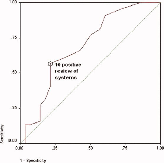
No differences were observed in the responses to the individual review of systems questions in women who did or did not have a lifetime history of IPV, with the exception that those with a positive history more commonly complained of difficulty sleeping and numbness and tingling in their hands or feet (although at best our study was sufficiently powered to detect only >20% differences in prevalences; Table 4). Although the sensitivity of having problems sleeping or experiencing numbness or tingling in patients with IPV was high, the specificity and positive and negative predictive values were not (Table 5).
| Review‐of‐Systems Questions | IPV History (n = 44) | No IPV History (n = 28) | P Value |
|---|---|---|---|
| |||
| 1. Shortness of breath | 25 (57) | 10 (36) | 0.081 |
| 2. Chest pain/pressure | 19 (43) | 9 (32) | 0.349 |
| 3. Abdominal pain | 17 (39) | 10 (36) | 0.803 |
| 4. Headaches | 24 (55) | 13 (46) | 0.502 |
| 5. Rashes | 15 (34) | 9 (32) | 0.864 |
| 6. Bruising | 32 (73) | 12 (43) | 0.011 |
| 7. Joint pain/stiffness | 27 (61) | 11 (39) | 0.067 |
| 8. Muscle pain/spasms | 22 (50) | 11 (39) | 0.374 |
| 9. Pain with intercourse | 8 (19) | 4 (14) | 0.753 |
| 10. Pelvic pain/cramps | 13 (30) | 5 (18) | 0.264 |
| 11. Nausea/vomiting | 19 (43) | 11 (39) | 0.744 |
| 12. Nervous/anxious | 28 (64) | 14 (50) | 0.253 |
| 13. Sad/crying | 21 (48) | 12 (43) | 0.686 |
| 14. Weight gain/loss | 26 (59) | 17 (61) | 0.891 |
| 15. Trouble sleeping | 37 (84) | 12 (43) | 0.000* |
| 16. Fever/chills | 19 (43) | 6 (21) | 0.059 |
| 17. Frequent/painful urination | 11 (25) | 6 (21) | 0.728 |
| 18. Pounding/emrregular heart beat | 14 (32) | 7 (25) | 0.535 |
| 19. Dizzy/passing out | 13 (30) | 7 (25) | 0.675 |
| 20. Memory problem | 19 (43) | 7 (25) | 0.117 |
| 21. Diarrhea/constipation | 27 (61) | 10 (36) | 0.034 |
| 22. Numbness/tingling | 35 (80) | 9 (32) | <0.0001* |
| 23. Pain chewing/swallowing | 8 (18) | 5 (18) | 0.972 |
| Trouble Sleeping | Numbness/Tingling | |
|---|---|---|
| Sensitivity (%) | 84 | 74 |
| Specificity (%) | 57 | 68 |
| Positive predictive value (%) | 76 | 78 |
| Negative predictive value (%) | 70 | 68 |
The admission history forms filled out by first‐year admitting residents showed that only 18 (25%) of the women were screened for IPV, even though the history and physical examination template used at Denver Health Medical Center includes a prompt in the social history section pertaining to a history of violence as a reminder.
DISCUSSION
The important findings of this study were that women admitted to the internal medicine service of a university‐affiliated public safety‐net hospital had a high prevalence of IPV (22% and 61% 1‐year and lifetime prevalences, respectively), that most women with a history of IPV had previously sought help for the problem, many from physicians, that women were more likely to have a history of IPV if they had >10 positive responses to questions asked in a routine review of systems (particularly problems sleeping and experiencing numbness or tingling in their extremities), and that routine screening for IPV was uncommon at the time of admission.
These conclusions should be interpreted with respect to a number of limitations in our study. First, although our study was designed to be a consecutive series, the interviewers did not have sufficient time to meet with and interview every woman admitted before they were discharged. This occurred in part because the interviewers were available only for a portion of each day, some patients were discharged within 24 hours of admission, and many were out of their rooms for ancillary testing. Within the interviewers' time constraints, however, all hospitalized women meeting entry criteria who were available were approached. Our data could, however, overrepresent the prevalence of IPV if hospitalized women with a history of IPV had longer hospital stays than those who did not or if those experiencing IPV were out of their rooms less frequently (eg, for diagnostic tests). On the other hand, our data could underrepresent the true prevalence of IPV if patients with a history of IPV had shorter hospital stays or if they received more ancillary testing that caused them to be out of their rooms more frequently. Second, none of our interviewers had specific training in interviewing techniques. Accordingly, our data could have underestimated the true prevalence of IPV if interviewers with advanced training in probing sensitive topics had more success in eliciting positive responses. Third, the relationship between a history of IPV and multiple positive responses to the review of systems may be confounded if some of these patients also had a history of adverse childhood experiences or other experiences resulting in posttraumatic stress disorder as these patients also have an increased prevalence of chronic and functional disorders.2527 Finally, as our numbers were small, we were not powered to detect clinically important differences in demographics or specific positive answers on the review of systems.
To the best of our knowledge, the only study presenting IPV prevalence data in patients hospitalized for other than psychiatric problems was performed by McKenzie and colleagues18 in 1997. In their group of 130 patients (61 on internal medicine, 59 on surgery, 7 on obstetrics, and 3 on psychiatry), the 1‐year and lifetime prevalences of IPV were only 5% and 26%, respectively. McKenzie and colleagues used only 1 question to screen for IPV, but that single question incorporated 2 of the 4 questions used in our survey. Forty‐three of our 44 patients (98%) with a history of IPV were discovered on the basis of these 2 questions. The hospitals in which the 2 studies were done were similar, as were the ages and levels of education of the 2 populations studied and the percentage of eligible patients who agreed to participate. The patients in the 2 studies were different with respect to race, language mix, and the percentage who were insured, but neither study found differences in the prevalence of IPV as a function of race or insurance (although others have found an association of IPV with being uninsured1, 3, 4, 12, 23). Our study was conducted in women admitted exclusively to an internal medicine service, whereas nearly half of the patients studied by McKenzie and colleagues were admitted to surgical, gynecologic, or psychiatric services. Although McKenzie and colleagues found no difference in the prevalence of IPV as a function of admitting service, others have suggested that the prevalence of IPV is higher in patients admitted for trauma or psychiatric problems.1517, 28 The percentage of patients who self‐administered the questionnaires was 57% in our study and 77% in the study by McKenzie and colleagues. Neither study, however, found a difference in the percentage of IPV in patients who self‐administered the survey versus those who were interviewed. Women may have become more comfortable discussing this issue in the 10‐year interval between these 2 studies, or the prevalence of IPV may have increased. The only other study of IPV in hospitalized patients of which we are aware reported a 90% 1‐year prevalence in suicidal women admitted to a psychiatric service.28
Several studies have reported that victims of IPV have multiple somatic complaints and an increased prevalence of chronic and functional illnesses.1923 We confirmed that women experiencing IPV have more positive responses to questions posed in a review of systems, but the low specificity and positive and negative predictive values of the responses make this association of little clinical utility.
For only 18 of the 72 patients (25%) in our study was there evidence that they were screened for a history of IPV by the admitting resident. If more women were screened without a response being recorded, or if women were screened only for a current history of violence, our data may not accurately reflect the true rate at which screening occurred; however, the rate of screening that we observed is consistent with a number of other studies.12, 22, 2931 Fourteen of 18 patients who were screened for IPV by the resident gave negative responses. Ten of these, however, gave positive responses to our interviewers. Accordingly, the sensitivity, specificity, and positive and negative predictive values of the information recorded by the admitting resident were 40%, 100%, 100%, and 57%, respectively (assuming that the responses given to the IPV survey represent the gold standard), and this confirms that routine screening underestimates the prevalence of this problem. Accordingly, we identified 2 problems pertaining to screening for IPV: (1) it is not routinely done at the time of hospital admission, and (2) responses reported during routine screening are frequently incorrect. A number of barriers to routine screening have been previously identified, as have interventions designed to increase screening.32 Providing specific screening questions increases the identification of victims of IPV, but simply educating healthcare providers does not.32 Our history and physical templates have a prompt for violence victim to facilitate the screening, but as a result of this study, we are changing our prompting question and indicating what should be done if the response is positive.
The US Preventive Services Task Force and the Canadian Task Force on Preventive Health Care both concluded that there was insufficient evidence to recommend for or against routine screening for IPV.3335 Their rationale was that trials assessing the effectiveness of screening have not been published, that studies designed to assess the effectiveness of any resulting intervention are few in number, focused on pregnant women, and limited by problems in study design, that no studies have determined the accuracy of the screening tools, and that none have addressed the potential harm of screening.3335 The US Preventive Services Task Force did recommend screening if providers were concerned about IPV.34 Our data would suggest that there is little in the admission history that distinguishes women who might be victims of IPV from those who might not. Guidelines published by the American Medical Association, the American Academy of Family Physicians, and the American College of Obstetricians and Gynecologists promote routine screening of all patients.3638 Janssen and colleagues39 support the importance of screening on the basis that IPV is associated with numerous physical and mental health problems (eg, arthritis, migraines and other types of headaches, vaginal bleeding, ulcers, spastic colon, chronic pain, substance abuse, depression, and suicide ideation) and that establishing the link between these conditions and IPV could be important with respect to developing appropriate diagnostic and therapeutic approaches to patients' complaints. Screening also allows physicians to become more knowledgeable about their patients' lives, facilitating their ability to provide a supportive relationship that, in turn, increases women's likelihood of using an intervention method.39 We did not confirm an increased prevalence of any of the complaints noted by Janssen and colleagues in the women experiencing a history of IPV, but we did find an increased prevalence of insomnia and extremity numbness in women admitting to IPV as well as an overall increase in the number of positive responses to the review of systems. Screening identifies women who should receive information about reporting IPV, obtaining available assistance, planning for personal safety, and formal counseling as these have all been shown to reduce the severity of IPV and to improve the quality of life in rather large, randomized controlled trials.4043
As previously observed by others,13, 22, 29, 4446 the large majority of women that we approached welcomed screening for IPV. Over half of those with a history of IPV had previously sought help for the problem, over one‐third of these sought help from physicians, and most took the resource card that we offered, regardless of whether they did or did not have a history of IPV (this suggests either that our data may actually underestimate the true prevalence of IPV or that patients taking the information knew of others experiencing this problem). Accordingly, regardless of whether physicians believe that routine screening is warranted, patients see physicians and other healthcare workers as a resource for this problem.
We have confirmed that a history of IPV is very common in women admitted to an internal medicine service of a university‐affiliated public hospital and that female victims of IPV have more positive responses on the review of systems (particularly difficulty sleeping and extremity numbness or tingling) than those who have not. Although we initially hypothesized that finding numerous somatic complaints might serve as a marker for IPV, thereby identifying patients for whom more careful screening should occur, finding such a high prevalence of IPV argues that screening should be a routine part of the history for all women admitted to internal medicine inpatient services.
Acknowledgements
The authors thank the patients who agreed to participate in this study during their hospitalization. They also thank Cheri Maestas and Debbie Rodriquez for their support and help in interviewing patients.
- ,,.Prevalence and determinants of intimate partner abuse among public hospital primary care patients.J Gen Intern Med.2000;15:811–817.
- ,,,.Women's experiences with violence: a national study.Womens Health Issues.2007;17:3–12.
- ,,,.Multistate analysis of factors associated with intimate partner violence.Am J Prev Med.2002;22:156–164.
- ,,,.Frequency and correlates of intimate partner violence by type: physical, sexual, and psychological battering.Am J Public Health.2000;90:553–559.
- ,,,,.Prevalence of domestic violence among patients in three ambulatory care internal medicine clinics.J Gen Intern Med.1991;6:317–322.
- ,,,.Prevalence of partner violence against 7,443 African American, White and Hispanic women receiving care at urban public primary care clinics.Public Health Nurs.2005;22:98–107.
- ,,,,.Evaluating domestic partner abuse in a family practice clinic.Fam Med.1997;29:492–495.
- ,.Prevalence and predictors of physical partner abuse among Mexican American women.Am J Public Health.2001;91:441–445.
- ,,.Rates of intimate partner violence in the United States.Am J Public Health.1998;88:1702–1704.
- ,,,.Domestic violence against women incidence and prevalence in an emergency department population.JAMA.1995;273:1763–1767.
- ,,, et al.Accuracy of 3 brief screening questions for detecting partner violence in the emergency department.JAMA.1997;277:1357–1361.
- ,,.A prevalence survey of abuse and screening for abuse in urgent care patients.Obstet Gynecol.1998;91:511–514.
- Morbidity and Mortality Weekly Report.Use of medical care, police assistance and restraining orders by women reporting intimate partner violence—Massachusetts, 1996–1997.JAMA.2000;284:558.
- ,,.Interpersonal violence among women seeking welfare: unraveling lives.Am J Public Health.2006;96:1409–1415.
- ,.A 5‐year follow‐up study of 117 battered women.Am J Public Health.1991;81:1486–1488.
- ,,.Rates and relative risk of hospital admission among women in violent intimate partner relationships.Am J Public Health.2000;90:1416–1420.
- ,,,.Intimate partner violence against women: do victims cost health plans more?J Fam Pract.1999;48:439–443.
- ,,,.Prevalence of domestic violence in an inpatient female population.J Gen Intern Med.1998;13:277–279.
- ,,, et al.Intimate partner violence and physical health consequences.Arch Intern Med.2002;162:1157–1163.
- ,,,,.Physical health consequences of physical and psychological intimate partner violence.Arch Fam Med.2000;9:451–457.
- ,,, et al.Sexual and physical abuse in women with functional or organic gastrointestinal disorders.Ann Intern Med.1990;113:828–833.
- ,,.Prevalence of intimate partner violence and health implications for women using emergency departments and primary care clinics.Womens Health Issues.2004;14:19–29.
- ,,, et al.The “battering syndrome”: prevalence and clinical characteristics of domestic violence in primary care internal medicine practices.Ann Intern Med.1995;123:737–746.
- ,.DeGowin and DeGowin's Bedside Diagnostic Examination.5th ed.New York, NY:Macmillan Publishing;1987:18–29.
- ,,, et al.Relationship of childhood abuse and household dysfunction to many of the leading causes of death in adults. The Adverse Childhood Experiences (ACE) Study.Am J Prev Med.1998;14:245–258.
- ,,,,,.Posttraumatic stress disorder and health status among female and male medical patients.J Trauma Stress.2004;17:1–9.
- ,,,,.Posttraumatic stress disorder and physical comorbidity among female children and adolescents: results from service‐use data.Pediatrics.2005:116;e767–e776.
- ,,,,.Prevalence and severity of intimate partner violence and associations with family functioning and alcohol abuse in psychiatric inpatients with suicidal intent.J Clin Psychiatry.2006;67:23–29.
- ,,.Intimate partner violence screening and intervention: data from eleven Pennsylvania and California community hospital emergency departments.J Emerg Nurs.2001;27:141–149.
- ,.Missed opportunities: emergency department visits by police‐identified victims of intimate partner violence.Emerg Med.2006;47:190–199.
- ,,, et al.Intimate partner violence and patient screening across medical specialties.Acad Emerg Med.2005;12:712–722.
- ,,,,.Screening for intimate partner violence by health care providers: barriers and interventions.Am J Prev Med.2000;19:230–237.
- ,,,.Screening women and elderly adults for family and intimate partner violence: a review of the evidence for the U.S. Preventive Services Task Force.Ann Intern Med.2004;140:387–396.
- U.S. Preventive Services Task Force.Screening for family and intimate partner violence: recommendation statement.Ann Intern Med.2004;140:382–386.
- ,.Interventions for violence against women: scientific review.JAMA.2003;289:589–600.
- American Medical Association. Policy H‐515.965: family and intimate partner violence. Available at: http://www.ama‐assn.org. Accessed May2007.
- American Academy of Family Physicians. Family and intimate partner violence and abuse. Available at: www.aafp.org/x16506.xml. Accessed May2007.
- Domestic Violence.Washington, DC:American College of Obstetrics and Gynecology;1999. Educational Bulletin Number; No. 257.
- ,,.Assessment for intimate partner violence: where do we stand?J Am Board Fam Med.2006;19:413–415.
- ,,,,,.What happens when health care providers ask about intimate partner violence? A description of consequences from the perspectives of female survivors.JAMA.2003;58:76–81.
- ,,,,,.Assessing intimate partner violence in health care settings leads to women's receipt of interventions and improved health.Public Health Rep.2006;121:435–444.
- ,,.An evaluation of interventions to decrease intimate partner violence to pregnant women.Public Health Nurs.2000;17:443–451.
- ,.Reducing violence using community‐based advocacy for women with abusive partners.J Consult Clin Psychol.1999;67:43–53.
- ,,,,.Help‐seeking for intimate partner violence and forced sex in South Carolina.Am J Prev Med.2000;19:316–320.
- ,,, et al.Women's opinions about domestic violence screening and mandatory reporting.Am J Prev Med.2000;19:279–285.
- ,,,.The factors associated with disclosure of intimate partner abuse to clinicians.J Fam Pract.2001;50:338–344.
The prevalence of intimate partner violence (IPV; defined as mental and/or physical violence directed from 1 person in an intimate relationship to the other) varies widely, depending on the population sampled and method of data collection. In the United States, IPV against women, occurring within the year prior to contact with a healthcare professional, ranges from 2% to 15% in surveys done by telephone, in primary care clinics, or in face‐to‐face home interviews19 and from 10% to 30% in surveys of patients visiting urgent care or emergency departments.1012 The prevalence of IPV occurring at any time during the life of the patient ranges from 18% in the aforementioned settings to as high as 88% in women applying for welfare.1, 2, 4, 5, 10, 1214
Although reports indicate that victims of IPV are more likely to be hospitalized,1517 the only study assessing the prevalence of IPV in hospitalized patients included women on medical, surgical, and obstetrical services and reported 1‐year and lifetime prevalences of only 5% and 23%, respectively.18
We hypothesized that the prevalence of IPV in hospitalized patients would be at least as high as that reported from emergency departments and sought to measure the 1‐year and lifetime prevalences of IPV in women admitted to a general internal medicine service. In addition, because studies done in various outpatient settings have reported that victims of IPV have a variety of somatic complaints and an increased prevalence of chronic and functional illnesses,1923 we also sought to determine whether women with a history of IPV and women without a history of IPV had different numbers or types of positive responses to questions asked on the review of systems.
PATIENTS AND METHODS
This study was approved by the Colorado Multiple Institution Review Board, and informed consent was obtained from all participants.
Women between the ages of 18 and 60 who were admitted to the internal medicine floor service of Denver Health Medical Center (a university‐affiliated public safety‐net hospital) between January 1 and February 28, 2004 and between October 1 and October 30, 2004 were approached to participate. These dates were selected on the basis of the availability of our interviewers. Patients older than 60 were excluded to avoid overlap between IPV and the problem of elder abuse. Women were excluded if they were unable to give informed consent, were pregnant, were incarcerated, were on contact precautions, or spoke a language other than English or Spanish. Although IPV is common in pregnant women and may occur in women who are incarcerated, these are considered vulnerable populations with respect to obtaining approval from internal review boards.
The questionnaire consisted of 23 review‐of‐systems questions,24 4 questions adapted from a previously validated screen for IPV11 (Table 1), and 1 question about attempts to seek help (Table 1). Women were considered to have experienced IPV if they gave positive responses to any of the 4 questions targeting IPV. According to patient preference, the combined questionnaire was either read and filled out by each subject independently or was read to her by a female interviewer who then recorded the subject's verbal responses. All interviewers were women with a shared common concern about, and interest in, IPV. Although none had advanced training in psychology, social work, or other formal discipline that involved interviewing skills, all interviews were scripted so that interactions with subjects and completion of the questionnaires would be uniform. Responses indicating sometimes were considered to be positive. Responses that were not answered, left blank, or marked as not applicable were considered to be negative.
| 1. Have you ever been hit, kicked, punched, or otherwise hurt by someone? If so, by whom? Friend, boyfriend, girlfriend, husband, family member, somebody you do not know, other |
| 2. Within the last year, have you been hit, kicked, or otherwise hurt by someone? If so, by whom? Friend, boyfriend, girlfriend, husband, family member, somebody you do not know, other |
| 3. Do you feel safe in your current relationship? |
| 4. Is there a partner from a previous relationship who is making you feel unsafe now? |
| 5. If you answered yes to any of the above, have you ever asked for help from police, shelter, counselor, physician? If so, how long ago? |
Each patient's medical record was reviewed to determine her age, race, number of previous hospital admissions, visits to the emergency department and walk‐in clinic, visits to primary care and subspecialty physicians, and whether the patient had been screened for IPV as recorded on the admission history and physical template. Admission diagnosis was obtained from the history and physical template, and the discharge diagnosis was obtained from the discharge paperwork. Functional diagnoses were considered to be symptoms (eg, shortness of breath) or problems (eg, constipation) that could not clearly be linked to a specific disease process. All participants were offered a card containing a list of resources for victims of IPV.
Data were analyzed with SAS 8.1 (SAS Institute, Cary, NC) and SPSS 11.5 (SPSS, Chicago, IL). The Student t test was used to compare continuous variables. Data are reported as means standard deviation. Chi‐square analysis was used to test associations between race, primary language, level of education, insurance status, admitting diagnosis, discharge diagnosis, number of previous hospital admissions, visit type, and the presence of IPV. For these, P < 0.05 was considered to be significant. The association of positive review‐of‐systems responses with the presence of IPV was also tested by chi‐square analysis, but P < 0.002 was considered to be significant on the basis of a Bonferroni adjustment for multiple comparisons. A receiver operating characteristic curve was used to assess the relationship between the number of positive responses to the questions included in the review of systems and a history of IPV. The odds ratio and confidence intervals were calculated to test the association between the number of positive responses to the review‐of‐systems questions and a lifetime history of IPV.
RESULTS
Throughout the dates of the study, 245 women were admitted to the internal medicine service, and 106 were excluded (Figure 1). Of the 139 eligible women, 78 were available to the interviewers and asked to participate, and 72 (92%) agreed. IPV occurring within the year prior to the interview or at any point in the patient's lifetime was reported by 16 (22%) and 44 (61%) subjects, respectively. No significant differences were seen in women who did or did not experience IPV at anytime in their life with respect to age, race, insurance status, education, number of scheduled outpatient, urgent, or emergent visits, or admission or discharge diagnosis even when the diagnoses were grouped into a functional category (although at best our study was powered to detect only >35% differences in prevalences; Tables 2 and 3). Of women reporting a lifetime history of IPV, 26 of 44 (59%) had previously sought help, and 9 of those 26 (35%) said that they sought help from a physician.
| IPV History | No IPV History | |
|---|---|---|
| ||
| Number (%) | 44 (61) | 28 (39) |
| Age (mean standard deviation) | 44 10 | 45 12 |
| Race [n, (%)] | ||
| Caucasian | 18 (41) | 6 (21) |
| Hispanic | 13 (30) | 15 (54) |
| African American | 12 (27) | 6 (21) |
| Other | 1 (2) | 1 (4) |
| Insurance status [n (%)] | ||
| Insured | 12 (27) | 5 (18) |
| Uninsured | 32 (73) | 23 (82) |
| Education [n (%)] | ||
| Grade school | 4 (9) | 3 (11) |
| Some high school | 13 (30) | 5 (18) |
| High school diploma | 15 (34) | 9 (32) |
| Some college | 9 (20) | 7 (25) |
| College degree | 2 (5) | 2 (7) |
| Postgraduate | 1 (2) | 2 (7) |
| Previous visit type (median, IQR) | ||
| Scheduled outpatient (includes primary care and subspecialty) | 2 (8) | 1.5 (7) |
| Emergency department and walk‐in clinic | 2 (3.5) | 1 (3) |
| Previous hospital admissions [n (%)] | ||
| 0 | 24 (55) | 16 (57) |
| 1 | 16 (36) | 4 (14) |
| 2 | 0 (0) | 4 (14) |
| 3 | 2 (5) | 2 (7) |
| >3 | 2 (5) | 2 (7) |
| Admission or Discharge Diagnosis | Admission | Discharge | ||
|---|---|---|---|---|
| IPV (n = 44) | No IPV (n = 28) | IPV (n = 44) | No IPV (n = 28) | |
| ||||
| Cardiovascular | ||||
| Chest pain (%)* | 8 (18) | 5 (18) | 6 (14) | 4 (14) |
| Cardiomyopathy | 0 | 0 | 1 | 0 |
| Cerebrovascular accident | 1 | 0 | 1 | 0 |
| Deep venous thrombosis | 0 | 0 | 1 | 0 |
| Hypertensive emergency | 0 | 0 | 1 | 0 |
| Palpitations* | 0 | 1 | 0 | 1 |
| Valvular disease | 0 | 0 | 1 | 0 |
| Venous stasis | 0 | 1 | 0 | 1 |
| Total (%) | 9 (20) | 7 (25) | 11 (25) | 6 (21) |
| Gastrointestinal | ||||
| Abdominal pain (%)* | 7 (16) | 4 (14) | 2 | 1 |
| Ascites | 0 | 1 | 0 | 0 |
| Constipation* | 0 | 0 | 1 | 0 |
| End‐stage liver disease | 1 | 1 | 1 | 2 |
| Esophagitis | 0 | 0 | 1 | 0 |
| Hepatitis | 1 | 0 | 1 | 0 |
| Nausea/vomiting* | 2 | 0 | 1 | 0 |
| Pancreatitis | 0 | 1 | 3 | 2 |
| Peptic ulcer disease | 1 | 0 | 1 | 0 |
| Upper gastrointestinal bleeding | 2 | 0 | 1 | 0 |
| Total (%) | 14 (32) | 7 (25) | 12 (27) | 5 (18) |
| Hematology/oncology | ||||
| Abdominal mass | 0 | 0 | 0 | 1 |
| Anemia | 1 | 0 | 1 | 0 |
| Breast cancer | 0 | 1 | 0 | 1 |
| Cervical cancer | 1 | 0 | 1 | 0 |
| Colon cancer | 0 | 1 | 0 | 1 |
| Sickle cell anemia | 1 | 0 | 1 | 0 |
| Thrombocytosis | 1 | 0 | 1 | 0 |
| Total (%) | 4 (9) | 2 (7) | 4 (9) | 3 (11) |
| Infectious disease | ||||
| Bacteremia/sepsis | 3 | 0 | 3 | 0 |
| Cellulitis | 1 | 0 | 1 | 1 |
| Cholangitis | 0 | 0 | 1 | 0 |
| Community‐acquired pneumonia | 2 | 2 | 2 | 1 |
| Endocarditis | 1 | 0 | 1 | 0 |
| Fever | 0 | 1 | 0 | 1 |
| Pelvic inflammatory disease | 0 | 0 | 0 | 1 |
| Urinary tract infection | 1 | 0 | 1 | 0 |
| Total (%) | 8 (18) | 3 (11) | 9 (20) | 4 (14) |
| Pulmonary | ||||
| Acute exacerbation of COPD | 0 | 0 | 1 | 0 |
| Asthma exacerbation | 1 | 1 | 1 | 2 |
| Pleuritic chest pain* | 0 | 0 | 1 | 0 |
| Pulmonary embolism | 0 | 0 | 1 | 0 |
| Shortness of breath* | 4 | 0 | 1 | 0 |
| Total (%) | 5 (11) | 1 (4) | 5 (11) | 2 (7) |
| Renal/genitourinary | ||||
| Acute renal failure | 0 | 1 | 0 | 1 |
| End‐stage renal disease | 1 | 2 | 1 | 2 |
| Nephrotic syndrome | 0 | 1 | 0 | 2 |
| Vaginal bleeding | 1 | 0 | 1 | 0 |
| Total (%) | 2 (5) | 4 (14) | 2 (5) | 5 (18) |
| Other | ||||
| Diabetic ketoacidosis | 0 | 1 | 0 | 1 |
| Extremity pain* | 0 | 1 | 0 | 0 |
| Mediastinal thickening | 0 | 0 | 0 | 1 |
| Hyponatremia | 0 | 1 | 0 | 1 |
| Lower extremity swelling | 2 | 1 | 0 | 0 |
| Somatization* | 0 | 0 | 1 | 0 |
| Total (%) | 2 (5) | 4 (14) | 1 (2) | 3 (11) |
| Total functional diagnoses (%) | 21 (48) | 11 (39) | 12 (27) | 6 (21) |
Women with a 1‐year history of IPV and women without a 1‐year history of IPV had 11.4 4.7 and 7.7 5.4 positive responses to the review of systems (P < 0.01), respectively. Women with a lifetime history of IPV and women without a lifetime history of IPV had 10.9 4.4 and 7.7 5.4 positive responses (P < 0.01), respectively. The receiver operating characteristic curve of the number of positive responses versus a lifetime history of IPV is presented in Figure 2. Subjects with 10 or more positive responses were 4.8 times more likely to report a lifetime history of IPV than subjects with 9 or fewer positive responses (confidence interval = 1.614.2, P = 0.003). The c‐statistic indicating the ability of the review of systems to properly classify cases when there were 10 or more positive responses was 0.692.
No differences were observed in the responses to the individual review of systems questions in women who did or did not have a lifetime history of IPV, with the exception that those with a positive history more commonly complained of difficulty sleeping and numbness and tingling in their hands or feet (although at best our study was sufficiently powered to detect only >20% differences in prevalences; Table 4). Although the sensitivity of having problems sleeping or experiencing numbness or tingling in patients with IPV was high, the specificity and positive and negative predictive values were not (Table 5).
| Review‐of‐Systems Questions | IPV History (n = 44) | No IPV History (n = 28) | P Value |
|---|---|---|---|
| |||
| 1. Shortness of breath | 25 (57) | 10 (36) | 0.081 |
| 2. Chest pain/pressure | 19 (43) | 9 (32) | 0.349 |
| 3. Abdominal pain | 17 (39) | 10 (36) | 0.803 |
| 4. Headaches | 24 (55) | 13 (46) | 0.502 |
| 5. Rashes | 15 (34) | 9 (32) | 0.864 |
| 6. Bruising | 32 (73) | 12 (43) | 0.011 |
| 7. Joint pain/stiffness | 27 (61) | 11 (39) | 0.067 |
| 8. Muscle pain/spasms | 22 (50) | 11 (39) | 0.374 |
| 9. Pain with intercourse | 8 (19) | 4 (14) | 0.753 |
| 10. Pelvic pain/cramps | 13 (30) | 5 (18) | 0.264 |
| 11. Nausea/vomiting | 19 (43) | 11 (39) | 0.744 |
| 12. Nervous/anxious | 28 (64) | 14 (50) | 0.253 |
| 13. Sad/crying | 21 (48) | 12 (43) | 0.686 |
| 14. Weight gain/loss | 26 (59) | 17 (61) | 0.891 |
| 15. Trouble sleeping | 37 (84) | 12 (43) | 0.000* |
| 16. Fever/chills | 19 (43) | 6 (21) | 0.059 |
| 17. Frequent/painful urination | 11 (25) | 6 (21) | 0.728 |
| 18. Pounding/emrregular heart beat | 14 (32) | 7 (25) | 0.535 |
| 19. Dizzy/passing out | 13 (30) | 7 (25) | 0.675 |
| 20. Memory problem | 19 (43) | 7 (25) | 0.117 |
| 21. Diarrhea/constipation | 27 (61) | 10 (36) | 0.034 |
| 22. Numbness/tingling | 35 (80) | 9 (32) | <0.0001* |
| 23. Pain chewing/swallowing | 8 (18) | 5 (18) | 0.972 |
| Trouble Sleeping | Numbness/Tingling | |
|---|---|---|
| Sensitivity (%) | 84 | 74 |
| Specificity (%) | 57 | 68 |
| Positive predictive value (%) | 76 | 78 |
| Negative predictive value (%) | 70 | 68 |
The admission history forms filled out by first‐year admitting residents showed that only 18 (25%) of the women were screened for IPV, even though the history and physical examination template used at Denver Health Medical Center includes a prompt in the social history section pertaining to a history of violence as a reminder.
DISCUSSION
The important findings of this study were that women admitted to the internal medicine service of a university‐affiliated public safety‐net hospital had a high prevalence of IPV (22% and 61% 1‐year and lifetime prevalences, respectively), that most women with a history of IPV had previously sought help for the problem, many from physicians, that women were more likely to have a history of IPV if they had >10 positive responses to questions asked in a routine review of systems (particularly problems sleeping and experiencing numbness or tingling in their extremities), and that routine screening for IPV was uncommon at the time of admission.
These conclusions should be interpreted with respect to a number of limitations in our study. First, although our study was designed to be a consecutive series, the interviewers did not have sufficient time to meet with and interview every woman admitted before they were discharged. This occurred in part because the interviewers were available only for a portion of each day, some patients were discharged within 24 hours of admission, and many were out of their rooms for ancillary testing. Within the interviewers' time constraints, however, all hospitalized women meeting entry criteria who were available were approached. Our data could, however, overrepresent the prevalence of IPV if hospitalized women with a history of IPV had longer hospital stays than those who did not or if those experiencing IPV were out of their rooms less frequently (eg, for diagnostic tests). On the other hand, our data could underrepresent the true prevalence of IPV if patients with a history of IPV had shorter hospital stays or if they received more ancillary testing that caused them to be out of their rooms more frequently. Second, none of our interviewers had specific training in interviewing techniques. Accordingly, our data could have underestimated the true prevalence of IPV if interviewers with advanced training in probing sensitive topics had more success in eliciting positive responses. Third, the relationship between a history of IPV and multiple positive responses to the review of systems may be confounded if some of these patients also had a history of adverse childhood experiences or other experiences resulting in posttraumatic stress disorder as these patients also have an increased prevalence of chronic and functional disorders.2527 Finally, as our numbers were small, we were not powered to detect clinically important differences in demographics or specific positive answers on the review of systems.
To the best of our knowledge, the only study presenting IPV prevalence data in patients hospitalized for other than psychiatric problems was performed by McKenzie and colleagues18 in 1997. In their group of 130 patients (61 on internal medicine, 59 on surgery, 7 on obstetrics, and 3 on psychiatry), the 1‐year and lifetime prevalences of IPV were only 5% and 26%, respectively. McKenzie and colleagues used only 1 question to screen for IPV, but that single question incorporated 2 of the 4 questions used in our survey. Forty‐three of our 44 patients (98%) with a history of IPV were discovered on the basis of these 2 questions. The hospitals in which the 2 studies were done were similar, as were the ages and levels of education of the 2 populations studied and the percentage of eligible patients who agreed to participate. The patients in the 2 studies were different with respect to race, language mix, and the percentage who were insured, but neither study found differences in the prevalence of IPV as a function of race or insurance (although others have found an association of IPV with being uninsured1, 3, 4, 12, 23). Our study was conducted in women admitted exclusively to an internal medicine service, whereas nearly half of the patients studied by McKenzie and colleagues were admitted to surgical, gynecologic, or psychiatric services. Although McKenzie and colleagues found no difference in the prevalence of IPV as a function of admitting service, others have suggested that the prevalence of IPV is higher in patients admitted for trauma or psychiatric problems.1517, 28 The percentage of patients who self‐administered the questionnaires was 57% in our study and 77% in the study by McKenzie and colleagues. Neither study, however, found a difference in the percentage of IPV in patients who self‐administered the survey versus those who were interviewed. Women may have become more comfortable discussing this issue in the 10‐year interval between these 2 studies, or the prevalence of IPV may have increased. The only other study of IPV in hospitalized patients of which we are aware reported a 90% 1‐year prevalence in suicidal women admitted to a psychiatric service.28
Several studies have reported that victims of IPV have multiple somatic complaints and an increased prevalence of chronic and functional illnesses.1923 We confirmed that women experiencing IPV have more positive responses to questions posed in a review of systems, but the low specificity and positive and negative predictive values of the responses make this association of little clinical utility.
For only 18 of the 72 patients (25%) in our study was there evidence that they were screened for a history of IPV by the admitting resident. If more women were screened without a response being recorded, or if women were screened only for a current history of violence, our data may not accurately reflect the true rate at which screening occurred; however, the rate of screening that we observed is consistent with a number of other studies.12, 22, 2931 Fourteen of 18 patients who were screened for IPV by the resident gave negative responses. Ten of these, however, gave positive responses to our interviewers. Accordingly, the sensitivity, specificity, and positive and negative predictive values of the information recorded by the admitting resident were 40%, 100%, 100%, and 57%, respectively (assuming that the responses given to the IPV survey represent the gold standard), and this confirms that routine screening underestimates the prevalence of this problem. Accordingly, we identified 2 problems pertaining to screening for IPV: (1) it is not routinely done at the time of hospital admission, and (2) responses reported during routine screening are frequently incorrect. A number of barriers to routine screening have been previously identified, as have interventions designed to increase screening.32 Providing specific screening questions increases the identification of victims of IPV, but simply educating healthcare providers does not.32 Our history and physical templates have a prompt for violence victim to facilitate the screening, but as a result of this study, we are changing our prompting question and indicating what should be done if the response is positive.
The US Preventive Services Task Force and the Canadian Task Force on Preventive Health Care both concluded that there was insufficient evidence to recommend for or against routine screening for IPV.3335 Their rationale was that trials assessing the effectiveness of screening have not been published, that studies designed to assess the effectiveness of any resulting intervention are few in number, focused on pregnant women, and limited by problems in study design, that no studies have determined the accuracy of the screening tools, and that none have addressed the potential harm of screening.3335 The US Preventive Services Task Force did recommend screening if providers were concerned about IPV.34 Our data would suggest that there is little in the admission history that distinguishes women who might be victims of IPV from those who might not. Guidelines published by the American Medical Association, the American Academy of Family Physicians, and the American College of Obstetricians and Gynecologists promote routine screening of all patients.3638 Janssen and colleagues39 support the importance of screening on the basis that IPV is associated with numerous physical and mental health problems (eg, arthritis, migraines and other types of headaches, vaginal bleeding, ulcers, spastic colon, chronic pain, substance abuse, depression, and suicide ideation) and that establishing the link between these conditions and IPV could be important with respect to developing appropriate diagnostic and therapeutic approaches to patients' complaints. Screening also allows physicians to become more knowledgeable about their patients' lives, facilitating their ability to provide a supportive relationship that, in turn, increases women's likelihood of using an intervention method.39 We did not confirm an increased prevalence of any of the complaints noted by Janssen and colleagues in the women experiencing a history of IPV, but we did find an increased prevalence of insomnia and extremity numbness in women admitting to IPV as well as an overall increase in the number of positive responses to the review of systems. Screening identifies women who should receive information about reporting IPV, obtaining available assistance, planning for personal safety, and formal counseling as these have all been shown to reduce the severity of IPV and to improve the quality of life in rather large, randomized controlled trials.4043
As previously observed by others,13, 22, 29, 4446 the large majority of women that we approached welcomed screening for IPV. Over half of those with a history of IPV had previously sought help for the problem, over one‐third of these sought help from physicians, and most took the resource card that we offered, regardless of whether they did or did not have a history of IPV (this suggests either that our data may actually underestimate the true prevalence of IPV or that patients taking the information knew of others experiencing this problem). Accordingly, regardless of whether physicians believe that routine screening is warranted, patients see physicians and other healthcare workers as a resource for this problem.
We have confirmed that a history of IPV is very common in women admitted to an internal medicine service of a university‐affiliated public hospital and that female victims of IPV have more positive responses on the review of systems (particularly difficulty sleeping and extremity numbness or tingling) than those who have not. Although we initially hypothesized that finding numerous somatic complaints might serve as a marker for IPV, thereby identifying patients for whom more careful screening should occur, finding such a high prevalence of IPV argues that screening should be a routine part of the history for all women admitted to internal medicine inpatient services.
Acknowledgements
The authors thank the patients who agreed to participate in this study during their hospitalization. They also thank Cheri Maestas and Debbie Rodriquez for their support and help in interviewing patients.
- ,,.Prevalence and determinants of intimate partner abuse among public hospital primary care patients.J Gen Intern Med.2000;15:811–817.
- ,,,.Women's experiences with violence: a national study.Womens Health Issues.2007;17:3–12.
- ,,,.Multistate analysis of factors associated with intimate partner violence.Am J Prev Med.2002;22:156–164.
- ,,,.Frequency and correlates of intimate partner violence by type: physical, sexual, and psychological battering.Am J Public Health.2000;90:553–559.
- ,,,,.Prevalence of domestic violence among patients in three ambulatory care internal medicine clinics.J Gen Intern Med.1991;6:317–322.
- ,,,.Prevalence of partner violence against 7,443 African American, White and Hispanic women receiving care at urban public primary care clinics.Public Health Nurs.2005;22:98–107.
- ,,,,.Evaluating domestic partner abuse in a family practice clinic.Fam Med.1997;29:492–495.
- ,.Prevalence and predictors of physical partner abuse among Mexican American women.Am J Public Health.2001;91:441–445.
- ,,.Rates of intimate partner violence in the United States.Am J Public Health.1998;88:1702–1704.
- ,,,.Domestic violence against women incidence and prevalence in an emergency department population.JAMA.1995;273:1763–1767.
- ,,, et al.Accuracy of 3 brief screening questions for detecting partner violence in the emergency department.JAMA.1997;277:1357–1361.
- ,,.A prevalence survey of abuse and screening for abuse in urgent care patients.Obstet Gynecol.1998;91:511–514.
- Morbidity and Mortality Weekly Report.Use of medical care, police assistance and restraining orders by women reporting intimate partner violence—Massachusetts, 1996–1997.JAMA.2000;284:558.
- ,,.Interpersonal violence among women seeking welfare: unraveling lives.Am J Public Health.2006;96:1409–1415.
- ,.A 5‐year follow‐up study of 117 battered women.Am J Public Health.1991;81:1486–1488.
- ,,.Rates and relative risk of hospital admission among women in violent intimate partner relationships.Am J Public Health.2000;90:1416–1420.
- ,,,.Intimate partner violence against women: do victims cost health plans more?J Fam Pract.1999;48:439–443.
- ,,,.Prevalence of domestic violence in an inpatient female population.J Gen Intern Med.1998;13:277–279.
- ,,, et al.Intimate partner violence and physical health consequences.Arch Intern Med.2002;162:1157–1163.
- ,,,,.Physical health consequences of physical and psychological intimate partner violence.Arch Fam Med.2000;9:451–457.
- ,,, et al.Sexual and physical abuse in women with functional or organic gastrointestinal disorders.Ann Intern Med.1990;113:828–833.
- ,,.Prevalence of intimate partner violence and health implications for women using emergency departments and primary care clinics.Womens Health Issues.2004;14:19–29.
- ,,, et al.The “battering syndrome”: prevalence and clinical characteristics of domestic violence in primary care internal medicine practices.Ann Intern Med.1995;123:737–746.
- ,.DeGowin and DeGowin's Bedside Diagnostic Examination.5th ed.New York, NY:Macmillan Publishing;1987:18–29.
- ,,, et al.Relationship of childhood abuse and household dysfunction to many of the leading causes of death in adults. The Adverse Childhood Experiences (ACE) Study.Am J Prev Med.1998;14:245–258.
- ,,,,,.Posttraumatic stress disorder and health status among female and male medical patients.J Trauma Stress.2004;17:1–9.
- ,,,,.Posttraumatic stress disorder and physical comorbidity among female children and adolescents: results from service‐use data.Pediatrics.2005:116;e767–e776.
- ,,,,.Prevalence and severity of intimate partner violence and associations with family functioning and alcohol abuse in psychiatric inpatients with suicidal intent.J Clin Psychiatry.2006;67:23–29.
- ,,.Intimate partner violence screening and intervention: data from eleven Pennsylvania and California community hospital emergency departments.J Emerg Nurs.2001;27:141–149.
- ,.Missed opportunities: emergency department visits by police‐identified victims of intimate partner violence.Emerg Med.2006;47:190–199.
- ,,, et al.Intimate partner violence and patient screening across medical specialties.Acad Emerg Med.2005;12:712–722.
- ,,,,.Screening for intimate partner violence by health care providers: barriers and interventions.Am J Prev Med.2000;19:230–237.
- ,,,.Screening women and elderly adults for family and intimate partner violence: a review of the evidence for the U.S. Preventive Services Task Force.Ann Intern Med.2004;140:387–396.
- U.S. Preventive Services Task Force.Screening for family and intimate partner violence: recommendation statement.Ann Intern Med.2004;140:382–386.
- ,.Interventions for violence against women: scientific review.JAMA.2003;289:589–600.
- American Medical Association. Policy H‐515.965: family and intimate partner violence. Available at: http://www.ama‐assn.org. Accessed May2007.
- American Academy of Family Physicians. Family and intimate partner violence and abuse. Available at: www.aafp.org/x16506.xml. Accessed May2007.
- Domestic Violence.Washington, DC:American College of Obstetrics and Gynecology;1999. Educational Bulletin Number; No. 257.
- ,,.Assessment for intimate partner violence: where do we stand?J Am Board Fam Med.2006;19:413–415.
- ,,,,,.What happens when health care providers ask about intimate partner violence? A description of consequences from the perspectives of female survivors.JAMA.2003;58:76–81.
- ,,,,,.Assessing intimate partner violence in health care settings leads to women's receipt of interventions and improved health.Public Health Rep.2006;121:435–444.
- ,,.An evaluation of interventions to decrease intimate partner violence to pregnant women.Public Health Nurs.2000;17:443–451.
- ,.Reducing violence using community‐based advocacy for women with abusive partners.J Consult Clin Psychol.1999;67:43–53.
- ,,,,.Help‐seeking for intimate partner violence and forced sex in South Carolina.Am J Prev Med.2000;19:316–320.
- ,,, et al.Women's opinions about domestic violence screening and mandatory reporting.Am J Prev Med.2000;19:279–285.
- ,,,.The factors associated with disclosure of intimate partner abuse to clinicians.J Fam Pract.2001;50:338–344.
Copyright © 2008 Society of Hospital Medicine
Editorial
In a minute or two the Caterpillar got down off the mushroom, and crawled away in the grass, merely remarking as it went, One side will make you grow taller, and the other side will make you grow shorter.
One side of WHAT? The other side of WHAT? thought Alice to herself.
Of the mushroom, said the Caterpillar.1
As a hospitalist of about 6 years, I enjoy hospital medicine and hope, over the course of my career, to see it develop into an increasingly respected, diverse, and influential specialty. There is abundant evidence that this is occurring, primarily through the praiseworthy efforts of the leadership and members of the Society of Hospital Medicine (SHM). Efforts to prove our value to inpatient care and align ourselves with quality improvement, as promoted early in the hospitalist movement,2 are coming to fruition. However, I would like to raise a flag of concern; and this is based on my experience working as a hospitalist in 10 community hospitals in 5 states, including positions as a locum tenens hospitalist, staff hospitalist, medical director of a hospitalist group, and full‐time teaching hospitalist for a community hospital residency program. I believe that hospitalists, particularly those working in community hospitals (approximately 80% of all hospitalists),3 are currently at a critical crossroad, with the option of either actively expanding their clinical, administrative, and quality improvement roles or allowing these roles to stagnate or atrophy. As in any career, we are, like Alice, perched on a mushroom, one side of which will make us grow taller and the other side of which will make us grow shorter. Which side are we choosing in our careers as hospitalists?
Hospitalists currently have numerous opportunities to expand their clinical, administrative, and quality improvement roles and responsibilities (Table 1), and these opportunities are in full alignment with the mission statement of SHM: to promote the highest quality of care for all hospitalized patients.4 My concern is that, for one reason or another, hospitalists in some settings are shrinking away from roles that they could or should fill, and this is a trend that I believe could affect our specialty adversely over time and that we, as an organization, should find ways to prevent. Although family medicine and traditional internal medicine physicians who work in the hospital face similar challenges, if we as hospitalists wish to qualify one day as board‐certified hospital medicine specialists, we are obligated to develop knowledge and skill sets that are truly unique to our profession.5 Holding to this goal, we cannot settle into a narrow comfort zone. I believe that the development of the hospital medicine core competencies by SHM6 was an important step in helping us define our intended reach, but even so, what are the specific growth factors or inhibitors that are influencing the expansion or shrinking of hospitalists and hospital medicine groups?
| 1. Quality improvement |
| a. Participating in quality assessments, making and implementing plans for improvement, and assessing effects of interventions |
| b. Assessing patient and family satisfaction with inpatient care and making and implementing plans for improvement |
| c. Assessing primary care physician, emergency room, subspecialist, and hospital staff satisfaction with inpatient care and making and implementing plans for improvement |
| d. Participating in the development and revision of clinical guidelines, pathways, and order sets to improve efficiency and uniformity of care on the basis of current evidence |
| e. Developing multidisciplinary hospitalist rounds to improve the coordination and quality of care |
| 2. Professional development |
| a. Developing new areas of knowledge and skill, such as certification in geriatric or palliative care medicine |
| b. Developing processes of peer review (including chart review or case review) to ensure quality and uniformity of care within the hospitalist group |
| c. Developing a system of continuing medical education for the hospitalist group to keep abreast of the latest evidence‐based guidelines |
| 3. Expansion of services |
| a. Developing an in‐house procedure team to perform bedside procedures for other physicians |
| b. Providing cross‐coverage for intensivists or other subspecialists at night or on weekends |
| c. Developing, participating in, and improving rapid response teams and cardiac arrest teams |
| d. Providing care or coverage for additional clinical areas, such as long‐term acute care hospital units or transitional care units |
| e. Meeting with subspecialist groups to identify any inpatient needs they have that could be filled by hospitalists |
| 4. Teaching |
| a. Participating in the medical education of residents and medical students |
| b. Participating in nursing education efforts |
| c. Promoting hospital medicine topics by speaking at hospital grand rounds or other local continuing medical education venues |
| d. Promoting community health by participating in community education talks or workshops |
| 5. Utilization management |
| a. Participating in utilization management committees |
| b. Evaluating the length of stay and cost per case for specific diagnosis‐related groups and making and implementing plans for improvement |
| c. Demonstrating cost savings and overall value to the hospital |
| d. Reviewing and improving clinical documentation to optimize hospital billing processes |
| 6. Information technology |
| a. Participating in the development and improvement of the electronic medical record system and the computerized physician order entry system |
| 7. Administrative |
| a. Strategically planning with hospital administration to determine areas of highest priority |
| 8. Research |
| a. Performing and publishing clinical research unique to the hospital setting |
On the basis of my observations, I believe that this problem is due in large part to a misalignment of incentives. Specifically, I believe that the expansion of hospitalist roles and responsibilities is often counteraligned with the bottom‐line productivity goals of the group. That is, to maintain high productivity, a hospitalist has a tendency to minimize his or her role in ways that save time. For example, there may be a tendency to overuse subspecialty consultations, which can take away some of the burden of complex clinical decision making, or to quickly transfer patients that are sicker and require more time to a higher level of care (if available). There may also be a tendency to avoid performing inpatient procedures (a significant part of the core competencies) because of time constraints and the demands of a higher census. Excessively rapid rounding results, and this diminishes other claimed benefits of the hospitalist model of care: patient satisfaction, safety, quality, and communication. Length‐of‐stay measures also suffer as productivity exceeds the limits of efficient care. Moreover, in such a productivity‐based environment, there is certainly no incentive for hospitalists to become enthusiastically involved in hospital committees, education, or quality improvement efforts, all of which are critical to the development of hospital medicine as a unique subspecialty. In essence, the incentive to expand one's role as a hospitalist in such a setting is almost completely absent, and I believe that this puts the future influence and reach of our specialty at significant risk.
Particularly as hospitals face increasing scrutiny about their quality and safety, and especially as the costs of hospital care increase and reimbursements threaten to decline, the value of hospitalists to the hospital has become different from that of all other physicians. Their value lies not in sheer productivity but in their ability to improve the cost, quality, efficiency, and safety of inpatient care simultaneously. If hospitalists settle into or are forced into a lesser role, hospital medicine will not be worthy of consideration as a unique subspecialty. Some of the remaining roles of the shrunken hospitalist may, at some point and in some settings, shift to nonphysicians,7 with a decline in the ratio of physicians to mid‐level providers in hospital medicine programs, and the jobs of some hospitalists will be effectively eliminated. Market forces will lead to improved training of mid‐level providers, allowing hospitals to fill inpatient care needs in a more cost‐effective way.
Having worked with some very capable nurse practitioners in 4 different community hospitals, I believe that a well‐trained mid‐level provider, with appropriate physician backup, can effectively manage many of the typical general medical admissions and surgical consultations seen in a community hospital setting. I admit that this may not be the case in larger referral centers or academic medical centers.
In developing and defining this new specialty and also in training new physicians for the field, we do not want to lose this transient opportunity to define ourselves as broadly as possible, pushing beyond traditional internal medicine to new areas of inpatient care and management and managing more complex conditions than a traditional primary care physician would typically manage, conditions that have always fallen within the broad spectrum of inpatient internal medicine (Table 2). If we instead develop a tendency to admit, consult, and walk away and do not have the time or appropriate incentives to expand our roles in other important ways (noted in Table 1) because of a focus on productivity, what is our specialty destined to become?
| Medical Condition | Potential Consult |
|---|---|
| Instructions: For each clinical condition, describe what testing and management of the condition that you, as a hospital medicine specialist, would independently perform before consulting the associated subspecialist. Identify what specific clinical findings would prompt a consultation. Also, ask yourself into which areas you could reasonably expand your clinical practice as a hospitalist with additional experience, training, or study. | |
| Abdominal pain | Gastroenterology |
| Surgery | |
| Abnormal electrocardiogram | Cardiology |
| Abnormal thyroid‐stimulating hormone | Endocrinology |
| Acute renal failure | Nephrology |
| Anemia | Hematology |
| Gastroenterology | |
| Ascites | Gastroenterology |
| Atrial fibrillation, new or uncontrolled | Cardiology |
| Bacteremia | Infectious disease |
| Central venous access | Surgery |
| Anesthesiology | |
| Chest pain | Cardiology |
| Chronic obstructive pulmonary disease | Pulmonary |
| Delirium/mental status change | Neurology |
| Psychiatry | |
| Depression/anxiety | Psychiatry |
| Diabetes, uncontrolled | Endocrinology |
| Diabetic ketoacidosis | Endocrinology |
| Diarrhea | Gastroenterology |
| End‐of‐life care | Palliative care |
| Fever | Infectious disease |
| Gastrointestinal bleed | Gastroenterology |
| Grief | Chaplain |
| Heart murmur | Cardiology |
| Hematuria | Urology |
| Hypercalcemia | Endocrine |
| Hypertension, uncontrolled | Cardiology |
| Nephrology | |
| Hyponatremia | Nephrology |
| Hypoxia/respiratory failure | Pulmonary |
| Infection | Infectious disease |
| Joint effusion | Orthopedics |
| Rheumatology | |
| Kidney stone | Urology |
| Meningitis | Infectious disease |
| Neutropenic fever | Hematology/oncology |
| Nonsustained ventricular tachycardia | Cardiology |
| Nose bleed | Ear, nose, and throat |
| Pain | Pain management |
| Paroxysmal supraventricular tachycardia | Cardiology |
| Pleural effusion | Pulmonary |
| Preoperative clearance | Cardiology |
| Pulmonary | |
| Pulmonary embolism | Pulmonary |
| Hematology | |
| Rash | Dermatology |
| Stroke | Neurology |
| Syncope | Neurology |
| Cardiology | |
| Thrombocytopenia | Hematology |
| Unstable angina | Cardiology |
| Urinary retention | Urology |
| Venous thromboembolism | Hematology |
That said, how can incentives be restructured to encourage hospitalists to expand their universe? Perhaps the simplest way of influencing the incentive structure of hospital medicine programs is more selectivity in the choice of jobs: seeking out jobs that offer us clear incentives (typically financial) to expand our universe by rewarding efforts to improve the quality, safety, and efficiency of inpatient care. According to the SHM 20052006 survey, about two‐thirds of responding hospital medicine programs reimbursed their physicians with a mix of salary and productivity/performance bonuses, with productivity being the dominant incentive (more than 80%). However, bonuses based on quality/efficiency measures were also being rewarded (about 60%), as well as bonuses for committee or project work (about 25%). Of all responding groups, that leaves about 60% of programs with no financial incentives for quality/efficiency measures. There is certainly room for progress in this area, and we can influence the process positively by requesting that such incentives be added to our contract before making a final commitment to a job or by negotiating changes to our current incentive structure at the time of contract renewal. This would be in the best interest of our individual careers as well as our specialty.
As we consider different job opportunities, we may also wish to consider the possible effect of the employment model on the incentive structure. Although it may seem logical that hospital‐employed groups would have broader goals than independent groups and thus might be more motivated to provide proper incentives, I do not believe that this is the case universally. Conversely, private groups who might be expected to focus more on productivity measures may actually offer excellent growth‐promoting incentives. In either case, careful consideration of the incentive structure is warranted when we choose to work in a given employment model.
Perhaps another way of encouraging hospitalists to expand their role would be through a program of national recognition, potentially established by SHM, that would allow individual hospitalists to formally claim specialization in a particular area of hospital medicine and benefit from such distinctions. For example, a hospitalist that was particularly proficient with inpatient procedures could submit documentation of procedures completed in a given time period and subsequently receive a formal designation as a certified procedural hospitalist or something similar. Alternatively, a hospitalist who preferred to focus on quality improvement efforts could submit information regarding his involvement with quality improvement initiatives and results and, on the basis of defined criteria, receive a formal designation as a quality improvement hospitalist. This approach could apply to any area of focus, and more than one designation could be achieved by each hospitalist. As the specialty of hospital medicine matures, these designations (similar to academic rank) could eventually correlate with salary ranges or incentive bonuses as hospitals learned to value the diverse skills of individual hospitalists.
Discouraging overconsultation of subspecialists while concurrently encouraging the broadening of our clinical skills is particularly difficult to address. The only solution to this issue that I can imagine would be to somehow align physician reimbursement more closely to the actual complexity of and time spent in managing patients with multiple comorbidities. Currently, the actual hospitalist physician reimbursement for subsequent visits of patients, with or without subspecialists involved, likely does not vary much. However, if hospitalists knew their extra effort in managing more complex conditions would be reimbursed differently (ie, billing for critical care time), they would certainly tend to broaden their practice to the benefit of their careers and the future of the specialty.
In summary, I believe that misaligned incentives are causing some hospitalists to underestimate their potential; this has the potential to adversely affect the future of the specialty of hospital medicine. I hope that this opinion will serve to generate discussion on the potential origins of and solutions to this problem and ultimately promote the future expansion of our hospital medicine universe, so that we do not find ourselves in Alice's predicament:
Well, I should like to be a LITTLE larger, sir, if you wouldn't mind said Alice: three inches is such a wretched height to be.1
- .Alice's Adventures in Wonderland.London, England:McMillan 1865.
- .Reflections: the hospitalist movement a decade later.J Hosp Med.2006;1:248–252.
- Society of Hospital Medicine. 2005‐2006 SHM Survey: State of the Hospital Medicine Movement. Available at: http://www.hospitalmedicine.org/AM/Template.cfm?Section=Surveys22:102–104.
- ,,,,.Core competencies of hospital medicine: development and methodology.J Hosp Med.2006;1:48–56.
- ,,,,.Trends in care by nonphysician clinicians in the United States.N Engl J Med.2003;348(2):130–137.
In a minute or two the Caterpillar got down off the mushroom, and crawled away in the grass, merely remarking as it went, One side will make you grow taller, and the other side will make you grow shorter.
One side of WHAT? The other side of WHAT? thought Alice to herself.
Of the mushroom, said the Caterpillar.1
As a hospitalist of about 6 years, I enjoy hospital medicine and hope, over the course of my career, to see it develop into an increasingly respected, diverse, and influential specialty. There is abundant evidence that this is occurring, primarily through the praiseworthy efforts of the leadership and members of the Society of Hospital Medicine (SHM). Efforts to prove our value to inpatient care and align ourselves with quality improvement, as promoted early in the hospitalist movement,2 are coming to fruition. However, I would like to raise a flag of concern; and this is based on my experience working as a hospitalist in 10 community hospitals in 5 states, including positions as a locum tenens hospitalist, staff hospitalist, medical director of a hospitalist group, and full‐time teaching hospitalist for a community hospital residency program. I believe that hospitalists, particularly those working in community hospitals (approximately 80% of all hospitalists),3 are currently at a critical crossroad, with the option of either actively expanding their clinical, administrative, and quality improvement roles or allowing these roles to stagnate or atrophy. As in any career, we are, like Alice, perched on a mushroom, one side of which will make us grow taller and the other side of which will make us grow shorter. Which side are we choosing in our careers as hospitalists?
Hospitalists currently have numerous opportunities to expand their clinical, administrative, and quality improvement roles and responsibilities (Table 1), and these opportunities are in full alignment with the mission statement of SHM: to promote the highest quality of care for all hospitalized patients.4 My concern is that, for one reason or another, hospitalists in some settings are shrinking away from roles that they could or should fill, and this is a trend that I believe could affect our specialty adversely over time and that we, as an organization, should find ways to prevent. Although family medicine and traditional internal medicine physicians who work in the hospital face similar challenges, if we as hospitalists wish to qualify one day as board‐certified hospital medicine specialists, we are obligated to develop knowledge and skill sets that are truly unique to our profession.5 Holding to this goal, we cannot settle into a narrow comfort zone. I believe that the development of the hospital medicine core competencies by SHM6 was an important step in helping us define our intended reach, but even so, what are the specific growth factors or inhibitors that are influencing the expansion or shrinking of hospitalists and hospital medicine groups?
| 1. Quality improvement |
| a. Participating in quality assessments, making and implementing plans for improvement, and assessing effects of interventions |
| b. Assessing patient and family satisfaction with inpatient care and making and implementing plans for improvement |
| c. Assessing primary care physician, emergency room, subspecialist, and hospital staff satisfaction with inpatient care and making and implementing plans for improvement |
| d. Participating in the development and revision of clinical guidelines, pathways, and order sets to improve efficiency and uniformity of care on the basis of current evidence |
| e. Developing multidisciplinary hospitalist rounds to improve the coordination and quality of care |
| 2. Professional development |
| a. Developing new areas of knowledge and skill, such as certification in geriatric or palliative care medicine |
| b. Developing processes of peer review (including chart review or case review) to ensure quality and uniformity of care within the hospitalist group |
| c. Developing a system of continuing medical education for the hospitalist group to keep abreast of the latest evidence‐based guidelines |
| 3. Expansion of services |
| a. Developing an in‐house procedure team to perform bedside procedures for other physicians |
| b. Providing cross‐coverage for intensivists or other subspecialists at night or on weekends |
| c. Developing, participating in, and improving rapid response teams and cardiac arrest teams |
| d. Providing care or coverage for additional clinical areas, such as long‐term acute care hospital units or transitional care units |
| e. Meeting with subspecialist groups to identify any inpatient needs they have that could be filled by hospitalists |
| 4. Teaching |
| a. Participating in the medical education of residents and medical students |
| b. Participating in nursing education efforts |
| c. Promoting hospital medicine topics by speaking at hospital grand rounds or other local continuing medical education venues |
| d. Promoting community health by participating in community education talks or workshops |
| 5. Utilization management |
| a. Participating in utilization management committees |
| b. Evaluating the length of stay and cost per case for specific diagnosis‐related groups and making and implementing plans for improvement |
| c. Demonstrating cost savings and overall value to the hospital |
| d. Reviewing and improving clinical documentation to optimize hospital billing processes |
| 6. Information technology |
| a. Participating in the development and improvement of the electronic medical record system and the computerized physician order entry system |
| 7. Administrative |
| a. Strategically planning with hospital administration to determine areas of highest priority |
| 8. Research |
| a. Performing and publishing clinical research unique to the hospital setting |
On the basis of my observations, I believe that this problem is due in large part to a misalignment of incentives. Specifically, I believe that the expansion of hospitalist roles and responsibilities is often counteraligned with the bottom‐line productivity goals of the group. That is, to maintain high productivity, a hospitalist has a tendency to minimize his or her role in ways that save time. For example, there may be a tendency to overuse subspecialty consultations, which can take away some of the burden of complex clinical decision making, or to quickly transfer patients that are sicker and require more time to a higher level of care (if available). There may also be a tendency to avoid performing inpatient procedures (a significant part of the core competencies) because of time constraints and the demands of a higher census. Excessively rapid rounding results, and this diminishes other claimed benefits of the hospitalist model of care: patient satisfaction, safety, quality, and communication. Length‐of‐stay measures also suffer as productivity exceeds the limits of efficient care. Moreover, in such a productivity‐based environment, there is certainly no incentive for hospitalists to become enthusiastically involved in hospital committees, education, or quality improvement efforts, all of which are critical to the development of hospital medicine as a unique subspecialty. In essence, the incentive to expand one's role as a hospitalist in such a setting is almost completely absent, and I believe that this puts the future influence and reach of our specialty at significant risk.
Particularly as hospitals face increasing scrutiny about their quality and safety, and especially as the costs of hospital care increase and reimbursements threaten to decline, the value of hospitalists to the hospital has become different from that of all other physicians. Their value lies not in sheer productivity but in their ability to improve the cost, quality, efficiency, and safety of inpatient care simultaneously. If hospitalists settle into or are forced into a lesser role, hospital medicine will not be worthy of consideration as a unique subspecialty. Some of the remaining roles of the shrunken hospitalist may, at some point and in some settings, shift to nonphysicians,7 with a decline in the ratio of physicians to mid‐level providers in hospital medicine programs, and the jobs of some hospitalists will be effectively eliminated. Market forces will lead to improved training of mid‐level providers, allowing hospitals to fill inpatient care needs in a more cost‐effective way.
Having worked with some very capable nurse practitioners in 4 different community hospitals, I believe that a well‐trained mid‐level provider, with appropriate physician backup, can effectively manage many of the typical general medical admissions and surgical consultations seen in a community hospital setting. I admit that this may not be the case in larger referral centers or academic medical centers.
In developing and defining this new specialty and also in training new physicians for the field, we do not want to lose this transient opportunity to define ourselves as broadly as possible, pushing beyond traditional internal medicine to new areas of inpatient care and management and managing more complex conditions than a traditional primary care physician would typically manage, conditions that have always fallen within the broad spectrum of inpatient internal medicine (Table 2). If we instead develop a tendency to admit, consult, and walk away and do not have the time or appropriate incentives to expand our roles in other important ways (noted in Table 1) because of a focus on productivity, what is our specialty destined to become?
| Medical Condition | Potential Consult |
|---|---|
| Instructions: For each clinical condition, describe what testing and management of the condition that you, as a hospital medicine specialist, would independently perform before consulting the associated subspecialist. Identify what specific clinical findings would prompt a consultation. Also, ask yourself into which areas you could reasonably expand your clinical practice as a hospitalist with additional experience, training, or study. | |
| Abdominal pain | Gastroenterology |
| Surgery | |
| Abnormal electrocardiogram | Cardiology |
| Abnormal thyroid‐stimulating hormone | Endocrinology |
| Acute renal failure | Nephrology |
| Anemia | Hematology |
| Gastroenterology | |
| Ascites | Gastroenterology |
| Atrial fibrillation, new or uncontrolled | Cardiology |
| Bacteremia | Infectious disease |
| Central venous access | Surgery |
| Anesthesiology | |
| Chest pain | Cardiology |
| Chronic obstructive pulmonary disease | Pulmonary |
| Delirium/mental status change | Neurology |
| Psychiatry | |
| Depression/anxiety | Psychiatry |
| Diabetes, uncontrolled | Endocrinology |
| Diabetic ketoacidosis | Endocrinology |
| Diarrhea | Gastroenterology |
| End‐of‐life care | Palliative care |
| Fever | Infectious disease |
| Gastrointestinal bleed | Gastroenterology |
| Grief | Chaplain |
| Heart murmur | Cardiology |
| Hematuria | Urology |
| Hypercalcemia | Endocrine |
| Hypertension, uncontrolled | Cardiology |
| Nephrology | |
| Hyponatremia | Nephrology |
| Hypoxia/respiratory failure | Pulmonary |
| Infection | Infectious disease |
| Joint effusion | Orthopedics |
| Rheumatology | |
| Kidney stone | Urology |
| Meningitis | Infectious disease |
| Neutropenic fever | Hematology/oncology |
| Nonsustained ventricular tachycardia | Cardiology |
| Nose bleed | Ear, nose, and throat |
| Pain | Pain management |
| Paroxysmal supraventricular tachycardia | Cardiology |
| Pleural effusion | Pulmonary |
| Preoperative clearance | Cardiology |
| Pulmonary | |
| Pulmonary embolism | Pulmonary |
| Hematology | |
| Rash | Dermatology |
| Stroke | Neurology |
| Syncope | Neurology |
| Cardiology | |
| Thrombocytopenia | Hematology |
| Unstable angina | Cardiology |
| Urinary retention | Urology |
| Venous thromboembolism | Hematology |
That said, how can incentives be restructured to encourage hospitalists to expand their universe? Perhaps the simplest way of influencing the incentive structure of hospital medicine programs is more selectivity in the choice of jobs: seeking out jobs that offer us clear incentives (typically financial) to expand our universe by rewarding efforts to improve the quality, safety, and efficiency of inpatient care. According to the SHM 20052006 survey, about two‐thirds of responding hospital medicine programs reimbursed their physicians with a mix of salary and productivity/performance bonuses, with productivity being the dominant incentive (more than 80%). However, bonuses based on quality/efficiency measures were also being rewarded (about 60%), as well as bonuses for committee or project work (about 25%). Of all responding groups, that leaves about 60% of programs with no financial incentives for quality/efficiency measures. There is certainly room for progress in this area, and we can influence the process positively by requesting that such incentives be added to our contract before making a final commitment to a job or by negotiating changes to our current incentive structure at the time of contract renewal. This would be in the best interest of our individual careers as well as our specialty.
As we consider different job opportunities, we may also wish to consider the possible effect of the employment model on the incentive structure. Although it may seem logical that hospital‐employed groups would have broader goals than independent groups and thus might be more motivated to provide proper incentives, I do not believe that this is the case universally. Conversely, private groups who might be expected to focus more on productivity measures may actually offer excellent growth‐promoting incentives. In either case, careful consideration of the incentive structure is warranted when we choose to work in a given employment model.
Perhaps another way of encouraging hospitalists to expand their role would be through a program of national recognition, potentially established by SHM, that would allow individual hospitalists to formally claim specialization in a particular area of hospital medicine and benefit from such distinctions. For example, a hospitalist that was particularly proficient with inpatient procedures could submit documentation of procedures completed in a given time period and subsequently receive a formal designation as a certified procedural hospitalist or something similar. Alternatively, a hospitalist who preferred to focus on quality improvement efforts could submit information regarding his involvement with quality improvement initiatives and results and, on the basis of defined criteria, receive a formal designation as a quality improvement hospitalist. This approach could apply to any area of focus, and more than one designation could be achieved by each hospitalist. As the specialty of hospital medicine matures, these designations (similar to academic rank) could eventually correlate with salary ranges or incentive bonuses as hospitals learned to value the diverse skills of individual hospitalists.
Discouraging overconsultation of subspecialists while concurrently encouraging the broadening of our clinical skills is particularly difficult to address. The only solution to this issue that I can imagine would be to somehow align physician reimbursement more closely to the actual complexity of and time spent in managing patients with multiple comorbidities. Currently, the actual hospitalist physician reimbursement for subsequent visits of patients, with or without subspecialists involved, likely does not vary much. However, if hospitalists knew their extra effort in managing more complex conditions would be reimbursed differently (ie, billing for critical care time), they would certainly tend to broaden their practice to the benefit of their careers and the future of the specialty.
In summary, I believe that misaligned incentives are causing some hospitalists to underestimate their potential; this has the potential to adversely affect the future of the specialty of hospital medicine. I hope that this opinion will serve to generate discussion on the potential origins of and solutions to this problem and ultimately promote the future expansion of our hospital medicine universe, so that we do not find ourselves in Alice's predicament:
Well, I should like to be a LITTLE larger, sir, if you wouldn't mind said Alice: three inches is such a wretched height to be.1
- .Alice's Adventures in Wonderland.London, England:McMillan 1865.
- .Reflections: the hospitalist movement a decade later.J Hosp Med.2006;1:248–252.
- Society of Hospital Medicine. 2005‐2006 SHM Survey: State of the Hospital Medicine Movement. Available at: http://www.hospitalmedicine.org/AM/Template.cfm?Section=Surveys22:102–104.
- ,,,,.Core competencies of hospital medicine: development and methodology.J Hosp Med.2006;1:48–56.
- ,,,,.Trends in care by nonphysician clinicians in the United States.N Engl J Med.2003;348(2):130–137.
Editorial
Accelerating the development of clinical research in academic hospitalist programs is a worthwhile goal if pursued with clarity, objectivity, and a thorough understanding of the process and its implications. In their articles, Flanders et al.1 and Wright et al.2 identify major barriers to growing academic hospitalist programs. These barriers include the need for protected time, the shortage of trained research faculty, the lack of infrastructure, and the limited availability of senior mentors. Both Flanders et al. and Wright et al. offer smart and innovative ways of addressing these issues. However, building an academic program from the ground up is more complex and challenging than it may seem at first glance. It takes time, patience, creativity, diplomacy, and the ability to recruit collaborators and advocates who are willing to share infrastructure and resources.
Although both articles add significantly to the discussion of strategies for creating an academic hospitalist program, they are unclear about the definition of academic in this context. The term academic is often misunderstood to be synonymous with research. However, research is just one component of an academic program, which also includes education, quality improvement (QI), administration, and program development. It may be helpful, therefore, to replace academic with scholarship, which can be defined as a process that involves peer review and dissemination of ideas at local, regional, and national levels. Scholarship also goes beyond research, encompassing education and other areas such as QI. Although academic programs are not necessarily involved with funded research, there is usually an expectation of peer review, through either presentations at regional and national meetings or publication. For the purposes of this discussion, the term academic hospitalist program will be defined broadly to include any program affiliated with a university that is involved in the teaching of residents and medical students and whose faculty is required to participate in a promotions process.
All members of an academic division should be expected to participate in scholarship, whether it is education, QI projects, or research. If there is a strong expectation that traditional National Institute of Health (NIH) funded research will take place, this expectation must come with sufficient resources. Without infrastructure for research and investment in research faculty, procuring NIH funds for research is not a reasonable expectation. Organizers of hospitalist programs currently within academic divisions of general internal medicine should consider ways to better integrate programs into the existing research infrastructure in their divisions. For either freestanding hospitalist programs or programs within academic divisions of general internal medicine, investments in infrastructure and faculty are needed to nurture this area of research and build an academic focus in hospital medicine. However, if obtaining NIH research funds is not the expectation and resources are not available for hospitalists or for any other division or department at that institution, then academic expectations should focus on other pursuits. Examples include participation in the education and QI initiatives.
For programs with expectations of both funded research and other scholarship, a successful program will most likely include a small core of skilled clinical researchers working closely with well‐trained clinical educators, all of whom are involved in scholarship. Both clinical educators and researchers need to be continuously developing, and to reach their full potential, all should have access to infrastructure that supports these activities, including resources such as MPH‐level project managers, research assistants, database managers, and, most importantly, appropriate mentors.
Clinician educators must be both proficient clinicians and dedicated teachers. Ideally, they should have strong familiarity with educational theory in addition to skills in hands‐on teaching. Their responsibilities include mastering the skills that students need, staying up to the minute in their areas of expertise, and serving as role models in their attitudes toward patients, colleagues, and their work.3 Many hospitalists may not have these skills when they begin, often fresh out of residency, and will need help developing them.
PROTECTED TIME
Protected time is crucial to the success of any academic program. Finding this time presents a challenge for any clinical group, but the challenge is exacerbated for hospitalists, who face tremendous pressure to serve full‐time clinical jobs with little emphasis on academic elements such as education, QI, and participation in funded research. As both Wright et al.2 and Flanders et al.1 point out, to build a group of well‐developed academic clinician educators, academic hospitalists not on track for funding need to be given adequate protected time to participate in committees, sharpen their teaching skills, develop QI projects that can be converted to scholarships, participate in research, and present at national and regional meetings.
The requirements of protected time for researchers are more challenging than those for educators. Building a newly funded research unit within a hospitalist group, as with any group, will entail hiring fellowship trained faculty with significant protected time (approximately 80%) to give them time to obtain funding such as a K award and eventually become independent investigators with RO1 grant funding. Building research units requires support from collaborators with infrastructure and mentors already in place that can be tapped during the incubation stage of the academic program. For most hospitalist programs, infrastructure and mentors will be found in their divisions of general internal medicine.
Protected time should be considered an integral element of academic hospitalist positionsnot a perkas long as the time is used responsibly and productively. As both Wright et al.2 and Flanders et al.1 correctly point out, herein lies the major challenge of creating any kind of academic program: How will the program support protected time for both educators and researchers? In most instances, significant seed money will be needed to support junior faculty over the first few years of their careers. It is noteworthy that building a federally funded hospital medicine research program will be particularly difficult in today's economy because funding levels at the NIH, Agency for Healthcare Research and Quality, Health Resources and Services Administration, and other traditional funders of clinical research either are flat or have been reduced dramatically.
To many Academic Medical Centers (AMC), it may not be immediately obvious why a strong Academic Hospitalist program is in their economic best interest. Hospitalist programs may confront consistently high levels of turnover and a shrinking supply of general internists. The associated high costs of hiring new, junior faculty include the time and effort needed to interview, credential, train, and most importantly build familiarity with the complex systems encountered in maneuvering through a hospital, especially one with widespread dissemination of electronic medical records for documentation and order entry. However, hospitalists provided with opportunities for academic development are more likely to stay on the job longer and perform at a higher level, providing convincing motivation for hospitals to invest in their academic hospitalist programs. Retaining high‐quality hospitalists may be one of the most cost‐efficient methods for an AMC to support a hospital medicine program.
STRATEGIES IN ACTION
Flanders et al.1 point to a shortage of well‐trained clinician investigators with a focus on inpatient research as a barrier to the development of academic hospitalist programming. They describe a strategy of collaboration with specialty groups. This highlights the importance of collaboration with more well‐established research units as a key ingredient to building a new academic unit. Wright et al.2 describe their mentorship program and how they created protected time for scholarship for hospitalists, including supporting mentors' time. These examples highlight another key benchmark of a viable academic program, mentoring, and the importance of ensuring that mentors have time for this essential effort.
However, it is critically important to remember that all politics is local, to quote the late Tip O'Neill, long‐time speaker of the US House of Representatives. What works in one setting may not work as well in a different contexthence the need for creativity and political acumen.
For example, although the specialty‐group collaboration described by Flanders et al.1 may be helpful in one setting, other strategic alliances may work on a larger scale and over a longer time period. Most hospital medicine groups are currently within academic divisions of general internal medicine, where infrastructure and mentoring may already exist for both research and educational scholarship. In those cases, fostering interaction between the division's hospitalists and its researchers would be a critical first step. The programming and growth developed in this way can be leveraged to support ongoing academic activities by hospitalists rather than being limited to a single project.
In the Division of General Internal Medicine at Mount Sinai, which houses the academic hospitalists, building research entailed collaboration with the well‐established Departments of Geriatrics and Health Policy, which had preexisting research infrastructure and mentors. At the same time, we developed a research fellowship program by applying jointly with the Division of General Pediatrics for federal grant support. Such diversity of collaboration enhanced our application.
LOOKING AHEAD
Putting the academic into academic hospitalist programs is the key to the future of hospital medicine. To be successful, one must consider all the issues described in light of available resources and the local and federal political landscape. As Flanders et al.1 and Wright et al.2 emphasize, collaboration will be the main component for success in the current academic landscape.
- ,,,.The University of Michigan Specialist‐Hospitalist Allied Research Program (SHARP): jumpstarting hospital medicine research.J Hosp Med.2008;3(4):308–313.
- , ,,.An innovative approach to supporting hospitalist physicians towards academic success.J Hosp Med.2008;3(4):314–318.
- ,,.The clinician‐educator—present and future roles.J Gen Intern Med.1997;12 (suppl 2):S1–S4.
Accelerating the development of clinical research in academic hospitalist programs is a worthwhile goal if pursued with clarity, objectivity, and a thorough understanding of the process and its implications. In their articles, Flanders et al.1 and Wright et al.2 identify major barriers to growing academic hospitalist programs. These barriers include the need for protected time, the shortage of trained research faculty, the lack of infrastructure, and the limited availability of senior mentors. Both Flanders et al. and Wright et al. offer smart and innovative ways of addressing these issues. However, building an academic program from the ground up is more complex and challenging than it may seem at first glance. It takes time, patience, creativity, diplomacy, and the ability to recruit collaborators and advocates who are willing to share infrastructure and resources.
Although both articles add significantly to the discussion of strategies for creating an academic hospitalist program, they are unclear about the definition of academic in this context. The term academic is often misunderstood to be synonymous with research. However, research is just one component of an academic program, which also includes education, quality improvement (QI), administration, and program development. It may be helpful, therefore, to replace academic with scholarship, which can be defined as a process that involves peer review and dissemination of ideas at local, regional, and national levels. Scholarship also goes beyond research, encompassing education and other areas such as QI. Although academic programs are not necessarily involved with funded research, there is usually an expectation of peer review, through either presentations at regional and national meetings or publication. For the purposes of this discussion, the term academic hospitalist program will be defined broadly to include any program affiliated with a university that is involved in the teaching of residents and medical students and whose faculty is required to participate in a promotions process.
All members of an academic division should be expected to participate in scholarship, whether it is education, QI projects, or research. If there is a strong expectation that traditional National Institute of Health (NIH) funded research will take place, this expectation must come with sufficient resources. Without infrastructure for research and investment in research faculty, procuring NIH funds for research is not a reasonable expectation. Organizers of hospitalist programs currently within academic divisions of general internal medicine should consider ways to better integrate programs into the existing research infrastructure in their divisions. For either freestanding hospitalist programs or programs within academic divisions of general internal medicine, investments in infrastructure and faculty are needed to nurture this area of research and build an academic focus in hospital medicine. However, if obtaining NIH research funds is not the expectation and resources are not available for hospitalists or for any other division or department at that institution, then academic expectations should focus on other pursuits. Examples include participation in the education and QI initiatives.
For programs with expectations of both funded research and other scholarship, a successful program will most likely include a small core of skilled clinical researchers working closely with well‐trained clinical educators, all of whom are involved in scholarship. Both clinical educators and researchers need to be continuously developing, and to reach their full potential, all should have access to infrastructure that supports these activities, including resources such as MPH‐level project managers, research assistants, database managers, and, most importantly, appropriate mentors.
Clinician educators must be both proficient clinicians and dedicated teachers. Ideally, they should have strong familiarity with educational theory in addition to skills in hands‐on teaching. Their responsibilities include mastering the skills that students need, staying up to the minute in their areas of expertise, and serving as role models in their attitudes toward patients, colleagues, and their work.3 Many hospitalists may not have these skills when they begin, often fresh out of residency, and will need help developing them.
PROTECTED TIME
Protected time is crucial to the success of any academic program. Finding this time presents a challenge for any clinical group, but the challenge is exacerbated for hospitalists, who face tremendous pressure to serve full‐time clinical jobs with little emphasis on academic elements such as education, QI, and participation in funded research. As both Wright et al.2 and Flanders et al.1 point out, to build a group of well‐developed academic clinician educators, academic hospitalists not on track for funding need to be given adequate protected time to participate in committees, sharpen their teaching skills, develop QI projects that can be converted to scholarships, participate in research, and present at national and regional meetings.
The requirements of protected time for researchers are more challenging than those for educators. Building a newly funded research unit within a hospitalist group, as with any group, will entail hiring fellowship trained faculty with significant protected time (approximately 80%) to give them time to obtain funding such as a K award and eventually become independent investigators with RO1 grant funding. Building research units requires support from collaborators with infrastructure and mentors already in place that can be tapped during the incubation stage of the academic program. For most hospitalist programs, infrastructure and mentors will be found in their divisions of general internal medicine.
Protected time should be considered an integral element of academic hospitalist positionsnot a perkas long as the time is used responsibly and productively. As both Wright et al.2 and Flanders et al.1 correctly point out, herein lies the major challenge of creating any kind of academic program: How will the program support protected time for both educators and researchers? In most instances, significant seed money will be needed to support junior faculty over the first few years of their careers. It is noteworthy that building a federally funded hospital medicine research program will be particularly difficult in today's economy because funding levels at the NIH, Agency for Healthcare Research and Quality, Health Resources and Services Administration, and other traditional funders of clinical research either are flat or have been reduced dramatically.
To many Academic Medical Centers (AMC), it may not be immediately obvious why a strong Academic Hospitalist program is in their economic best interest. Hospitalist programs may confront consistently high levels of turnover and a shrinking supply of general internists. The associated high costs of hiring new, junior faculty include the time and effort needed to interview, credential, train, and most importantly build familiarity with the complex systems encountered in maneuvering through a hospital, especially one with widespread dissemination of electronic medical records for documentation and order entry. However, hospitalists provided with opportunities for academic development are more likely to stay on the job longer and perform at a higher level, providing convincing motivation for hospitals to invest in their academic hospitalist programs. Retaining high‐quality hospitalists may be one of the most cost‐efficient methods for an AMC to support a hospital medicine program.
STRATEGIES IN ACTION
Flanders et al.1 point to a shortage of well‐trained clinician investigators with a focus on inpatient research as a barrier to the development of academic hospitalist programming. They describe a strategy of collaboration with specialty groups. This highlights the importance of collaboration with more well‐established research units as a key ingredient to building a new academic unit. Wright et al.2 describe their mentorship program and how they created protected time for scholarship for hospitalists, including supporting mentors' time. These examples highlight another key benchmark of a viable academic program, mentoring, and the importance of ensuring that mentors have time for this essential effort.
However, it is critically important to remember that all politics is local, to quote the late Tip O'Neill, long‐time speaker of the US House of Representatives. What works in one setting may not work as well in a different contexthence the need for creativity and political acumen.
For example, although the specialty‐group collaboration described by Flanders et al.1 may be helpful in one setting, other strategic alliances may work on a larger scale and over a longer time period. Most hospital medicine groups are currently within academic divisions of general internal medicine, where infrastructure and mentoring may already exist for both research and educational scholarship. In those cases, fostering interaction between the division's hospitalists and its researchers would be a critical first step. The programming and growth developed in this way can be leveraged to support ongoing academic activities by hospitalists rather than being limited to a single project.
In the Division of General Internal Medicine at Mount Sinai, which houses the academic hospitalists, building research entailed collaboration with the well‐established Departments of Geriatrics and Health Policy, which had preexisting research infrastructure and mentors. At the same time, we developed a research fellowship program by applying jointly with the Division of General Pediatrics for federal grant support. Such diversity of collaboration enhanced our application.
LOOKING AHEAD
Putting the academic into academic hospitalist programs is the key to the future of hospital medicine. To be successful, one must consider all the issues described in light of available resources and the local and federal political landscape. As Flanders et al.1 and Wright et al.2 emphasize, collaboration will be the main component for success in the current academic landscape.
- ,,,.The University of Michigan Specialist‐Hospitalist Allied Research Program (SHARP): jumpstarting hospital medicine research.J Hosp Med.2008;3(4):308–313.
- , ,,.An innovative approach to supporting hospitalist physicians towards academic success.J Hosp Med.2008;3(4):314–318.
- ,,.The clinician‐educator—present and future roles.J Gen Intern Med.1997;12 (suppl 2):S1–S4.
Academic Suppport
Promotion through the ranks is the hallmark of success in academia. The support and infrastructure necessary to develop junior faculty members at academic medical centers may be inadequate.1, 2 Academic hospitalists are particularly vulnerable and at high risk for failure because of their heavy clinical commitment and limited time to pursue scholarly interests. Further, relatively few have pursued fellowship training, which means that many hospitalists must learn research‐related skills and the nuances of academia after joining the faculty.
Top‐notch mentors are believed to be integral to the success of the academic physician.36 Among other responsibilities, mentors (1) direct mentees toward promising opportunities, (2) serve as advocates for mentees, and (3) lend expertise to mentees' studies and scholarship. In general, there is concern that the cadre of talented, committed, and capable mentors is dwindling such that they are insufficient in number to satisfy and support the needs of the faculty.7, 8 In hospital medicine, experienced mentorship is particularly in short supply because the field is relatively new and there has been tremendous growth in the number of academic hospitalists, producing a large demand.
Like many hospitalist groups, our hospitalist division, the Collaborative Inpatient Medicine Service (CIMS), has experienced significant growth. It became apparent that the faculty needed and deserved a well‐designed academic support program to foster the development of skills necessary for academic success. The remainder of this article discusses our approach toward fulfilling these needs and the results to date.
DEVELOPING THE HOSPITALIST ACADEMIC SUPPORT PROGRAM
Problem Identification
Johns Hopkins Bayview Medical Center (JHBMC) is a 700‐bed urban university‐affiliated hospital. The CIMS hospital group is a distinct division separate from the hospitalist group at Johns Hopkins Hospital. All faculty are employed by the Johns Hopkins University School of Medicine (JHUSOM), and there is a single promotion track for the faculty. Specific requirements for promotion may be found in the Johns Hopkins University School of Medicine silver book at
CIMS had been growing in numbers from 4 full‐time equivalent (FTE) physicians in fiscal year (FY) 01 to 11.8 FTE physicians in FY06.
Most had limited training in research.
The physicians had little protected time for skill development and for working on scholarly projects.
Attempts to recruit a professor‐/associate professorlevel hospitalist from another institution to mentor our faculty members had been unsuccessful.
The hospitalists in our group had diverse interests such that we needed to find a flexible mentor who was willing and able to work across a breadth of content areas and methodologies.
Preliminary attempts to link up our hospitalists with clinician‐investigators at our institution were not fruitful.
Needs Assessment
In soliciting input from the hospitalists themselves and other stakeholders (including institutional leadership and leaders in hospital medicine), the following needs were identified:
Each CIMS faculty member must have a body of scholarship to support promotion and long‐term academic success.
Each CIMS faculty member needs appropriate mentorship.
Each CIMS faculty member needs protected time for scholarly work.
The CIMS faculty members need to support one another and be collaborative in their scholarly work.
The scholarly activities of the CIMS faculty need to support the mission of the division.
The mission of our division had been established to value and encourage the diverse interests and talents within the group:
The Collaborative Inpatient Medical Service (CIMS) is dedicated to serving the public trust by advancing the field of Hospital Medicine through the realization of excellence in patient care, education, research, leadership, and systems‐improvement.
Objectives
The objectives of the academic support program were organized into those for the CIMS Division as well as specific individual faculty goals and are outlined below:
Objectives for the division:
To increase the number and quality of peer‐reviewed publications produced by CIMS faculty.
To increase the amount of scholarly time available to CIMS faculty. In addition to external funding sources, we were committed to exploring nontraditional funding sources such as hospital administration and partnerships with other divisions or departments (including information technology) in need of clinically savvy physicians to help with projects.
To augment the leadership roles of the CIMS faculty with our institution and on a national level.
To support the CIMS faculty members such that they can be promoted at Johns Hopkins University School of Medicine (JHUSOM) and thereby retained.
Goals for individuals:
Each CIMS faculty member will advance his or her skill set to be moving toward producing scholarly work independently.
Each faculty member will lead at least 1 scholarly project at all times and will be involved as a team‐member in others.
Each faculty member will understand the criteria for promotion at our institution and will reflect on plans and strategies to realize success.
Strategies for Achieving the Objectives and Goals
Establish a Strong Mentoring System for the CIMS
The CIMS identified a primary mentor for the group, a faculty member within the Division of General Internal Medicine who was an experienced mentor with formidable management skills and an excellent track record in publishing scholarly work. Twenty‐percent of the mentor's time was set aside so he would have sufficient time to spend with CIMS faculty members in developing scholarly activities.
The mentor meets individually with each CIMS faculty member at the beginning of each academic year to identify career objectives; review current activities, interests, and skills; identify career development needs that require additional training or resources; set priorities for scholarly work; identify opportunities for collaboration internally and externally; and identify additional potential mentors to support specific projects. Regular follow‐up meetings are arranged, as needed to review progress and encourage advancing the work. The mentor uses resources to stay abreast of relevant funding opportunities and shares them with the group. The mentor reports regularly to the director of the CIMS regarding progress. The process as outlined remains ongoing.
Investing the Requisite Resources
A major decision was made that CIMS hospitalists would have 30% of their time protected for academic work, without the need for external funding. The expectation that the faculty had to use this time to effectively advance their career goals, which in turn would support the mission of CIMS, was clearly and explicitly expressed. The faculty would also be permitted to decrease their clinical time further on obtaining external funding. Additionally, in conjunction with a specific grant, the group hired a research assistant to permanently support the scholarly work of the faculty.
Leaders in both hospital administration and the Department of Medicine agreed that the only way to maintain a stable group of mature hospitalists who could serve as champions for change and help develop functional quality improvement projects was to support them in their academic efforts, including protected academic time irrespective of external funding.
The funding to protect the scholarly commitment (the mentor, the protected time of CIMS faculty, and the research assistant) has come primarily from divisional funds, although the CIMS budget is subsidized by the Department of Medicine and the medical center.
Recruit Faculty with Fellowship Training
It is our goal to reach a critical mass of hospitalists with experience and advanced training in scholarship. Fellowship‐trained faculty members are best positioned to realize academic success and can impart their knowledge and skills to others. Fellowship‐trained faculty members hired to date have come from either general internal medicine (n = 1) or geriatric (n = 2) fellowship programs, and none have been trained in a hospitalist fellowship program. It is hoped that these fellowship‐trained faculty and some of the other more experienced members of the group will be able to share in the mentoring responsibilities so that mentoring outsourcing can ultimately be replaced by CIMS faculty members.
EVALUATION DATA
In the 2 years since implementation of the scholarly support program, individual faculty in the CIMS have been meeting the above‐mentioned goals. Specifically, with respect to acquiring knowledge and skills, 2 faculty members have completed their master's degrees, and 6 others have made use of select courses to augment their knowledge and skills. All faculty members (100%) have a scholarly project they are leading, and most have reached out to a colleague in the CIMS to assist them, such that nearly all are team members on at least 1 other scholarly project. Through informal mentoring sessions and a once‐yearly formal meeting related to academic promotion, all members (100%) of the faculty are aware of the expectations and requirements for promotion.
Table 1 shows the accomplishment of the 5 faculty members in the academic track who have been division members for 3 years or more. Among the 5 faculty in the academic track, publications and extramural funding are improving. In the 5 years before the initiative, CIMS faculty averaged approximately 0.5 publications per person per year; in the first 2 years of this initiative, that number has increased to 1.3 publications per person per year. The 1 physician who has not yet been published has completed projects and has several article in process. External funding (largely in the form of 3 extramural grants from private foundations) has increased dramatically from an average of 4% per FTE before the intervention to approximately 15% per FTE afterward. In addition, all faculty members have secured a source of additional funding to reduce their clinical efforts since the implementation of this program. One foundation funded project that involved all division members, whose goal was to develop mechanisms to improve the discharge process of elderly patients to their homes, won the award at the SGIM 2007 National Meeting for the best clinical innovation. As illustrated in Table 1, 1 of the founding CIMS members transferred out of the academic track in 2003 in alignment with this physician's personal and professional goals and preferences. Two faculty members have moved up an academic rank, and several others are poised to do so.
| Dr. A* | Dr. B | Dr. C | Dr. D | Dr. E | Dr. F | |
|---|---|---|---|---|---|---|
| ||||||
| Years on faculty | 7 | 7 | 7 | 5 | 3 | 3 |
| Clinical % FTE before ASP | 70% | 60% | 60% | 70% | 70% | 70% |
| Clinical % FTE after ASP | Not applicable | 30% | 60% | 60% | 50% | 45% |
| Number of publications per year before ASP | Not applicable | 0.75 | 0.75 | 0 | 0 | 0 |
| Number of publications per year after ASP | Not applicable | 2.5 | 2 | 1 | 1 | 0 |
| Leadership role and title before ASP: | Not applicable | |||||
| a. within institution | Yes | No | No | No | No | |
| b. national level | No | No | No | No | No | |
| Leadership role and title after ASP: | Not applicable | |||||
| a. within institution | Yes | Yes | Yes | Yes | No | |
| b. national level | Yes | No | No | No | Yes | |
Thus, the divisional objectives (increasing number of publications, securing funding to increase the time devoted to scholarship, new leadership roles, and progression toward promotion) are being met as well.
CONCLUSIONS
Our rapidly growing hospitalist division recognized that several factors threatened the ability of the division and individuals to succeed academically. Divisional, departmental, and medical center leadership was committed to creating a supportive structure that would be available to all hospitalists as opposed to expecting each individual to unearth the necessary resources on their own. The innovative approach to foster individual, and therefore divisional, academic and scholarly success was designed around the following strategies: retention of an expert mentor (who is a not a hospitalist) and securing 20% of his time, investing in scholarship by protecting 30% nonclinical time for academic pursuits, and attempting to seek out fellowship‐trained hospitalists when hiring.
Although quality mentorship, protected time, and recruiting the best‐available talent to fill needs may not seem all that innovative, we believe the systematic approach to the problem and our steadfast application of the strategic plan is unique, innovative, and may present a model to be emulated by other divisions. Some may contend that it is impossible to protect 30% FTE of academic hospitalists indefinitely. Our group has made substantial investment in supporting the academic pursuits of our physicians, and we believe this is essential to maintaining their satisfaction and commitment to scholarship. This amount of protected time is offered to the entire physician faculty and continues even as our division has almost tripled in size. This initiative represents a carefully calculated investment that has influenced our ability to recruit and retain excellent people. Ongoing prospective study of this intervention over time will provide additional perspective on its value and shortcomings. Nonetheless, early data suggest that the plan is indeed working and that our group is satisfied with the return on investment to date.
- ,,,.2001.Status of clinical research in academic health centers: views from the research leadership.JAMA.286:800–806.
- ,,,.Contemporary (post‐Wills) survey of the views of Australian medical researchers: importance of funding, infrastructure and motivators for a research career.Med J Aust.2005;183:604–605.
- ,.Mentors, Advisors, and Role Models in Graduate and Professional Education.Washington DC:Association of Academic Health Centers;1996.
- ,.Characteristics of the successful researcher and implications for faculty development.J Med Educ.1986;61:22–31.
- .On mentoring.J R Soc Med.1997;90:347–349.
- ,,, et al.Junior faculty members' mentoring relationships and their professional development in U.S. medical schools.Acad Med.1998;73:318–323.
- AAMC (Association of American Medical Colleges).For the Health of the Public: Ensuring the Future of Clinical Research.Washington, DC:AAMC;1999.
- .2002.Clinical research career development: the individual perspective.Acad Med.77:1084–1088.
Promotion through the ranks is the hallmark of success in academia. The support and infrastructure necessary to develop junior faculty members at academic medical centers may be inadequate.1, 2 Academic hospitalists are particularly vulnerable and at high risk for failure because of their heavy clinical commitment and limited time to pursue scholarly interests. Further, relatively few have pursued fellowship training, which means that many hospitalists must learn research‐related skills and the nuances of academia after joining the faculty.
Top‐notch mentors are believed to be integral to the success of the academic physician.36 Among other responsibilities, mentors (1) direct mentees toward promising opportunities, (2) serve as advocates for mentees, and (3) lend expertise to mentees' studies and scholarship. In general, there is concern that the cadre of talented, committed, and capable mentors is dwindling such that they are insufficient in number to satisfy and support the needs of the faculty.7, 8 In hospital medicine, experienced mentorship is particularly in short supply because the field is relatively new and there has been tremendous growth in the number of academic hospitalists, producing a large demand.
Like many hospitalist groups, our hospitalist division, the Collaborative Inpatient Medicine Service (CIMS), has experienced significant growth. It became apparent that the faculty needed and deserved a well‐designed academic support program to foster the development of skills necessary for academic success. The remainder of this article discusses our approach toward fulfilling these needs and the results to date.
DEVELOPING THE HOSPITALIST ACADEMIC SUPPORT PROGRAM
Problem Identification
Johns Hopkins Bayview Medical Center (JHBMC) is a 700‐bed urban university‐affiliated hospital. The CIMS hospital group is a distinct division separate from the hospitalist group at Johns Hopkins Hospital. All faculty are employed by the Johns Hopkins University School of Medicine (JHUSOM), and there is a single promotion track for the faculty. Specific requirements for promotion may be found in the Johns Hopkins University School of Medicine silver book at
CIMS had been growing in numbers from 4 full‐time equivalent (FTE) physicians in fiscal year (FY) 01 to 11.8 FTE physicians in FY06.
Most had limited training in research.
The physicians had little protected time for skill development and for working on scholarly projects.
Attempts to recruit a professor‐/associate professorlevel hospitalist from another institution to mentor our faculty members had been unsuccessful.
The hospitalists in our group had diverse interests such that we needed to find a flexible mentor who was willing and able to work across a breadth of content areas and methodologies.
Preliminary attempts to link up our hospitalists with clinician‐investigators at our institution were not fruitful.
Needs Assessment
In soliciting input from the hospitalists themselves and other stakeholders (including institutional leadership and leaders in hospital medicine), the following needs were identified:
Each CIMS faculty member must have a body of scholarship to support promotion and long‐term academic success.
Each CIMS faculty member needs appropriate mentorship.
Each CIMS faculty member needs protected time for scholarly work.
The CIMS faculty members need to support one another and be collaborative in their scholarly work.
The scholarly activities of the CIMS faculty need to support the mission of the division.
The mission of our division had been established to value and encourage the diverse interests and talents within the group:
The Collaborative Inpatient Medical Service (CIMS) is dedicated to serving the public trust by advancing the field of Hospital Medicine through the realization of excellence in patient care, education, research, leadership, and systems‐improvement.
Objectives
The objectives of the academic support program were organized into those for the CIMS Division as well as specific individual faculty goals and are outlined below:
Objectives for the division:
To increase the number and quality of peer‐reviewed publications produced by CIMS faculty.
To increase the amount of scholarly time available to CIMS faculty. In addition to external funding sources, we were committed to exploring nontraditional funding sources such as hospital administration and partnerships with other divisions or departments (including information technology) in need of clinically savvy physicians to help with projects.
To augment the leadership roles of the CIMS faculty with our institution and on a national level.
To support the CIMS faculty members such that they can be promoted at Johns Hopkins University School of Medicine (JHUSOM) and thereby retained.
Goals for individuals:
Each CIMS faculty member will advance his or her skill set to be moving toward producing scholarly work independently.
Each faculty member will lead at least 1 scholarly project at all times and will be involved as a team‐member in others.
Each faculty member will understand the criteria for promotion at our institution and will reflect on plans and strategies to realize success.
Strategies for Achieving the Objectives and Goals
Establish a Strong Mentoring System for the CIMS
The CIMS identified a primary mentor for the group, a faculty member within the Division of General Internal Medicine who was an experienced mentor with formidable management skills and an excellent track record in publishing scholarly work. Twenty‐percent of the mentor's time was set aside so he would have sufficient time to spend with CIMS faculty members in developing scholarly activities.
The mentor meets individually with each CIMS faculty member at the beginning of each academic year to identify career objectives; review current activities, interests, and skills; identify career development needs that require additional training or resources; set priorities for scholarly work; identify opportunities for collaboration internally and externally; and identify additional potential mentors to support specific projects. Regular follow‐up meetings are arranged, as needed to review progress and encourage advancing the work. The mentor uses resources to stay abreast of relevant funding opportunities and shares them with the group. The mentor reports regularly to the director of the CIMS regarding progress. The process as outlined remains ongoing.
Investing the Requisite Resources
A major decision was made that CIMS hospitalists would have 30% of their time protected for academic work, without the need for external funding. The expectation that the faculty had to use this time to effectively advance their career goals, which in turn would support the mission of CIMS, was clearly and explicitly expressed. The faculty would also be permitted to decrease their clinical time further on obtaining external funding. Additionally, in conjunction with a specific grant, the group hired a research assistant to permanently support the scholarly work of the faculty.
Leaders in both hospital administration and the Department of Medicine agreed that the only way to maintain a stable group of mature hospitalists who could serve as champions for change and help develop functional quality improvement projects was to support them in their academic efforts, including protected academic time irrespective of external funding.
The funding to protect the scholarly commitment (the mentor, the protected time of CIMS faculty, and the research assistant) has come primarily from divisional funds, although the CIMS budget is subsidized by the Department of Medicine and the medical center.
Recruit Faculty with Fellowship Training
It is our goal to reach a critical mass of hospitalists with experience and advanced training in scholarship. Fellowship‐trained faculty members are best positioned to realize academic success and can impart their knowledge and skills to others. Fellowship‐trained faculty members hired to date have come from either general internal medicine (n = 1) or geriatric (n = 2) fellowship programs, and none have been trained in a hospitalist fellowship program. It is hoped that these fellowship‐trained faculty and some of the other more experienced members of the group will be able to share in the mentoring responsibilities so that mentoring outsourcing can ultimately be replaced by CIMS faculty members.
EVALUATION DATA
In the 2 years since implementation of the scholarly support program, individual faculty in the CIMS have been meeting the above‐mentioned goals. Specifically, with respect to acquiring knowledge and skills, 2 faculty members have completed their master's degrees, and 6 others have made use of select courses to augment their knowledge and skills. All faculty members (100%) have a scholarly project they are leading, and most have reached out to a colleague in the CIMS to assist them, such that nearly all are team members on at least 1 other scholarly project. Through informal mentoring sessions and a once‐yearly formal meeting related to academic promotion, all members (100%) of the faculty are aware of the expectations and requirements for promotion.
Table 1 shows the accomplishment of the 5 faculty members in the academic track who have been division members for 3 years or more. Among the 5 faculty in the academic track, publications and extramural funding are improving. In the 5 years before the initiative, CIMS faculty averaged approximately 0.5 publications per person per year; in the first 2 years of this initiative, that number has increased to 1.3 publications per person per year. The 1 physician who has not yet been published has completed projects and has several article in process. External funding (largely in the form of 3 extramural grants from private foundations) has increased dramatically from an average of 4% per FTE before the intervention to approximately 15% per FTE afterward. In addition, all faculty members have secured a source of additional funding to reduce their clinical efforts since the implementation of this program. One foundation funded project that involved all division members, whose goal was to develop mechanisms to improve the discharge process of elderly patients to their homes, won the award at the SGIM 2007 National Meeting for the best clinical innovation. As illustrated in Table 1, 1 of the founding CIMS members transferred out of the academic track in 2003 in alignment with this physician's personal and professional goals and preferences. Two faculty members have moved up an academic rank, and several others are poised to do so.
| Dr. A* | Dr. B | Dr. C | Dr. D | Dr. E | Dr. F | |
|---|---|---|---|---|---|---|
| ||||||
| Years on faculty | 7 | 7 | 7 | 5 | 3 | 3 |
| Clinical % FTE before ASP | 70% | 60% | 60% | 70% | 70% | 70% |
| Clinical % FTE after ASP | Not applicable | 30% | 60% | 60% | 50% | 45% |
| Number of publications per year before ASP | Not applicable | 0.75 | 0.75 | 0 | 0 | 0 |
| Number of publications per year after ASP | Not applicable | 2.5 | 2 | 1 | 1 | 0 |
| Leadership role and title before ASP: | Not applicable | |||||
| a. within institution | Yes | No | No | No | No | |
| b. national level | No | No | No | No | No | |
| Leadership role and title after ASP: | Not applicable | |||||
| a. within institution | Yes | Yes | Yes | Yes | No | |
| b. national level | Yes | No | No | No | Yes | |
Thus, the divisional objectives (increasing number of publications, securing funding to increase the time devoted to scholarship, new leadership roles, and progression toward promotion) are being met as well.
CONCLUSIONS
Our rapidly growing hospitalist division recognized that several factors threatened the ability of the division and individuals to succeed academically. Divisional, departmental, and medical center leadership was committed to creating a supportive structure that would be available to all hospitalists as opposed to expecting each individual to unearth the necessary resources on their own. The innovative approach to foster individual, and therefore divisional, academic and scholarly success was designed around the following strategies: retention of an expert mentor (who is a not a hospitalist) and securing 20% of his time, investing in scholarship by protecting 30% nonclinical time for academic pursuits, and attempting to seek out fellowship‐trained hospitalists when hiring.
Although quality mentorship, protected time, and recruiting the best‐available talent to fill needs may not seem all that innovative, we believe the systematic approach to the problem and our steadfast application of the strategic plan is unique, innovative, and may present a model to be emulated by other divisions. Some may contend that it is impossible to protect 30% FTE of academic hospitalists indefinitely. Our group has made substantial investment in supporting the academic pursuits of our physicians, and we believe this is essential to maintaining their satisfaction and commitment to scholarship. This amount of protected time is offered to the entire physician faculty and continues even as our division has almost tripled in size. This initiative represents a carefully calculated investment that has influenced our ability to recruit and retain excellent people. Ongoing prospective study of this intervention over time will provide additional perspective on its value and shortcomings. Nonetheless, early data suggest that the plan is indeed working and that our group is satisfied with the return on investment to date.
- ,,,.2001.Status of clinical research in academic health centers: views from the research leadership.JAMA.286:800–806.
- ,,,.Contemporary (post‐Wills) survey of the views of Australian medical researchers: importance of funding, infrastructure and motivators for a research career.Med J Aust.2005;183:604–605.
- ,.Mentors, Advisors, and Role Models in Graduate and Professional Education.Washington DC:Association of Academic Health Centers;1996.
- ,.Characteristics of the successful researcher and implications for faculty development.J Med Educ.1986;61:22–31.
- .On mentoring.J R Soc Med.1997;90:347–349.
- ,,, et al.Junior faculty members' mentoring relationships and their professional development in U.S. medical schools.Acad Med.1998;73:318–323.
- AAMC (Association of American Medical Colleges).For the Health of the Public: Ensuring the Future of Clinical Research.Washington, DC:AAMC;1999.
- .2002.Clinical research career development: the individual perspective.Acad Med.77:1084–1088.
Copyright © 2008 Society of Hospital Medicine
Should ACEIs/ARAs Be Continued Before Surgery?
Clinicians commonly use renin‐angiotensin‐aldosterone‐system (RAAS) antagonists such as angiotensin‐converting enzyme inhibitors (ACEIs) and angiotensin II receptor subtype 1 antagonists (ARAs) to treat hypertension, congestive heart failure, and diabetic nephropathy. Hospitalists and other clinicians involved in the preoperative care of patients treated chronically with these agents are faced with the uncertainty of whether to continue these medications immediately prior to surgery.
The concern among those who recommend holding therapy is that pharmacologic suppression of the RAAS in patients undergoing general anesthesia may lead to severe or refractory (to intravenous fluid support) hypotension requiring vasopressors. On the other hand, if complications are no more likely when continuing one of these agents up to the day of surgery, withholding it could represent an unnecessary and potentially harmful intervention (eg, when a clinician caring postoperatively for a patient forgets to restart it). Although several studies have attempted to address this dilemma, a systematic and comprehensive summary of the pertinent evidence has not been published.
In this systematic review and meta‐analysis, we sought to summarize the best available evidence about the relative incidence of patient‐important outcomes1 in patients who do or do not receive ACEI/ARA therapy on the day of their nonemergent surgery.
METHODS
We report this protocol‐driven review in accordance with the Quality of Reporting of Meta‐analyses (QUOROM) standards for reporting systematic reviews of randomized trials.2
Search Strategy
In collaboration with an expert reference librarian (P.J.E.), we designed a search strategy that included the electronic databases MEDLINE, EMBASE, CINAHL, Web of Science, Current Contents, CENTRAL, DARE, and SCOPUS from 1981 (when captopril, the first ACEI, was approved by the FDA) until March 2006. We also reviewed the reference lists of included articles, retrieved articles from our personal files, and consulted with anesthesiologists and hospitalists with an interest in perioperative care in order to identify unpublished studies or studies missed by our strategy.
Study Selection
Eligible studies were prospective cohort studies or randomized controlled trials enrolling adult patients (ie, most patients > 18 years) undergoing nonemergent surgery and using ACEI or ARA chronically and assessing the effect of withdrawing or continuing these agents up to the morning of surgery. Eligible studies measured and reported either events of great patient importance (death, myocardial infarction, transient ischemic attack or stroke, and hepatic or renal failure) or of potentially less importance such as unplanned admission to the intensive care unit or treatment‐requiring hypotension, arrhythmias, or hyperkalemia.
Study Selection
Two reviewers (D.J.R. and F.S.M.) independently screened the titles and abstracts for potential inclusion and retrieved potentially eligible articles for full‐text evaluation. Two reviewers (D.J.R. and M.L.B.) working in duplicate independently selected studies for inclusion. The reviewers were in agreement for full text inclusion 100% of the time.
Data Extraction
Two hospitalists with experience in perioperative care and trained in clinical research (D.J.R. and F.S.M.) working independently and in duplicate extracted data from each eligible article using a standardized structured data extraction form. We extracted information about the study authors and publication, the patients (numbers in each group, indications for chronic ACEI/ARA therapy, type of surgery, agents used for anesthesia), event rates of surgical and perioperative complications (death, stroke, myocardial infarction, unplanned admission to the intensive care unit, treatment‐requiring hypotension, arrhythmias, or hyperkalemia), and relevant periods (e.g., between last dose of ACEI/ARA and surgery, between surgery and clinical end points, total follow‐up). When key information was not available in the published report, we contacted authors by electronic mail. We made 2 attempts to contact authors who failed to respond. Three of the 4 authors contacted responded with the requested information.
Quality Assessment
For randomized trials, we noted whether authors reported adequate allocation concealment, blinding of patients, clinicians, data collectors, data analysts, outcome assessors, and loss to follow‐up. The same reviewers (D.J.R. and F.S.M.) assessed study quality and were in agreement for each article and each domain of quality (kappa statistic in each case was 1.0). For cohort studies we noted details of cohort selection and comparability according to the Newcastle‐Ottawa approach.3
Statistical Analysis
We used a DerSimonian and Laird random effects method4 to conduct meta‐analyses across eligible outcomes. Random effects meta‐analysis incorporates both within‐study and between‐study variability. We chose a random effects approach because of the important degree of clinical heterogeneity expected between the included studies. For rare events we followed the approach by Sweeting et al. for the choice of a continuity correction factor.5 We report the pooled relative risk and the associated 95% confidence interval.
Inconsistency and Subgroup Analyses
To ascertain the magnitude of inconsistency across trials, we measured the I2 statistic, an estimate of the proportion of the overall between‐study variability that is not a result of random error or chance but of true clinical heterogeneity.6 When possible, we explored subgroup analyses to explain heterogeneity, including subgroups defined by type of surgery (cardiovascular versus noncardiovascular), timing of measurement of outcomes (in relation to anesthesia induction postoperatively), and type of agent (ACEI or ARA). We estimated the difference in treatment effects between subgroups by testing the hypothesis of treatmentsubgroup interaction with a nominal significance level of 5%.7
RESULTS
Search Results
The 509 titles reviewed included 410 titles produced by electronic searches and an additional 99 titles from other sources (Fig. 1).

Study Characteristics
Table 1 summarizes the characteristics of the 5 included studies (n = 434 patients). Myocardial infarction was an end point in 3 studies (Brabant, Bertrand, and Comfere); 1 event was reported in the withheld arm of each of these studies (none in the continuing arms). Hypotension requiring vasopressors was reported in all 5 studies. The other end points of interest were reported sparsely. There was considerable heterogeneity across studies regarding follow‐up period, which ranged from ending at incision to ending at dismissal from the hospital.
| Author/Year | Patients (n) | Indication for ACEI/ARA | Type of surgery | End points measured |
|---|---|---|---|---|
| ||||
| Randomized trials | ||||
| Bertrand, 200111 | 19 continued 18 withheld | Hypertension | Elective major vascular | Hypotension, need for vasoactive drugs (at or shortly after induction) |
| Coriat, 19948 | 21 continued 30 withheld | Hypertension | Peripheral vascular (>2 hours) | Systolic blood pressure (at or shortly after induction), plasma ACEI and catecholamine levels |
| Pigott, 199917 | 20 continued 20 withheld | Hypertension (n = 17); previous myocardial infarction (n = 23) | Coronary artery bypass graft | Arterial pressure (at or shortly after induction), cardiac index, systemic vascular resistance, use of vasoactive drugs |
| Observational studies | ||||
| Brabant, 199910 | 12 continued 27 withheld | Previous myocardial infarction (n = 6); diabetes mellitus (n = 6; n with diabetic nephropathy unknown); hypertension (n = unknown) | Elective vascular surgery | Blood pressure (at or shortly after induction) |
| Comfere, 20059 | 144 continued 123 withheld | Hypertension | Noncardiovascular | Blood pressure (at or shortly after induction), unplanned ICU admissions, hemodynamic instability in the PACU (ABP or HR out of range), acute renal impairment, TIA, stroke, myocardial ischemia/emnfarction, and death |
Methodological Quality of Included Studies
Table 2 describes the methodological quality, as reported, of the included studies. Allocation concealment was unclear in 2 of the 3 randomized trials. Details of blinding either were not reported or otherwise were unclear in 2 of these 3 studies. Only 1 study specified the extent of loss to follow‐up.8 In 1 of the observational studies,9 details of cohort selection were generally appropriate. The 12 patients examined in another study10 had been scheduled consecutively for surgery. Both studies controlled for a variety of demographic and other key variables. Duration of follow‐up ranged from 3 days after surgery (for ECG)10 to as long as duration of hospitalization.9
| Randomized trials | |||
|---|---|---|---|
| Allocation concealment | Blinding | Loss to follow‐up | |
| Bertrand, 200111 | Unclear | Unclear | Not reported |
| Coriat, 19948 | Unclear | None | 19% |
| Pigott, 199917 | Adequate | Investigator, cardiac anesthetists, perfusionists, and recovery staff were blinded to allocation. Blinding not reported for other data collectors, assessors of outcome, or data analysts | Not reported |
| Observational studies | |||
| Details of cohort selection | Comparability of cohorts | ||
| Brabant, 199910 | Appropriate Cohort somewhat representative of the adult population undergoing nonemergent surgery. The unexposed cohort was drawn from the same community as the exposed cohort | Similar with 2 exceptions: compared with the ACEI‐withheld group, the ARA‐given group contained more than twice the proportion of patients with previous myocardial revascularization Compared with the ARA‐given group, the ACEI‐withheld group contained more than twice the proportion of patients with diabetes mellitus | |
| Comfere, 20059 | Appropriate Cohort somewhat representative of the adult population undergoing nonemergent surgery (referral center population may not truly represent overall population). The unexposed cohort was drawn from the same community as the exposed cohort. Data were extracted from a secure record | Adequate This study controls for a variety of demographic and other variables | |
Meta‐analyses
Pooled results suggested that patients receiving the immediate preoperative ACEI/ARA dose were more likely (RR 1.51, 95% CI 1.14‐2.01) to develop hypotension requiring vasopressors at or shortly after induction of anesthesia (Fig. 2A). There was important inconsistency between studies (I2 = 59%). The pooled effect derived from randomized trials (RR = 2.26, 95% CI 0.84‐6.12) seemed greater than that derived from the 2 observational studies (RR = 1.33, 95% CI 1.02‐1.73), but the treatment‐study design interaction was not significant (P = .3). Similarly, other subgroup explorations were not contributory.
The incidence of perioperative myocardial infarction was not significantly different between continuing and withheld groups (Fig. 2B); the results were consistent across trials (I2 = 0%) but were imprecise (RR = 0.41, 95% CI 0.07‐2.53). Data were insufficient for subgroup analyses.
DISCUSSION
Statement of Principle Findings
Our systematic review identified 3 randomized trials and 2 observational studies examining the clinical consequences of continuing versus deliberately withholding the immediate preoperative dose of a renin‐angiotensin‐aldosterone system antagonist in patients treated chronically with these agents and scheduled to undergo nonemergent surgery.
Results from pooled estimates suggest that continuing chronic therapy up until surgery may increase the risk of perioperative hypotension requiring vasopressors (Fig. 3). Otherwise, this systematic review did not identify any clinically significant consequences associated either with preoperatively withholding or continuing RAAS antagonists. We do note that all 3 of the myocardial infarctions reported occurred in patients from whom the immediate preoperative ACEI/ARA dose was withheld, although no meaningful conclusion can be inferred from so few data points.
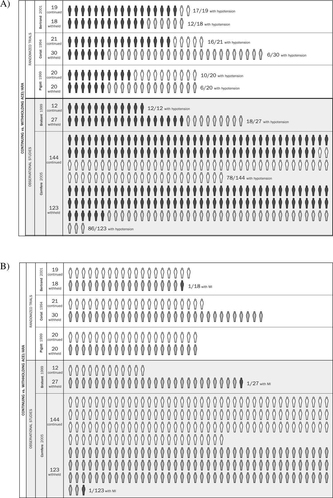
Strengths and Weaknesses of This Review
We observed considerable variation in design quality from study to study. With the exception of hypotension, other end points were not examined uniformly in the studies comprising this review. This was due either to study design (retrospective) or to the belief that the outcomes were not likely. With 1 exception,11 patient‐important end points such as myocardial infarction were noted if they occurred but not explicitly sought. Without active surveillance (serial electrocardiographic and biomarker testing), events such as myocardial infarction may remain undetected. Pain from myocardial ischemia, for example, may be masked by postoperative analgesia. Creatine kinase with muscle and brain subunits (CK‐MB) may be elevated in response to extracardiac injury. Postoperative ECG findings often are nonspecific.12 Furthermore, these studies examined the immediate and short‐term postoperative periods, possibly missing late‐manifesting hypotension‐induced or other end‐organ damage. Thus, truly reliable conclusions regarding the frequency of myocardial infarction, cerebrovascular events, and other patient‐important outcomes cannot be reached. Because this review includes small studies, it is particularly vulnerable to the effects of publication bias. The overall quality of the evidence we summarized makes it likely that larger rigorous trials may fail to confirm our findings.1315 Notably, this is to our knowledge the first systematic review addressing the clinical consequences of continuing or withholding the immediate preoperative dose of ACEI/ARAs.
Meaning of the Study
Evidence exists that perioperative ACEI/ARA therapy can impair the body's already anesthesia‐ suppressed blood pressure regulation system. Patients scheduled to undergo cardiovascular surgery may be at increased risk for the development of perioperative hypotension requiring vasopressors if the immediate preoperative ACEI/ARA dose is given. The results of this reviewa review of studies that were relatively small and generally not powered to observe clinically significant consequencesdo not provide sufficient evidence to support the systematic withholding or the systematic continuation of RAAS antagonists. Patients will be served best by hospitalists and other clinicians involved in perioperative care who take into account situation‐specific details in making this decision. A patient at particularly high risk for the complications of a blood pressure extreme (either hyper‐ or hypotension) represents such an example.
For patients who receive the immediate preoperative ACEI/ARA dose and do develop perioperative hypotension, there is inadequate evidence to determine whether that hypotension leads to patient‐important adverse outcomes. In fact, data from literature presently available are insufficient to reach any conclusion about long‐term clinical consequences of continuing or not continuing chronic ACEI/ARA therapy. The available studies were relatively small, reported few if any events, and were not designed to measure accurately the incidence of patient‐important end points.
Unanswered Questions and Future Research
Large and rigorous randomized trials could help to clarify the relationships suggested in this meta‐analysis and provide valid data about the consequences of continuing versus withholding preoperative ACEI/ARA therapy. Such trials are required before strong evidence‐based recommendations can be formulated.
Acknowledgements
The authors are indebted to James M. Naessens, ScD, David R. Danielson, MD, and David O. Warner, MD, for their advice during the conduct of this study. We also gratefully acknowledge Amanda Ebright, MD, for asking the original question that led to this review and Mr. Matthew Maleska for his design of summary Figure 3.
- ,,,,.Patients at the center: in our practice, and in our use of language.ACP J Club.2004;140:A11–A12.
- ,,,,,.Improving the quality of reports of meta‐analyses of randomised controlled trials: the QUOROM statement.Lancet.1999;354:1896–1900.
- ,,, et al. The Newcastle‐Ottawa Scale (NOS) for assessing the quality of nonrandomized studies in meta‐analysis. Ottawa Health Research Institute, University of Ottawa, Ontario, Canada. http://www.ohri.ca/programs/clinical_epidemiology/oxford.htm. Accessed: March 21,2006.
- ,.Meta‐analysis in clinical trials.Control Clin Trials. Sep1986;7:177–188.
- .,..What to add to nothing? Use and avoidance of continuity corrections in meta‐analysis of sparse data.Stat Med.2004;23:1351–1375.
- ,.Quantifying heterogeneity in a meta‐analysis.Stat Med.2002;21:1539–1558.
- ,.Statistics notes: Interaction revisited: the difference between two estimatesBMJ.2003;326:219
- ,,, et al.Influence of chronic angiotensin‐converting enzyme inhibition on anesthetic induction.Anesthesiology.1994;81:299–307.
- ,,, et al.Angiotensin system inhibitors in a general surgical population.Anesth Analg.2005;100:636–644.
- ,,,,.The hemodynamic effects of anesthetic induction in vascular surgical patients chronically treated with angiotensin II receptor antagonists.Anesth Analg.1999;89:1388–1392.
- ,,,,,.Should the angiotensin II antagonists be discontinued before surgery? [see comment].Anesth Analg.2001;92:26–30.
- Zipes.Braunwald's Heart Disease: A Textbook of Cardiovascular Medicine.7th ed.New York:Saunders, an imprint of Elsevier;2005.
- ,.Summarizing the Evidence: Publication Bias.Chicago:AMA Press;2002.
- ,,, et al.Large trials vsmeta‐analysis of smaller trials: how do their results compare? [see comment].JAMA.1996;276:1332–1338.
- ,,,.Bias in meta‐analysis detected by a simple, graphical test.BMJ.1997;315:629–634.
- .Preoperative evaluation of the patient with hypertension.JAMA.2002;287:2043–2046.
- ,,,,.Effect of omitting regular ACE inhibitor medication before cardiac surgery on haemodynamic variables and vasoactive drug requirements.Br J Anaesth.1999;83:715–720.
Clinicians commonly use renin‐angiotensin‐aldosterone‐system (RAAS) antagonists such as angiotensin‐converting enzyme inhibitors (ACEIs) and angiotensin II receptor subtype 1 antagonists (ARAs) to treat hypertension, congestive heart failure, and diabetic nephropathy. Hospitalists and other clinicians involved in the preoperative care of patients treated chronically with these agents are faced with the uncertainty of whether to continue these medications immediately prior to surgery.
The concern among those who recommend holding therapy is that pharmacologic suppression of the RAAS in patients undergoing general anesthesia may lead to severe or refractory (to intravenous fluid support) hypotension requiring vasopressors. On the other hand, if complications are no more likely when continuing one of these agents up to the day of surgery, withholding it could represent an unnecessary and potentially harmful intervention (eg, when a clinician caring postoperatively for a patient forgets to restart it). Although several studies have attempted to address this dilemma, a systematic and comprehensive summary of the pertinent evidence has not been published.
In this systematic review and meta‐analysis, we sought to summarize the best available evidence about the relative incidence of patient‐important outcomes1 in patients who do or do not receive ACEI/ARA therapy on the day of their nonemergent surgery.
METHODS
We report this protocol‐driven review in accordance with the Quality of Reporting of Meta‐analyses (QUOROM) standards for reporting systematic reviews of randomized trials.2
Search Strategy
In collaboration with an expert reference librarian (P.J.E.), we designed a search strategy that included the electronic databases MEDLINE, EMBASE, CINAHL, Web of Science, Current Contents, CENTRAL, DARE, and SCOPUS from 1981 (when captopril, the first ACEI, was approved by the FDA) until March 2006. We also reviewed the reference lists of included articles, retrieved articles from our personal files, and consulted with anesthesiologists and hospitalists with an interest in perioperative care in order to identify unpublished studies or studies missed by our strategy.
Study Selection
Eligible studies were prospective cohort studies or randomized controlled trials enrolling adult patients (ie, most patients > 18 years) undergoing nonemergent surgery and using ACEI or ARA chronically and assessing the effect of withdrawing or continuing these agents up to the morning of surgery. Eligible studies measured and reported either events of great patient importance (death, myocardial infarction, transient ischemic attack or stroke, and hepatic or renal failure) or of potentially less importance such as unplanned admission to the intensive care unit or treatment‐requiring hypotension, arrhythmias, or hyperkalemia.
Study Selection
Two reviewers (D.J.R. and F.S.M.) independently screened the titles and abstracts for potential inclusion and retrieved potentially eligible articles for full‐text evaluation. Two reviewers (D.J.R. and M.L.B.) working in duplicate independently selected studies for inclusion. The reviewers were in agreement for full text inclusion 100% of the time.
Data Extraction
Two hospitalists with experience in perioperative care and trained in clinical research (D.J.R. and F.S.M.) working independently and in duplicate extracted data from each eligible article using a standardized structured data extraction form. We extracted information about the study authors and publication, the patients (numbers in each group, indications for chronic ACEI/ARA therapy, type of surgery, agents used for anesthesia), event rates of surgical and perioperative complications (death, stroke, myocardial infarction, unplanned admission to the intensive care unit, treatment‐requiring hypotension, arrhythmias, or hyperkalemia), and relevant periods (e.g., between last dose of ACEI/ARA and surgery, between surgery and clinical end points, total follow‐up). When key information was not available in the published report, we contacted authors by electronic mail. We made 2 attempts to contact authors who failed to respond. Three of the 4 authors contacted responded with the requested information.
Quality Assessment
For randomized trials, we noted whether authors reported adequate allocation concealment, blinding of patients, clinicians, data collectors, data analysts, outcome assessors, and loss to follow‐up. The same reviewers (D.J.R. and F.S.M.) assessed study quality and were in agreement for each article and each domain of quality (kappa statistic in each case was 1.0). For cohort studies we noted details of cohort selection and comparability according to the Newcastle‐Ottawa approach.3
Statistical Analysis
We used a DerSimonian and Laird random effects method4 to conduct meta‐analyses across eligible outcomes. Random effects meta‐analysis incorporates both within‐study and between‐study variability. We chose a random effects approach because of the important degree of clinical heterogeneity expected between the included studies. For rare events we followed the approach by Sweeting et al. for the choice of a continuity correction factor.5 We report the pooled relative risk and the associated 95% confidence interval.
Inconsistency and Subgroup Analyses
To ascertain the magnitude of inconsistency across trials, we measured the I2 statistic, an estimate of the proportion of the overall between‐study variability that is not a result of random error or chance but of true clinical heterogeneity.6 When possible, we explored subgroup analyses to explain heterogeneity, including subgroups defined by type of surgery (cardiovascular versus noncardiovascular), timing of measurement of outcomes (in relation to anesthesia induction postoperatively), and type of agent (ACEI or ARA). We estimated the difference in treatment effects between subgroups by testing the hypothesis of treatmentsubgroup interaction with a nominal significance level of 5%.7
RESULTS
Search Results
The 509 titles reviewed included 410 titles produced by electronic searches and an additional 99 titles from other sources (Fig. 1).
Study Characteristics
Table 1 summarizes the characteristics of the 5 included studies (n = 434 patients). Myocardial infarction was an end point in 3 studies (Brabant, Bertrand, and Comfere); 1 event was reported in the withheld arm of each of these studies (none in the continuing arms). Hypotension requiring vasopressors was reported in all 5 studies. The other end points of interest were reported sparsely. There was considerable heterogeneity across studies regarding follow‐up period, which ranged from ending at incision to ending at dismissal from the hospital.
| Author/Year | Patients (n) | Indication for ACEI/ARA | Type of surgery | End points measured |
|---|---|---|---|---|
| ||||
| Randomized trials | ||||
| Bertrand, 200111 | 19 continued 18 withheld | Hypertension | Elective major vascular | Hypotension, need for vasoactive drugs (at or shortly after induction) |
| Coriat, 19948 | 21 continued 30 withheld | Hypertension | Peripheral vascular (>2 hours) | Systolic blood pressure (at or shortly after induction), plasma ACEI and catecholamine levels |
| Pigott, 199917 | 20 continued 20 withheld | Hypertension (n = 17); previous myocardial infarction (n = 23) | Coronary artery bypass graft | Arterial pressure (at or shortly after induction), cardiac index, systemic vascular resistance, use of vasoactive drugs |
| Observational studies | ||||
| Brabant, 199910 | 12 continued 27 withheld | Previous myocardial infarction (n = 6); diabetes mellitus (n = 6; n with diabetic nephropathy unknown); hypertension (n = unknown) | Elective vascular surgery | Blood pressure (at or shortly after induction) |
| Comfere, 20059 | 144 continued 123 withheld | Hypertension | Noncardiovascular | Blood pressure (at or shortly after induction), unplanned ICU admissions, hemodynamic instability in the PACU (ABP or HR out of range), acute renal impairment, TIA, stroke, myocardial ischemia/emnfarction, and death |
Methodological Quality of Included Studies
Table 2 describes the methodological quality, as reported, of the included studies. Allocation concealment was unclear in 2 of the 3 randomized trials. Details of blinding either were not reported or otherwise were unclear in 2 of these 3 studies. Only 1 study specified the extent of loss to follow‐up.8 In 1 of the observational studies,9 details of cohort selection were generally appropriate. The 12 patients examined in another study10 had been scheduled consecutively for surgery. Both studies controlled for a variety of demographic and other key variables. Duration of follow‐up ranged from 3 days after surgery (for ECG)10 to as long as duration of hospitalization.9
| Randomized trials | |||
|---|---|---|---|
| Allocation concealment | Blinding | Loss to follow‐up | |
| Bertrand, 200111 | Unclear | Unclear | Not reported |
| Coriat, 19948 | Unclear | None | 19% |
| Pigott, 199917 | Adequate | Investigator, cardiac anesthetists, perfusionists, and recovery staff were blinded to allocation. Blinding not reported for other data collectors, assessors of outcome, or data analysts | Not reported |
| Observational studies | |||
| Details of cohort selection | Comparability of cohorts | ||
| Brabant, 199910 | Appropriate Cohort somewhat representative of the adult population undergoing nonemergent surgery. The unexposed cohort was drawn from the same community as the exposed cohort | Similar with 2 exceptions: compared with the ACEI‐withheld group, the ARA‐given group contained more than twice the proportion of patients with previous myocardial revascularization Compared with the ARA‐given group, the ACEI‐withheld group contained more than twice the proportion of patients with diabetes mellitus | |
| Comfere, 20059 | Appropriate Cohort somewhat representative of the adult population undergoing nonemergent surgery (referral center population may not truly represent overall population). The unexposed cohort was drawn from the same community as the exposed cohort. Data were extracted from a secure record | Adequate This study controls for a variety of demographic and other variables | |
Meta‐analyses
Pooled results suggested that patients receiving the immediate preoperative ACEI/ARA dose were more likely (RR 1.51, 95% CI 1.14‐2.01) to develop hypotension requiring vasopressors at or shortly after induction of anesthesia (Fig. 2A). There was important inconsistency between studies (I2 = 59%). The pooled effect derived from randomized trials (RR = 2.26, 95% CI 0.84‐6.12) seemed greater than that derived from the 2 observational studies (RR = 1.33, 95% CI 1.02‐1.73), but the treatment‐study design interaction was not significant (P = .3). Similarly, other subgroup explorations were not contributory.
The incidence of perioperative myocardial infarction was not significantly different between continuing and withheld groups (Fig. 2B); the results were consistent across trials (I2 = 0%) but were imprecise (RR = 0.41, 95% CI 0.07‐2.53). Data were insufficient for subgroup analyses.
DISCUSSION
Statement of Principle Findings
Our systematic review identified 3 randomized trials and 2 observational studies examining the clinical consequences of continuing versus deliberately withholding the immediate preoperative dose of a renin‐angiotensin‐aldosterone system antagonist in patients treated chronically with these agents and scheduled to undergo nonemergent surgery.
Results from pooled estimates suggest that continuing chronic therapy up until surgery may increase the risk of perioperative hypotension requiring vasopressors (Fig. 3). Otherwise, this systematic review did not identify any clinically significant consequences associated either with preoperatively withholding or continuing RAAS antagonists. We do note that all 3 of the myocardial infarctions reported occurred in patients from whom the immediate preoperative ACEI/ARA dose was withheld, although no meaningful conclusion can be inferred from so few data points.
Strengths and Weaknesses of This Review
We observed considerable variation in design quality from study to study. With the exception of hypotension, other end points were not examined uniformly in the studies comprising this review. This was due either to study design (retrospective) or to the belief that the outcomes were not likely. With 1 exception,11 patient‐important end points such as myocardial infarction were noted if they occurred but not explicitly sought. Without active surveillance (serial electrocardiographic and biomarker testing), events such as myocardial infarction may remain undetected. Pain from myocardial ischemia, for example, may be masked by postoperative analgesia. Creatine kinase with muscle and brain subunits (CK‐MB) may be elevated in response to extracardiac injury. Postoperative ECG findings often are nonspecific.12 Furthermore, these studies examined the immediate and short‐term postoperative periods, possibly missing late‐manifesting hypotension‐induced or other end‐organ damage. Thus, truly reliable conclusions regarding the frequency of myocardial infarction, cerebrovascular events, and other patient‐important outcomes cannot be reached. Because this review includes small studies, it is particularly vulnerable to the effects of publication bias. The overall quality of the evidence we summarized makes it likely that larger rigorous trials may fail to confirm our findings.1315 Notably, this is to our knowledge the first systematic review addressing the clinical consequences of continuing or withholding the immediate preoperative dose of ACEI/ARAs.
Meaning of the Study
Evidence exists that perioperative ACEI/ARA therapy can impair the body's already anesthesia‐ suppressed blood pressure regulation system. Patients scheduled to undergo cardiovascular surgery may be at increased risk for the development of perioperative hypotension requiring vasopressors if the immediate preoperative ACEI/ARA dose is given. The results of this reviewa review of studies that were relatively small and generally not powered to observe clinically significant consequencesdo not provide sufficient evidence to support the systematic withholding or the systematic continuation of RAAS antagonists. Patients will be served best by hospitalists and other clinicians involved in perioperative care who take into account situation‐specific details in making this decision. A patient at particularly high risk for the complications of a blood pressure extreme (either hyper‐ or hypotension) represents such an example.
For patients who receive the immediate preoperative ACEI/ARA dose and do develop perioperative hypotension, there is inadequate evidence to determine whether that hypotension leads to patient‐important adverse outcomes. In fact, data from literature presently available are insufficient to reach any conclusion about long‐term clinical consequences of continuing or not continuing chronic ACEI/ARA therapy. The available studies were relatively small, reported few if any events, and were not designed to measure accurately the incidence of patient‐important end points.
Unanswered Questions and Future Research
Large and rigorous randomized trials could help to clarify the relationships suggested in this meta‐analysis and provide valid data about the consequences of continuing versus withholding preoperative ACEI/ARA therapy. Such trials are required before strong evidence‐based recommendations can be formulated.
Acknowledgements
The authors are indebted to James M. Naessens, ScD, David R. Danielson, MD, and David O. Warner, MD, for their advice during the conduct of this study. We also gratefully acknowledge Amanda Ebright, MD, for asking the original question that led to this review and Mr. Matthew Maleska for his design of summary Figure 3.
- ,,,,.Patients at the center: in our practice, and in our use of language.ACP J Club.2004;140:A11–A12.
- ,,,,,.Improving the quality of reports of meta‐analyses of randomised controlled trials: the QUOROM statement.Lancet.1999;354:1896–1900.
- ,,, et al. The Newcastle‐Ottawa Scale (NOS) for assessing the quality of nonrandomized studies in meta‐analysis. Ottawa Health Research Institute, University of Ottawa, Ontario, Canada. http://www.ohri.ca/programs/clinical_epidemiology/oxford.htm. Accessed: March 21,2006.
- ,.Meta‐analysis in clinical trials.Control Clin Trials. Sep1986;7:177–188.
- .,..What to add to nothing? Use and avoidance of continuity corrections in meta‐analysis of sparse data.Stat Med.2004;23:1351–1375.
- ,.Quantifying heterogeneity in a meta‐analysis.Stat Med.2002;21:1539–1558.
- ,.Statistics notes: Interaction revisited: the difference between two estimatesBMJ.2003;326:219
- ,,, et al.Influence of chronic angiotensin‐converting enzyme inhibition on anesthetic induction.Anesthesiology.1994;81:299–307.
- ,,, et al.Angiotensin system inhibitors in a general surgical population.Anesth Analg.2005;100:636–644.
- ,,,,.The hemodynamic effects of anesthetic induction in vascular surgical patients chronically treated with angiotensin II receptor antagonists.Anesth Analg.1999;89:1388–1392.
- ,,,,,.Should the angiotensin II antagonists be discontinued before surgery? [see comment].Anesth Analg.2001;92:26–30.
- Zipes.Braunwald's Heart Disease: A Textbook of Cardiovascular Medicine.7th ed.New York:Saunders, an imprint of Elsevier;2005.
- ,.Summarizing the Evidence: Publication Bias.Chicago:AMA Press;2002.
- ,,, et al.Large trials vsmeta‐analysis of smaller trials: how do their results compare? [see comment].JAMA.1996;276:1332–1338.
- ,,,.Bias in meta‐analysis detected by a simple, graphical test.BMJ.1997;315:629–634.
- .Preoperative evaluation of the patient with hypertension.JAMA.2002;287:2043–2046.
- ,,,,.Effect of omitting regular ACE inhibitor medication before cardiac surgery on haemodynamic variables and vasoactive drug requirements.Br J Anaesth.1999;83:715–720.
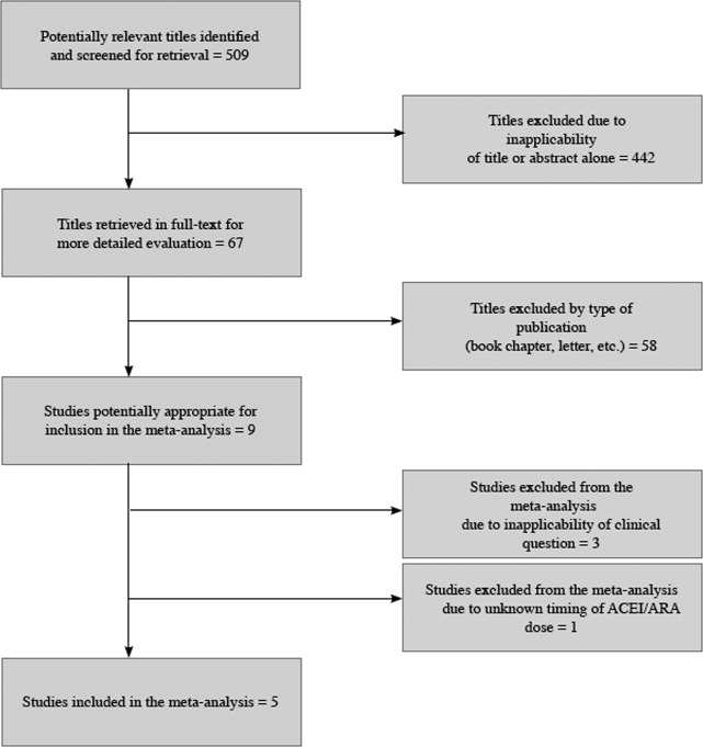
Pediatric Hospitalist Variation in Care
Reduction of undesirable variation in care has been a major focus of systematic efforts to improve the quality of the healthcare system.13 The emergence of hospitalists, physicians specializing in the care of hospitalized patients, was spurred by a desire to streamline care and reduce variability in hospital management of common diseases.4, 5 Over the past decade, hospitalist systems have become a leading vehicle for care delivery.4, 6, 7 It remains unclear, however, whether implementation of hospitalist systems has lessened undesirable variation in the inpatient management of common diseases.
While systematic reviews have found costs and hospital length of stay to be 10‐15% lower in both pediatric and internal medicine hospitalist systems, few studies have adequately assessed the processes or quality of care in hospitalist systems.8, 9 Two internal medicine studies have found decreased mortality in hospitalist systems, but the mechanism by which hospitalists apparently achieved these gains is unclear.10, 11 Even less is known about care processes or quality in pediatric hospitalist systems. Death is a rare occurrence in pediatric ward settings, and the seven studies conducted to date comparing pediatric hospitalist and traditional systems have been universally underpowered to detect differences in mortality.9, 1218 There is a need to better understand care processes as a first step in understanding and improving quality of care in hospitalist systems.19
The Pediatric Research in Inpatient Settings (PRIS) Network was formed to improve the quality of care for hospitalized children through collaborative clinical research. In this study, we sought to study variation in the care of common pediatric conditions among a cohort of pediatric hospitalists. We have previously reported that less variability exists in hospitalists' reported management of inpatient conditions than in the reported management of these same conditions by community‐based pediatricians,20 but we were concerned that substantial undesirable variation (ie, variation in practice due to uncertainty or unsubstantiated local practice traditions, rather than justified variation in care based on different risks of harms or benefits in different patients) may still exist among hospitalists. We therefore conducted a study: 1) to investigate variation in hospitalists' reported use of common inpatient therapies, and 2) to test the hypothesis that greater variation exists in hospitalists' reported use of inpatient therapies of unproven benefit than in those therapies proven to be beneficial.
METHODS
Survey Design and Administration
In 2003, we designed the PRIS Survey to collect data on hospitalists' backgrounds, practices, and training needs, as well as their management of common pediatric conditions. For the current study, we chose a priori to evaluate hospitalists' use of 14 therapies in the management of 4 common conditions: asthma, bronchiolitis, gastroenteritis, and gastro‐esophageal reflux disease (GERD) (Table 1). These four conditions were chosen for study because they were among the top discharge diagnoses (primary and secondary) from the inpatient services at 2 of the authors' institutions (Children's Hospital Boston and Children's Hospital San Diego) during the year before administration of the survey, and because a discrete set of therapeutic agents are commonly used in their management. Respondents were asked to report the frequency with which they used each of the 14 therapies of interest on 5‐point Likert scales (from 1=never to 5=almost always). The survey initially developed was piloted with a small group of hospitalists and pediatricians, and a final version incorporating revisions was subsequently administered to all pediatric hospitalists in the US and Canada identified through any of 3 sources: 1) the Pediatric Research in Inpatient Settings (PRIS) list of participants; 2) the Society for Hospital Medicine (SHM) pediatric hospital medicine e‐mail listserv; and 3) the list of all attendees of the first national pediatric hospitalist conference sponsored by the Ambulatory Pediatrics Association (APA), SHM, and American Academy of Pediatrics (AAP); this meeting was held in San Antonio, Texas, USA in November 2003. Individuals identified through more than 1 of these groups were counted only once. Potential participants were assured that individual responses would be kept confidential, and were e‐mailed an access code to participate in the online survey, using a secure web‐based interface; a paper‐based version was also made available to those who preferred to respond in this manner. Regular reminder notices were sent to all non‐responders. Further details regarding PRIS Survey recruitment and study methods have been published previously.20
| Condition | Therapy | BMJ clinical evidence Treatment effect categorization* | Study classification |
|---|---|---|---|
| |||
| Asthma | Inhaled albuterol | Beneficial | Proven |
| Systemic corticosteroids | Beneficial | Proven | |
| Inhaled ipratropium in the first 24 hours of hospitalization | Beneficial | Proven | |
| Inhaled ipratropium after the first 24 hours of hospitalization | Unknown effectiveness | Unproven | |
| Bronchiolitis | Inhaled albuterol | Unknown effectiveness | Unproven |
| Inhaled epinephrine | Unknown effectiveness | Unproven | |
| Systemic corticosteroids | Unknown effectiveness | Unproven | |
| Gastroenteritis | Intravenous hydration | Beneficial | Proven |
| Lactobacillus | Not assessed | Unproven | |
| Ondansetron | Not assessed | Unproven | |
| Gastro‐Esophageal Reflux Disease (GERD) | H2 histamine‐receptor antagonists | Unknown effectiveness | Unproven |
| Thickened feeds | Unknown effectiveness Likely to be beneficial | Unproven Proven | |
| Metoclopramide | Unknown effectiveness | Unproven | |
| Proton‐pump inhibitors | Unknown effectiveness | Unproven | |
DefinitionsReference Responses and Percent Variation
To measure variation in reported management, we first sought to determine a reference response for each therapy of interest. Since the evidence base for most of the therapies we studied is weak, it was not possible to determine a gold standard response for each therapy. Instead, we sought to measure the degree of divergence from a reference response for each therapy in the following manner. First, to simplify analyses, we collapsed our five‐category Likert scale into three categories (never/rarely, sometimes, and often/almost always). We then defined the reference response for each therapy to be never/rarely or often/almost always, whichever of the 2 was more frequently selected by respondents; sometimes was not used as a reference category, as reporting use of a particular therapy sometimes indicated substantial variability even within an individual's own practice.
Classification of therapies as proven or unproven.
To classify each of the 14 studied therapies as being of proven or unproven, we used the British Medical Journal's publication Clinical Evidence.19 We chose to use Clinical Evidence as an evidence‐based reference because it provides rigorously developed, systematic analyses of therapeutic management options for multiple common pediatric conditions, and organizes recommendations in a straightforward manner. Four of the 14 therapies had been determined on systematic review to be proven beneficial at the time of study design: systemic corticosteroids, inhaled albuterol, and ipratropium (in the first 24 h) in the care of children with asthma; and IV hydration in the care of children with acute gastroenteritis. The remaining 10 therapies were either considered to be of unknown effectiveness or had not been formally evaluated by Clinical Evidence, and were hence considered unproven for this study (Table 1). Of note, the use of thickened feeds in the treatment of children with GERD had been determined to be of unknown effectiveness at the time of study design, but was reclassified as likely to be beneficial during the course of the study.
Analyses
Descriptive statistics were used to report respondents' demographic characteristics and work environments, as well as variation in their reported use of each of the 14 therapies. Variation in hospitalists' use of proven versus unproven therapies was compared using the Wilcoxon rank sum test, as it was distributed non‐normally. For our primary analysis, the use of thickened feeds in GERD was considered unproven, but a sensitivity analysis was conducted reclassifying it as proven in light of the evolving literature on its use and its consequent reclassification in Clinical Evidence.(SAS Version 9.1, Cary, NC) was used for statistical analyses.
RESULTS
213 of the 320 individuals identified through the 3 lists of pediatric hospitalists (67%) responded to the survey. Of these, 198 (93%) identified themselves as hospitalists and were therefore included. As previously reported,20 53% of respondents were male, 55% worked in academic training environments, and 47% had completed advanced training (fellowship) beyond their core pediatric training (residency training); respondents reported completing residency training 11 9 (mean, standard deviation) years prior to the survey, and spending 176 72 days per year in the care of hospitalized patients.
Variation in reported management: asthma
(Figure 1, Panel A). Relatively little variation existed in reported use of the 4 asthma therapies studied. Only 4.4% (95% CI, 1.4‐7.4%) of respondents did not provide the reference response of using inhaled albuterol often or almost always in the care of inpatients with asthma, and only 6.0% (2.5‐9.5%) of respondents did not report using systemic corticosteroids often or almost always. Variation in reported use of ipratropium was somewhat higher.
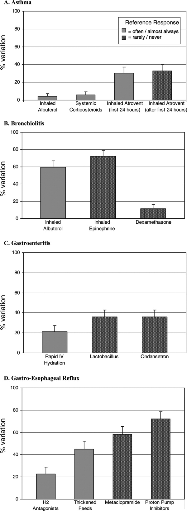
Bronchiolitis
(Figure 1B). By contrast, variation in reported use of inhaled therapies for bronchiolitis was high, with many respondents reporting that they often or always used inhaled albuterol or epinephrine, while many others reported rarely or never using them. There was 59.6% (52.4‐66.8%) variation from the reference response of often/almost always using inhaled albuterol, and 72.2% (65.6‐78.8%) variation from the reference response of never/rarely using inhaled epinephrine. Only 11.6% (6.9‐16.3%) of respondents, however, varied from the reference response of using dexamethasone more than rarely in the care of children with bronchiolitis.
Gastroenteritis
(Figure 1C). Moderate variability existed in the reported use of the 3 studied therapies for children hospitalized with gastroenteritis. 21.1% (15.1‐27.1%) of respondents did not provide the reference response of often/almost always using IV hydration; 35.9% (28.9‐42.9%) did not provide the reference response of never or rarely using lactobacillus; likewise, 35.9% (28.9‐42.9%) did not provide the reference response of never or rarely using ondansetron.
Gastro‐Esophageal Reflux Disease
(Figure 1, Panel D). There was moderate to high variability in the reported management of GERD. 22.8% (16.7‐28.9%) of respondents did not provide the reference response of often/almost always using H2 antagonists, and 44.9% (37.6‐52.2%) did not report often/almost always using thickened feeds in the care of these children. 58.3% (51.1‐65.5%) and 72.1% (65.5‐78.7%) of respondents did not provide the reference response of never/rarely using metoclopramide and proton pump inhibitors, respectively.
Proven vs. Unproven Therapies
(Figure 2). Variation in reported use of therapies of unproven benefit was significantly higher than variation in reported use of the 4 proven therapies (albuterol, corticosteroids, and ipratropium in the first 24 h for asthma; IV re‐hydration for gastroenteritis). The mean variation in reported use of unproven therapies was 44.6 20.5%, compared with 15.5 12.5% variation in reported use of therapies of proven benefit (p = 0.02).
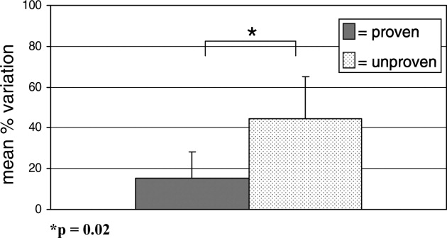
As a sensitivity analysis, the use of thickened feeds as a therapy for GERD was re‐categorized as proven and the above analysis repeated, for the reasons outlined in the methods section. This did not alter the identified relationship between variability and the evidence base fundamentally; hospitalists' reported variation in use of therapies of unproven benefit in this sensitivity analysis was 44.6 21.7%, compared with 21.4 17.0% variation in reported use of proven therapies (p = 0.05).
DISCUSSION
Substantial variation exists in the inpatient management of common pediatric diseases. Although we have previously found less reported variability in pediatric hospitalists' practices than in those of community‐based pediatricians,20 the current study demonstrates a high degree of reported variation even among a cohort of inpatient specialists. Importantly, however, reported variation was found to be significantly less for those inpatient therapies supported by a robust evidence base.
Bronchiolitis, gastroenteritis, asthma, and GERD are extremely common causes of pediatric hospitalization throughout the developed world.2125 Our finding of high reported variability in the routine care of all of these conditions except asthma is concerning, as it suggests that experts do not agree on how to manage children hospitalized with even the most common childhood diseases. While we hypothesized that there would be some variation in the use of therapies whose benefit has not been well established, the high degree of variation observed is of concern because it indicates that an insufficient evidentiary base exists to support much of our day‐to‐day practice. Some variation in practice in response to differing clinical presentations is both expected and desirable, but it is remarkable that variance in practice was significantly less for the most evidence‐based therapies than for those grounded less firmly in science, suggesting that the variation identified here is not justifiable variation based on appropriate responses to atypical clinical presentations, but uncertainty in the absence of clear data. Such undesired variability may decrease system reliability (introducing avoidable opportunity for error),26 and lead to under‐use of needed therapies as well as overuse of unnecessary therapies.1
Our work extends prior research that has identified wide variation in patterns of hospital admission, use of hospital resources, and processes of inpatient care,2732 by documenting reported variation in the use of common inpatient therapies. Rates of hospital admission may vary by as much as 7‐fold across regions.33 Our study demonstrates that wide variation exists not only in admission rates, but in reported inpatient care processes for some of the most common diseases seen in pediatric hospitals. Our study also supports the hypothesis that variation in care may be driven by gaps in knowledge.32 Among hospitalists, we found the strength of the evidence base to be a major determinant of reported variability.
Our study has several limitations. First, the data presented here are derived from provider self‐reports, which may not fully reflect actual practice. In the case of the few proven therapies studied, reporting bias could lead to an over‐reporting of adherence to evidence‐based standards of care. Like our study, however, prior studies have found that hospital‐based providers fairly consistently comply with evidence‐based practice recommendations for acute asthma care,34, 35 supporting our finding that variation in acute asthma care (which represented 3 of our 4 proven therapies) is low in this setting.
Another limitation is that classifications of therapies as proven or unproven change as the evidence base evolves. Of particular relevance to this study, the use of thickened feeds as a therapy for GERD, originally classified as being of unknown effectiveness, was reclassified by Clinical Evidence during the course of the study as likely to be beneficial. The relationship we identified between proven therapies and degree of variability in care did not change when we conducted a sensitivity analysis re‐categorizing this therapy as proven, but precisely quantifying variation is complicated by continuous changes in the state of the evidence.
Pediatric hospitalist systems have been found consistently to improve the efficiency of care,9 yet this study suggests that considerable variation in hospitalists' management of key conditions remains. The Pediatric Research in Inpatient Settings (PRIS) Network was formed in 2002 to improve the care of hospitalized children and the quality of inpatient practice by developing an evidence base for inpatient pediatric care. Ongoing multi‐center research efforts through PRIS and other research networks are beginning to critically evaluate therapies used in the management of common pediatric conditions. Rigorous studies of the processes and outcomes of pediatric hospital care will inform inpatient pediatric practice, and ultimately improve the care of hospitalized children. The current study strongly affirms the urgent need to establish such an evidence base. Without data to inform optimal care, efforts to reduce undesirable variation in care and improve care quality cannot be fully realized.
Acknowledgements
The authors would like to extend their thanks to the hospitalists and members of the Pediatric Research in Inpatient Settings Network who participated in this research, as well as the Children's National Medical Center and Children's Hospital Boston Inpatient Pediatrics Services, who provided funding to support this study. Special thanks to the Ambulatory Pediatrics Association (APA), for its core support of the PRIS Network. Dr. Landrigan is the recipient of a career development award from the Agency for Healthcare Research and Quality (AHRQ K08 HS13333). Dr. Conway is the recipient of a Robert Wood Johnson Clinical Scholars Grant. All researchers were independent from the funding agencies; the academic medical centers named above, APA, and AHRQ had no role in the design and conduct of the study; collection, management, analysis, and interpretation of the data; or preparation, review, or approval of the manuscript.
- Institute of Medicine: Crossing the Quality Chasm: A New Health System for the 21st Century.Washington, D.C.:National Academic Press,2001.
- ..Reducing variation in surgical care.BMJ2005;330:1401–1402.
- ,,,.Variation in use of video assisted thoracic surgery in the United Kingdom.BMJ2004;329:1011–1012.
- ,..The emerging role of “hospitalists” in the American health care system.N. Engl J Med1996;335:514–517.
- ,..Hospitalism in the USA.Lancet1999;353:1902.
- Society of Hospital Medicine. Growth of Hospital Medicine Nationwide. Available at: http://www.hospitalmedicine.org/Content/NavigationMenu/Media/GrowthofHospitalMedicineNationwide/Growth_of_Hospital_M.htm. Accessed April 11,2007.
- .The changing face of hospital practice.Med Econ2002;79:72–79.
- ,..The hospitalist movement 5 years later.JAMA2002;287:487–494.
- ,,,.Pediatric hospitalists: a systematic review of the literature.Pediatrics2006;117:1736–1744.
- ,,,,,.Implementation of a voluntary hospitalist service at a community teaching hospital: improved clinical efficiency and patient outcomes.Ann Intern Med2002;137:859–865.
- ,,,,,, et al.Effects of physician experience on costs and outcomes on an academic general medicine service: results of a trial of hospitalists.Ann Intern Med2002;137:866–874.
- ,.Evaluation of a pediatric hospitalist service: impact on length of stay and hospital charges.Pediatrics2000;105:478–484.
- ,,,,,,, and .Impact of an HMO hospitalist system in academic pediatrics.Pediatrics2002;110:720–728.
- ,, and .Evaluation of a pediatric hospitalist service by APR‐DRG's: impact on length of stay and hospital charges.Pediatr Research2001;49(suppl),691.
- ,,.Pediatric hospitalists: quality care for the underserved?Am J Med Qual2001;16:174–180.
- ,,,,,.Restructuring an academic pediatric inpatient service using concepts developed by hospitalists.Clin Pediatr (Phila)2001;40:653–660.
- ,,, and .Hospitalist care of medically complex children.Pediatr Research2004;55(suppl),1789.
- ,,.Hospital‐based and community pediatricians: comparing outcomes for asthma and bronchiolitis.J Clin Outcomes Manage1997;4:21–24.
- Godlee F,Tovey D,Bedford M, et al., eds.Clinical Evidence: The International Source of the Best Available Evidence for Effective Health Care.London, United Kingdom:BMJ Publishing Group;2004.
- ,,,,,.Variations in management of common inpatient pediatric illnesses: hospitalists and community pediatricians.Pediatrics2006;118:441–447.
- ,,,,.Contribution of RSV to bronchiolitis and pneumonia‐associated hospitalizations in English children, April 1995‐March 1998.Epidemiol Infect2002;129:99–106.
- ,,.Direct medical costs of bronchiolitis hospitalizations in the United States.Pediatrics2006;118:2418–2423.
- ,,,,,.Multicenter Prospective Study of the Burden of Rotavirus Acute Gastroenteritis in Europe, 2004‐2005: The REVEAL Study.J Infect Dis2007;195Suppl 1:S4–S16.
- .The state of childhood asthma, United States, 1980‐2005.Adv.Data.2006;1–24.
- ,.Gastroesophageal reflux in children: pathogenesis, prevalence, diagnosis, and role of proton pump inhibitors in treatment.Paediatr Drugs2002;4:673–685.
- ,,,.Reliability science and patient safety.Pediatr Clin North Am2006;53:1121–1133.
- Wennberg JE and McAndrew Cooper M, eds.The Dartmouth Atlas of Health Care in the United States.Hanover, NH, USA:Health Forum, Inc.,1999.
- ,,,,,.Variations in rates of hospitalization of children in three urban communities.N Engl J Med1989;320:1183–1187.
- ,,,,,.Use of hospitals, physician visits, and hospice care during last six months of life among cohorts loyal to highly respected hospitals in the United States.BMJ2004;328:607.
- ,,,,,, et al.Variations in practice and outcomes in the Canadian NICU network: 1996‐1997.Pediatrics2000;106:1070–1079.
- ,,,.Evaluation of febrile children with petechial rashes: is there consensus among pediatricians?Pediatr Infect Dis J1998;17:1135–1140.
- ,,,,,, et al.Practice variation among pediatric emergency departments in the treatment of bronchiolitis.Acad Emerg Med2004;11:353–360.
- ,,,.Paediatric inpatient utilisation in a district general hospital.Arch Dis Child1994;70:488–492.
- ,,,.Emergency department asthma: compliance with an evidence‐based management algorithm.Ann Acad Med Singapore2002;31:419–424.
- ,,.Is the practice of paediatric inpatient medicine evidence‐based?J Paediatr Child Health2002;38:347–351.
Reduction of undesirable variation in care has been a major focus of systematic efforts to improve the quality of the healthcare system.13 The emergence of hospitalists, physicians specializing in the care of hospitalized patients, was spurred by a desire to streamline care and reduce variability in hospital management of common diseases.4, 5 Over the past decade, hospitalist systems have become a leading vehicle for care delivery.4, 6, 7 It remains unclear, however, whether implementation of hospitalist systems has lessened undesirable variation in the inpatient management of common diseases.
While systematic reviews have found costs and hospital length of stay to be 10‐15% lower in both pediatric and internal medicine hospitalist systems, few studies have adequately assessed the processes or quality of care in hospitalist systems.8, 9 Two internal medicine studies have found decreased mortality in hospitalist systems, but the mechanism by which hospitalists apparently achieved these gains is unclear.10, 11 Even less is known about care processes or quality in pediatric hospitalist systems. Death is a rare occurrence in pediatric ward settings, and the seven studies conducted to date comparing pediatric hospitalist and traditional systems have been universally underpowered to detect differences in mortality.9, 1218 There is a need to better understand care processes as a first step in understanding and improving quality of care in hospitalist systems.19
The Pediatric Research in Inpatient Settings (PRIS) Network was formed to improve the quality of care for hospitalized children through collaborative clinical research. In this study, we sought to study variation in the care of common pediatric conditions among a cohort of pediatric hospitalists. We have previously reported that less variability exists in hospitalists' reported management of inpatient conditions than in the reported management of these same conditions by community‐based pediatricians,20 but we were concerned that substantial undesirable variation (ie, variation in practice due to uncertainty or unsubstantiated local practice traditions, rather than justified variation in care based on different risks of harms or benefits in different patients) may still exist among hospitalists. We therefore conducted a study: 1) to investigate variation in hospitalists' reported use of common inpatient therapies, and 2) to test the hypothesis that greater variation exists in hospitalists' reported use of inpatient therapies of unproven benefit than in those therapies proven to be beneficial.
METHODS
Survey Design and Administration
In 2003, we designed the PRIS Survey to collect data on hospitalists' backgrounds, practices, and training needs, as well as their management of common pediatric conditions. For the current study, we chose a priori to evaluate hospitalists' use of 14 therapies in the management of 4 common conditions: asthma, bronchiolitis, gastroenteritis, and gastro‐esophageal reflux disease (GERD) (Table 1). These four conditions were chosen for study because they were among the top discharge diagnoses (primary and secondary) from the inpatient services at 2 of the authors' institutions (Children's Hospital Boston and Children's Hospital San Diego) during the year before administration of the survey, and because a discrete set of therapeutic agents are commonly used in their management. Respondents were asked to report the frequency with which they used each of the 14 therapies of interest on 5‐point Likert scales (from 1=never to 5=almost always). The survey initially developed was piloted with a small group of hospitalists and pediatricians, and a final version incorporating revisions was subsequently administered to all pediatric hospitalists in the US and Canada identified through any of 3 sources: 1) the Pediatric Research in Inpatient Settings (PRIS) list of participants; 2) the Society for Hospital Medicine (SHM) pediatric hospital medicine e‐mail listserv; and 3) the list of all attendees of the first national pediatric hospitalist conference sponsored by the Ambulatory Pediatrics Association (APA), SHM, and American Academy of Pediatrics (AAP); this meeting was held in San Antonio, Texas, USA in November 2003. Individuals identified through more than 1 of these groups were counted only once. Potential participants were assured that individual responses would be kept confidential, and were e‐mailed an access code to participate in the online survey, using a secure web‐based interface; a paper‐based version was also made available to those who preferred to respond in this manner. Regular reminder notices were sent to all non‐responders. Further details regarding PRIS Survey recruitment and study methods have been published previously.20
| Condition | Therapy | BMJ clinical evidence Treatment effect categorization* | Study classification |
|---|---|---|---|
| |||
| Asthma | Inhaled albuterol | Beneficial | Proven |
| Systemic corticosteroids | Beneficial | Proven | |
| Inhaled ipratropium in the first 24 hours of hospitalization | Beneficial | Proven | |
| Inhaled ipratropium after the first 24 hours of hospitalization | Unknown effectiveness | Unproven | |
| Bronchiolitis | Inhaled albuterol | Unknown effectiveness | Unproven |
| Inhaled epinephrine | Unknown effectiveness | Unproven | |
| Systemic corticosteroids | Unknown effectiveness | Unproven | |
| Gastroenteritis | Intravenous hydration | Beneficial | Proven |
| Lactobacillus | Not assessed | Unproven | |
| Ondansetron | Not assessed | Unproven | |
| Gastro‐Esophageal Reflux Disease (GERD) | H2 histamine‐receptor antagonists | Unknown effectiveness | Unproven |
| Thickened feeds | Unknown effectiveness Likely to be beneficial | Unproven Proven | |
| Metoclopramide | Unknown effectiveness | Unproven | |
| Proton‐pump inhibitors | Unknown effectiveness | Unproven | |
DefinitionsReference Responses and Percent Variation
To measure variation in reported management, we first sought to determine a reference response for each therapy of interest. Since the evidence base for most of the therapies we studied is weak, it was not possible to determine a gold standard response for each therapy. Instead, we sought to measure the degree of divergence from a reference response for each therapy in the following manner. First, to simplify analyses, we collapsed our five‐category Likert scale into three categories (never/rarely, sometimes, and often/almost always). We then defined the reference response for each therapy to be never/rarely or often/almost always, whichever of the 2 was more frequently selected by respondents; sometimes was not used as a reference category, as reporting use of a particular therapy sometimes indicated substantial variability even within an individual's own practice.
Classification of therapies as proven or unproven.
To classify each of the 14 studied therapies as being of proven or unproven, we used the British Medical Journal's publication Clinical Evidence.19 We chose to use Clinical Evidence as an evidence‐based reference because it provides rigorously developed, systematic analyses of therapeutic management options for multiple common pediatric conditions, and organizes recommendations in a straightforward manner. Four of the 14 therapies had been determined on systematic review to be proven beneficial at the time of study design: systemic corticosteroids, inhaled albuterol, and ipratropium (in the first 24 h) in the care of children with asthma; and IV hydration in the care of children with acute gastroenteritis. The remaining 10 therapies were either considered to be of unknown effectiveness or had not been formally evaluated by Clinical Evidence, and were hence considered unproven for this study (Table 1). Of note, the use of thickened feeds in the treatment of children with GERD had been determined to be of unknown effectiveness at the time of study design, but was reclassified as likely to be beneficial during the course of the study.
Analyses
Descriptive statistics were used to report respondents' demographic characteristics and work environments, as well as variation in their reported use of each of the 14 therapies. Variation in hospitalists' use of proven versus unproven therapies was compared using the Wilcoxon rank sum test, as it was distributed non‐normally. For our primary analysis, the use of thickened feeds in GERD was considered unproven, but a sensitivity analysis was conducted reclassifying it as proven in light of the evolving literature on its use and its consequent reclassification in Clinical Evidence.(SAS Version 9.1, Cary, NC) was used for statistical analyses.
RESULTS
213 of the 320 individuals identified through the 3 lists of pediatric hospitalists (67%) responded to the survey. Of these, 198 (93%) identified themselves as hospitalists and were therefore included. As previously reported,20 53% of respondents were male, 55% worked in academic training environments, and 47% had completed advanced training (fellowship) beyond their core pediatric training (residency training); respondents reported completing residency training 11 9 (mean, standard deviation) years prior to the survey, and spending 176 72 days per year in the care of hospitalized patients.
Variation in reported management: asthma
(Figure 1, Panel A). Relatively little variation existed in reported use of the 4 asthma therapies studied. Only 4.4% (95% CI, 1.4‐7.4%) of respondents did not provide the reference response of using inhaled albuterol often or almost always in the care of inpatients with asthma, and only 6.0% (2.5‐9.5%) of respondents did not report using systemic corticosteroids often or almost always. Variation in reported use of ipratropium was somewhat higher.
Bronchiolitis
(Figure 1B). By contrast, variation in reported use of inhaled therapies for bronchiolitis was high, with many respondents reporting that they often or always used inhaled albuterol or epinephrine, while many others reported rarely or never using them. There was 59.6% (52.4‐66.8%) variation from the reference response of often/almost always using inhaled albuterol, and 72.2% (65.6‐78.8%) variation from the reference response of never/rarely using inhaled epinephrine. Only 11.6% (6.9‐16.3%) of respondents, however, varied from the reference response of using dexamethasone more than rarely in the care of children with bronchiolitis.
Gastroenteritis
(Figure 1C). Moderate variability existed in the reported use of the 3 studied therapies for children hospitalized with gastroenteritis. 21.1% (15.1‐27.1%) of respondents did not provide the reference response of often/almost always using IV hydration; 35.9% (28.9‐42.9%) did not provide the reference response of never or rarely using lactobacillus; likewise, 35.9% (28.9‐42.9%) did not provide the reference response of never or rarely using ondansetron.
Gastro‐Esophageal Reflux Disease
(Figure 1, Panel D). There was moderate to high variability in the reported management of GERD. 22.8% (16.7‐28.9%) of respondents did not provide the reference response of often/almost always using H2 antagonists, and 44.9% (37.6‐52.2%) did not report often/almost always using thickened feeds in the care of these children. 58.3% (51.1‐65.5%) and 72.1% (65.5‐78.7%) of respondents did not provide the reference response of never/rarely using metoclopramide and proton pump inhibitors, respectively.
Proven vs. Unproven Therapies
(Figure 2). Variation in reported use of therapies of unproven benefit was significantly higher than variation in reported use of the 4 proven therapies (albuterol, corticosteroids, and ipratropium in the first 24 h for asthma; IV re‐hydration for gastroenteritis). The mean variation in reported use of unproven therapies was 44.6 20.5%, compared with 15.5 12.5% variation in reported use of therapies of proven benefit (p = 0.02).
As a sensitivity analysis, the use of thickened feeds as a therapy for GERD was re‐categorized as proven and the above analysis repeated, for the reasons outlined in the methods section. This did not alter the identified relationship between variability and the evidence base fundamentally; hospitalists' reported variation in use of therapies of unproven benefit in this sensitivity analysis was 44.6 21.7%, compared with 21.4 17.0% variation in reported use of proven therapies (p = 0.05).
DISCUSSION
Substantial variation exists in the inpatient management of common pediatric diseases. Although we have previously found less reported variability in pediatric hospitalists' practices than in those of community‐based pediatricians,20 the current study demonstrates a high degree of reported variation even among a cohort of inpatient specialists. Importantly, however, reported variation was found to be significantly less for those inpatient therapies supported by a robust evidence base.
Bronchiolitis, gastroenteritis, asthma, and GERD are extremely common causes of pediatric hospitalization throughout the developed world.2125 Our finding of high reported variability in the routine care of all of these conditions except asthma is concerning, as it suggests that experts do not agree on how to manage children hospitalized with even the most common childhood diseases. While we hypothesized that there would be some variation in the use of therapies whose benefit has not been well established, the high degree of variation observed is of concern because it indicates that an insufficient evidentiary base exists to support much of our day‐to‐day practice. Some variation in practice in response to differing clinical presentations is both expected and desirable, but it is remarkable that variance in practice was significantly less for the most evidence‐based therapies than for those grounded less firmly in science, suggesting that the variation identified here is not justifiable variation based on appropriate responses to atypical clinical presentations, but uncertainty in the absence of clear data. Such undesired variability may decrease system reliability (introducing avoidable opportunity for error),26 and lead to under‐use of needed therapies as well as overuse of unnecessary therapies.1
Our work extends prior research that has identified wide variation in patterns of hospital admission, use of hospital resources, and processes of inpatient care,2732 by documenting reported variation in the use of common inpatient therapies. Rates of hospital admission may vary by as much as 7‐fold across regions.33 Our study demonstrates that wide variation exists not only in admission rates, but in reported inpatient care processes for some of the most common diseases seen in pediatric hospitals. Our study also supports the hypothesis that variation in care may be driven by gaps in knowledge.32 Among hospitalists, we found the strength of the evidence base to be a major determinant of reported variability.
Our study has several limitations. First, the data presented here are derived from provider self‐reports, which may not fully reflect actual practice. In the case of the few proven therapies studied, reporting bias could lead to an over‐reporting of adherence to evidence‐based standards of care. Like our study, however, prior studies have found that hospital‐based providers fairly consistently comply with evidence‐based practice recommendations for acute asthma care,34, 35 supporting our finding that variation in acute asthma care (which represented 3 of our 4 proven therapies) is low in this setting.
Another limitation is that classifications of therapies as proven or unproven change as the evidence base evolves. Of particular relevance to this study, the use of thickened feeds as a therapy for GERD, originally classified as being of unknown effectiveness, was reclassified by Clinical Evidence during the course of the study as likely to be beneficial. The relationship we identified between proven therapies and degree of variability in care did not change when we conducted a sensitivity analysis re‐categorizing this therapy as proven, but precisely quantifying variation is complicated by continuous changes in the state of the evidence.
Pediatric hospitalist systems have been found consistently to improve the efficiency of care,9 yet this study suggests that considerable variation in hospitalists' management of key conditions remains. The Pediatric Research in Inpatient Settings (PRIS) Network was formed in 2002 to improve the care of hospitalized children and the quality of inpatient practice by developing an evidence base for inpatient pediatric care. Ongoing multi‐center research efforts through PRIS and other research networks are beginning to critically evaluate therapies used in the management of common pediatric conditions. Rigorous studies of the processes and outcomes of pediatric hospital care will inform inpatient pediatric practice, and ultimately improve the care of hospitalized children. The current study strongly affirms the urgent need to establish such an evidence base. Without data to inform optimal care, efforts to reduce undesirable variation in care and improve care quality cannot be fully realized.
Acknowledgements
The authors would like to extend their thanks to the hospitalists and members of the Pediatric Research in Inpatient Settings Network who participated in this research, as well as the Children's National Medical Center and Children's Hospital Boston Inpatient Pediatrics Services, who provided funding to support this study. Special thanks to the Ambulatory Pediatrics Association (APA), for its core support of the PRIS Network. Dr. Landrigan is the recipient of a career development award from the Agency for Healthcare Research and Quality (AHRQ K08 HS13333). Dr. Conway is the recipient of a Robert Wood Johnson Clinical Scholars Grant. All researchers were independent from the funding agencies; the academic medical centers named above, APA, and AHRQ had no role in the design and conduct of the study; collection, management, analysis, and interpretation of the data; or preparation, review, or approval of the manuscript.
- Institute of Medicine: Crossing the Quality Chasm: A New Health System for the 21st Century.Washington, D.C.:National Academic Press,2001.
- ..Reducing variation in surgical care.BMJ2005;330:1401–1402.
- ,,,.Variation in use of video assisted thoracic surgery in the United Kingdom.BMJ2004;329:1011–1012.
- ,..The emerging role of “hospitalists” in the American health care system.N. Engl J Med1996;335:514–517.
- ,..Hospitalism in the USA.Lancet1999;353:1902.
- Society of Hospital Medicine. Growth of Hospital Medicine Nationwide. Available at: http://www.hospitalmedicine.org/Content/NavigationMenu/Media/GrowthofHospitalMedicineNationwide/Growth_of_Hospital_M.htm. Accessed April 11,2007.
- .The changing face of hospital practice.Med Econ2002;79:72–79.
- ,..The hospitalist movement 5 years later.JAMA2002;287:487–494.
- ,,,.Pediatric hospitalists: a systematic review of the literature.Pediatrics2006;117:1736–1744.
- ,,,,,.Implementation of a voluntary hospitalist service at a community teaching hospital: improved clinical efficiency and patient outcomes.Ann Intern Med2002;137:859–865.
- ,,,,,, et al.Effects of physician experience on costs and outcomes on an academic general medicine service: results of a trial of hospitalists.Ann Intern Med2002;137:866–874.
- ,.Evaluation of a pediatric hospitalist service: impact on length of stay and hospital charges.Pediatrics2000;105:478–484.
- ,,,,,,, and .Impact of an HMO hospitalist system in academic pediatrics.Pediatrics2002;110:720–728.
- ,, and .Evaluation of a pediatric hospitalist service by APR‐DRG's: impact on length of stay and hospital charges.Pediatr Research2001;49(suppl),691.
- ,,.Pediatric hospitalists: quality care for the underserved?Am J Med Qual2001;16:174–180.
- ,,,,,.Restructuring an academic pediatric inpatient service using concepts developed by hospitalists.Clin Pediatr (Phila)2001;40:653–660.
- ,,, and .Hospitalist care of medically complex children.Pediatr Research2004;55(suppl),1789.
- ,,.Hospital‐based and community pediatricians: comparing outcomes for asthma and bronchiolitis.J Clin Outcomes Manage1997;4:21–24.
- Godlee F,Tovey D,Bedford M, et al., eds.Clinical Evidence: The International Source of the Best Available Evidence for Effective Health Care.London, United Kingdom:BMJ Publishing Group;2004.
- ,,,,,.Variations in management of common inpatient pediatric illnesses: hospitalists and community pediatricians.Pediatrics2006;118:441–447.
- ,,,,.Contribution of RSV to bronchiolitis and pneumonia‐associated hospitalizations in English children, April 1995‐March 1998.Epidemiol Infect2002;129:99–106.
- ,,.Direct medical costs of bronchiolitis hospitalizations in the United States.Pediatrics2006;118:2418–2423.
- ,,,,,.Multicenter Prospective Study of the Burden of Rotavirus Acute Gastroenteritis in Europe, 2004‐2005: The REVEAL Study.J Infect Dis2007;195Suppl 1:S4–S16.
- .The state of childhood asthma, United States, 1980‐2005.Adv.Data.2006;1–24.
- ,.Gastroesophageal reflux in children: pathogenesis, prevalence, diagnosis, and role of proton pump inhibitors in treatment.Paediatr Drugs2002;4:673–685.
- ,,,.Reliability science and patient safety.Pediatr Clin North Am2006;53:1121–1133.
- Wennberg JE and McAndrew Cooper M, eds.The Dartmouth Atlas of Health Care in the United States.Hanover, NH, USA:Health Forum, Inc.,1999.
- ,,,,,.Variations in rates of hospitalization of children in three urban communities.N Engl J Med1989;320:1183–1187.
- ,,,,,.Use of hospitals, physician visits, and hospice care during last six months of life among cohorts loyal to highly respected hospitals in the United States.BMJ2004;328:607.
- ,,,,,, et al.Variations in practice and outcomes in the Canadian NICU network: 1996‐1997.Pediatrics2000;106:1070–1079.
- ,,,.Evaluation of febrile children with petechial rashes: is there consensus among pediatricians?Pediatr Infect Dis J1998;17:1135–1140.
- ,,,,,, et al.Practice variation among pediatric emergency departments in the treatment of bronchiolitis.Acad Emerg Med2004;11:353–360.
- ,,,.Paediatric inpatient utilisation in a district general hospital.Arch Dis Child1994;70:488–492.
- ,,,.Emergency department asthma: compliance with an evidence‐based management algorithm.Ann Acad Med Singapore2002;31:419–424.
- ,,.Is the practice of paediatric inpatient medicine evidence‐based?J Paediatr Child Health2002;38:347–351.
Copyright © 2008 Society of Hospital Medicine
