User login
Off spells and dyskinesias: Pharmacologic management of motor complications
Dopaminergic treatment is extremely beneficial in inducing symptom improvement in early Parkinson disease (PD). Patients typically experience a smooth and even response to the early stages of levodopa treatment. With disease progression, however, the effect of levodopa begins to weaken approximately 4 hours after each dose, leaving patients anticipating the need for their next dose and vulnerable to motor fluctuations and dyskinesias.
Motor fluctuations refer to the unanticipated loss of effect of a given dose of levodopa; instead of a smooth, predictable symptomatic benefit, the patient may lose benefit earlier than usual (termed “wearing off”) or may suddenly switch from “on” (symptoms controlled) to “off” (symptoms return). Dyskinesias, or involuntary movements, occur when dopamine levels are too high.
Motor complications are a major cause of disability in PD. They affect 60% to 90% of PD patients after 5 to 10 years of treatment. Moreover, in one study of 143 PD patients, motor complications diminished quality of life; the most strongly affected dimensions were mobility, activities of daily living, communication, and stigma.1
PATHOGENESIS OF MOTOR COMPLICATIONS
Under physiologic conditions, dopamine stimulation of the striatal dopamine receptors occurs in a sustained fashion. In early PD, the pool of remaining neurons of the substantia nigra is believed to be sufficiently active to smooth out changes in levodopa levels, providing a relatively constant amount of dopamine. Many PD patients therefore have several years of trouble-free treatment following diagnosis. In the advanced disease states, however, the number of presynaptic dopaminergic neurons progressively decreases. With fewer dopaminergic neurons, a constant dopaminergic concentration cannot be sustained. As PD advances, the progressive loss of dopaminergic neurons leads to impaired dopamine storage. Thus, the buffering capacity of dopaminergic neurons decreases and synaptic dopamine levels begin to reflect systemic or exogenous levodopa levels.
According to current views, the total motor response to levodopa results from the combination of endogenous dopamine production along with the short-duration response (SDR) and the long-duration response (LDR) to exogenous levodopa.2 The SDR represents an improvement in parkinsonian symptoms and signs, lasting minutes to hours, that is closely related to the rise and fall of plasma levodopa concentrations. The SDR parallels the fluctuations in motor response and has received the most attention in the literature. The LDR is an improvement in parkinsonism that builds up over days and likewise decays over days. The LDR decays more rapidly in severely affected patients. Negative response, or “super off,” is a transient worsening of motor function to below the baseline level that may occur as the effects of the SDR dissipate.
The proportions of the SDR and LDR can vary according to disease progression. The LDR is more prominent in early stages, accounting for the stable response seen in the honeymoon period of treatment.
Peripheral factors
Additional peripheral factors such as changes in gastric motility and absorption contribute to motor complications. Levodopa is transported by a saturable active transporter system, the large neutral amino acid system, in the gut, and across the blood-brain barrier. Levodopa absorption is thus affected by food intake, especially protein. Levodopa and dietary amino acids compete with each other for absorption at the intestinal and blood-brain levels. Levodopa and other dopaminergic therapies further chronically reduce gastric emptying.
Pulsatile dopamine stimulation
The latency from the time of levodopa administration to the onset of motor improvement is typically 30 to 90 minutes. Latency is longer in late stages when the striatal buffer is weakened and the plasma concentration of levodopa fluctuates.
TYPES OF MOTOR FLUCTUATIONS
Fluctuating motor response in levodopa-treated patients refers to clinically apparent oscillations in motor function. Management of the fluctuating response may require frequent daily dosing of levodopa.
Motor fluctuations in PD take four forms: wearing off, off, delayed on/no on, and dyskinesias.
Wearing off
Wearing off refers to the premature loss of benefit from a given dose of levodopa, causing a predictable return of parkinsonian symptoms (bradykinesia, tremors, rigidity, and gait problems) in advance of the next scheduled dose. Observed in early and moderate PD, wearing off is the most common type of motor fluctuation. Its pathophysiology relates to disease progression and pharmacokinetics of levodopa. It can be sudden or gradual, predictable or unpredictable.
Off state
The off state is the unpredictable reappearance of parkinsonian symptoms at a time when central levels of antiparkinsonian drugs are expected to be within the target therapeutic range. Such symptoms include pain, stiffness, paresthesia, cognitive symptoms (depression, anxiety, difficulty with concentration, and mental slowing), inner restlessness, and inner tremulousness. The off state can be sudden or gradual, predictable or unpredictable.
Delayed on/no on
Delayed on is a prolongation of the time required for the central antiparkinsonian drug effect to appear. As the disease progresses, wearing off becomes more complicated and more unpredictable. The dosing responses vary, and patients sometimes report delayed on. The causes of delayed on or no on can be an insufficient dose, dosing with high-protein meals, or delayed gastric emptying. Metoclopramide or domperidone can help with gastric emptying. Metoclopramide can cross the blood-brain barrier and thus may cause adverse effects related to dopaminergic blockade; domperidone does not cross the blood-brain barrier.
Dyskinesias
Dyskinesias are hyperkinetic movements related to dopaminergic effects that are greater or less than the therapeutic threshold. They are common with long-term levodopa therapy and have three patterns:
Off dystonia occurs when levodopa concentrations are low and the SDR has dissipated. Dystonic states may be a manifestation of too little or too much levodopa; differentiating the two is important. Off dystonia occurs mostly in the early mornings, when plasma levodopa levels are low, and mostly involves the more affected side first.
Peak-dose dyskinesia, which occurs during the SDR, is the most common type of dykinesia and is related to peak plasma levodopa levels. It is characterized by stereotypic, choreic abnormal movements involving the head, neck, trunk, and limbs, and possibly hemidyskinesia in young-onset PD. Peak-dose dyskinesias are sometimes severe enough to be disabling.
Diphasic dyskinesias are stereotyped, dystonic, or choreic movements that occur at the beginning of the SDR and again as the SDR dissipates. They predominantly affect the legs and spare the trunk, neck, and arms.
TREATMENT OF OFF AND WEARING OFF
Food and tolerance
The patient should not take levodopa with protein-containing meals, particularly if his or her PD is at an advanced stage. If excessive nausea, vomiting, or lightheadedness prevents the patient from taking an adequate dose, adding carbidopa (up to 75 mg) to the regimen will be helpful.
MAO-B inhibitors
By inhibiting one of the central dopamine catabolic pathways, MAO-B inhibitors (selegiline, rasagiline, and Zydis selegiline) prolong the half-life of dopamine in the brain and increase on time.
Improvement in off time with rasagiline is comparable to that seen with the COMT inhibitor entacapone. In an 18-week, double-blind trial of 687 patients randomized to receive once-daily rasagiline, entacapone, or placebo as an adjunct to levodopa, both rasagiline and entacapone reduced off time by 1.2 hours compared with placebo.3
In a 26-week placebo-controlled study, rasagiline decreased off time by 29% when added to levodopa in patients with PD and motor fluctuations, compared with a 15% reduction in the placebo group.4 This study confirmed the benefit of adding rasagiline to the regimens of patients who were already optimally treated with levodopa, dopamine agonists, amantadine, anticholinergics, and entacapone before enrolling in the study.
An orally disintegrating selegiline (Zydis selegiline) tablet is particularly useful for patients who have difficulty swallowing. The bioavailability of Zydis selegiline is 80% compared with 10% for selegiline, resulting in faster absorption. Pregastric absorption of Zydis selegiline avoids extensive first-pass metabolism in the liver and, therefore, the concentration of amphetamine-like metabolites is much lower.
In a 3-month, placebo-controlled study of patients with PD who were experiencing levodopa-related motor fluctuations, Zydis selegiline was associated with a 2.2-hour reduction in the total number of off hours compared with 0.6 hours in the placebo group, and dyskinesia-free on hours increased by 1.8 hours.5
The use of MAO-B inhibitors with tricyclic antidepressants or selective serotonin reuptake inhibitors has been reported to induce the serotonin syndrome by activation of 5HT1a and 5HT2 receptors. Serotonin syndrome is a potentially life-threatening accumulation of serotonin that can cause encephalopathy, severe rigidity of the legs, dysautonomia (diarrhea, mydriasis, and excessive lacrimation), myoclonus, hyperreflexia, and seizures.
COMT inhibitors
Catechol-O-methyltransferase inhibitors (entacapone and tolcapone) block peripheral degradation of levodopa. Tolcapone also blocks central degradation of levodopa and dopamine. These mechanisms increase central levodopa and dopamine levels and prolong levodopa half-life and bioavailability. Tolcapone has more powerful COMT inhibition than entacapone because tolcapone crosses the blood-brain barrier and inhibits the peripheral and central pathways of levodopa degradation. Use of COMT inhibitors can increase daily on time, but diarrhea is a common side effect and leads to withdrawal of these agents in about 3% of patients.
Tolcapone-treated patients show significant improvement in off time with improvement in motor fluctuations.6 Because tolcapone causes rare instances of fulminant hepatitis, liver function needs to be monitored every other week. For this reason, tolcapone should be reserved for patients in whom other treatments, including entacapone, have failed.
Controlled-release levodopa
Controlled-release levodopa was developed to provide more constant delivery of levodopa to the striatum. The benefit of controlled-release levodopa is only mild, however, as absorption of this formulation is variable. In advanced PD cases, the effects of controlled-release levodopa are more unpredictable than those with standard levodopa. Controlled-release levodopa is effective in patients with less severe wearing off, but it is not as effective in patients with a less predictable pattern of fluctuations.
Dopamine agonists
Dopamine agonists (pramipexole, ropinirole, apomorphine, and bromocriptine) have shown beneficial effects as adjunctive therapy to reduce wearing off. Side effects of dopamine agonists include ankle edema, hallucinations, somnolence, and impulse control disorders. These side effects should be discussed with patients before instituting therapy, and therapy should be discontinued if any of them occur.
In patients with advanced PD, pramipexole was shown to improve motor function during on and off periods, decrease the total off time, and decrease the severity of off time. Further, a larger reduction in the dosage of levodopa was possible in the pramipexole-treated patients than in the placebo-treated patients.7
In a comparison of pramipexole with levodopa on the end point of motor complications of PD in 300 patients, the incidences of wearing off and dyskinesia were significantly lower in the patients randomized to pramipexole with follow-up over 4 years.8 Only 25% of patients initially treated with pramipexole exhibited dyskinesia compared with 54% of patients initially treated with levodopa. Forty-seven percent of patients in the pramipexole group experienced wearing off compared with 63% initially treated with levodopa. Pramipexole is available as tablets ranging from 0.125 mg to 1.5 mg in size. It is given in three divided daily doses with gradual increments of 0.25 mg three times a day every week. Pramipexole is now also available in an extended-release formulation for once-a-day dosing in tablets ranging in size from 0.375 mg to 4.5 mg.
Ropinirole adds clinical benefit in PD patients with motor fluctuations and also permits a reduction in the dosage of levodopa.9
In one study, ropinirole monotherapy was compared with levodopa therapy in 268 patients with early PD. By the end of the 5-year study, 45% of the levodopa patients experienced dyskinesias versus 20% of the ropinirole patients.10
Ropinirole is available as tablets ranging in size from 0.25 mg to 5 mg. It is now also available in an extended-release (XL) formulation, with tablet sizes ranging from 2 mg to 12 mg. Ropinirole XL is taken once a day.
Bromocriptine is an old ergot-derived dopamine agonist that has also been studied for monotherapy and add-on treatment in PD. Due to the potential risks of pulmonary, retroperitoneal, and heart valve fibrosis, bromocriptine is not commonly used.
Apomorphine was approved by the US Food and Drug Administration in 2004 as an acute, intermittent, subcutaneous injection for the treatment of hypomobility off episodes (end-of-dose wearing off and unpredictable on-off episodes) associated with advanced PD. Apomorphine has been shown to be beneficial in patients with unpredictable off periods.11 Its onset of action is 10 to 15 minutes, and the effects of each dose last for 60 to 90 minutes. The best tolerated dose is 4 mg to 10 mg. Apomorphine appears to be most useful as as rescue medication in the refractory off periods with severe bradykinesia and unpredictable off periods.
TREATMENT OF DYSKINESIAS
Reduction of levodopa doses will reduce the frequency of dyskinesias, but at a cost of worsened parkinsonism and increased numbers of off periods. An alternative is to spread out the doses of levodopa (more frequent smaller doses), but this practice has not achieved good results. Replacing levodopa with dopamine agonists can also reduce the frequency of dyskinesias, but control of PD symptoms is less optimal than with levodopa. Amantadine and clozapine both have been shown to reduce dyskinesias.
Amantadine
Amantadine is an N-methyl-d-aspartate antagonist with antidyskinesia effects. Metman et al12 demonstrated that amantadine reduced dyskinesia severity by 60%, without exacerbation of motor function, in a randomized placebo-controlled crossover study. Dose-response studies with amantadine have not been conducted, but 100 mg two or three times daily is used in practice. In some studies, a short duration of benefit has been a concern. Side effects of amantadine include leg edema, hallucinations, confusion, and rash.
Clozapine
Clozapine is an atypical antipsychotic that has been shown in open-label trials and a randomized, double-blind, placebo-controlled trial to reduce the duration and severity of levodopa-induced dyskinesias without worsening of parkinsonian features and with no change in motor fluctuation.13 No benefit of clozapine was observed during activation dyskinesia, however. Cloza pine carries the inconvenience of weekly blood draws to monitor for the development of agranulocytosis, which occurs rarely.
GAIT FREEZING
Gait freezing, most commonly a manifestation of off states, causes substantial disability. It has been thought to occur as a result of a loss of noradrenaline due to locus ceruleus degeneration. Improvement in gait freezing has been shown with apomorphine and methylphenidate.
CONCEPT OF CONTINUOUS DOPAMINE STIMULATION
Short-acting dopaminergic drugs have the potential for nonphysiologic pulsatile stimulation of postsynaptic receptors, leading to motor complications. Continuous dopaminergic stimulation to prevent this pulsatile stimulation would theoretically avoid motor complications.14 Continuous dopaminergic stimulation can be achieved by using the extended-release formulation of ropinirole or pramipexole or by continuous delivery of levodopa or dopamine agonists. Several double-blind controlled trials have shown that treatment with long-acting dopamine agonists lowers the risk of motor complications compared with short-acting levodopa treatment.
In 2005, Stocchi concluded that in patients with advanced PD, a continuous infusion of levodopa was more effective in reducing motor complications than standard oral formulations.15 The reduction in motor complications was attributed to avoidance of low plasma levodopa trough levels; motor complications were not affected by relatively high plasma levodopa concentrations. The authors of this study speculated that if oral levodopa could be given “in a manner that mirrors the pharmacokinetic pattern of infusion,” it might be able to reduce motor complications.
This hypothesis led to an interest in treatment with levodopa plus entacapone. A regimen of levodopa-carbidopa-entacapone, four times daily at 3.5-hour intervals, was compared with levodopa-carbidopa in 747 patients with early PD over 134 weeks.16 Initiating levodopa therapy with levodopa-carbidopa in combination with entacapone did not delay the induction of dyskinesia compared with levodopa-carbidopa alone. In fact, levodopa-carbidopa-entacapone was associated with a shorter time to onset and an increased frequency of dyskinesia compared with levodopa-carbidopa.
Potential future treatment options
An intrajejunal pump system delivers a constant-rate infusion of levodopa. A double-blind study of this system is being conducted in the United States. Implantation of the system is an invasive procedure with the potential for infection, kinking dislocation, and occlusion and reposition of the catheter.
Miniature pumps for continuous subcutaneous delivery of apomorphine, currently available only in Europe, have been shown to reverse dyskinesias and motor fluctuations. Limitations of the minipumps are the development of red itchy nodules, ulcerations, and abscesses at infusion sites.
Extended-release dopamine agonists
Extended-release formulations of the dopamine agonists ropinirole and pramipexole are easy to administer, and they maintain therapeutic plasma levels for up to 24 hours. They are unlikely to replace stronger continuous dopamine stimulation with levodopa and apomorphine.
SUMMARY
Motor complications in PD result from progression of the disease and limitations of levodopa. Although the effects of levodopa on PD eventually wane, leaving patients vulnerable to motor complications, clinicians should not undertreat patients.
Effective options for the management of motor complications include prolonging the efficacy of levodopa through the use of selective MAO-B inhibitors and COMT inhibitors as adjuncts to levodopa or continuous dopaminergic stimulation achieved by the use of long-acting dopamine agonists or continuous intraduodenal levodopa.
Emerging therapies will be more efficient for continuous delivery of dopaminergic drugs. Pump delivery systems and extended-release formulations have shown promise.
- Chapuis S, Ouchchane L, Metz O, Gerbaud L, Durif F. Impact of the motor complications of Parkinson’s disease on the quality of life. Mov Disord 2005; 20:224–230.
- Nutt JG, Holford NHG. The response to levodopa in Parkinson’s disease: imposing pharmacological law and order. Ann Neurol 1996; 39:561–573.
- Rascol O, Brooks DJ, Oertel W, et al. Rasagiline as an adjunct to levodopa in patients with Parkinson’s disease and motor fluctuations (LARGO, Lasting effect in Adjunct therapy with Rasagiline Given Once daily, study): a randomised, double-blind, parallel-group trial. Lancet 2005; 365:947–954.
- Parkinson Study Group. A randomized placebo-controlled trial of rasagiline in levodopa-treated patients with Parkinson disease and motor fluctuations: the PRESTO study. Arch Neurol 2005; 62:241–248.
- Waters CH, Sethi KD, Hauser RA, et al. Zydis selegiline reduces off time in Parkinson’s disease patients with motor fluctuations: a 3-month, randomized, placebo-controlled study. Mov Disord 2004; 19:426–432.
- Rajput AH, Martin W, Saint-Hilaire MH, Dorflinger E, Pedder S. Tolcapone improves motor function in parkinsonian patients with the “wearing-off” phenomenon: a double-blind, placebo-controlled, multicenter trial. Neurology 1997; 49:1066–1071.
- Lieberman A, Ranhosky A, Korts D. Clinical evaluation of pramipexole in advanced Parkinson’s disease: results of a double-blind, placebo-controlled, parallel-group study. Neurology 1997; 49:162–168.
- Parkinson Study Group. Pramipexole vs levodopa as initial treatment for Parkinson disease: a 4-year randomized controlled trial. JAMA 2004; 61:1044–1053.
- Lieberman A, Olanow CW, Sethi K, et al. A multicenter trial of ropinirole as adjunct treatment for Parkinson’s disease. Neurology 1998; 51:1057–1062.
- Rascol O, Brooks DJ, Korczyn AD, et al. A five-year study of the incidence of dyskinesia in patients with early Parkinson’s disease who were treated with ropinirole or levodopa. N Engl J Med 2000; 342:1484–1491.
- Dewey RB, Hutton JT, LeWitt PA, Factor SA. A randomized, double-blind, placebo-controlled trial of subcutaneously injected apomorphine for parkinsonian off-state events. Arch Neurol 2001; 58:1385–1392.
- Metman LV, Del Dotto P, van den Munckhof P, Fang J, Mouradian MM, Chase TN. Amantadine as treatment for dyskinesias and motor fluctuations in Parkinson’s disease. Neurology 1998; 50:1323–1326.
- Durif F, Debilly B, Galitzky M, et al. Clozapine improves dyskinesias in Parkinson disease: a double-blind, placebo-controlled study. Neurology 2004; 62:381–388.
- Olanow CW, Obeso JA, Stocchi F. Continuous dopamine-receptor treatment of Parkinson’s disease: scientific rationale and clinical implications. Lancet Neurol 2006; 5:677–687.
- Stocchi F, Vacca L, Ruggieri S, Olanow CW. Intermittent vs continuous levodopa administration in patients with advanced Parkinson disease: a clinical and pharmacokinetic study. Arch Neurol 2005; 62:905–910.
- Stocchi F, Rascol O, Kieburtz K, et al. Initiating levodopa/carbidopa therapy with and without entacapone in early Parkinson disease: The STRIDE-PD study. Ann Neurol 2010; 68:18–27.
Dopaminergic treatment is extremely beneficial in inducing symptom improvement in early Parkinson disease (PD). Patients typically experience a smooth and even response to the early stages of levodopa treatment. With disease progression, however, the effect of levodopa begins to weaken approximately 4 hours after each dose, leaving patients anticipating the need for their next dose and vulnerable to motor fluctuations and dyskinesias.
Motor fluctuations refer to the unanticipated loss of effect of a given dose of levodopa; instead of a smooth, predictable symptomatic benefit, the patient may lose benefit earlier than usual (termed “wearing off”) or may suddenly switch from “on” (symptoms controlled) to “off” (symptoms return). Dyskinesias, or involuntary movements, occur when dopamine levels are too high.
Motor complications are a major cause of disability in PD. They affect 60% to 90% of PD patients after 5 to 10 years of treatment. Moreover, in one study of 143 PD patients, motor complications diminished quality of life; the most strongly affected dimensions were mobility, activities of daily living, communication, and stigma.1
PATHOGENESIS OF MOTOR COMPLICATIONS
Under physiologic conditions, dopamine stimulation of the striatal dopamine receptors occurs in a sustained fashion. In early PD, the pool of remaining neurons of the substantia nigra is believed to be sufficiently active to smooth out changes in levodopa levels, providing a relatively constant amount of dopamine. Many PD patients therefore have several years of trouble-free treatment following diagnosis. In the advanced disease states, however, the number of presynaptic dopaminergic neurons progressively decreases. With fewer dopaminergic neurons, a constant dopaminergic concentration cannot be sustained. As PD advances, the progressive loss of dopaminergic neurons leads to impaired dopamine storage. Thus, the buffering capacity of dopaminergic neurons decreases and synaptic dopamine levels begin to reflect systemic or exogenous levodopa levels.
According to current views, the total motor response to levodopa results from the combination of endogenous dopamine production along with the short-duration response (SDR) and the long-duration response (LDR) to exogenous levodopa.2 The SDR represents an improvement in parkinsonian symptoms and signs, lasting minutes to hours, that is closely related to the rise and fall of plasma levodopa concentrations. The SDR parallels the fluctuations in motor response and has received the most attention in the literature. The LDR is an improvement in parkinsonism that builds up over days and likewise decays over days. The LDR decays more rapidly in severely affected patients. Negative response, or “super off,” is a transient worsening of motor function to below the baseline level that may occur as the effects of the SDR dissipate.
The proportions of the SDR and LDR can vary according to disease progression. The LDR is more prominent in early stages, accounting for the stable response seen in the honeymoon period of treatment.
Peripheral factors
Additional peripheral factors such as changes in gastric motility and absorption contribute to motor complications. Levodopa is transported by a saturable active transporter system, the large neutral amino acid system, in the gut, and across the blood-brain barrier. Levodopa absorption is thus affected by food intake, especially protein. Levodopa and dietary amino acids compete with each other for absorption at the intestinal and blood-brain levels. Levodopa and other dopaminergic therapies further chronically reduce gastric emptying.
Pulsatile dopamine stimulation
The latency from the time of levodopa administration to the onset of motor improvement is typically 30 to 90 minutes. Latency is longer in late stages when the striatal buffer is weakened and the plasma concentration of levodopa fluctuates.
TYPES OF MOTOR FLUCTUATIONS
Fluctuating motor response in levodopa-treated patients refers to clinically apparent oscillations in motor function. Management of the fluctuating response may require frequent daily dosing of levodopa.
Motor fluctuations in PD take four forms: wearing off, off, delayed on/no on, and dyskinesias.
Wearing off
Wearing off refers to the premature loss of benefit from a given dose of levodopa, causing a predictable return of parkinsonian symptoms (bradykinesia, tremors, rigidity, and gait problems) in advance of the next scheduled dose. Observed in early and moderate PD, wearing off is the most common type of motor fluctuation. Its pathophysiology relates to disease progression and pharmacokinetics of levodopa. It can be sudden or gradual, predictable or unpredictable.
Off state
The off state is the unpredictable reappearance of parkinsonian symptoms at a time when central levels of antiparkinsonian drugs are expected to be within the target therapeutic range. Such symptoms include pain, stiffness, paresthesia, cognitive symptoms (depression, anxiety, difficulty with concentration, and mental slowing), inner restlessness, and inner tremulousness. The off state can be sudden or gradual, predictable or unpredictable.
Delayed on/no on
Delayed on is a prolongation of the time required for the central antiparkinsonian drug effect to appear. As the disease progresses, wearing off becomes more complicated and more unpredictable. The dosing responses vary, and patients sometimes report delayed on. The causes of delayed on or no on can be an insufficient dose, dosing with high-protein meals, or delayed gastric emptying. Metoclopramide or domperidone can help with gastric emptying. Metoclopramide can cross the blood-brain barrier and thus may cause adverse effects related to dopaminergic blockade; domperidone does not cross the blood-brain barrier.
Dyskinesias
Dyskinesias are hyperkinetic movements related to dopaminergic effects that are greater or less than the therapeutic threshold. They are common with long-term levodopa therapy and have three patterns:
Off dystonia occurs when levodopa concentrations are low and the SDR has dissipated. Dystonic states may be a manifestation of too little or too much levodopa; differentiating the two is important. Off dystonia occurs mostly in the early mornings, when plasma levodopa levels are low, and mostly involves the more affected side first.
Peak-dose dyskinesia, which occurs during the SDR, is the most common type of dykinesia and is related to peak plasma levodopa levels. It is characterized by stereotypic, choreic abnormal movements involving the head, neck, trunk, and limbs, and possibly hemidyskinesia in young-onset PD. Peak-dose dyskinesias are sometimes severe enough to be disabling.
Diphasic dyskinesias are stereotyped, dystonic, or choreic movements that occur at the beginning of the SDR and again as the SDR dissipates. They predominantly affect the legs and spare the trunk, neck, and arms.
TREATMENT OF OFF AND WEARING OFF
Food and tolerance
The patient should not take levodopa with protein-containing meals, particularly if his or her PD is at an advanced stage. If excessive nausea, vomiting, or lightheadedness prevents the patient from taking an adequate dose, adding carbidopa (up to 75 mg) to the regimen will be helpful.
MAO-B inhibitors
By inhibiting one of the central dopamine catabolic pathways, MAO-B inhibitors (selegiline, rasagiline, and Zydis selegiline) prolong the half-life of dopamine in the brain and increase on time.
Improvement in off time with rasagiline is comparable to that seen with the COMT inhibitor entacapone. In an 18-week, double-blind trial of 687 patients randomized to receive once-daily rasagiline, entacapone, or placebo as an adjunct to levodopa, both rasagiline and entacapone reduced off time by 1.2 hours compared with placebo.3
In a 26-week placebo-controlled study, rasagiline decreased off time by 29% when added to levodopa in patients with PD and motor fluctuations, compared with a 15% reduction in the placebo group.4 This study confirmed the benefit of adding rasagiline to the regimens of patients who were already optimally treated with levodopa, dopamine agonists, amantadine, anticholinergics, and entacapone before enrolling in the study.
An orally disintegrating selegiline (Zydis selegiline) tablet is particularly useful for patients who have difficulty swallowing. The bioavailability of Zydis selegiline is 80% compared with 10% for selegiline, resulting in faster absorption. Pregastric absorption of Zydis selegiline avoids extensive first-pass metabolism in the liver and, therefore, the concentration of amphetamine-like metabolites is much lower.
In a 3-month, placebo-controlled study of patients with PD who were experiencing levodopa-related motor fluctuations, Zydis selegiline was associated with a 2.2-hour reduction in the total number of off hours compared with 0.6 hours in the placebo group, and dyskinesia-free on hours increased by 1.8 hours.5
The use of MAO-B inhibitors with tricyclic antidepressants or selective serotonin reuptake inhibitors has been reported to induce the serotonin syndrome by activation of 5HT1a and 5HT2 receptors. Serotonin syndrome is a potentially life-threatening accumulation of serotonin that can cause encephalopathy, severe rigidity of the legs, dysautonomia (diarrhea, mydriasis, and excessive lacrimation), myoclonus, hyperreflexia, and seizures.
COMT inhibitors
Catechol-O-methyltransferase inhibitors (entacapone and tolcapone) block peripheral degradation of levodopa. Tolcapone also blocks central degradation of levodopa and dopamine. These mechanisms increase central levodopa and dopamine levels and prolong levodopa half-life and bioavailability. Tolcapone has more powerful COMT inhibition than entacapone because tolcapone crosses the blood-brain barrier and inhibits the peripheral and central pathways of levodopa degradation. Use of COMT inhibitors can increase daily on time, but diarrhea is a common side effect and leads to withdrawal of these agents in about 3% of patients.
Tolcapone-treated patients show significant improvement in off time with improvement in motor fluctuations.6 Because tolcapone causes rare instances of fulminant hepatitis, liver function needs to be monitored every other week. For this reason, tolcapone should be reserved for patients in whom other treatments, including entacapone, have failed.
Controlled-release levodopa
Controlled-release levodopa was developed to provide more constant delivery of levodopa to the striatum. The benefit of controlled-release levodopa is only mild, however, as absorption of this formulation is variable. In advanced PD cases, the effects of controlled-release levodopa are more unpredictable than those with standard levodopa. Controlled-release levodopa is effective in patients with less severe wearing off, but it is not as effective in patients with a less predictable pattern of fluctuations.
Dopamine agonists
Dopamine agonists (pramipexole, ropinirole, apomorphine, and bromocriptine) have shown beneficial effects as adjunctive therapy to reduce wearing off. Side effects of dopamine agonists include ankle edema, hallucinations, somnolence, and impulse control disorders. These side effects should be discussed with patients before instituting therapy, and therapy should be discontinued if any of them occur.
In patients with advanced PD, pramipexole was shown to improve motor function during on and off periods, decrease the total off time, and decrease the severity of off time. Further, a larger reduction in the dosage of levodopa was possible in the pramipexole-treated patients than in the placebo-treated patients.7
In a comparison of pramipexole with levodopa on the end point of motor complications of PD in 300 patients, the incidences of wearing off and dyskinesia were significantly lower in the patients randomized to pramipexole with follow-up over 4 years.8 Only 25% of patients initially treated with pramipexole exhibited dyskinesia compared with 54% of patients initially treated with levodopa. Forty-seven percent of patients in the pramipexole group experienced wearing off compared with 63% initially treated with levodopa. Pramipexole is available as tablets ranging from 0.125 mg to 1.5 mg in size. It is given in three divided daily doses with gradual increments of 0.25 mg three times a day every week. Pramipexole is now also available in an extended-release formulation for once-a-day dosing in tablets ranging in size from 0.375 mg to 4.5 mg.
Ropinirole adds clinical benefit in PD patients with motor fluctuations and also permits a reduction in the dosage of levodopa.9
In one study, ropinirole monotherapy was compared with levodopa therapy in 268 patients with early PD. By the end of the 5-year study, 45% of the levodopa patients experienced dyskinesias versus 20% of the ropinirole patients.10
Ropinirole is available as tablets ranging in size from 0.25 mg to 5 mg. It is now also available in an extended-release (XL) formulation, with tablet sizes ranging from 2 mg to 12 mg. Ropinirole XL is taken once a day.
Bromocriptine is an old ergot-derived dopamine agonist that has also been studied for monotherapy and add-on treatment in PD. Due to the potential risks of pulmonary, retroperitoneal, and heart valve fibrosis, bromocriptine is not commonly used.
Apomorphine was approved by the US Food and Drug Administration in 2004 as an acute, intermittent, subcutaneous injection for the treatment of hypomobility off episodes (end-of-dose wearing off and unpredictable on-off episodes) associated with advanced PD. Apomorphine has been shown to be beneficial in patients with unpredictable off periods.11 Its onset of action is 10 to 15 minutes, and the effects of each dose last for 60 to 90 minutes. The best tolerated dose is 4 mg to 10 mg. Apomorphine appears to be most useful as as rescue medication in the refractory off periods with severe bradykinesia and unpredictable off periods.
TREATMENT OF DYSKINESIAS
Reduction of levodopa doses will reduce the frequency of dyskinesias, but at a cost of worsened parkinsonism and increased numbers of off periods. An alternative is to spread out the doses of levodopa (more frequent smaller doses), but this practice has not achieved good results. Replacing levodopa with dopamine agonists can also reduce the frequency of dyskinesias, but control of PD symptoms is less optimal than with levodopa. Amantadine and clozapine both have been shown to reduce dyskinesias.
Amantadine
Amantadine is an N-methyl-d-aspartate antagonist with antidyskinesia effects. Metman et al12 demonstrated that amantadine reduced dyskinesia severity by 60%, without exacerbation of motor function, in a randomized placebo-controlled crossover study. Dose-response studies with amantadine have not been conducted, but 100 mg two or three times daily is used in practice. In some studies, a short duration of benefit has been a concern. Side effects of amantadine include leg edema, hallucinations, confusion, and rash.
Clozapine
Clozapine is an atypical antipsychotic that has been shown in open-label trials and a randomized, double-blind, placebo-controlled trial to reduce the duration and severity of levodopa-induced dyskinesias without worsening of parkinsonian features and with no change in motor fluctuation.13 No benefit of clozapine was observed during activation dyskinesia, however. Cloza pine carries the inconvenience of weekly blood draws to monitor for the development of agranulocytosis, which occurs rarely.
GAIT FREEZING
Gait freezing, most commonly a manifestation of off states, causes substantial disability. It has been thought to occur as a result of a loss of noradrenaline due to locus ceruleus degeneration. Improvement in gait freezing has been shown with apomorphine and methylphenidate.
CONCEPT OF CONTINUOUS DOPAMINE STIMULATION
Short-acting dopaminergic drugs have the potential for nonphysiologic pulsatile stimulation of postsynaptic receptors, leading to motor complications. Continuous dopaminergic stimulation to prevent this pulsatile stimulation would theoretically avoid motor complications.14 Continuous dopaminergic stimulation can be achieved by using the extended-release formulation of ropinirole or pramipexole or by continuous delivery of levodopa or dopamine agonists. Several double-blind controlled trials have shown that treatment with long-acting dopamine agonists lowers the risk of motor complications compared with short-acting levodopa treatment.
In 2005, Stocchi concluded that in patients with advanced PD, a continuous infusion of levodopa was more effective in reducing motor complications than standard oral formulations.15 The reduction in motor complications was attributed to avoidance of low plasma levodopa trough levels; motor complications were not affected by relatively high plasma levodopa concentrations. The authors of this study speculated that if oral levodopa could be given “in a manner that mirrors the pharmacokinetic pattern of infusion,” it might be able to reduce motor complications.
This hypothesis led to an interest in treatment with levodopa plus entacapone. A regimen of levodopa-carbidopa-entacapone, four times daily at 3.5-hour intervals, was compared with levodopa-carbidopa in 747 patients with early PD over 134 weeks.16 Initiating levodopa therapy with levodopa-carbidopa in combination with entacapone did not delay the induction of dyskinesia compared with levodopa-carbidopa alone. In fact, levodopa-carbidopa-entacapone was associated with a shorter time to onset and an increased frequency of dyskinesia compared with levodopa-carbidopa.
Potential future treatment options
An intrajejunal pump system delivers a constant-rate infusion of levodopa. A double-blind study of this system is being conducted in the United States. Implantation of the system is an invasive procedure with the potential for infection, kinking dislocation, and occlusion and reposition of the catheter.
Miniature pumps for continuous subcutaneous delivery of apomorphine, currently available only in Europe, have been shown to reverse dyskinesias and motor fluctuations. Limitations of the minipumps are the development of red itchy nodules, ulcerations, and abscesses at infusion sites.
Extended-release dopamine agonists
Extended-release formulations of the dopamine agonists ropinirole and pramipexole are easy to administer, and they maintain therapeutic plasma levels for up to 24 hours. They are unlikely to replace stronger continuous dopamine stimulation with levodopa and apomorphine.
SUMMARY
Motor complications in PD result from progression of the disease and limitations of levodopa. Although the effects of levodopa on PD eventually wane, leaving patients vulnerable to motor complications, clinicians should not undertreat patients.
Effective options for the management of motor complications include prolonging the efficacy of levodopa through the use of selective MAO-B inhibitors and COMT inhibitors as adjuncts to levodopa or continuous dopaminergic stimulation achieved by the use of long-acting dopamine agonists or continuous intraduodenal levodopa.
Emerging therapies will be more efficient for continuous delivery of dopaminergic drugs. Pump delivery systems and extended-release formulations have shown promise.
- Chapuis S, Ouchchane L, Metz O, Gerbaud L, Durif F. Impact of the motor complications of Parkinson’s disease on the quality of life. Mov Disord 2005; 20:224–230.
- Nutt JG, Holford NHG. The response to levodopa in Parkinson’s disease: imposing pharmacological law and order. Ann Neurol 1996; 39:561–573.
- Rascol O, Brooks DJ, Oertel W, et al. Rasagiline as an adjunct to levodopa in patients with Parkinson’s disease and motor fluctuations (LARGO, Lasting effect in Adjunct therapy with Rasagiline Given Once daily, study): a randomised, double-blind, parallel-group trial. Lancet 2005; 365:947–954.
- Parkinson Study Group. A randomized placebo-controlled trial of rasagiline in levodopa-treated patients with Parkinson disease and motor fluctuations: the PRESTO study. Arch Neurol 2005; 62:241–248.
- Waters CH, Sethi KD, Hauser RA, et al. Zydis selegiline reduces off time in Parkinson’s disease patients with motor fluctuations: a 3-month, randomized, placebo-controlled study. Mov Disord 2004; 19:426–432.
- Rajput AH, Martin W, Saint-Hilaire MH, Dorflinger E, Pedder S. Tolcapone improves motor function in parkinsonian patients with the “wearing-off” phenomenon: a double-blind, placebo-controlled, multicenter trial. Neurology 1997; 49:1066–1071.
- Lieberman A, Ranhosky A, Korts D. Clinical evaluation of pramipexole in advanced Parkinson’s disease: results of a double-blind, placebo-controlled, parallel-group study. Neurology 1997; 49:162–168.
- Parkinson Study Group. Pramipexole vs levodopa as initial treatment for Parkinson disease: a 4-year randomized controlled trial. JAMA 2004; 61:1044–1053.
- Lieberman A, Olanow CW, Sethi K, et al. A multicenter trial of ropinirole as adjunct treatment for Parkinson’s disease. Neurology 1998; 51:1057–1062.
- Rascol O, Brooks DJ, Korczyn AD, et al. A five-year study of the incidence of dyskinesia in patients with early Parkinson’s disease who were treated with ropinirole or levodopa. N Engl J Med 2000; 342:1484–1491.
- Dewey RB, Hutton JT, LeWitt PA, Factor SA. A randomized, double-blind, placebo-controlled trial of subcutaneously injected apomorphine for parkinsonian off-state events. Arch Neurol 2001; 58:1385–1392.
- Metman LV, Del Dotto P, van den Munckhof P, Fang J, Mouradian MM, Chase TN. Amantadine as treatment for dyskinesias and motor fluctuations in Parkinson’s disease. Neurology 1998; 50:1323–1326.
- Durif F, Debilly B, Galitzky M, et al. Clozapine improves dyskinesias in Parkinson disease: a double-blind, placebo-controlled study. Neurology 2004; 62:381–388.
- Olanow CW, Obeso JA, Stocchi F. Continuous dopamine-receptor treatment of Parkinson’s disease: scientific rationale and clinical implications. Lancet Neurol 2006; 5:677–687.
- Stocchi F, Vacca L, Ruggieri S, Olanow CW. Intermittent vs continuous levodopa administration in patients with advanced Parkinson disease: a clinical and pharmacokinetic study. Arch Neurol 2005; 62:905–910.
- Stocchi F, Rascol O, Kieburtz K, et al. Initiating levodopa/carbidopa therapy with and without entacapone in early Parkinson disease: The STRIDE-PD study. Ann Neurol 2010; 68:18–27.
Nonmotor complications of Parkinson disease
Although the definition of Parkinson disease (PD) is based on the presence of motor features, these are just the “tip of the iceberg.” Nonmotor manifestations are nearly ubiquitous in PD, with behavior problems often being the most malignant. Almost all patients with PD have nonmotor and neuropsychiatric features, including sleep disturbances, compulsive and impulsive behaviors, autonomic dysfunction, and psychosis.
The neuropsychiatric and behavioral features of PD can be classified as intrinsic features, which occur as part of PD, and iatrogenic features, which are complications that arise from treatments used to manage the motor symptoms of PD.
DEMENTIA IN PD
An intrinsic nonmotor feature of PD is dementia, which occurs at a rate four to six times greater in patients with PD than in age-matched controls without PD.1 The prevalence of dementia in PD varies among studies and depends on the demographics of the population being studied. The cross-sectional prevalence of dementia is 40% in patients with PD.2 Seventy-eight percent of a population-based, representative cohort of patients with PD developed dementia during an 8-year study period.3
Dementia is a burden to the caregiver, the patient, and society. Cognitive and behavioral symptoms in patients with PD are the greatest contributors to caregiver distress.4 Dementia and associated behavioral symptoms (ie, hallucinations) hasten nursing home placement, contributing to the financial burden of caring for patients with PD.5 The risk of mortality is increased when dementia develops.6
PSYCHOTIC SYMPTOMS IN PD: AN EFFECT OF EXCESS DOPAMINE STIMULATION
Most of the complications observed in PD can be explained by the dopamine effect of medications and by dopamine deficiencies. An excess of dopamine stimulation caused by administration of prodopaminergic agents manifests as dyskinesias, hallucinations, or delusions. Withdrawal of levodopa will reverse these complications but leads to dopamine deficiency and thus a worsening of PD symptoms. Most patients with PD will tolerate mild dyskinesias or hallucinations if their PD symptoms are well controlled.
The hallucinations in PD tend to be visual as opposed to auditory (as in schizophrenia). They are usually benign and involve figures of people, furry animals, or complex scenes. About 10% to 40% of hallucinations in PD are secondary auditory hallucinations, which tend to be nondistinct, non-paranoid, and often incomprehensible (ie, voices in a crowd).
In the same way, the delusions experienced in patients with PD are distinct from those in schizophrenia. The delusions in PD are usually paranoid in nature and involve stereotyped themes (ie, spousal infidelity, feelings of abandonment) rather than the grandiose delusions that are common in schizophrenia.
The reported prevalence of psychotic symptoms in PD, including hallucinations and delusions, ranges from 20% to 50%.8,9 Auditory hallucinations are a feature in about 10%, and they usually occur with visual hallucinations. Less common are delusions and hallucinations with loss of insight, which are more likely with increasing severity of dementia.
Once a PD patient experiences hallucinations, they are likely to continue. In a 6-year longitudinal study, the prevalence of hallucinations increased from 33% at baseline to 55% at 72 months.10 Persistent psychosis was found in 69% of participants in the Psychosis and Clozapine in PD Study (PSYCLOPS) with 26 months of follow-up.11
High caregiver burden
Psychotic symptoms in PD are associated with high caregiver stress and increased rates of nursing home placement. Goetz et al12 showed that PD patients with psychosis had a much greater risk of nursing home placement than those without psychosis. The prognosis for PD patients in extended-care facilities is worse for those with psychotic symptoms.13
Management of psychotic symptoms
The first step in managing psychosis in PD is to rule out other causes of changes in mental status, such as infection, electrolyte imbalance, or introduction of new medications.
Adjusting anti-PD medications to a tolerable yet effective dose may help to reduce the incidence and severity of psychotic complications. If necessary, selective discontinuation of anti-PD medications may be tried in the following sequence: anticholinergics, amantadine, monoamine oxidase B inhibitors, dopamine agonists, catechol-O-methyltransferase inhibitors, and levodopa/carbidopa.
If motor symptoms prevent dosage minimization or discontinuation of some medications, then the addition of an atypical antipsychotic medication should be considered. Before the advent of atypical antipsychotics, the management of psychosis and hallucinations in PD was unsatisfactory, reflected by a mortality of 100% within 2 years among psychotic PD patients placed in nursing homes compared with 32% among age-matched community dwellers.13 The introduction of atypical antipsychotics has improved survival among PD patients with psychosis. In one study, mortality over 5 years was 44% among PD patients taking long-term clozapine for the treatment of psychosis.14 Recurrence of psychosis is rapid (within 8 weeks) even when PD patients are slowly weaned from atypical antipsychotics.15
Receptor affinities differ among antipsychotics. Because dopamine has been implicated as the principal neurotransmitter in the development of PD psychosis, atypical antipsychotics, with milder dopamine-blocking action, have played a central role in the treatment of PD psychosis. The dopamine D2 receptor is the main target for conventional antipsychotic drugs to exert their clinical effects. Atypical antipsychotics have different affinities for the D2 receptors.16 Occupancy of D2 receptors with atypical antipsychotics is 40% to 70% (risperidone and olanzapine have higher affinity for the D2 receptor than clozapine and quetiapine), and affinity for 5-HT2A receptors can be as high as 70%. This affinity for 5-HT2A receptors relative to D2 receptors may be important for therapeutic efficacy of the atypical antipsychotics. Antagonism of muscarinic, histaminergic, noradrenergic, and other serotonergic receptors also differs among the atypical antipsychotics.
Clozapine remains the gold standard atypical antipsychotic agent, based on results from three relatively small (N = 6 to 60) double-blind, placebo-controlled studies in PD patients with dopaminergic drug-induced psychosis.17–19 Quetiapine improved psychotic symptoms associated with PD in several open-label studies, but has not demonstrated the same success in double-blind clinical trials.20,21
Loss of cholinergic neurons and implications for treatment. In autopsy studies, the loss of cholinergic neurons is more profound in PD than in Alzheimer disease, which suggests that procholinergic drugs may improve symptoms of PD dementia, a major risk factor for hallucinations. In open-label studies, acetylcholinesterase inhibitors have reduced the frequency of hallucinations in patients who have dementia with Lewy bodies (DLB) and in patients with PD dementia. Double-blind trials of patients with DLB and PD dementia concentrated on the effect of cholinesterase inhibitors on dementia and not hallucinations. One concern with the use of a procholinergic drug in patients with PD has been worsening of parkinsonism, but studies of acetylcholinesterase inhibitors have shown no worsening of parkinsonism and only transient worsening of tremor.
Ondansetron, a 5-HT3 receptor antagonist used as an antinausea medication, produced moderate improvements in hallucinations and delusions in an open-label trial for the treatment of psychosis in advanced PD.22 For PD patients with psychosis and comorbid depression, antidepressant therapy and electroconvulsive therapy may be effective options.23,24
MOOD DISTURBANCES IN PD
Depression and apathy occur more frequently in patients with PD than in those who do not have PD.
Depression
Challenges in the management of depression in PD include recognition of depression and distinguishing depressive disorders from mood fluctuations. Whereas a depressive disorder lasts from weeks to years and can occur at any stage of illness, mood fluctuations can change many times daily and appear as nonmotor manifestations during the “off” medication state. Mood fluctuations occur mostly in patients who have developed motor fluctuations. The implication for treatment is that the treatment strategy for a depressive disorder is antidepressant therapy, whereas the strategy for mood fluctuations in PD is to increase the levodopa dose.
Recognition of depression in PD is confounded by the depression criteria of the Diagnostic and Statistical Manual of Mental Disorders, Fourth Edition; many of these criteria can be intrinsic features of PD itself—for example, anhedonia, weight/appetite loss or gain, insomnia or hypersomnia, psychomotor retardation, and fatigue. Questions such as “are you feeling sad” or “are you feeling blue” may be superior to questions about associative symptoms when evaluating PD patients for depression.
Most of the medications used for the treatment of depression also work well for depression in patients with PD. Double-blind controlled studies have demonstrated superiority of nortriptyline, citalopram, desipramine, and pramipexole over placebo in improving mood.26–29
Apathy
The overlap between apathy and depressive symptoms can also complicate recognition of apathy, which can be described as a lack of motivation or failure to initiate goal-directed behavior. Apathy involves three domains30:
- Cognitive: expressed as a loss of interest in new experience or a lack of concern about a personal problem
- Diminished affect: flattened affect or a lack of reaction to positive or negative events
- Final: diminished goal-directed cognition, as indicated by a lack of effort or requiring others to structure activities.
Unlike depression, which is similarly representative of PD and other episodic conditions such as dystonia, apathy is more common in PD than in dystonia. In fact, the occurrence of apathy alone distinguishes PD from dystonia. Apathy in PD has no known treatment. If it is associated with depression, apathy may respond to antidepressants.
Repetitive transcranial magnetic stimulation (rTMS) manipulates activity in specific brain neural circuits through the skull to induce changes in behavior. Some studies suggest that modulation of behavior may last beyond the actual stimulation. A randomized, sham-controlled trial of rTMS over the middorsolateral frontal cortex has been conducted with the primary aim of improving apathy in PD. Unfortunately, while patients who were randomized to rTMS experienced some improvement in apathy during the study, the improvement was not significantly different from that observed in patients who received sham treatment.31
IMPULSE CONTROL AND COMPULSIVE DISORDERS IN PD
Impulse control disorders are characterized by the inability to resist an urge to act; the resulting irrational desire to pursue self-gratification may inflict suffering on friends and relatives that compromises relationships and impairs social- and work-related functioning.
Examples of impulse control disorders in PD are pathologic gambling, hypersexuality, compulsive shopping, excessive spending, and binge eating. Patients taking dopamine agonists are two to three times more likely to develop impulse control disorders than those receiving other treatments for PD. Dopamine agonists with relative selectivity for D3 receptors have been implicated in impulse control disorders in PD because D3 receptors are abundant in a region of the brain (ventral striatum) associated with behavioral and substance addictions. Higher levodopa dosages were also associated with impulse control disorders.
Factors associated with impulse control disorders in PD are young age, being single, a family history of impulse control disorders, and levodopa treatment.32 Modifications to dopamine agonist or levodopa therapy are important in the treatment of dopamine agonist–induced impulse disorders.
Compulsive disorders have been described as a class distinct from impulse control disorders and involve repetitive stereotypes and well-ordered acts to decrease inner anxiety and avoid harm. Punding is the engagement of stereotyped behaviors that are repeated compulsively—for example, repetitive manipulation of technical equipment; continual handling, sorting, and examining of objects; grooming; and hoarding. The punder has poor insight into the disruptive and senseless nature of his or her acts. Punding has consistently been related to dopaminergic therapy. Its prevalence in PD patients on dopaminergic therapy ranges from 1.4%33 to 14%.34 An improvement in behavior is observed with a reduction in dosage or discontinuation of levodopa.
Pathologic gambling, or the inability to control gambling, can result in lying to obtain money for gambling, thereby complicating relationships. It can affect up to 8% of patients with PD.35
SUMMARY
Dementia, psychotic symptoms, mood disturbances, and impulse control disorders are important nonmotor manifestations of PD that present management challenges. Some of these manifestations are intrinsic to PD, and some are complications of therapies used to treat the motor manifestations of PD.
Dementia and psychotic symptoms extract a considerable toll on the patient, caregivers, and society. Psychotic symptoms generally manifest as hallucinations (mostly visual) and other sensory disturbances. Initial management involves adjustment of anti-PD medications. The use of atypical antipsychotic drugs has been shown to improve survival among patients with PD. Clozapine is the preferred agent.
Mood disturbances such as depression and apathy may be difficult to diagnose. Depression may be treated similarly to depression unassociated with PD.
Dopamine agonists and levodopa have been associated with impulse control disorders in PD. Compulsive disorders, which are distinct from impulse control disorders, may improve with reduction or discontinuation of levodopa therapy.
- Aarsland D, Andersen K, Larsen JP, Lolk A, Nielsen H, Kragh-Sørensen P. Risk of dementia in Parkinson’s disease: a community-based, prospective study. Neurology 2001; 56:730–736.
- Cummings JL. Intellectual impairment in Parkinson’s disease: clinical, pathologic, and biochemical correlates. J Geriatr Psychiatry Neurol 1988; 1:24–36.
- Aarsland D, Andersen K, Larsen JP, Lolk A, Kragh-Sørensen P. Prevalence and characteristics of dementia in Parkinson disease: an 8-year prospective study. Arch Neurol 2003; 60:387–392.
- Aarsland D, Larsen JP, Karlsen K, Lim NG, Tandberg E. Mental symptoms in Parkinson’s disease are important contributors to caregiver distress. Int J Geriatr Psychiatry 1999; 14:866–874.
- Aarsland D, Larsen JP, Tandberg E, Laake K. Predictors of nursing home placement in Parkinson’s disease: a population-based, prospective study. J Am Geriatr Soc 2000; 48:938–942.
- Hughes TA, Ross HF, Mindham RH, Spokes EG. Mortality in Parkinson’s disease and its association with dementia and depression. Acta Neurol Scand 2004; 110:118–123.
- Emre M, Aarsland D, Albanese A, et al. Rivastigmine for dementia associated with Parkinson’s disease. N Engl J Med 2004; 351:2509–2518.
- Fénelon G, Mahieux F, Huon R, Ziégler M. Hallucinations in Parkinson’s disease: prevalence, phenomenology and risk factors. Brain 2000; 123:733–745.
- Zahodne LB, Fernandez HH. Pathophysiology and treatment of psychosis in Parkinson’s disease: a review. Drugs Aging 2008; 25:665–682.
- Goetz CG, Wuu J, Curgian LM, Leurgans S. Hallucinations and sleep disorders in PD: six-year prospective longitudinal study. Neurology 2005; 64:81–86.
- Factor SA, Feustel PJ, Friedman JH, et al. Longitudinal outcome of Parkinson’s disease patients with psychosis. Neurology 2003; 60:1756–1761.
- Goetz CG, Stebbins GT. Risk factors for nursing home placement in advanced Parkinson’s disease. Neurology 1993; 43:2227–2229.
- Goetz CG, Stebbins GT. Mortality and hallucinations in nursing home patients with advanced Parkinson’s disease. Neurology 1995; 45:669–671.
- Fernandez HH, Donnelly EM, Friedman JH. Long-term outcome of clozapine use for psychosis in parkinsonian patients. Mov Disord 2004; 19:831–833.
- Fernandez HH, Trieschmann ME, Okun MS. Rebound psychosis: effect of discontinuation of antipsychotics in Parkinson’s disease. Mov Disord 2005; 20:104–115.
- Goldstein JM. Atypical antipsychotic drugs: beyond acute psychosis, new directions. Emerging Drugs 1999; 4:127–151.
- Pollak P, Tison F, Rascol O, et al; on behalf of the French Clozapine Parkinson Study Group. Clozapine in drug induced psychosis in Parkinson’s disease: a randomised, placebo controlled study with open follow up. J Neurol Neurosurg Psychiatry 2004; 75:689–695.
- The Parkinson Study Group. Low-dose clozapine for the treatment of drug-induced psychosis in Parkinson’s disease. N Engl J Med 1999; 340:757–763.
- Wolters ECh, Hurwitz TA, Mak E, et al. Clozapine in the treatment of parkinsonian patients with dopaminomimetic psychosis. Neurology 1990; 40:832–834.
- Ondo WG, Tintner R, Voung KD, Lai D, Ringholz G. Double-blind, placebo-controlled, unforced titration parallel trial of quetiapine for dopaminergic-induced hallucinations in Parkinson’s disease. Mov Disord 2005; 20:958–963.
- Fernandez HH, Okun MS, Rodriguez RL, Malaty IA, Romrell J. Quetiapine improves visual hallucinations in Parkinson disease but not through normalization of sleep architecture: results from a double-blind clinical-polysomnography study. Int J Neurosci 2009; 119:2196–2205.
- Zoldan J, Friedberg G, Livneh M, Melamed E. Psychosis in advanced Parkinson’s disease: treatment with ondansetron, a 5-HT3 receptor antagonist. Neurology 1995; 45:1305–1308.
- Voon V, Lang AE. Antidepressants in the treatment of psychosis with comorbid depression in Parkinson disease. Clin Neuropharmacol 2004; 27:90–92.
- Ozer F, Meral H, Aydin B, Hanoglu L, Aydemir T, Oral T. Electroconvulsive therapy in drug-induced psychiatric states and neuroleptic malignant syndrome. J ECT 2005; 21:125–127.
- Shulman LM, Taback RL, Rabinstein AA, Weiner WJ. Non-recognition of depression and other non-motor symptoms in Parkinson’s disease. Parkinsonism Relat Disord 2002; 8:193–197.
- Devos D, Dujardin K, Poirot I, et al. Comparison of desipramine and citalopram treatments for depression in Parkinson’s disease: a double-blind, randomized, placebo-controlled study. Mov Disord 2008; 23:850–857.
- Menza M, Dobkin RD, Marin H, et al. A controlled trial of antidepressants in patients with Parkinson disease and depression. Neurology 2009; 72:886–892.
- Barone P, Poewe W, Albrecht S, et al. Pramipexole for the treatment of depressive symptoms in patients with Parkinson’s disease: a randomised, double-blind, placebo-controlled trial. Lancet Neurol 2010; 9:573–580.
- Fernandez HH, Merello M. Pramipexole for depression and motor symptoms in Parkinson disease: can we kill two birds with one stone? Lancet Neurol 2010; 9:556–557.
- Marin RS. Apathy: a neuropsychiatric syndrome. J Neuropsychiatry Clin Neurosci 1991; 3:243–254.
- Fernandez HH, Bowers D, Triggs WJ, et al. Repetitive transcranial magnetic stimulation for the treatment of apathy in Parkinson’s disease: results from a double-blind, sham-controlled, randomized, controlled trial. Neurology 2010; 74( suppl 2):352.
- Weintraub D, Koester J, Potenza MN, et al. Impulse control disorders in Parkinson disease: a cross-sectional study of 3090 patients. Arch Neurol 2010; 67:589–595.
- Miyasaki JM, Al Hassan K, Lang AE, Voon V. Punding prevalence in Parkinson’s disease. Mov Disord 2007; 22:1179–1181.
- Evans AH, Katzenschlager R, Paviour D, et al. Punding in Parkinson’s disease: its relation to the dopamine dysregulation syndrome. Mov Disord 2004; 19:397–405.
- Grosset KA, Macphee G, Pal G, et al. Problematic gambling on dopamine agonists: not such a rarity. Mov Disord 2006; 21:2206–2208.
Although the definition of Parkinson disease (PD) is based on the presence of motor features, these are just the “tip of the iceberg.” Nonmotor manifestations are nearly ubiquitous in PD, with behavior problems often being the most malignant. Almost all patients with PD have nonmotor and neuropsychiatric features, including sleep disturbances, compulsive and impulsive behaviors, autonomic dysfunction, and psychosis.
The neuropsychiatric and behavioral features of PD can be classified as intrinsic features, which occur as part of PD, and iatrogenic features, which are complications that arise from treatments used to manage the motor symptoms of PD.
DEMENTIA IN PD
An intrinsic nonmotor feature of PD is dementia, which occurs at a rate four to six times greater in patients with PD than in age-matched controls without PD.1 The prevalence of dementia in PD varies among studies and depends on the demographics of the population being studied. The cross-sectional prevalence of dementia is 40% in patients with PD.2 Seventy-eight percent of a population-based, representative cohort of patients with PD developed dementia during an 8-year study period.3
Dementia is a burden to the caregiver, the patient, and society. Cognitive and behavioral symptoms in patients with PD are the greatest contributors to caregiver distress.4 Dementia and associated behavioral symptoms (ie, hallucinations) hasten nursing home placement, contributing to the financial burden of caring for patients with PD.5 The risk of mortality is increased when dementia develops.6
PSYCHOTIC SYMPTOMS IN PD: AN EFFECT OF EXCESS DOPAMINE STIMULATION
Most of the complications observed in PD can be explained by the dopamine effect of medications and by dopamine deficiencies. An excess of dopamine stimulation caused by administration of prodopaminergic agents manifests as dyskinesias, hallucinations, or delusions. Withdrawal of levodopa will reverse these complications but leads to dopamine deficiency and thus a worsening of PD symptoms. Most patients with PD will tolerate mild dyskinesias or hallucinations if their PD symptoms are well controlled.
The hallucinations in PD tend to be visual as opposed to auditory (as in schizophrenia). They are usually benign and involve figures of people, furry animals, or complex scenes. About 10% to 40% of hallucinations in PD are secondary auditory hallucinations, which tend to be nondistinct, non-paranoid, and often incomprehensible (ie, voices in a crowd).
In the same way, the delusions experienced in patients with PD are distinct from those in schizophrenia. The delusions in PD are usually paranoid in nature and involve stereotyped themes (ie, spousal infidelity, feelings of abandonment) rather than the grandiose delusions that are common in schizophrenia.
The reported prevalence of psychotic symptoms in PD, including hallucinations and delusions, ranges from 20% to 50%.8,9 Auditory hallucinations are a feature in about 10%, and they usually occur with visual hallucinations. Less common are delusions and hallucinations with loss of insight, which are more likely with increasing severity of dementia.
Once a PD patient experiences hallucinations, they are likely to continue. In a 6-year longitudinal study, the prevalence of hallucinations increased from 33% at baseline to 55% at 72 months.10 Persistent psychosis was found in 69% of participants in the Psychosis and Clozapine in PD Study (PSYCLOPS) with 26 months of follow-up.11
High caregiver burden
Psychotic symptoms in PD are associated with high caregiver stress and increased rates of nursing home placement. Goetz et al12 showed that PD patients with psychosis had a much greater risk of nursing home placement than those without psychosis. The prognosis for PD patients in extended-care facilities is worse for those with psychotic symptoms.13
Management of psychotic symptoms
The first step in managing psychosis in PD is to rule out other causes of changes in mental status, such as infection, electrolyte imbalance, or introduction of new medications.
Adjusting anti-PD medications to a tolerable yet effective dose may help to reduce the incidence and severity of psychotic complications. If necessary, selective discontinuation of anti-PD medications may be tried in the following sequence: anticholinergics, amantadine, monoamine oxidase B inhibitors, dopamine agonists, catechol-O-methyltransferase inhibitors, and levodopa/carbidopa.
If motor symptoms prevent dosage minimization or discontinuation of some medications, then the addition of an atypical antipsychotic medication should be considered. Before the advent of atypical antipsychotics, the management of psychosis and hallucinations in PD was unsatisfactory, reflected by a mortality of 100% within 2 years among psychotic PD patients placed in nursing homes compared with 32% among age-matched community dwellers.13 The introduction of atypical antipsychotics has improved survival among PD patients with psychosis. In one study, mortality over 5 years was 44% among PD patients taking long-term clozapine for the treatment of psychosis.14 Recurrence of psychosis is rapid (within 8 weeks) even when PD patients are slowly weaned from atypical antipsychotics.15
Receptor affinities differ among antipsychotics. Because dopamine has been implicated as the principal neurotransmitter in the development of PD psychosis, atypical antipsychotics, with milder dopamine-blocking action, have played a central role in the treatment of PD psychosis. The dopamine D2 receptor is the main target for conventional antipsychotic drugs to exert their clinical effects. Atypical antipsychotics have different affinities for the D2 receptors.16 Occupancy of D2 receptors with atypical antipsychotics is 40% to 70% (risperidone and olanzapine have higher affinity for the D2 receptor than clozapine and quetiapine), and affinity for 5-HT2A receptors can be as high as 70%. This affinity for 5-HT2A receptors relative to D2 receptors may be important for therapeutic efficacy of the atypical antipsychotics. Antagonism of muscarinic, histaminergic, noradrenergic, and other serotonergic receptors also differs among the atypical antipsychotics.
Clozapine remains the gold standard atypical antipsychotic agent, based on results from three relatively small (N = 6 to 60) double-blind, placebo-controlled studies in PD patients with dopaminergic drug-induced psychosis.17–19 Quetiapine improved psychotic symptoms associated with PD in several open-label studies, but has not demonstrated the same success in double-blind clinical trials.20,21
Loss of cholinergic neurons and implications for treatment. In autopsy studies, the loss of cholinergic neurons is more profound in PD than in Alzheimer disease, which suggests that procholinergic drugs may improve symptoms of PD dementia, a major risk factor for hallucinations. In open-label studies, acetylcholinesterase inhibitors have reduced the frequency of hallucinations in patients who have dementia with Lewy bodies (DLB) and in patients with PD dementia. Double-blind trials of patients with DLB and PD dementia concentrated on the effect of cholinesterase inhibitors on dementia and not hallucinations. One concern with the use of a procholinergic drug in patients with PD has been worsening of parkinsonism, but studies of acetylcholinesterase inhibitors have shown no worsening of parkinsonism and only transient worsening of tremor.
Ondansetron, a 5-HT3 receptor antagonist used as an antinausea medication, produced moderate improvements in hallucinations and delusions in an open-label trial for the treatment of psychosis in advanced PD.22 For PD patients with psychosis and comorbid depression, antidepressant therapy and electroconvulsive therapy may be effective options.23,24
MOOD DISTURBANCES IN PD
Depression and apathy occur more frequently in patients with PD than in those who do not have PD.
Depression
Challenges in the management of depression in PD include recognition of depression and distinguishing depressive disorders from mood fluctuations. Whereas a depressive disorder lasts from weeks to years and can occur at any stage of illness, mood fluctuations can change many times daily and appear as nonmotor manifestations during the “off” medication state. Mood fluctuations occur mostly in patients who have developed motor fluctuations. The implication for treatment is that the treatment strategy for a depressive disorder is antidepressant therapy, whereas the strategy for mood fluctuations in PD is to increase the levodopa dose.
Recognition of depression in PD is confounded by the depression criteria of the Diagnostic and Statistical Manual of Mental Disorders, Fourth Edition; many of these criteria can be intrinsic features of PD itself—for example, anhedonia, weight/appetite loss or gain, insomnia or hypersomnia, psychomotor retardation, and fatigue. Questions such as “are you feeling sad” or “are you feeling blue” may be superior to questions about associative symptoms when evaluating PD patients for depression.
Most of the medications used for the treatment of depression also work well for depression in patients with PD. Double-blind controlled studies have demonstrated superiority of nortriptyline, citalopram, desipramine, and pramipexole over placebo in improving mood.26–29
Apathy
The overlap between apathy and depressive symptoms can also complicate recognition of apathy, which can be described as a lack of motivation or failure to initiate goal-directed behavior. Apathy involves three domains30:
- Cognitive: expressed as a loss of interest in new experience or a lack of concern about a personal problem
- Diminished affect: flattened affect or a lack of reaction to positive or negative events
- Final: diminished goal-directed cognition, as indicated by a lack of effort or requiring others to structure activities.
Unlike depression, which is similarly representative of PD and other episodic conditions such as dystonia, apathy is more common in PD than in dystonia. In fact, the occurrence of apathy alone distinguishes PD from dystonia. Apathy in PD has no known treatment. If it is associated with depression, apathy may respond to antidepressants.
Repetitive transcranial magnetic stimulation (rTMS) manipulates activity in specific brain neural circuits through the skull to induce changes in behavior. Some studies suggest that modulation of behavior may last beyond the actual stimulation. A randomized, sham-controlled trial of rTMS over the middorsolateral frontal cortex has been conducted with the primary aim of improving apathy in PD. Unfortunately, while patients who were randomized to rTMS experienced some improvement in apathy during the study, the improvement was not significantly different from that observed in patients who received sham treatment.31
IMPULSE CONTROL AND COMPULSIVE DISORDERS IN PD
Impulse control disorders are characterized by the inability to resist an urge to act; the resulting irrational desire to pursue self-gratification may inflict suffering on friends and relatives that compromises relationships and impairs social- and work-related functioning.
Examples of impulse control disorders in PD are pathologic gambling, hypersexuality, compulsive shopping, excessive spending, and binge eating. Patients taking dopamine agonists are two to three times more likely to develop impulse control disorders than those receiving other treatments for PD. Dopamine agonists with relative selectivity for D3 receptors have been implicated in impulse control disorders in PD because D3 receptors are abundant in a region of the brain (ventral striatum) associated with behavioral and substance addictions. Higher levodopa dosages were also associated with impulse control disorders.
Factors associated with impulse control disorders in PD are young age, being single, a family history of impulse control disorders, and levodopa treatment.32 Modifications to dopamine agonist or levodopa therapy are important in the treatment of dopamine agonist–induced impulse disorders.
Compulsive disorders have been described as a class distinct from impulse control disorders and involve repetitive stereotypes and well-ordered acts to decrease inner anxiety and avoid harm. Punding is the engagement of stereotyped behaviors that are repeated compulsively—for example, repetitive manipulation of technical equipment; continual handling, sorting, and examining of objects; grooming; and hoarding. The punder has poor insight into the disruptive and senseless nature of his or her acts. Punding has consistently been related to dopaminergic therapy. Its prevalence in PD patients on dopaminergic therapy ranges from 1.4%33 to 14%.34 An improvement in behavior is observed with a reduction in dosage or discontinuation of levodopa.
Pathologic gambling, or the inability to control gambling, can result in lying to obtain money for gambling, thereby complicating relationships. It can affect up to 8% of patients with PD.35
SUMMARY
Dementia, psychotic symptoms, mood disturbances, and impulse control disorders are important nonmotor manifestations of PD that present management challenges. Some of these manifestations are intrinsic to PD, and some are complications of therapies used to treat the motor manifestations of PD.
Dementia and psychotic symptoms extract a considerable toll on the patient, caregivers, and society. Psychotic symptoms generally manifest as hallucinations (mostly visual) and other sensory disturbances. Initial management involves adjustment of anti-PD medications. The use of atypical antipsychotic drugs has been shown to improve survival among patients with PD. Clozapine is the preferred agent.
Mood disturbances such as depression and apathy may be difficult to diagnose. Depression may be treated similarly to depression unassociated with PD.
Dopamine agonists and levodopa have been associated with impulse control disorders in PD. Compulsive disorders, which are distinct from impulse control disorders, may improve with reduction or discontinuation of levodopa therapy.
- Aarsland D, Andersen K, Larsen JP, Lolk A, Nielsen H, Kragh-Sørensen P. Risk of dementia in Parkinson’s disease: a community-based, prospective study. Neurology 2001; 56:730–736.
- Cummings JL. Intellectual impairment in Parkinson’s disease: clinical, pathologic, and biochemical correlates. J Geriatr Psychiatry Neurol 1988; 1:24–36.
- Aarsland D, Andersen K, Larsen JP, Lolk A, Kragh-Sørensen P. Prevalence and characteristics of dementia in Parkinson disease: an 8-year prospective study. Arch Neurol 2003; 60:387–392.
- Aarsland D, Larsen JP, Karlsen K, Lim NG, Tandberg E. Mental symptoms in Parkinson’s disease are important contributors to caregiver distress. Int J Geriatr Psychiatry 1999; 14:866–874.
- Aarsland D, Larsen JP, Tandberg E, Laake K. Predictors of nursing home placement in Parkinson’s disease: a population-based, prospective study. J Am Geriatr Soc 2000; 48:938–942.
- Hughes TA, Ross HF, Mindham RH, Spokes EG. Mortality in Parkinson’s disease and its association with dementia and depression. Acta Neurol Scand 2004; 110:118–123.
- Emre M, Aarsland D, Albanese A, et al. Rivastigmine for dementia associated with Parkinson’s disease. N Engl J Med 2004; 351:2509–2518.
- Fénelon G, Mahieux F, Huon R, Ziégler M. Hallucinations in Parkinson’s disease: prevalence, phenomenology and risk factors. Brain 2000; 123:733–745.
- Zahodne LB, Fernandez HH. Pathophysiology and treatment of psychosis in Parkinson’s disease: a review. Drugs Aging 2008; 25:665–682.
- Goetz CG, Wuu J, Curgian LM, Leurgans S. Hallucinations and sleep disorders in PD: six-year prospective longitudinal study. Neurology 2005; 64:81–86.
- Factor SA, Feustel PJ, Friedman JH, et al. Longitudinal outcome of Parkinson’s disease patients with psychosis. Neurology 2003; 60:1756–1761.
- Goetz CG, Stebbins GT. Risk factors for nursing home placement in advanced Parkinson’s disease. Neurology 1993; 43:2227–2229.
- Goetz CG, Stebbins GT. Mortality and hallucinations in nursing home patients with advanced Parkinson’s disease. Neurology 1995; 45:669–671.
- Fernandez HH, Donnelly EM, Friedman JH. Long-term outcome of clozapine use for psychosis in parkinsonian patients. Mov Disord 2004; 19:831–833.
- Fernandez HH, Trieschmann ME, Okun MS. Rebound psychosis: effect of discontinuation of antipsychotics in Parkinson’s disease. Mov Disord 2005; 20:104–115.
- Goldstein JM. Atypical antipsychotic drugs: beyond acute psychosis, new directions. Emerging Drugs 1999; 4:127–151.
- Pollak P, Tison F, Rascol O, et al; on behalf of the French Clozapine Parkinson Study Group. Clozapine in drug induced psychosis in Parkinson’s disease: a randomised, placebo controlled study with open follow up. J Neurol Neurosurg Psychiatry 2004; 75:689–695.
- The Parkinson Study Group. Low-dose clozapine for the treatment of drug-induced psychosis in Parkinson’s disease. N Engl J Med 1999; 340:757–763.
- Wolters ECh, Hurwitz TA, Mak E, et al. Clozapine in the treatment of parkinsonian patients with dopaminomimetic psychosis. Neurology 1990; 40:832–834.
- Ondo WG, Tintner R, Voung KD, Lai D, Ringholz G. Double-blind, placebo-controlled, unforced titration parallel trial of quetiapine for dopaminergic-induced hallucinations in Parkinson’s disease. Mov Disord 2005; 20:958–963.
- Fernandez HH, Okun MS, Rodriguez RL, Malaty IA, Romrell J. Quetiapine improves visual hallucinations in Parkinson disease but not through normalization of sleep architecture: results from a double-blind clinical-polysomnography study. Int J Neurosci 2009; 119:2196–2205.
- Zoldan J, Friedberg G, Livneh M, Melamed E. Psychosis in advanced Parkinson’s disease: treatment with ondansetron, a 5-HT3 receptor antagonist. Neurology 1995; 45:1305–1308.
- Voon V, Lang AE. Antidepressants in the treatment of psychosis with comorbid depression in Parkinson disease. Clin Neuropharmacol 2004; 27:90–92.
- Ozer F, Meral H, Aydin B, Hanoglu L, Aydemir T, Oral T. Electroconvulsive therapy in drug-induced psychiatric states and neuroleptic malignant syndrome. J ECT 2005; 21:125–127.
- Shulman LM, Taback RL, Rabinstein AA, Weiner WJ. Non-recognition of depression and other non-motor symptoms in Parkinson’s disease. Parkinsonism Relat Disord 2002; 8:193–197.
- Devos D, Dujardin K, Poirot I, et al. Comparison of desipramine and citalopram treatments for depression in Parkinson’s disease: a double-blind, randomized, placebo-controlled study. Mov Disord 2008; 23:850–857.
- Menza M, Dobkin RD, Marin H, et al. A controlled trial of antidepressants in patients with Parkinson disease and depression. Neurology 2009; 72:886–892.
- Barone P, Poewe W, Albrecht S, et al. Pramipexole for the treatment of depressive symptoms in patients with Parkinson’s disease: a randomised, double-blind, placebo-controlled trial. Lancet Neurol 2010; 9:573–580.
- Fernandez HH, Merello M. Pramipexole for depression and motor symptoms in Parkinson disease: can we kill two birds with one stone? Lancet Neurol 2010; 9:556–557.
- Marin RS. Apathy: a neuropsychiatric syndrome. J Neuropsychiatry Clin Neurosci 1991; 3:243–254.
- Fernandez HH, Bowers D, Triggs WJ, et al. Repetitive transcranial magnetic stimulation for the treatment of apathy in Parkinson’s disease: results from a double-blind, sham-controlled, randomized, controlled trial. Neurology 2010; 74( suppl 2):352.
- Weintraub D, Koester J, Potenza MN, et al. Impulse control disorders in Parkinson disease: a cross-sectional study of 3090 patients. Arch Neurol 2010; 67:589–595.
- Miyasaki JM, Al Hassan K, Lang AE, Voon V. Punding prevalence in Parkinson’s disease. Mov Disord 2007; 22:1179–1181.
- Evans AH, Katzenschlager R, Paviour D, et al. Punding in Parkinson’s disease: its relation to the dopamine dysregulation syndrome. Mov Disord 2004; 19:397–405.
- Grosset KA, Macphee G, Pal G, et al. Problematic gambling on dopamine agonists: not such a rarity. Mov Disord 2006; 21:2206–2208.
Deep brain stimulation for movement disorders: Patient selection and technical options
Implantation of a deep brain stimulator is the most common surgical procedure performed in the United States and industrialized world for the management of advanced movement disorders. These procedures are US Food and Drug Administration (FDA)–approved for the management of the symptoms of Parkinson disease (PD) and essential tremor. Deep brain stimulation (DBS) is also approved for managing primary generalized dystonia and torticollis under a humanitarian device exemption.
Deep brain stimulation has largely replaced ablative procedures such as thalamotomy and pallidotomy. While ablative procedures can be effective for the symptoms of movement disorders, they cause a permanent lesion in the targeted nuclei and are therefore not reversible. DBS is considered safer because it can be adjusted over time and the location of the leads can be revised.1 On the other hand, regular maintenance of implanted hardware may be considered a disadvantage of DBS.
HARDWARE AND TARGETS
While ablative procedures do not require implantable hardware, DBS consists of permanently implanted neurostimulation systems. The battery-powered pulse generators typically last for several years but require multiple replacements during a lifetime. In addition, if other hardware components fail, surgical revision may be required to maintain treatment efficacy. Surgery involving implantation of hardware carries a higher risk of infection than does a nonimplantation procedure. If infections occur, removal of the hardware is often required, with reimplantation performed after the infection clears. In addition, the expense of DBS hardware may limit availability in some cases.
Three components
Target nuclei
Several nodes or nuclei can serve as targets for DBS. In patients with PD, the most common surgical target is the subthalamic nucleus (STN), either unilaterally or bilaterally.2 The globus pallidus pars interna (GPi) is also a viable target and is preferred for some patients with PD. The most common target for managing essential tremor is the ventral intermediate nucleus (VIM) of the thalamus, which can also be the target of choice for patients with tremor-predominant PD. However, the GPi and STN are usually preferred over the VIM in patients with PD because stimulation of these targets can relieve symptoms other than tremor, such as rigidity and bradykinesia. Bilateral stimulation of the GPi is the most frequent approach in patients with generalized torsion dystonia and torticollis, although the STN and thalamic nuclei (off-label) are also considered options.
PATIENT SELECTION
Patients are evaluated in our center at Cleveland Clinic by a multidisciplinary team that includes a movement disorder neurologist, a subspecialized neurosurgeon, a movement disorder neuropsychologist, and a psychiatrist with special interest in the behavioral comorbidities of movement disorders.3 Neuroimaging is included in this assessment. We have also included physical therapy as part of the initial evaluation in order to gain insight into the patient’s limitations and develop rehabilitation strategies that may enhance the outcomes of surgery or provide alternatives should surgery not be indicated. This evaluation provides extensive data that are then reviewed by the team in a conference dedicated to discussing candidacy for DBS or options for managing the symptoms of advanced movement disorders. Behavioral and cognitive issues are assessed in detail and, in our experience, are the most common reasons for not recommending DBS.
An important part of the evaluation of patients with PD is a formal test with rating of the motor section of the Unified Parkinson’s Disease Rating Scale (UPDRS) with the patient off medications for 8 to 12 hours and then after a test dose of levodopa. At our center, this off/on test is videotaped so that the responsiveness of individual symptoms to levodopa can be reviewed later in conference.
Risk of cognitive decline
While DBS is considered safe and effective, there is a risk of cognitive decline in some patients. In most patients, long-term stimulation-related cognitive decline may be detected with formal measures but is not clinically significant and is outweighed by the motor and quality-of-life benefits of surgery. In some patients, long-term cognitive decline can be significant and can limit function. Cognitive neuropsychologic testing provides valuable information in this regard. Patients with preserved cognitive function seldom experience significant decline with DBS while those with substantial baseline impairment are thought to be at greater risk. Patients who meet criteria for dementia are usually not considered candidates for DBS, but exceptions exist. Transient perioperative cognitive difficulties are more common than persistent deficits, and typically resolve within a few weeks (see “Complications of deep brain stimulation”).
Benefits in Parkinson disease
Deep brain stimulation can address several symptoms of PD but with varying effects. Tremor, rigidity, and bradykinesia usually improve substantially. Gait has a more variable response, and balance is typically refractory. A general rule is that symptoms that improve with a single dose of levodopa should also improve with DBS. (Tremor, however, will most often respond to DBS even if refractory to medication.) Good candidates for surgery typically have a greater than 30% improvement in UPDRS motor score with levodopa challenge, but sometimes, improvement in the total score is less informative than evaluation of the effects of levodopa on particular symptoms. Treatment effects can be compared with the patient’s expectations for surgery in order to infer whether the goals for symptom improvement are realistic.
Treatment outcomes depend on etiology
After programming, DBS can provide PD symptom control similar to that of medication “on time,” but with fewer on-off fluctuations and less on-time dyskinesia. Good surgical candidates are patients who once responded well to dopaminergic medications but who, after several years with the disease, present with increased duration of “off time,” unpredictable duration of on time, and medication side effects such as on-time dyskinesia. Patients who do not respond well to levodopa even in subscores of the UPDRS may not be good candidates for DBS, and in some cases the diagnosis itself needs to be reviewed.
Deep brain stimulation can improve quality of life and alleviate symptoms of essential tremor. Tremor control is best for the upper extremities and tends to be better for distal tremors than for proximal ones. Patients who are good candidates for surgery often have severe tremors. A substantial improvement in these symptoms often has a dramatic, positive effect on work and quality of life. In some patients, surgery is considered for mild tremor if it seriously disrupts the patient’s lifestyle or occupation and cannot be well controlled with medications. Often, in these cases, tremor that appears relatively mild to the examiner is significantly limiting for the patient.
Very severe and proximal tremor is more refractory, though it may also improve. The changes can be well documented with objective measures. In these cases, however, residual tremor can still be moderate to severe and can be functionally limiting. Head or vocal tremors are typically refractory. They may be improved with bilateral implantation, but this cannot be accurately predicted. Patients who present with head-only or head-predominant tremor are thought to be less likely to benefit than those with limb tremor. Nonetheless, tremors of the head can severely impair quality of life. Because there are few other treatment options, some patients choose DBS with the understanding that the outcome is uncertain and the benefit may be limited.
Tremor resulting from multiple sclerosis or other causes can be medically refractory and disabling. In our experience, DBS can be an off-label option for managing secondary tremors and good outcomes have been observed. However, outcomes are much less predictable and tremor control less effective than in patients with essential tremor.
Patients with primary generalized dystonia can be considered candidates for DBS and may experience improved symptom control and quality of life.4 Patients with the DYT1 mutation are more likely to respond well to DBS, as are those with other forms of primary generalized dystonia. In contrast to that seen in patients with PD and tremor, symptomatic improvement is frequently not observed during intraoperative testing. Several months of stimulation and programming may be required before significant improvements are detected.5 Surgery can also be considered for off-label use in the treatment of patients with secondary dystonia—such as that following injury or associated with cerebral palsy—but outcomes are less predictable and usually more limited. A possible exception may be seen in cases of tardive dystonia, for which there is increasing evidence6 for the effectiveness of DBS. This remains an off-label use of DBS.
Realistic expectations
An important aspect of the multidisciplinary evaluation includes a discussion of the expectations for surgery, the risks, and the requirements for postoperative care. As discussed above, DBS is reversible and adjustable, so outcomes depend not only on accurate implantation of the hardware but also on postoperative programming. Also, monitoring and maintenance of the implanted hardware are required in these patients. It is important that patients and families appreciate the fact that specialized, long-term postoperative follow-up is as much a part of the treatment as is the implantation itself.
UNILATERAL VERSUS BILATERAL DBS
Most patients with generalized dystonia undergo bilateral DBS. However, patients with PD or essential tremor may receive bilateral, staged, or unilateral implants. Some patients with PD present with either near-complete predominance of symptoms on one side or with symptoms that affect mostly the dominant extremity. In these patients, unilateral implantation is often recommended because it has less risk than the bilateral approach and may be sufficient to address the most limiting symptoms.
As the disease advances, an additional surgery may be required to accomplish bilateral symptom control. Nevertheless, we do not routinely recommend preventive implantation because it is not known whether second-side symptoms will become severe enough to require it. This strategy allows for deferring surgical risk, which is in itself advantageous. In our experience, bilateral implantation is often recommended to PD patients who present with symptoms such as freezing of gait.
Patients who have essential tremor often present with bilateral symptoms. Although many patients will indicate that they need symptom relief on both upper extremities in order to perform activities of daily living, our practice is to recommend surgery on one side at first and to suggest the patient consider contralateral implantation after weeks or months. Bilateral implantation may carry a risk for dysarthria and the risk is thought to be reduced if bilateral procedures are staged. Although high rates of dysarthria have been reported following bilateral surgery for tremor, its occurrence has been infrequent in our experience with bilateral staged DBS. Benefits of treating tremor in the dominant extremity usually exceed those of treating nondominant tremor, so most patients prefer that the dominant side be the first one treated.
TECHNICAL OPTIONS
There are several technical options for implantation of DBS systems. Stereotactic procedures rely on co-registration of preoperative imaging with external and internal fiducials, or points of reference. Targeting of the intended structures is performed by combining direct and indirect methods. Direct methods rely on identification of the target structures with imaging, such as visualization of the STN and GPi on preoperative magnetic resonance imaging (MRI). Indirect targeting relies on cadaveric anatomic atlases and coordinate systems that infer the location of the intended structures in relation to anatomical points of reference.
Frame-based systems
Frameless systems
The key advantage of the frameless system over the frame-based system is greater mobility of the head. Another important advantage is easier access to the airway, should an emergency situation occur. In our practice, patients with experience of both frameless and frame-based systems did not report significantly less discomfort with the frameless system.
The frameless system also has disadvantages, including less secure fixation of the head, which can add risk to the procedure. In addition, because of its lightweight, plastic construction, it provides less robust support to the instrumentation entering the brain than do metallic head frames and, in some cases, there is less flexibility for adjusting targets if needed during surgery. In addition, frameless systems are nonreusable and represent a substantial additional cost.
Microelectrode recording
Physiologic verification of anatomic targets identified by imaging can be accomplished with microelectrode recording (MER). This technique involves placing fine, high-impedance electrodes through the target area, so that anatomic structures can be recognized by characteristic electrical activity of individual neurons or groups of neurons. The locations of the structures are identified and the lengths of the electrode trajectories through the different structures—as well as the gaps between these structures—are recorded. The distances are then compared with the anatomy and a best-fit model is created to infer the location of the trajectory in the target area. Additional MER penetrations are made in order to further delineate the anatomy. Once a location for implantation has been selected, the DBS lead is inserted into the target area.
Electrode implantation
Lead implantation is often performed under fluoroscopic guidance in order to ensure accuracy and stability. When implanted, the electrode may cause a microlesional effect, manifested by transient improvement in symptoms.
The DBS leads are then connected to external pulse generators and assessed for clinical benefits and side effects. Amplitude, pulse width, and frequency are adjusted to test the therapeutic window of stimulation (clinical improvement thresholds versus side effect thresholds). Some PD patients develop dyskinesia during test stimulation, which may be a positive indicator for lead location. If good effects and a therapeutic window are observed, the location of the lead is considered to be satisfactory and the procedure is completed.
Pulse generator implantation
During the final step of surgery, performed under general anesthesia, the pulse generator is implanted. The extension cable that connects the DBS lead to the implantable pulse generator is tunneled subcutaneously, connecting the DBS lead to the pulse generator in the chest.
Intraoperative, real-time MRI stereotaxis
Real-time intraoperative MRI has become available for DBS implantation with devices recently cleared for use by the FDA. The procedure, typically performed in a diagnostic MRI suite, uses MR images acquired during surgery to guide DBS lead implantation in the target area and to verify implantation accuracy.8
- Rezai AR, Machado AG, Azmi H, Kubu C, Boulis NM. Surgery for movement disorders. Neurosurg 2008; 62(SHC suppl 2):SHC809–SHC839.
- Krack P, Batir A, Van Blercom N, et al. Five-year follow-up of bilateral stimulation of the subthalamic nucleus in advanced Parkinson’s disease. N Engl J Med 2003; 349:1925–1934.
- Machado A, Fernandez HH, Deogaonkar M. Deep brain stimulation: what can patients expect from it? Cleve Clin J Med 2012; 79:113–120.
- Vidailhet M, Vercueil L, Houeto JL, et al. Bilateral deep-brain stimulation of the globus pallidus in primary generalized dystonia. N Engl J Med 2005; 352:459–467.
- Kupsch A, Benecke R, Müller J, et al. Pallidal deep-brain stimulation in primary generalized or segmental dystonia. N Engl J Med 2006; 355:1978–1990.
- Gruber D, Trottenberg T, Kivi A, et al. Long-term effects of pallidal deep brain stimulation in tardive dystonia. Neurology 2009; 73:53–58.
- Gross RE, Sharan AD, Benabid AL. Deep brain stimulation for Parkinson’s disease: surgical technique and perioperative management. Mov Disord 2006; 21( suppl 14):S247–S258.
- Starr PA, Martin AJ, Ostrem JL, et al. Subthalamic nucleus deep brain stimulator placement using high-field interventional magnetic resonance imaging and a skull-mounted aiming device: technique and application accuracy. J Neurosurg 2010; 112:479–490.
Implantation of a deep brain stimulator is the most common surgical procedure performed in the United States and industrialized world for the management of advanced movement disorders. These procedures are US Food and Drug Administration (FDA)–approved for the management of the symptoms of Parkinson disease (PD) and essential tremor. Deep brain stimulation (DBS) is also approved for managing primary generalized dystonia and torticollis under a humanitarian device exemption.
Deep brain stimulation has largely replaced ablative procedures such as thalamotomy and pallidotomy. While ablative procedures can be effective for the symptoms of movement disorders, they cause a permanent lesion in the targeted nuclei and are therefore not reversible. DBS is considered safer because it can be adjusted over time and the location of the leads can be revised.1 On the other hand, regular maintenance of implanted hardware may be considered a disadvantage of DBS.
HARDWARE AND TARGETS
While ablative procedures do not require implantable hardware, DBS consists of permanently implanted neurostimulation systems. The battery-powered pulse generators typically last for several years but require multiple replacements during a lifetime. In addition, if other hardware components fail, surgical revision may be required to maintain treatment efficacy. Surgery involving implantation of hardware carries a higher risk of infection than does a nonimplantation procedure. If infections occur, removal of the hardware is often required, with reimplantation performed after the infection clears. In addition, the expense of DBS hardware may limit availability in some cases.
Three components
Target nuclei
Several nodes or nuclei can serve as targets for DBS. In patients with PD, the most common surgical target is the subthalamic nucleus (STN), either unilaterally or bilaterally.2 The globus pallidus pars interna (GPi) is also a viable target and is preferred for some patients with PD. The most common target for managing essential tremor is the ventral intermediate nucleus (VIM) of the thalamus, which can also be the target of choice for patients with tremor-predominant PD. However, the GPi and STN are usually preferred over the VIM in patients with PD because stimulation of these targets can relieve symptoms other than tremor, such as rigidity and bradykinesia. Bilateral stimulation of the GPi is the most frequent approach in patients with generalized torsion dystonia and torticollis, although the STN and thalamic nuclei (off-label) are also considered options.
PATIENT SELECTION
Patients are evaluated in our center at Cleveland Clinic by a multidisciplinary team that includes a movement disorder neurologist, a subspecialized neurosurgeon, a movement disorder neuropsychologist, and a psychiatrist with special interest in the behavioral comorbidities of movement disorders.3 Neuroimaging is included in this assessment. We have also included physical therapy as part of the initial evaluation in order to gain insight into the patient’s limitations and develop rehabilitation strategies that may enhance the outcomes of surgery or provide alternatives should surgery not be indicated. This evaluation provides extensive data that are then reviewed by the team in a conference dedicated to discussing candidacy for DBS or options for managing the symptoms of advanced movement disorders. Behavioral and cognitive issues are assessed in detail and, in our experience, are the most common reasons for not recommending DBS.
An important part of the evaluation of patients with PD is a formal test with rating of the motor section of the Unified Parkinson’s Disease Rating Scale (UPDRS) with the patient off medications for 8 to 12 hours and then after a test dose of levodopa. At our center, this off/on test is videotaped so that the responsiveness of individual symptoms to levodopa can be reviewed later in conference.
Risk of cognitive decline
While DBS is considered safe and effective, there is a risk of cognitive decline in some patients. In most patients, long-term stimulation-related cognitive decline may be detected with formal measures but is not clinically significant and is outweighed by the motor and quality-of-life benefits of surgery. In some patients, long-term cognitive decline can be significant and can limit function. Cognitive neuropsychologic testing provides valuable information in this regard. Patients with preserved cognitive function seldom experience significant decline with DBS while those with substantial baseline impairment are thought to be at greater risk. Patients who meet criteria for dementia are usually not considered candidates for DBS, but exceptions exist. Transient perioperative cognitive difficulties are more common than persistent deficits, and typically resolve within a few weeks (see “Complications of deep brain stimulation”).
Benefits in Parkinson disease
Deep brain stimulation can address several symptoms of PD but with varying effects. Tremor, rigidity, and bradykinesia usually improve substantially. Gait has a more variable response, and balance is typically refractory. A general rule is that symptoms that improve with a single dose of levodopa should also improve with DBS. (Tremor, however, will most often respond to DBS even if refractory to medication.) Good candidates for surgery typically have a greater than 30% improvement in UPDRS motor score with levodopa challenge, but sometimes, improvement in the total score is less informative than evaluation of the effects of levodopa on particular symptoms. Treatment effects can be compared with the patient’s expectations for surgery in order to infer whether the goals for symptom improvement are realistic.
Treatment outcomes depend on etiology
After programming, DBS can provide PD symptom control similar to that of medication “on time,” but with fewer on-off fluctuations and less on-time dyskinesia. Good surgical candidates are patients who once responded well to dopaminergic medications but who, after several years with the disease, present with increased duration of “off time,” unpredictable duration of on time, and medication side effects such as on-time dyskinesia. Patients who do not respond well to levodopa even in subscores of the UPDRS may not be good candidates for DBS, and in some cases the diagnosis itself needs to be reviewed.
Deep brain stimulation can improve quality of life and alleviate symptoms of essential tremor. Tremor control is best for the upper extremities and tends to be better for distal tremors than for proximal ones. Patients who are good candidates for surgery often have severe tremors. A substantial improvement in these symptoms often has a dramatic, positive effect on work and quality of life. In some patients, surgery is considered for mild tremor if it seriously disrupts the patient’s lifestyle or occupation and cannot be well controlled with medications. Often, in these cases, tremor that appears relatively mild to the examiner is significantly limiting for the patient.
Very severe and proximal tremor is more refractory, though it may also improve. The changes can be well documented with objective measures. In these cases, however, residual tremor can still be moderate to severe and can be functionally limiting. Head or vocal tremors are typically refractory. They may be improved with bilateral implantation, but this cannot be accurately predicted. Patients who present with head-only or head-predominant tremor are thought to be less likely to benefit than those with limb tremor. Nonetheless, tremors of the head can severely impair quality of life. Because there are few other treatment options, some patients choose DBS with the understanding that the outcome is uncertain and the benefit may be limited.
Tremor resulting from multiple sclerosis or other causes can be medically refractory and disabling. In our experience, DBS can be an off-label option for managing secondary tremors and good outcomes have been observed. However, outcomes are much less predictable and tremor control less effective than in patients with essential tremor.
Patients with primary generalized dystonia can be considered candidates for DBS and may experience improved symptom control and quality of life.4 Patients with the DYT1 mutation are more likely to respond well to DBS, as are those with other forms of primary generalized dystonia. In contrast to that seen in patients with PD and tremor, symptomatic improvement is frequently not observed during intraoperative testing. Several months of stimulation and programming may be required before significant improvements are detected.5 Surgery can also be considered for off-label use in the treatment of patients with secondary dystonia—such as that following injury or associated with cerebral palsy—but outcomes are less predictable and usually more limited. A possible exception may be seen in cases of tardive dystonia, for which there is increasing evidence6 for the effectiveness of DBS. This remains an off-label use of DBS.
Realistic expectations
An important aspect of the multidisciplinary evaluation includes a discussion of the expectations for surgery, the risks, and the requirements for postoperative care. As discussed above, DBS is reversible and adjustable, so outcomes depend not only on accurate implantation of the hardware but also on postoperative programming. Also, monitoring and maintenance of the implanted hardware are required in these patients. It is important that patients and families appreciate the fact that specialized, long-term postoperative follow-up is as much a part of the treatment as is the implantation itself.
UNILATERAL VERSUS BILATERAL DBS
Most patients with generalized dystonia undergo bilateral DBS. However, patients with PD or essential tremor may receive bilateral, staged, or unilateral implants. Some patients with PD present with either near-complete predominance of symptoms on one side or with symptoms that affect mostly the dominant extremity. In these patients, unilateral implantation is often recommended because it has less risk than the bilateral approach and may be sufficient to address the most limiting symptoms.
As the disease advances, an additional surgery may be required to accomplish bilateral symptom control. Nevertheless, we do not routinely recommend preventive implantation because it is not known whether second-side symptoms will become severe enough to require it. This strategy allows for deferring surgical risk, which is in itself advantageous. In our experience, bilateral implantation is often recommended to PD patients who present with symptoms such as freezing of gait.
Patients who have essential tremor often present with bilateral symptoms. Although many patients will indicate that they need symptom relief on both upper extremities in order to perform activities of daily living, our practice is to recommend surgery on one side at first and to suggest the patient consider contralateral implantation after weeks or months. Bilateral implantation may carry a risk for dysarthria and the risk is thought to be reduced if bilateral procedures are staged. Although high rates of dysarthria have been reported following bilateral surgery for tremor, its occurrence has been infrequent in our experience with bilateral staged DBS. Benefits of treating tremor in the dominant extremity usually exceed those of treating nondominant tremor, so most patients prefer that the dominant side be the first one treated.
TECHNICAL OPTIONS
There are several technical options for implantation of DBS systems. Stereotactic procedures rely on co-registration of preoperative imaging with external and internal fiducials, or points of reference. Targeting of the intended structures is performed by combining direct and indirect methods. Direct methods rely on identification of the target structures with imaging, such as visualization of the STN and GPi on preoperative magnetic resonance imaging (MRI). Indirect targeting relies on cadaveric anatomic atlases and coordinate systems that infer the location of the intended structures in relation to anatomical points of reference.
Frame-based systems
Frameless systems
The key advantage of the frameless system over the frame-based system is greater mobility of the head. Another important advantage is easier access to the airway, should an emergency situation occur. In our practice, patients with experience of both frameless and frame-based systems did not report significantly less discomfort with the frameless system.
The frameless system also has disadvantages, including less secure fixation of the head, which can add risk to the procedure. In addition, because of its lightweight, plastic construction, it provides less robust support to the instrumentation entering the brain than do metallic head frames and, in some cases, there is less flexibility for adjusting targets if needed during surgery. In addition, frameless systems are nonreusable and represent a substantial additional cost.
Microelectrode recording
Physiologic verification of anatomic targets identified by imaging can be accomplished with microelectrode recording (MER). This technique involves placing fine, high-impedance electrodes through the target area, so that anatomic structures can be recognized by characteristic electrical activity of individual neurons or groups of neurons. The locations of the structures are identified and the lengths of the electrode trajectories through the different structures—as well as the gaps between these structures—are recorded. The distances are then compared with the anatomy and a best-fit model is created to infer the location of the trajectory in the target area. Additional MER penetrations are made in order to further delineate the anatomy. Once a location for implantation has been selected, the DBS lead is inserted into the target area.
Electrode implantation
Lead implantation is often performed under fluoroscopic guidance in order to ensure accuracy and stability. When implanted, the electrode may cause a microlesional effect, manifested by transient improvement in symptoms.
The DBS leads are then connected to external pulse generators and assessed for clinical benefits and side effects. Amplitude, pulse width, and frequency are adjusted to test the therapeutic window of stimulation (clinical improvement thresholds versus side effect thresholds). Some PD patients develop dyskinesia during test stimulation, which may be a positive indicator for lead location. If good effects and a therapeutic window are observed, the location of the lead is considered to be satisfactory and the procedure is completed.
Pulse generator implantation
During the final step of surgery, performed under general anesthesia, the pulse generator is implanted. The extension cable that connects the DBS lead to the implantable pulse generator is tunneled subcutaneously, connecting the DBS lead to the pulse generator in the chest.
Intraoperative, real-time MRI stereotaxis
Real-time intraoperative MRI has become available for DBS implantation with devices recently cleared for use by the FDA. The procedure, typically performed in a diagnostic MRI suite, uses MR images acquired during surgery to guide DBS lead implantation in the target area and to verify implantation accuracy.8
- Rezai AR, Machado AG, Azmi H, Kubu C, Boulis NM. Surgery for movement disorders. Neurosurg 2008; 62(SHC suppl 2):SHC809–SHC839.
- Krack P, Batir A, Van Blercom N, et al. Five-year follow-up of bilateral stimulation of the subthalamic nucleus in advanced Parkinson’s disease. N Engl J Med 2003; 349:1925–1934.
- Machado A, Fernandez HH, Deogaonkar M. Deep brain stimulation: what can patients expect from it? Cleve Clin J Med 2012; 79:113–120.
- Vidailhet M, Vercueil L, Houeto JL, et al. Bilateral deep-brain stimulation of the globus pallidus in primary generalized dystonia. N Engl J Med 2005; 352:459–467.
- Kupsch A, Benecke R, Müller J, et al. Pallidal deep-brain stimulation in primary generalized or segmental dystonia. N Engl J Med 2006; 355:1978–1990.
- Gruber D, Trottenberg T, Kivi A, et al. Long-term effects of pallidal deep brain stimulation in tardive dystonia. Neurology 2009; 73:53–58.
- Gross RE, Sharan AD, Benabid AL. Deep brain stimulation for Parkinson’s disease: surgical technique and perioperative management. Mov Disord 2006; 21( suppl 14):S247–S258.
- Starr PA, Martin AJ, Ostrem JL, et al. Subthalamic nucleus deep brain stimulator placement using high-field interventional magnetic resonance imaging and a skull-mounted aiming device: technique and application accuracy. J Neurosurg 2010; 112:479–490.
Use of chemodenervation in dystonic conditions
Dystonia is a movement disorder in which involuntary sustained muscle contractions cause twisting movements that place the body in abnormal, sometimes painful, positions. Dystonia is believed to arise from an abnormality in the basal ganglia and an inherent or acquired defect in the processing of neurotransmitters.1
Dystonia is uncommon, although its exact prevalence is unknown. Nutt et al concluded that at least 250,000 people were affected by idiopathic dystonia in the United States, but prevalence is likely higher because misdiagnosis is not uncommon.2 A more recent European study found the prevalence of primary dystonia in the general population aged 50 years or more to be 732 per 100,000.3 The Epidemiological Study of Dystonia in Europe (ESDE) Collaborative Group found that the estimated prevalence of cervical dystonia was 50 to 200 per 1 million individuals.4 Also known as spasmodic torticollis, this is the most commonly diagnosed form of focal dystonia.
CLASSIFICATION OF DYSTONIA
Accurate classification of dystonia is important, since this informs approaches to management as well as prognosis. The three most important means by which dystonia is classified are (1) etiology, including primary dystonia, which encompasses a variety of genetic variables, and secondary dystonia; (2) bodily distribution of symptoms; and (3) age at onset.
Etiology
Most primary or idiopathic dystonia appears to be hereditary. Early-onset primary dystonia is most frequently caused by a mutation in the DYT1 gene, although other genetic mutations are possible.5 Patients with primary dystonia have no other underlying disorder; involuntary muscle contractions are the sole symptom. A thorough history should include a review of perinatal and early developmental history, prior neurologic illness, and exposure to drugs known to cause acquired dystonia. Physical examinations (encompassing intellectual, pyramidal, cerebellar, and sensory domains) and laboratory tests reveal no specific cause for the dystonic symptoms. Primary dystonia is also most frequently action-induced; at rest, the affected body region may appear to be normal.
Secondary dystonia occurs as a symptom of another disease process. Multiple sclerosis or any one of several hereditary neurologic disorders, such as Wilson disease, may be implicated. Secondary dystonia also may result from trauma to the brain, as might occur during an automobile accident; from heavy-metal or carbon monoxide poisoning; or as an adverse effect of medication. It may be psychogenic or related to Parkinson disease or Parkinson-plus syndromes, a group of neurodegenerative disorders with parkinsonian features. Tardive dystonia, the most common adult form of secondary dystonia, may occur follow ing exposure to certain neuroleptic drugs; tardive dystonia is a type of tardive dyskinesia that describes any involuntary neurologic movement disorder.
Bodily distribution
Dystonia is further classified by location of symptoms. Focal dystonias, which are usually primary dystonias, describe symptoms that are limited to a region of the body, such as a specific arm. There are several variations. Cervical dystonia affects the head and neck, is the most common adult-onset dystonia, and affects more women than men. Blepharospasm, or involuntary contractions of the eyelids, potentially leads to extended eye closure and functional blindness and often involves other facial muscles. Laryngeal dystonia affects the muscles in the larynx. Limb dystonia, such as writer’s or musician’s cramp, affects muscles in the arm, hand, leg, or foot. Limb dystonia is often task-specific action dystonia, and can be primary or secondary.
Segmental dystonia describes a group of involved muscles that are contiguous, such as cranial to neck to cervical to arm. Oromandibular dystonia, affecting the face, mouth, and jaw, often with unusual tongue movements (ie, lingual dystonia), is a type of segmental dystonia, although some consider it a focal dystonia. Meige syndrome is the combination of blepharospasm and oromandibular dystonia. Certain limb and cranial dystonias are considered segmental dystonias. Dystonia that affects two or more noncontiguous muscle groups in different parts of the body is multifocal. Hemi dystonia describes unilateral symptoms.
Symptoms that have advanced from a focal presentation to affect additional regions of the body characterize generalized dystonia. The symptoms potentially advance to include the trunk and limbs. The muscular contractions are usually sustained, are often both repetitive and painful, and worsen with activity.6 In severe cases, muscular contractions may occur even while resting. Early-onset myoclonus dystonia is a generalized hereditary dystonia whose symptoms include dystonic contractions of the neck and shoulders and rapid jerking movements.7 Of note diagnostically, early-onset dystonia in a leg typically begins at age 8 to 9 years and is more likely than other early-onset presentations to progress to generalized dystonia. Early-onset dystonia that begins in an arm typically presents later, at age 12 to 14 years, and is less likely to progress to generalized dystonia. Late-onset dystonia (> 27 years of age), by contrast, rarely begins in a leg and tends to remain either focal or segmental.8
Age of onset
A third useful classification scheme identifies early-onset (childhood to young adult) and late-onset varieties of dystonia.
THE DIAGNOSTIC CHALLENGE
Consider primary dystonia if perinatal and developmental histories, intellect, strength, and perception of sensations are normal. There should be no prior history of neurologic illness or exposure to neuroleptic drugs whose adverse effects include secondary dystonia. In primary dystonia, diagnostic studies are negative and dystonia is the only symptom. If onset of symptoms is associated with activity, then primary dystonia should be considered. In the case of early- or late-onset limb dystonia, testing should be performed for the DYT1 gene. If the results are negative, then a trial for dopa-responsive dystonia should be undertaken with levodopa.
Consider secondary dystonia if the patient has been exposed to neuroleptic drugs, symptoms are distributed unilaterally, or the presentation is unusual for age or distribution of symptoms. For example, cranial dystonia in a child would raise the index of suspicion for secondary dystonia. If tardive dystonia is part of the differential diagnosis, consider magnetic resonance imaging (MRI), serum ceruloplasmin measurement, or slit-lamp diagnostic testing. Suspicion of a structural lesion affecting the central nervous system warrants examination with MRI, computed tomography, or angiography. Certain metabolic and neurologic hereditary disorders cause secondary dystonia, in which case dopa-responsive dystonia should be ruled out. Psychometric testing should also be considered.
SYMPTOMATIC TREATMENT WITH CHEMODENERVATION
In the absence of a cure, treatment options for dystonia are necessarily symptomatic and supportive. Titratable chemo denervation agents are injected directly into the muscle or motor nerve, temporarily weakening the local muscle and easing dystonia symptoms. Chemo denervation agents include phenol, ethyl alcohol, and botulinum toxin types A (BTX-A; onabotulinumtoxinA, abo botulinumtoxinA, and incobotulinumtoxinA) and B (BTX-B; rimabotulinum toxinB).
Phenol and ethyl alcohol injections targeted perineurally or as a motor point block have been employed for dystonia and cause nonselective tissue destruction, muscle necrosis, and highly variable durations of response. Perineural microcirculation may be damaged, possibly leading to long-term defects.
Clostridium botulinum bacteria produce seven serologically distinct neuroparalytic toxins. They are the most powerful such toxins currently known and temporarily prevent acetylcholine vesicles from docking into the presynaptic neuromuscular junction. Use of BTX-A for treatment of dystonia was recommended in a National Institutes of Health consensus statement in 1990.9 It has been studied for a variety of dystonias, including blepharospasm, hemifacial spasm, laryngeal dystonia, oromandibular dystonia, and cervical dystonia, among other focal dystonias. Lew et al reported in 1997 on the successful use of BTX-B for cervical dystonia in a double-blind, single-treatment study,10 and confirmatory studies followed.11,12
Varying indications for botulinum toxin
US Food and Drug Administration–approved indications for the toxins vary. The three BTX-A products and the single BTX-B product are approved for the treatment of cervical dystonia in adults to reduce the severity of abnormal head position and neck pain. OnabotulinumtoxinA is approved for treatment of blepharospasm and strabismus associated with dystonia; and incobotulinumtoxinA is approved for blepharospasm in patients who have previously been treated with onabotulinumtoxinA. BTX-A has also been found to be safe and effective for the management of focal dystonias. These botulinum toxin agents are not equivalent in dosing units, so caution must be observed when switching brands.
Patients selected to receive BTX for dystonia should meet three criteria:
- The dystonia should interfere with their functioning, comfort, or care to the degree that causes impairment and affects activities of daily living;
- Focal weakening following administration of the drug should not decrease their level of function; and
- The patient should understand that use of BTX may not completely address positioning, posturing, or secondary deformities.
Contraindications include pregnancy, lactation, comorbid neuromuscular disease (eg, amyotrophic lateral sclerosis or myasthenia gravis), and use of an aminoglycoside.
The need for BTX therapy should be reevaluated prior to each treatment; clinical benefit lasts 3 months or more. Electromyography may facilitate the location of target muscles, particularly since involved musculature may not be palpable and is often not superficial.13 In-office tools that help document baseline and posttreatment results, including videotaping dystonic limb movements and the use of rating scales, can be important for evaluating the patient’s progress.14
Relief for cervical dystonia
The treatment of choice for focal dystonias and focal aspects of generalized dystonia is BTX. Both BTX-A and BTX-B offer effective palliative treatments for cervical dystonia by improving neck position, reducing pain, and decreasing disability in sufferers.11,15–18 The BTX solution is injected directly into the dystonic muscle at several locations, temporarily weakening the overactive muscle. The BTX dose is approximately proportional to the size of the muscle, although smaller muscles typically responsible for precision movement may require a relatively larger dose (Table 2). Doses may be modified according to clinical factors such as muscle bulk and severity of dystonia (Table 3).
Relief following BTX injection for cervical dystonia occurs about 1 week later, with the greatest effect seen at about 2 to 6 weeks following injection; relief may last 12 to 16 weeks. Reinjections are not normally administered prior to 12 weeks’ duration in order to reduce the possibility of antibody formation. Concomitant interventions addressing depression and anxiety may have a significant effect on overall quality of life.19 Patients may also try several sensory tricks, called gestes antagoniste, which may temporarily reduce or alleviate the dystonia. However, these tactile procedures—such as placing a hand on top of the head—lose their effectiveness over time.
Treatment of blepharospasm, focal limb dystonia
The use of BTX-A for blepharospasm is a significant improvement over the former clinical reliance on various oral medications, which, with the exception of baclofen, proved largely ineffective.20 Surgical treatments result in damage to muscular and nervous tissues, and so are reserved only for nonresponders to BTX-A therapy.21
BTX-A can provide effective relief and is the treatment of choice for focal limb dystonias.22 Goals of treatment include functional improvement, correction of abnormal posture, and relief from discomfort. Although a variety of oral medications may also be prescribed, drug toxicity and adverse effects can outweigh the benefit and are usually only used in cases of severe dystonia. Oral medications used for limb dystonia include anticholinergics, dopamine agonists and antagonists, baclofen, clonazepam or other benzodiazepines, and muscle relaxants.
Antibodies may bind to the drug in a small percentage of patients who regularly receive injections of BTX, rendering additional injections of that specific serotype of BTX ineffective. This immunoresistance can be avoided if clinicians inject only the smallest quantity of BTX that achieves clinical efficacy, avoid administering booster injections before the end of the minimum 12-week lockout period, and extend the period between treatments as long as possible. If immunoresistance does occur, the BTX should be exchanged for a different serotype.
Testing for nonresponse
Patients are said to be nonresponders to BTX therapy if at 4 to 6 weeks following injection they show no reduction in muscle tone. A functional test for nonresponse is to inject a small amount of BTX into either the frontalis or sternocleidomastoid muscle prior to starting treatment; asymmetric weakness demonstrates a response, indicating that either injection technique or muscle selection is the problem. In addition to the development of neutralizing antibodies, other possible reasons for nonresponse include a dose that is too low or an alteration in the pattern of muscles involved in the dystonic movement.
- Dystonia fact sheet. National Institute of Neurological Disorders and Stroke Web Site. http://www.ninds.nih.gov/disorders/dystonias/detail_dystonias.htm. Updated October 27, 2011. Accessed April 17, 2012.
- Nutt JG, Muenter MD, Aronson A, Kurland LT, Melton LJ. Epidemiology of focal and generalized dystonia in Rochester, Minnesota. Mov Disord 1988; 3:188–194.
- Muller J, Kiechl S, Wenning GK, et al. The prevalence of primary dystonia in the general community. Neurology 2002; 59:941–943.
- Epidemiological Study of Dystonia in Europe Collaborative Group. A prevalence study of primary dystonia in eight European countries. J Neurol 2000; 24:787–793.
- Klein C, Kann M, Kis B, et al Genetics of dystonia. Nervenarz 2000; 71:431–441.
- Fahn S, Marsden CD, Calne DB. Classification and investigation of dystonia. In:Marsden CD, Fahn S, eds. Movement Disorders 2. London, UK: Butterworth; 1987:332–358.
- Doheny D, Danisi F, Smith C, et al. Clinical findings of a myoclonus-dystonia family with two distinct mutations. Neurology 2002; 59:1130–1131.
- Greene P, Kang UJ, Fahn S. Spread of symptoms in idiopathic torsion dystonia. Mov Disord 1995; 10:143–152.
- Clinical use of botulinum toxin. NIH Consens Statement 1990; 8:1–20.
- Lew MF, Adornato BT, Duane DD, et al. Botulinum toxin type B: a double-blind, placebo-controlled, safety and efficacy study in cervical dystonia. Neurology 1997; 49:701–707.
- Brin MF, Lew MF, Adler CH, et al. Safety and efficacy of Neuro-Bloc (botulinum toxin type B) in type A-resistant cervical dystonia. Neurology 1999; 53:1431–1438.
- Brashear A, Lew MF, Dykstra DD, et al. Safety and efficacy of NeuroBloc (botulinum toxin type B) in type A-responsive cervical dystonia. Neurology 1999; 53:1439–1446.
- Dressler D. Electromyographic evaluation of cervical dystonia for planning of botulinum toxin therapy. Eur J Neurol 2000; 7:713–718.
- Dystonia rating scales and scoring sheets. Movement Disorders Virtual University Web site. http://www.mdvu.org/library/ratingscales/dystonia/. Updated April 21, 2008. Accessed April 18, 2012.
- Brashear A. The botulinum toxins in the treatment of cervical dystonia. Semin Neurol 2001; 21:85–90.
- Brashear A, Watts MW, Marchetti A, Magar R, Lau H, Wang L. Duration of effect of botulinum toxin type A in adult patients with cervical dystonia: a retrospective chart review. Clin Ther 2000; 22:1516–1524.
- Ceballos-Baumann AO. Evidence-based medicine in botulinum toxin therapy for cervical dystonia. J Neurol 2001; 248( suppl 1):14–20.
- Giladi N, Meer J, Kidan H, Honigman S. Long-term remission of idiopathic cervical dystonia after treatment with botulinum toxin. Eur Neurol 2000; 44:144–146.
- Ben-Shlomo Y, Camfield L, Warner T, ESDE Collaborative Group. What are the determinants of quality of life in people with cervical dystonia? J Neurol Neurosurg Psychiatry 2002; 72:608–614.
- Fahn S, Hening WA, Bressman S, et al. Long-term usefulness of baclofen in the treatment of essential blepharospasm. Adv Ophthal Plastic Reconstr Surg 1985; 4:219–226.
- Callahan A. Blepharospasm with resection of part of orbicularis nerve supply. Arch Ophthalmol 1963; 70:508–511.
- Yoshimura DM, Aminoff MJ, Olney RK. Botulinum toxin therapy for limb dystonias. Neurology 1992; 42:627–630.
Dystonia is a movement disorder in which involuntary sustained muscle contractions cause twisting movements that place the body in abnormal, sometimes painful, positions. Dystonia is believed to arise from an abnormality in the basal ganglia and an inherent or acquired defect in the processing of neurotransmitters.1
Dystonia is uncommon, although its exact prevalence is unknown. Nutt et al concluded that at least 250,000 people were affected by idiopathic dystonia in the United States, but prevalence is likely higher because misdiagnosis is not uncommon.2 A more recent European study found the prevalence of primary dystonia in the general population aged 50 years or more to be 732 per 100,000.3 The Epidemiological Study of Dystonia in Europe (ESDE) Collaborative Group found that the estimated prevalence of cervical dystonia was 50 to 200 per 1 million individuals.4 Also known as spasmodic torticollis, this is the most commonly diagnosed form of focal dystonia.
CLASSIFICATION OF DYSTONIA
Accurate classification of dystonia is important, since this informs approaches to management as well as prognosis. The three most important means by which dystonia is classified are (1) etiology, including primary dystonia, which encompasses a variety of genetic variables, and secondary dystonia; (2) bodily distribution of symptoms; and (3) age at onset.
Etiology
Most primary or idiopathic dystonia appears to be hereditary. Early-onset primary dystonia is most frequently caused by a mutation in the DYT1 gene, although other genetic mutations are possible.5 Patients with primary dystonia have no other underlying disorder; involuntary muscle contractions are the sole symptom. A thorough history should include a review of perinatal and early developmental history, prior neurologic illness, and exposure to drugs known to cause acquired dystonia. Physical examinations (encompassing intellectual, pyramidal, cerebellar, and sensory domains) and laboratory tests reveal no specific cause for the dystonic symptoms. Primary dystonia is also most frequently action-induced; at rest, the affected body region may appear to be normal.
Secondary dystonia occurs as a symptom of another disease process. Multiple sclerosis or any one of several hereditary neurologic disorders, such as Wilson disease, may be implicated. Secondary dystonia also may result from trauma to the brain, as might occur during an automobile accident; from heavy-metal or carbon monoxide poisoning; or as an adverse effect of medication. It may be psychogenic or related to Parkinson disease or Parkinson-plus syndromes, a group of neurodegenerative disorders with parkinsonian features. Tardive dystonia, the most common adult form of secondary dystonia, may occur follow ing exposure to certain neuroleptic drugs; tardive dystonia is a type of tardive dyskinesia that describes any involuntary neurologic movement disorder.
Bodily distribution
Dystonia is further classified by location of symptoms. Focal dystonias, which are usually primary dystonias, describe symptoms that are limited to a region of the body, such as a specific arm. There are several variations. Cervical dystonia affects the head and neck, is the most common adult-onset dystonia, and affects more women than men. Blepharospasm, or involuntary contractions of the eyelids, potentially leads to extended eye closure and functional blindness and often involves other facial muscles. Laryngeal dystonia affects the muscles in the larynx. Limb dystonia, such as writer’s or musician’s cramp, affects muscles in the arm, hand, leg, or foot. Limb dystonia is often task-specific action dystonia, and can be primary or secondary.
Segmental dystonia describes a group of involved muscles that are contiguous, such as cranial to neck to cervical to arm. Oromandibular dystonia, affecting the face, mouth, and jaw, often with unusual tongue movements (ie, lingual dystonia), is a type of segmental dystonia, although some consider it a focal dystonia. Meige syndrome is the combination of blepharospasm and oromandibular dystonia. Certain limb and cranial dystonias are considered segmental dystonias. Dystonia that affects two or more noncontiguous muscle groups in different parts of the body is multifocal. Hemi dystonia describes unilateral symptoms.
Symptoms that have advanced from a focal presentation to affect additional regions of the body characterize generalized dystonia. The symptoms potentially advance to include the trunk and limbs. The muscular contractions are usually sustained, are often both repetitive and painful, and worsen with activity.6 In severe cases, muscular contractions may occur even while resting. Early-onset myoclonus dystonia is a generalized hereditary dystonia whose symptoms include dystonic contractions of the neck and shoulders and rapid jerking movements.7 Of note diagnostically, early-onset dystonia in a leg typically begins at age 8 to 9 years and is more likely than other early-onset presentations to progress to generalized dystonia. Early-onset dystonia that begins in an arm typically presents later, at age 12 to 14 years, and is less likely to progress to generalized dystonia. Late-onset dystonia (> 27 years of age), by contrast, rarely begins in a leg and tends to remain either focal or segmental.8
Age of onset
A third useful classification scheme identifies early-onset (childhood to young adult) and late-onset varieties of dystonia.
THE DIAGNOSTIC CHALLENGE
Consider primary dystonia if perinatal and developmental histories, intellect, strength, and perception of sensations are normal. There should be no prior history of neurologic illness or exposure to neuroleptic drugs whose adverse effects include secondary dystonia. In primary dystonia, diagnostic studies are negative and dystonia is the only symptom. If onset of symptoms is associated with activity, then primary dystonia should be considered. In the case of early- or late-onset limb dystonia, testing should be performed for the DYT1 gene. If the results are negative, then a trial for dopa-responsive dystonia should be undertaken with levodopa.
Consider secondary dystonia if the patient has been exposed to neuroleptic drugs, symptoms are distributed unilaterally, or the presentation is unusual for age or distribution of symptoms. For example, cranial dystonia in a child would raise the index of suspicion for secondary dystonia. If tardive dystonia is part of the differential diagnosis, consider magnetic resonance imaging (MRI), serum ceruloplasmin measurement, or slit-lamp diagnostic testing. Suspicion of a structural lesion affecting the central nervous system warrants examination with MRI, computed tomography, or angiography. Certain metabolic and neurologic hereditary disorders cause secondary dystonia, in which case dopa-responsive dystonia should be ruled out. Psychometric testing should also be considered.
SYMPTOMATIC TREATMENT WITH CHEMODENERVATION
In the absence of a cure, treatment options for dystonia are necessarily symptomatic and supportive. Titratable chemo denervation agents are injected directly into the muscle or motor nerve, temporarily weakening the local muscle and easing dystonia symptoms. Chemo denervation agents include phenol, ethyl alcohol, and botulinum toxin types A (BTX-A; onabotulinumtoxinA, abo botulinumtoxinA, and incobotulinumtoxinA) and B (BTX-B; rimabotulinum toxinB).
Phenol and ethyl alcohol injections targeted perineurally or as a motor point block have been employed for dystonia and cause nonselective tissue destruction, muscle necrosis, and highly variable durations of response. Perineural microcirculation may be damaged, possibly leading to long-term defects.
Clostridium botulinum bacteria produce seven serologically distinct neuroparalytic toxins. They are the most powerful such toxins currently known and temporarily prevent acetylcholine vesicles from docking into the presynaptic neuromuscular junction. Use of BTX-A for treatment of dystonia was recommended in a National Institutes of Health consensus statement in 1990.9 It has been studied for a variety of dystonias, including blepharospasm, hemifacial spasm, laryngeal dystonia, oromandibular dystonia, and cervical dystonia, among other focal dystonias. Lew et al reported in 1997 on the successful use of BTX-B for cervical dystonia in a double-blind, single-treatment study,10 and confirmatory studies followed.11,12
Varying indications for botulinum toxin
US Food and Drug Administration–approved indications for the toxins vary. The three BTX-A products and the single BTX-B product are approved for the treatment of cervical dystonia in adults to reduce the severity of abnormal head position and neck pain. OnabotulinumtoxinA is approved for treatment of blepharospasm and strabismus associated with dystonia; and incobotulinumtoxinA is approved for blepharospasm in patients who have previously been treated with onabotulinumtoxinA. BTX-A has also been found to be safe and effective for the management of focal dystonias. These botulinum toxin agents are not equivalent in dosing units, so caution must be observed when switching brands.
Patients selected to receive BTX for dystonia should meet three criteria:
- The dystonia should interfere with their functioning, comfort, or care to the degree that causes impairment and affects activities of daily living;
- Focal weakening following administration of the drug should not decrease their level of function; and
- The patient should understand that use of BTX may not completely address positioning, posturing, or secondary deformities.
Contraindications include pregnancy, lactation, comorbid neuromuscular disease (eg, amyotrophic lateral sclerosis or myasthenia gravis), and use of an aminoglycoside.
The need for BTX therapy should be reevaluated prior to each treatment; clinical benefit lasts 3 months or more. Electromyography may facilitate the location of target muscles, particularly since involved musculature may not be palpable and is often not superficial.13 In-office tools that help document baseline and posttreatment results, including videotaping dystonic limb movements and the use of rating scales, can be important for evaluating the patient’s progress.14
Relief for cervical dystonia
The treatment of choice for focal dystonias and focal aspects of generalized dystonia is BTX. Both BTX-A and BTX-B offer effective palliative treatments for cervical dystonia by improving neck position, reducing pain, and decreasing disability in sufferers.11,15–18 The BTX solution is injected directly into the dystonic muscle at several locations, temporarily weakening the overactive muscle. The BTX dose is approximately proportional to the size of the muscle, although smaller muscles typically responsible for precision movement may require a relatively larger dose (Table 2). Doses may be modified according to clinical factors such as muscle bulk and severity of dystonia (Table 3).
Relief following BTX injection for cervical dystonia occurs about 1 week later, with the greatest effect seen at about 2 to 6 weeks following injection; relief may last 12 to 16 weeks. Reinjections are not normally administered prior to 12 weeks’ duration in order to reduce the possibility of antibody formation. Concomitant interventions addressing depression and anxiety may have a significant effect on overall quality of life.19 Patients may also try several sensory tricks, called gestes antagoniste, which may temporarily reduce or alleviate the dystonia. However, these tactile procedures—such as placing a hand on top of the head—lose their effectiveness over time.
Treatment of blepharospasm, focal limb dystonia
The use of BTX-A for blepharospasm is a significant improvement over the former clinical reliance on various oral medications, which, with the exception of baclofen, proved largely ineffective.20 Surgical treatments result in damage to muscular and nervous tissues, and so are reserved only for nonresponders to BTX-A therapy.21
BTX-A can provide effective relief and is the treatment of choice for focal limb dystonias.22 Goals of treatment include functional improvement, correction of abnormal posture, and relief from discomfort. Although a variety of oral medications may also be prescribed, drug toxicity and adverse effects can outweigh the benefit and are usually only used in cases of severe dystonia. Oral medications used for limb dystonia include anticholinergics, dopamine agonists and antagonists, baclofen, clonazepam or other benzodiazepines, and muscle relaxants.
Antibodies may bind to the drug in a small percentage of patients who regularly receive injections of BTX, rendering additional injections of that specific serotype of BTX ineffective. This immunoresistance can be avoided if clinicians inject only the smallest quantity of BTX that achieves clinical efficacy, avoid administering booster injections before the end of the minimum 12-week lockout period, and extend the period between treatments as long as possible. If immunoresistance does occur, the BTX should be exchanged for a different serotype.
Testing for nonresponse
Patients are said to be nonresponders to BTX therapy if at 4 to 6 weeks following injection they show no reduction in muscle tone. A functional test for nonresponse is to inject a small amount of BTX into either the frontalis or sternocleidomastoid muscle prior to starting treatment; asymmetric weakness demonstrates a response, indicating that either injection technique or muscle selection is the problem. In addition to the development of neutralizing antibodies, other possible reasons for nonresponse include a dose that is too low or an alteration in the pattern of muscles involved in the dystonic movement.
- Dystonia fact sheet. National Institute of Neurological Disorders and Stroke Web Site. http://www.ninds.nih.gov/disorders/dystonias/detail_dystonias.htm. Updated October 27, 2011. Accessed April 17, 2012.
- Nutt JG, Muenter MD, Aronson A, Kurland LT, Melton LJ. Epidemiology of focal and generalized dystonia in Rochester, Minnesota. Mov Disord 1988; 3:188–194.
- Muller J, Kiechl S, Wenning GK, et al. The prevalence of primary dystonia in the general community. Neurology 2002; 59:941–943.
- Epidemiological Study of Dystonia in Europe Collaborative Group. A prevalence study of primary dystonia in eight European countries. J Neurol 2000; 24:787–793.
- Klein C, Kann M, Kis B, et al Genetics of dystonia. Nervenarz 2000; 71:431–441.
- Fahn S, Marsden CD, Calne DB. Classification and investigation of dystonia. In:Marsden CD, Fahn S, eds. Movement Disorders 2. London, UK: Butterworth; 1987:332–358.
- Doheny D, Danisi F, Smith C, et al. Clinical findings of a myoclonus-dystonia family with two distinct mutations. Neurology 2002; 59:1130–1131.
- Greene P, Kang UJ, Fahn S. Spread of symptoms in idiopathic torsion dystonia. Mov Disord 1995; 10:143–152.
- Clinical use of botulinum toxin. NIH Consens Statement 1990; 8:1–20.
- Lew MF, Adornato BT, Duane DD, et al. Botulinum toxin type B: a double-blind, placebo-controlled, safety and efficacy study in cervical dystonia. Neurology 1997; 49:701–707.
- Brin MF, Lew MF, Adler CH, et al. Safety and efficacy of Neuro-Bloc (botulinum toxin type B) in type A-resistant cervical dystonia. Neurology 1999; 53:1431–1438.
- Brashear A, Lew MF, Dykstra DD, et al. Safety and efficacy of NeuroBloc (botulinum toxin type B) in type A-responsive cervical dystonia. Neurology 1999; 53:1439–1446.
- Dressler D. Electromyographic evaluation of cervical dystonia for planning of botulinum toxin therapy. Eur J Neurol 2000; 7:713–718.
- Dystonia rating scales and scoring sheets. Movement Disorders Virtual University Web site. http://www.mdvu.org/library/ratingscales/dystonia/. Updated April 21, 2008. Accessed April 18, 2012.
- Brashear A. The botulinum toxins in the treatment of cervical dystonia. Semin Neurol 2001; 21:85–90.
- Brashear A, Watts MW, Marchetti A, Magar R, Lau H, Wang L. Duration of effect of botulinum toxin type A in adult patients with cervical dystonia: a retrospective chart review. Clin Ther 2000; 22:1516–1524.
- Ceballos-Baumann AO. Evidence-based medicine in botulinum toxin therapy for cervical dystonia. J Neurol 2001; 248( suppl 1):14–20.
- Giladi N, Meer J, Kidan H, Honigman S. Long-term remission of idiopathic cervical dystonia after treatment with botulinum toxin. Eur Neurol 2000; 44:144–146.
- Ben-Shlomo Y, Camfield L, Warner T, ESDE Collaborative Group. What are the determinants of quality of life in people with cervical dystonia? J Neurol Neurosurg Psychiatry 2002; 72:608–614.
- Fahn S, Hening WA, Bressman S, et al. Long-term usefulness of baclofen in the treatment of essential blepharospasm. Adv Ophthal Plastic Reconstr Surg 1985; 4:219–226.
- Callahan A. Blepharospasm with resection of part of orbicularis nerve supply. Arch Ophthalmol 1963; 70:508–511.
- Yoshimura DM, Aminoff MJ, Olney RK. Botulinum toxin therapy for limb dystonias. Neurology 1992; 42:627–630.
Comprehensive treatment of Huntington disease and other choreic disorders
Chorea is characterized by continuous, random, brief, involuntary muscle contractions that result from a variety of causes.1 These involuntary movements are nonstereotyped and irregular. Although choreic disorders are among the most common involuntary movement disorders, their diagnosis and treatment present several important challenges, including identifying and removing the cause if possible, controlling and preventing motor symptoms, and managing neuropsychologic complications.1 This article provides an overview of the diagnosis and treatment of choreic disorders, using Sydenham chorea to illustrate the management of autoimmune choreas and Huntington disease as the model for the management of heritable choreas.
Management of choreic disorders begins with a first-pass diagnosis and the use of symptomatic therapies. Even if this first pass yields no firm diagnosis, it at least rules out causes that have the most practical significance. A subsequent second-pass evaluation can be undertaken to look for rarer causes. Symptomatic therapies are continued throughout the diagnostic period. More specific therapies can be administered if an etiologic or pathogenic mechanism is determined (eg, postinfectious, autoimmune, metabolic).
CHOREIC DISORDERS: A PRACTICAL DIAGNOSTIC APPROACH
In general, choreic disorders may be subdivided into six categories:
1. Heredodegenerative disorders, such as Huntington disease and other genetically heterogeneous choreas, include Huntington disease–like 2 (HDL2) and benign hereditary chorea.1 Sporadic cases include those of unknown paternity; X-linked disorders (eg, McLeod syndrome); and autosomal-recessive disorders such as chorea-acanthocytosis, which is characterized by chorea, dystonia with prominent orofacial involvement, self-mutilation, myopathy, and neuropathy.2
2. Drug-induced choreas include neurolepticinduced tardive dyskinesia and nontardive hyperkinetic drug-related choreas, the most common of which is levodopa-induced dyskinesias.3 Tardive drug-induced choreas may occur while using the culprit drug, while tapering the drug, or after it has been discontinued. The culprit drugs are represented by dopamine-receptor blockers and include the first-generation neuroleptics (eg, phenothiazines, haloperidol), antidepressants (lox-apine), and gastrointestinal agents (metoclopramide, prochlorperazine). Drug-induced choreas are possible with a wide range of pharmacologic agents, including antiparkinsonian drugs (eg, levodopa, dopamine agonists, anticholinergics), sympathomimetics (eg, amphetamines, cocaine), anticonvulsants, calcium channel blockers, and oral contraceptives.1
3. Autoimmune choreas include Sydenham chorea, systemic lupus erythematosus, and antiphospholipid antibody syndromes. The latter encompass lupus anticoagulant and anticardiolipin antibodies.
4. Metabolic choreas are most often associated with hyperthyroidism, although case reports have described choreas in patients with vitamin B12 deficiency. A variety of hereditary metabolic diseases are also included in this category.
5. Vascular choreas include polycythemia vera and cerebrovascular accidents, the latter frequently presenting as hemiballismus. Polycythemia vera is associated with a high incidence of neurologic symptoms, including a reported incidence of chorea of 0.5% to 5%,4 and should be considered as a potential cause of chorea.
6. Other choreic disorders include a variety of entities such as rare paraneoplastic disorder/syndrome, and posttraumatic and postanoxic presentations.
The first-pass diagnostic approach includes a family history, drug history, and brain magnetic resonance imaging to identify potential structural causes of chorea. Genetic testing for Huntington disease or other choreic disorders may also be performed, although it is essential to consider the potential implications of a positive test result. Intensive pretest and posttest counseling is important both for the patient and for currently asymptomatic family members who may also be affected.1
Other testing includes:
- Complete blood count
- Creatine phosphokinase
- Peripheral smear for acanthocytes
- Comprehensive metabolic panel
- Ceruloplasmin level
- Measurement of thyroxine (T4) and triiodothyronine (T3)
- B12 tests
- Antinuclear antibody sedimentation rate
- Lupus anticoagulant-anticardiolipin antibodies
- Antistreptolysin O (ASO) titer
- Anti-DNase-B titer.
GENERAL CONSIDERATIONS FOR THE TREATMENT OF CHOREIC DISORDERS
In some cases, choreic disorders have a treatable underlying etiology, such as thyroid disease or vitamin B12 deficiency. Tardive syndromes may require treatment beyond drug discontinuation, including use of dopamine depleters for the classic tardive dyskinetic syndromes and anticholinergics for the tardive dystonic syndromes. Levodopa dyskinesia may be treated using amantadine, clozapine, or deep brain stimulation.5,6 The treatment of patients with autoimmune choreas is not well defined. It may include anticoagulation in patients with positive anticardiolipin antibodies to prevent venous or arterial thromboembolism,7 but the risk of arterial thromboembolism is uncertain, and it is unclear whether chorea is truly a harbinger of vascular events.
A negative ASO titer does not exclude Sydenham chorea, a result of childhood infection with group-A beta-hemolytic streptococcus, and antibiotics should be considered in the appropriate context. Some researchers have argued that immune responses associated with acute infections may result in autoimmune neuropsychiatric symptoms. In pediatric patients, this has been referred to as pediatric autoimmune neuropsychiatric disorders associated with streptococcal infections (PANDAS).8
A related phenomenon has been proposed as a potential mechanism of some types of chorea, although the relationship between acute infection and chorea is controversial. Patients with elevated ASO titer or anti-DNase-B titers may be candidates for antibiotics. By 6 weeks after the onset of infection, these titers will fall and a diagnosis of Sydenham chorea can be postulated or based exclusively on clinical judgment.
SYDENHAM CHOREA
Unique to Sydenham chorea is the use of penicillin as prophylaxis. Other than that, the management of Sydenham chorea exemplifies the management approach for the larger category of autoimmune choreic disorders. Pathogenic-based treatment options include immune modulation with cortico steroids, intravenous immunoglobulin (IVIG), and plasma exchange; all treatments must be administered in the appropriate clinical context.
One double-blind clinical trial examined the effectiveness of corticosteroid treatment in children with Sydenham chorea randomly assigned to receive either prednisone (n = 22) or placebo (n = 15).9 Prednisone was administered at a dose of 2 mg/kg/day for 4 weeks, followed by gradual tapering and discontinuation. The median time to remission of chorea was significantly lower for patients in the prednisone group (54.3 days) compared with those in the placebo group (119.9 days; P < .001). Patients in the prednisone group also exhibited significantly better scores on a chorea intensity rating scale at 8 weeks and 12 weeks (P < .001). Potential limitations of this approach include relapse of chorea symptoms and corticosteroid-related adverse events (eg, Cushing syndrome, hypertension).
A second study compared the effectiveness of three modalities: IVIG at a dose of 1 g/kg/day for 2 days (n = 4), plasma exchange (n = 8), and prednisone (n = 6).10 Although differences between treatment groups were not statistically significant, the authors noted that the clinical improvement in chorea symptoms tended to be greater for patients who received IVIG or plasma exchange than for those who received prednisone. Mean chorea scores improved from baseline by 72% for the IVIG group, 50% for the plasma exchange group, and 29% for the prednisone group.
After etiology-dependent treatments have been considered, several other options may be effective regardless of the specific etiology. These include symptomatic treatments such as haloperidol, atypical neuroleptics, and amantadine.11 Antiepileptic medications or benzodiazepines may also help to control symptoms, although less information is available about the use of these agents for the treatment of Sydenham chorea. Tetrabenazine may be considered for patients who will require long-term treatment.
HUNTINGTON DISEASE
Pharmacotherapy of Huntington disease may be unnecessary if symptoms are mild or not bothersome. Symptomatic treatment options include tetrabenazine, amantadine, and either first-generation neuroleptics (eg, haloperidol) or second-generation atypical neuroleptics (eg, olanzapine, quetiapine, risperidone, ziprasidone).
Treating choreas with tetrabenazine or amantadine
Considerable recent attention has focused on the efficacy and safety of tetrabenazine for the treatment of Huntington disease and other choreic disorders. Tetrabenazine is a central monoamine depleter that reversibly binds to the type-2 vesicular monoamine transporter.12 The TETRA-HD study examined the efficacy and safety of tetrabenazine for the short- and long-term control of Huntington disease.12 An initial study compared tetrabenazine with placebo in 75 patients who were treated for up to 13 weeks. In an extension study, all patients received individualized tetrabenazine doses for up to 80 weeks.
The mean total maximal chorea (TMC) scores from the Unified Huntington Disease Rating Scale (UHDRS) decreased markedly during the first 10 weeks of tetrabenazine treatment, remained lower than baseline throughout 80 weeks, and then returned to baseline levels after tetrabenazine discontinuation (Figure 1). At week 80, the mean TMC score was reduced by 4.6 UHDRS units compared with baseline (P < .001) The long-term extension phase was completed by 45 of 75 patients. Treatment-related adverse events that prompted discontinuation included depression, delusions, and vocal tics. The most commonly reported adverse events included sedation or somnolence (n = 18), depressed mood (n = 17), anxiety (n = 13), insomnia (n = 10), and akathisia (n = 9). Scores of parkinsonism and dysphagia increased significantly from baseline over the 80-week study.
Amantadine is an option for patients who cannot tolerate tetrabenazine. A double-blind, placebo-controlled study performed by researchers at the National Institutes of Health (NIH) examined the efficacy and safety of amantadine in 24 patients with Huntington disease.13 Patients were treated with oral amantadine 400 mg/day or placebo for 2 weeks, and were then crossed over to the other treatment. Amantadine was associated with a median reduction in extremity chorea score at rest of 36% from baseline (P = .04), versus 0% improvement with placebo. The mean improvement with amantadine was 56% for the 10 patients with the highest drug plasma levels.
Improvement in chorea scores from baseline for amantadine compared with placebo was rated with four different methods: (1) maximal chorea severity measured from video recordings; (2) maximal chorea severity measured by live raters; (3), chorea severity at rest measured from video recordings; and (4) extremity chorea at rest measured from video recordings. Amantadine was superior to placebo according to all four rating methods. Treatment was generally safe and well tolerated, and no consistent changes in cognitive function were noted with amantadine therapy.
Managing nonmotor complications
In addition to addressing chorea, it is also important to manage nonmotor complications of Huntington disease, including cognition, mood, and thought disorders. Rivastigmine was assessed for the treatment of motor symptoms, functional disability, and cognitive impairment associated with Huntington disease in an open-label study of 18 patients; 11 received rivastigmine 6 mg/day and 7 control patients did not.15 Motor and cognitive function were assessed for up to 2 years by raters who were blinded to treatment assignment. Ratings on a global motor performance scale were significantly better for patients who received rivastigmine than for control subjects. Rivastigmine treatment was also associated with trends toward improvements in functional disability and cognitive impairment, although these differences were not statistically significant.
A small open-label study examined the effects of donepezil for movement and cognitive symptoms associated with Huntington disease.16 Donepezil did not significantly improve cognitive symptoms, although the study enrolled only eight patients. All patients tolerated oral donepezil at a dose of 5 mg/day, but four patients withdrew from the study when the dose was increased to 10 mg/day. In two patients, chorea worsened and falls increased, moderate to severe diarrhea developed in three patients, and one patient reported anxiety and irritability.
Depression is another common complication of Huntington disease. The incidence of depression among patients with Huntington disease is approximately 40%, and the risk of suicide is at least eightfold greater than that among the general population.17 Treatment must be guided by clinical judgment. Selective serotonin reuptake inhibitor antidepressants have been recommended. Other options to manage depression include mirtazapine, monoamine oxidase inhibitors, or electroshock therapy. Mood-stabilizing agents (eg, carbamazepine, lamotrigine, valproate) may also be indicated in helping with impulse control. Haloperidol and second-generation antipsychotics are used for the treatment of a broad range of psychiatric conditions, many of which may overlap with Huntington disease, including schizophrenia and schizophreniform disorder, schizoaffective disorder, bipolar disorder, dementia, and disruptive behavior.18 The risk of tardive dyskinesia may be as much as fivefold lower with second-generation antipsychotics.18 Many patients with Huntington disease require treatment for aggression. A variety of approaches are available, including behavior modification, the antidepressant sertraline, buspirone, antipsychotic agents (eg, risperidone, olanzapine), propranolol, and lithium (combined with haloperidol).
Long-term care considerations
As a consequence of the diverse clinical manifestations of choreic disorders in movement, function, mood, and cognition, the treatment of Huntington disease requires a multidisciplinary approach that involves a number of different health care specialties across the long-term course of the disorder. Members of the Huntington disease treatment team may include neurologists, psychiatrists, nurses, social workers, geneticists, physical therapists, occupational therapists, speech therapists, dietitians, and other supporting groups or professional societies. The clinical manifestations of Huntington disease may evolve over time, as symptoms such as bradykinesia, dystonia, rigidity, cognitive decline, and gait instability become more significant.19 As a result, optimal management strategies for patients with Huntington disease may change significantly across the long-term course of the disease. During the early course of the disease, the typical clinical presentation is largely hyperkinesis, irritability, and distractibility. These patients will require initiation of drug therapy and linkage to sources of support. In the later stages of the disease, the presentation shifts to a more hypokinetic and apathetic profile, and patients are more likely to require drug regimen review and modification, nursing home placement, and palliative care services.19
Another important concern in Huntington disease treatment is care of the caregiver. Surveys show that the key concerns of caregivers include the expertise of the health care professionals who are treating the patient and the availability of sufficient services in the community.20 Several resources are available for Huntington disease caregivers, including local support groups, the Huntington’s Disease Society of America, Q Foundation, and the Huntington Study Group. The Lundbeck pharmaceutical company operates a patient assistance program (LundbeckShare. com) as well as an information center that can be accessed toll free at (888)457-4273. Approximately 90% of patients who request copayment assistance qualify for aid, regardless of the type of insurance they carry.
SUMMARY AND CONCLUSIONS
The approach to a patient with chorea starts with a search for specifically treatable etiologies. Autoimmune, metabolic, and vascular causes should be sought first and treated. The symptomatic treatment of all choreas is based on the model described here for Huntington disease, and includes attention to cognitive, psychiatric, and social support issues. The recommended approach is multidisciplinary, with a change in the mix of services as the disease progresses. It is also important to recognize the burden of Huntington disease on the caregiver and consider steps to make this burden more manageable.
- Cardoso F, Seppi K, Mair KJ, Wenning GK, Poewe W. Seminar on choreas. Lancet Neurol 2006; 5:589–602.
- Schneider SA, Walker RH, Bhatia KP. The Huntington’s disease-like syndromes: what to consider in patients with a negative Huntington’s disease gene test. Nat Clin Pract Neurol 2007; 3:517–525.
- Zesiewicz TA, Sullivan KL. Drug-induced hyperkinetic movement disorders by nonneuroleptic agents. Handb Clin Neurol 2011; 100:347–363.
- Margolis RL, Ross CA. Diagnosis of Huntington disease. Clin Chem 2003; 49:1726–1732.
- Rascol O. The pharmacological therapeutic management of levodopa-induced dyskinesias in patients with Parkinson’s disease. J Neurol 2000; 247 (suppl 2):II51–II57.
- Fabbrini G, Brotchie JM, Grandas F, Nomoto M, Goetz CG. Levodopa-induced dyskinesias. Mov Disord 2007; 22:1379–1389.
- Lim W, Crowther MA, Eifelboom JW. Management of antiphospholipid antibody syndrome: a systematic review. JAMA 2006; 295:1050–1057.
- Mink J, Kurlan R. Acute postinfectious movement and psychiatric disorders in children and adolescents. J Child Neurol 2011; 26:214–217.
- Paz JA, Silva CA, Marques-Dias MJ. Randomized double-blind study with prednisone in Sydenham’s chorea. Pediatr Neurol 2006; 34:264–269.
- Garvey MA, Snider LA, Leitman SF, Werden R, Swedo SE. Treatment of Sydenham’s chorea with intravenous immunoglobulin, plasma exchange, or prednisone. J Child Neurol 2005; 20:424–429.
- Walker KG, Wilmshurst JM. An update on the treatment of Sydenham’s chorea: the evidence for established and evolving interventions. Ther Adv Neurol Disord 2010; 3:301–309.
- Frank S. Tetrabenazine as anti-chorea therapy in Huntington disease: an open-label continuation study. Huntington Study Group/TETRA-HD Investigators. BMC Neurol 2009; 9:62.
- Verhagen Metman L, Morris MJ, Farmer C, et al. Huntington’s disease: a randomized, controlled trial using the NMDA-antagonist amantadine. Neurology 2002; 59:694–699.
- Lucetti C, Del Dotto P, Gamabaccini G, et al. IV amantadine improves chorea in Huntington’s disease: an acute randomized, controlled study. Neurology 2003; 60:1995–1997.
- de Tommaso M, Difruscolo O, Scirruicchio V, Specchio N, Livrea P. Two years’ follow-up of rivastigmine treatment in Huntington disease. Clin Neuropharmacol 2007; 30:43–46.
- Fernandez HH, Friedman JH, Grace J, Beasin-Hazen S. Donepezil for Huntington’s disease. Mov Disord 2000; 15:173–176.
- Schoenfeld M, Myers RH, Cupples LA, Berkman B, Sax DS, Clark E. Increased rate of suicide among patients with Huntington’s disease. J Neurol Neurosurg Psychiatry 1984; 47:1283–1287.
- Correll CU, Leucht S, Kane JM. Lower risk for tardive dyskinesia associated with second-generation antipsychotics: a systematic review of 1-year studies. Am J Psychiatry 2004; 161:414–425.
- Phillips W, Shannon KM, Barker RA. The current clinical management of Huntington’s disease. Mov Disord 2008; 23:1491–1504.
- Skirton H, Williams JK, Jackson Barnette J, Paulsen JS. Huntington disease: families’ experiences of healthcare services. J Adv Nursing 2010; 66:500–510.
Chorea is characterized by continuous, random, brief, involuntary muscle contractions that result from a variety of causes.1 These involuntary movements are nonstereotyped and irregular. Although choreic disorders are among the most common involuntary movement disorders, their diagnosis and treatment present several important challenges, including identifying and removing the cause if possible, controlling and preventing motor symptoms, and managing neuropsychologic complications.1 This article provides an overview of the diagnosis and treatment of choreic disorders, using Sydenham chorea to illustrate the management of autoimmune choreas and Huntington disease as the model for the management of heritable choreas.
Management of choreic disorders begins with a first-pass diagnosis and the use of symptomatic therapies. Even if this first pass yields no firm diagnosis, it at least rules out causes that have the most practical significance. A subsequent second-pass evaluation can be undertaken to look for rarer causes. Symptomatic therapies are continued throughout the diagnostic period. More specific therapies can be administered if an etiologic or pathogenic mechanism is determined (eg, postinfectious, autoimmune, metabolic).
CHOREIC DISORDERS: A PRACTICAL DIAGNOSTIC APPROACH
In general, choreic disorders may be subdivided into six categories:
1. Heredodegenerative disorders, such as Huntington disease and other genetically heterogeneous choreas, include Huntington disease–like 2 (HDL2) and benign hereditary chorea.1 Sporadic cases include those of unknown paternity; X-linked disorders (eg, McLeod syndrome); and autosomal-recessive disorders such as chorea-acanthocytosis, which is characterized by chorea, dystonia with prominent orofacial involvement, self-mutilation, myopathy, and neuropathy.2
2. Drug-induced choreas include neurolepticinduced tardive dyskinesia and nontardive hyperkinetic drug-related choreas, the most common of which is levodopa-induced dyskinesias.3 Tardive drug-induced choreas may occur while using the culprit drug, while tapering the drug, or after it has been discontinued. The culprit drugs are represented by dopamine-receptor blockers and include the first-generation neuroleptics (eg, phenothiazines, haloperidol), antidepressants (lox-apine), and gastrointestinal agents (metoclopramide, prochlorperazine). Drug-induced choreas are possible with a wide range of pharmacologic agents, including antiparkinsonian drugs (eg, levodopa, dopamine agonists, anticholinergics), sympathomimetics (eg, amphetamines, cocaine), anticonvulsants, calcium channel blockers, and oral contraceptives.1
3. Autoimmune choreas include Sydenham chorea, systemic lupus erythematosus, and antiphospholipid antibody syndromes. The latter encompass lupus anticoagulant and anticardiolipin antibodies.
4. Metabolic choreas are most often associated with hyperthyroidism, although case reports have described choreas in patients with vitamin B12 deficiency. A variety of hereditary metabolic diseases are also included in this category.
5. Vascular choreas include polycythemia vera and cerebrovascular accidents, the latter frequently presenting as hemiballismus. Polycythemia vera is associated with a high incidence of neurologic symptoms, including a reported incidence of chorea of 0.5% to 5%,4 and should be considered as a potential cause of chorea.
6. Other choreic disorders include a variety of entities such as rare paraneoplastic disorder/syndrome, and posttraumatic and postanoxic presentations.
The first-pass diagnostic approach includes a family history, drug history, and brain magnetic resonance imaging to identify potential structural causes of chorea. Genetic testing for Huntington disease or other choreic disorders may also be performed, although it is essential to consider the potential implications of a positive test result. Intensive pretest and posttest counseling is important both for the patient and for currently asymptomatic family members who may also be affected.1
Other testing includes:
- Complete blood count
- Creatine phosphokinase
- Peripheral smear for acanthocytes
- Comprehensive metabolic panel
- Ceruloplasmin level
- Measurement of thyroxine (T4) and triiodothyronine (T3)
- B12 tests
- Antinuclear antibody sedimentation rate
- Lupus anticoagulant-anticardiolipin antibodies
- Antistreptolysin O (ASO) titer
- Anti-DNase-B titer.
GENERAL CONSIDERATIONS FOR THE TREATMENT OF CHOREIC DISORDERS
In some cases, choreic disorders have a treatable underlying etiology, such as thyroid disease or vitamin B12 deficiency. Tardive syndromes may require treatment beyond drug discontinuation, including use of dopamine depleters for the classic tardive dyskinetic syndromes and anticholinergics for the tardive dystonic syndromes. Levodopa dyskinesia may be treated using amantadine, clozapine, or deep brain stimulation.5,6 The treatment of patients with autoimmune choreas is not well defined. It may include anticoagulation in patients with positive anticardiolipin antibodies to prevent venous or arterial thromboembolism,7 but the risk of arterial thromboembolism is uncertain, and it is unclear whether chorea is truly a harbinger of vascular events.
A negative ASO titer does not exclude Sydenham chorea, a result of childhood infection with group-A beta-hemolytic streptococcus, and antibiotics should be considered in the appropriate context. Some researchers have argued that immune responses associated with acute infections may result in autoimmune neuropsychiatric symptoms. In pediatric patients, this has been referred to as pediatric autoimmune neuropsychiatric disorders associated with streptococcal infections (PANDAS).8
A related phenomenon has been proposed as a potential mechanism of some types of chorea, although the relationship between acute infection and chorea is controversial. Patients with elevated ASO titer or anti-DNase-B titers may be candidates for antibiotics. By 6 weeks after the onset of infection, these titers will fall and a diagnosis of Sydenham chorea can be postulated or based exclusively on clinical judgment.
SYDENHAM CHOREA
Unique to Sydenham chorea is the use of penicillin as prophylaxis. Other than that, the management of Sydenham chorea exemplifies the management approach for the larger category of autoimmune choreic disorders. Pathogenic-based treatment options include immune modulation with cortico steroids, intravenous immunoglobulin (IVIG), and plasma exchange; all treatments must be administered in the appropriate clinical context.
One double-blind clinical trial examined the effectiveness of corticosteroid treatment in children with Sydenham chorea randomly assigned to receive either prednisone (n = 22) or placebo (n = 15).9 Prednisone was administered at a dose of 2 mg/kg/day for 4 weeks, followed by gradual tapering and discontinuation. The median time to remission of chorea was significantly lower for patients in the prednisone group (54.3 days) compared with those in the placebo group (119.9 days; P < .001). Patients in the prednisone group also exhibited significantly better scores on a chorea intensity rating scale at 8 weeks and 12 weeks (P < .001). Potential limitations of this approach include relapse of chorea symptoms and corticosteroid-related adverse events (eg, Cushing syndrome, hypertension).
A second study compared the effectiveness of three modalities: IVIG at a dose of 1 g/kg/day for 2 days (n = 4), plasma exchange (n = 8), and prednisone (n = 6).10 Although differences between treatment groups were not statistically significant, the authors noted that the clinical improvement in chorea symptoms tended to be greater for patients who received IVIG or plasma exchange than for those who received prednisone. Mean chorea scores improved from baseline by 72% for the IVIG group, 50% for the plasma exchange group, and 29% for the prednisone group.
After etiology-dependent treatments have been considered, several other options may be effective regardless of the specific etiology. These include symptomatic treatments such as haloperidol, atypical neuroleptics, and amantadine.11 Antiepileptic medications or benzodiazepines may also help to control symptoms, although less information is available about the use of these agents for the treatment of Sydenham chorea. Tetrabenazine may be considered for patients who will require long-term treatment.
HUNTINGTON DISEASE
Pharmacotherapy of Huntington disease may be unnecessary if symptoms are mild or not bothersome. Symptomatic treatment options include tetrabenazine, amantadine, and either first-generation neuroleptics (eg, haloperidol) or second-generation atypical neuroleptics (eg, olanzapine, quetiapine, risperidone, ziprasidone).
Treating choreas with tetrabenazine or amantadine
Considerable recent attention has focused on the efficacy and safety of tetrabenazine for the treatment of Huntington disease and other choreic disorders. Tetrabenazine is a central monoamine depleter that reversibly binds to the type-2 vesicular monoamine transporter.12 The TETRA-HD study examined the efficacy and safety of tetrabenazine for the short- and long-term control of Huntington disease.12 An initial study compared tetrabenazine with placebo in 75 patients who were treated for up to 13 weeks. In an extension study, all patients received individualized tetrabenazine doses for up to 80 weeks.
The mean total maximal chorea (TMC) scores from the Unified Huntington Disease Rating Scale (UHDRS) decreased markedly during the first 10 weeks of tetrabenazine treatment, remained lower than baseline throughout 80 weeks, and then returned to baseline levels after tetrabenazine discontinuation (Figure 1). At week 80, the mean TMC score was reduced by 4.6 UHDRS units compared with baseline (P < .001) The long-term extension phase was completed by 45 of 75 patients. Treatment-related adverse events that prompted discontinuation included depression, delusions, and vocal tics. The most commonly reported adverse events included sedation or somnolence (n = 18), depressed mood (n = 17), anxiety (n = 13), insomnia (n = 10), and akathisia (n = 9). Scores of parkinsonism and dysphagia increased significantly from baseline over the 80-week study.
Amantadine is an option for patients who cannot tolerate tetrabenazine. A double-blind, placebo-controlled study performed by researchers at the National Institutes of Health (NIH) examined the efficacy and safety of amantadine in 24 patients with Huntington disease.13 Patients were treated with oral amantadine 400 mg/day or placebo for 2 weeks, and were then crossed over to the other treatment. Amantadine was associated with a median reduction in extremity chorea score at rest of 36% from baseline (P = .04), versus 0% improvement with placebo. The mean improvement with amantadine was 56% for the 10 patients with the highest drug plasma levels.
Improvement in chorea scores from baseline for amantadine compared with placebo was rated with four different methods: (1) maximal chorea severity measured from video recordings; (2) maximal chorea severity measured by live raters; (3), chorea severity at rest measured from video recordings; and (4) extremity chorea at rest measured from video recordings. Amantadine was superior to placebo according to all four rating methods. Treatment was generally safe and well tolerated, and no consistent changes in cognitive function were noted with amantadine therapy.
Managing nonmotor complications
In addition to addressing chorea, it is also important to manage nonmotor complications of Huntington disease, including cognition, mood, and thought disorders. Rivastigmine was assessed for the treatment of motor symptoms, functional disability, and cognitive impairment associated with Huntington disease in an open-label study of 18 patients; 11 received rivastigmine 6 mg/day and 7 control patients did not.15 Motor and cognitive function were assessed for up to 2 years by raters who were blinded to treatment assignment. Ratings on a global motor performance scale were significantly better for patients who received rivastigmine than for control subjects. Rivastigmine treatment was also associated with trends toward improvements in functional disability and cognitive impairment, although these differences were not statistically significant.
A small open-label study examined the effects of donepezil for movement and cognitive symptoms associated with Huntington disease.16 Donepezil did not significantly improve cognitive symptoms, although the study enrolled only eight patients. All patients tolerated oral donepezil at a dose of 5 mg/day, but four patients withdrew from the study when the dose was increased to 10 mg/day. In two patients, chorea worsened and falls increased, moderate to severe diarrhea developed in three patients, and one patient reported anxiety and irritability.
Depression is another common complication of Huntington disease. The incidence of depression among patients with Huntington disease is approximately 40%, and the risk of suicide is at least eightfold greater than that among the general population.17 Treatment must be guided by clinical judgment. Selective serotonin reuptake inhibitor antidepressants have been recommended. Other options to manage depression include mirtazapine, monoamine oxidase inhibitors, or electroshock therapy. Mood-stabilizing agents (eg, carbamazepine, lamotrigine, valproate) may also be indicated in helping with impulse control. Haloperidol and second-generation antipsychotics are used for the treatment of a broad range of psychiatric conditions, many of which may overlap with Huntington disease, including schizophrenia and schizophreniform disorder, schizoaffective disorder, bipolar disorder, dementia, and disruptive behavior.18 The risk of tardive dyskinesia may be as much as fivefold lower with second-generation antipsychotics.18 Many patients with Huntington disease require treatment for aggression. A variety of approaches are available, including behavior modification, the antidepressant sertraline, buspirone, antipsychotic agents (eg, risperidone, olanzapine), propranolol, and lithium (combined with haloperidol).
Long-term care considerations
As a consequence of the diverse clinical manifestations of choreic disorders in movement, function, mood, and cognition, the treatment of Huntington disease requires a multidisciplinary approach that involves a number of different health care specialties across the long-term course of the disorder. Members of the Huntington disease treatment team may include neurologists, psychiatrists, nurses, social workers, geneticists, physical therapists, occupational therapists, speech therapists, dietitians, and other supporting groups or professional societies. The clinical manifestations of Huntington disease may evolve over time, as symptoms such as bradykinesia, dystonia, rigidity, cognitive decline, and gait instability become more significant.19 As a result, optimal management strategies for patients with Huntington disease may change significantly across the long-term course of the disease. During the early course of the disease, the typical clinical presentation is largely hyperkinesis, irritability, and distractibility. These patients will require initiation of drug therapy and linkage to sources of support. In the later stages of the disease, the presentation shifts to a more hypokinetic and apathetic profile, and patients are more likely to require drug regimen review and modification, nursing home placement, and palliative care services.19
Another important concern in Huntington disease treatment is care of the caregiver. Surveys show that the key concerns of caregivers include the expertise of the health care professionals who are treating the patient and the availability of sufficient services in the community.20 Several resources are available for Huntington disease caregivers, including local support groups, the Huntington’s Disease Society of America, Q Foundation, and the Huntington Study Group. The Lundbeck pharmaceutical company operates a patient assistance program (LundbeckShare. com) as well as an information center that can be accessed toll free at (888)457-4273. Approximately 90% of patients who request copayment assistance qualify for aid, regardless of the type of insurance they carry.
SUMMARY AND CONCLUSIONS
The approach to a patient with chorea starts with a search for specifically treatable etiologies. Autoimmune, metabolic, and vascular causes should be sought first and treated. The symptomatic treatment of all choreas is based on the model described here for Huntington disease, and includes attention to cognitive, psychiatric, and social support issues. The recommended approach is multidisciplinary, with a change in the mix of services as the disease progresses. It is also important to recognize the burden of Huntington disease on the caregiver and consider steps to make this burden more manageable.
- Cardoso F, Seppi K, Mair KJ, Wenning GK, Poewe W. Seminar on choreas. Lancet Neurol 2006; 5:589–602.
- Schneider SA, Walker RH, Bhatia KP. The Huntington’s disease-like syndromes: what to consider in patients with a negative Huntington’s disease gene test. Nat Clin Pract Neurol 2007; 3:517–525.
- Zesiewicz TA, Sullivan KL. Drug-induced hyperkinetic movement disorders by nonneuroleptic agents. Handb Clin Neurol 2011; 100:347–363.
- Margolis RL, Ross CA. Diagnosis of Huntington disease. Clin Chem 2003; 49:1726–1732.
- Rascol O. The pharmacological therapeutic management of levodopa-induced dyskinesias in patients with Parkinson’s disease. J Neurol 2000; 247 (suppl 2):II51–II57.
- Fabbrini G, Brotchie JM, Grandas F, Nomoto M, Goetz CG. Levodopa-induced dyskinesias. Mov Disord 2007; 22:1379–1389.
- Lim W, Crowther MA, Eifelboom JW. Management of antiphospholipid antibody syndrome: a systematic review. JAMA 2006; 295:1050–1057.
- Mink J, Kurlan R. Acute postinfectious movement and psychiatric disorders in children and adolescents. J Child Neurol 2011; 26:214–217.
- Paz JA, Silva CA, Marques-Dias MJ. Randomized double-blind study with prednisone in Sydenham’s chorea. Pediatr Neurol 2006; 34:264–269.
- Garvey MA, Snider LA, Leitman SF, Werden R, Swedo SE. Treatment of Sydenham’s chorea with intravenous immunoglobulin, plasma exchange, or prednisone. J Child Neurol 2005; 20:424–429.
- Walker KG, Wilmshurst JM. An update on the treatment of Sydenham’s chorea: the evidence for established and evolving interventions. Ther Adv Neurol Disord 2010; 3:301–309.
- Frank S. Tetrabenazine as anti-chorea therapy in Huntington disease: an open-label continuation study. Huntington Study Group/TETRA-HD Investigators. BMC Neurol 2009; 9:62.
- Verhagen Metman L, Morris MJ, Farmer C, et al. Huntington’s disease: a randomized, controlled trial using the NMDA-antagonist amantadine. Neurology 2002; 59:694–699.
- Lucetti C, Del Dotto P, Gamabaccini G, et al. IV amantadine improves chorea in Huntington’s disease: an acute randomized, controlled study. Neurology 2003; 60:1995–1997.
- de Tommaso M, Difruscolo O, Scirruicchio V, Specchio N, Livrea P. Two years’ follow-up of rivastigmine treatment in Huntington disease. Clin Neuropharmacol 2007; 30:43–46.
- Fernandez HH, Friedman JH, Grace J, Beasin-Hazen S. Donepezil for Huntington’s disease. Mov Disord 2000; 15:173–176.
- Schoenfeld M, Myers RH, Cupples LA, Berkman B, Sax DS, Clark E. Increased rate of suicide among patients with Huntington’s disease. J Neurol Neurosurg Psychiatry 1984; 47:1283–1287.
- Correll CU, Leucht S, Kane JM. Lower risk for tardive dyskinesia associated with second-generation antipsychotics: a systematic review of 1-year studies. Am J Psychiatry 2004; 161:414–425.
- Phillips W, Shannon KM, Barker RA. The current clinical management of Huntington’s disease. Mov Disord 2008; 23:1491–1504.
- Skirton H, Williams JK, Jackson Barnette J, Paulsen JS. Huntington disease: families’ experiences of healthcare services. J Adv Nursing 2010; 66:500–510.
Tics and Tourette syndrome: An adult perspective
Tourette syndrome (TS) is part of a spectrum of tic disorders. Tics are sudden, rapid, stereotyped, repetitive, nonrhythmic movements or vocalizations affecting discrete muscle groups, and are preceded by a sensory component. Patients in whom tic suppression is attempted report the experience of a sensation of inner pressure that must be released. This eventually results in the performance of motor movement or vocal sounds. TS is a disorder of childhood onset that is characterized by multiple motor and vocal tics. In some cases, there are features of obsessive compulsive disorder (OCD), attention deficit hyperactivity disorder (ADHD), or other behavioral manifestations such as coprolalia, echopraxia, palilalia, and self-injury.1,2 The spectrum of tic disorders includes:
- Transient tics of childhood (tic duration less than 12 months)
- Chronic motor or vocal tics (lasting more than 12 months), and
- TS (variable motor and vocal tics lasting more than 12 months).
Many children meet the diagnostic criteria for TS between the ages of 6 and 9 years, but symptoms may improve by adulthood. The eventual loss of tics over time reflects the maturation of brain systems that control ballistic action.3
The tics that accompany TS may be defined as simple or complex, and as motor or vocal. Simple motor tics involve only a few muscles, such as eye blinking, shoulder shrugging, or facial grimacing. Complex motor tics involve multiple groups of muscles that are recruited in orchestrated bouts (eg, hand gestures, jumping, touching, or pressing), and may include copropraxia (a sudden tic-like vulgar, sexual, or obscene gesture) or echopraxia (involuntary, spontaneous imitation of someone else’s movements). Simple vocal tics are meaningless sounds such as throat clearing, grunting, sniffing, snorting, and chirping. Complex vocal tics involve speech and language such as sudden, spontaneous expression of single words or phrases, or speech blocking.4
Tics may be acquired as a consequence of other disorders, including head trauma, encephalitis, stroke, carbon monoxide poisoning, Creutzfeldt-Jakob disease, neurosyphilis, hypoglycemia, or Sydenham chorea.5 Genetic disorders such as Huntington disease may be associated with tics. Tics may also occur with certain chromosomal abnormalities or be associated with some neuropsychiatric disorders. Finally, tics may be caused by a large number of medications or illicit drugs, including cocaine, amphetamines, antipsychotics, and antidepressants.
The prevalence of all types of tics in childhood is approximately 6% to 12%, although the prevalence of chronic vocal tics is approximately 1 to 10 per 1,000 children and adolescents.6 TS is especially common among autistic children and in those with Asperger syndrome and other autistic spectrum disorders. A survey of patients at Cleveland Clinic Florida found that tics and TS accounted for 8% of all patients with movement disorders. Of patients with tics or TS who were older than 18 years, 70% were male.
PATHOPHYSIOLOGY OF TOURETTE SYNDROME: ROLE OF THE BASAL GANGLIA
Although the pathophysiology of TS is not completely understood, abnormal function of the basal ganglia is thought to be a central component of the disorder. The basal ganglia normally act to facilitate voluntary movements while suppressing competing involuntary ones. Abnormalities of basal ganglia activity are important in several disorders of motor function.7 Output neurons from the basal ganglia inhibit thalamic motor nuclei and midbrain neurons of the extrapyramidal motor system, and act to inhibit motor pattern generators in the cerebral cortex and brainstem. Hyperkinetic disorders, including tics, chorea, and dystonia, are thought to result at least in part from impaired inhibition of unwanted motor activity from the basal ganglia to downstream motor centers.7
Family heritability studies provide strong support that TS is a genetic disorder. For example, the concordance rate is 86% for monozygotic twins versus 20% for dizygotic twins.8 Chromosomes linked to TS include 2p32.2 and 13q31.1.9,10 Interactions between genetics and environment are also thought to play a significant role. The concept of pediatric autoimmune neuropsychiatric disorders associated with streptococcal (PANDAS) infections has been proposed to explain an apparent temporal association between streptococcal infections and exacerbation of tics. According to this model, molecular mimicry between streptococcal antigens and endogenous brain antigens results in an autoimmune attack.11 However, the identification of specific antibodies against basal ganglia cells remains controversial.12
MANAGEMENT OVERVIEW
Accurate diagnosis of TS is essential, and includes a complete history and neurologic examination. The tic phenomenology (complex vs simple) should be characterized, and the patient should be carefully questioned to identify the symptoms that are most bothersome (eg, motor or vocal tics, OCD, or ADHD). Pharmacotherapy should be reserved for problems that are functionally disabling and not remediable by nonpharmacologic interventions.
Treatment may also be required for other neuropsychiatric symptoms. Anxiety and depression have been reported in 19% to 80% of patients with tics, and depression is strongly correlated with the duration and severity of tics.13,14 Episodic outburst (rage), self-injurious, OCD, antisocial, and oppositional behaviors are all more common among individuals with tic disorders.15 Personality disorders may be related to OCD, ADHD, or to family or economic issues. Tic disorders are also associated with an increased incidence of somatic complaints, as well as higher rates of academic difficulties, which may be related to ADHD or medications. Sleep disturbances affect an estimated 20% to 50% of patients, and may include difficulty initiating or maintaining sleep, restlessness, movement-related arousal, or parasomnia.16
Education is an important part of treatment, and may include the patient, family members, teachers or other school staff, and work colleagues. A number of behavioral or psychosocial approaches may help to improve tics, including conditioning techniques, relaxation training, biofeedback, habit reversal, awareness training, and hypnosis.17
PHARMACOLOGIC TREATMENT: THREE TIERS
First-tier therapies
The alpha-adrenergic blockers clonidine and guanfacine are first-tier therapies. Treatment should be initiated at a low dose and escalated gradually according to response, which is determined by the severity, and not the presence, of tics. Clonidine may be administered at a dose of 0.025 mg two or three times daily or, for maintenance, 0.1 mg three times daily; another option is 0.1, 0.2, or 0.3 mg weekly by transdermal administration. Guanfacine may be administered at a dose of 1 mg once daily. Alpha-adrenergic blockers are useful for the treatment of mild tics, and are considered first-line therapy for tic suppression. Side effects may include dry mouth, somnolence, and, rarely, blood pressure fluctuations.
Agents that affect gamma-amino butyric acid (GABA) neurotransmission have been associated with improved symptoms of tic disorders.18 For example, both clonazepam and diazepam have been reported to reduce TS symptoms.18 Both of these benzodiazepines are associated with sedation, blunting of cognition, and exacerbation of depression, however.19,20
Second-tier therapies
Second-tier therapies, consisting of neuroleptics, induce a rapid treatment response. Haloperidol may be started at a dose of 0.25 mg once daily, with a maintenance dosage of 0.5 to 3.0 mg/day. Cognitive blunting or extrapyramidal side effects are rare in patients with TS, but the potential for these side effects should be thoroughly discussed with the patient or parent/guardian before treatment. Pimozide 0.5 mg (2 to 6 mg/day for maintenance) may be associated with tremor or parkinsonian symptoms (predominantly akinesia). Risperidone 0.25 mg/day (0.5 to 4 mg/day for maintenance), olanzapine 2.5 mg/day (5 to 10 mg/day for maintenance), and quetiapine 25 mg twice daily (100 to 300 mg/day for maintenance) are associated with potential adverse effects of extrapyramidal symptoms, weight gain, and diabetes.
Third-tier therapies
Dopamine agonists (reserpine and tetrabenazine) and botulinum toxin are third-tier therapies. Reserpine, although rarely used in current clinical practice, may be administered at doses of 0.1 to 0.25 mg/day, titrating upward on the basis of clinical response. Tetrabenazine may be administered at a starting dose of 12.5 mg/day, with higher doses as needed depending on the response to treatment. Adverse effects include hypotension, sedation, extrapyramidal symptoms (predominantly parkinsonism), and depression.21
The exact mechanism by which tetrabenazine produces this suppression effect is unknown, but it is believed to be related to its effect of reversibly depleting monoamines. At least three neuronal protein classes regulate the effects of dopamine on voluntary and involuntary movement.22 The two presynaptic proteins are vesicular monoamine transporter subtype 2 (VMAT2) and dopamine transporter (DAT). Postsynaptically, dopamine activity is regulated by G-protein–linked dopamine receptors (eg, the D2 receptor). Tetrabenazine reduces the uptake of monoamines (including dopamine) into synaptic vesicles by reversibly binding to VMAT2, resulting in degradation of dopamine within axon terminals by monoamine oxidases.23 By blocking dopamine transport, tetrabenazine depletes dopamine with greater selectivity than it does other monoamines.24
The dosage of tetrabenazine for the treatment of motor disorders, particularly chorea, was established in the Huntington Study Group (HSG) clinical trial.25 In the HSG trial, a starting dose of 12.5 mg on day 1 was increased to 12.5 mg twice daily on days 2 to 7, and then by 12.5 mg/day at weekly intervals until the desired clinical effect, intolerable adverse effects, or a maximum dose of 100 mg/day was reached. Daily dosages of 37.5 mg or more are administered in three divided doses. Adverse events (reported in 70% of patients who received placebo and 91% of patients who received tetrabenazine) include sedation or somnolence, insomnia, and fatigue.21 These findings may be carried over to patients with tics.
Botulinum toxin may also help to control tics—especially dystonic tics. The premonitory symptoms of TS are usually unaffected by botulinum toxin.26 The adverse effect profile for patients with TS is similar to that of patients with dystonia or facial dyskinesia, and may include soreness, transient weakness, ptosis (if injected for eye blinking), and mild transient dysphagia (if injected into the larynx).27
MANAGING COMORBID CONDITIONS
Approximately 30% of patients with TS also have OCD.28 Treatment options include selective serotonin reuptake inhibitors at standard doses, and the tricyclic antidepressant clomipramine (25 mg once or twice daily, or 75 mg/day in sustained-release form). Trazodone, a serotonin antagonist and reuptake inhibitor that is associated with a lower incidence of anticholinergic effects, may be initiated at a dose of 50 mg/day and slowly increased to 150 to 400 mg/day depending on clinical response.
As many as 60% of patients with TS may also have ADHD.28 Methylphenidate is helpful for the treatment of ADHD and does not exacerbate tics, but it is a restricted medication. The recommended dose is 20 mg once daily, titrated upward as needed based on response. Atomoxetine carries a warning regarding increased risk of suicide. It has also been associated with an increased risk of sexual dysfunction and behavioral changes, including aggressive behaviors, agitation, and irratibility.29
DEEP BRAIN STIMULATION
Deep brain stimulation (DBS) has been shown to improve TS in single-case studies and in small series, although the long-term benefit is unclear. Potential targets of stimulation include midline thalamic centromedian-parafascicular (CM-PF) nuclei, the ventralis oralis complex of the thalamus, motor and limbic globus pallidus pars interna (GPi), and the anterior limb of the internal capsule.30 In particular, stimulation of the sensorimotor GPi may ameliorate hyperkinetic states.
One report described the results of DBS implantation in a 15-year-old boy with TS who had not responded to several pharmacologic treatment options.31 Six months after implantation, the patient exhibited markedly improved tic severity as measured using the Yale Global Tic Severity Scale, including a 76% reduction in motor tic severity, 68% reduction in vocal tics, and a complete resolution of impairment.31
Published consensus criteria for the selection of suitable candidates for DBS include age greater than 25 years, chronic and severe tics with severe functional impairment for at least 12 months, tics that are frequent and noticeable in most situations most of the time, failure of conventional medical therapy, medical stability for 6 months, and willingness to participate in ongoing psychologic interventions.32 Exclusion criteria include the presence of another medical condition that could explain the tics, an unstable medical condition, being considered likely to benefit from psychologic interventions, psychosocial factors that may complicate the recovery process or make it difficult to assess outcome, and unwillingness to participate in ongoing treatment for psychosocial problems or risk factors. Other factors that should be considered include comorbidities, the variability in tic severity over time, the involvement of a multidisciplinary treatment team, results of a thorough neuropsychologic assessment, expertise of the surgical team, and access to imaging facilities for presurgical mapping and postsurgical evaluation.
SUMMARY AND CONCLUSIONS
Tourette syndrome is not uncommon among the adult population of a typical neurology practice, and should not be considered exclusively a pediatric diagnosis. Several treatment options are available, including behavioral approaches and several medications. Treatment should focus on the most disabling symptoms. Neuropsychologic assessment and psychiatric support may be necessary for some patients. The same comorbidities that are encountered in children are usually evident in adult patients as well. In medically refractory cases, DBS surgery may be helpful.
- Robertson MM. Annotation: Gilles de la Tourette syndrome—an update. J Child Psychol Psychiatry 1994; 35:597–611.
- Robertson MM, Althoff RR, Hafez A, Pauls DL. Principal components analysis of a large cohort with Tourette syndrome. Br J Psychiatry 2008; 193:31–36.
- Flaherty AW. Movement disorders. In: Stern TA, Rosenbaum JF, Fava M, Biederman J, Rauch SL, eds. Massachusetts General Hospital: Comprehensive Clinical Psychiatry E-Book. Philadelphia, PA: Mosby Elsevier; 2008.
- Müller N. Tourette’s syndrome: clinical features, pathophysiology, and therapeutic approaches. Dialogues Clin Neurosci 2007; 9:161–171.
- Bagheri MM, Kerbeshian J, Burd L. Recognition and management of Tourette’s syndrome and tic disorders. Am Fam Physician 1999; 59:2263–2272,2274.
- Lombroso PJ, Scahill L. Tourette syndrome and obsessive–compulsive disorder [published online ahead of print October 15, 2007]. Brain Dev 2008; 30:231–237. 10.1016/j.braindev.2007.09.001
- Mink JW. The basal ganglia and involuntary movements: impaired inhibition of competing motor patterns. Arch Neurol 2003; 60:1365–1368.
- Singer HS, Smith-Hicks C, Lieberman D. Tourette syndrome. In: LeDoux M, ed. Animal Models of Movement Disorders. Academic Press; 2005.
- Tourette Syndrome Association International Consortium for Genetics. Genome scan for Tourette disorder in affected-sibling-pair and multigenerational families. Am J Hum Genet 2007; 80:265–272.
- Abelson JF, Kwan KY, O’Roak BJ, et al. Sequence variants in SLITRK1 are associated with Tourette’s syndrome. Science 2005; 310:317–320.
- Kurlan R. Tourette’s syndrome and ‘PANDAS’: will the relation bear out? Pediatric autoimmune neuropsychiatric disorders associated with streptococcal infection. Neurology 1998; 50:1530–1534.
- Morris CM, Pardo-Villamizar C, Gause CD, Singer HS. Serum autoantibodies measured by immunofluorescence confirm a failure to differentiate PANDAS and Tourette syndrome from controls [published online ahead of print September 27, 2008]. J Neurol Sci 2009; 276:45–48. 10.1016/j.jns.2008.08.032
- Comings BG, Comings DE. A controlled study of Tourette syndrome. V. Depression and mania. Am J Hum Genet 1987; 41:804–821.
- Robertson MM, Williamson F, Eapen V. Depressive symptomatology in young people with Gilles de la Tourette syndrome—a comparison of self-report scales [published online ahead of print February 7, 2006]. J Affect Disord 2006; 91:265–268. 10.1016/j.jad.2005.12.046
- Budman CL, Bruun RD, Park KS, Lesser M, Olson M. Explosive outbursts in children with Tourette’s disorder. J Am Acad Child Adolesc Psychiatry 2000; 39:1270–1276.
- Kostanecka-Endress T, Banaschewski T, Kinkelbur J, et al. Disturbed sleep in children with Tourette syndrome: a polysomnographic study. J Psychosom Res 2003; 55:23–29.
- Peterson AL. Psychosocial management of tics and intentional repetitive behaviors associated with Tourette syndrome. In:Woods DW, Piacentini J, Walkup JT, eds. Treating Tourette Syndrome and Tic Disorders: A Guide for Practitioners. Guilford Press; 2007.
- Robertson MM. Tourette syndrome, associated conditions and the complexities of treatment. Brain 2000; 123:425–462.
- Klonopin [package insert]. South San Francisco, CA: Genentech USA, Inc.; 2010.
- Valium [package insert]. Nutley, NJ: Roche Laboratories, Inc.; 2008.
- Xenazine [package insert]. Deerfield, IL: Lundbeck Inc.; 2011.
- Schmitz Y, Benoit-Marand M, Gonon F, Sulzer D. Presynaptic regulation of dopaminergic neurotransmission. J Neurochem 2003; 87:273–289.
- Morrow T. Gene therapy offers HD patients relief from some symptoms. Tetrabenazine inhibits the transport of a molecule called vesicular monoamine transporter type 2 or VMAT2. Manag Care 2008; 17:46–47.
- Pearson SJ, Reynolds GP. Depletion of monoamine transmitters by tetrabenazine in brain tissue in Huntington’s disease. Neuropharmacology 1988; 27:717–719.
- Huntington Study Group. Tetrabenazine as antichorea therapy in Huntington disease: a randomized controlled trial. Neurology 2006; 66:366–372.
- Jankovic J, Kurlan R. Tourette syndrome: evolving concepts [published online ahead of print April 11, 2011]. Mov Disord 2011; 26:1149–1156. 10.1002/mds.23618
- Swain JE, Leckman JF. Tourette syndrome and tic disorders: overview and practical guide to diagnosis and treatment. Psychiatry (Edgmont) 2005; 2:26–36.
- Gorman DA, Thompson N, Plessen KJ, Robertson MM, Leckman JF, Peterson BS. Psychosocial outcome and psychiatric comorbidity in older adolescents with Tourette syndrome: controlled study. Br J Psychiatry 2010; 197:36–44.
- Strattera [package insert]. Indianapolis, IN: Eli Lilly and Company; 2011.
- Ackermans L, Temel Y, Visser-Vandewalle V. Deep brain stimulation in Tourette’s syndrome. Neurotherapeutics 2008; 5:339–344.
- Shahed J, Poysky J, Kenney C, Simpson R, Jankovic J. GPi deep brain stimulation for Tourette syndrome improves tics and psychiatric comorbidities. Neurology 2007; 68:159–160.
- Mink JW, Walkup J, Frey KA, et al .Patient selection and assessment recommendations for deep brain stimulation in Tourette syndrome. Mov Disord 2006; 21:1831–1838.
Tourette syndrome (TS) is part of a spectrum of tic disorders. Tics are sudden, rapid, stereotyped, repetitive, nonrhythmic movements or vocalizations affecting discrete muscle groups, and are preceded by a sensory component. Patients in whom tic suppression is attempted report the experience of a sensation of inner pressure that must be released. This eventually results in the performance of motor movement or vocal sounds. TS is a disorder of childhood onset that is characterized by multiple motor and vocal tics. In some cases, there are features of obsessive compulsive disorder (OCD), attention deficit hyperactivity disorder (ADHD), or other behavioral manifestations such as coprolalia, echopraxia, palilalia, and self-injury.1,2 The spectrum of tic disorders includes:
- Transient tics of childhood (tic duration less than 12 months)
- Chronic motor or vocal tics (lasting more than 12 months), and
- TS (variable motor and vocal tics lasting more than 12 months).
Many children meet the diagnostic criteria for TS between the ages of 6 and 9 years, but symptoms may improve by adulthood. The eventual loss of tics over time reflects the maturation of brain systems that control ballistic action.3
The tics that accompany TS may be defined as simple or complex, and as motor or vocal. Simple motor tics involve only a few muscles, such as eye blinking, shoulder shrugging, or facial grimacing. Complex motor tics involve multiple groups of muscles that are recruited in orchestrated bouts (eg, hand gestures, jumping, touching, or pressing), and may include copropraxia (a sudden tic-like vulgar, sexual, or obscene gesture) or echopraxia (involuntary, spontaneous imitation of someone else’s movements). Simple vocal tics are meaningless sounds such as throat clearing, grunting, sniffing, snorting, and chirping. Complex vocal tics involve speech and language such as sudden, spontaneous expression of single words or phrases, or speech blocking.4
Tics may be acquired as a consequence of other disorders, including head trauma, encephalitis, stroke, carbon monoxide poisoning, Creutzfeldt-Jakob disease, neurosyphilis, hypoglycemia, or Sydenham chorea.5 Genetic disorders such as Huntington disease may be associated with tics. Tics may also occur with certain chromosomal abnormalities or be associated with some neuropsychiatric disorders. Finally, tics may be caused by a large number of medications or illicit drugs, including cocaine, amphetamines, antipsychotics, and antidepressants.
The prevalence of all types of tics in childhood is approximately 6% to 12%, although the prevalence of chronic vocal tics is approximately 1 to 10 per 1,000 children and adolescents.6 TS is especially common among autistic children and in those with Asperger syndrome and other autistic spectrum disorders. A survey of patients at Cleveland Clinic Florida found that tics and TS accounted for 8% of all patients with movement disorders. Of patients with tics or TS who were older than 18 years, 70% were male.
PATHOPHYSIOLOGY OF TOURETTE SYNDROME: ROLE OF THE BASAL GANGLIA
Although the pathophysiology of TS is not completely understood, abnormal function of the basal ganglia is thought to be a central component of the disorder. The basal ganglia normally act to facilitate voluntary movements while suppressing competing involuntary ones. Abnormalities of basal ganglia activity are important in several disorders of motor function.7 Output neurons from the basal ganglia inhibit thalamic motor nuclei and midbrain neurons of the extrapyramidal motor system, and act to inhibit motor pattern generators in the cerebral cortex and brainstem. Hyperkinetic disorders, including tics, chorea, and dystonia, are thought to result at least in part from impaired inhibition of unwanted motor activity from the basal ganglia to downstream motor centers.7
Family heritability studies provide strong support that TS is a genetic disorder. For example, the concordance rate is 86% for monozygotic twins versus 20% for dizygotic twins.8 Chromosomes linked to TS include 2p32.2 and 13q31.1.9,10 Interactions between genetics and environment are also thought to play a significant role. The concept of pediatric autoimmune neuropsychiatric disorders associated with streptococcal (PANDAS) infections has been proposed to explain an apparent temporal association between streptococcal infections and exacerbation of tics. According to this model, molecular mimicry between streptococcal antigens and endogenous brain antigens results in an autoimmune attack.11 However, the identification of specific antibodies against basal ganglia cells remains controversial.12
MANAGEMENT OVERVIEW
Accurate diagnosis of TS is essential, and includes a complete history and neurologic examination. The tic phenomenology (complex vs simple) should be characterized, and the patient should be carefully questioned to identify the symptoms that are most bothersome (eg, motor or vocal tics, OCD, or ADHD). Pharmacotherapy should be reserved for problems that are functionally disabling and not remediable by nonpharmacologic interventions.
Treatment may also be required for other neuropsychiatric symptoms. Anxiety and depression have been reported in 19% to 80% of patients with tics, and depression is strongly correlated with the duration and severity of tics.13,14 Episodic outburst (rage), self-injurious, OCD, antisocial, and oppositional behaviors are all more common among individuals with tic disorders.15 Personality disorders may be related to OCD, ADHD, or to family or economic issues. Tic disorders are also associated with an increased incidence of somatic complaints, as well as higher rates of academic difficulties, which may be related to ADHD or medications. Sleep disturbances affect an estimated 20% to 50% of patients, and may include difficulty initiating or maintaining sleep, restlessness, movement-related arousal, or parasomnia.16
Education is an important part of treatment, and may include the patient, family members, teachers or other school staff, and work colleagues. A number of behavioral or psychosocial approaches may help to improve tics, including conditioning techniques, relaxation training, biofeedback, habit reversal, awareness training, and hypnosis.17
PHARMACOLOGIC TREATMENT: THREE TIERS
First-tier therapies
The alpha-adrenergic blockers clonidine and guanfacine are first-tier therapies. Treatment should be initiated at a low dose and escalated gradually according to response, which is determined by the severity, and not the presence, of tics. Clonidine may be administered at a dose of 0.025 mg two or three times daily or, for maintenance, 0.1 mg three times daily; another option is 0.1, 0.2, or 0.3 mg weekly by transdermal administration. Guanfacine may be administered at a dose of 1 mg once daily. Alpha-adrenergic blockers are useful for the treatment of mild tics, and are considered first-line therapy for tic suppression. Side effects may include dry mouth, somnolence, and, rarely, blood pressure fluctuations.
Agents that affect gamma-amino butyric acid (GABA) neurotransmission have been associated with improved symptoms of tic disorders.18 For example, both clonazepam and diazepam have been reported to reduce TS symptoms.18 Both of these benzodiazepines are associated with sedation, blunting of cognition, and exacerbation of depression, however.19,20
Second-tier therapies
Second-tier therapies, consisting of neuroleptics, induce a rapid treatment response. Haloperidol may be started at a dose of 0.25 mg once daily, with a maintenance dosage of 0.5 to 3.0 mg/day. Cognitive blunting or extrapyramidal side effects are rare in patients with TS, but the potential for these side effects should be thoroughly discussed with the patient or parent/guardian before treatment. Pimozide 0.5 mg (2 to 6 mg/day for maintenance) may be associated with tremor or parkinsonian symptoms (predominantly akinesia). Risperidone 0.25 mg/day (0.5 to 4 mg/day for maintenance), olanzapine 2.5 mg/day (5 to 10 mg/day for maintenance), and quetiapine 25 mg twice daily (100 to 300 mg/day for maintenance) are associated with potential adverse effects of extrapyramidal symptoms, weight gain, and diabetes.
Third-tier therapies
Dopamine agonists (reserpine and tetrabenazine) and botulinum toxin are third-tier therapies. Reserpine, although rarely used in current clinical practice, may be administered at doses of 0.1 to 0.25 mg/day, titrating upward on the basis of clinical response. Tetrabenazine may be administered at a starting dose of 12.5 mg/day, with higher doses as needed depending on the response to treatment. Adverse effects include hypotension, sedation, extrapyramidal symptoms (predominantly parkinsonism), and depression.21
The exact mechanism by which tetrabenazine produces this suppression effect is unknown, but it is believed to be related to its effect of reversibly depleting monoamines. At least three neuronal protein classes regulate the effects of dopamine on voluntary and involuntary movement.22 The two presynaptic proteins are vesicular monoamine transporter subtype 2 (VMAT2) and dopamine transporter (DAT). Postsynaptically, dopamine activity is regulated by G-protein–linked dopamine receptors (eg, the D2 receptor). Tetrabenazine reduces the uptake of monoamines (including dopamine) into synaptic vesicles by reversibly binding to VMAT2, resulting in degradation of dopamine within axon terminals by monoamine oxidases.23 By blocking dopamine transport, tetrabenazine depletes dopamine with greater selectivity than it does other monoamines.24
The dosage of tetrabenazine for the treatment of motor disorders, particularly chorea, was established in the Huntington Study Group (HSG) clinical trial.25 In the HSG trial, a starting dose of 12.5 mg on day 1 was increased to 12.5 mg twice daily on days 2 to 7, and then by 12.5 mg/day at weekly intervals until the desired clinical effect, intolerable adverse effects, or a maximum dose of 100 mg/day was reached. Daily dosages of 37.5 mg or more are administered in three divided doses. Adverse events (reported in 70% of patients who received placebo and 91% of patients who received tetrabenazine) include sedation or somnolence, insomnia, and fatigue.21 These findings may be carried over to patients with tics.
Botulinum toxin may also help to control tics—especially dystonic tics. The premonitory symptoms of TS are usually unaffected by botulinum toxin.26 The adverse effect profile for patients with TS is similar to that of patients with dystonia or facial dyskinesia, and may include soreness, transient weakness, ptosis (if injected for eye blinking), and mild transient dysphagia (if injected into the larynx).27
MANAGING COMORBID CONDITIONS
Approximately 30% of patients with TS also have OCD.28 Treatment options include selective serotonin reuptake inhibitors at standard doses, and the tricyclic antidepressant clomipramine (25 mg once or twice daily, or 75 mg/day in sustained-release form). Trazodone, a serotonin antagonist and reuptake inhibitor that is associated with a lower incidence of anticholinergic effects, may be initiated at a dose of 50 mg/day and slowly increased to 150 to 400 mg/day depending on clinical response.
As many as 60% of patients with TS may also have ADHD.28 Methylphenidate is helpful for the treatment of ADHD and does not exacerbate tics, but it is a restricted medication. The recommended dose is 20 mg once daily, titrated upward as needed based on response. Atomoxetine carries a warning regarding increased risk of suicide. It has also been associated with an increased risk of sexual dysfunction and behavioral changes, including aggressive behaviors, agitation, and irratibility.29
DEEP BRAIN STIMULATION
Deep brain stimulation (DBS) has been shown to improve TS in single-case studies and in small series, although the long-term benefit is unclear. Potential targets of stimulation include midline thalamic centromedian-parafascicular (CM-PF) nuclei, the ventralis oralis complex of the thalamus, motor and limbic globus pallidus pars interna (GPi), and the anterior limb of the internal capsule.30 In particular, stimulation of the sensorimotor GPi may ameliorate hyperkinetic states.
One report described the results of DBS implantation in a 15-year-old boy with TS who had not responded to several pharmacologic treatment options.31 Six months after implantation, the patient exhibited markedly improved tic severity as measured using the Yale Global Tic Severity Scale, including a 76% reduction in motor tic severity, 68% reduction in vocal tics, and a complete resolution of impairment.31
Published consensus criteria for the selection of suitable candidates for DBS include age greater than 25 years, chronic and severe tics with severe functional impairment for at least 12 months, tics that are frequent and noticeable in most situations most of the time, failure of conventional medical therapy, medical stability for 6 months, and willingness to participate in ongoing psychologic interventions.32 Exclusion criteria include the presence of another medical condition that could explain the tics, an unstable medical condition, being considered likely to benefit from psychologic interventions, psychosocial factors that may complicate the recovery process or make it difficult to assess outcome, and unwillingness to participate in ongoing treatment for psychosocial problems or risk factors. Other factors that should be considered include comorbidities, the variability in tic severity over time, the involvement of a multidisciplinary treatment team, results of a thorough neuropsychologic assessment, expertise of the surgical team, and access to imaging facilities for presurgical mapping and postsurgical evaluation.
SUMMARY AND CONCLUSIONS
Tourette syndrome is not uncommon among the adult population of a typical neurology practice, and should not be considered exclusively a pediatric diagnosis. Several treatment options are available, including behavioral approaches and several medications. Treatment should focus on the most disabling symptoms. Neuropsychologic assessment and psychiatric support may be necessary for some patients. The same comorbidities that are encountered in children are usually evident in adult patients as well. In medically refractory cases, DBS surgery may be helpful.
- Robertson MM. Annotation: Gilles de la Tourette syndrome—an update. J Child Psychol Psychiatry 1994; 35:597–611.
- Robertson MM, Althoff RR, Hafez A, Pauls DL. Principal components analysis of a large cohort with Tourette syndrome. Br J Psychiatry 2008; 193:31–36.
- Flaherty AW. Movement disorders. In: Stern TA, Rosenbaum JF, Fava M, Biederman J, Rauch SL, eds. Massachusetts General Hospital: Comprehensive Clinical Psychiatry E-Book. Philadelphia, PA: Mosby Elsevier; 2008.
- Müller N. Tourette’s syndrome: clinical features, pathophysiology, and therapeutic approaches. Dialogues Clin Neurosci 2007; 9:161–171.
- Bagheri MM, Kerbeshian J, Burd L. Recognition and management of Tourette’s syndrome and tic disorders. Am Fam Physician 1999; 59:2263–2272,2274.
- Lombroso PJ, Scahill L. Tourette syndrome and obsessive–compulsive disorder [published online ahead of print October 15, 2007]. Brain Dev 2008; 30:231–237. 10.1016/j.braindev.2007.09.001
- Mink JW. The basal ganglia and involuntary movements: impaired inhibition of competing motor patterns. Arch Neurol 2003; 60:1365–1368.
- Singer HS, Smith-Hicks C, Lieberman D. Tourette syndrome. In: LeDoux M, ed. Animal Models of Movement Disorders. Academic Press; 2005.
- Tourette Syndrome Association International Consortium for Genetics. Genome scan for Tourette disorder in affected-sibling-pair and multigenerational families. Am J Hum Genet 2007; 80:265–272.
- Abelson JF, Kwan KY, O’Roak BJ, et al. Sequence variants in SLITRK1 are associated with Tourette’s syndrome. Science 2005; 310:317–320.
- Kurlan R. Tourette’s syndrome and ‘PANDAS’: will the relation bear out? Pediatric autoimmune neuropsychiatric disorders associated with streptococcal infection. Neurology 1998; 50:1530–1534.
- Morris CM, Pardo-Villamizar C, Gause CD, Singer HS. Serum autoantibodies measured by immunofluorescence confirm a failure to differentiate PANDAS and Tourette syndrome from controls [published online ahead of print September 27, 2008]. J Neurol Sci 2009; 276:45–48. 10.1016/j.jns.2008.08.032
- Comings BG, Comings DE. A controlled study of Tourette syndrome. V. Depression and mania. Am J Hum Genet 1987; 41:804–821.
- Robertson MM, Williamson F, Eapen V. Depressive symptomatology in young people with Gilles de la Tourette syndrome—a comparison of self-report scales [published online ahead of print February 7, 2006]. J Affect Disord 2006; 91:265–268. 10.1016/j.jad.2005.12.046
- Budman CL, Bruun RD, Park KS, Lesser M, Olson M. Explosive outbursts in children with Tourette’s disorder. J Am Acad Child Adolesc Psychiatry 2000; 39:1270–1276.
- Kostanecka-Endress T, Banaschewski T, Kinkelbur J, et al. Disturbed sleep in children with Tourette syndrome: a polysomnographic study. J Psychosom Res 2003; 55:23–29.
- Peterson AL. Psychosocial management of tics and intentional repetitive behaviors associated with Tourette syndrome. In:Woods DW, Piacentini J, Walkup JT, eds. Treating Tourette Syndrome and Tic Disorders: A Guide for Practitioners. Guilford Press; 2007.
- Robertson MM. Tourette syndrome, associated conditions and the complexities of treatment. Brain 2000; 123:425–462.
- Klonopin [package insert]. South San Francisco, CA: Genentech USA, Inc.; 2010.
- Valium [package insert]. Nutley, NJ: Roche Laboratories, Inc.; 2008.
- Xenazine [package insert]. Deerfield, IL: Lundbeck Inc.; 2011.
- Schmitz Y, Benoit-Marand M, Gonon F, Sulzer D. Presynaptic regulation of dopaminergic neurotransmission. J Neurochem 2003; 87:273–289.
- Morrow T. Gene therapy offers HD patients relief from some symptoms. Tetrabenazine inhibits the transport of a molecule called vesicular monoamine transporter type 2 or VMAT2. Manag Care 2008; 17:46–47.
- Pearson SJ, Reynolds GP. Depletion of monoamine transmitters by tetrabenazine in brain tissue in Huntington’s disease. Neuropharmacology 1988; 27:717–719.
- Huntington Study Group. Tetrabenazine as antichorea therapy in Huntington disease: a randomized controlled trial. Neurology 2006; 66:366–372.
- Jankovic J, Kurlan R. Tourette syndrome: evolving concepts [published online ahead of print April 11, 2011]. Mov Disord 2011; 26:1149–1156. 10.1002/mds.23618
- Swain JE, Leckman JF. Tourette syndrome and tic disorders: overview and practical guide to diagnosis and treatment. Psychiatry (Edgmont) 2005; 2:26–36.
- Gorman DA, Thompson N, Plessen KJ, Robertson MM, Leckman JF, Peterson BS. Psychosocial outcome and psychiatric comorbidity in older adolescents with Tourette syndrome: controlled study. Br J Psychiatry 2010; 197:36–44.
- Strattera [package insert]. Indianapolis, IN: Eli Lilly and Company; 2011.
- Ackermans L, Temel Y, Visser-Vandewalle V. Deep brain stimulation in Tourette’s syndrome. Neurotherapeutics 2008; 5:339–344.
- Shahed J, Poysky J, Kenney C, Simpson R, Jankovic J. GPi deep brain stimulation for Tourette syndrome improves tics and psychiatric comorbidities. Neurology 2007; 68:159–160.
- Mink JW, Walkup J, Frey KA, et al .Patient selection and assessment recommendations for deep brain stimulation in Tourette syndrome. Mov Disord 2006; 21:1831–1838.
Surgical considerations for tremor and dystonia
Over the last decade, several studies have demonstrated that deep brain stimulation (DBS) is among the most effective approaches for the treatment of patients with advanced movement disorders, including chorea, levodopa-induced dyskinesia, tremor, and dystonia.1 The goal of DBS is to restore function or relieve pain by stimulating neuronal activity through surgically implanted electrodes. DBS produces marked and persistent reductions in abnormal movements in patients with common hyperkinetic disorders, with a generally low incidence of serious adverse events in pediatric patients and adults.
DEEP BRAIN STIMULATION FOR ESSENTIAL TREMOR

Improvement with thalamic DBS
The ventral intermediate nucleus (VIM) of the thalamus is the most common target for DBS treatment of essential tremor. Several studies have demonstrated significant long-term improvement in tremor following thalamic DBS.3 Most studies enrolled 20 to 30 patients, who were followed for 1 to 5 years after device implantation. On average, these studies reported an improvement in overall tremor of approximately 50% from baseline with thalamic DBS.
Patient selection and stimulation parameters
Symptoms targeted for DBS treatment include unilateral and sometimes bilateral limb tremor. Some evidence exists for effectiveness in axial and vocal tremor as well. Factors to consider in patient selection for DBS surgery include tremor severity, degree of refractoriness to medication, and type of tremor. In addition, individual patient characteristics should be considered, including age, comorbid conditions, surgical risk, patient preference, social and employment factors, and social support.
DEEP BRAIN STIMULATION FOR DYSTONIA
Dystonia is characterized by involuntary twisting muscle contractions causing abnormal postures sometimes accompanied by jerky or repetitive involuntary movements. It may be classified according to the body part affected as generalized, segmental, or focal; in some cases it may be classified as multifocal dystonia or hemidystonia. Dystonia is also classified as primary or secondary, according to etiology. Primary dystonias are those not caused by any other identifiable condition and not associated with other neurologic abnormalities. These include idiopathic and some genetic dystonias, such as the DYT1 torsinA gene mutation. DBS of the globus pallidus internus (GPi) or subthalamic nucleus (STN) was approved by the US Food and Drug Administration under a humanitarian device exemption in 2003 for the treatment of primary generalized dystonia (PGD) in patients aged 7 years and older; GPi is the more common target).1
Evidence of efficacy
Several clinical studies have demonstrated the efficacy of DBS for patients with disabling PGD that is unresponsive to pharmacotherapy.
Long-term efficacy. Isaias and colleagues examined long-term safety and efficacy of DBS in 30 consecutive patients with PGD who were followed for at least 3 years after pallidal DBS surgery.6 DBS was delivered bilaterally in 28 patients and unilaterally in 2 patients. Clinical rating scales of motor function improved by a mean of 82.5% after 2 years, and dystonia-related disability improved by a mean of 75.2%. Improvement in motor function from baseline was noted for all 30 subjects. In five patients who were followed for 7 years, improvement in motor function remained greater than 80% at the last follow-up visit. Transient regressions were noted for patients with hardware failures or whose batteries had reached the end of life. Stimulation-related adverse events were reported for three patients and included speech difficulties and, in one patient, transient blepharospasm.
Vidailhet and colleagues examined the efficacy of bilateral pallidal stimulation in 22 patients with PGD who were followed prospectively for 3 years.7 Mean improvement from baseline in motor function on a dystonia rating scale was 51% after 1 year and 58% after 3 years (P = .03). Significant improvement was noted for individual ratings of upper and lower limb function scores. Health-related quality of life was also significantly improved at 3-year follow-up (P = .05). Serious adverse events were reported for three patients, including two lead fractures and one infection.
Results from double-blind trial. Kupsch and colleagues performed a randomized, double-blind clinical trial comparing pallidal DBS versus device implantation and sham stimulation in 40 patients with primary segmental or generalized dystonia.8 After 3 months, the mean change from baseline in severity of dystonia was 15.8% for patients who received DBS versus 1.4% with sham stimulation (P < .001). At the conclusion of the double-blind treatment phase, patients entered an open-label extension phase in which all patients received DBS for another 3 months. The initial benefit of treatment was sustained across the entire 6-month study period for patients initially randomized to DBS, whereas patients who were initially randomized to sham stimulation exhibited improved motor function during the open-label extension phase. Ratings of disability and quality of life also improved for patients receiving DBS at the end of the 6-month study. Adverse events included dysarthria (five patients), serious infections (four patients), and lead dislodgement (one patient).
Response with DYT1mutation. Coubes and colleagues examined the long-term efficacy and safety of bilateral DBS in 31 children and adults with PGD.9 PGD is associated with autosomal DYT1 mutations in approximately 30% of cases, and these authors examined the effects of treatment in patients with and without the DYT1 mutation. After 2 years of treatment, mean scores on a dystonia clinical rating scale decreased by 79% from baseline, and mean disability ratings decreased by 65%. The improvement in clinical dystonia rating scale scores was significantly greater for children than adults after 2 years (84.7% vs 70.1%; P = .04). In children, functional improvement was greater after 2 years in the subset of patients with DYT1 mutations than in the subset of patients without (76.1% vs 44.5%; P = .03), whereas in adults, DYT1 mutation status did not significantly influence response to treatment. One case of unilateral infection was noted, which required removal of the implant with successful reimplantation 6 months later. No other adverse events were reported.
Patient selection
Appropriate patients for DBS include those with an unequivocal diagnosis of dystonia and significant disability. Etiology and type of dystonia should also be considered. Patients with secondary dystonia (eg, due to structural brain lesions or heredodegenerative disorders) generally do not respond to DBS as well as patients with primary dystonias. A possible exception is tardive dystonia, which is caused by past exposure to dopamine receptor–blocking drugs. Although it is a secondary dystonia, tardive dystonia may respond well to DBS. Data on this point remain limited. Moreover, with tardive dystonia (as well as Sydenham chorea and poststroke hemiballismus), there may be spontaneous remission. DBS in these conditions should therefore be considered when enough time has elapsed that the likelihood of spontaneous remission is low.1
Not all dystonic symptoms have been shown to respond equally to DBS. Evidence of effectiveness is stronger and more consistent for limb and axial dystonia than for dystonic impairment of speech and swallowing. Phasic dystonia (jerky or rhythmic movements) appears to respond better than fixed postures. A critical point is that fixed postures not caused by electrically active muscle contraction will not respond to DBS. For example, bony deformity of the spine, joint disease, or tendon shortening cannot be expected to improve with DBS. The situation is complicated, since such conditions may develop as secondary consequences of dystonia. The potential for their development may warrant earlier rather than later DBS surgery in childhood-onset PGD.10
UNRESOLVED ISSUES IN DBS FOR DYSTONIA
How aggressively should other therapies be tried before starting DBS?
Pharmacologic options include a range of oral, intramuscular, and intrathecal agents. Injection of botulinum toxin to denervate affected muscles is a mainstay of treatment for focal or segmental dystonia, but often fails to improve symptoms because of the involvement of a large number of muscles, complexity of the movement pattern, or the development of neutralizing antibodies.8 With the exception of levodopa-responsive PGD, other pharmacologic therapy for PGD is generally of limited effectiveness for controlling symptoms of dystonia.9 Oral or intrathecal baclofen may improve symptoms, but often produces unacceptable sedation.
How important is intraoperative microelectrophysiology?
Although contemporary imaging techniques are important in the correct placement of stimulating electrodes, the available techniques do not always provide sufficient resolution to delineate the STN or GPi. The accuracy of electrode placement may also be influenced by distortions caused by lack of homogeneity among magnetic resonance images, brain shift, and signal deflections from cannulae or electrodes.14 These errors may result in significant deviation of electrode placement from the intended target. Microelectrode recording and micro-stimulation may be used to map the target region and refine the surgical target. It is widely, but not universally, held that this strategy contributes to superior accuracy and outcomes; it ordinarily requires an awake procedure, which is not always feasible in patients with severe dystonia or in pediatric patients.11
How should be programming (stimulator adjustment) be performed?
Research continues to refine our understanding of how electrical parameters such as voltage, frequency, and pulse width affect clinical outcomes in patients undergoing DBS for dystonia. Some programming approaches, such as long pulse width and high frequency, that were once generally accepted are now widely questioned. Another major unresolved question is: “How long should it take to see the results of stimulation?” In the clinical studies described above, continued improvement was generally observed over months or even years, and, in most patients, stimulators are incrementally adjusted over an extended period. However, some patients may experience much more rapid onset of benefit.
Long-term DBS management of dystonia
Unlike DBS for Parkinson disease or even essential tremor, DBS for dystonia is performed in young patients. This creates special challenges in pediatric patients, who can be expected to grow and develop after device implantation. As a result, children may require additional surgeries to reposition devices.
In addition, the most widely used devices require repeated battery replacement surgeries, although newer rechargeable devices are becoming available.
Finally, there is a nontrivial incidence of hardware-related complications when devices are used continuously for many years. Although individual dystonia patients vary in the acuity of their response to the cessation of stimulation,6 deterioration can be acute and dramatic. In long-term studies of bilateral pallidal stimulation described above, hardware failures were the most commonly reported adverse events, including unilateral or bilateral lead fracture.7,9 These appear to be more frequent in patients with dystonia than in other movement disorders.
SUMMARY AND CONCLUSIONS
Deep brain stimulation produces marked and long-lasting improvement in motor function and disability in patients with hyperkinetic disorders. In patients with essential tremor, stimulation usually targets the VIM of the thalamus. Reduction in tremor is most closely related to stimulation frequency and voltage, whereas pulse width has little effect on treatment outcome. In patients with dystonia, stimulation typically targets the GPi or STN. Long-term prospective clinical trials demonstrated significant reductions in motor severity rating scale scores. Selecting patients for DBS requires careful consideration of a range of factors, including the specific clinical presentation, treatment history, and social support. Areas of current investigation include optimal stimulation programming, intraoperative mapping, and the long-term efficacy and safety of stimulation.
- Montgomery EB. Deep brain stimulation for hyperkinetic disorders. Neurosurg Focus 2004; 17:E1.
- Deuschl G, Bain P, Brin M. Consensus statement of the Movement Disorder Society on tremor. Ad Hoc Scientific Committee. Mov Disord 1998; 13 (suppl 3):2–23.
- Lyons KE, Pahwa R. Deep brain stimulation and tremor. Neurotherapeutics 2008; 5:331–338.
- Koller WC, Lyons KE, Wilkinson SB, Pahwa R. Efficacy of unilateral deep brain stimulation of the VIM nucleus of the thalamus for essential head tremor. Mov Disord 1999; 14:847–850.
- Cooper SE, Kuncel AM, Wolgamuth BR, Rezai AR, Grill WM. A model predicting optimal parameters for deep brain stimulation in essential tremor. J Clin Neurophysiol 2008; 25:265–273.
- Isaias IU, Alterman RL, Tagliati M. Deep brain stimulation for primary generalized dystonia: long-term outcomes. Arch Neurol 2009; 66:465–470.
- Vidailhet M, Vercueil L, Houeto JL, et al. Bilateral, pallidal, deepbrain stimulation in primary generalised dystonia: a prospective 3 year follow-up study. Lancet Neurol 2007; 6:223–229.
- Kupsch A, Benecke R, Müller J, et al. Pallidal deep-brain stimulation in primary generalized or segmental dystonia. N Engl J Med 2006; 355:1978–1990.
- Coubes P, Cif L, El Fertit H, et al. Electrical stimulation of the globus pallidus internus in patients with primary generalized dystonia: long-term results. J Neurosurg 2004; 101:189–194.
- Loher TJ, Capelle HH, Kaelin-Lang A, et al. Deep brain stimulation for dystonia: outcome at long-term follow-up. J Neurol 2008; 255:881–884.
- Lozano AM, Snyder BJ, Hamani C, Hutchison WD, Dostrovsky JO. Basal ganglia physiology and deep brain stimulation. Mov Disord 2010; 25 (suppl 1):S71–S75.
Over the last decade, several studies have demonstrated that deep brain stimulation (DBS) is among the most effective approaches for the treatment of patients with advanced movement disorders, including chorea, levodopa-induced dyskinesia, tremor, and dystonia.1 The goal of DBS is to restore function or relieve pain by stimulating neuronal activity through surgically implanted electrodes. DBS produces marked and persistent reductions in abnormal movements in patients with common hyperkinetic disorders, with a generally low incidence of serious adverse events in pediatric patients and adults.
DEEP BRAIN STIMULATION FOR ESSENTIAL TREMOR
Improvement with thalamic DBS
The ventral intermediate nucleus (VIM) of the thalamus is the most common target for DBS treatment of essential tremor. Several studies have demonstrated significant long-term improvement in tremor following thalamic DBS.3 Most studies enrolled 20 to 30 patients, who were followed for 1 to 5 years after device implantation. On average, these studies reported an improvement in overall tremor of approximately 50% from baseline with thalamic DBS.
Patient selection and stimulation parameters
Symptoms targeted for DBS treatment include unilateral and sometimes bilateral limb tremor. Some evidence exists for effectiveness in axial and vocal tremor as well. Factors to consider in patient selection for DBS surgery include tremor severity, degree of refractoriness to medication, and type of tremor. In addition, individual patient characteristics should be considered, including age, comorbid conditions, surgical risk, patient preference, social and employment factors, and social support.
DEEP BRAIN STIMULATION FOR DYSTONIA
Dystonia is characterized by involuntary twisting muscle contractions causing abnormal postures sometimes accompanied by jerky or repetitive involuntary movements. It may be classified according to the body part affected as generalized, segmental, or focal; in some cases it may be classified as multifocal dystonia or hemidystonia. Dystonia is also classified as primary or secondary, according to etiology. Primary dystonias are those not caused by any other identifiable condition and not associated with other neurologic abnormalities. These include idiopathic and some genetic dystonias, such as the DYT1 torsinA gene mutation. DBS of the globus pallidus internus (GPi) or subthalamic nucleus (STN) was approved by the US Food and Drug Administration under a humanitarian device exemption in 2003 for the treatment of primary generalized dystonia (PGD) in patients aged 7 years and older; GPi is the more common target).1
Evidence of efficacy
Several clinical studies have demonstrated the efficacy of DBS for patients with disabling PGD that is unresponsive to pharmacotherapy.
Long-term efficacy. Isaias and colleagues examined long-term safety and efficacy of DBS in 30 consecutive patients with PGD who were followed for at least 3 years after pallidal DBS surgery.6 DBS was delivered bilaterally in 28 patients and unilaterally in 2 patients. Clinical rating scales of motor function improved by a mean of 82.5% after 2 years, and dystonia-related disability improved by a mean of 75.2%. Improvement in motor function from baseline was noted for all 30 subjects. In five patients who were followed for 7 years, improvement in motor function remained greater than 80% at the last follow-up visit. Transient regressions were noted for patients with hardware failures or whose batteries had reached the end of life. Stimulation-related adverse events were reported for three patients and included speech difficulties and, in one patient, transient blepharospasm.
Vidailhet and colleagues examined the efficacy of bilateral pallidal stimulation in 22 patients with PGD who were followed prospectively for 3 years.7 Mean improvement from baseline in motor function on a dystonia rating scale was 51% after 1 year and 58% after 3 years (P = .03). Significant improvement was noted for individual ratings of upper and lower limb function scores. Health-related quality of life was also significantly improved at 3-year follow-up (P = .05). Serious adverse events were reported for three patients, including two lead fractures and one infection.
Results from double-blind trial. Kupsch and colleagues performed a randomized, double-blind clinical trial comparing pallidal DBS versus device implantation and sham stimulation in 40 patients with primary segmental or generalized dystonia.8 After 3 months, the mean change from baseline in severity of dystonia was 15.8% for patients who received DBS versus 1.4% with sham stimulation (P < .001). At the conclusion of the double-blind treatment phase, patients entered an open-label extension phase in which all patients received DBS for another 3 months. The initial benefit of treatment was sustained across the entire 6-month study period for patients initially randomized to DBS, whereas patients who were initially randomized to sham stimulation exhibited improved motor function during the open-label extension phase. Ratings of disability and quality of life also improved for patients receiving DBS at the end of the 6-month study. Adverse events included dysarthria (five patients), serious infections (four patients), and lead dislodgement (one patient).
Response with DYT1mutation. Coubes and colleagues examined the long-term efficacy and safety of bilateral DBS in 31 children and adults with PGD.9 PGD is associated with autosomal DYT1 mutations in approximately 30% of cases, and these authors examined the effects of treatment in patients with and without the DYT1 mutation. After 2 years of treatment, mean scores on a dystonia clinical rating scale decreased by 79% from baseline, and mean disability ratings decreased by 65%. The improvement in clinical dystonia rating scale scores was significantly greater for children than adults after 2 years (84.7% vs 70.1%; P = .04). In children, functional improvement was greater after 2 years in the subset of patients with DYT1 mutations than in the subset of patients without (76.1% vs 44.5%; P = .03), whereas in adults, DYT1 mutation status did not significantly influence response to treatment. One case of unilateral infection was noted, which required removal of the implant with successful reimplantation 6 months later. No other adverse events were reported.
Patient selection
Appropriate patients for DBS include those with an unequivocal diagnosis of dystonia and significant disability. Etiology and type of dystonia should also be considered. Patients with secondary dystonia (eg, due to structural brain lesions or heredodegenerative disorders) generally do not respond to DBS as well as patients with primary dystonias. A possible exception is tardive dystonia, which is caused by past exposure to dopamine receptor–blocking drugs. Although it is a secondary dystonia, tardive dystonia may respond well to DBS. Data on this point remain limited. Moreover, with tardive dystonia (as well as Sydenham chorea and poststroke hemiballismus), there may be spontaneous remission. DBS in these conditions should therefore be considered when enough time has elapsed that the likelihood of spontaneous remission is low.1
Not all dystonic symptoms have been shown to respond equally to DBS. Evidence of effectiveness is stronger and more consistent for limb and axial dystonia than for dystonic impairment of speech and swallowing. Phasic dystonia (jerky or rhythmic movements) appears to respond better than fixed postures. A critical point is that fixed postures not caused by electrically active muscle contraction will not respond to DBS. For example, bony deformity of the spine, joint disease, or tendon shortening cannot be expected to improve with DBS. The situation is complicated, since such conditions may develop as secondary consequences of dystonia. The potential for their development may warrant earlier rather than later DBS surgery in childhood-onset PGD.10
UNRESOLVED ISSUES IN DBS FOR DYSTONIA
How aggressively should other therapies be tried before starting DBS?
Pharmacologic options include a range of oral, intramuscular, and intrathecal agents. Injection of botulinum toxin to denervate affected muscles is a mainstay of treatment for focal or segmental dystonia, but often fails to improve symptoms because of the involvement of a large number of muscles, complexity of the movement pattern, or the development of neutralizing antibodies.8 With the exception of levodopa-responsive PGD, other pharmacologic therapy for PGD is generally of limited effectiveness for controlling symptoms of dystonia.9 Oral or intrathecal baclofen may improve symptoms, but often produces unacceptable sedation.
How important is intraoperative microelectrophysiology?
Although contemporary imaging techniques are important in the correct placement of stimulating electrodes, the available techniques do not always provide sufficient resolution to delineate the STN or GPi. The accuracy of electrode placement may also be influenced by distortions caused by lack of homogeneity among magnetic resonance images, brain shift, and signal deflections from cannulae or electrodes.14 These errors may result in significant deviation of electrode placement from the intended target. Microelectrode recording and micro-stimulation may be used to map the target region and refine the surgical target. It is widely, but not universally, held that this strategy contributes to superior accuracy and outcomes; it ordinarily requires an awake procedure, which is not always feasible in patients with severe dystonia or in pediatric patients.11
How should be programming (stimulator adjustment) be performed?
Research continues to refine our understanding of how electrical parameters such as voltage, frequency, and pulse width affect clinical outcomes in patients undergoing DBS for dystonia. Some programming approaches, such as long pulse width and high frequency, that were once generally accepted are now widely questioned. Another major unresolved question is: “How long should it take to see the results of stimulation?” In the clinical studies described above, continued improvement was generally observed over months or even years, and, in most patients, stimulators are incrementally adjusted over an extended period. However, some patients may experience much more rapid onset of benefit.
Long-term DBS management of dystonia
Unlike DBS for Parkinson disease or even essential tremor, DBS for dystonia is performed in young patients. This creates special challenges in pediatric patients, who can be expected to grow and develop after device implantation. As a result, children may require additional surgeries to reposition devices.
In addition, the most widely used devices require repeated battery replacement surgeries, although newer rechargeable devices are becoming available.
Finally, there is a nontrivial incidence of hardware-related complications when devices are used continuously for many years. Although individual dystonia patients vary in the acuity of their response to the cessation of stimulation,6 deterioration can be acute and dramatic. In long-term studies of bilateral pallidal stimulation described above, hardware failures were the most commonly reported adverse events, including unilateral or bilateral lead fracture.7,9 These appear to be more frequent in patients with dystonia than in other movement disorders.
SUMMARY AND CONCLUSIONS
Deep brain stimulation produces marked and long-lasting improvement in motor function and disability in patients with hyperkinetic disorders. In patients with essential tremor, stimulation usually targets the VIM of the thalamus. Reduction in tremor is most closely related to stimulation frequency and voltage, whereas pulse width has little effect on treatment outcome. In patients with dystonia, stimulation typically targets the GPi or STN. Long-term prospective clinical trials demonstrated significant reductions in motor severity rating scale scores. Selecting patients for DBS requires careful consideration of a range of factors, including the specific clinical presentation, treatment history, and social support. Areas of current investigation include optimal stimulation programming, intraoperative mapping, and the long-term efficacy and safety of stimulation.
- Montgomery EB. Deep brain stimulation for hyperkinetic disorders. Neurosurg Focus 2004; 17:E1.
- Deuschl G, Bain P, Brin M. Consensus statement of the Movement Disorder Society on tremor. Ad Hoc Scientific Committee. Mov Disord 1998; 13 (suppl 3):2–23.
- Lyons KE, Pahwa R. Deep brain stimulation and tremor. Neurotherapeutics 2008; 5:331–338.
- Koller WC, Lyons KE, Wilkinson SB, Pahwa R. Efficacy of unilateral deep brain stimulation of the VIM nucleus of the thalamus for essential head tremor. Mov Disord 1999; 14:847–850.
- Cooper SE, Kuncel AM, Wolgamuth BR, Rezai AR, Grill WM. A model predicting optimal parameters for deep brain stimulation in essential tremor. J Clin Neurophysiol 2008; 25:265–273.
- Isaias IU, Alterman RL, Tagliati M. Deep brain stimulation for primary generalized dystonia: long-term outcomes. Arch Neurol 2009; 66:465–470.
- Vidailhet M, Vercueil L, Houeto JL, et al. Bilateral, pallidal, deepbrain stimulation in primary generalised dystonia: a prospective 3 year follow-up study. Lancet Neurol 2007; 6:223–229.
- Kupsch A, Benecke R, Müller J, et al. Pallidal deep-brain stimulation in primary generalized or segmental dystonia. N Engl J Med 2006; 355:1978–1990.
- Coubes P, Cif L, El Fertit H, et al. Electrical stimulation of the globus pallidus internus in patients with primary generalized dystonia: long-term results. J Neurosurg 2004; 101:189–194.
- Loher TJ, Capelle HH, Kaelin-Lang A, et al. Deep brain stimulation for dystonia: outcome at long-term follow-up. J Neurol 2008; 255:881–884.
- Lozano AM, Snyder BJ, Hamani C, Hutchison WD, Dostrovsky JO. Basal ganglia physiology and deep brain stimulation. Mov Disord 2010; 25 (suppl 1):S71–S75.
Challenges in the management of aortic stenosis
The classic case of aortic stenosis is in an otherwise healthy middle-aged patient with symptomatic severe disease who is referred to a cardiac surgeon for surgical aortic valve replacement. Unfortunately, physicians who manage valvular heart disease do not encounter this straightforward scenario on a regular basis. Rather, patients come with comorbidities such as advanced age, pulmonary disease, renal dysfunction, coronary artery disease, and significant left ventricular dysfunction. They also come with severe aortic stenosis without symptoms.
In this issue of the Cleveland Clinic Journal of Medicine, Sawaya and colleagues1 review the management of aortic stenosis, focusing on clinically challenging scenarios such as low-flow, low-gradient aortic stenosis, low-gradient severe aortic stenosis with a normal ejection fraction, aortic stenosis in elderly patients, moderate aortic stenosis in patients undergoing other cardiac surgery, and transcatheter aortic valve replacement, according to the guidelines from the American College of Cardiology and American Heart Association.2
In addition to the situations covered in their review, a few other complicated situations in patients with severe aortic stenosis also merit discussion. We discuss these below.
ASYMPTOMATIC SEVERE AORTIC STENOSIS AND A NORMAL EJECTION FRACTION
Patients with aortic stenosis may be unaware of their decline in functional capacity, since the illness is gradually progressive. In these patients, exercise testing is often done, as it can uncover limitations and determine the need for aortic valve replacement. Another group of patients with asymptomatic severe aortic stenosis who need aortic valve replacement are those whose ejection fraction is less than 50%.
However, many patients with asymptomatic aortic stenosis pass the stress test with flying colors—no symptoms, no blood pressure changes, no arrhythmias—and have a normal ejection fraction. Managing these patients can be more complicated.
Lancellotti et al3 described a group of patients with asymptomatic severe aortic stenosis, a normal ejection fraction, an aortic valve area smaller than 1 cm2, and normal results on exercise testing. Rates of the primary end point (cardiovascular death or need for aortic valve replacement due to symptoms or left ventricular dysfunction) were assessed in subsets of patients grouped on the basis of two variables:
- Left ventricular stroke volume index (flow)—either normal or low (< 35 mL/m2) and
- Mean gradient—either high or low (< 40 mm Hg).
The prevalence rates and 2-year event rates (which were substantial) were as follows:
- Normal flow, high gradient—51% of patients; event rate 56%
- Normal flow, low gradient—31% of patients; event rate 17%
- Low flow, high gradient—10% of patients; event rate 70%
- Low flow, low gradient—7% of patients; event rate 73%.
Mihaljevic et al4 at our institution found that left ventricular hypertrophy at the time of surgery for aortic stenosis may have lasting negative consequences. In an observational study of 3,049 patients who underwent aortic valve replacement, severe left ventricular hypertrophy preceded symptoms in 17%. Additionally, the survival rate at 10 years in the group whose left ventricular mass was greater than 185 g/m2 was 45% at 10 years, compared with 65% in patients whose left ventricular mass was less than 100 g/m2. Left ventricular hypertrophy may, therefore, eventually become another factor that we use in defining the appropriateness of surgery in patients with severe but asymptomatic aortic stenosis.
Comment. Not all patients who have severe aortic stenosis, no symptoms, and a “normal” ejection fraction are the same. Our view of what constitutes appropriate left ventricular function in aortic stenosis has changed and now encompasses diastolic filling values, myocardial velocity, and patterns of hypertrophy in addition to ejection fraction. Surgery is already considered reasonable for patients with asymptomatic but “extremely severe” aortic stenosis (aortic valve area < 0.6 cm2, jet velocity > 5 m/sec, mean gradient > 60 mm Hg), and it may improve long-term survival.2,5
However, closer inspection of left ventricular mechanics may also identify another group of patients whose prognosis is worse than in the rest. It is possible that a more thorough evaluation of left ventricular mechanics, including strain imaging, will provide a more elegant way to risk-stratify patients and help clinicians decide when to intervene in this difficult group of patients.6
While these factors are not yet a part of the diagnostic algorithm, the work by Lancellotti et al3 and Mihaljevic et al4 sheds light on the idea that evaluation of advanced echocardiographic variables may provide clinical insights into whether patients should undergo aortic valve replacement.
PCI FOR CONCOMITANT SEVERE CORONARY ARTERY DISEASE
The risk factors for aortic stenosis are similar to those for coronary artery disease, and many patients with moderate or severe aortic stenosis also have significant coronary disease. These patients are traditionally referred for combined surgical aortic valve replacement and coronary artery bypass grafting.
Patients who have the combination of both diseases have a worse prognosis, and adding coronary artery bypass grafting to surgical aortic valve replacement increases the perioperative mortality rate.7
With advances in transcatheter aortic valve replacement, attention has turned to managing concomitant coronary artery disease percutaneously as well. Until recently, however, there were few data on the safety of percutaneous coronary intervention (PCI) in patients with severe aortic stenosis.
Goel et al8 analyzed the outcomes of 254 patients with severe aortic stenosis who underwent PCI at our institution, compared with a propensity-matched group of 508 patients without aortic stenosis undergoing PCI. Overall, the 30-day mortality rate did not differ significantly between the two groups (4.3% vs 4.7%, P = .20), nor did the rate of complications such as contrast nephropathy, periprocedural myocardial infarction, and hemodynamic compromise during the procedure. In subgroup analysis, patients who had severe aortic stenosis and ejection fractions of 30% or less had a significantly higher risk of death than those with ejection fractions greater than 30% (15.4% vs 1.2%, P < .001).
Comment. This information is important, since many patients with severe aortic stenosis also have coronary artery disease. Certainly, for patients with significant coronary artery disease and severe aortic stenosis who cannot undergo surgery, the findings are especially encouraging with respect to the safety of PCI.
The findings also suggest that in patients for whom transcatheter aortic valve replacement can be performed in a timely fashion, a completely percutaneous approach to treating aortic stenosis and coronary artery disease may be reasonable. This hypothesis must be further investigated, but the preliminary data are encouraging.
TRANSCATHETER AORTIC VALVE REPLACEMENT IN LOWER-RISK PATIENTS
The PARTNER (Placement of Aortic Transcatheter Valves) trial showed that transcatheter aortic valve replacement was superior to medical therapy alone for patients who cannot undergo surgery, and not inferior to surgical aortic valve replacement for patients at high surgical risk, ie, a Society of Thoracic Surgeons (STS) mortality risk score greater than 10%.9
Given these encouraging results, the PARTNER II trial is now randomizing patients who are at moderate surgical risk (STS score > 4%) to surgical vs transcatheter aortic valve replacement.
Since transcatheter aortic valve replacement has been performed in Europe under the Conformité Européenne (CE) marking since 2007, diffusion of the procedure there has occurred in a more rapid fashion than in the United States. As a result, a number of patients with low or moderate surgical risk have undergone this procedure.
Lange et al10 summarized their experience at a single center in Munich, Germany, with an eye toward patient selection and surgical risk. Between 2007 and 2010, 420 patients underwent transcatheter aortic valve replacement. When the authors divided the cases into quartiles according to the sequence in which they were seen, they found a statistically significant decline in the STS score over time, from 7.1% in the earliest quartile to 4.8% in the latest quartile (P < .001), indicating the procedure was diffusing into lower-risk groups. With respect to outcome, the 6-month mortality rate declined from 23.5% to 12.4%; this was likely due to a combination of patient-related factors (more patients at lower risk over time), device advances, and greater operator experience. Also of note, only 70% of patients in the latest quartile were intubated for the procedure.
Comment. Diffusion of transcatheter aortic valve replacement in the United States is following a thoughtful path, with patients being assessed by “heart teams” of clinical cardiologists, interventional cardiologists, imaging cardiologists, and cardiac surgeons, and with strict criteria for site approval to perform commercial placement of the Edwards Sapien valve. In keeping with this controlled process, future randomized studies (such as PARTNER II) of transcatheter aortic valve replacement in lower-risk patients will be necessary before this procedure can be widely applied to this patient group. The results are, therefore, eagerly anticipated, but preliminary experience from Europe is encouraging.
BALLOON AORTIC VALVULOPLASTY IS SEEING A RESURGENCE
In large part due to rising interest in managing aortic stenosis and to the anticipated diffusion of transcatheter aortic valve replacement, balloon aortic valvuloplasty has seen a resurgence in recent years.
This procedure can be considered in a number of situations. In patients with severe aortic stenosis who are hemodynamically unstable and for whom urgent aortic valve replacement is not feasible, balloon valvuloplasty may serve as a “bridge” to valve replacement. Similarly, we have seen significant functional improvement in patients after balloon aortic valvuloplasty, so that some who initially were unable to undergo aortic valve replacement have improved to a point that either transcatheter or surgical replacement could be performed safely. In patients who need urgent noncardiac surgery, balloon valvuloplasty may be considered as a temporizing measure in the hope of reducing the risks of perioperative hemodynamic changes associated with anesthesia.
Many patients with severe aortic stenosis have comorbidities such as chronic obstructive pulmonary disease or liver or kidney disease that make it difficult to discern the degree to which aortic stenosis contributes to their symptoms. In such cases, the balloon procedure may provide a therapeutic answer; improvement of symptoms points to aortic stenosis as the driver of symptoms and may push for a more definitive valve replacement option.
Finally, in patients with no option for either transcatheter or surgical aortic valve replacement, balloon aortic valvuloplasty may be considered as a palliative measure.
The benefit of this procedure is only temporary, and restenosis generally occurs within 6 months. Therefore, its value as a stand-alone procedure is limited, and the overall survival rate is significantly improved only when it is used as a bridge to valve replacement.
It should be noted that balloon aortic valvuloplasty carries significant risk. The 30-day mortality rate may be as high as 10%, usually due to either aortic regurgitation (as a complication of the procedure) or persistent heart failure. Other complications occur in up to 15% of cases and include stroke, peripheral vascular complications (due to the size of the devices used and concomitant incidence of peripheral arterial disease), coronary occlusion, need for permanent pacemaker implantation, cardiac tamponade, and cardiac arrest. In patients who require a repeat procedure, it entails similar risks and outcomes as the first procedure.
Comment. Balloon aortic valvuloplasty holds an important place in the treatment of patients with severe aortic stenosis. In our experience, it is most often performed to bridge severely symptomatic patients to transcatheter or surgical aortic valve replacement, or to better understand the contribution of aortic stenosis to functional limitation in patients with multiple comorbidities. It has tremendous potential to alleviate symptoms and provide an opportunity for functional improvement, in turn allowing definitive treatment with aortic valve replacement and improved quality and quantity of life in patients with severe aortic stenosis.
MANAGING SEVERE STENOSIS IS FULFILLING, BUT CHALLENGING
Managing patients with severe aortic stenosis is very fulfilling but at the same time can be extraordinarily challenging. It requires a patient-by-patient analysis of clinical, echocardiographic, and hemodynamic data. In some cases, the relationship between aortic stenosis and current symptoms or future outcomes may be in doubt, and provocative testing or balloon aortic valvuloplasty may be necessary to provide further direction. A meticulous assessment, requiring the expertise of clinicians, imagers, interventionalists, and surgeons is often necessary to deliver optimal care to this group of patients.
- Sawaya F, Stewart J, Babaliaros V. Aortic stenosis: who should undergo surgery, transcatheter valve replacement? Cleve Clin J Med 2012; 79:487–497.
- Bonow RO, Carabello BA, Chatterjee K, et al; 2006 Writing Committee Members; American College of Cardiology/American Heart Association Task Force. 2008 Focused update incorporated into the ACC/AHA 2006 guidelines for the management of patients with valvular heart disease: a report of the American College of Cardiology/American Heart Association Task Force on Practice Guidelines (Writing Committee to Revise the 1998 Guidelines for the Management of Patients With Valvular Heart Disease): endorsed by the Society of Cardiovascular Anesthesiologists, Society for Cardiovascular Angiography and Interventions, and Society of Thoracic Surgeons. Circulation 2008; 118:e523–e661
- Lancellotti P, Magne J, Donal E, et al. Clinical outcome in asymptomatic severe aortic stenosis: insights from the new proposed aortic stenosis grading classification. J Am Coll Cardiol 2012; 59:235–243.
- Mihaljevic T, Nowicki ER, Rajeswaran J, et al. Survival after valve replacement for aortic stenosis: implications for decision making. J Thorac Cardiovasc Surg 2008; 135:1270–1278; discussion 1278–1279.
- Kang DH, Park SJ, Rim JH, et al. Early surgery versus conventional treatment in asymptomatic very severe aortic stenosis. Circulation 2010; 121:1502–1509.
- Ozkan A, Kapadia S, Tuzcu M, Marwick TH. Assessment of left ventricular function in aortic stenosis. Nat Rev Cardiol 2011; 8:494–501.
- Nowicki ER, Birkmeyer NJ, Weintraub RW, et al; Northern New England Cardiovascular Disease Study Group and the Center for Evaluative Clinical Sciences, Dartmouth Medical School. Multivariable prediction of in-hospital mortality associated with aortic and mitral valve surgery in Northern New England. Ann Thorac Surg 2004; 77:1966–1977.
- Goel SS, Agarwal S, Tuzcu EM, et al. Percutaneous coronary intervention in patients with severe aortic stenosis: implications for transcatheter aortic valve replacement. Circulation 2012; 125:1005–1013.
- Smith CR, Leon MB, Mack MJ, et al; PARTNER Trial Investigators. Transcatheter versus surgical aortic-valve replacement in high-risk patients. N Engl J Med 2011; 364:2187–2198.
- Lange R, Bleiziffer S, Mazzitelli D, et al. Improvements in transcatheter aortic valve implantation outcomes in lower surgical risk patients: a glimpse into the future. J Am Coll Cardiol 2012; 59:280–287.
The classic case of aortic stenosis is in an otherwise healthy middle-aged patient with symptomatic severe disease who is referred to a cardiac surgeon for surgical aortic valve replacement. Unfortunately, physicians who manage valvular heart disease do not encounter this straightforward scenario on a regular basis. Rather, patients come with comorbidities such as advanced age, pulmonary disease, renal dysfunction, coronary artery disease, and significant left ventricular dysfunction. They also come with severe aortic stenosis without symptoms.
In this issue of the Cleveland Clinic Journal of Medicine, Sawaya and colleagues1 review the management of aortic stenosis, focusing on clinically challenging scenarios such as low-flow, low-gradient aortic stenosis, low-gradient severe aortic stenosis with a normal ejection fraction, aortic stenosis in elderly patients, moderate aortic stenosis in patients undergoing other cardiac surgery, and transcatheter aortic valve replacement, according to the guidelines from the American College of Cardiology and American Heart Association.2
In addition to the situations covered in their review, a few other complicated situations in patients with severe aortic stenosis also merit discussion. We discuss these below.
ASYMPTOMATIC SEVERE AORTIC STENOSIS AND A NORMAL EJECTION FRACTION
Patients with aortic stenosis may be unaware of their decline in functional capacity, since the illness is gradually progressive. In these patients, exercise testing is often done, as it can uncover limitations and determine the need for aortic valve replacement. Another group of patients with asymptomatic severe aortic stenosis who need aortic valve replacement are those whose ejection fraction is less than 50%.
However, many patients with asymptomatic aortic stenosis pass the stress test with flying colors—no symptoms, no blood pressure changes, no arrhythmias—and have a normal ejection fraction. Managing these patients can be more complicated.
Lancellotti et al3 described a group of patients with asymptomatic severe aortic stenosis, a normal ejection fraction, an aortic valve area smaller than 1 cm2, and normal results on exercise testing. Rates of the primary end point (cardiovascular death or need for aortic valve replacement due to symptoms or left ventricular dysfunction) were assessed in subsets of patients grouped on the basis of two variables:
- Left ventricular stroke volume index (flow)—either normal or low (< 35 mL/m2) and
- Mean gradient—either high or low (< 40 mm Hg).
The prevalence rates and 2-year event rates (which were substantial) were as follows:
- Normal flow, high gradient—51% of patients; event rate 56%
- Normal flow, low gradient—31% of patients; event rate 17%
- Low flow, high gradient—10% of patients; event rate 70%
- Low flow, low gradient—7% of patients; event rate 73%.
Mihaljevic et al4 at our institution found that left ventricular hypertrophy at the time of surgery for aortic stenosis may have lasting negative consequences. In an observational study of 3,049 patients who underwent aortic valve replacement, severe left ventricular hypertrophy preceded symptoms in 17%. Additionally, the survival rate at 10 years in the group whose left ventricular mass was greater than 185 g/m2 was 45% at 10 years, compared with 65% in patients whose left ventricular mass was less than 100 g/m2. Left ventricular hypertrophy may, therefore, eventually become another factor that we use in defining the appropriateness of surgery in patients with severe but asymptomatic aortic stenosis.
Comment. Not all patients who have severe aortic stenosis, no symptoms, and a “normal” ejection fraction are the same. Our view of what constitutes appropriate left ventricular function in aortic stenosis has changed and now encompasses diastolic filling values, myocardial velocity, and patterns of hypertrophy in addition to ejection fraction. Surgery is already considered reasonable for patients with asymptomatic but “extremely severe” aortic stenosis (aortic valve area < 0.6 cm2, jet velocity > 5 m/sec, mean gradient > 60 mm Hg), and it may improve long-term survival.2,5
However, closer inspection of left ventricular mechanics may also identify another group of patients whose prognosis is worse than in the rest. It is possible that a more thorough evaluation of left ventricular mechanics, including strain imaging, will provide a more elegant way to risk-stratify patients and help clinicians decide when to intervene in this difficult group of patients.6
While these factors are not yet a part of the diagnostic algorithm, the work by Lancellotti et al3 and Mihaljevic et al4 sheds light on the idea that evaluation of advanced echocardiographic variables may provide clinical insights into whether patients should undergo aortic valve replacement.
PCI FOR CONCOMITANT SEVERE CORONARY ARTERY DISEASE
The risk factors for aortic stenosis are similar to those for coronary artery disease, and many patients with moderate or severe aortic stenosis also have significant coronary disease. These patients are traditionally referred for combined surgical aortic valve replacement and coronary artery bypass grafting.
Patients who have the combination of both diseases have a worse prognosis, and adding coronary artery bypass grafting to surgical aortic valve replacement increases the perioperative mortality rate.7
With advances in transcatheter aortic valve replacement, attention has turned to managing concomitant coronary artery disease percutaneously as well. Until recently, however, there were few data on the safety of percutaneous coronary intervention (PCI) in patients with severe aortic stenosis.
Goel et al8 analyzed the outcomes of 254 patients with severe aortic stenosis who underwent PCI at our institution, compared with a propensity-matched group of 508 patients without aortic stenosis undergoing PCI. Overall, the 30-day mortality rate did not differ significantly between the two groups (4.3% vs 4.7%, P = .20), nor did the rate of complications such as contrast nephropathy, periprocedural myocardial infarction, and hemodynamic compromise during the procedure. In subgroup analysis, patients who had severe aortic stenosis and ejection fractions of 30% or less had a significantly higher risk of death than those with ejection fractions greater than 30% (15.4% vs 1.2%, P < .001).
Comment. This information is important, since many patients with severe aortic stenosis also have coronary artery disease. Certainly, for patients with significant coronary artery disease and severe aortic stenosis who cannot undergo surgery, the findings are especially encouraging with respect to the safety of PCI.
The findings also suggest that in patients for whom transcatheter aortic valve replacement can be performed in a timely fashion, a completely percutaneous approach to treating aortic stenosis and coronary artery disease may be reasonable. This hypothesis must be further investigated, but the preliminary data are encouraging.
TRANSCATHETER AORTIC VALVE REPLACEMENT IN LOWER-RISK PATIENTS
The PARTNER (Placement of Aortic Transcatheter Valves) trial showed that transcatheter aortic valve replacement was superior to medical therapy alone for patients who cannot undergo surgery, and not inferior to surgical aortic valve replacement for patients at high surgical risk, ie, a Society of Thoracic Surgeons (STS) mortality risk score greater than 10%.9
Given these encouraging results, the PARTNER II trial is now randomizing patients who are at moderate surgical risk (STS score > 4%) to surgical vs transcatheter aortic valve replacement.
Since transcatheter aortic valve replacement has been performed in Europe under the Conformité Européenne (CE) marking since 2007, diffusion of the procedure there has occurred in a more rapid fashion than in the United States. As a result, a number of patients with low or moderate surgical risk have undergone this procedure.
Lange et al10 summarized their experience at a single center in Munich, Germany, with an eye toward patient selection and surgical risk. Between 2007 and 2010, 420 patients underwent transcatheter aortic valve replacement. When the authors divided the cases into quartiles according to the sequence in which they were seen, they found a statistically significant decline in the STS score over time, from 7.1% in the earliest quartile to 4.8% in the latest quartile (P < .001), indicating the procedure was diffusing into lower-risk groups. With respect to outcome, the 6-month mortality rate declined from 23.5% to 12.4%; this was likely due to a combination of patient-related factors (more patients at lower risk over time), device advances, and greater operator experience. Also of note, only 70% of patients in the latest quartile were intubated for the procedure.
Comment. Diffusion of transcatheter aortic valve replacement in the United States is following a thoughtful path, with patients being assessed by “heart teams” of clinical cardiologists, interventional cardiologists, imaging cardiologists, and cardiac surgeons, and with strict criteria for site approval to perform commercial placement of the Edwards Sapien valve. In keeping with this controlled process, future randomized studies (such as PARTNER II) of transcatheter aortic valve replacement in lower-risk patients will be necessary before this procedure can be widely applied to this patient group. The results are, therefore, eagerly anticipated, but preliminary experience from Europe is encouraging.
BALLOON AORTIC VALVULOPLASTY IS SEEING A RESURGENCE
In large part due to rising interest in managing aortic stenosis and to the anticipated diffusion of transcatheter aortic valve replacement, balloon aortic valvuloplasty has seen a resurgence in recent years.
This procedure can be considered in a number of situations. In patients with severe aortic stenosis who are hemodynamically unstable and for whom urgent aortic valve replacement is not feasible, balloon valvuloplasty may serve as a “bridge” to valve replacement. Similarly, we have seen significant functional improvement in patients after balloon aortic valvuloplasty, so that some who initially were unable to undergo aortic valve replacement have improved to a point that either transcatheter or surgical replacement could be performed safely. In patients who need urgent noncardiac surgery, balloon valvuloplasty may be considered as a temporizing measure in the hope of reducing the risks of perioperative hemodynamic changes associated with anesthesia.
Many patients with severe aortic stenosis have comorbidities such as chronic obstructive pulmonary disease or liver or kidney disease that make it difficult to discern the degree to which aortic stenosis contributes to their symptoms. In such cases, the balloon procedure may provide a therapeutic answer; improvement of symptoms points to aortic stenosis as the driver of symptoms and may push for a more definitive valve replacement option.
Finally, in patients with no option for either transcatheter or surgical aortic valve replacement, balloon aortic valvuloplasty may be considered as a palliative measure.
The benefit of this procedure is only temporary, and restenosis generally occurs within 6 months. Therefore, its value as a stand-alone procedure is limited, and the overall survival rate is significantly improved only when it is used as a bridge to valve replacement.
It should be noted that balloon aortic valvuloplasty carries significant risk. The 30-day mortality rate may be as high as 10%, usually due to either aortic regurgitation (as a complication of the procedure) or persistent heart failure. Other complications occur in up to 15% of cases and include stroke, peripheral vascular complications (due to the size of the devices used and concomitant incidence of peripheral arterial disease), coronary occlusion, need for permanent pacemaker implantation, cardiac tamponade, and cardiac arrest. In patients who require a repeat procedure, it entails similar risks and outcomes as the first procedure.
Comment. Balloon aortic valvuloplasty holds an important place in the treatment of patients with severe aortic stenosis. In our experience, it is most often performed to bridge severely symptomatic patients to transcatheter or surgical aortic valve replacement, or to better understand the contribution of aortic stenosis to functional limitation in patients with multiple comorbidities. It has tremendous potential to alleviate symptoms and provide an opportunity for functional improvement, in turn allowing definitive treatment with aortic valve replacement and improved quality and quantity of life in patients with severe aortic stenosis.
MANAGING SEVERE STENOSIS IS FULFILLING, BUT CHALLENGING
Managing patients with severe aortic stenosis is very fulfilling but at the same time can be extraordinarily challenging. It requires a patient-by-patient analysis of clinical, echocardiographic, and hemodynamic data. In some cases, the relationship between aortic stenosis and current symptoms or future outcomes may be in doubt, and provocative testing or balloon aortic valvuloplasty may be necessary to provide further direction. A meticulous assessment, requiring the expertise of clinicians, imagers, interventionalists, and surgeons is often necessary to deliver optimal care to this group of patients.
- Sawaya F, Stewart J, Babaliaros V. Aortic stenosis: who should undergo surgery, transcatheter valve replacement? Cleve Clin J Med 2012; 79:487–497.
- Bonow RO, Carabello BA, Chatterjee K, et al; 2006 Writing Committee Members; American College of Cardiology/American Heart Association Task Force. 2008 Focused update incorporated into the ACC/AHA 2006 guidelines for the management of patients with valvular heart disease: a report of the American College of Cardiology/American Heart Association Task Force on Practice Guidelines (Writing Committee to Revise the 1998 Guidelines for the Management of Patients With Valvular Heart Disease): endorsed by the Society of Cardiovascular Anesthesiologists, Society for Cardiovascular Angiography and Interventions, and Society of Thoracic Surgeons. Circulation 2008; 118:e523–e661
- Lancellotti P, Magne J, Donal E, et al. Clinical outcome in asymptomatic severe aortic stenosis: insights from the new proposed aortic stenosis grading classification. J Am Coll Cardiol 2012; 59:235–243.
- Mihaljevic T, Nowicki ER, Rajeswaran J, et al. Survival after valve replacement for aortic stenosis: implications for decision making. J Thorac Cardiovasc Surg 2008; 135:1270–1278; discussion 1278–1279.
- Kang DH, Park SJ, Rim JH, et al. Early surgery versus conventional treatment in asymptomatic very severe aortic stenosis. Circulation 2010; 121:1502–1509.
- Ozkan A, Kapadia S, Tuzcu M, Marwick TH. Assessment of left ventricular function in aortic stenosis. Nat Rev Cardiol 2011; 8:494–501.
- Nowicki ER, Birkmeyer NJ, Weintraub RW, et al; Northern New England Cardiovascular Disease Study Group and the Center for Evaluative Clinical Sciences, Dartmouth Medical School. Multivariable prediction of in-hospital mortality associated with aortic and mitral valve surgery in Northern New England. Ann Thorac Surg 2004; 77:1966–1977.
- Goel SS, Agarwal S, Tuzcu EM, et al. Percutaneous coronary intervention in patients with severe aortic stenosis: implications for transcatheter aortic valve replacement. Circulation 2012; 125:1005–1013.
- Smith CR, Leon MB, Mack MJ, et al; PARTNER Trial Investigators. Transcatheter versus surgical aortic-valve replacement in high-risk patients. N Engl J Med 2011; 364:2187–2198.
- Lange R, Bleiziffer S, Mazzitelli D, et al. Improvements in transcatheter aortic valve implantation outcomes in lower surgical risk patients: a glimpse into the future. J Am Coll Cardiol 2012; 59:280–287.
Aortic stenosis: Who should undergo surgery, transcatheter valve replacement?
For some patients with aortic stenosis, the choice of management is simple; for others it is less so. Patients who have severe, symptomatic stenosis and who have low surgical risk should undergo aortic valve replacement. But if the stenosis is severe but asymptomatic, or if the patient is at higher surgical risk, or if there seems to be a mismatch in the hemodynamic variables, the situation is more complicated.
Fortunately, we have evidence and guidelines to go on. In this paper we review the indications for surgical and transcatheter aortic valve replacement, focusing on the areas of less certainty.
AN INDOLENT DISEASE, UNTIL IT ISN’T
Aortic stenosis is the most common valvular disease and the third most prevalent form of cardiovascular disease in the Western world, after hypertension and coronary artery disease. It is largely a disease of the elderly; its prevalence increases with age, and it is present in 2% to 7% of patients over age 65.1,2
At first, its course is indolent, as it progresses slowly over years to decades. However, this is followed by rapid clinical deterioration and a high death rate after symptoms develop.
SURGICAL AORTIC VALVE REPLACEMENT FOR SEVERE SYMPTOMATIC STENOSIS
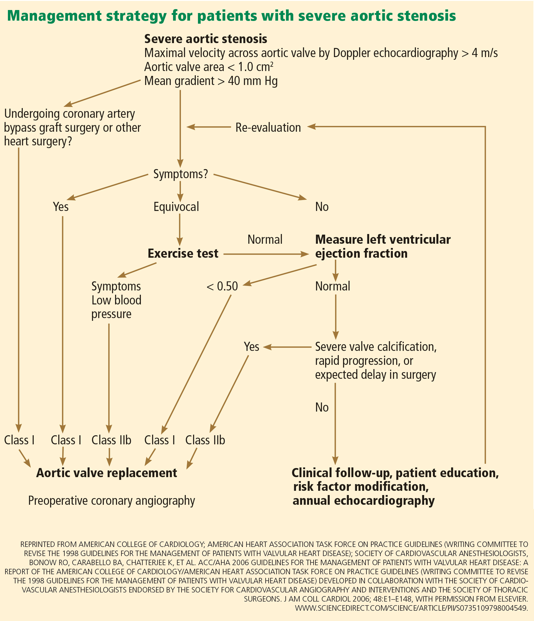
Classic symptoms of aortic stenosis include angina, heart failure, and syncope. Once symptoms appear, patients with severe aortic stenosis should be promptly referred for surgical aortic valve replacement, as survival is poor unless outflow obstruction is relieved (Figure 1). The onset of symptoms confers a poor prognosis: patients die within an average of 5 years after the onset of angina, 3 years after the onset of syncope, and 2 years after the onset of heart failure symptoms. The overall mortality rate is 75% at 3 years without surgery.3,4 Furthermore, 8% to 34% of patients with symptoms die suddenly.
Advances in prosthetic-valve design, cardiopulmonary bypass, surgical technique, and anesthesia have steadily improved the outcomes of aortic valve surgery. An analysis of the Society of Thoracic Surgeons (STS) database in 2006 showed that during the previous decade the death rate during isolated aortic valve replacement decreased from 3.4% to 2.6%. For patients under age 70 at the time of surgery, the rate of death was 1.3%, and in those ages 80 to 85, the 30-day mortality rate was less than 5%.5
Patients who survive surgery enjoy a near-normal life expectancy: 99% survive at least 5 years, 85% at least 10 years, and 82% at least 15 years.6,7 Nearly all have improvement in their ejection fraction and heart failure symptoms, and those who had more advanced symptoms before surgery enjoy the most benefit afterward.8,9
Recommendation. Surgical valve replacement for symptomatic severe aortic stenosis receives a class I recommendation, level of evidence B, in the current guidelines from the American College of Cardiology (ACC) and the American Heart Association (AHA).10,11 (See Table 1 for an explanation of the classes of recommendations and levels of evidence.)
TWO RISK-ASSESSMENT SCORES
There are two widely used scores for assessing the risk of aortic valve replacement: the European System for Cardiac Operative Risk Evaluation (EuroSCORE) and the STS score. Each has limitations.
The EuroSCORE was developed to predict the risk of dying in the hospital after adult cardiac surgery. It has been shown to predict the short-term and the long-term risk of death after heart valve surgery.12 Unfortunately, it overestimates the dangers of isolated aortic valve replacement in the patients at highest risk.13,14
The STS score, a logistic model, reflects more closely the operative and 30-day mortality rates for the patients at highest risk undergoing surgical aortic valve replacement.15,16 It was used to assess patients for surgical or transcatheter aortic valve replacement in the Placement of Aortic Transcatheter Valves (PARTNER) trial.17
These risk scores, though not perfect, are helpful as part of an overall estimation of risk that includes functional status, cardiac function, and comorbidities.
OTHER INDICATIONS FOR SURGICAL AORTIC VALVE REPLACEMENT
For patients with severe but asymptomatic aortic stenosis, surgical referral is standard practice in several circumstances.
Asymptomatic severe aortic stenosis with a low ejection fraction
Early studies found significant differences in survival beginning as early as 3 years after valve replacement between those whose preoperative ejection fraction was greater than 50% and those with a lower ejection fraction.4 Delaying surgery in these patients may lead to irreversible left ventricular dysfunction and worse survival.
Recommendation. The AHA and the ACC recommend surgical aortic valve replacement for patients who have no symptoms and whose left ventricular ejection fraction is less than 50% (class I indication, level of evidence C).10,11
Asymptomatic severe aortic stenosis in patients undergoing other cardiac surgery
Recommendation. Even if it is causing no symptoms, a severely stenotic aortic valve ought to be replaced if the ejection fraction is greater than 50% and the patient is undergoing another type of heart surgery, such as coronary artery bypass grafting, aortic surgery, or surgery on other heart valves (class I indication, level of evidence B).10,11
Asymptomatic moderate aortic stenosis in patients undergoing other cardiac surgery
When patients with a mildly or moderately stenotic aortic valve undergo other types of cardiac surgery, the decision to replace the valve is more difficult. Clinicians have to consider the increase in risk caused by adding aortic valve replacement to the planned surgery compared with the future likelihood of aortic stenosis progressing to a severe symptomatic state and eventually requiring a second cardiac surgery.
We have no evidence from a large prospective randomized controlled trial regarding prophylactic valve replacement at the time of coronary bypass surgery. However, a review of outcomes from the STS database between 1995 and 2000 found that patients under age 70 with a peak aortic gradient greater than “about 28 mm Hg” (correlating with a moderate degree of stenosis) benefited from prophylactic valve replacement at the time of coronary artery bypass surgery.18
These conclusions were supported by a subsequent retrospective analysis that found a significant survival advantage at 8 years in favor of prophylactic valve replacement at the time of bypass surgery for those with moderate (but not mild) aortic stenosis.19
Recommendation. The AHA and ACC give a class IIb endorsement, level of evidence B, for aortic valve replacement in patients with asymptomatic moderate aortic stenosis undergoing coronary bypass, valve, or aortic surgery.10,11
SEVERE ASYMPTOMATIC STENOSIS: WHICH TESTS HELP IN DECIDING?
A patient without symptoms presents a greater challenge than one with symptoms.
If surgery is deferred, the prognosis is usually excellent in such patients. Pellikka et al20 found that patients with severe asymptomatic aortic stenosis who did not undergo surgery had a rate of sudden cardiac death of about 1% per year of follow-up. However, physicians worry about missing the rapid development of symptoms of aortic stenosis in patients who previously had none. Pallikka et al also found that, at 5 years, only 20% of patients had not undergone aortic valve replacement or had not died of cardiovascular causes.20
Many researchers advocate surgical aortic valve replacement for severe asymptomatic aortic stenosis. However, the operative risk is 3% overall and has to be weighed against the 1%-per-year risk of death in patients who do not undergo surgery. Therefore, we need a way to identify a subgroup of patients without symptoms who are at higher risk.
Exercise stress testing
Some patients might subconsciously adapt to aortic stenosis by reducing their physical activity. In these “asymptomatic” patients, exercise stress testing can uncover symptoms in around 40%.21
In a group of people with severe asymptomatic aortic stenosis, a positive treadmill test (defined as an abnormal blood pressure response, ST segment changes, symptoms such as limiting dyspnea, chest discomfort, or dizziness on a modified Bruce protocol, or complex ventricular arrhythmias) strongly predicted the onset of symptoms or the need for surgery. At 24 months, only 19% of those who had had a positive exercise test result remained alive, symptom-free, and without valve replacement, compared with 85% of those who had had a negative test result.22
Subsequent study found that symptoms with exercise were the strongest predictor of the onset of symptoms of aortic stenosis, especially among patients under age 70, in whom the symptoms of fatigue and breathlessness are more specific than in the elderly.23
Recommendation. Exercise testing is recommended in patients with severe asymptomatic aortic stenosis (class IIa indication, level of evidence B) as a means of identifying those who are likely to develop symptoms or who might benefit from surgery. Surgery for those who have an abnormal exercise stress response receives a class IIb, level of evidence C recommendation from the ACC/AHA and a class IC from the European Society of Cardiology.24,25
Exercise stress echocardiography to measure change in transvalvular gradient
Emerging data suggest that exercise stress echocardiography may provide incremental prognostic information in patients with severe asymptomatic aortic stenosis. In fact, two studies showed that an exercise-induced increase in the transvalvular gradient of more than 20 mm Hg26 or 18 mm Hg27 predicts future cardiac events. This increase reflects fixed valve stenosis with limited valve compliance.
Other echocardiographic variables
Additional data have shown that severe aortic stenosis (valve area < 0.6 cm2), aortic velocity greater than 4.0 m/s, and severe calcification confer a higher risk of developing symptoms.28,29
Recommendation. The ACC and AHA say that surgical aortic valve replacement may be considered in patients without symptoms who have a high likelihood of rapid progression of aortic stenosis (ie, who are older or have severe calcification or coronary artery disease) or if surgery might be delayed at the time of symptom onset (class IIb, level of evidence C).
Aortic valve replacement can also be considered for extremely severe aortic stenosis (valve area < 0.6 cm2), mean gradient > 60 mm Hg, and velocity > 5.0 m/s if the operative mortality rate is 1.0% or less (class IIb, level of evidence C).
Brain natriuretic peptide levels
Measuring the brain natriuretic peptide (BNP) level may help if symptoms are unclear; higher levels suggest cardiac decompensation.28
One study showed that BNP levels are higher in patients with symptomatic aortic stenosis than in those with asymptomatic severe disease, and correlate with symptom severity.30 In addition, in two other studies, higher BNP and N-terminal BNP levels were shown to predict disease progression, symptom onset, and poorer event-free survival.31,32
In severe asymptomatic aortic stenosis, natriuretic peptides may provide important prognostic information beyond clinical and echocardiographic evaluation. Furthermore, in a recent study, Monin et al33 proposed a risk score that integrates peak aortic jet velocity, BNP level, and sex (women being at higher risk) in predicting who would benefit from early surgery in patients with severe asymptomatic aortic stenosis.33
SPECIAL CONSIDERATIONS
Low-output, low-gradient aortic stenosis: True severe stenosis vs pseudostenosis
Patients with a low ejection fraction (< 50%) and a high mean transvalvular gradient (> 30 or 40 mm Hg) pose no therapeutic dilemma. They have true afterload mismatch and improve markedly with surgery.34 However, patients with an even lower ejection fraction (< 35% or 40%) and a low mean transvalvular gradient (< 30 or 40 mm Hg) pose more of a problem.
It is hard to tell if these patients have true severe aortic stenosis or pseudostenosis due to primary myocardial dysfunction. In pseudostenosis, the aortic valves are moderately diseased, and leaflet opening is reduced by a failing ventricle. When cardiac output is low, the formulae used to calculate the aortic valve area become less accurate, so that patients with cardiomyopathy who have only mild or moderate aortic stenosis may appear to have severe stenosis.
Patients with pseudostenosis have a high risk of dying during surgical aortic valve replacement, approaching 50%, and benefit more from evidence-based heart failure management.35,36 In patients with true stenosis, ventricular dysfunction is mainly a result of severe stenosis and should improve after aortic valve replacement.
Dobutamine stress echocardiography can be used in patients with low-flow, low-gradient aortic stenosis to distinguish true severe stenosis from pseudostenosis. Dobutamine, an inotropic drug, increases the stroke volume so that patients with true severe aortic stenosis increase their transvalvular gradient and velocity with no or minimal change in the valve area. Conversely, in patients with pseudostenosis, the increase in stroke volume will open the aortic valve further and cause no or minimal increase in transvalvular gradient and velocity, but will increase the calculated valve area, confirming that aortic stenosis only is mild to moderate.37
Patients with low-flow, low-gradient aortic stenosis are at higher risk during surgical aortic valve replacement. Many studies have reported a 30-day mortality rate between 9% and 18%, although risks vary considerably within this population.38,39
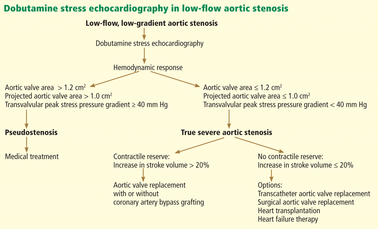
Contractile reserve. Dobutamine stress echocardiography has also been used to identify patients with severe aortic stenosis who can increase their ejection fraction and stroke volume (Figure 2).40,41 These patients are said to have “contractile reserve” and do better with surgery than those who lack adequate contractile reserve. Contractile reserve is defined as an increase of more than 20% in stroke volume during low-dose dobutamine infusion.42,43 In one small nonrandomized study, patients with contractile reserve had a 5% mortality rate at 30 days, compared with 32% in patients with no contractile reserve.44,45
In fact, patients with no contractile reserve have a high operative mortality rate during aortic valve replacement, but those who survive the operation have improvements in symptoms, functional class, and ejection fraction similar to those in patients who do have contractile reserve.46
On the other hand, if patients with no contractile reserve are treated conservatively, they have a much worse prognosis than those managed surgically.47 While it is true that patients without contractile reserve did not have a statistically significant difference in mortality rates with aortic valve replacement (P = .07) in a study by Monin et al,44 the difference was staggering between the group who underwent aortic valve replacement and the group who received medical treatment alone (hazard ratio = 0.47, 95% confidence interval 0.31–1.05, P = .07). The difference in the mortality rates may not have reached statistical significance because of the study’s small sample size.
A few years later, the same group published a similar paper with a larger study sample, focusing on patients with no contractile reserve. Using 42 propensity-matched patients, they found a statistically significantly higher 5-year survival rate in patients with no contractile reserve who underwent aortic valve replacement than in similar patients who received medical management (65% ± 11% vs 11 ± 7%, P = .019).47
Hence, surgery may be a better option than medical treatment for this select high-risk group despite the higher operative mortality risk. Transcatheter aortic valve implantation may also offer an interesting alternative to surgical aortic valve replacement in this particular subset of patients.48
Low-gradient ‘severe’ aortic stenosis with preserved ejection fraction or ‘paradoxically low-flow aortic stenosis’
Low-gradient “severe” aortic stenosis with a preserved left ventricular ejection fraction is a recently recognized clinical entity in patients with severe aortic stenosis who present with a lower-than-expected transvalvular gradient on the basis of generally accepted values.49 (A patient with severe aortic stenosis and preserved ejection fraction is expected to generate a mean transaortic gradient greater than 40 mm Hg.24) This situation remains incompletely understood but has been shown in retrospective studies to foretell a poor prognosis.50–52
This subgroup of patients has pronounced left ventricular concentric remodeling with a small left ventricular cavity, impaired left ventricular filling, and reduced systolic longitudinal myocardial shortening.44
Herrmann et al53 provided more insight into the pathophysiology by showing that patients with this condition exhibit more pronounced myocardial fibrosis on myocardial biopsy and more pronounced late subendocardial enhancement on magnetic resonance imaging. These patients also displayed a significant decrease in mitral ring displacement and systolic strain. These abnormalities result in a low stroke volume despite a preserved ejection fraction and consequently a lower transvalvular gradient (< 40 mm Hg).
This disease pattern, in which the low gradient is interpreted as mild to moderate aortic stenosis, may lead to underestimation of stenosis severity and, thus, to inappropriate delay of aortic valve replacement.
However, other conditions can cause this hemodynamic situation with a lower-than-expected gradient. It can arise from a small left ventricle that correlates with a small body size, yielding a lower-than-normal stroke volume, measurement errors in determining stroke volume and valve area by Doppler echocardiography, systemic hypertension (which can influence estimation of the gradient by Doppler echocardiography), and inconsistency in the definition of severe aortic stenosis in the current guidelines relating to cutoffs of valve area in relation to those of jet velocity and gradient.54
This subgroup of patients seems to be at a more advanced stage and has a poorer prognosis if treated medically rather than surgically. When symptomatic, low-gradient severe aortic stenosis should be treated surgically, with one study showing excellent outcomes with aortic valve replacement.50
However, a recent study by Jander et al55 showed that patients with low-gradient severe aortic stenosis and normal ejection fraction have outcomes similar to those in patients with moderate aortic stenosis, suggesting a strategy of medical therapy and close monitoring.55 Of note, the subset of patients reported in this substudy of the Simvastatin and Ezetimibe in Aortic Stenosis (SEAS) trial did not really fit the pattern of low-gradient severe aortic stenosis described by Hachicha et al50 and other groups.51,56 These patients had aortic valve areas in the severe range but mean transaortic gradients in the moderate range, and in light of the other echocardiographic findings in these patients, the area-gradient discordances were predominantly due to small body surface area and measurement errors. These patients indeed had near-normal left ventricular size, no left ventricular hypertrophy, and no evidence of concentric remodeling.
Finally, the findings of the study by Jander et al55 are discordant with those of another substudy of the SEAS trial,57 which reported that paradoxical low-flow aortic stenosis occurred in about 7% of the cohort (compared with 52% in the study by Jander et al55) and was associated with more pronounced concentric remodeling and more severe impairment of myocardial function.
Whether intervention in patients with low-gradient severe aortic stenosis and valve area less than 1.0 cm2 improves outcomes remains to be confirmed and reproduced in future prospective studies.
Elderly patients
The risks of cardiac surgery increase with age. Older patients may be more deconditioned and have more comorbidities than younger patients, placing them at greater risk of a poor outcome.
Several retrospective studies of valve replacement in octogenarians have found that operative mortality rates range from 5.7% to 9% during isolated aortic valve replacement.58–60 Note that, using the STS score, the operative mortality risk increases only from 1.2% in a 70-year-old man with no comorbidities to 1.8% in an 80-year-old man undergoing aortic valve replacement plus coronary artery bypass grafting.61
As in younger patients, valve replacement results in a significant survival benefit and symptomatic improvement. Yet up to 30% of patients with severe aortic stenosis are not referred for surgery because surgery is believed to be too risky.62 The conditions most frequently cited by physicians when declining to refer patients for surgery include a low ejection fraction, advanced age, and advanced comorbidities. None of these is an absolute contraindication to surgery.
A recent retrospective study of 443 elderly patients (mean age 79.5) showed that those with left ventricular concentric remodeling, lower stroke volume, elevated left ventricular filling pressures, and mildly elevated pulmonary artery pressures have a very bad prognosis, with a mortality rate of 50.5% at 3.3 ± 2.7 years.63
Despite the higher operative mortality risk, these patients face a dismal prognosis when treated medically and should be referred to a cardiologist or cardiothoracic surgeon for an assessment of their operative risk and, potentially, for referral for catheter-based valve replacement.
Acutely ill patients
In critically ill patients with aortic stenosis and cardiogenic shock, the use of intravenous sodium nitroprusside increases cardiac output and decreases pulmonary artery wedge pressure, allowing patients to transition to surgery or vasodilator therapy. The mechanism seems to be an increase in myocardial contractility rather than a decrease in peripheral resistance. The reduction in filling pressure and concurrent increase in coronary blood flow relieves ischemia and subsequently enhances contractility.64
TRANSCATHETER AORTIC VALVE REPLACEMENT
Until recently, patients with severe aortic stenosis who were deemed to be at high surgical risk were referred for balloon valvuloplasty as a palliative option. The procedure consists of balloon inflation across the aortic valve to relieve the stenosis.
Most patients have improved symptoms and a decrease in pressure gradient immediately after the procedure, but the results are not durable, with a high restenosis rate within 6 to 12 months and no decrease in the mortality rate.65 (There is some evidence that serial balloon dilation improves survival.66)
The procedure has several limitations, including a risk of embolic stroke, myocardial infarction, and, sometimes, perforation of the left ventricle. It is only used in people who do not wish to have surgery or as a bridge to surgical aortic valve replacement in hemodynamically unstable patients.
Advances in transcatheter technologies have made nonsurgical valve replacement a reality that is increasingly available to a broader population of patients. The first percutaneous valve replacement in a human was performed in 2002.67 Since then, multiple registries from centers around the world, especially in Europe, have shown that it can be performed in high-risk patients with outcomes very comparable to those of surgical aortic valve replacement as predicted by the STS score and EuroSCORE.68,69 Procedural success rates have increased from around 80% in the initial experience to over 95% in the most current series.70
Results from randomized trials
The long-awaited PARTNER A and B trials have been published.
The PARTNER B trial17 randomized patients with severe aortic stenosis who were not considered by the STS score to be suitable candidates for surgery to standard therapy (which included balloon valvoplasty in 84%) or transcatheter aortic valve replacement. There was a dramatic 20% absolute improvement in survival at 1 year with transcatheter replacement, with the survival curve continuing to diverge at 1 year. The rate of death from any cause was 30.7% with transcatheter aortic valve replacement vs 50.7% with standard therapy (hazard ratio with transcatheter replacement 0.55; P < .001).
The major concerns about transcatheter aortic valve replacement borne out in the study are procedural complications, namely stroke and vascular events. At 30 days, transcatheter replacement, as compared with standard therapy, was associated with a higher incidence of major stroke (5.0% vs 1.1%, P = .06) and major vascular complications (16.2% vs 1.1%, P < .001).17
On the other hand, the PARTNER A trial randomized high-risk patients deemed operable by the STS score to either transcatheter or surgical aortic valve replacement. The rate of death at 1 year from any cause was similar in both groups (24.2% vs 26.8%; P = .44), but again at the expense of higher rates of vascular complications (11.0% vs 3.2%, P < .001 at 30 days) and stroke (5.1% vs 2.4%; P = .07 at 1 year) in the transcatheter group. However, the surgical group had higher rates of major bleeding (19.5% vs 9.3%; P < .001) and new-onset atrial fibrillation (16.0% vs 8.6%, P = .06).71
Transcatheter aortic valve replacement has modernized the way we treat aortic stenosis and without a shred of doubt will become the standard of therapy for severe symptomatic aortic stenosis in patients who are not candidates for surgery. For the high-risk operable patient, the benefit of avoiding a sternotomy should be weighed against the higher risk of stroke and vascular complications with the transcatheter procedure. The availability of smaller delivery systems, better expertise, and better vascular access selection should decrease the rate of complications in the future.
- Stewart BF, Siscovick D, Lind BK, et al. Clinical factors associated with calcific aortic valve disease. Cardiovascular Health Study. J Am Coll Cardiol 1997; 29:630–634.
- Iung B, Baron G, Butchart EG, et al. A prospective survey of patients with valvular heart disease in Europe: The Euro Heart Survey on Valvular Heart Disease. Eur Heart J 2003; 24:1231–1243.
- Ross J, Braunwald E. Aortic stenosis. Circulation 1968; 38(suppl 1):61–67.
- Schwarz F, Baumann P, Manthey J, et al. The effect of aortic valve replacement on survival. Circulation 1982; 66:1105–1110.
- Brown JM, O’Brien SM, Wu C, Sikora JA, Griffith BP, Gammie JS. Isolated aortic valve replacement in North America comprising 108,687 patients in 10 years: changes in risks, valve types, and outcomes in the Society of Thoracic Surgeons National Database. J Thorac Cardiovasc Surg 2009; 137:82–90.
- Kvidal P, Bergström R, Malm T, Ståhle E. Long-term follow-up of morbidity and mortality after aortic valve replacement with a mechanical valve prosthesis. Eur Heart J 2000; 21:1099–1111.
- Ståhle E, Kvidal P, Nyström SO, Bergström R. Long-term relative survival after primary heart valve replacement. Eur J Cardiothorac Surg 1997; 11:81–91.
- Sharma UC, Barenbrug P, Pokharel S, Dassen WR, Pinto YM, Maessen JG. Systematic review of the outcome of aortic valve replacement in patients with aortic stenosis. Ann Thorac Surg 2004; 78:90–95.
- Vaquette B, Corbineau H, Laurent M, et al. Valve replacement in patients with critical aortic stenosis and depressed left ventricular function: predictors of operative risk, left ventricular function recovery, and long term outcome. Heart 2005; 91:1324–1329.
- American College of Cardiology/American Heart Association Task Force on Practice Guidelines; Society of Cardiovascular Anesthesiologists; Society for Cardiovascular Angiography and Interventions; Society of Thoracic Surgeons, Bonow RO, Carabello BA, Kanu C, et al. ACC/AHA 2006 guidelines for the management of patients with valvular heart disease: a report of the American College of Cardiology/American Heart Association Task Force on Practice Guidelines (writing committee to revise the 1998 Guidelines for the Management of Patients With Valvular Heart Disease): developed in collaboration with the Society of Cardiovascular Anesthesiologists: endorsed by the Society for Cardiovascular Angiography and Interventions and the Society of Thoracic Surgeons. Circulation 2006; 114:e84–e231.
- Bonow RO, Carabello BA, Chatterjee K, et al; 2006 Writing Committee Members; American College of Cardiology/American Heart Association Task Force. 2008 Focused update incorporated into the ACC/AHA 2006 guidelines for the management of patients with valvular heart disease: a report of the American College of Cardiology/American Heart Association Task Force on Practice Guidelines (Writing Committee to Revise the 1998 Guidelines for the Management of Patients With Valvular Heart Disease): endorsed by the Society of Cardiovascular Anesthesiologists, Society for Cardiovascular Angiography and Interventions, and Society of Thoracic Surgeons. Circulation 2008; 118:e523–e661.
- Nashef SA, Roques F, Hammill BG, et al; EuroSCORE Project Group. Validation of European System for Cardiac Operative Risk Evaluation (EuroSCORE) in North American cardiac surgery. Eur J Cardiothorac Surg 2002; 22:101–105.
- Grossi EA, Schwartz CF, Yu PJ, et al. High-risk aortic valve replacement: are the outcomes as bad as predicted? Ann Thorac Surg 2008; 85:102–106.
- Kalavrouziotis D, Li D, Buth KJ, Légaré JF. The European System for Cardiac Operative Risk Evaluation (EuroSCORE) is not appropriate for withholding surgery in high-risk patients with aortic stenosis: a retrospective cohort study. J Cardiothorac Surg 2009; 4:32.
- Dewey TM, Brown D, Ryan WH, Herbert MA, Prince SL, Mack MJ. Reliability of risk algorithms in predicting early and late operative outcomes in high-risk patients undergoing aortic valve replacement. J Thorac Cardiovasc Surg 2008; 135:180–187.
- Wendt D, Osswald BR, Kayser K, et al. Society of Thoracic Surgeons score is superior to the EuroSCORE determining mortality in high risk patients undergoing isolated aortic valve replacement. Ann Thorac Surg 2009; 88:468–474.
- Leon MB, Smith CR, Mack M, et al; PARTNER Trial Investigators. Transcatheter aortic-valve implantation for aortic stenosis in patients who cannot undergo surgery. N Engl J Med 2010; 363:1597–1607.
- Smith WT, Ferguson TB, Ryan T, Landolfo CK, Peterson ED. Should coronary artery bypass graft surgery patients with mild or moderate aortic stenosis undergo concomitant aortic valve replacement? A decision analysis approach to the surgical dilemma. J Am Coll Cardiol 2004; 44:1241–1247.
- Pereira JJ, Balaban K, Lauer MS, Lytle B, Thomas JD, Garcia MJ. Aortic valve replacement in patients with mild or moderate aortic stenosis and coronary bypass surgery. Am J Med 2005; 118:735–742.
- Pellikka PA, Sarano ME, Nishimura RA, et al. Outcome of 622 adults with asymptomatic, hemodynamically significant aortic stenosis during prolonged follow-up. Circulation 2005; 111:3290–3295.
- Ennezat PV, Maréchaux S, Iung B, Chauvel C, LeJemtel TH, Pibarot P. Exercise testing and exercise stress echocardiography in asymptomatic aortic valve stenosis. Heart 2009; 95:877–884.
- Amato MC, Moffa PJ, Werner KE, Ramires JA. Treatment decision in asymptomatic aortic valve stenosis: role of exercise testing. Heart 2001; 86:381–386.
- Das P, Rimington H, Chambers J. Exercise testing to stratify risk in aortic stenosis. Eur Heart J 2005; 26:1309–1313.
- American College of Cardiology; American Heart Association Task Force on Practice Guidelines (Writing Committee to revise the 1998 guidelines for the management of patients with valvular heart disease); Society of Cardiovascular Anesthesiologists, Bonow RO, Carabello BA, Chatterjee K, et al. ACC/AHA 2006 guidelines for the management of patients with valvular heart disease: a report of the American College of Cardiology/American Heart Association Task Force on Practice Guidelines (writing Committee to Revise the 1998 guidelines for the management of patients with valvular heart disease) developed in collaboration with the Society of Cardiovascular Anesthesiologists endorsed by the Society for Cardiovascular Angiography and Interventions and the Society of Thoracic Surgeons. J Am Coll Cardiol 2006; 48:e1–e148.
- Vahanian A, Baumgartner H, Bax J, et al; Task Force on the Management of Valvular Hearth Disease of the European Society of Cardiology; ESC Committee for Practice Guidelines. Guidelines on the management of valvular heart disease: The Task Force on the Management of Valvular Heart Disease of the European Society of Cardiology. Eur Heart J 2007; 28:230–268.
- Maréchaux S, Hachicha Z, Bellouin A, et al. Usefulness of exercise-stress echocardiography for risk stratification of true asymptomatic patients with aortic valve stenosis. Eur Heart J 2010; 31:1390–1397.
- Lancellotti P, Lebois F, Simon M, Tombeux C, Chauvel C, Pierard LA. Prognostic importance of quantitative exercise Doppler echocardiography in asymptomatic valvular aortic stenosis. Circulation 2005; 112(suppl 9):I377–1382.
- Otto CM. Valvular aortic stenosis: disease severity and timing of intervention. J Am Coll Cardiol 2006; 47:2141–2151.
- Rosenhek R, Binder T, Porenta G, et al. Predictors of outcome in severe, asymptomatic aortic stenosis. N Engl J Med 2000; 343:611–617.
- Lim P, Monin JL, Monchi M, et al. Predictors of outcome in patients with severe aortic stenosis and normal left ventricular function: role of B-type natriuretic peptide. Eur Heart J 2004; 25:2048–2053.
- Gerber IL, Legget ME, West TM, Richards AM, Stewart RA. Usefulness of serial measurement of N-terminal pro-brain natriuretic peptide plasma levels in asymptomatic patients with aortic stenosis to predict symptomatic deterioration. Am J Cardiol 2005; 95:898–901.
- Bergler-Klein J, Klaar U, Heger M, et al. Natriuretic peptides predict symptom-free survival and postoperative outcome in severe aortic stenosis. Circulation 2004; 109:2302–2308.
- Monin JL, Lancellotti P, Monchi M, et al. Risk score for predicting outcome in patients with asymptomatic aortic stenosis. Circulation 2009; 120:69–75.
- Carabello BA, Green LH, Grossman W, Cohn LH, Koster JK, Collins JJ. Hemodynamic determinants of prognosis of aortic valve replacement in critical aortic stenosis and advanced congestive heart failure. Circulation 1980; 62:42–48.
- Connolly HM, Oh JK, Schaff HV, et al. Severe aortic stenosis with low transvalvular gradient and severe left ventricular dysfunction: result of aortic valve replacement in 52 patients. Circulation 2000; 101:1940–1946.
- Brogan WC, Grayburn PA, Lange RA, Hillis LD. Prognosis after valve replacement in patients with severe aortic stenosis and a low transvalvular pressure gradient. J Am Coll Cardiol 1993; 21:1657–1660.
- Burwash IG. Low-flow, low-gradient aortic stenosis: from evaluation to treatment. Curr Opin Cardiol 2007; 22:84–91.
- Connolly HM, Oh JK, Orszulak TA, et al. Aortic valve replacement for aortic stenosis with severe left ventricular dysfunction. Prognostic indicators. Circulation 1997; 95:2395–2400.
- Pai RG, Varadarajan P, Razzouk A. Survival benefit of aortic valve replacement in patients with severe aortic stenosis with low ejection fraction and low gradient with normal ejection fraction. Ann Thorac Surg 2008; 86:1781–1789.
- Blais C, Burwash IG, Mundigler G, et al. Projected valve area at normal flow rate improves the assessment of stenosis severity in patients with low-flow, low-gradient aortic stenosis: the multicenter TOPAS (Truly or Pseudo-Severe Aortic Stenosis) study. Circulation 2006; 113:711–721.
- Clavel MA, Burwash IG, Mundigler G, et al. Validation of conventional and simplified methods to calculate projected valve area at normal flow rate in patients with low flow, low gradient aortic stenosis: the multicenter TOPAS (True or Pseudo Severe Aortic Stenosis) study. J Am Soc Echocardiogr 2010; 23:380–386.
- Monin JL, Monchi M, Gest V, Duval-Moulin AM, Dubois-Rande JL, Gueret P. Aortic stenosis with severe left ventricular dysfunction and low transvalvular pressure gradients: risk stratification by low-dose dobutamine echocardiography. J Am Coll Cardiol 2001; 37:2101–2107.
- Nishimura RA, Grantham JA, Connolly HM, Schaff HV, Higano ST, Holmes DR. Low-output, low-gradient aortic stenosis in patients with depressed left ventricular systolic function: the clinical utility of the dobutamine challenge in the catheterization laboratory. Circulation 2002; 106:809–813.
- Monin JL, Quéré JP, Monchi M, et al. Low-gradient aortic stenosis: operative risk stratification and predictors for long-term outcome: a multicenter study using dobutamine stress hemodynamics. Circulation 2003; 108:319–324.
- Monin JL, Guéret P. Calcified aortic stenosis with left ventricular dysfunction and low transvalvular gradients. Must one reject surgery in certain cases?. (In French.) Arch Mal Coeur Vaiss 2003; 96:864–870.
- Quere JP, Monin JL, Levy F, et al. Influence of preoperative left ventricular contractile reserve on postoperative ejection fraction in low-gradient aortic stenosis. Circulation 2006; 113:1738–1744.
- Tribouilloy C, Lévy F, Rusinaru D, et al. Outcome after aortic valve replacement for low-flow/low-gradient aortic stenosis without contractile reserve on dobutamine stress echocardiography. J Am Coll Cardiol 2009; 53:1865–1873.
- Clavel MA, Webb JG, Rodés-Cabau J, et al. Comparison between transcatheter and surgical prosthetic valve implantation in patients with severe aortic stenosis and reduced left ventricular ejection fraction. Circulation 2010; 122:1928–1936.
- Dumesnil JG, Pibarot P, Carabello B. Paradoxical low flow and/or low gradient severe aortic stenosis despite preserved left ventricular ejection fraction: implications for diagnosis and treatment. Eur Heart J 2010; 31:281–289.
- Hachicha Z, Dumesnil JG, Bogaty P, Pibarot P. Paradoxical low-flow, low-gradient severe aortic stenosis despite preserved ejection fraction is associated with higher afterload and reduced survival. Circulation 2007; 115:2856–2864.
- Barasch E, Fan D, Chukwu EO, et al. Severe isolated aortic stenosis with normal left ventricular systolic function and low transvalvular gradients: pathophysiologic and prognostic insights. J Heart Valve Dis 2008; 17:81–88.
- Dumesnil JG, Pibarot P, Carabello B. Paradoxical low flow and/or low gradient severe aortic stenosis despite preserved left ventricular ejection fraction: implications for diagnosis and treatment. Eur Heart J 2010; 31:281–289.
- Herrmann S, Störk S, Niemann M, et al. Low-gradient aortic valve stenosis myocardial fibrosis and its influence on function and outcome. J Am Coll Cardiol 2011; 58:402–412.
- Minners J, Allgeier M, Gohlke-Baerwolf C, Kienzle RP, Neumann FJ, Jander N. Inconsistent grading of aortic valve stenosis by current guidelines: haemodynamic studies in patients with apparently normal left ventricular function. Heart 2010; 96:1463–1468.
- Jander N, Minners J, Holme I, et al. Outcome of patients with low-gradient “severe” aortic stenosis and preserved ejection fraction. Circulation 2011; 123:887–895.
- Lancellotti P, Donal E, Magne J, et al. Impact of global left ventricular afterload on left ventricular function in asymptomatic severe aortic stenosis: a two-dimensional speckle-tracking study. Eur J Echocardiogr 2010; 11:537–543.
- Cramariuc D, Cioffi G, Rieck AE, et al. Low-flow aortic stenosis in asymptomatic patients: valvular-arterial impedance and systolic function from the SEAS Substudy. JACC Cardiovasc Imaging 2009; 2:390–399.
- Craver JM, Puskas JD, Weintraub WW, et al. 601 octogenarians undergoing cardiac surgery: outcome and comparison with younger age groups. Ann Thorac Surg 1999; 67:1104–1110.
- Alexander KP, Anstrom KJ, Muhlbaier LH, et al. Outcomes of cardiac surgery in patients > or = 80 years: results from the National Cardiovascular Network. J Am Coll Cardiol 2000; 35:731–738.
- Collart F, Feier H, Kerbaul F, et al. Valvular surgery in octogenarians: operative risks factors, evaluation of Euroscore and long term results. Eur J Cardiothorac Surg 2005; 27:276–280.
- Kurtz CE, Otto CM. Aortic stenosis: clinical aspects of diagnosis and management, with 10 illustrative case reports from a 25-year experience. Medicine (Baltimore) 2010; 89:349–379.
- Iung B, Cachier A, Baron G, et al. Decision-making in elderly patients with severe aortic stenosis: why are so many denied surgery? Eur Heart J 2005; 26:2714–2720.
- Kahn J, Petillo F, Rhee PDY, et al. Echocardiographic predictors of mortality in patients with severe isolated aortic stenosis and normal left ventricular ejection fraction who do not undergo aortic valve replacement. American Society of Echocardiography 2011 Scientific Sessions; June 13, 2011; Montreal, QC. http://www.abstractsonline.com/Plan/ViewAbstract.aspx?sKey=845e6287-66e1-4df5-8aef-8f5da16ef94a&cKey=5e5438dd-20df-48bfbee7-5f867fce66e6&mKey=%7bAE58A7EE-7140-41D6-9C7ED375E33DDABD%7d. Accessed May 27, 2012.
- Popovic ZB, Khot UN, Novaro GM, et al. Effects of sodium nitroprusside in aortic stenosis associated with severe heart failure: pressure-volume loop analysis using a numerical model. Am J Physiol Heart Circ Physiol 2005; 288:H416–H423.
- Otto CM, Mickel MC, Kennedy JW, et al. Three-year outcome after balloon aortic valvuloplasty. Insights into prognosis of valvular aortic stenosis. Circulation 1994; 89:642–650.
- Letac B, Cribier A, Eltchaninoff H, Koning R, Derumeaux G. Evaluation of restenosis after balloon dilatation in adult aortic stenosis by repeat catheterization. Am Heart J 1991; 122:55–60.
- Cribier A, Eltchaninoff H, Bash A, et al. Percutaneous transcatheter implantation of an aortic valve prosthesis for calcific aortic stenosis: first human case description. Circulation 2002; 106:3006–3008.
- Grube E, Schuler G, Buellesfeld L, et al. Percutaneous aortic valve replacement for severe aortic stenosis in high-risk patients using the second- and current third-generation self-expanding CoreValve prosthesis: device success and 30-day clinical outcome. J Am Coll Cardiol 2007; 50:69–76.
- Webb JG, Altwegg L, Masson JB, Al Bugami S, Al Ali A, Boone RA. A new transcatheter aortic valve and percutaneous valve delivery system. J Am Coll Cardiol 2009; 53:1855–1858.
- Clavel MA, Webb JG, Pibarot P, et al. Comparison of the hemodynamic performance of percutaneous and surgical bioprostheses for the treatment of severe aortic stenosis. J Am Coll Cardiol 2009; 53:1883–1891.
- Smith CR, Leon MB, Mack MJ, et al; PARTNER Trial Investigators. Transcatheter versus surgical aortic-valve replacement in high-risk patients. N Engl J Med. 2011; 364:2187–2198.
For some patients with aortic stenosis, the choice of management is simple; for others it is less so. Patients who have severe, symptomatic stenosis and who have low surgical risk should undergo aortic valve replacement. But if the stenosis is severe but asymptomatic, or if the patient is at higher surgical risk, or if there seems to be a mismatch in the hemodynamic variables, the situation is more complicated.
Fortunately, we have evidence and guidelines to go on. In this paper we review the indications for surgical and transcatheter aortic valve replacement, focusing on the areas of less certainty.
AN INDOLENT DISEASE, UNTIL IT ISN’T
Aortic stenosis is the most common valvular disease and the third most prevalent form of cardiovascular disease in the Western world, after hypertension and coronary artery disease. It is largely a disease of the elderly; its prevalence increases with age, and it is present in 2% to 7% of patients over age 65.1,2
At first, its course is indolent, as it progresses slowly over years to decades. However, this is followed by rapid clinical deterioration and a high death rate after symptoms develop.
SURGICAL AORTIC VALVE REPLACEMENT FOR SEVERE SYMPTOMATIC STENOSIS
Classic symptoms of aortic stenosis include angina, heart failure, and syncope. Once symptoms appear, patients with severe aortic stenosis should be promptly referred for surgical aortic valve replacement, as survival is poor unless outflow obstruction is relieved (Figure 1). The onset of symptoms confers a poor prognosis: patients die within an average of 5 years after the onset of angina, 3 years after the onset of syncope, and 2 years after the onset of heart failure symptoms. The overall mortality rate is 75% at 3 years without surgery.3,4 Furthermore, 8% to 34% of patients with symptoms die suddenly.
Advances in prosthetic-valve design, cardiopulmonary bypass, surgical technique, and anesthesia have steadily improved the outcomes of aortic valve surgery. An analysis of the Society of Thoracic Surgeons (STS) database in 2006 showed that during the previous decade the death rate during isolated aortic valve replacement decreased from 3.4% to 2.6%. For patients under age 70 at the time of surgery, the rate of death was 1.3%, and in those ages 80 to 85, the 30-day mortality rate was less than 5%.5
Patients who survive surgery enjoy a near-normal life expectancy: 99% survive at least 5 years, 85% at least 10 years, and 82% at least 15 years.6,7 Nearly all have improvement in their ejection fraction and heart failure symptoms, and those who had more advanced symptoms before surgery enjoy the most benefit afterward.8,9
Recommendation. Surgical valve replacement for symptomatic severe aortic stenosis receives a class I recommendation, level of evidence B, in the current guidelines from the American College of Cardiology (ACC) and the American Heart Association (AHA).10,11 (See Table 1 for an explanation of the classes of recommendations and levels of evidence.)
TWO RISK-ASSESSMENT SCORES
There are two widely used scores for assessing the risk of aortic valve replacement: the European System for Cardiac Operative Risk Evaluation (EuroSCORE) and the STS score. Each has limitations.
The EuroSCORE was developed to predict the risk of dying in the hospital after adult cardiac surgery. It has been shown to predict the short-term and the long-term risk of death after heart valve surgery.12 Unfortunately, it overestimates the dangers of isolated aortic valve replacement in the patients at highest risk.13,14
The STS score, a logistic model, reflects more closely the operative and 30-day mortality rates for the patients at highest risk undergoing surgical aortic valve replacement.15,16 It was used to assess patients for surgical or transcatheter aortic valve replacement in the Placement of Aortic Transcatheter Valves (PARTNER) trial.17
These risk scores, though not perfect, are helpful as part of an overall estimation of risk that includes functional status, cardiac function, and comorbidities.
OTHER INDICATIONS FOR SURGICAL AORTIC VALVE REPLACEMENT
For patients with severe but asymptomatic aortic stenosis, surgical referral is standard practice in several circumstances.
Asymptomatic severe aortic stenosis with a low ejection fraction
Early studies found significant differences in survival beginning as early as 3 years after valve replacement between those whose preoperative ejection fraction was greater than 50% and those with a lower ejection fraction.4 Delaying surgery in these patients may lead to irreversible left ventricular dysfunction and worse survival.
Recommendation. The AHA and the ACC recommend surgical aortic valve replacement for patients who have no symptoms and whose left ventricular ejection fraction is less than 50% (class I indication, level of evidence C).10,11
Asymptomatic severe aortic stenosis in patients undergoing other cardiac surgery
Recommendation. Even if it is causing no symptoms, a severely stenotic aortic valve ought to be replaced if the ejection fraction is greater than 50% and the patient is undergoing another type of heart surgery, such as coronary artery bypass grafting, aortic surgery, or surgery on other heart valves (class I indication, level of evidence B).10,11
Asymptomatic moderate aortic stenosis in patients undergoing other cardiac surgery
When patients with a mildly or moderately stenotic aortic valve undergo other types of cardiac surgery, the decision to replace the valve is more difficult. Clinicians have to consider the increase in risk caused by adding aortic valve replacement to the planned surgery compared with the future likelihood of aortic stenosis progressing to a severe symptomatic state and eventually requiring a second cardiac surgery.
We have no evidence from a large prospective randomized controlled trial regarding prophylactic valve replacement at the time of coronary bypass surgery. However, a review of outcomes from the STS database between 1995 and 2000 found that patients under age 70 with a peak aortic gradient greater than “about 28 mm Hg” (correlating with a moderate degree of stenosis) benefited from prophylactic valve replacement at the time of coronary artery bypass surgery.18
These conclusions were supported by a subsequent retrospective analysis that found a significant survival advantage at 8 years in favor of prophylactic valve replacement at the time of bypass surgery for those with moderate (but not mild) aortic stenosis.19
Recommendation. The AHA and ACC give a class IIb endorsement, level of evidence B, for aortic valve replacement in patients with asymptomatic moderate aortic stenosis undergoing coronary bypass, valve, or aortic surgery.10,11
SEVERE ASYMPTOMATIC STENOSIS: WHICH TESTS HELP IN DECIDING?
A patient without symptoms presents a greater challenge than one with symptoms.
If surgery is deferred, the prognosis is usually excellent in such patients. Pellikka et al20 found that patients with severe asymptomatic aortic stenosis who did not undergo surgery had a rate of sudden cardiac death of about 1% per year of follow-up. However, physicians worry about missing the rapid development of symptoms of aortic stenosis in patients who previously had none. Pallikka et al also found that, at 5 years, only 20% of patients had not undergone aortic valve replacement or had not died of cardiovascular causes.20
Many researchers advocate surgical aortic valve replacement for severe asymptomatic aortic stenosis. However, the operative risk is 3% overall and has to be weighed against the 1%-per-year risk of death in patients who do not undergo surgery. Therefore, we need a way to identify a subgroup of patients without symptoms who are at higher risk.
Exercise stress testing
Some patients might subconsciously adapt to aortic stenosis by reducing their physical activity. In these “asymptomatic” patients, exercise stress testing can uncover symptoms in around 40%.21
In a group of people with severe asymptomatic aortic stenosis, a positive treadmill test (defined as an abnormal blood pressure response, ST segment changes, symptoms such as limiting dyspnea, chest discomfort, or dizziness on a modified Bruce protocol, or complex ventricular arrhythmias) strongly predicted the onset of symptoms or the need for surgery. At 24 months, only 19% of those who had had a positive exercise test result remained alive, symptom-free, and without valve replacement, compared with 85% of those who had had a negative test result.22
Subsequent study found that symptoms with exercise were the strongest predictor of the onset of symptoms of aortic stenosis, especially among patients under age 70, in whom the symptoms of fatigue and breathlessness are more specific than in the elderly.23
Recommendation. Exercise testing is recommended in patients with severe asymptomatic aortic stenosis (class IIa indication, level of evidence B) as a means of identifying those who are likely to develop symptoms or who might benefit from surgery. Surgery for those who have an abnormal exercise stress response receives a class IIb, level of evidence C recommendation from the ACC/AHA and a class IC from the European Society of Cardiology.24,25
Exercise stress echocardiography to measure change in transvalvular gradient
Emerging data suggest that exercise stress echocardiography may provide incremental prognostic information in patients with severe asymptomatic aortic stenosis. In fact, two studies showed that an exercise-induced increase in the transvalvular gradient of more than 20 mm Hg26 or 18 mm Hg27 predicts future cardiac events. This increase reflects fixed valve stenosis with limited valve compliance.
Other echocardiographic variables
Additional data have shown that severe aortic stenosis (valve area < 0.6 cm2), aortic velocity greater than 4.0 m/s, and severe calcification confer a higher risk of developing symptoms.28,29
Recommendation. The ACC and AHA say that surgical aortic valve replacement may be considered in patients without symptoms who have a high likelihood of rapid progression of aortic stenosis (ie, who are older or have severe calcification or coronary artery disease) or if surgery might be delayed at the time of symptom onset (class IIb, level of evidence C).
Aortic valve replacement can also be considered for extremely severe aortic stenosis (valve area < 0.6 cm2), mean gradient > 60 mm Hg, and velocity > 5.0 m/s if the operative mortality rate is 1.0% or less (class IIb, level of evidence C).
Brain natriuretic peptide levels
Measuring the brain natriuretic peptide (BNP) level may help if symptoms are unclear; higher levels suggest cardiac decompensation.28
One study showed that BNP levels are higher in patients with symptomatic aortic stenosis than in those with asymptomatic severe disease, and correlate with symptom severity.30 In addition, in two other studies, higher BNP and N-terminal BNP levels were shown to predict disease progression, symptom onset, and poorer event-free survival.31,32
In severe asymptomatic aortic stenosis, natriuretic peptides may provide important prognostic information beyond clinical and echocardiographic evaluation. Furthermore, in a recent study, Monin et al33 proposed a risk score that integrates peak aortic jet velocity, BNP level, and sex (women being at higher risk) in predicting who would benefit from early surgery in patients with severe asymptomatic aortic stenosis.33
SPECIAL CONSIDERATIONS
Low-output, low-gradient aortic stenosis: True severe stenosis vs pseudostenosis
Patients with a low ejection fraction (< 50%) and a high mean transvalvular gradient (> 30 or 40 mm Hg) pose no therapeutic dilemma. They have true afterload mismatch and improve markedly with surgery.34 However, patients with an even lower ejection fraction (< 35% or 40%) and a low mean transvalvular gradient (< 30 or 40 mm Hg) pose more of a problem.
It is hard to tell if these patients have true severe aortic stenosis or pseudostenosis due to primary myocardial dysfunction. In pseudostenosis, the aortic valves are moderately diseased, and leaflet opening is reduced by a failing ventricle. When cardiac output is low, the formulae used to calculate the aortic valve area become less accurate, so that patients with cardiomyopathy who have only mild or moderate aortic stenosis may appear to have severe stenosis.
Patients with pseudostenosis have a high risk of dying during surgical aortic valve replacement, approaching 50%, and benefit more from evidence-based heart failure management.35,36 In patients with true stenosis, ventricular dysfunction is mainly a result of severe stenosis and should improve after aortic valve replacement.
Dobutamine stress echocardiography can be used in patients with low-flow, low-gradient aortic stenosis to distinguish true severe stenosis from pseudostenosis. Dobutamine, an inotropic drug, increases the stroke volume so that patients with true severe aortic stenosis increase their transvalvular gradient and velocity with no or minimal change in the valve area. Conversely, in patients with pseudostenosis, the increase in stroke volume will open the aortic valve further and cause no or minimal increase in transvalvular gradient and velocity, but will increase the calculated valve area, confirming that aortic stenosis only is mild to moderate.37
Patients with low-flow, low-gradient aortic stenosis are at higher risk during surgical aortic valve replacement. Many studies have reported a 30-day mortality rate between 9% and 18%, although risks vary considerably within this population.38,39
Contractile reserve. Dobutamine stress echocardiography has also been used to identify patients with severe aortic stenosis who can increase their ejection fraction and stroke volume (Figure 2).40,41 These patients are said to have “contractile reserve” and do better with surgery than those who lack adequate contractile reserve. Contractile reserve is defined as an increase of more than 20% in stroke volume during low-dose dobutamine infusion.42,43 In one small nonrandomized study, patients with contractile reserve had a 5% mortality rate at 30 days, compared with 32% in patients with no contractile reserve.44,45
In fact, patients with no contractile reserve have a high operative mortality rate during aortic valve replacement, but those who survive the operation have improvements in symptoms, functional class, and ejection fraction similar to those in patients who do have contractile reserve.46
On the other hand, if patients with no contractile reserve are treated conservatively, they have a much worse prognosis than those managed surgically.47 While it is true that patients without contractile reserve did not have a statistically significant difference in mortality rates with aortic valve replacement (P = .07) in a study by Monin et al,44 the difference was staggering between the group who underwent aortic valve replacement and the group who received medical treatment alone (hazard ratio = 0.47, 95% confidence interval 0.31–1.05, P = .07). The difference in the mortality rates may not have reached statistical significance because of the study’s small sample size.
A few years later, the same group published a similar paper with a larger study sample, focusing on patients with no contractile reserve. Using 42 propensity-matched patients, they found a statistically significantly higher 5-year survival rate in patients with no contractile reserve who underwent aortic valve replacement than in similar patients who received medical management (65% ± 11% vs 11 ± 7%, P = .019).47
Hence, surgery may be a better option than medical treatment for this select high-risk group despite the higher operative mortality risk. Transcatheter aortic valve implantation may also offer an interesting alternative to surgical aortic valve replacement in this particular subset of patients.48
Low-gradient ‘severe’ aortic stenosis with preserved ejection fraction or ‘paradoxically low-flow aortic stenosis’
Low-gradient “severe” aortic stenosis with a preserved left ventricular ejection fraction is a recently recognized clinical entity in patients with severe aortic stenosis who present with a lower-than-expected transvalvular gradient on the basis of generally accepted values.49 (A patient with severe aortic stenosis and preserved ejection fraction is expected to generate a mean transaortic gradient greater than 40 mm Hg.24) This situation remains incompletely understood but has been shown in retrospective studies to foretell a poor prognosis.50–52
This subgroup of patients has pronounced left ventricular concentric remodeling with a small left ventricular cavity, impaired left ventricular filling, and reduced systolic longitudinal myocardial shortening.44
Herrmann et al53 provided more insight into the pathophysiology by showing that patients with this condition exhibit more pronounced myocardial fibrosis on myocardial biopsy and more pronounced late subendocardial enhancement on magnetic resonance imaging. These patients also displayed a significant decrease in mitral ring displacement and systolic strain. These abnormalities result in a low stroke volume despite a preserved ejection fraction and consequently a lower transvalvular gradient (< 40 mm Hg).
This disease pattern, in which the low gradient is interpreted as mild to moderate aortic stenosis, may lead to underestimation of stenosis severity and, thus, to inappropriate delay of aortic valve replacement.
However, other conditions can cause this hemodynamic situation with a lower-than-expected gradient. It can arise from a small left ventricle that correlates with a small body size, yielding a lower-than-normal stroke volume, measurement errors in determining stroke volume and valve area by Doppler echocardiography, systemic hypertension (which can influence estimation of the gradient by Doppler echocardiography), and inconsistency in the definition of severe aortic stenosis in the current guidelines relating to cutoffs of valve area in relation to those of jet velocity and gradient.54
This subgroup of patients seems to be at a more advanced stage and has a poorer prognosis if treated medically rather than surgically. When symptomatic, low-gradient severe aortic stenosis should be treated surgically, with one study showing excellent outcomes with aortic valve replacement.50
However, a recent study by Jander et al55 showed that patients with low-gradient severe aortic stenosis and normal ejection fraction have outcomes similar to those in patients with moderate aortic stenosis, suggesting a strategy of medical therapy and close monitoring.55 Of note, the subset of patients reported in this substudy of the Simvastatin and Ezetimibe in Aortic Stenosis (SEAS) trial did not really fit the pattern of low-gradient severe aortic stenosis described by Hachicha et al50 and other groups.51,56 These patients had aortic valve areas in the severe range but mean transaortic gradients in the moderate range, and in light of the other echocardiographic findings in these patients, the area-gradient discordances were predominantly due to small body surface area and measurement errors. These patients indeed had near-normal left ventricular size, no left ventricular hypertrophy, and no evidence of concentric remodeling.
Finally, the findings of the study by Jander et al55 are discordant with those of another substudy of the SEAS trial,57 which reported that paradoxical low-flow aortic stenosis occurred in about 7% of the cohort (compared with 52% in the study by Jander et al55) and was associated with more pronounced concentric remodeling and more severe impairment of myocardial function.
Whether intervention in patients with low-gradient severe aortic stenosis and valve area less than 1.0 cm2 improves outcomes remains to be confirmed and reproduced in future prospective studies.
Elderly patients
The risks of cardiac surgery increase with age. Older patients may be more deconditioned and have more comorbidities than younger patients, placing them at greater risk of a poor outcome.
Several retrospective studies of valve replacement in octogenarians have found that operative mortality rates range from 5.7% to 9% during isolated aortic valve replacement.58–60 Note that, using the STS score, the operative mortality risk increases only from 1.2% in a 70-year-old man with no comorbidities to 1.8% in an 80-year-old man undergoing aortic valve replacement plus coronary artery bypass grafting.61
As in younger patients, valve replacement results in a significant survival benefit and symptomatic improvement. Yet up to 30% of patients with severe aortic stenosis are not referred for surgery because surgery is believed to be too risky.62 The conditions most frequently cited by physicians when declining to refer patients for surgery include a low ejection fraction, advanced age, and advanced comorbidities. None of these is an absolute contraindication to surgery.
A recent retrospective study of 443 elderly patients (mean age 79.5) showed that those with left ventricular concentric remodeling, lower stroke volume, elevated left ventricular filling pressures, and mildly elevated pulmonary artery pressures have a very bad prognosis, with a mortality rate of 50.5% at 3.3 ± 2.7 years.63
Despite the higher operative mortality risk, these patients face a dismal prognosis when treated medically and should be referred to a cardiologist or cardiothoracic surgeon for an assessment of their operative risk and, potentially, for referral for catheter-based valve replacement.
Acutely ill patients
In critically ill patients with aortic stenosis and cardiogenic shock, the use of intravenous sodium nitroprusside increases cardiac output and decreases pulmonary artery wedge pressure, allowing patients to transition to surgery or vasodilator therapy. The mechanism seems to be an increase in myocardial contractility rather than a decrease in peripheral resistance. The reduction in filling pressure and concurrent increase in coronary blood flow relieves ischemia and subsequently enhances contractility.64
TRANSCATHETER AORTIC VALVE REPLACEMENT
Until recently, patients with severe aortic stenosis who were deemed to be at high surgical risk were referred for balloon valvuloplasty as a palliative option. The procedure consists of balloon inflation across the aortic valve to relieve the stenosis.
Most patients have improved symptoms and a decrease in pressure gradient immediately after the procedure, but the results are not durable, with a high restenosis rate within 6 to 12 months and no decrease in the mortality rate.65 (There is some evidence that serial balloon dilation improves survival.66)
The procedure has several limitations, including a risk of embolic stroke, myocardial infarction, and, sometimes, perforation of the left ventricle. It is only used in people who do not wish to have surgery or as a bridge to surgical aortic valve replacement in hemodynamically unstable patients.
Advances in transcatheter technologies have made nonsurgical valve replacement a reality that is increasingly available to a broader population of patients. The first percutaneous valve replacement in a human was performed in 2002.67 Since then, multiple registries from centers around the world, especially in Europe, have shown that it can be performed in high-risk patients with outcomes very comparable to those of surgical aortic valve replacement as predicted by the STS score and EuroSCORE.68,69 Procedural success rates have increased from around 80% in the initial experience to over 95% in the most current series.70
Results from randomized trials
The long-awaited PARTNER A and B trials have been published.
The PARTNER B trial17 randomized patients with severe aortic stenosis who were not considered by the STS score to be suitable candidates for surgery to standard therapy (which included balloon valvoplasty in 84%) or transcatheter aortic valve replacement. There was a dramatic 20% absolute improvement in survival at 1 year with transcatheter replacement, with the survival curve continuing to diverge at 1 year. The rate of death from any cause was 30.7% with transcatheter aortic valve replacement vs 50.7% with standard therapy (hazard ratio with transcatheter replacement 0.55; P < .001).
The major concerns about transcatheter aortic valve replacement borne out in the study are procedural complications, namely stroke and vascular events. At 30 days, transcatheter replacement, as compared with standard therapy, was associated with a higher incidence of major stroke (5.0% vs 1.1%, P = .06) and major vascular complications (16.2% vs 1.1%, P < .001).17
On the other hand, the PARTNER A trial randomized high-risk patients deemed operable by the STS score to either transcatheter or surgical aortic valve replacement. The rate of death at 1 year from any cause was similar in both groups (24.2% vs 26.8%; P = .44), but again at the expense of higher rates of vascular complications (11.0% vs 3.2%, P < .001 at 30 days) and stroke (5.1% vs 2.4%; P = .07 at 1 year) in the transcatheter group. However, the surgical group had higher rates of major bleeding (19.5% vs 9.3%; P < .001) and new-onset atrial fibrillation (16.0% vs 8.6%, P = .06).71
Transcatheter aortic valve replacement has modernized the way we treat aortic stenosis and without a shred of doubt will become the standard of therapy for severe symptomatic aortic stenosis in patients who are not candidates for surgery. For the high-risk operable patient, the benefit of avoiding a sternotomy should be weighed against the higher risk of stroke and vascular complications with the transcatheter procedure. The availability of smaller delivery systems, better expertise, and better vascular access selection should decrease the rate of complications in the future.
- Stewart BF, Siscovick D, Lind BK, et al. Clinical factors associated with calcific aortic valve disease. Cardiovascular Health Study. J Am Coll Cardiol 1997; 29:630–634.
- Iung B, Baron G, Butchart EG, et al. A prospective survey of patients with valvular heart disease in Europe: The Euro Heart Survey on Valvular Heart Disease. Eur Heart J 2003; 24:1231–1243.
- Ross J, Braunwald E. Aortic stenosis. Circulation 1968; 38(suppl 1):61–67.
- Schwarz F, Baumann P, Manthey J, et al. The effect of aortic valve replacement on survival. Circulation 1982; 66:1105–1110.
- Brown JM, O’Brien SM, Wu C, Sikora JA, Griffith BP, Gammie JS. Isolated aortic valve replacement in North America comprising 108,687 patients in 10 years: changes in risks, valve types, and outcomes in the Society of Thoracic Surgeons National Database. J Thorac Cardiovasc Surg 2009; 137:82–90.
- Kvidal P, Bergström R, Malm T, Ståhle E. Long-term follow-up of morbidity and mortality after aortic valve replacement with a mechanical valve prosthesis. Eur Heart J 2000; 21:1099–1111.
- Ståhle E, Kvidal P, Nyström SO, Bergström R. Long-term relative survival after primary heart valve replacement. Eur J Cardiothorac Surg 1997; 11:81–91.
- Sharma UC, Barenbrug P, Pokharel S, Dassen WR, Pinto YM, Maessen JG. Systematic review of the outcome of aortic valve replacement in patients with aortic stenosis. Ann Thorac Surg 2004; 78:90–95.
- Vaquette B, Corbineau H, Laurent M, et al. Valve replacement in patients with critical aortic stenosis and depressed left ventricular function: predictors of operative risk, left ventricular function recovery, and long term outcome. Heart 2005; 91:1324–1329.
- American College of Cardiology/American Heart Association Task Force on Practice Guidelines; Society of Cardiovascular Anesthesiologists; Society for Cardiovascular Angiography and Interventions; Society of Thoracic Surgeons, Bonow RO, Carabello BA, Kanu C, et al. ACC/AHA 2006 guidelines for the management of patients with valvular heart disease: a report of the American College of Cardiology/American Heart Association Task Force on Practice Guidelines (writing committee to revise the 1998 Guidelines for the Management of Patients With Valvular Heart Disease): developed in collaboration with the Society of Cardiovascular Anesthesiologists: endorsed by the Society for Cardiovascular Angiography and Interventions and the Society of Thoracic Surgeons. Circulation 2006; 114:e84–e231.
- Bonow RO, Carabello BA, Chatterjee K, et al; 2006 Writing Committee Members; American College of Cardiology/American Heart Association Task Force. 2008 Focused update incorporated into the ACC/AHA 2006 guidelines for the management of patients with valvular heart disease: a report of the American College of Cardiology/American Heart Association Task Force on Practice Guidelines (Writing Committee to Revise the 1998 Guidelines for the Management of Patients With Valvular Heart Disease): endorsed by the Society of Cardiovascular Anesthesiologists, Society for Cardiovascular Angiography and Interventions, and Society of Thoracic Surgeons. Circulation 2008; 118:e523–e661.
- Nashef SA, Roques F, Hammill BG, et al; EuroSCORE Project Group. Validation of European System for Cardiac Operative Risk Evaluation (EuroSCORE) in North American cardiac surgery. Eur J Cardiothorac Surg 2002; 22:101–105.
- Grossi EA, Schwartz CF, Yu PJ, et al. High-risk aortic valve replacement: are the outcomes as bad as predicted? Ann Thorac Surg 2008; 85:102–106.
- Kalavrouziotis D, Li D, Buth KJ, Légaré JF. The European System for Cardiac Operative Risk Evaluation (EuroSCORE) is not appropriate for withholding surgery in high-risk patients with aortic stenosis: a retrospective cohort study. J Cardiothorac Surg 2009; 4:32.
- Dewey TM, Brown D, Ryan WH, Herbert MA, Prince SL, Mack MJ. Reliability of risk algorithms in predicting early and late operative outcomes in high-risk patients undergoing aortic valve replacement. J Thorac Cardiovasc Surg 2008; 135:180–187.
- Wendt D, Osswald BR, Kayser K, et al. Society of Thoracic Surgeons score is superior to the EuroSCORE determining mortality in high risk patients undergoing isolated aortic valve replacement. Ann Thorac Surg 2009; 88:468–474.
- Leon MB, Smith CR, Mack M, et al; PARTNER Trial Investigators. Transcatheter aortic-valve implantation for aortic stenosis in patients who cannot undergo surgery. N Engl J Med 2010; 363:1597–1607.
- Smith WT, Ferguson TB, Ryan T, Landolfo CK, Peterson ED. Should coronary artery bypass graft surgery patients with mild or moderate aortic stenosis undergo concomitant aortic valve replacement? A decision analysis approach to the surgical dilemma. J Am Coll Cardiol 2004; 44:1241–1247.
- Pereira JJ, Balaban K, Lauer MS, Lytle B, Thomas JD, Garcia MJ. Aortic valve replacement in patients with mild or moderate aortic stenosis and coronary bypass surgery. Am J Med 2005; 118:735–742.
- Pellikka PA, Sarano ME, Nishimura RA, et al. Outcome of 622 adults with asymptomatic, hemodynamically significant aortic stenosis during prolonged follow-up. Circulation 2005; 111:3290–3295.
- Ennezat PV, Maréchaux S, Iung B, Chauvel C, LeJemtel TH, Pibarot P. Exercise testing and exercise stress echocardiography in asymptomatic aortic valve stenosis. Heart 2009; 95:877–884.
- Amato MC, Moffa PJ, Werner KE, Ramires JA. Treatment decision in asymptomatic aortic valve stenosis: role of exercise testing. Heart 2001; 86:381–386.
- Das P, Rimington H, Chambers J. Exercise testing to stratify risk in aortic stenosis. Eur Heart J 2005; 26:1309–1313.
- American College of Cardiology; American Heart Association Task Force on Practice Guidelines (Writing Committee to revise the 1998 guidelines for the management of patients with valvular heart disease); Society of Cardiovascular Anesthesiologists, Bonow RO, Carabello BA, Chatterjee K, et al. ACC/AHA 2006 guidelines for the management of patients with valvular heart disease: a report of the American College of Cardiology/American Heart Association Task Force on Practice Guidelines (writing Committee to Revise the 1998 guidelines for the management of patients with valvular heart disease) developed in collaboration with the Society of Cardiovascular Anesthesiologists endorsed by the Society for Cardiovascular Angiography and Interventions and the Society of Thoracic Surgeons. J Am Coll Cardiol 2006; 48:e1–e148.
- Vahanian A, Baumgartner H, Bax J, et al; Task Force on the Management of Valvular Hearth Disease of the European Society of Cardiology; ESC Committee for Practice Guidelines. Guidelines on the management of valvular heart disease: The Task Force on the Management of Valvular Heart Disease of the European Society of Cardiology. Eur Heart J 2007; 28:230–268.
- Maréchaux S, Hachicha Z, Bellouin A, et al. Usefulness of exercise-stress echocardiography for risk stratification of true asymptomatic patients with aortic valve stenosis. Eur Heart J 2010; 31:1390–1397.
- Lancellotti P, Lebois F, Simon M, Tombeux C, Chauvel C, Pierard LA. Prognostic importance of quantitative exercise Doppler echocardiography in asymptomatic valvular aortic stenosis. Circulation 2005; 112(suppl 9):I377–1382.
- Otto CM. Valvular aortic stenosis: disease severity and timing of intervention. J Am Coll Cardiol 2006; 47:2141–2151.
- Rosenhek R, Binder T, Porenta G, et al. Predictors of outcome in severe, asymptomatic aortic stenosis. N Engl J Med 2000; 343:611–617.
- Lim P, Monin JL, Monchi M, et al. Predictors of outcome in patients with severe aortic stenosis and normal left ventricular function: role of B-type natriuretic peptide. Eur Heart J 2004; 25:2048–2053.
- Gerber IL, Legget ME, West TM, Richards AM, Stewart RA. Usefulness of serial measurement of N-terminal pro-brain natriuretic peptide plasma levels in asymptomatic patients with aortic stenosis to predict symptomatic deterioration. Am J Cardiol 2005; 95:898–901.
- Bergler-Klein J, Klaar U, Heger M, et al. Natriuretic peptides predict symptom-free survival and postoperative outcome in severe aortic stenosis. Circulation 2004; 109:2302–2308.
- Monin JL, Lancellotti P, Monchi M, et al. Risk score for predicting outcome in patients with asymptomatic aortic stenosis. Circulation 2009; 120:69–75.
- Carabello BA, Green LH, Grossman W, Cohn LH, Koster JK, Collins JJ. Hemodynamic determinants of prognosis of aortic valve replacement in critical aortic stenosis and advanced congestive heart failure. Circulation 1980; 62:42–48.
- Connolly HM, Oh JK, Schaff HV, et al. Severe aortic stenosis with low transvalvular gradient and severe left ventricular dysfunction: result of aortic valve replacement in 52 patients. Circulation 2000; 101:1940–1946.
- Brogan WC, Grayburn PA, Lange RA, Hillis LD. Prognosis after valve replacement in patients with severe aortic stenosis and a low transvalvular pressure gradient. J Am Coll Cardiol 1993; 21:1657–1660.
- Burwash IG. Low-flow, low-gradient aortic stenosis: from evaluation to treatment. Curr Opin Cardiol 2007; 22:84–91.
- Connolly HM, Oh JK, Orszulak TA, et al. Aortic valve replacement for aortic stenosis with severe left ventricular dysfunction. Prognostic indicators. Circulation 1997; 95:2395–2400.
- Pai RG, Varadarajan P, Razzouk A. Survival benefit of aortic valve replacement in patients with severe aortic stenosis with low ejection fraction and low gradient with normal ejection fraction. Ann Thorac Surg 2008; 86:1781–1789.
- Blais C, Burwash IG, Mundigler G, et al. Projected valve area at normal flow rate improves the assessment of stenosis severity in patients with low-flow, low-gradient aortic stenosis: the multicenter TOPAS (Truly or Pseudo-Severe Aortic Stenosis) study. Circulation 2006; 113:711–721.
- Clavel MA, Burwash IG, Mundigler G, et al. Validation of conventional and simplified methods to calculate projected valve area at normal flow rate in patients with low flow, low gradient aortic stenosis: the multicenter TOPAS (True or Pseudo Severe Aortic Stenosis) study. J Am Soc Echocardiogr 2010; 23:380–386.
- Monin JL, Monchi M, Gest V, Duval-Moulin AM, Dubois-Rande JL, Gueret P. Aortic stenosis with severe left ventricular dysfunction and low transvalvular pressure gradients: risk stratification by low-dose dobutamine echocardiography. J Am Coll Cardiol 2001; 37:2101–2107.
- Nishimura RA, Grantham JA, Connolly HM, Schaff HV, Higano ST, Holmes DR. Low-output, low-gradient aortic stenosis in patients with depressed left ventricular systolic function: the clinical utility of the dobutamine challenge in the catheterization laboratory. Circulation 2002; 106:809–813.
- Monin JL, Quéré JP, Monchi M, et al. Low-gradient aortic stenosis: operative risk stratification and predictors for long-term outcome: a multicenter study using dobutamine stress hemodynamics. Circulation 2003; 108:319–324.
- Monin JL, Guéret P. Calcified aortic stenosis with left ventricular dysfunction and low transvalvular gradients. Must one reject surgery in certain cases?. (In French.) Arch Mal Coeur Vaiss 2003; 96:864–870.
- Quere JP, Monin JL, Levy F, et al. Influence of preoperative left ventricular contractile reserve on postoperative ejection fraction in low-gradient aortic stenosis. Circulation 2006; 113:1738–1744.
- Tribouilloy C, Lévy F, Rusinaru D, et al. Outcome after aortic valve replacement for low-flow/low-gradient aortic stenosis without contractile reserve on dobutamine stress echocardiography. J Am Coll Cardiol 2009; 53:1865–1873.
- Clavel MA, Webb JG, Rodés-Cabau J, et al. Comparison between transcatheter and surgical prosthetic valve implantation in patients with severe aortic stenosis and reduced left ventricular ejection fraction. Circulation 2010; 122:1928–1936.
- Dumesnil JG, Pibarot P, Carabello B. Paradoxical low flow and/or low gradient severe aortic stenosis despite preserved left ventricular ejection fraction: implications for diagnosis and treatment. Eur Heart J 2010; 31:281–289.
- Hachicha Z, Dumesnil JG, Bogaty P, Pibarot P. Paradoxical low-flow, low-gradient severe aortic stenosis despite preserved ejection fraction is associated with higher afterload and reduced survival. Circulation 2007; 115:2856–2864.
- Barasch E, Fan D, Chukwu EO, et al. Severe isolated aortic stenosis with normal left ventricular systolic function and low transvalvular gradients: pathophysiologic and prognostic insights. J Heart Valve Dis 2008; 17:81–88.
- Dumesnil JG, Pibarot P, Carabello B. Paradoxical low flow and/or low gradient severe aortic stenosis despite preserved left ventricular ejection fraction: implications for diagnosis and treatment. Eur Heart J 2010; 31:281–289.
- Herrmann S, Störk S, Niemann M, et al. Low-gradient aortic valve stenosis myocardial fibrosis and its influence on function and outcome. J Am Coll Cardiol 2011; 58:402–412.
- Minners J, Allgeier M, Gohlke-Baerwolf C, Kienzle RP, Neumann FJ, Jander N. Inconsistent grading of aortic valve stenosis by current guidelines: haemodynamic studies in patients with apparently normal left ventricular function. Heart 2010; 96:1463–1468.
- Jander N, Minners J, Holme I, et al. Outcome of patients with low-gradient “severe” aortic stenosis and preserved ejection fraction. Circulation 2011; 123:887–895.
- Lancellotti P, Donal E, Magne J, et al. Impact of global left ventricular afterload on left ventricular function in asymptomatic severe aortic stenosis: a two-dimensional speckle-tracking study. Eur J Echocardiogr 2010; 11:537–543.
- Cramariuc D, Cioffi G, Rieck AE, et al. Low-flow aortic stenosis in asymptomatic patients: valvular-arterial impedance and systolic function from the SEAS Substudy. JACC Cardiovasc Imaging 2009; 2:390–399.
- Craver JM, Puskas JD, Weintraub WW, et al. 601 octogenarians undergoing cardiac surgery: outcome and comparison with younger age groups. Ann Thorac Surg 1999; 67:1104–1110.
- Alexander KP, Anstrom KJ, Muhlbaier LH, et al. Outcomes of cardiac surgery in patients > or = 80 years: results from the National Cardiovascular Network. J Am Coll Cardiol 2000; 35:731–738.
- Collart F, Feier H, Kerbaul F, et al. Valvular surgery in octogenarians: operative risks factors, evaluation of Euroscore and long term results. Eur J Cardiothorac Surg 2005; 27:276–280.
- Kurtz CE, Otto CM. Aortic stenosis: clinical aspects of diagnosis and management, with 10 illustrative case reports from a 25-year experience. Medicine (Baltimore) 2010; 89:349–379.
- Iung B, Cachier A, Baron G, et al. Decision-making in elderly patients with severe aortic stenosis: why are so many denied surgery? Eur Heart J 2005; 26:2714–2720.
- Kahn J, Petillo F, Rhee PDY, et al. Echocardiographic predictors of mortality in patients with severe isolated aortic stenosis and normal left ventricular ejection fraction who do not undergo aortic valve replacement. American Society of Echocardiography 2011 Scientific Sessions; June 13, 2011; Montreal, QC. http://www.abstractsonline.com/Plan/ViewAbstract.aspx?sKey=845e6287-66e1-4df5-8aef-8f5da16ef94a&cKey=5e5438dd-20df-48bfbee7-5f867fce66e6&mKey=%7bAE58A7EE-7140-41D6-9C7ED375E33DDABD%7d. Accessed May 27, 2012.
- Popovic ZB, Khot UN, Novaro GM, et al. Effects of sodium nitroprusside in aortic stenosis associated with severe heart failure: pressure-volume loop analysis using a numerical model. Am J Physiol Heart Circ Physiol 2005; 288:H416–H423.
- Otto CM, Mickel MC, Kennedy JW, et al. Three-year outcome after balloon aortic valvuloplasty. Insights into prognosis of valvular aortic stenosis. Circulation 1994; 89:642–650.
- Letac B, Cribier A, Eltchaninoff H, Koning R, Derumeaux G. Evaluation of restenosis after balloon dilatation in adult aortic stenosis by repeat catheterization. Am Heart J 1991; 122:55–60.
- Cribier A, Eltchaninoff H, Bash A, et al. Percutaneous transcatheter implantation of an aortic valve prosthesis for calcific aortic stenosis: first human case description. Circulation 2002; 106:3006–3008.
- Grube E, Schuler G, Buellesfeld L, et al. Percutaneous aortic valve replacement for severe aortic stenosis in high-risk patients using the second- and current third-generation self-expanding CoreValve prosthesis: device success and 30-day clinical outcome. J Am Coll Cardiol 2007; 50:69–76.
- Webb JG, Altwegg L, Masson JB, Al Bugami S, Al Ali A, Boone RA. A new transcatheter aortic valve and percutaneous valve delivery system. J Am Coll Cardiol 2009; 53:1855–1858.
- Clavel MA, Webb JG, Pibarot P, et al. Comparison of the hemodynamic performance of percutaneous and surgical bioprostheses for the treatment of severe aortic stenosis. J Am Coll Cardiol 2009; 53:1883–1891.
- Smith CR, Leon MB, Mack MJ, et al; PARTNER Trial Investigators. Transcatheter versus surgical aortic-valve replacement in high-risk patients. N Engl J Med. 2011; 364:2187–2198.
KEY POINTS
- The management of severe but asymptomatic aortic stenosis is challenging. An abnormal response to exercise stress testing and elevated biomarkers may identify a higher-risk group that might benefit from closer followup and earlier surgery.
- Even patients with impaired left ventricular function and advanced disease can have a good outcome from surgery.
- Dobutamine infusion can help ascertain which patients with low-flow, low-gradient aortic valve stenosis have true severe stenosis (as opposed to pseudostenosis) and are most likely to benefit from aortic valve replacement.
- Transcatheter aortic valve implantation will soon become the procedure of choice for patients at high risk for whom surgery is not feasible, and it may be an alternative to surgery in other patients at high risk even if they can undergo surgery.
Dengue: A reemerging concern for travelers
Why do primary care physicians in nontropical parts of the world need to be on the lookout for tropical diseases such as dengue?
First, more people are traveling than ever before, and second, more people are traveling to parts of the world where dengue and other tropical diseases are endemic. Thus, dengue should now be included in the differential diagnosis of fever in anyone returning from travel to a part of the world where dengue is endemic (Table 1).1
The number of cases of dengue in returning US travelers has been increasing steadily over the past 25 years.2,3 Dengue is a more common cause of febrile illness than malaria in US travelers returning from all tropical and subtropical regions except Africa.4
Moreover, dengue may be gaining a permanent foothold in the United States, and in areas of the country where mosquitoes that can transmit the virus are found, primary care physicians are the first line of defense in public health. Specifically, to prevent the virus from becoming established locally, primary care physicians need to quickly identify and report cases to public health authorities, who can promptly follow up and initiate prevention measures.5
Underscoring the importance of dengue, this infection was added in 2009 to the list of nationally notifiable infectious diseases in the United States.6
A COMMON INFECTION WORLDWIDE
Dengue causes up to 100 million new infections, 500,000 hospitalizations, and 25,000 deaths every year in the 2.5 billion people who live in subtropical and tropical areas of the world.7 It is transmitted by mosquitoes of the genus Aedes (which, unlike most other mosquitoes, often bite in the daytime), and it is the most common arboviral infection (ie, transmitted mainly by arthropods) worldwide.
The dengue virus belongs to the family of flaviviruses, which includes West Nile virus, St. Louis encephalitis, and yellow fever. It has four closely related but antigenically distinct serotypes, designated DENV-1, DENV-2, DENV-3, and DENV-4. Infection with one serotype induces lifetime homotypic immunity but only short-lived heterotypic immunity to the other dengue serotypes.8 Hence, a person can be infected over time with each of the four serotypes. The first infection is termed the primary infection; subsequent infection with any of the remaining three serotypes is termed a secondary infection.

Dengue virus transmission has been expanding since the end of World War II in Asia and since the 1980s in the Americas following the end of many regional vector-control programs.9 Although dengue is known to occur in tropical Africa, its epidemiology is less well defined on that continent (Figure 1).10
Explosive epidemics of dengue occur when there are enough mosquitoes and a susceptible population across a broad age range, ie, both children and adults.11,12 Transmission can be halted with vigorous vector-control programs, or it slows and stops when the pool of susceptible people is exhausted.13–15
On the other hand, hyperendemic transmission occurs in areas in which multiple virus serotypes continuously circulate in a large pool of susceptible people. In these areas, dengue seroprevalence increases with age, and most adults are immune.
Anyone of any age who travels from a nonendemic area to an epidemic or hyperendemic area is at risk of infection.16
DENGUE IN THE UNITED STATES
Reported cases of dengue in South and Central America, the Caribbean, and Mexico, common destinations for US travelers, have increased more than fourfold since the 1980s. There were a total of approximately 1 million cases in the 10-year period ending in 1989, compared with more than 4.5 million in the 8-year period from 2000 to 2007.11
The geographic proximity of these areas to the continental United States and the large numbers of US residents travelling to these areas have raised concern that dengue could emerge in the continental United States in areas where potential vectors exist (see below).17 Furthermore, several US territories and former territories where tourism is an economic mainstay, including the Commonwealth of Puerto Rico,18 the US Virgin Islands,19 American Samoa, and other smaller Pacific island jurisdictions such as Palau,20 have reported dengue virus circulation.

Adding to the concern that dengue could gain a persistent foothold in the United States, competent dengue vectors are found here. Two mosquito vectors, Aedes aegypti21 and Aedes albopictus,22,23 live in some areas of the southwestern and southeastern United States (Figure 2), with Aedes aegypti being the more competent transmitter. Both vectors may be abundant in warmer months. This raises a concern that a returning dengue-infected traveler could initiate an outbreak of autochthonous transmission.24
Notably, endemic dengue transmission has occurred in the past in the United States, and the virus is circulating here again at low levels. From 1946 to 1980, no cases of dengue were acquired in the continental United States. However, since 1980 there have been seven outbreaks of laboratory-confirmed, locally acquired dengue along the Texas-Mexico border.25–27 More recently, local transmission emerged in Key West, Florida,28,29 the first outbreak since 1945 of dengue in the continental United States not to occur near the Texas-Mexico border. And in early 2011, nonsustained but locally acquired transmission was confirmed in Hawaii after a transmission-free decade.30
THE CLINICAL SPECTRUM OF DENGUE VIRUS INFECTION
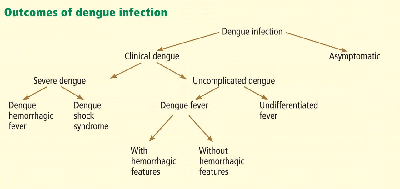
Most primary and secondary dengue infections are asymptomatic.8 The common forms of clinically apparent disease include self-limited, undifferentiated fever and classic dengue fever (Figure 3). Severe disease, manifesting as either dengue hemorrhagic fever or dengue shock syndrome, is a rare outcome of dengue virus infection, estimated to occur in 1% of cases worldwide.31 However, the true proportion of severe infection among all dengue cases seen in travelers is difficult to assess reliably.
Asymptomatic infection
Clinical dengue disease is relatively uncommon, as between 60% and 80% of infections are asymptomatic, particularly in children and adults who never have been infected before.32
In the recent outbreak in Key West, 28 symptomatic cases of locally acquired dengue were detected. The US Centers for Disease Control and Prevention (CDC) conducted a serologic survey of 240 healthy, randomly selected residents of Key West and found evidence of recent infection in 5.4% of those tested.28 Based on this finding, the CDC estimated that 1,000 people had been infected, of whom more than 90% had no symptoms. However, no attempt was made to differentiate primary from secondary infection in this serosurvey. (Previous infection with one dengue serotype places some individuals at risk for severe dengue if infected with a different serotype in the future.)
Uncomplicated dengue infection
Undifferentiated fever33 and classic dengue fever are the most common manifestations of clinical dengue infection. Also known as breakbone fever, classic dengue fever is a fever-arthralgia-rash syndrome.1
The onset is acute, with a high fever (though rarely greater than 40.5°C [104.9°F]) 3 to 14 days (usually 5 to 9 days) after the patient was bitten by an Aedes mosquito. Therefore, a febrile illness beginning more than 2 weeks after returning from travel to an endemic area is unlikely to be dengue, and another diagnosis should be sought.
A prodrome of headache, backache, fatigue, chills, anorexia, and occasionally a rash may precede the onset of fever by about 12 hours.
With fever comes a severe frontal headache, associated retro-orbital pain with eye movement, and conjunctival injection.
Some patients develop a bright, erythematous, maculopapular eruption 2 to 6 days into the illness that appears first on the trunk and then spreads to the face and extremities, with characteristic islands of unaffected skin throughout the involved area.34
Severe back or groin pain occurs in about 60% of adult patients.35
Anorexia, nausea, and vomiting are common.
Patients remain febrile for about 5 days, although some experience a biphasic (saddleback) fever that declines after 2 to 3 days, only to recur in about 24 hours.
In some patients, relative bradycardia is seen 2 to 3 days after fever onset.
Lymphadenopathy, sore throat, diarrhea or constipation, cutaneous hypesthesia, dysuria, dysgeusia, hepatitis, aseptic meningitis, and encephalopathy with delirium have been reported. Splenomegaly is rare.
In classic dengue fever, initial neutropenia and lymphopenia with subsequent lymphocytosis and monocytosis are often noted.
Mild hepatitis can be seen; aspartate aminotransferase and alanine aminotransferase levels can be two to three times the upper limit of normal.
Hemorrhagic manifestations, eg, petechiae, gingival bleeding, and epistaxis, may be seen in patients with mild thrombocytopenia even if they have no evidence of hemoconcentration or evidence of vascular instability.18
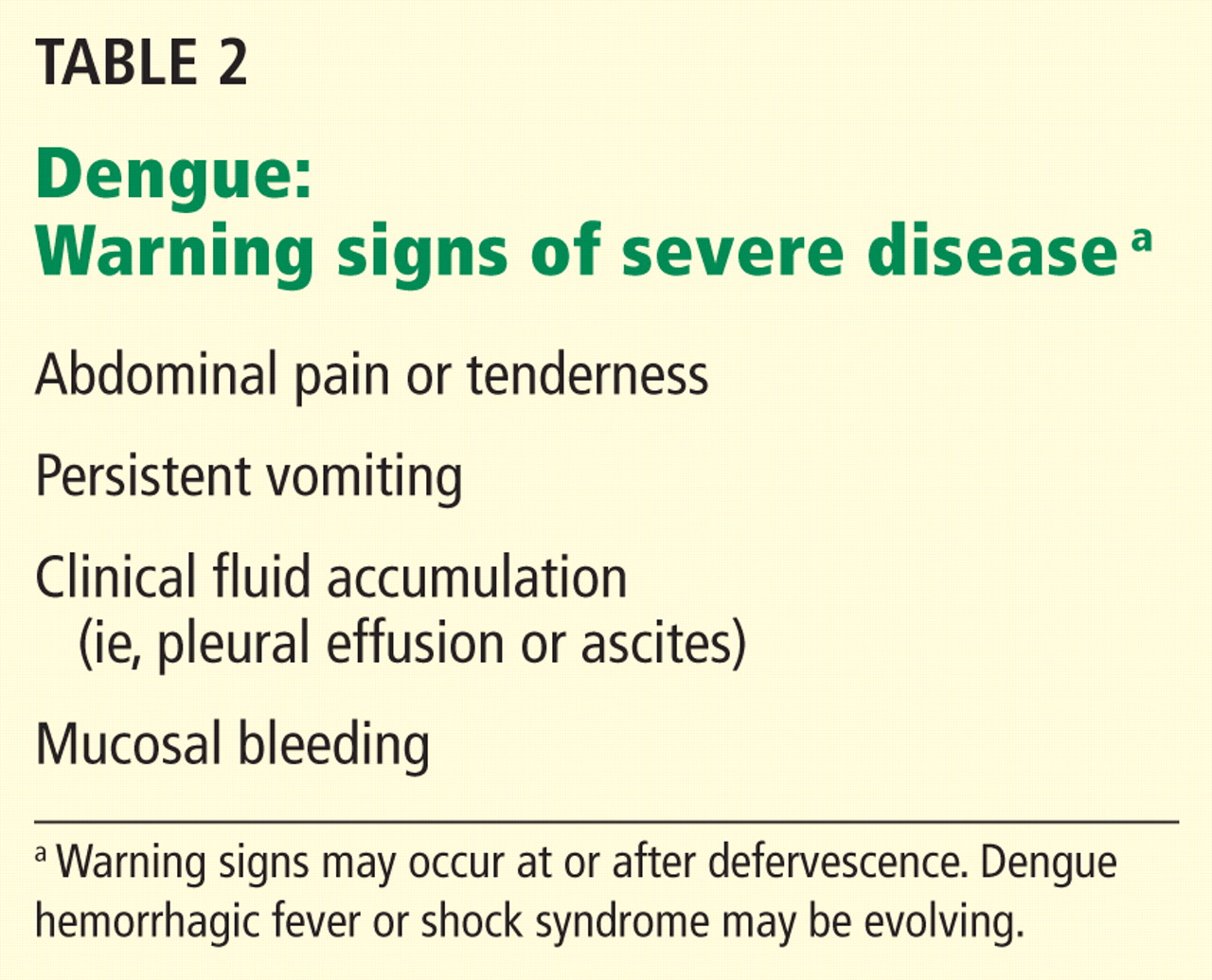
Although clinical dengue infection is usually self-limiting, acute symptoms can be incapacitating and can require hospitalization,36 and convalescence may take several months because of ongoing asthenia or depression.37 Furthermore, certain critical findings should alert the clinician to possible impending severe dengue and should lead to hospitalization for further observation until evolution to severe dengue has been ruled out (Table 2).
Severe dengue: Hemorrhagic fever and shock syndrome
In a very small subset of patients, dengue infection develops into a severe, potentially lifethreatening illness. Fortunately, this has rarely been reported in travelers.38
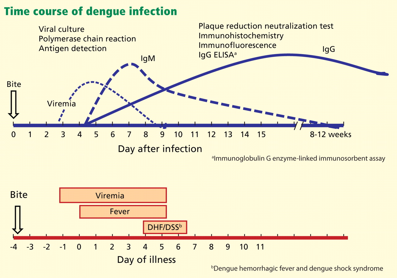
Dengue hemorrhagic fever and dengue shock syndrome arise just as fever is subsiding. They constitute a spectrum of severe illness (Figure 4). Dengue hemorrhagic fever is poorly understood because its hemorrhagic manifestations are not of themselves diagnostic of the condition, as petechiae, epistaxis, and gingival bleeding may be seen in classic dengue fever (Figure 3) without progression to more severe illness.
The differentiating characteristic of severe dengue, in addition to hemorrhagic manifestations, is objective evidence of plasma leakage.39 Impending shock is suggested by the new onset of severe abdominal pain, restlessness, hepatomegaly, hypothermia, and diaphoresis.
The mechanisms causing the severe hemorrhagic manifestations characteristic of dengue hemorrhagic fever and the sudden onset of vascular permeability underlying dengue shock syndrome are not understood.40 Many hypotheses have been generated and risk factors identified from observational and retrospective analyses. These include T-cell immune-pathologic responses involving receptors, antibodies, and cytokines,41 as well as specific host-genetic characteristics,42,43 age,44,45 sex,46 comorbid conditions,47–49 dengue virus virulence factors,50 sequence of dengue infection, and infection parity.51 One hypothesis is that antibody-dependent enhancement of virus occurs during infection with a second dengue serotype after infection with a different serotype in the past, and that this may be the root cause of dengue hemorrhagic fever and dengue shock syndrome. However, this has not been proven.40
DIAGNOSTIC TESTS FOR VIRUS, ANTIGENIC FRAGMENTS, ANTIBODIES
The appropriate test to confirm dengue virus infection is based on the natural history of the infection (Figure 4) coupled with the exposure risk in the returned traveler. These tests include isolation of the virus using cell culture, identification of antigenic fragments (test not available in the United States), and serologic tests for specific immunoglobulin M (IgM) and IgG antibodies using enzyme-linked immunosorbent assay (ELISA) or neutralization assays.52 During primary infection, viremia and antigenemia usually parallel fever, but when a person is later infected with a different dengue serotype, the period of viremia may be as short as 2 to 3 days, with antigens persisting in the serum for several more days.40 Virus isolation is not routinely available but is both sensitive and specific for the diagnosis of dengue virus infection during the viremic period.
If polymerase chain reaction testing, dengue antigen capture ELISA, or virus isolation testing is not available, the ideal confirmatory procedure is to test for dengue IgM (looking for conversion from negative to positive), IgG (looking for a fourfold rise in antibody), or both, in paired serum samples collected 2 weeks apart, with the initial sample collected less than 5 days after the onset of symptoms. A presumptive diagnosis can be made if a single blood sample collected more than 7 days after symptom onset is found to have dengue IgM antibody. A single blood sample for IgM collected earlier than 7 days after the onset of illness may give a false-negative result (Figure 4) in infected persons.
DIFFERENTIAL DIAGNOSES: INFECTIOUS AND NONINFECTIOUS
The differential diagnosis of uncomplicated dengue in a traveler returning from an endemic area includes viral, bacterial, and protozoal infections as well as noninfectious conditions (Table 1).53
Although most dengue virus infections are self-limiting, the clinical presentation may be severe enough to warrant hospitalization so that potentially life-threatening conditions can be systematically dismissed from the differential diagnosis.
Infections that can be rapidly fatal, such as malaria and enteric fever, need to be considered in patients who have traveled to endemic areas who present with undifferentiated fever. In cases of fever and maculopapular eruption, the differential diagnosis should include other causes of rash illness, such as measles and rubella. If hemorrhagic features are present, potentially fatal conditions need to be considered, including the classic viral hemorrhagic fevers caused by the Ebola and Marburg viruses, meningococcemia, the icterohemorrhagic form of leptospirosis, or other causes of bacterial sepsis. Other nonfatal infections should also be considered.
TREATMENT IS SUPPORTIVE
There is no antiviral treatment for dengue across the spectrum of disease presentations. Treatment is supportive and based on clinical presentation.
Acetaminophen (Tylenol) can be used to control fever, but aspirin and nonsteroidal anti-inflammatory drugs should not be used because they can make bleeding worse. Corticosteroids do not improve the outcome in severe dengue.2

Scrupulous attention to fluid and electrolyte balance is critical in severe dengue cases. Proper support and fluid resuscitation, including blood transfusion if needed, result in rapid recovery from dengue hemorrhagic fever with or without shock.
Suspected, probable, or confirmed cases of dengue should be reported to the local health department on the basis of published criteria (Table 3).
ADVICE TO TRAVELERS: DON’T GET BITTEN
There is currently no commercially available dengue vaccine, although several are under development.54 Therefore, pretravel counseling on how to avoid mosquito bites when traveling to dengue-endemic areas is the key dengue prevention strategy. Proactive prevention strategies include use of insect repellents such as those containing diethyltoluamide (DEET) or permethrin55 and elimination of outdoor locations where mosquitoes lay eggs, such as flower planter dishes, to reduce local mosquito breeding.56
Patients who have had a previous dengue infection should be counseled about the possible increased risk of severe disease if infected with a second dengue serotype.
Acknowledgment: The author thanks Chester G. Moore, PhD, of Colorado State University for assistance in creating Figure 2, based on data contained in the CDC, ArboNET, and Exotic/Invasive databases.
- Leggat PA. Assessment of febrile illness in the returned traveller. Aust Fam Physician 2007; 36:328–332.
- Wilder-Smith A, Schwartz E. Dengue in travelers. N Engl J Med 2005; 353:924–932.
- Mohammed HP, Ramos MM, Rivera A, et al. Travel-associated dengue infections in the United States, 1996 to 2005. J Travel Med 2010; 17:8–14.
- Centers for Disease Control and Prevention (CDC). Travel-associated dengue surveillance—United States, 2006–2008. MMWR Morb Mortal Wkly Rep 2010; 59:715–719.
- Ang KT, Rohani I, Look CH. Role of primary care providers in dengue prevention and control in the community. Med J Malaysia 2010; 65:58–62.
- Centers for Disease Control and Prevention. Notice to readers: Changes to the national notifiable infectious disease list and data presentation—January 2010. MMWR Morb Mortal Wkly Rep 2010; 59:11. www.cdc.gov/mmwr/preview/mmwrhtml/mm5901a7.htm. Accessed May 29, 2012.
- Guzman A, Istúriz RE. Update on the global spread of dengue. Int J Antimicrob Agents 2010; 36:(suppl 1):S40–S42.
- Midgley CM, Bajwa-Joseph M, Vasanawathana S, et al. An in-depth analysis of original antigenic sin in dengue virus infection. J Virol 2011; 85:410–421.
- Gubler DJ. Dengue/dengue haemorrhagic fever: history and current status. Novartis Found Symp 2006; 277:3–16.
- Franco L, Di Caro A, Carletti F, et al. Recent expansion of dengue virus serotype 3 in West Africa. Euro Surveill 2010; 15:19490.
- San Martín JL, Brathwaite O, Zambrano B, et al. The epidemiology of dengue in the Americas over the last three decades: a worrisome reality. Am J Trop Med Hyg 2010; 82:128–135.
- Ramos MM, Mohammed H, Zielinski-Gutierrez E, et al; Dengue Serosurvey Working Group. Epidemic dengue and dengue hemorrhagic fever at the Texas-Mexico border: results of a household-based seroepidemiologic survey, December 2005. Am J Trop Med Hyg 2008; 78:364–369.
- Ong DQ, Sitaram N, Rajakulendran M, et al. Knowledge and practice of household mosquito breeding control measures between a dengue hotspot and non-hotspot in Singapore. Ann Acad Med Singapore 2010; 39:146–149.
- Vazquez-Prokopec GM, Chaves LF, Ritchie SA, Davis J, Kitron U. Unforeseen costs of cutting mosquito surveillance budgets. PLoS Negl Trop Dis 2010; 4:e858.
- Ballenger-Browning KK, Elder JP. Multi-modal Aedes aegypti mosquito reduction interventions and dengue fever prevention. Trop Med Int Health 2009; 14:1542–1551.
- Courtney M, Shetty AK. Imported dengue fever: an important reemerging disease. Pediatr Emerg Care 2009; 25:769–772.
- Morens DM, Fauci AS. Dengue and hemorrhagic fever: a potential threat to public health in the United States. JAMA 2008; 299:214–216.
- Gregory CJ, Santiago LM, Argüello DF, Hunsperger E, Tomashek KM. Clinical and laboratory features that differentiate dengue from other febrile illnesses in an endemic area—Puerto Rico, 2007–2008. Am J Trop Med Hyg 2010; 82:922–929.
- Mohammed H, Ramos M, Armstrong J, et al. An outbreak of dengue fever in St. Croix (US Virgin Islands), 2005. PLoS One 2010; 5):e13729.
- Li DS, Liu W, Guigon A, Mostyn C, Grant R, Aaskov J. Rapid displacement of dengue virus type 1 by type 4, Pacific region, 2007–2009. Emerg Infect Dis 2010; 16:123–125.
- Hayden MH, Uejio CK, Walker K, et al. Microclimate and human factors in the divergent ecology of Aedes aegypti along the Arizona, US/Sonora, MX border. Ecohealth 2010; 7:64–77.
- Knudsen AB. The significance of the introduction of Aedes albopictus into the southeastern United States with implications for the Caribbean, and perspectives of the Pan American Health Organization. J Am Mosq Control Assoc 1986; 2:420–423.
- Gratz NG. Critical review of the vector status of Aedes albopictus. Med Vet Entomol 2004; 18:215–227.
- Franco C, Hynes NA, Bouri N, Henderson DA. The dengue threat to the United States. Biosecur Bioterror 2010; 8:273–276.
- Centers for Disease Control and Prevention (CDC). Dengue hemorrhagic fever—US-Mexico border, 2005. MMWR Morb Mortal Wkly Rep 2007; 56:785–789.
- Brunkard JM, Robles López JL, Ramirez J, et al. Dengue fever seroprevalence and risk factors, Texas-Mexico border, 2004. Emerg Infect Dis 2007; 13:1477–1483.
- Hafkin B, Kaplan JE, Reed C, et al. Reintroduction of dengue fever into the continental United States. I. Dengue surveillance in Texas, 1980. Am J Trop Med Hyg 1982; 31:1222–1228.
- Centers for Disease Control and Prevention (CDC). Locally acquired Dengue—Key West, Florida, 2009–2010. MMWR Morb Mortal Wkly Rep 2010; 59:577–581.
- Gill J, Stark LM, Clark GG. Dengue surveillance in Florida, 1997–98. Emerg Infect Dis 2000; 6:30–35.
- Department of Health. DOH investigates cases of dengue fever on Oahu and asks public & community to be vigilant. http://hawaii.gov/health/about/pr/2011/11-028.pdf. Accessed May 29, 2012.
- Wilder-Smith A, Earnest A, Tan SB, Ooi EE, Gubler DJ. Lack of association of dengue activity with haze. Epidemiol Infect 2010; 138:962–967.
- Kyle JL, Harris E. Global spread and persistence of dengue. Annu Rev Microbiol 2008; 62:71–92.
- Chrispal A, Boorugu H, Gopinath KG, et al. Acute undifferentiated febrile illness in adult hospitalized patients: the disease spectrum and diagnostic predictors—an experience from a tertiary care hospital in South India. Trop Doct 2010; 40:230–234.
- World Health Organization; the Special Programme for Research and Training in Tropical Diseases (TDR). Dengue: guidelines for diagnosis, treatment, prevention and control. www.who.int/rpc/guidelines/9789241547871/en/. Accessed May 29, 2012.
- Potts JA, Rothman AL. Clinical and laboratory features that distinguish dengue from other febrile illnesses in endemic populations. Trop Med Int Health 2008; 13:1328–1340.
- Streit JA, Yang M, Cavanaugh JE, Polgreen PM. Upward trend in dengue incidence among hospitalized patients, United States. Emerg Infect Dis 2011; 17:914–916.
- Jelinek T. Dengue fever in international travelers. Clin Infect Dis 2000; 31:144–147.
- Gibbons RV, Vaughn DW. Dengue: an escalating problem. BMJ 2002; 324:1563–1566.
- Gibbons RV. Dengue conundrums. Int J Antimicrob Agents 2010; 36(suppl 1):S36–S39.
- Halstead SB. Dengue. Lancet 2007; 370:1644–1652.
- Halstead SB. Antibodies determine virulence in dengue. Ann N Y Acad Sci 2009; 1171(suppl 1):E48–E56.
- Coffey LL, Mertens E, Brehin AC, et al. Human genetic determinants of dengue virus susceptibility. Microbes Infect 2009; 11:143–156.
- García G, Sierra B, Pérez AB, et al. Asymptomatic dengue infection in a Cuban population confirms the protective role of the RR variant of the FcgammaRIIa polymorphism. Am J Trop Med Hyg 2010; 82:1153–1156.
- Braga C, Luna CF, Martelli CM, et al. Seroprevalence and risk factors for dengue infection in socio-economically distinct areas of Recife, Brazil. Acta Trop 2010; 113:234–240.
- Jain A, Chaturvedi UC. Dengue in infants: an overview. FEMS Immunol Med Microbiol 2010; 59:119–130.
- Almas A, Parkash O, Akhter J. Clinical factors associated with mortality in dengue infection at a tertiary care center. Southeast Asian J Trop Med Public Health 2010; 41:333–340.
- Diaz-Quijano FA, Villar-Centeno LA, Martinez-Vega RA. Predictors of spontaneous bleeding in patients with acute febrile syndrome from a dengue endemic area. J Clin Virol 2010; 49:11–15.
- Marón GM, Clará AW, Diddle JW, et al. Association between nutritional status and severity of dengue infection in children in El Salvador. Am J Trop Med Hyg 2010; 82:324–329.
- Sierra B, Perez AB, Vogt K, et al. Secondary heterologous dengue infection risk: disequilibrium between immune regulation and inflammation? Cell Immunol 2010; 262:134–140.
- Brien JD, Austin SK, Sukupolvi-Petty S, et al. Genotype-specific neutralization and protection by antibodies against dengue virus type 3. J Virol 2010; 84:10630–10643.
- Humayoun MA, Waseem T, Jawa AA, Hashmi MS, Akram J. Multiple dengue serotypes and high frequency of dengue hemorrhagic fever at two tertiary care hospitals in Lahore during the 2008 dengue virus outbreak in Punjab, Pakistan. Int J Infect Dis 2010; 14(suppl 3):e54–e59.
- Guzman MG, Halstead SB, Artsob H, et al. Dengue: a continuing global threat. Nature Rev Microbiol 2010; 8:S7–S16.
- Crowell CS, Stamos JK. Evaluation of fever after international travel. Pediatr Ann 2011; 40:39–44.
- Durbin AP, Whitehead SS. Dengue vaccine candidates in development. Curr Top Microbiol Immunol 2010; 338:129–143.
- Chen LH, Wilson ME. Dengue and chikungunya infections in travelers. Curr Opin Infect Dis 2010; 23:438–444.
- Lambrechts L, Scott TW, Gubler DJ. Consequences of the expanding global distribution of Aedes albopictus for dengue virus transmission. PLoS Negl Trop Dis 2010; 4:e646.
Why do primary care physicians in nontropical parts of the world need to be on the lookout for tropical diseases such as dengue?
First, more people are traveling than ever before, and second, more people are traveling to parts of the world where dengue and other tropical diseases are endemic. Thus, dengue should now be included in the differential diagnosis of fever in anyone returning from travel to a part of the world where dengue is endemic (Table 1).1
The number of cases of dengue in returning US travelers has been increasing steadily over the past 25 years.2,3 Dengue is a more common cause of febrile illness than malaria in US travelers returning from all tropical and subtropical regions except Africa.4
Moreover, dengue may be gaining a permanent foothold in the United States, and in areas of the country where mosquitoes that can transmit the virus are found, primary care physicians are the first line of defense in public health. Specifically, to prevent the virus from becoming established locally, primary care physicians need to quickly identify and report cases to public health authorities, who can promptly follow up and initiate prevention measures.5
Underscoring the importance of dengue, this infection was added in 2009 to the list of nationally notifiable infectious diseases in the United States.6
A COMMON INFECTION WORLDWIDE
Dengue causes up to 100 million new infections, 500,000 hospitalizations, and 25,000 deaths every year in the 2.5 billion people who live in subtropical and tropical areas of the world.7 It is transmitted by mosquitoes of the genus Aedes (which, unlike most other mosquitoes, often bite in the daytime), and it is the most common arboviral infection (ie, transmitted mainly by arthropods) worldwide.
The dengue virus belongs to the family of flaviviruses, which includes West Nile virus, St. Louis encephalitis, and yellow fever. It has four closely related but antigenically distinct serotypes, designated DENV-1, DENV-2, DENV-3, and DENV-4. Infection with one serotype induces lifetime homotypic immunity but only short-lived heterotypic immunity to the other dengue serotypes.8 Hence, a person can be infected over time with each of the four serotypes. The first infection is termed the primary infection; subsequent infection with any of the remaining three serotypes is termed a secondary infection.
Dengue virus transmission has been expanding since the end of World War II in Asia and since the 1980s in the Americas following the end of many regional vector-control programs.9 Although dengue is known to occur in tropical Africa, its epidemiology is less well defined on that continent (Figure 1).10
Explosive epidemics of dengue occur when there are enough mosquitoes and a susceptible population across a broad age range, ie, both children and adults.11,12 Transmission can be halted with vigorous vector-control programs, or it slows and stops when the pool of susceptible people is exhausted.13–15
On the other hand, hyperendemic transmission occurs in areas in which multiple virus serotypes continuously circulate in a large pool of susceptible people. In these areas, dengue seroprevalence increases with age, and most adults are immune.
Anyone of any age who travels from a nonendemic area to an epidemic or hyperendemic area is at risk of infection.16
DENGUE IN THE UNITED STATES
Reported cases of dengue in South and Central America, the Caribbean, and Mexico, common destinations for US travelers, have increased more than fourfold since the 1980s. There were a total of approximately 1 million cases in the 10-year period ending in 1989, compared with more than 4.5 million in the 8-year period from 2000 to 2007.11
The geographic proximity of these areas to the continental United States and the large numbers of US residents travelling to these areas have raised concern that dengue could emerge in the continental United States in areas where potential vectors exist (see below).17 Furthermore, several US territories and former territories where tourism is an economic mainstay, including the Commonwealth of Puerto Rico,18 the US Virgin Islands,19 American Samoa, and other smaller Pacific island jurisdictions such as Palau,20 have reported dengue virus circulation.
Adding to the concern that dengue could gain a persistent foothold in the United States, competent dengue vectors are found here. Two mosquito vectors, Aedes aegypti21 and Aedes albopictus,22,23 live in some areas of the southwestern and southeastern United States (Figure 2), with Aedes aegypti being the more competent transmitter. Both vectors may be abundant in warmer months. This raises a concern that a returning dengue-infected traveler could initiate an outbreak of autochthonous transmission.24
Notably, endemic dengue transmission has occurred in the past in the United States, and the virus is circulating here again at low levels. From 1946 to 1980, no cases of dengue were acquired in the continental United States. However, since 1980 there have been seven outbreaks of laboratory-confirmed, locally acquired dengue along the Texas-Mexico border.25–27 More recently, local transmission emerged in Key West, Florida,28,29 the first outbreak since 1945 of dengue in the continental United States not to occur near the Texas-Mexico border. And in early 2011, nonsustained but locally acquired transmission was confirmed in Hawaii after a transmission-free decade.30
THE CLINICAL SPECTRUM OF DENGUE VIRUS INFECTION
Most primary and secondary dengue infections are asymptomatic.8 The common forms of clinically apparent disease include self-limited, undifferentiated fever and classic dengue fever (Figure 3). Severe disease, manifesting as either dengue hemorrhagic fever or dengue shock syndrome, is a rare outcome of dengue virus infection, estimated to occur in 1% of cases worldwide.31 However, the true proportion of severe infection among all dengue cases seen in travelers is difficult to assess reliably.
Asymptomatic infection
Clinical dengue disease is relatively uncommon, as between 60% and 80% of infections are asymptomatic, particularly in children and adults who never have been infected before.32
In the recent outbreak in Key West, 28 symptomatic cases of locally acquired dengue were detected. The US Centers for Disease Control and Prevention (CDC) conducted a serologic survey of 240 healthy, randomly selected residents of Key West and found evidence of recent infection in 5.4% of those tested.28 Based on this finding, the CDC estimated that 1,000 people had been infected, of whom more than 90% had no symptoms. However, no attempt was made to differentiate primary from secondary infection in this serosurvey. (Previous infection with one dengue serotype places some individuals at risk for severe dengue if infected with a different serotype in the future.)
Uncomplicated dengue infection
Undifferentiated fever33 and classic dengue fever are the most common manifestations of clinical dengue infection. Also known as breakbone fever, classic dengue fever is a fever-arthralgia-rash syndrome.1
The onset is acute, with a high fever (though rarely greater than 40.5°C [104.9°F]) 3 to 14 days (usually 5 to 9 days) after the patient was bitten by an Aedes mosquito. Therefore, a febrile illness beginning more than 2 weeks after returning from travel to an endemic area is unlikely to be dengue, and another diagnosis should be sought.
A prodrome of headache, backache, fatigue, chills, anorexia, and occasionally a rash may precede the onset of fever by about 12 hours.
With fever comes a severe frontal headache, associated retro-orbital pain with eye movement, and conjunctival injection.
Some patients develop a bright, erythematous, maculopapular eruption 2 to 6 days into the illness that appears first on the trunk and then spreads to the face and extremities, with characteristic islands of unaffected skin throughout the involved area.34
Severe back or groin pain occurs in about 60% of adult patients.35
Anorexia, nausea, and vomiting are common.
Patients remain febrile for about 5 days, although some experience a biphasic (saddleback) fever that declines after 2 to 3 days, only to recur in about 24 hours.
In some patients, relative bradycardia is seen 2 to 3 days after fever onset.
Lymphadenopathy, sore throat, diarrhea or constipation, cutaneous hypesthesia, dysuria, dysgeusia, hepatitis, aseptic meningitis, and encephalopathy with delirium have been reported. Splenomegaly is rare.
In classic dengue fever, initial neutropenia and lymphopenia with subsequent lymphocytosis and monocytosis are often noted.
Mild hepatitis can be seen; aspartate aminotransferase and alanine aminotransferase levels can be two to three times the upper limit of normal.
Hemorrhagic manifestations, eg, petechiae, gingival bleeding, and epistaxis, may be seen in patients with mild thrombocytopenia even if they have no evidence of hemoconcentration or evidence of vascular instability.18
Although clinical dengue infection is usually self-limiting, acute symptoms can be incapacitating and can require hospitalization,36 and convalescence may take several months because of ongoing asthenia or depression.37 Furthermore, certain critical findings should alert the clinician to possible impending severe dengue and should lead to hospitalization for further observation until evolution to severe dengue has been ruled out (Table 2).
Severe dengue: Hemorrhagic fever and shock syndrome
In a very small subset of patients, dengue infection develops into a severe, potentially lifethreatening illness. Fortunately, this has rarely been reported in travelers.38
Dengue hemorrhagic fever and dengue shock syndrome arise just as fever is subsiding. They constitute a spectrum of severe illness (Figure 4). Dengue hemorrhagic fever is poorly understood because its hemorrhagic manifestations are not of themselves diagnostic of the condition, as petechiae, epistaxis, and gingival bleeding may be seen in classic dengue fever (Figure 3) without progression to more severe illness.
The differentiating characteristic of severe dengue, in addition to hemorrhagic manifestations, is objective evidence of plasma leakage.39 Impending shock is suggested by the new onset of severe abdominal pain, restlessness, hepatomegaly, hypothermia, and diaphoresis.
The mechanisms causing the severe hemorrhagic manifestations characteristic of dengue hemorrhagic fever and the sudden onset of vascular permeability underlying dengue shock syndrome are not understood.40 Many hypotheses have been generated and risk factors identified from observational and retrospective analyses. These include T-cell immune-pathologic responses involving receptors, antibodies, and cytokines,41 as well as specific host-genetic characteristics,42,43 age,44,45 sex,46 comorbid conditions,47–49 dengue virus virulence factors,50 sequence of dengue infection, and infection parity.51 One hypothesis is that antibody-dependent enhancement of virus occurs during infection with a second dengue serotype after infection with a different serotype in the past, and that this may be the root cause of dengue hemorrhagic fever and dengue shock syndrome. However, this has not been proven.40
DIAGNOSTIC TESTS FOR VIRUS, ANTIGENIC FRAGMENTS, ANTIBODIES
The appropriate test to confirm dengue virus infection is based on the natural history of the infection (Figure 4) coupled with the exposure risk in the returned traveler. These tests include isolation of the virus using cell culture, identification of antigenic fragments (test not available in the United States), and serologic tests for specific immunoglobulin M (IgM) and IgG antibodies using enzyme-linked immunosorbent assay (ELISA) or neutralization assays.52 During primary infection, viremia and antigenemia usually parallel fever, but when a person is later infected with a different dengue serotype, the period of viremia may be as short as 2 to 3 days, with antigens persisting in the serum for several more days.40 Virus isolation is not routinely available but is both sensitive and specific for the diagnosis of dengue virus infection during the viremic period.
If polymerase chain reaction testing, dengue antigen capture ELISA, or virus isolation testing is not available, the ideal confirmatory procedure is to test for dengue IgM (looking for conversion from negative to positive), IgG (looking for a fourfold rise in antibody), or both, in paired serum samples collected 2 weeks apart, with the initial sample collected less than 5 days after the onset of symptoms. A presumptive diagnosis can be made if a single blood sample collected more than 7 days after symptom onset is found to have dengue IgM antibody. A single blood sample for IgM collected earlier than 7 days after the onset of illness may give a false-negative result (Figure 4) in infected persons.
DIFFERENTIAL DIAGNOSES: INFECTIOUS AND NONINFECTIOUS
The differential diagnosis of uncomplicated dengue in a traveler returning from an endemic area includes viral, bacterial, and protozoal infections as well as noninfectious conditions (Table 1).53
Although most dengue virus infections are self-limiting, the clinical presentation may be severe enough to warrant hospitalization so that potentially life-threatening conditions can be systematically dismissed from the differential diagnosis.
Infections that can be rapidly fatal, such as malaria and enteric fever, need to be considered in patients who have traveled to endemic areas who present with undifferentiated fever. In cases of fever and maculopapular eruption, the differential diagnosis should include other causes of rash illness, such as measles and rubella. If hemorrhagic features are present, potentially fatal conditions need to be considered, including the classic viral hemorrhagic fevers caused by the Ebola and Marburg viruses, meningococcemia, the icterohemorrhagic form of leptospirosis, or other causes of bacterial sepsis. Other nonfatal infections should also be considered.
TREATMENT IS SUPPORTIVE
There is no antiviral treatment for dengue across the spectrum of disease presentations. Treatment is supportive and based on clinical presentation.
Acetaminophen (Tylenol) can be used to control fever, but aspirin and nonsteroidal anti-inflammatory drugs should not be used because they can make bleeding worse. Corticosteroids do not improve the outcome in severe dengue.2
Scrupulous attention to fluid and electrolyte balance is critical in severe dengue cases. Proper support and fluid resuscitation, including blood transfusion if needed, result in rapid recovery from dengue hemorrhagic fever with or without shock.
Suspected, probable, or confirmed cases of dengue should be reported to the local health department on the basis of published criteria (Table 3).
ADVICE TO TRAVELERS: DON’T GET BITTEN
There is currently no commercially available dengue vaccine, although several are under development.54 Therefore, pretravel counseling on how to avoid mosquito bites when traveling to dengue-endemic areas is the key dengue prevention strategy. Proactive prevention strategies include use of insect repellents such as those containing diethyltoluamide (DEET) or permethrin55 and elimination of outdoor locations where mosquitoes lay eggs, such as flower planter dishes, to reduce local mosquito breeding.56
Patients who have had a previous dengue infection should be counseled about the possible increased risk of severe disease if infected with a second dengue serotype.
Acknowledgment: The author thanks Chester G. Moore, PhD, of Colorado State University for assistance in creating Figure 2, based on data contained in the CDC, ArboNET, and Exotic/Invasive databases.
- Leggat PA. Assessment of febrile illness in the returned traveller. Aust Fam Physician 2007; 36:328–332.
- Wilder-Smith A, Schwartz E. Dengue in travelers. N Engl J Med 2005; 353:924–932.
- Mohammed HP, Ramos MM, Rivera A, et al. Travel-associated dengue infections in the United States, 1996 to 2005. J Travel Med 2010; 17:8–14.
- Centers for Disease Control and Prevention (CDC). Travel-associated dengue surveillance—United States, 2006–2008. MMWR Morb Mortal Wkly Rep 2010; 59:715–719.
- Ang KT, Rohani I, Look CH. Role of primary care providers in dengue prevention and control in the community. Med J Malaysia 2010; 65:58–62.
- Centers for Disease Control and Prevention. Notice to readers: Changes to the national notifiable infectious disease list and data presentation—January 2010. MMWR Morb Mortal Wkly Rep 2010; 59:11. www.cdc.gov/mmwr/preview/mmwrhtml/mm5901a7.htm. Accessed May 29, 2012.
- Guzman A, Istúriz RE. Update on the global spread of dengue. Int J Antimicrob Agents 2010; 36:(suppl 1):S40–S42.
- Midgley CM, Bajwa-Joseph M, Vasanawathana S, et al. An in-depth analysis of original antigenic sin in dengue virus infection. J Virol 2011; 85:410–421.
- Gubler DJ. Dengue/dengue haemorrhagic fever: history and current status. Novartis Found Symp 2006; 277:3–16.
- Franco L, Di Caro A, Carletti F, et al. Recent expansion of dengue virus serotype 3 in West Africa. Euro Surveill 2010; 15:19490.
- San Martín JL, Brathwaite O, Zambrano B, et al. The epidemiology of dengue in the Americas over the last three decades: a worrisome reality. Am J Trop Med Hyg 2010; 82:128–135.
- Ramos MM, Mohammed H, Zielinski-Gutierrez E, et al; Dengue Serosurvey Working Group. Epidemic dengue and dengue hemorrhagic fever at the Texas-Mexico border: results of a household-based seroepidemiologic survey, December 2005. Am J Trop Med Hyg 2008; 78:364–369.
- Ong DQ, Sitaram N, Rajakulendran M, et al. Knowledge and practice of household mosquito breeding control measures between a dengue hotspot and non-hotspot in Singapore. Ann Acad Med Singapore 2010; 39:146–149.
- Vazquez-Prokopec GM, Chaves LF, Ritchie SA, Davis J, Kitron U. Unforeseen costs of cutting mosquito surveillance budgets. PLoS Negl Trop Dis 2010; 4:e858.
- Ballenger-Browning KK, Elder JP. Multi-modal Aedes aegypti mosquito reduction interventions and dengue fever prevention. Trop Med Int Health 2009; 14:1542–1551.
- Courtney M, Shetty AK. Imported dengue fever: an important reemerging disease. Pediatr Emerg Care 2009; 25:769–772.
- Morens DM, Fauci AS. Dengue and hemorrhagic fever: a potential threat to public health in the United States. JAMA 2008; 299:214–216.
- Gregory CJ, Santiago LM, Argüello DF, Hunsperger E, Tomashek KM. Clinical and laboratory features that differentiate dengue from other febrile illnesses in an endemic area—Puerto Rico, 2007–2008. Am J Trop Med Hyg 2010; 82:922–929.
- Mohammed H, Ramos M, Armstrong J, et al. An outbreak of dengue fever in St. Croix (US Virgin Islands), 2005. PLoS One 2010; 5):e13729.
- Li DS, Liu W, Guigon A, Mostyn C, Grant R, Aaskov J. Rapid displacement of dengue virus type 1 by type 4, Pacific region, 2007–2009. Emerg Infect Dis 2010; 16:123–125.
- Hayden MH, Uejio CK, Walker K, et al. Microclimate and human factors in the divergent ecology of Aedes aegypti along the Arizona, US/Sonora, MX border. Ecohealth 2010; 7:64–77.
- Knudsen AB. The significance of the introduction of Aedes albopictus into the southeastern United States with implications for the Caribbean, and perspectives of the Pan American Health Organization. J Am Mosq Control Assoc 1986; 2:420–423.
- Gratz NG. Critical review of the vector status of Aedes albopictus. Med Vet Entomol 2004; 18:215–227.
- Franco C, Hynes NA, Bouri N, Henderson DA. The dengue threat to the United States. Biosecur Bioterror 2010; 8:273–276.
- Centers for Disease Control and Prevention (CDC). Dengue hemorrhagic fever—US-Mexico border, 2005. MMWR Morb Mortal Wkly Rep 2007; 56:785–789.
- Brunkard JM, Robles López JL, Ramirez J, et al. Dengue fever seroprevalence and risk factors, Texas-Mexico border, 2004. Emerg Infect Dis 2007; 13:1477–1483.
- Hafkin B, Kaplan JE, Reed C, et al. Reintroduction of dengue fever into the continental United States. I. Dengue surveillance in Texas, 1980. Am J Trop Med Hyg 1982; 31:1222–1228.
- Centers for Disease Control and Prevention (CDC). Locally acquired Dengue—Key West, Florida, 2009–2010. MMWR Morb Mortal Wkly Rep 2010; 59:577–581.
- Gill J, Stark LM, Clark GG. Dengue surveillance in Florida, 1997–98. Emerg Infect Dis 2000; 6:30–35.
- Department of Health. DOH investigates cases of dengue fever on Oahu and asks public & community to be vigilant. http://hawaii.gov/health/about/pr/2011/11-028.pdf. Accessed May 29, 2012.
- Wilder-Smith A, Earnest A, Tan SB, Ooi EE, Gubler DJ. Lack of association of dengue activity with haze. Epidemiol Infect 2010; 138:962–967.
- Kyle JL, Harris E. Global spread and persistence of dengue. Annu Rev Microbiol 2008; 62:71–92.
- Chrispal A, Boorugu H, Gopinath KG, et al. Acute undifferentiated febrile illness in adult hospitalized patients: the disease spectrum and diagnostic predictors—an experience from a tertiary care hospital in South India. Trop Doct 2010; 40:230–234.
- World Health Organization; the Special Programme for Research and Training in Tropical Diseases (TDR). Dengue: guidelines for diagnosis, treatment, prevention and control. www.who.int/rpc/guidelines/9789241547871/en/. Accessed May 29, 2012.
- Potts JA, Rothman AL. Clinical and laboratory features that distinguish dengue from other febrile illnesses in endemic populations. Trop Med Int Health 2008; 13:1328–1340.
- Streit JA, Yang M, Cavanaugh JE, Polgreen PM. Upward trend in dengue incidence among hospitalized patients, United States. Emerg Infect Dis 2011; 17:914–916.
- Jelinek T. Dengue fever in international travelers. Clin Infect Dis 2000; 31:144–147.
- Gibbons RV, Vaughn DW. Dengue: an escalating problem. BMJ 2002; 324:1563–1566.
- Gibbons RV. Dengue conundrums. Int J Antimicrob Agents 2010; 36(suppl 1):S36–S39.
- Halstead SB. Dengue. Lancet 2007; 370:1644–1652.
- Halstead SB. Antibodies determine virulence in dengue. Ann N Y Acad Sci 2009; 1171(suppl 1):E48–E56.
- Coffey LL, Mertens E, Brehin AC, et al. Human genetic determinants of dengue virus susceptibility. Microbes Infect 2009; 11:143–156.
- García G, Sierra B, Pérez AB, et al. Asymptomatic dengue infection in a Cuban population confirms the protective role of the RR variant of the FcgammaRIIa polymorphism. Am J Trop Med Hyg 2010; 82:1153–1156.
- Braga C, Luna CF, Martelli CM, et al. Seroprevalence and risk factors for dengue infection in socio-economically distinct areas of Recife, Brazil. Acta Trop 2010; 113:234–240.
- Jain A, Chaturvedi UC. Dengue in infants: an overview. FEMS Immunol Med Microbiol 2010; 59:119–130.
- Almas A, Parkash O, Akhter J. Clinical factors associated with mortality in dengue infection at a tertiary care center. Southeast Asian J Trop Med Public Health 2010; 41:333–340.
- Diaz-Quijano FA, Villar-Centeno LA, Martinez-Vega RA. Predictors of spontaneous bleeding in patients with acute febrile syndrome from a dengue endemic area. J Clin Virol 2010; 49:11–15.
- Marón GM, Clará AW, Diddle JW, et al. Association between nutritional status and severity of dengue infection in children in El Salvador. Am J Trop Med Hyg 2010; 82:324–329.
- Sierra B, Perez AB, Vogt K, et al. Secondary heterologous dengue infection risk: disequilibrium between immune regulation and inflammation? Cell Immunol 2010; 262:134–140.
- Brien JD, Austin SK, Sukupolvi-Petty S, et al. Genotype-specific neutralization and protection by antibodies against dengue virus type 3. J Virol 2010; 84:10630–10643.
- Humayoun MA, Waseem T, Jawa AA, Hashmi MS, Akram J. Multiple dengue serotypes and high frequency of dengue hemorrhagic fever at two tertiary care hospitals in Lahore during the 2008 dengue virus outbreak in Punjab, Pakistan. Int J Infect Dis 2010; 14(suppl 3):e54–e59.
- Guzman MG, Halstead SB, Artsob H, et al. Dengue: a continuing global threat. Nature Rev Microbiol 2010; 8:S7–S16.
- Crowell CS, Stamos JK. Evaluation of fever after international travel. Pediatr Ann 2011; 40:39–44.
- Durbin AP, Whitehead SS. Dengue vaccine candidates in development. Curr Top Microbiol Immunol 2010; 338:129–143.
- Chen LH, Wilson ME. Dengue and chikungunya infections in travelers. Curr Opin Infect Dis 2010; 23:438–444.
- Lambrechts L, Scott TW, Gubler DJ. Consequences of the expanding global distribution of Aedes albopictus for dengue virus transmission. PLoS Negl Trop Dis 2010; 4:e646.
KEY POINTS
- Dengue results from infection with one of four distinct serotypes: DENV-1, DENV-2, DENV-3, and DENV-4.
- The most common outcome after infection by the bite of an Aedes mosquito (which bites in the daytime) is asymptomatic infection, a flulike illness, or classic self-limited dengue fever. Severe, life-threatening disease with hemorrhagic manifestations or shock is rare.
- Obtaining a history of recent travel to a dengue-endemic area is a key in evaluating a person presenting with undifferentiated fever or a fever-rash-arthralgia syndrome.
- Diagnostic testing is based on the natural history of infection; antibody levels begin to rise as levels of viremia begin to decline.
- Risk factors help predict who will develop severe dengue after primary or secondary infection.