User login
Uncorking the negative emotions of IBS
CHICAGO – For patients with irritable bowel syndrome, expressing rather than bottling up negative emotions may be just what the doctor ordered, according to Elyse R. Thakur, Ph.D.
A novel psychological intervention designed to elicit negative emotions was associated with a significantly greater reduction in IBS symptom severity at 4 weeks than standard medical care (mean, 3.62 vs. 4.68; P = .004) and reductions similar to those seen with relaxation training (mean, 3.62 vs. 4.16; P = .126).

By 12 weeks, there were no differences between groups, and all patients continued to improve, Dr. Thakur of the DeBakey VA Medical Center and Baylor College of Medicine in Houston reported at the meeting sponsored by the American Gastroenterological Association.
One patient who had IBS-related nausea off and on for years wrote after Emotional Awareness and Expression Training (EAET): “I feel lighter, and after the exercise to deal with a particularly traumatic event, I feel less angry and less tense. I can’t say that my IBS is completely gone, but the symptoms have definitely gotten better.”
Psychological interventions for IBS have traditionally emphasized suppressing negative emotions such as anxiety and sadness through psychophysiologic strategies such as relaxation training (RT).
Recent research, however, suggests this suppression may be counterproductive. In a study involving 47 healthy controls, self-reported anger suppression predicted greater pain intensity in response to the cold pressor ice water immersion test (Ann Behav Med. 2010 Jun;39[3]:211-21).
While at the Wayne State University stress and health lab in Detroit, Dr. Thakur and her then graduate school adviser Mark Lumley, Ph.D., opted to take a different tact and developed the EAET based on the principle that emotional awareness and suppression can lead to stress-related symptoms and a dysregulated brain-gut system.
The goal of the intervention is to help patients reduce stress by having them learn about connections between stressful life experiences and physical symptoms; by teaching them to identify, experience, and express their emotions related to these stressful situations; and by encouraging them to engage in healthy emotional and interpersonal behaviors in their daily lives, including assertive and genuine communication, Dr. Thakur explained.
To facilitate this process, patients undergo a life-history interview, which helps them connect their IBS episodes to their life experiences. The therapist then conducts experiential exercises such as role playing and imagery to help patients engage with their avoided feelings, behaviors, memories, and relationships through their tone of voice, words, and body language. Finally, patients are encouraged to communicate more genuinely in their relationships, she said.
To evaluate the intervention, 106 patients who met the Rome III IBS diagnostic criteria were recruited from the community and gastroenterologic clinics, and evenly randomized to standard medical care or three 50-minute individualized sessions per week of EAET or RT including progressive muscle relaxation, relaxed breathing skills, and guided imagery. Patients had to have at least moderately severe IBS symptoms at least 2 days per week at the time of screening. Their mean age was 36 years, 80.2% were female, and 65% were of European-American descent.
Outcomes were measured at 4 and 12 weeks by using the IBS Symptom Severity Scale, Brief Symptom Inventory, and IBS Quality of Life questionnaire.
At 4 weeks, EAET and RT significantly reduced anxiety (mean, 0.71 and 0.62 vs. 1.16; P = .003 and P = .001, respectively) and hostility (mean, 0.56 and 0.60 vs. 0.89; P = .013 and P = .029, respectively), compared with controls, Dr. Thakur reported in a poster at the meeting.
“These findings suggest that techniques that enhance awareness, experiencing, and expression of negative emotions resulting from life stress and psychological conflicts are as effective in reducing anxiety and hostility as somatic control techniques,” she said in an interview.
RT significantly reduced depression, compared with standard care (mean, 0.52 vs. 1.02; P = .002), while EAET did not (mean, 0.77 vs. 1.02; P = .119).
This finding was unexpected, “albeit in retrospect, not surprising because emotional processing interventions often negatively impact the moods of people, at least in the short term, as they deal with the newfound awareness of their conflicts,” Dr. Thakur explained.
At 12 weeks, EAET and relaxation training maintained the improvements in anxiety and hostility, but the differences were no longer statistically significant because the standard care group improved, she noted.
Poor quality of life was significantly less common among patients receiving EAET and RT than standard medical care at 4 weeks (mean, 2.12 vs. 2.22 vs. 2.61; both P values less than .001) and 12 weeks (mean, 1.98 vs. 2.04 vs. 2.39; P = .004 and P = .016, respectively).
“These findings have broadened my conceptualization of IBS patients and provided me with a viable treatment alternative for those patients who have difficulties with emotional awareness and expression,” Dr. Thakur said in the interview.
Future research goals are to determine the types of patients for whom EAET is best suited, explore whether EAET and RT work best when integrated, and identify the best ways to implement brief, psychological interventions in routine practice settings, she said.
The study was funded by Blue Cross Blue Shied of Michigan, American Psychological Association, and Wayne State University. Dr. Thakur reported having no financial disclosures.
On Twitter @pwendl
CHICAGO – For patients with irritable bowel syndrome, expressing rather than bottling up negative emotions may be just what the doctor ordered, according to Elyse R. Thakur, Ph.D.
A novel psychological intervention designed to elicit negative emotions was associated with a significantly greater reduction in IBS symptom severity at 4 weeks than standard medical care (mean, 3.62 vs. 4.68; P = .004) and reductions similar to those seen with relaxation training (mean, 3.62 vs. 4.16; P = .126).
By 12 weeks, there were no differences between groups, and all patients continued to improve, Dr. Thakur of the DeBakey VA Medical Center and Baylor College of Medicine in Houston reported at the meeting sponsored by the American Gastroenterological Association.
One patient who had IBS-related nausea off and on for years wrote after Emotional Awareness and Expression Training (EAET): “I feel lighter, and after the exercise to deal with a particularly traumatic event, I feel less angry and less tense. I can’t say that my IBS is completely gone, but the symptoms have definitely gotten better.”
Psychological interventions for IBS have traditionally emphasized suppressing negative emotions such as anxiety and sadness through psychophysiologic strategies such as relaxation training (RT).
Recent research, however, suggests this suppression may be counterproductive. In a study involving 47 healthy controls, self-reported anger suppression predicted greater pain intensity in response to the cold pressor ice water immersion test (Ann Behav Med. 2010 Jun;39[3]:211-21).
While at the Wayne State University stress and health lab in Detroit, Dr. Thakur and her then graduate school adviser Mark Lumley, Ph.D., opted to take a different tact and developed the EAET based on the principle that emotional awareness and suppression can lead to stress-related symptoms and a dysregulated brain-gut system.
The goal of the intervention is to help patients reduce stress by having them learn about connections between stressful life experiences and physical symptoms; by teaching them to identify, experience, and express their emotions related to these stressful situations; and by encouraging them to engage in healthy emotional and interpersonal behaviors in their daily lives, including assertive and genuine communication, Dr. Thakur explained.
To facilitate this process, patients undergo a life-history interview, which helps them connect their IBS episodes to their life experiences. The therapist then conducts experiential exercises such as role playing and imagery to help patients engage with their avoided feelings, behaviors, memories, and relationships through their tone of voice, words, and body language. Finally, patients are encouraged to communicate more genuinely in their relationships, she said.
To evaluate the intervention, 106 patients who met the Rome III IBS diagnostic criteria were recruited from the community and gastroenterologic clinics, and evenly randomized to standard medical care or three 50-minute individualized sessions per week of EAET or RT including progressive muscle relaxation, relaxed breathing skills, and guided imagery. Patients had to have at least moderately severe IBS symptoms at least 2 days per week at the time of screening. Their mean age was 36 years, 80.2% were female, and 65% were of European-American descent.
Outcomes were measured at 4 and 12 weeks by using the IBS Symptom Severity Scale, Brief Symptom Inventory, and IBS Quality of Life questionnaire.
At 4 weeks, EAET and RT significantly reduced anxiety (mean, 0.71 and 0.62 vs. 1.16; P = .003 and P = .001, respectively) and hostility (mean, 0.56 and 0.60 vs. 0.89; P = .013 and P = .029, respectively), compared with controls, Dr. Thakur reported in a poster at the meeting.
“These findings suggest that techniques that enhance awareness, experiencing, and expression of negative emotions resulting from life stress and psychological conflicts are as effective in reducing anxiety and hostility as somatic control techniques,” she said in an interview.
RT significantly reduced depression, compared with standard care (mean, 0.52 vs. 1.02; P = .002), while EAET did not (mean, 0.77 vs. 1.02; P = .119).
This finding was unexpected, “albeit in retrospect, not surprising because emotional processing interventions often negatively impact the moods of people, at least in the short term, as they deal with the newfound awareness of their conflicts,” Dr. Thakur explained.
At 12 weeks, EAET and relaxation training maintained the improvements in anxiety and hostility, but the differences were no longer statistically significant because the standard care group improved, she noted.
Poor quality of life was significantly less common among patients receiving EAET and RT than standard medical care at 4 weeks (mean, 2.12 vs. 2.22 vs. 2.61; both P values less than .001) and 12 weeks (mean, 1.98 vs. 2.04 vs. 2.39; P = .004 and P = .016, respectively).
“These findings have broadened my conceptualization of IBS patients and provided me with a viable treatment alternative for those patients who have difficulties with emotional awareness and expression,” Dr. Thakur said in the interview.
Future research goals are to determine the types of patients for whom EAET is best suited, explore whether EAET and RT work best when integrated, and identify the best ways to implement brief, psychological interventions in routine practice settings, she said.
The study was funded by Blue Cross Blue Shied of Michigan, American Psychological Association, and Wayne State University. Dr. Thakur reported having no financial disclosures.
On Twitter @pwendl
AT THE 2015 JAMES W. FRESTON CONFERENCE
Key clinical point: A psychological intervention designed to elicit negative emotions may have therapeutic advantages over somatic control techniques in patients with IBS.
Major finding: Emotional Awareness and Expression Training was associated with a significantly greater reduction in IBS symptom severity at 4 weeks than treatment as usual (mean, 3.62 vs. 4.68; P = .004) and reductions similar to those seen with relaxation training (mean, 3.62 vs. 4.16; P = .126).
Data source: A randomized clinical trial in 106 patients with IBS.
Disclosures: The study was funded by Blue Cross Blue Shield of Michigan, American Psychological Association, and Wayne State University. Dr. Thakur reported having no financial disclosures.

Hospitalists Play Vital Role in Patients’ View of Hospital Stay
Special Reports
Hospitalists are often perceived as the face of the hospital, whether that is their official responsibility or not. They are on the front lines of hearing, seeing, and understanding where gaps exist in a patient’s experience.
“Whenever I hear a patient complain, I can almost piece together what happened without having to interview other staff,” says Jairy C. Hunter III, MD, MBA, SFHM, associate CMO for care transitions at the Medical University of South Carolina in Charleston.
“Up to this point, there hasn’t been as much accountability regarding customer satisfaction in our industry compared to other industries,” Dr. Hunter says.
The paradigm shift has occurred because payers are demanding it. They want value and satisfaction in what they are paying for. In fact, there is a movement to try to standardize procedures whenever possible, such as the amount of time it takes someone to answer a call bell or the volume of noise in a hallway.
“Patients are being asked questions about such topics in surveys,” Dr. Hunter says. “Although these types of questions don’t involve medical decision-making or a course of treatment, they do include personal interactions that influence how patients feel about their hospital experience.”
Another reason for the shift is the significant increase in the use of electronic communication devices and the explosion of online ratings of consumer products and services. Naturally, consumers want access to accurate and easy-to-use information about the quality of healthcare services.
Patient experience surveys focus on how patients’ experienced or perceived key aspects of their care, not how satisfied they were with their care.1 One way a hospital can measure patient experience is with the Hospital Consumer Assessment of Healthcare Providers and Systems (HCAHPS) survey, which was developed by the Centers for Medicare and Medicaid Services (CMS) and the Agency for Healthcare Research and Quality (AHRQ).2 Although other patient satisfaction/experience vendors offer surveys, the Deficit Reduction Act of 2005 states that all Inpatient Prospective Payment Systems (IPPS) hospitals who wish to receive their full annual payment update must collect and submit HCAHPS data to CMS.
The HCAHPS survey, which employs standardized survey instrument and data collection methodology to measure patients’ perspectives on hospital care, is administered to a random sample of patients throughout the year. CMS cleans, adjusts, and analyzes the data and then publicly reports the results. All CAHPS products are available at no cost at www.cahps.ahrq.gov.2
Christine Crofton, PhD, director of CAHPS in Rockville, Md., notes that the HCAHPS survey focuses on patient experience measures because they are considered more understandable, unambiguous, actionable, and objective compared to general satisfaction ratings. Although CAHPS surveys do ask respondents to provide overall ratings (e.g. rate the physician on a scale of one to 10), their primary focus is to ask patients to report on their experiences with specific aspects of care in order to provide information that is not biased by different expectations.
For example, if a patient doesn’t understand what symptoms or problems to report to his or her provider after leaving the hospital, the lack of understanding could lead to a complication, a worsening condition, or readmission.
“A specific survey question about written discharge instructions will give hospital administrators more actionable information concerning an increase in readmission rates than a response to a 10-point satisfaction scale,” Dr. Crofton explains.
Efforts to Improve
At medical institutions across the nation, hospitalists and their team members are making conscious efforts to improve the patient experience in light of the growing importance of surveys. Baylor Scott and White Health in Round Rock, Texas, offers a lecture series and provider coaching as part of its continuing education program. The training, says Trina E. Dorrah, MD, MPH, a BSWH hospitalist and physician director for quality improvement, encompasses such topics as:
- Dealing with difficult patient scenarios;
- Patient experience improvement tips;
- Tips to improve providers’ explanations; and
- Tips to improve patients’ understanding.
Dr. Dorrah uses one-on-one shadowing to help providers improve the patient experience.
“I accompany the provider when visiting the patient and observe his or her interactions,” she says. “This enables me to help providers to see what skills they can incorporate to positively impact patient experience.”
Interdisciplinary rounds have also helped to improve the patient experience.
“Patients want to know that their entire healthcare team is focused on them and that they are working together to improve their experience,” Dr. Dorrah says. On weekdays, hospitalists lead interdisciplinary rounds with the rest of the care team, including case management, nursing, and therapy. “We discuss our patients and ensure that we are all on the same page regarding the plan.”
In addition, hospitalists round with nurses each morning. “Everyone benefits,” Dr. Dorrah says. “The patient gets more coordinated care and the nurse is better educated about the plan of care for the day. The number of pages from the nurse to the physician is also reduced because the nurse better understands the care plan.”
BSWH, which uses Press Ganey Associates to administer HCAHPS surveys, considers the scores for the doctor communication domain when establishing a hospitalist team goal for the year.
“If our team reaches the goal, the leadership/administrative team rewards the hospitalist team with a financial bonus,” Dr. Dorrah says.
Lawrence General Hospital, in Lawrence, Mass., which also uses Press Ganey Associates to administer and manage its HCAHPS satisfaction surveys, is working to increase the ability of hospitalists and other caregivers to proactively meet and exceed patients’ needs with its Five-to-Thrive program. The program consists of these five strategies:
- Care-Out-Loud: an initiative that charges every clinical and nonclinical staff member to be present, sensitive, and compassionate to the patient and explain each step of the clinical interaction;
- Manager rounding on staff and patients;
- Hourly staff rounding on patients;
- Interdisciplinary bedside rounding; and
- Senior leader rounding.

“It is based on best practice tactics that aim to improve the overall patient and family experience,” says Damaris Valera, MS, CMPE, director of the hospital’s Service Excellence Program.
Cogent Healthcare at University of Florida Health in Jacksonville, Fla., places a large emphasis on AIDET principles—acknowledge, introduce, duration, explanation, and thank you—during each patient encounter, says Larry Sharp, MD, SFHM, system medical director. AIDET principles entail offering a pleasant greeting and introducing yourself to patients, keeping patients abreast of wait times, explaining procedures, and thanking patients for the opportunity to participate in their care.
The medical director makes shadow rounds with providers and then ghost rounds by surveying the patients after rounds to get the patients’ direct feedback about encounters.

“We provide information to our providers from these rounds as a method to improve care,” Dr. Sharp says.
Northwestern University Feinberg School of Medicine in Chicago trains hospitalists on communication skills and consequently saw a trend toward improved satisfaction scores and used physician face cards to improve patients’ knowledge of the names and roles of physicians, which did not impact patient satisfaction, reports Kevin J. O’Leary, MD, MS, SFHM, associate professor of medicine, chief of the division of hospital medicine, and associate chair for quality in the department of medicine at Northwestern.3,4 Findings were published in the Journal of Hospital Medicine.
“These efforts have reinforced the need for multifaceted interventions,” Dr. O’Leary says. “Alone, each one has had little effect, but combined they may have a greater effect. The data is intended to be formative and to identify opportunities to learn.”
Additional improvements have been made due to a better understanding of drivers of low satisfaction.
“Unit medical directors [hospitalists] have started to visit patients to get a qualitative sense of what things affect patient experience,” Dr. O’Leary says. As a result, two previously unidentified issues—ED personnel making promises that can’t be kept to patients and patients receiving conflicting information from specialist consultants and hospitalists—surfaced which could now be addressed.”
Challenges and Limitations
Despite their best efforts to improve the patient experience, hospitalists face myriad obstacles. First, the HCAHPS survey asks about the collective care delivered by doctors during the hospitalization, as opposed to the care given by one particular hospitalist.
“One challenge hospitalists face by not having individual data is not knowing which hospitalists excel at the patient experience and which ones do not,” Dr. Dorrah says. “When no one feels that he or she is the problem, it is difficult to hold individual hospitalists accountable.”

Another problem, Dr. Dorrah reports, stems from the fact that patients may see more than one physician—perhaps several hospitalists or specialists—during their hospitalization. When the HCAHPS survey asks patients to assess the care given by all physicians, patients consider the care given by multiple different physicians.
“Therefore, it is difficult to hold a particular hospitalist accountable for the physician communication domain when he or she is not the only provider influencing patients’ perceptions.”
Some hospital systems still have chosen to attribute HCAHPS doctor communication scores to individual hospitalists. These health systems address the issue by attributing the survey results to the admitting physician, the discharging physician, or all hospitalists who participated in the patient’s care.
“None of these methods are perfect, but health systems are increasingly wanting to ensure their inpatient providers are as invested in the patient experience as their outpatient physicians,” Dr. Dorrah says.
Another obstacle hospitalist groups face is the fact that more attention is given to raising HCAHPS survey scores than to improving the overall patient experience.
“In an effort to raise survey scores, hospitals often lose sight of what truly matters to patients,” Dr. Dorrah says. “Many things contribute to a positive or negative patient experience that are not necessarily measured by the survey. If you only pay attention to the survey, your hospital may overlook things that truly matter to your patients.”
Finally, with the increasing focus on the patient experience, the focus on maintaining a good provider experience can fall short.
“While it’s tempting to ask hospitalists to do more—see more patients, take on more responsibility, and participate in more committees—if hospitals fail to provide a positive environment for their hospitalists, they will have a difficult time fully engaging their hospitalists with the patient experience,” Dr. Dorrah says.
Some situations are out of the hospitalists’ hands. A patient may get upset or angry, and the cause is outside of anyone’s control.
“They may have to spend a night in the emergency department or have an unfavorable outcome,” Dr. Hunter says. “In those instances, employ the art of personal interaction—try to empathize with patients and let them know that you care about them.”
Another limitation, Dr. Sharp says, is that you can’t specifically script encounters to “teach to the test,” by using verbiage with the patient that is verbatim from the satisfaction survey questions.
“Nor can we directly control the temperature in patients’ rooms or the quality of their food,” he says. “We also do not have direct control over a negative experience in the emergency department before patients are referred to us, and many surveys show that it is very difficult to overcome a bad experience.”
Tools at Your Fingertips
As a result of the growing emphasis on patient-centered care, SHM created a Patient Experience Committee this year. SHM defines patient experience as “everything we say and do that affects our patients’ thoughts, feelings, and well-being.” The committee is looking at the issues at hand and defining the patient experience and what makes it good.
“We are looking at success stories, as well as not so successful stories, from some of our members to identify what seems to work and what doesn’t work,” says Dr. Sharp, a member of the committee. “By identifying best practices, we can then share this knowledge with the rest of the society, along with methods to implement these practices. We can centralize the gathered knowledge and data and then analyze and make it available to SHM members for their implementation and use.”
The hospitalist plays a key role in the patient experience. Now, more than ever, it’s important to do what you can to make it positive. Consider initiatives you might want to participate in—and perhaps even start your own.
Karen Appold is a medical writer in Pennsylvania.
References
- Consumer Assessment of Healthcare Providers & Systems (CAHPS). CMS.gov. Accessed August 2, 2015.
- Survey of patients’ experiences (HCAHPS). Medicare.gov/Hospital Compare. Accessed August 2, 2015.
- O’Leary KJ, Darling TA, Rauworth J, Williams MV. Impact of hospitalist communication-skills training on patient-satisfaction scores. J Hosp Med. 2013;8(6):315-320.
- Simons Y, Caprio T, Furiasse N, Kriss M, Williams MV, O’Leary KJ. The impact of facecards on patients’ knowledge, satisfaction, trust, and agreement with hospital physicians: a pilot study. J Hosp Med. 2014;9(3):137-141.
Special Reports
Hospitalists are often perceived as the face of the hospital, whether that is their official responsibility or not. They are on the front lines of hearing, seeing, and understanding where gaps exist in a patient’s experience.
“Whenever I hear a patient complain, I can almost piece together what happened without having to interview other staff,” says Jairy C. Hunter III, MD, MBA, SFHM, associate CMO for care transitions at the Medical University of South Carolina in Charleston.
“Up to this point, there hasn’t been as much accountability regarding customer satisfaction in our industry compared to other industries,” Dr. Hunter says.
The paradigm shift has occurred because payers are demanding it. They want value and satisfaction in what they are paying for. In fact, there is a movement to try to standardize procedures whenever possible, such as the amount of time it takes someone to answer a call bell or the volume of noise in a hallway.
“Patients are being asked questions about such topics in surveys,” Dr. Hunter says. “Although these types of questions don’t involve medical decision-making or a course of treatment, they do include personal interactions that influence how patients feel about their hospital experience.”
Another reason for the shift is the significant increase in the use of electronic communication devices and the explosion of online ratings of consumer products and services. Naturally, consumers want access to accurate and easy-to-use information about the quality of healthcare services.
Patient experience surveys focus on how patients’ experienced or perceived key aspects of their care, not how satisfied they were with their care.1 One way a hospital can measure patient experience is with the Hospital Consumer Assessment of Healthcare Providers and Systems (HCAHPS) survey, which was developed by the Centers for Medicare and Medicaid Services (CMS) and the Agency for Healthcare Research and Quality (AHRQ).2 Although other patient satisfaction/experience vendors offer surveys, the Deficit Reduction Act of 2005 states that all Inpatient Prospective Payment Systems (IPPS) hospitals who wish to receive their full annual payment update must collect and submit HCAHPS data to CMS.
The HCAHPS survey, which employs standardized survey instrument and data collection methodology to measure patients’ perspectives on hospital care, is administered to a random sample of patients throughout the year. CMS cleans, adjusts, and analyzes the data and then publicly reports the results. All CAHPS products are available at no cost at www.cahps.ahrq.gov.2
Christine Crofton, PhD, director of CAHPS in Rockville, Md., notes that the HCAHPS survey focuses on patient experience measures because they are considered more understandable, unambiguous, actionable, and objective compared to general satisfaction ratings. Although CAHPS surveys do ask respondents to provide overall ratings (e.g. rate the physician on a scale of one to 10), their primary focus is to ask patients to report on their experiences with specific aspects of care in order to provide information that is not biased by different expectations.
For example, if a patient doesn’t understand what symptoms or problems to report to his or her provider after leaving the hospital, the lack of understanding could lead to a complication, a worsening condition, or readmission.
“A specific survey question about written discharge instructions will give hospital administrators more actionable information concerning an increase in readmission rates than a response to a 10-point satisfaction scale,” Dr. Crofton explains.
Efforts to Improve
At medical institutions across the nation, hospitalists and their team members are making conscious efforts to improve the patient experience in light of the growing importance of surveys. Baylor Scott and White Health in Round Rock, Texas, offers a lecture series and provider coaching as part of its continuing education program. The training, says Trina E. Dorrah, MD, MPH, a BSWH hospitalist and physician director for quality improvement, encompasses such topics as:
- Dealing with difficult patient scenarios;
- Patient experience improvement tips;
- Tips to improve providers’ explanations; and
- Tips to improve patients’ understanding.
Dr. Dorrah uses one-on-one shadowing to help providers improve the patient experience.
“I accompany the provider when visiting the patient and observe his or her interactions,” she says. “This enables me to help providers to see what skills they can incorporate to positively impact patient experience.”
Interdisciplinary rounds have also helped to improve the patient experience.
“Patients want to know that their entire healthcare team is focused on them and that they are working together to improve their experience,” Dr. Dorrah says. On weekdays, hospitalists lead interdisciplinary rounds with the rest of the care team, including case management, nursing, and therapy. “We discuss our patients and ensure that we are all on the same page regarding the plan.”
In addition, hospitalists round with nurses each morning. “Everyone benefits,” Dr. Dorrah says. “The patient gets more coordinated care and the nurse is better educated about the plan of care for the day. The number of pages from the nurse to the physician is also reduced because the nurse better understands the care plan.”
BSWH, which uses Press Ganey Associates to administer HCAHPS surveys, considers the scores for the doctor communication domain when establishing a hospitalist team goal for the year.
“If our team reaches the goal, the leadership/administrative team rewards the hospitalist team with a financial bonus,” Dr. Dorrah says.
Lawrence General Hospital, in Lawrence, Mass., which also uses Press Ganey Associates to administer and manage its HCAHPS satisfaction surveys, is working to increase the ability of hospitalists and other caregivers to proactively meet and exceed patients’ needs with its Five-to-Thrive program. The program consists of these five strategies:
- Care-Out-Loud: an initiative that charges every clinical and nonclinical staff member to be present, sensitive, and compassionate to the patient and explain each step of the clinical interaction;
- Manager rounding on staff and patients;
- Hourly staff rounding on patients;
- Interdisciplinary bedside rounding; and
- Senior leader rounding.
“It is based on best practice tactics that aim to improve the overall patient and family experience,” says Damaris Valera, MS, CMPE, director of the hospital’s Service Excellence Program.
Cogent Healthcare at University of Florida Health in Jacksonville, Fla., places a large emphasis on AIDET principles—acknowledge, introduce, duration, explanation, and thank you—during each patient encounter, says Larry Sharp, MD, SFHM, system medical director. AIDET principles entail offering a pleasant greeting and introducing yourself to patients, keeping patients abreast of wait times, explaining procedures, and thanking patients for the opportunity to participate in their care.
The medical director makes shadow rounds with providers and then ghost rounds by surveying the patients after rounds to get the patients’ direct feedback about encounters.
“We provide information to our providers from these rounds as a method to improve care,” Dr. Sharp says.
Northwestern University Feinberg School of Medicine in Chicago trains hospitalists on communication skills and consequently saw a trend toward improved satisfaction scores and used physician face cards to improve patients’ knowledge of the names and roles of physicians, which did not impact patient satisfaction, reports Kevin J. O’Leary, MD, MS, SFHM, associate professor of medicine, chief of the division of hospital medicine, and associate chair for quality in the department of medicine at Northwestern.3,4 Findings were published in the Journal of Hospital Medicine.
“These efforts have reinforced the need for multifaceted interventions,” Dr. O’Leary says. “Alone, each one has had little effect, but combined they may have a greater effect. The data is intended to be formative and to identify opportunities to learn.”
Additional improvements have been made due to a better understanding of drivers of low satisfaction.
“Unit medical directors [hospitalists] have started to visit patients to get a qualitative sense of what things affect patient experience,” Dr. O’Leary says. As a result, two previously unidentified issues—ED personnel making promises that can’t be kept to patients and patients receiving conflicting information from specialist consultants and hospitalists—surfaced which could now be addressed.”
Challenges and Limitations
Despite their best efforts to improve the patient experience, hospitalists face myriad obstacles. First, the HCAHPS survey asks about the collective care delivered by doctors during the hospitalization, as opposed to the care given by one particular hospitalist.
“One challenge hospitalists face by not having individual data is not knowing which hospitalists excel at the patient experience and which ones do not,” Dr. Dorrah says. “When no one feels that he or she is the problem, it is difficult to hold individual hospitalists accountable.”
Another problem, Dr. Dorrah reports, stems from the fact that patients may see more than one physician—perhaps several hospitalists or specialists—during their hospitalization. When the HCAHPS survey asks patients to assess the care given by all physicians, patients consider the care given by multiple different physicians.
“Therefore, it is difficult to hold a particular hospitalist accountable for the physician communication domain when he or she is not the only provider influencing patients’ perceptions.”
Some hospital systems still have chosen to attribute HCAHPS doctor communication scores to individual hospitalists. These health systems address the issue by attributing the survey results to the admitting physician, the discharging physician, or all hospitalists who participated in the patient’s care.
“None of these methods are perfect, but health systems are increasingly wanting to ensure their inpatient providers are as invested in the patient experience as their outpatient physicians,” Dr. Dorrah says.
Another obstacle hospitalist groups face is the fact that more attention is given to raising HCAHPS survey scores than to improving the overall patient experience.
“In an effort to raise survey scores, hospitals often lose sight of what truly matters to patients,” Dr. Dorrah says. “Many things contribute to a positive or negative patient experience that are not necessarily measured by the survey. If you only pay attention to the survey, your hospital may overlook things that truly matter to your patients.”
Finally, with the increasing focus on the patient experience, the focus on maintaining a good provider experience can fall short.
“While it’s tempting to ask hospitalists to do more—see more patients, take on more responsibility, and participate in more committees—if hospitals fail to provide a positive environment for their hospitalists, they will have a difficult time fully engaging their hospitalists with the patient experience,” Dr. Dorrah says.
Some situations are out of the hospitalists’ hands. A patient may get upset or angry, and the cause is outside of anyone’s control.
“They may have to spend a night in the emergency department or have an unfavorable outcome,” Dr. Hunter says. “In those instances, employ the art of personal interaction—try to empathize with patients and let them know that you care about them.”
Another limitation, Dr. Sharp says, is that you can’t specifically script encounters to “teach to the test,” by using verbiage with the patient that is verbatim from the satisfaction survey questions.
“Nor can we directly control the temperature in patients’ rooms or the quality of their food,” he says. “We also do not have direct control over a negative experience in the emergency department before patients are referred to us, and many surveys show that it is very difficult to overcome a bad experience.”
Tools at Your Fingertips
As a result of the growing emphasis on patient-centered care, SHM created a Patient Experience Committee this year. SHM defines patient experience as “everything we say and do that affects our patients’ thoughts, feelings, and well-being.” The committee is looking at the issues at hand and defining the patient experience and what makes it good.
“We are looking at success stories, as well as not so successful stories, from some of our members to identify what seems to work and what doesn’t work,” says Dr. Sharp, a member of the committee. “By identifying best practices, we can then share this knowledge with the rest of the society, along with methods to implement these practices. We can centralize the gathered knowledge and data and then analyze and make it available to SHM members for their implementation and use.”
The hospitalist plays a key role in the patient experience. Now, more than ever, it’s important to do what you can to make it positive. Consider initiatives you might want to participate in—and perhaps even start your own.
Karen Appold is a medical writer in Pennsylvania.
References
- Consumer Assessment of Healthcare Providers & Systems (CAHPS). CMS.gov. Accessed August 2, 2015.
- Survey of patients’ experiences (HCAHPS). Medicare.gov/Hospital Compare. Accessed August 2, 2015.
- O’Leary KJ, Darling TA, Rauworth J, Williams MV. Impact of hospitalist communication-skills training on patient-satisfaction scores. J Hosp Med. 2013;8(6):315-320.
- Simons Y, Caprio T, Furiasse N, Kriss M, Williams MV, O’Leary KJ. The impact of facecards on patients’ knowledge, satisfaction, trust, and agreement with hospital physicians: a pilot study. J Hosp Med. 2014;9(3):137-141.
Brain’s marvels pop up even in life’s simple experiences
This summer my daughter spent a week at Astrocamp. She wasn’t allowed to have her phone, so we went a week wondering what she was up to.
Each night the camp staff would upload 200-300 pictures of that day’s activities, so every morning I’d go to their website and scan through them. I’d see her launching rockets, blowing things up, and doing blacksmithing. (I’m not sure how the last got in there, but she came home with a big piece of metal she calls “the brother poker.”)
It took me maybe 5 minutes to go click through all the shots. A few were of just one person, but most were of a group working on something.
While doing so I became fascinated with the brain’s ability to almost instantaneously sort faces into those that were familiar and those that weren’t, picking my daughter out quickly. We all read about these things in training, and see them in practice all the time, but it’s still a marvel when you realize how fast and precise the system is. Even when she was in the background I quickly identified her (although her habitual hat and jacket helped). After seeing other faces just one or two times I quickly recognized them in later pictures, too.
After she got back, we went on a cruise. I’m not prone to seasickness, and it’s impressive how quickly the vestibular system adjusts to the constant motion. The complex four-way rocking as the ship pushes through water quickly fades into the background. The semicircular canals and their input centers in the brain rapidly adjust to the moving world around you.
And when I return to land … the world keeps moving. For 3-4 weeks after a cruise, I continue to have a constant, mild rocking sensation. In my case, the “mal de débarquement” is more interesting than bothersome. Perhaps even a bit relaxing. My brain and vestibular apparatus, after syncing themselves to the constant motion of the ship, have trouble returning to the everyday stability of land. So my home and office slowly roll and pitch around me, gradually decreasing with each passing day.
Even as a doctor who specializes in the brain, its abilities still strike me as something to be marveled at. We take its 2-3 pounds of highly specialized nerve tissue for granted, not noticing its functioning as it guides our every activity (such as writing and reading this article). Yet, some innocuous events of this past summer again reminded me what an amazing thing it is.
Dr. Block has a solo neurology practice in Scottsdale, Ariz.

Hospitalists Get Prep Tool for ABIM’s Hospital Medicine MOC Exam
Now, SHM is introducing hospital medicine’s first preparation tool for the exam.
For the hundreds of hospitalists considering taking the American Board of Internal Medicine’s Focused Practice in Hospital Medicine Maintenance of Certification exam in the near future, SHM’s SPARK exam prep tool is the perfect way to brush up on the skills and knowledge needed to pass.
SHM SPARK gives hospitalists the peace of mind that comes from knowing they are ready for the topics unique to hospitalists in the exam, including:
- Palliative care, medical ethics, and decision making;

- Peri-operative care and consultative comanagement; and
- Quality, safety, and clinical reasoning.
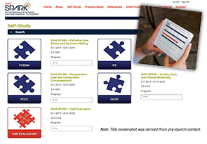
Best of all, SPARK is available exclusively online, making it easy to access from the hospital, the home, the coffee shop, or on the go.
SHM SPARK is a unique online self-assessment tool featuring 175 vignette-style, single-best-answer, multiple-choice questions, complete with answers, discussion, reasoning, references, and quizzing capabilities.
In addition to educating hospitalists with nearly 200 questions online, SHM SPARK empowers users to:
- Create customized practice quizzes in topic areas that meet your specific knowledge gaps/study needs;
- Progress at your own pace using the self-study reference feature; and
- Claim MOC Part II medical knowledge points through ABIM and download a CME certificate for earned AMA Physician’s Recognition Award (PRA) Category 1 Credit™.
The tool includes:
- Robust teaching points for each vignette;
- The option to save key questions for later review; and
- Question-level comparisons to the average response.
Access to SHM Spark is $199 for SHM members and $349 for nonmembers. Groups of 10 or more hospitalists receive a 10% discount.
Now, SHM is introducing hospital medicine’s first preparation tool for the exam.
For the hundreds of hospitalists considering taking the American Board of Internal Medicine’s Focused Practice in Hospital Medicine Maintenance of Certification exam in the near future, SHM’s SPARK exam prep tool is the perfect way to brush up on the skills and knowledge needed to pass.
SHM SPARK gives hospitalists the peace of mind that comes from knowing they are ready for the topics unique to hospitalists in the exam, including:
- Palliative care, medical ethics, and decision making;
- Peri-operative care and consultative comanagement; and
- Quality, safety, and clinical reasoning.
Best of all, SPARK is available exclusively online, making it easy to access from the hospital, the home, the coffee shop, or on the go.
SHM SPARK is a unique online self-assessment tool featuring 175 vignette-style, single-best-answer, multiple-choice questions, complete with answers, discussion, reasoning, references, and quizzing capabilities.
In addition to educating hospitalists with nearly 200 questions online, SHM SPARK empowers users to:
- Create customized practice quizzes in topic areas that meet your specific knowledge gaps/study needs;
- Progress at your own pace using the self-study reference feature; and
- Claim MOC Part II medical knowledge points through ABIM and download a CME certificate for earned AMA Physician’s Recognition Award (PRA) Category 1 Credit™.
The tool includes:
- Robust teaching points for each vignette;
- The option to save key questions for later review; and
- Question-level comparisons to the average response.
Access to SHM Spark is $199 for SHM members and $349 for nonmembers. Groups of 10 or more hospitalists receive a 10% discount.
Student Hospitalist Scholar Grant Winners Blog About Patient Safety
The following are excerpts of those posts.
Why We Should Care about Alarm Fatigue

When I arrived back at the Children’s Hospital of Philadelphia (CHOP) after my first year of medical school, I knew what was awaiting me: thousands of alarms from physiologic monitors, most of them inconsequential, lined up neatly in spreadsheets, splattered all over research databases, lighting up on video screens, chirping down hallways and up elevators. Of course, they were incessantly firing at the bedside, but when patient care is video recorded for Dr. Bonafide’s research study on alarm fatigue, those patient care hours turn into data points that live on hard drives and servers waiting to be classified, annotated, and cataloged by a team of research assistants, including me.
I began working at the CHOP while attending the University of Pennsylvania’s post-baccalaureate premed program. What started as a temporary summer research position turned into an almost three-year endeavor. The pilot that I helped design uses video cameras in hospitalized patient rooms to record patient care. We download the video, edit it so we can review multiple viewing angles at one time, download a spreadsheet of all of the alarms that fired during the study period, and then, with a little patience and some subtraction, we can line up every alarm that fired with the video clip. That’s the easy part.
This small pilot has transformed into a much larger study with a much larger volume of alarms. Since I started medical school last July, the research team has steadily collected video data all year. With support from SHM’s student scholar grant program, I have been able to return to CHOP for the summer. And now the video review begins.
The First Two Years—Pathways and Patient Outcomes
The first two years of our medical curriculum are an introduction to the human body’s normal and pathophysiology and an attempt to untangle the complex pathways involved in the interactions between self and nonself. We hope to make connections between our physical exam findings and the physiologic pathways we have at our educational foundation. We begin to realize that there is a fine line to walk when treating a patient: Altering the inputs of a single system can drastically affect the outputs of another.
If we place patient outcomes in the context of the dance that occurs in clinical care for patients on the wards, similar to the downstream effects of disrupting biological pathways in illness, there is a multifactorial system underlying hospitalized patient outcomes.
Prior to medical school, I worked for several years as a population health epidemiologist in the Democratic Republic of Congo and then as a research data analyst at the University of Chicago. During my work in both of these settings, I quickly learned the relevance of contextual clues in complex systems-based problem solving. Over the course of my first year of medical school, I realized that nowhere is this creative use of information more important than in the inpatient setting, where we attempt to distill out the most important available information when assessing a patient.
But there are caveats to our interpretations of data points: Are we recognizing the most relevant physiologic associations when making clinical decisions? Are patient data really telling us what we think they are? What systemic factors are at play when patients experience an adverse outcome?
In my exploration of the importance of contextualizing inpatient data, I have been incredibly fortunate to work with Dana Edelson, MD, MS, and Matthew Churpek, MD, PhD, MPH, two mentors at the University of Chicago who are equally passionate about asking these same questions surrounding clinical care. Using ward patient data, we have investigated the importance of physician judgment in clinical deterioration and documented the need for greater sensitivity in recognition of sepsis and organ dysfunction in ward patients. But what can be done to reorient clinicians who are overwhelmed by and desensitized to data streams and bedside alarms?
Improving Patient Care as a Trainee

Patient safety has always been a priority for me, but it is only recently that I became aware of the many issues that threaten quality of care for patients. As a medical student, I vividly remember shadowing at the hospital and being shocked at what I saw. I walked through patient rooms and noticed loud beeps, the constant chatter of hospital staff, and the automatic entrance into patients’ rooms without even a knock. I wondered whether all of the disruptions and commotion impacted patient recovery in the hospital and after discharge. After pondering this, I decided that I wanted to take action and see what I, as a medical student, could do to improve daily inpatient conditions.
In order to begin addressing my vision, I knew I needed to find the right mentor in the right location. As a medical student, I feel like my life is on pause at times. During the school year, when exams come in constant waves and my days are spent with my head in notes and books, I am pausing my time with friends and family, using my energy to pursue my dream of helping others improve their health. While I am blessed to have such an opportunity, I knew that during my time off between my first and second year of medical school, I wanted to be in Chicago, near my close friends. And, from there, it seemed as if everything fell into place perfectly. I found on the University of Chicago Medical School website that Dr. Vineet Arora is a prominent Society of Hospital Medicine member who was looking for medical students to conduct research on sleep/functional recovery during hospital stay and post-discharge. The minute I spoke to Dr. Arora, I knew that she would be a great mentor. Not only are her credentials and accomplishments unbelievable, but I could tell she is really passionate about the work she is doing. Her excitement for the project is contagious, and I started getting really excited to start!
The following are excerpts of those posts.
Why We Should Care about Alarm Fatigue
When I arrived back at the Children’s Hospital of Philadelphia (CHOP) after my first year of medical school, I knew what was awaiting me: thousands of alarms from physiologic monitors, most of them inconsequential, lined up neatly in spreadsheets, splattered all over research databases, lighting up on video screens, chirping down hallways and up elevators. Of course, they were incessantly firing at the bedside, but when patient care is video recorded for Dr. Bonafide’s research study on alarm fatigue, those patient care hours turn into data points that live on hard drives and servers waiting to be classified, annotated, and cataloged by a team of research assistants, including me.
I began working at the CHOP while attending the University of Pennsylvania’s post-baccalaureate premed program. What started as a temporary summer research position turned into an almost three-year endeavor. The pilot that I helped design uses video cameras in hospitalized patient rooms to record patient care. We download the video, edit it so we can review multiple viewing angles at one time, download a spreadsheet of all of the alarms that fired during the study period, and then, with a little patience and some subtraction, we can line up every alarm that fired with the video clip. That’s the easy part.
This small pilot has transformed into a much larger study with a much larger volume of alarms. Since I started medical school last July, the research team has steadily collected video data all year. With support from SHM’s student scholar grant program, I have been able to return to CHOP for the summer. And now the video review begins.
The First Two Years—Pathways and Patient Outcomes
The first two years of our medical curriculum are an introduction to the human body’s normal and pathophysiology and an attempt to untangle the complex pathways involved in the interactions between self and nonself. We hope to make connections between our physical exam findings and the physiologic pathways we have at our educational foundation. We begin to realize that there is a fine line to walk when treating a patient: Altering the inputs of a single system can drastically affect the outputs of another.
If we place patient outcomes in the context of the dance that occurs in clinical care for patients on the wards, similar to the downstream effects of disrupting biological pathways in illness, there is a multifactorial system underlying hospitalized patient outcomes.
Prior to medical school, I worked for several years as a population health epidemiologist in the Democratic Republic of Congo and then as a research data analyst at the University of Chicago. During my work in both of these settings, I quickly learned the relevance of contextual clues in complex systems-based problem solving. Over the course of my first year of medical school, I realized that nowhere is this creative use of information more important than in the inpatient setting, where we attempt to distill out the most important available information when assessing a patient.
But there are caveats to our interpretations of data points: Are we recognizing the most relevant physiologic associations when making clinical decisions? Are patient data really telling us what we think they are? What systemic factors are at play when patients experience an adverse outcome?
In my exploration of the importance of contextualizing inpatient data, I have been incredibly fortunate to work with Dana Edelson, MD, MS, and Matthew Churpek, MD, PhD, MPH, two mentors at the University of Chicago who are equally passionate about asking these same questions surrounding clinical care. Using ward patient data, we have investigated the importance of physician judgment in clinical deterioration and documented the need for greater sensitivity in recognition of sepsis and organ dysfunction in ward patients. But what can be done to reorient clinicians who are overwhelmed by and desensitized to data streams and bedside alarms?
Improving Patient Care as a Trainee
Patient safety has always been a priority for me, but it is only recently that I became aware of the many issues that threaten quality of care for patients. As a medical student, I vividly remember shadowing at the hospital and being shocked at what I saw. I walked through patient rooms and noticed loud beeps, the constant chatter of hospital staff, and the automatic entrance into patients’ rooms without even a knock. I wondered whether all of the disruptions and commotion impacted patient recovery in the hospital and after discharge. After pondering this, I decided that I wanted to take action and see what I, as a medical student, could do to improve daily inpatient conditions.
In order to begin addressing my vision, I knew I needed to find the right mentor in the right location. As a medical student, I feel like my life is on pause at times. During the school year, when exams come in constant waves and my days are spent with my head in notes and books, I am pausing my time with friends and family, using my energy to pursue my dream of helping others improve their health. While I am blessed to have such an opportunity, I knew that during my time off between my first and second year of medical school, I wanted to be in Chicago, near my close friends. And, from there, it seemed as if everything fell into place perfectly. I found on the University of Chicago Medical School website that Dr. Vineet Arora is a prominent Society of Hospital Medicine member who was looking for medical students to conduct research on sleep/functional recovery during hospital stay and post-discharge. The minute I spoke to Dr. Arora, I knew that she would be a great mentor. Not only are her credentials and accomplishments unbelievable, but I could tell she is really passionate about the work she is doing. Her excitement for the project is contagious, and I started getting really excited to start!
Primary and Secondary Headache During Pregnancy
Among pregnant women receiving inpatient neurologic consultation, more than one-third have secondary headache, according to a study of 140 pregnant women presenting with acute headache. Researchers found:
• The women had a mean age of 29 years and 56.4% presented in the third trimester.
• Diagnoses were 65.0% primary and 35% secondary disorders.
• Most common primary headache disorder was migraine (91.2%) and secondary headache disorders were hypertensive disorders (51.0%).
• Groups were similar in demographics, gestational ages, and most headache features.
• In univariate analysis, secondary headaches were associated with: lack of headache history, seizures, elevated blood pressure, fever, and an abnormal neurological exam.
• In multivariate logistic regression, elevated blood pressure and a lack of headache history had an increased association with secondary headache, while psychiatric comorbidity and phonophobia had a reduced association with secondary headache.
Citation: Robbins MS, Farmakidis C, Dayal AK, Lipton RB. Acute headache diagnosis in pregnant women: A hospital-based study. [Published online ahead of print August 19, 2015]. Neurology. doi: http://dx.doi.org/10.1212/WNL.0000000000001954.
Palliative Care, Advance Care Planning Conversations Needed Between Patients, Hospitalists

Last week, the Centers for Medicare and Medicaid Services (CMS), the nation’s largest payer of healthcare services and the 800-pound gorilla in setting medical necessity and coverage policies, announced a proposal to begin paying for goals of care and advance care planning (ACP) discussions between medical providers and patients. Sound familiar? It should. This is the same, seemingly no-brainer proposal that in 2009 was stricken from the eventually approved Patient Protection and Affordable Care Act (PPACA, aka the ACA, aka “Obamacare”) in response to the intentional and patently false accusations of government-run “death panels,” in the hopes of salvaging some measure of bipartisan support. As we all know, the bill eventually passed the following year without a single Republican voting in favor in either the House or Senate, and without funding for ACP sessions!
The need for ACP and access to primary and specialty palliative care is so great and accepted in the healthcare community. In their Choosing Wisely recommendations, numerous medical specialty societies, including ACEP [American College of Emergency Physicians], AAHPM [American Academy of Hospice and Palliative Medicine], AGS [American Geriatrics Society], and AMDA [The Society for Post-Acute and Long-Term Care Medicine], have included early and reliable access to palliative care and avoidance of non-value added care, such as placement of feeding tubes in patients with advanced dementia, calling out the gap between quality, evidence-based, patient and family-centered care, and “usual care” (e.g. medical and disease-focused care) that patients receive too often near the end of life.
So where is the disconnect between what people want and what actually happens to them at end of life?
The answer is clear: We’re not having “The Conversation.”
And, though our primary care and even specialty care colleagues are involved regularly in the care of these patients, they may be inclined to postpone or avoid ACP with patients and families in the outpatient setting due to lack of comfort [or] skill or even recognizing that the person they’ve been trying valiantly to cure or at least prolong the inevitable [for] is on that downslope of life we all eventually experience—it’s called dying.
Our current reimbursement system throws another barrier in front of providers. Like many other “nonprocedural” activities, ACP is not only undervalued; there is currently a lack of value assigned to this important cognitive, empathic, and communication-based “procedure.” And I refer to it as a procedure because, like a surgical or invasive vascular procedure, when it goes badly, the consequences and sequelae can be just as damaging, and even irreparable.
Thus, intentionally or not, the can is kicked further down the proverbial road until the patient reaches the hospital in a state of crisis—sometimes in extremis—and the hospitalist is left to make sense of all the clinical, emotional, psychological, spiritual, and frequently familial history (baggage?) leading up to that hospital admission. We are expected to develop instant rapport and trust while simultaneously attempting to develop (in collaboration with our specialty care providers and, preferably, the patient’s primary care provider) a plan of care that takes into account the personal values and treatment preferences for that individual within the clinical realities of the patient’s illness and disease trajectory as they lie before us.
Sound familiar?
For the full blog post, including Dr. Epstein’s recommendations for what hospitalists can do to support the CMS proposal, visit Hospital Leader.
ICEID: Population-level data support ACIP flu vaccine recommendations
ATLANTA – Expanded influenza vaccination coverage among children between 2002 and 2012 appears to have provided direct benefit with respect to influenza-related hospitalizations among vaccinated children, according to an analysis of vaccination and hospitalization data.
Additionally, the coverage among children appears to have provided indirect benefits in adults, Cecile Viboud, Ph.D. of the National Institutes of Health, Bethesda, Md., reported at the International Conference on Emerging Infectious Diseases.

Between 2006-2007 and 2010-2011, the U.S. Advisory Committee on Immunization Practices (ACIP) broadened vaccination recommendations to include not only children aged 6-23 months, but also those aged 24-59 months, then those aged 5-18 years, and eventually all those over age 6 months. Consequently, the vaccine coverage rate increased from less than 5% in 2002 to about 52% in 2012 (and to about 70% in those under age 5 years). Modeling of weekly influenza-related hospitalization outcomes (pneumonia and influenza outcomes and respiratory and circulatory outcomes) provided solid evidence of a direct and significant protective effect of vaccination both in children under age 5 years and in those aged 5-19 years. This finding was consistent across disease outcomes, and remained significant in those under age 5 after adjusting for state, but the association was weaker with stratification by season, Dr. Viboud noted.
Further, hospitalization rates among working-age adults and seniors aged 65-74 years declined with increasing pediatric vaccine coverage, suggesting an indirect protective effect in that population, she said, noting that the vaccination rate among older adults remained stable across the study period.
No evidence was seen for an indirect protective effect among adults over age 74 years, she said.
Dr. Viboud and her colleagues used age-specific annual vaccination rates derived from the National Immunization Survey and the Behavioral Risk Factor Surveillance System. Age-specific rates of influenza-associated hospitalizations were estimated for each season during 1989-2012 by modeling weekly pneumonia and influenza outcomes plus respiratory and circulatory outcomes from the State Inpatient Databases of the Agency for Healthcare Research & Quality.
“In a nutshell, we see strong statistical evidence for the direct protective effects of the influenza vaccination program in children on the basis of analyses of population-level hospitalization data, which supports the expansion of the ACIP flu vaccine recommendations in the past decade,” Dr. Viboud said in an interview. “We also find weak evidence of herd immunity effects, whereby hospitalization rates are reduced in adults. That the evidence is weak is perhaps not surprising given that vaccine uptake in children remains moderate (60% in most highly vaccinated states) and vaccine effectiveness is modest at 40%-60% depending on the season. Independent information from mathematical transmission models confirms that herd immunity benefits are expected to be low given these levels of vaccine coverage.”
The indirect effects may become clearer with increasing vaccine uptake, she added.
Dr. Viboud reported having no disclosures.
ATLANTA – Expanded influenza vaccination coverage among children between 2002 and 2012 appears to have provided direct benefit with respect to influenza-related hospitalizations among vaccinated children, according to an analysis of vaccination and hospitalization data.
Additionally, the coverage among children appears to have provided indirect benefits in adults, Cecile Viboud, Ph.D. of the National Institutes of Health, Bethesda, Md., reported at the International Conference on Emerging Infectious Diseases.
Between 2006-2007 and 2010-2011, the U.S. Advisory Committee on Immunization Practices (ACIP) broadened vaccination recommendations to include not only children aged 6-23 months, but also those aged 24-59 months, then those aged 5-18 years, and eventually all those over age 6 months. Consequently, the vaccine coverage rate increased from less than 5% in 2002 to about 52% in 2012 (and to about 70% in those under age 5 years). Modeling of weekly influenza-related hospitalization outcomes (pneumonia and influenza outcomes and respiratory and circulatory outcomes) provided solid evidence of a direct and significant protective effect of vaccination both in children under age 5 years and in those aged 5-19 years. This finding was consistent across disease outcomes, and remained significant in those under age 5 after adjusting for state, but the association was weaker with stratification by season, Dr. Viboud noted.
Further, hospitalization rates among working-age adults and seniors aged 65-74 years declined with increasing pediatric vaccine coverage, suggesting an indirect protective effect in that population, she said, noting that the vaccination rate among older adults remained stable across the study period.
No evidence was seen for an indirect protective effect among adults over age 74 years, she said.
Dr. Viboud and her colleagues used age-specific annual vaccination rates derived from the National Immunization Survey and the Behavioral Risk Factor Surveillance System. Age-specific rates of influenza-associated hospitalizations were estimated for each season during 1989-2012 by modeling weekly pneumonia and influenza outcomes plus respiratory and circulatory outcomes from the State Inpatient Databases of the Agency for Healthcare Research & Quality.
“In a nutshell, we see strong statistical evidence for the direct protective effects of the influenza vaccination program in children on the basis of analyses of population-level hospitalization data, which supports the expansion of the ACIP flu vaccine recommendations in the past decade,” Dr. Viboud said in an interview. “We also find weak evidence of herd immunity effects, whereby hospitalization rates are reduced in adults. That the evidence is weak is perhaps not surprising given that vaccine uptake in children remains moderate (60% in most highly vaccinated states) and vaccine effectiveness is modest at 40%-60% depending on the season. Independent information from mathematical transmission models confirms that herd immunity benefits are expected to be low given these levels of vaccine coverage.”
The indirect effects may become clearer with increasing vaccine uptake, she added.
Dr. Viboud reported having no disclosures.
AT ICEID 2015
Key clinical point: Expanded influenza vaccination coverage in children provided direct benefits with respect to hospitalizations.
Major finding: Vaccine coverage rate increased from less than 5% in 2002 to about 52% in 2012 .
Data source: An analysis of vaccination and hospitalization data.
Disclosures: Dr. Viboud reported having no disclosures.

Society of Hospital Medicine Posts Quality Improvement Resources Online

Glycemic Control Webinar
Subcutaneous Insulin Order Sets in the Inpatient Setting: Design and Implementation
Presenter: Kristi Kulasa, MD
Date: September 17
Time: 4 p.m. EDT
General QI Webinars
Quality Improvement for Hospital Medicine Groups: Self-Assessment and Self-Improvement Using the SHM Key Characteristics
Presenter: Steve Deitelzweig, MD, SFHM
Date: September 16
Time: 2 p.m. EDT
Other online resources at www.hospitalmedicine.org:
- New FREE clinical topics and guide: chronic obstructive pulmonary disease.
- Coming soon: antibiotic stewardship.
- And now, all of SHM’s popular SHMConsults modules are available on the Learning Portal.
With new quality improvement resources available online every month, Hospital Medicine is THE source for hospitalists ready to improve their hospitals.
Start Planning for State of Hospital Medicine Survey 2016
SHM’s practice analysis committee has geared up over the last month or two in preparation for January’s biannual State of Hospital Medicine survey. We’ve been reviewing topics included in the survey with an eye toward making sure the content stays relevant to what hospitalists and hospital leaders want to know. And we’ve been parsing the language and construction of each question with the goal of making it as clear as possible.
Once the committee finalizes the survey questions, it will be time for our intrepid SHM staffer Patrick Vulgamore, MPH, to build and test the survey instrument. At the same time, the committee will complete supporting materials like FAQs, the survey guide, and the communication plan.
But not all the work is on our side. There are a number of things that you can do now to begin preparing for your survey participation.
First, Commit to Participating
The survey is only as good as the number and quality of responses that we receive. We need everyone to participate, whether your hospital medicine group works in an academic or community setting, whether you serve adult or pediatric patients or both, and whether you are employed by a hospital/system, a management company, or a private group.
Go ask the leaders of your group whether they plan to participate, and lobby heavily to be part of the process. In addition to helping define the current state of hospital medicine, your group will also receive a free copy of the survey report.
Make a Plan
Your next step is to identify who will be responsible for pulling together the required information and completing the survey instrument. You’ll also want to be thinking now about where the various pieces of information you’ll need will come from. The survey includes topics such as scope of clinical practice, schedule, skill mix and work allocation, compensation methodology and benefits, CPT code distribution, and amount of financial support received. Then make sure you set aside time in the January-March period to complete the survey, and check to see that any necessary approvals have been obtained.
Look for Survey Communications in January 2016
The practice analysis committee tries hard to cover every possible communication avenue, but invariably people will say, “We didn’t know it was time for the survey.” So this is your heads up to be on the lookout for your survey invitation—in the regular mail, in both targeted and general emails, in The Hospitalist and other SHM publications, and on the SHM website.

Participate in the MGMA Survey, Too
The SoHM survey doesn’t include questions about individual provider productivity and compensation, but those are some of the most often looked for data points. Instead, SHM partners with the Medical Group Management Association (MGMA) to encourage hospital medicine groups to participate in the MGMA compensation and production survey—which will be conducted concurrently with SHM’s survey for both academic and non-academic groups. SHM then licenses this information for inclusion in the SoHM report.
Full participation in the SHM survey process means not just completing SHM’s SoHM survey but also completing the applicable MGMA survey. This is the only way we can obtain robust information about trends in hospitalist compensation and productivity to share with you.
So don’t wait until January. Start working now to ensure your group is well positioned to contribute to what we learn about the state of hospital medicine!
Leslie Flores is a member of SHM’s Practice Analysis Committee and a partner in Nelson Flores Hospital Medicine Consultants.
SHM’s practice analysis committee has geared up over the last month or two in preparation for January’s biannual State of Hospital Medicine survey. We’ve been reviewing topics included in the survey with an eye toward making sure the content stays relevant to what hospitalists and hospital leaders want to know. And we’ve been parsing the language and construction of each question with the goal of making it as clear as possible.
Once the committee finalizes the survey questions, it will be time for our intrepid SHM staffer Patrick Vulgamore, MPH, to build and test the survey instrument. At the same time, the committee will complete supporting materials like FAQs, the survey guide, and the communication plan.
But not all the work is on our side. There are a number of things that you can do now to begin preparing for your survey participation.
First, Commit to Participating
The survey is only as good as the number and quality of responses that we receive. We need everyone to participate, whether your hospital medicine group works in an academic or community setting, whether you serve adult or pediatric patients or both, and whether you are employed by a hospital/system, a management company, or a private group.
Go ask the leaders of your group whether they plan to participate, and lobby heavily to be part of the process. In addition to helping define the current state of hospital medicine, your group will also receive a free copy of the survey report.
Make a Plan
Your next step is to identify who will be responsible for pulling together the required information and completing the survey instrument. You’ll also want to be thinking now about where the various pieces of information you’ll need will come from. The survey includes topics such as scope of clinical practice, schedule, skill mix and work allocation, compensation methodology and benefits, CPT code distribution, and amount of financial support received. Then make sure you set aside time in the January-March period to complete the survey, and check to see that any necessary approvals have been obtained.
Look for Survey Communications in January 2016
The practice analysis committee tries hard to cover every possible communication avenue, but invariably people will say, “We didn’t know it was time for the survey.” So this is your heads up to be on the lookout for your survey invitation—in the regular mail, in both targeted and general emails, in The Hospitalist and other SHM publications, and on the SHM website.
Participate in the MGMA Survey, Too
The SoHM survey doesn’t include questions about individual provider productivity and compensation, but those are some of the most often looked for data points. Instead, SHM partners with the Medical Group Management Association (MGMA) to encourage hospital medicine groups to participate in the MGMA compensation and production survey—which will be conducted concurrently with SHM’s survey for both academic and non-academic groups. SHM then licenses this information for inclusion in the SoHM report.
Full participation in the SHM survey process means not just completing SHM’s SoHM survey but also completing the applicable MGMA survey. This is the only way we can obtain robust information about trends in hospitalist compensation and productivity to share with you.
So don’t wait until January. Start working now to ensure your group is well positioned to contribute to what we learn about the state of hospital medicine!
Leslie Flores is a member of SHM’s Practice Analysis Committee and a partner in Nelson Flores Hospital Medicine Consultants.