User login
High-fat diet linked to RBC dysfunction

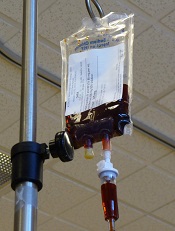
Preclinical research suggests a high-fat diet is associated with red blood cell (RBC) dysfunction and helps explain how these dysfunctional cells may mediate atherosclerosis.
The researchers believe their findings may have implications for the pathogenesis of atherosclerosis in obesity, but the work may aid the study of other health conditions as well, such as thrombosis in the context of cancer.
The team detailed their findings in Circulation.
“Obesity caused by chronic consumption of a high-calorie, high-fat diet is a worldwide epidemic, representing one of the greatest threats to global health,” said principal investigator Vladimir Bogdanov, PhD, of the University of Cincinnati in Ohio.
“White blood cells play a key role in fueling adipose tissue inflammation and insulin resistance in obesity and also promote the clogging of arteries, or atherosclerosis, setting the stage for heart attack and stroke. While these outcomes linked with a high-fat diet and fat in the blood on white blood cells have been shown in animal models and humans, the impact of high-fat diets on other bone marrow-derived cells, like red blood cells, is not well-defined.”
“Evidence is emerging that red blood cells play an important regulatory role in the development of atherosclerosis, binding pro-inflammatory proteins that cause dysfunction in the inner lining of the blood vessel wall—the endothelium. We explored how a high fat-diet causes red blood cell dysfunction in this study.”
Dr Bogdanov and his team fed a 60% high-fat diet to mice for 12 weeks and compared these animals to control mice that received a normal diet.
There was an increase in the level of chemokines bound to the RBCs of mice that received the high-fat diet. These chemokines were bound to RBCs via the Duffy antigen receptor for chemokines.
The researchers exposed RBCs from mice on the high-fat diet to an endothelial monolayer in vitro, and they observed “significantly enhanced” macrophage transendothelial migration. They said this confirms the functional importance of RBC-bound chemokines in the setting of a high-fat diet.
“In red blood cells from animal models fed a high-fat diet, there was an increase in cholesterol found in the cell membrane and phosphatidylserine levels, promoting inflammatory reactions,” Dr Bogdanov added.
“Phosphatidylserine is a phospholipid membrane component which plays a key role in the cycle of cells. When red blood cells from the animals being fed the high-fat diet were injected into a control group, eating a normal diet, there was a 3-fold increase in their spleens’ uptake of red blood cells. The spleen is involved in the removal of blood cells, as well as systemic inflammation.”
“All of these findings show that the dysfunction of red blood cells, corresponding with dysfunction of the lining of blood vessels, occurs very early in diet-induced obesity and may play a part in the formation of atherosclerosis. Diets high in saturated fat have long been associated with endothelial dysfunction, the precursor to atherosclerosis, but, to our knowledge, the effects of high-fat diet on red blood cells have not been rigorously examined.”
Dr Bogdanov noted that, in humans, high cholesterol is associated with alterations in RBCs that are improved by treatment with statins. But the majority of obese humans do not have severe high cholesterol, as was the case with the animals in this study.
The researchers are now working on translating their findings to humans. ![]()
Berry-derived compound can fight AML

A compound derived from the berries of the Bloodhorn tree has demonstrated activity against acute myeloid leukemia (AML), according to preclinical research published in Investigational New Drugs.
The compound, 7-formyl-10-methylisoellipticine, induced apoptosis in AML cells in a dose- and time-dependent manner.
It also significantly slowed tumor growth and reduced tumor mass in a mouse model of AML.
7-formyl-10-methylisoellipticine is derived from an ellipticine, which has been isolated from the berries of the Ochrosia Elliptica tree. The tree, also known as the Bloodhorn tree due to the shape and color of the berries, grows on the northeast coast of Australia and in the rainforests of Brazil.
“[We have] taken the natural product and restyled it with unique features to improve the potency and solubility,” explained Florence McCarthy, PhD, of University College Cork in Ireland.
“What is truly exceptional is that these features are not common in drugs, and so we aim to exploit this fully. There is also significant potential to apply this approach to other drugs in a similar fashion.”
For this study, Dr McCarthy and his colleagues first tested 7-formyl-10-methylisoellipticine in the AML cell line MV4-11. They tested a range of concentrations in an attempt to identify the minimum concentration that would cause significant cytotoxicity. It turned out to be 5 μM.
Over a period of 24 hours, 5 μM of 7-formyl-10-methylisoellipticine killed up to 40% of MV4-11 cells. And over 96 hours, 5 μM of 7-formyl-10-methylisoellipticine killed more than 90% of cells.
Further investigation revealed that 5 μM of 7-formyl-10-methylisoellipticine increases the sub-G1 phase of the MV4-11 cell cycle. And the compound functions, at least in part, by generating mitochondrial-derived reactive oxygen species.
The researchers then found that 7-formyl-10-methylisoellipticine is not toxic to BALB/c mice. The team injected the mice with 7-formyl-10-methylisoellipticine at a range of doses—5 mg/kg, 10 mg/kg, 25 mg/kg, and 50 mg/kg.
Regardless of the dose, the compound did not cause a change in body weight, significantly increase levels of alanine aminotransferase or aspartate aminotransferase relative to negative control, or significantly change cell morphology or tissue structure in specified major organs.
Finally, the researchers found that 7-formyl-10-methylisoellipticine has antitumor activity in an AML xenograft mouse model. Based on the toxicity experiments, the team used a dose of 25 mg/kg in these mice.
At this dose, 7-formyl-10-methylisoellipticine significantly slowed tumor growth and reduced tumor mass. Tumor growth was 4 times slower in mice treated with 7-formyl-10-methylisoellipticine than in control mice. And tumor mass was up to 7 times greater in controls than it was in treated mice.
Based on these results, the researchers said they plan to continue investigating the mechanism of action of ellipticines, which “have a clear potential clinical application.” ![]()
Risk of anaphylaxis with IV iron products
Researchers have compared the risk of anaphylaxis with different intravenous (IV) iron products and found evidence to suggest that iron dextran poses the greatest risk.
Compared with nondextran formulations, iron dextran was associated with a higher cumulative risk of anaphylaxis and an increased risk of anaphylaxis at first administration.
Iron sucrose was associated with the lowest risk of anaphylaxis, both cumulative and at first administration.
Cunlin Wang, MD, PhD, of the US Food and Drug Administration in Silver Spring, Maryland, and his colleagues conducted this research and reported the results in JAMA.
The researchers studied 688,183 recipients of IV iron enrolled in the fee-for-service Medicare program from January 2003 to December 2013.
The team examined administrations of IV iron dextran, gluconate, sucrose, or ferumoxytol. They identified 247,500 iron dextran and 440,683 nondextran users during the study period.
Overall, there were 274 cases of anaphylaxis at first exposure to IV iron and an additional 170 cases during subsequent iron administrations.
At first administration, iron dextran was associated with a higher anaphylaxis risk than nondextran formulations. The risk of anaphylaxis was 68 per 100,000 persons for iron dextran and 24 per 100,000 persons for all nondextran products combined. The odds ratio—adjusted for age, indication, history of coronary heart disease, and hypertension—was 2.6 (P<0.001).
Among the nondextran products, the risk of anaphylaxis at first administration was higher with both iron gluconate and ferumoxytol than with iron sucrose. When compared with iron sucrose, the adjusted odds ratio of anaphylaxis was 3.6 for iron dextran, 2.0 for iron gluconate, and 2.2 for ferumoxytol.
Because each IV iron product has a specific recommended dose and schedule of administration, the researchers also calculated the cumulative risk of anaphylaxis based on both the number of administrations and the clinically relevant repletion level of iron (1000 mg) achieved within 12 weeks.
The cumulative risk of anaphylaxis over multiple administrations was highest for iron dextran, followed by ferumoxytol, iron gluconate, and iron sucrose.
The estimated cumulative anaphylaxis risk following total iron repletion of 1000 mg administered within a 12-week period was highest with iron dextran (82 per 100,000 persons) and lowest with iron sucrose (21 per 100,000 persons).
The researchers noted that the mechanism of anaphylactic reaction after IV iron remains unknown. ![]()
Project BOOST Study Is Journal of Hospital Medicine’s Top-Cited Article in 2014
A study that examines Project BOOST’s effectiveness at decreasing rehospitalization rates was the top-cited article from the Journal of Hospital Medicine (JHM) in 2014. Titled “Project BOOST: Effectiveness of a Multihospital Effort to Reduce Rehospitalization,” the study has been cited 33 times since its publication in July 2013. The article concludes that hospitals participating in SHM’s Project BOOST (Better Outcomes for Older adults through Safe Transitions) experienced lower readmission rates.
“Project BOOST showed the effectiveness of physician-mentored implementation at reducing rehospitalization rates by improving the quality of patient care,” the study’s senior author, Mark V. Williams, MD, MHM, of Northwestern University Feinberg School of Medicine in Chicago, writes in an email to The Hospitalist eWire.
While researching the article, Dr. Williams says he knew it would be especially interesting to hospitalists. “I know hospitalists want to do the best job possible and not have patients be forced to return to the hospital because of problems with the hospital discharge process,” he writes. “Also, since hospitalists led this research as a nationwide quality improvement initiative, it is of particular interest to them.”
JHM Editor in Chief Andrew Auerbach, MD, MPH, and his editorial team publish some 30% of the 40-odd submissions they receive on average each month. “It was a very good paper,” Dr. Auerbach says of the Project BOOST study. Because of the importance of Project BOOST transitional care interventions, Dr. Auerbach and his team “knew it was going to be important to the field,” he adds.
In addition to its 33 citations, the Project BOOST article has received significant online attention. With an Altmetric score of 72, it is “one of the highest-scoring articles from [JHM] (#9 of 686),” according to its Altmetric page. This score reflects the article’s mentions in social media, newspapers, policy documents, and other sources.
Other factors such as the “number of tweets and downloads, the number of times people go to our website, those are also things that we look at very carefully to make sure that the journal is providing a service to people who may not be citing the papers but who want to use it just to read and to use in clinical care,” Dr. Auerbach says.
The four other top-cited articles discuss reducing inpatient falls, predicting mortality in ward patients through emergency medical records, detecting delirium to reduce hospitalization of dementia patients, and decreasing the use of non–evidence-based theories in treating bronchiolitis in pediatric patients.
The quality of researched published in JHM has changed since the journal’s debut in 2006, Dr. Auerbach says. “I think the field has developed quite a bit,” he adds. “I think the quality of research that’s happening in the field of hospital medicine is improving quite a bit, which is reflected in the type of papers we’re getting at the journal.”
In addition to 2014’s top-cited articles, the editorial team highlighted JHM’s new impact factor (IF) of 2.304, up from last year’s IF of 2.081. An IF indicates how many times the articles in a journal are cited elsewhere. “It is a very important metric for the journal, it’s very important for our authors, it’s important to our field,” Dr. Auerbach says. “It talks about how important the things we’re publishing are to other researchers.”
This increased IF ranks JHM 37 out of 153 journals in the General and Internal Medicine category of professional, peer-reviewed journals. Dr. Auerbach, whose five-year term will end in 2016, says he is “very happy with the pace of [JHM’s] improvement” and hopeful of the journal’s continued success. “We’re confident in our strategies,” he says. “I think if we keep focusing on really great papers and continue to grow the number of papers that come to the journal, we’ll be on track.”
Visit our website for more information on the Project BOOST study.
Merger options
The ongoing sea change in medicine has led to a substantial erosion of physician autonomy and to ever-increasing administrative burdens that hit small practices the hardest. Does this mean that the independent private physician practice model is doomed, as some predict? Absolutely not; but it will force many solo practitioners and small groups to join forces to protect themselves.
Those practices that offer unique services, or fill an unmet niche, may be able to remain small; but most smaller practices will need to consider a larger alternative. In my last column, I outlined the basics of arriving at a fair market value for a private practice; once that has been accomplished, you will be in a position to consider the various merger options that are available.

One attractive and relatively straightforward strategy is the formation of a cooperative group. In most areas, there are very likely several small practices in similar predicaments that might be receptive to discussing a collaboration on billing and purchasing. This allows each participant to maintain independence as a private practice, while pooling resources to ease the administrative burdens of all. Once that arrangement is in place, the group can consider more ambitious projects, such as the joint purchase of an integrated electronic health records (EHR) network, sharing personnel to lower staffing costs, and an integrated scheduling system. The latter will be particularly attractive to participants in later stages of their careers who are considering an intermediate option, somewhere between full-time work and complete retirement.
After a time, when the structure is stabilized and everyone agrees that his or her individual and shared interests and goals are being met, an outright merger can be contemplated. Obviously, projects of this scope require careful planning and implementation, and should not be undertaken without the help of competent legal counsel and an experienced business consultant.
A more complex but increasingly popular option is to join other small practices and providers in an independent practice association. An IPA is a legal entity, organized and directed by physicians for the purpose of negotiating contracts with insurance companies on their behalf. Because of its structure, an IPA is better positioned to enter into such financial arrangements and to counterbalance the leverage of insurers; but there are legal issues to consider. Many IPAs are vulnerable to antitrust charges because they include competing health care providers. You should check with legal counsel before signing on to an IPA, to make sure that it abides by antitrust and price fixing laws. IPAs have also been known to fail, particularly in states where they are not adequately regulated.
One proposed successor to the IPA is the accountable care organization (ACO), an entity born as a component of the Affordable Care Act. While the official definition remains nebulous, an ACO is basically a network of doctors and hospitals that shares financial and medical responsibility for providing coordinated and efficient care to patients. The goal of ACO participators is to limit unnecessary spending, both individually and collectively, according to criteria established by the Centers for Medicare & Medicaid Services (CMS), without compromising quality of care in the process. More than 600 ACOs had been approved by the CMS as of the beginning of 2014.
As the name implies, ACOs make providers jointly accountable for the health of their patients; they offer financial incentives to cooperate and to save money by avoiding unnecessary tests and procedures. A key component is the sharing of information. Providers that save money while also meeting quality targets are theoretically entitled to a portion of the savings.
As with IPAs, ACO ventures involve a measure of risk. ACOs that fail to meet the CMS performance and savings benchmarks can be stuck with the bill for investments made to improve care, such as equipment and computer purchases, and the hiring of mid-level providers and managers, and may be assessed monetary penalties as well. ACOs sponsored by physicians or rural providers, however, can apply to receive payments in advance to help finance infrastructure investments – a concession the Obama administration made after receiving complaints from rural hospitals. It is important to remember that the ACO model remains very much a work in progress.
Clearly, the price of remaining autonomous will be significant, and many private practitioners will be unwilling to pay it: Only 36% of physicians remained in independent practice at the end of the 2013, according to data from the American Medical Association – down from 57% in 2000; but those of us who remain committed to independence will find ways to preserve it, by mergers or other methods. In medicine, as in life, those most responsive to change will survive and flourish.
Dr. Eastern practices dermatology and dermatologic surgery in Belleville, N.J. He is the author of numerous articles and textbook chapters, and is a longtime monthly columnist for Dermatology News. Write to him at [email protected].
The ongoing sea change in medicine has led to a substantial erosion of physician autonomy and to ever-increasing administrative burdens that hit small practices the hardest. Does this mean that the independent private physician practice model is doomed, as some predict? Absolutely not; but it will force many solo practitioners and small groups to join forces to protect themselves.
Those practices that offer unique services, or fill an unmet niche, may be able to remain small; but most smaller practices will need to consider a larger alternative. In my last column, I outlined the basics of arriving at a fair market value for a private practice; once that has been accomplished, you will be in a position to consider the various merger options that are available.
One attractive and relatively straightforward strategy is the formation of a cooperative group. In most areas, there are very likely several small practices in similar predicaments that might be receptive to discussing a collaboration on billing and purchasing. This allows each participant to maintain independence as a private practice, while pooling resources to ease the administrative burdens of all. Once that arrangement is in place, the group can consider more ambitious projects, such as the joint purchase of an integrated electronic health records (EHR) network, sharing personnel to lower staffing costs, and an integrated scheduling system. The latter will be particularly attractive to participants in later stages of their careers who are considering an intermediate option, somewhere between full-time work and complete retirement.
After a time, when the structure is stabilized and everyone agrees that his or her individual and shared interests and goals are being met, an outright merger can be contemplated. Obviously, projects of this scope require careful planning and implementation, and should not be undertaken without the help of competent legal counsel and an experienced business consultant.
A more complex but increasingly popular option is to join other small practices and providers in an independent practice association. An IPA is a legal entity, organized and directed by physicians for the purpose of negotiating contracts with insurance companies on their behalf. Because of its structure, an IPA is better positioned to enter into such financial arrangements and to counterbalance the leverage of insurers; but there are legal issues to consider. Many IPAs are vulnerable to antitrust charges because they include competing health care providers. You should check with legal counsel before signing on to an IPA, to make sure that it abides by antitrust and price fixing laws. IPAs have also been known to fail, particularly in states where they are not adequately regulated.
One proposed successor to the IPA is the accountable care organization (ACO), an entity born as a component of the Affordable Care Act. While the official definition remains nebulous, an ACO is basically a network of doctors and hospitals that shares financial and medical responsibility for providing coordinated and efficient care to patients. The goal of ACO participators is to limit unnecessary spending, both individually and collectively, according to criteria established by the Centers for Medicare & Medicaid Services (CMS), without compromising quality of care in the process. More than 600 ACOs had been approved by the CMS as of the beginning of 2014.
As the name implies, ACOs make providers jointly accountable for the health of their patients; they offer financial incentives to cooperate and to save money by avoiding unnecessary tests and procedures. A key component is the sharing of information. Providers that save money while also meeting quality targets are theoretically entitled to a portion of the savings.
As with IPAs, ACO ventures involve a measure of risk. ACOs that fail to meet the CMS performance and savings benchmarks can be stuck with the bill for investments made to improve care, such as equipment and computer purchases, and the hiring of mid-level providers and managers, and may be assessed monetary penalties as well. ACOs sponsored by physicians or rural providers, however, can apply to receive payments in advance to help finance infrastructure investments – a concession the Obama administration made after receiving complaints from rural hospitals. It is important to remember that the ACO model remains very much a work in progress.
Clearly, the price of remaining autonomous will be significant, and many private practitioners will be unwilling to pay it: Only 36% of physicians remained in independent practice at the end of the 2013, according to data from the American Medical Association – down from 57% in 2000; but those of us who remain committed to independence will find ways to preserve it, by mergers or other methods. In medicine, as in life, those most responsive to change will survive and flourish.
Dr. Eastern practices dermatology and dermatologic surgery in Belleville, N.J. He is the author of numerous articles and textbook chapters, and is a longtime monthly columnist for Dermatology News. Write to him at [email protected].

Nursing Care Top-Ranked Factor in Pediatric Inpatient Satisfaction Survey
A recent patient satisfaction survey that ranked nursing care as the most important factor within inpatient pediatric care settings comes as no surprise to the chair of SHM’s Nurse Practitioner/Physician Assistant Committee.
Published online last month in the American Journal of Medical Quality, the retrospective study found that patient satisfaction for pediatric care varies widely depending on “which departmental setting patients receive treatment within a healthcare system,” study authors noted.
“Communication is one of the most important things,” says committee chair Tracy Cardin, ACNP-BC, FHM. “And nursing is sort of our designee sometimes in the communication of the plan of care. You can go over something with a patient and the family for a minute or so, or five, or even 10. But, really, it’s the nurses who reinforce the message.”
For the study, researchers at Nemours/Alfred I. duPont Hospital for Children in Delaware reviewed more than 27,000 patient satisfaction survey results over a three-year period at facilities of the Nemours Children’s Health System in the Delaware Valley and Jefferson University Hospitals in Philadelphia. Families rated their satisfaction on a five-point scale for various factors including physician care, nurse care, and personal concern.
The researchers say knowing what kinds of care expectations patients have in different settings, be it the emergency department or an inpatient ward, could help tailor interventions.
Cardin is hopeful that’s true, but she notes that patient satisfaction is a complex issue. A hospitalist could deliver perfect care, but if the patient had a bad experience at the hospital’s front door, the actual care delivery scores may be affected.
“It would be nice if there were just one variable to manipulate,” she says. “There are so many different impacts on [satisfaction]. What do we really have the ability to control?”
View our website for more information on inpatient satisfaction.
Difference in the timing of cessation of palliative chemotherapy between patients with incurable cancer receiving therapy only in a local hospital and those transitioned from a tertiary medical center to a local hospital
Click on the PDF icon at the top of this introduction to read the full article.
Click on the PDF icon at the top of this introduction to read the full article.
Click on the PDF icon at the top of this introduction to read the full article.
Impact of inpatient radiation on length of stay and health care costs
Background Health care costs are rising. Identifying areas for health care utilization savings may reduce costs.
Objective To identify oncology patients receiving inpatient radiotherapy with the purpose of measuring length of stay (LoS) and hospital charges.
Methods During July 2013 the oncology service physicians at Mount Sinai Medical Center in New York City were surveyed daily to identify patients receiving inpatient radiation. Actual LoS, acuity LoS were determined from the chart review. Expected LoS was calculated using the University Healthsystem Consortium database. Charges associated with actual LoS, acuity LoS, and expected LoS were then reported. Actual and expected LoS were compared for inpatient radiotherapy and nonradiotherapy groups.
Results 7 patients were identified as having remained in the hospital to receive radiation treatment. In that cohort, the average actual LoS and charges per patient were 40.1 and $48,724, compared with acuity LoS and charges of 25.6 days and $34,089 and expected LoS and charges of 7.7 days and $10,028. Mean LoS and charges attributed to radiation alone amounted to 11 days and $12,514. The mean actual LoS of oncology patients admitted during the same time period who did not receive radiation was 6.7 days, compared with 40.1 days for patients who received radiation (P < .0001).
Limitations Inability to access actual reimbursement data prevented exact cost calculations, small sample size, and single-institution focus.
Conclusion Delivery of radiation therapy during inpatient hospitalization extends LoS and contributes to higher health care costs. Methods to facilitate the delivery of outpatient radiotherapy may result in cost savings.
Click on the PDF icon at the top of this introduction to read the full article.
Caregivers’ attitudes toward promoting exercise among patients with late-stage lung cancer
Click on the PDF icon at the top of this introduction to read the full article.
Click on the PDF icon at the top of this introduction to read the full article.
Click on the PDF icon at the top of this introduction to read the full article.
Gout hospitalizations, costs surpass those for rheumatoid arthritis
SAN FRANCISCO – Hospitalization rates for gout doubled between 1993 and 2011, while those for rheumatoid arthritis fell by 67%, Dr. Sian Yik Lim reported at the annual meeting of the American College of Rheumatology.
The trends reveal marked progress in the treatment of rheumatoid arthritis, but “also highlight a critical need to improve gout care” and to implement measures to combat its rising prevalence, said Dr. Lim, a research fellow in medicine at Massachusetts General Hospital, Boston.
The study is “a very nice piece of work,” added Dr. James Galloway, who helped moderate the session at the meeting and is a lecturer in rheumatology at King’s College Hospital in London. “It makes us wonder whether we should spend more time teaching our primary care physician colleagues more about gout than rheumatoid arthritis.”

Gout and rheumatoid arthritis are the two most common inflammatory joint diseases, but few longitudinal studies have examined trends in related hospital admissions or costs, Dr. Lim said. Therefore, he and his associates extracted primary hospital discharge ICD-9 diagnosis codes from the National Inpatient Sample and also studied primary procedure codes for total knee replacement, total hip replacement, and other major joint surgeries for 1993 through 2011. In addition, they merged these with cost data from the Healthcare Cost and Utilization Project for 2001 to 2011.
Hospitalizations for gout and rheumatoid arthritis “crisscrossed” during the study period, Dr. Lim said. While there were more than three times as many hospitalizations for rheumatoid arthritis (26,712) as for gout (8,485) in 1993, by 2011, there were nearly twice as many admissions for gout (20,949) as for rheumatoid arthritis (11,015). Likewise, hospitalization rates for gout rose from 4.5 to 9 per 100,000 adults, while rheumatoid arthritis admissions fell by about two-thirds, from 14 to 5 per 100,000 adults. For the entire study period, hospital admissions for gout totaled 254,982, compared with 323,649 for rheumatoid arthritis, Dr. Lim added.
The investigators also documented a steady drop in admissions for total knee replacement, total hip replacement, and other major joint surgeries among rheumatoid arthritis patients, mirroring the results of recent multicenter studies in California (Ann Rheum Dis. 2009 Jul 5. doi: 10.1136/ard.2009.112474), the United Kingdom (Rheumatology [Oxford]. 2015 Apr;54[4]:666-71), and Germany (Ann Rheum Dis. 2015;74:738-45), Dr. Lim said. “This finding contrasts with the overall dramatic increase in these surgeries in the general United States population in recent years,” he added.
Inflation-adjusted hospital costs for gout and rheumatoid arthritis also converged during the study period, and by 2011, costs for gout actually exceeded those for rheumatoid arthritis, Dr. Lim said.
Overall, the findings reflect stark differences in care for gout and rheumatoid arthritis in the United States, he added. “New and potent drugs, effective combination regimens, and management strategies are increasingly being adopted for rheumatoid arthritis, but care for gout remains suboptimal, even though its prevalence and incidence are increasing,” he said. “Although the vast majority of gout patients are indicated for urate-lowering therapy, only a small proportion retrieve treatment, and the level of uric acid is not even measured in the vast majority of patients.”
Surveys also indicate that patients with gout often do not receive clear explanations about their disease, which may explain why as few as 10% follow treatment recommendations, according to Dr. Lim. He pointed to a hospital-based, retrospective cohort study reported at last year’s ACR annual meeting in which 89% of hospitalizations for gout were considered preventable. “These data support the need to improve gout prevention and care,” he said.
Dr. Lim had no disclosures. Dr. Hyon Choi, the senior author, reported having served on advisory boards for Takeda and AstraZeneca.
SAN FRANCISCO – Hospitalization rates for gout doubled between 1993 and 2011, while those for rheumatoid arthritis fell by 67%, Dr. Sian Yik Lim reported at the annual meeting of the American College of Rheumatology.
The trends reveal marked progress in the treatment of rheumatoid arthritis, but “also highlight a critical need to improve gout care” and to implement measures to combat its rising prevalence, said Dr. Lim, a research fellow in medicine at Massachusetts General Hospital, Boston.
The study is “a very nice piece of work,” added Dr. James Galloway, who helped moderate the session at the meeting and is a lecturer in rheumatology at King’s College Hospital in London. “It makes us wonder whether we should spend more time teaching our primary care physician colleagues more about gout than rheumatoid arthritis.”
Gout and rheumatoid arthritis are the two most common inflammatory joint diseases, but few longitudinal studies have examined trends in related hospital admissions or costs, Dr. Lim said. Therefore, he and his associates extracted primary hospital discharge ICD-9 diagnosis codes from the National Inpatient Sample and also studied primary procedure codes for total knee replacement, total hip replacement, and other major joint surgeries for 1993 through 2011. In addition, they merged these with cost data from the Healthcare Cost and Utilization Project for 2001 to 2011.
Hospitalizations for gout and rheumatoid arthritis “crisscrossed” during the study period, Dr. Lim said. While there were more than three times as many hospitalizations for rheumatoid arthritis (26,712) as for gout (8,485) in 1993, by 2011, there were nearly twice as many admissions for gout (20,949) as for rheumatoid arthritis (11,015). Likewise, hospitalization rates for gout rose from 4.5 to 9 per 100,000 adults, while rheumatoid arthritis admissions fell by about two-thirds, from 14 to 5 per 100,000 adults. For the entire study period, hospital admissions for gout totaled 254,982, compared with 323,649 for rheumatoid arthritis, Dr. Lim added.
The investigators also documented a steady drop in admissions for total knee replacement, total hip replacement, and other major joint surgeries among rheumatoid arthritis patients, mirroring the results of recent multicenter studies in California (Ann Rheum Dis. 2009 Jul 5. doi: 10.1136/ard.2009.112474), the United Kingdom (Rheumatology [Oxford]. 2015 Apr;54[4]:666-71), and Germany (Ann Rheum Dis. 2015;74:738-45), Dr. Lim said. “This finding contrasts with the overall dramatic increase in these surgeries in the general United States population in recent years,” he added.
Inflation-adjusted hospital costs for gout and rheumatoid arthritis also converged during the study period, and by 2011, costs for gout actually exceeded those for rheumatoid arthritis, Dr. Lim said.
Overall, the findings reflect stark differences in care for gout and rheumatoid arthritis in the United States, he added. “New and potent drugs, effective combination regimens, and management strategies are increasingly being adopted for rheumatoid arthritis, but care for gout remains suboptimal, even though its prevalence and incidence are increasing,” he said. “Although the vast majority of gout patients are indicated for urate-lowering therapy, only a small proportion retrieve treatment, and the level of uric acid is not even measured in the vast majority of patients.”
Surveys also indicate that patients with gout often do not receive clear explanations about their disease, which may explain why as few as 10% follow treatment recommendations, according to Dr. Lim. He pointed to a hospital-based, retrospective cohort study reported at last year’s ACR annual meeting in which 89% of hospitalizations for gout were considered preventable. “These data support the need to improve gout prevention and care,” he said.
Dr. Lim had no disclosures. Dr. Hyon Choi, the senior author, reported having served on advisory boards for Takeda and AstraZeneca.
AT THE ACR ANNUAL MEETING
Key clinical point: Hospitalizations and costs for gout in the United States have surpassed those for rheumatoid arthritis, reflecting stark differences in the state of care for these diseases.
Major finding: Between 1993 and 2011, hospitalizations for gout rose from 4.5 to 9 per 100,000 adults, while rheumatoid arthritis admissions fell from 14 to 5 per 100,000 adults.
Data source: Analysis of primary discharge diagnoses from the National Inpatient Sample and cost data from the Healthcare Cost and Utilization Project.
Disclosures: Dr. Lim had no disclosures. Dr. Hyon Choi, the senior author, reported having served on advisory boards for Takeda and AstraZeneca.
