User login
David Henry's JCSO podcast, November 2015
In his November podcast for The Journal of Community and Supportive Oncology, Dr David Henry discusses an article and accompanying commentary on the US Food and Drug Administration’s re-approval of gefitinib for the treatment of patients with EGFR-mutated lung cancer. (The drug’s original 2003 approval had been withdrawn in 2011 after subsequent findings failed to show a survival advantage.) He also comments on a line-up of clinical and supportive oncology articles reporting on a modified olanzapine regimen for the prevention of chemotherapy-induced nausea and vomiting; caregivers’ attitudes about their roles in promoting exercise among patients with late-stage lung cancer; the impact of inpatient radiation on length of stay and health care costs; and a study of patients with incurable cancer by Japanese investigators who examined differences in the timing of palliative chemotherapy cessation between patients in a local hospital and patients who transitioned to a local hospital from a tertiary medical center. The final item details a case of treatment-related MDS/AML in a patient after receiving therapy for large-cell neuroendocrine lung cancer.
Click on the download icon at the top of this introduction to listen to the podcast.
FDA approves pegylated product for hemophilia A

The US Food and Drug Administration (FDA) has approved Adynovate, a recombinant pegylated factor VIII (FVIII) product, for use in patients age 12 and older with hemophilia A.
The product can be used as routine prophylaxis and for on-demand treatment and control of bleeding episodes.
Adynovate will be available in the US in the coming weeks, according to Baxalta US Inc., the company developing the product.
Adynovate (formerly BAX 855) is built on the full-length Advate, a recombinant antihemophilic factor product that was approved by the FDA in 2003.
Adynovate consists of the full-length FVIII molecule linked to other molecules, known as polyethylene glycol (pegylated). This link extends the circulating half-life of the product and therefore extends the time between treatments.
So patients on Adynovate can receive twice-weekly doses rather than the 3 to 4 weekly doses typically required with other full-length FVIII products.
“The approval of Adynovate provides an important therapeutic option for use in the care of patients with hemophilia A and reduces the frequency of FVIII infusions needed to avoid bleeding,” said Karen Midthun, MD, director of the FDA’s Center for Biologics Evaluation and Research.
The FDA approved Adynovate based on results of a phase 2/3 trial. The study included 137 previously treated hemophilia A patients who were 12 to 65 years of age.
Patients were assigned to either twice-weekly prophylaxis (40-50 IU/kg, n=120) or on-demand treatment (10-60 IU/kg, n=17) with Adynovate.
Patients in the twice-weekly prophylaxis arm had far fewer annual bleeds than patients treated on-demand. The median annual bleed rates were 1.9 and 41.5, respectively.
Nearly all (96%) bleeding episodes (n=591) were controlled with 1 or 2 infusions of Adynovate.
None of the patients developed inhibitors to the treatment. However, there were 171 adverse events in the 73 patients who received Adynovate for about 6 months.
There were 7 events (occurring in 6 patients) that were considered possibly related to Adynovate. These included diarrhea, nausea, headache, and flushing.
Studies of Adynovate are ongoing in previously treated patients with severe hemophilia A undergoing surgery and in previously treated patients with severe hemophilia A who are under the age of 12. Baxalta is also planning to initiate a study in previously untreated patients with severe hemophilia A.
The company has filed for regulatory approval of Adynovate in Japan and expects to file for marketing authorization in Europe once the pediatric study is complete. ![]()
Patients may benefit from managing own VKA treatment

Photo courtesy of NIGMS
Allowing patients to self-manage anticoagulant therapy after heart valve surgery may lower the risk of death, according to a study published in the Annals of Thoracic Surgery.
Some of the patients studied were required to complete an educational program before they could manage their own treatment with a vitamin K antagonist (VKA).
For the remaining patients, VKA treatment was managed by a healthcare provider.
At 5 years, patients who managed their own treatment had a lower risk of death than patients on standard management.
“Oral anticoagulation therapy is usually monitored by laboratory analysis of the patient’s blood, which healthcare providers use to determine the appropriate dosage of medication,” said study author Thomas Decker Christensen, MD, PhD, of Aarhus University Hospital in Denmark.
“We believe that allowing patients to have more control over their own treatment can improve the standard of care in this patient group.”
Dr Christensen and his colleagues evaluated patients treated at 2 hospitals in Denmark between 1996 and 2012. There were 615 patients with mechanical heart valves who self-managed VKA treatment and 3075 control patients (a 1:5 ratio) who received standard VKA management following valve surgery.
Patients were required to attend an educational program consisting of a minimum of 3 lessons to be eligible for self-management. Over a period of 27 weeks following the educational training, patients gradually became self-managed.
“Once patients were approved for self-management, they were allowed to continue as such but could contact the treatment center with questions or concerns,” Dr Christensen noted.
“Patients were provided with a portable coagulometer, which they used to analyze medication levels and monitor dosage. Blood levels were measured and tracked weekly.”
Patients compared their own measurements against the International Normalized Ratio and adjusted their dosage levels according to what they had been taught in the educational training.
Bleeding and VTE
Over 5 years of follow-up, the risk of venous thromboembolism (VTE) and major bleeding was similar between self-managed patients and patients on standard management.
The rate of thromboembolic events requiring hospitalization was 1.6 events per 100 patient-years in the self-management group and 2.0 per 100 patient-years in the standard group. At 1 year of follow-up, the rates were 2.0 and 2.1, respectively.
When the researchers adjusted for potential confounders, there was a statistically nonsignificant higher risk of VTE among self-managed patients after 1 year (hazard ratio [HR]=1.29) and a statistically nonsignificant lower risk after 5 years (HR=0.91).
The rate of major bleeding was about 1.0 per 100 patient-years in the self-management group at both 1 year and 5 years. The corresponding rates in the standard management group were 1.9 at 1 year and 1.4 at 5 years.
When the researchers adjusted for potential confounders, there was a statistically nonsignificant lower risk of major bleeding in the self-management group at 1 year (HR=0.54) and at 5 years (HR=0.83).
Mortality
After 5 years, the rate of all-cause mortality was 1.1 per 100 patient-years in the self-management group and 2.5 in the standard management group. The adjusted HR was 0.49 in favor of the self-management group.
A landmark analysis restricting the survival analysis to patients alive 1 year after study inclusion resulted in an adjusted HR of 0.57.
“There are several reasons that patients who self-manage treatment have better outcomes than those who follow standard management,” Dr Christensen explained.
“Self-management patients receive more detailed information about oral anticoagulation therapy. They also learn more about the influence that diet, infectious diseases, alcohol, and other drug interactions can have on their treatment than do patients receiving standard management.”
He added that he hopes this study will have an impact on the management and care of heart valve patients.
“We believe that the majority of patients who have a mechanical heart valve inserted during surgery should be able to manage their oral anticoagulant therapy and recommend this as the standard treatment approach for these patients,” he concluded. ![]()
Invasive candidiasis hospitalizations down overall
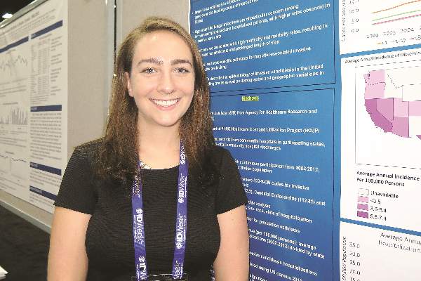
SAN DIEGO – The incidence of hospitalizations associated with invasive candidiasis decreased between 2007 and 2012, but both elderly and black patients remain at greatest risk for the infection, according to an analysis of national data.
“It’s been noted previously that the incidence of neonatal candidiasis seems to be going down, but we wanted to focus on older populations,” Sara Strollo, M.P.H., a trainee in the division of intramural research at the National Institute of Allergy and Infectious Diseases, Rockville., Md., said in an interview at an annual scientific meeting on infectious diseases.

For the study, which is the first of its kind, Ms. Strollo and her associates analyzed data from the State Inpatient Database from the Agency for Healthcare Research and Quality, which represents 97% of all community hospital discharges. They excluded neonatal cases.
The age-adjusted annual incidence of hospitalizations associated with invasive candidiasis ranged from 4.3 to 6.0 per 100,000 persons between 2002 and 2012. The incidence increased from 2002-2005, was stable through 2007, and decreased significantly between 2007 and 2012 -- by 6.7% among men and by 7.4% among women.
The highest incidence of hospitalization for invasive candidiasis occurred among the oldest age groups and among men. For example, compared with persons aged 50-64, the average annual incidence among those over age 80 years old was 2.6-fold higher among women (7.6 vs. 19.7 per 100,000 persons) and 3.9-fold higher among men (7.6 vs. 30 per 100,000 persons).
The researchers also found that among persons older than 50 years of age, black men and women had more than a two-fold higher incidence, compared with white men and women (23.7 vs. 11.7 per 100,000 persons and 22 vs. 10.4 per 100,000 persons, respectively).
During the overall study period, Ms. Strollo and her associates observed a nearly three-fold variation in the average annual incidence of hospital discharges for candidiasis per 100,000 persons, from 2.7 in Oregon to 7.2 in Florida. States with the highest incidence were Florida, Maryland, Missouri, Michigan, California, and Texas, but temporal trends were similar across states and no clear regional patterns among states were observed.
The investigators limited their analysis to 24 states with continuous reporting from 2002 through 2012, which represents 65% of the United States population. The researchers extracted records for discharges where ICD-9 codes for invasive candidiasis were listed in the primary or secondary discharge fields, including disseminated candidiasis (112.5), candidal endocarditis (112.81), and candidal meningitis (112.83). Age, gender, hospitalization year, and state data were extracted, and U.S. Census Bureau data were used as the denominator for state hospitalization incidence and trends. Poisson regression was used to assess significance of trends.
IDWeek marks the combined annual meetings of the Infectious Diseases Society of America, the Society for Healthcare Epidemiology of America, the HIV Medicine Association, and the Pediatric Infectious Diseases Society. The study was supported by a training grant from the National Institute of Child Health and Human Development. The researchers reported having no financial disclosures.
SAN DIEGO – The incidence of hospitalizations associated with invasive candidiasis decreased between 2007 and 2012, but both elderly and black patients remain at greatest risk for the infection, according to an analysis of national data.
“It’s been noted previously that the incidence of neonatal candidiasis seems to be going down, but we wanted to focus on older populations,” Sara Strollo, M.P.H., a trainee in the division of intramural research at the National Institute of Allergy and Infectious Diseases, Rockville., Md., said in an interview at an annual scientific meeting on infectious diseases.
For the study, which is the first of its kind, Ms. Strollo and her associates analyzed data from the State Inpatient Database from the Agency for Healthcare Research and Quality, which represents 97% of all community hospital discharges. They excluded neonatal cases.
The age-adjusted annual incidence of hospitalizations associated with invasive candidiasis ranged from 4.3 to 6.0 per 100,000 persons between 2002 and 2012. The incidence increased from 2002-2005, was stable through 2007, and decreased significantly between 2007 and 2012 -- by 6.7% among men and by 7.4% among women.
The highest incidence of hospitalization for invasive candidiasis occurred among the oldest age groups and among men. For example, compared with persons aged 50-64, the average annual incidence among those over age 80 years old was 2.6-fold higher among women (7.6 vs. 19.7 per 100,000 persons) and 3.9-fold higher among men (7.6 vs. 30 per 100,000 persons).
The researchers also found that among persons older than 50 years of age, black men and women had more than a two-fold higher incidence, compared with white men and women (23.7 vs. 11.7 per 100,000 persons and 22 vs. 10.4 per 100,000 persons, respectively).
During the overall study period, Ms. Strollo and her associates observed a nearly three-fold variation in the average annual incidence of hospital discharges for candidiasis per 100,000 persons, from 2.7 in Oregon to 7.2 in Florida. States with the highest incidence were Florida, Maryland, Missouri, Michigan, California, and Texas, but temporal trends were similar across states and no clear regional patterns among states were observed.
The investigators limited their analysis to 24 states with continuous reporting from 2002 through 2012, which represents 65% of the United States population. The researchers extracted records for discharges where ICD-9 codes for invasive candidiasis were listed in the primary or secondary discharge fields, including disseminated candidiasis (112.5), candidal endocarditis (112.81), and candidal meningitis (112.83). Age, gender, hospitalization year, and state data were extracted, and U.S. Census Bureau data were used as the denominator for state hospitalization incidence and trends. Poisson regression was used to assess significance of trends.
IDWeek marks the combined annual meetings of the Infectious Diseases Society of America, the Society for Healthcare Epidemiology of America, the HIV Medicine Association, and the Pediatric Infectious Diseases Society. The study was supported by a training grant from the National Institute of Child Health and Human Development. The researchers reported having no financial disclosures.
AT IDWEEK 2015
Key clinical point: As of 2007, the incidence of hospital-associated invasive candidiasis appears to be decreasing.
Major finding: Between 2007 and 2012, the age-adjusted annual incidence of hospitalizations associated with invasive candidiasis in the United States decreased by 6.7% among men and by 7.4% among women.
Data source: A long-term analysis of data from the State Inpatient Database from the Agency for Healthcare Research and Quality.
Disclosures: The researchers reported having no financial disclosures.
Daily cookie makes no dent in ADHD diet effect
SAN ANTONIO – Though restriction and elimination diets have been studied for decades in the treatment of children with attention deficit hyperactive disorder, their therapeutic role and level of efficacy remain controversial in part because of blinding difficulties in dietary studies.
In a study presented at the annual meeting of the American Academy of Child & Adolescent Psychiatry Dr. Steven Pliszka of the University of Texas Health Center in San Antonio reported that among children with ADHD on a gluten- and additive-free diet, those given a daily snack inconsistent with the prescribed diet still showed behavioral improvement over 5 weeks.

In the study, Dr. Pliszka and colleagues randomized 29 children with ADHD, ages 8-12, to a restricted diet with a daily cookie or with a snack consistent with the diet. Stimulants were the only psychotropic medications allowed in the study. “The hypothesis was that if you’re on the restriction diet and getting better, and you get a snack that has gluten in it, you ought to deteriorate,” Dr. Pliszka said in an interview. “What we thought we would see was only one group would improve, but we saw improvement across the board.” This could mean that one cookie was not enough to corrupt the beneficial effects of the diet, he said, or that something besides the diet, such as a placebo effect, was responsible for the children’s improvement.
ADHD behavioral rating scores for both groups of children improved over the course of the 5 weeks; however, neither group improved faster. Children were also screened on functional magnetic resonance imaging (fMRI) at baseline and 5 weeks to evaluate activity in the ventral striatum related to images of high and low-calorie foods. Because not enough children were available for fMRI screening at the study’s end, Dr. Pliszka and colleagues combined the cohorts for analysis. Both groups also showed imaging evidence of increased activity in the ventral striatum, the part of the brain associated with reward response, and where activity is known to be lower in children with ADHD.
The researchers found regional activation within the ventral striatum to be significantly greater at follow-up than baseline for the cohort as a whole.
Dr. Pliszka said that the findings required more follow-up to understand.
“What we need to do for a follow-up study is get these groups to separate. If it is a low-calorie gluten free [diet], then probably we need to get the snack to be even more inconsistent than it was in this study, or give it several times a day, for example.”
If more snacks produced different results, “we could say it’s a dose-response issue. If there’s still no difference, we’d be able to say the diet’s not working. Because you give people all the bad stuff, but they think they’re getting the good stuff and they do ok, it’s clearly not the diet. “
Dr. Pliszka disclosed research support from Shire and Purdue Pharma and a consulting relationship with Ironshore Pharma. His co-authors in the study disclosed no conflicts of interest.
SAN ANTONIO – Though restriction and elimination diets have been studied for decades in the treatment of children with attention deficit hyperactive disorder, their therapeutic role and level of efficacy remain controversial in part because of blinding difficulties in dietary studies.
In a study presented at the annual meeting of the American Academy of Child & Adolescent Psychiatry Dr. Steven Pliszka of the University of Texas Health Center in San Antonio reported that among children with ADHD on a gluten- and additive-free diet, those given a daily snack inconsistent with the prescribed diet still showed behavioral improvement over 5 weeks.
In the study, Dr. Pliszka and colleagues randomized 29 children with ADHD, ages 8-12, to a restricted diet with a daily cookie or with a snack consistent with the diet. Stimulants were the only psychotropic medications allowed in the study. “The hypothesis was that if you’re on the restriction diet and getting better, and you get a snack that has gluten in it, you ought to deteriorate,” Dr. Pliszka said in an interview. “What we thought we would see was only one group would improve, but we saw improvement across the board.” This could mean that one cookie was not enough to corrupt the beneficial effects of the diet, he said, or that something besides the diet, such as a placebo effect, was responsible for the children’s improvement.
ADHD behavioral rating scores for both groups of children improved over the course of the 5 weeks; however, neither group improved faster. Children were also screened on functional magnetic resonance imaging (fMRI) at baseline and 5 weeks to evaluate activity in the ventral striatum related to images of high and low-calorie foods. Because not enough children were available for fMRI screening at the study’s end, Dr. Pliszka and colleagues combined the cohorts for analysis. Both groups also showed imaging evidence of increased activity in the ventral striatum, the part of the brain associated with reward response, and where activity is known to be lower in children with ADHD.
The researchers found regional activation within the ventral striatum to be significantly greater at follow-up than baseline for the cohort as a whole.
Dr. Pliszka said that the findings required more follow-up to understand.
“What we need to do for a follow-up study is get these groups to separate. If it is a low-calorie gluten free [diet], then probably we need to get the snack to be even more inconsistent than it was in this study, or give it several times a day, for example.”
If more snacks produced different results, “we could say it’s a dose-response issue. If there’s still no difference, we’d be able to say the diet’s not working. Because you give people all the bad stuff, but they think they’re getting the good stuff and they do ok, it’s clearly not the diet. “
Dr. Pliszka disclosed research support from Shire and Purdue Pharma and a consulting relationship with Ironshore Pharma. His co-authors in the study disclosed no conflicts of interest.
AT THE AACAP ANNUAL MEETING
Key clinical point:An inconsistent snack food does not appear to alter effects of restricted diet in children with ADHD.
Major finding: Children with ADHD who were allowed a cookie inconsistent with their restricted diets still improved on behavioral scores as well as those who received a diet-consistent snack; no significant differences found in rate and degree of improvement between groups.
Data source: A non-blinded trial enrolling 29 children and randomizing them to a restricted calorie and gluten diet plus a consistent or inconsistent snack, behavioral measures and fMRI taken at baseline and 5 weeks.
Disclosures: Lead investigator disclosed financial relationships with Shire, Purdue Pharma and Ironshore Pharma.

EC approves drug for acquired hemophilia A
Photo courtesy of
Baxter International Inc.
The European Commission (EC) has approved a recombinant porcine factor VIII (FVIII) product, Obizur, to treat bleeding episodes in adults with acquired hemophilia A caused by autoantibodies to FVIII.
Obizur is the first recombinant porcine treatment to be made available for acquired hemophilia A in Europe.
It is specifically designed so physicians can monitor treatment response by measuring FVIII activity levels in addition to making clinical assessments.
The EC’s approval is based on a phase 2/3 trial in which patients with acquired hemophilia A received Obizur as treatment for serious bleeding episodes.
Twenty-nine patients were enrolled in this trial and evaluated for safety. Twenty-eight patients were evaluated for efficacy, as researchers determined that one of the patients did not actually have acquired hemophilia A.
At 24 hours after the initial infusion, all 28 patients in the efficacy analysis had a positive response to Obizur. This meant that bleeding stopped or decreased, the patients experienced clinical stabilization or improvement, and FVIII levels were 20% or higher.
Eighty-six percent of patients (24/28) had successful treatment of their initial bleeding episode. The overall treatment success was determined by the investigator based on the ability to discontinue or reduce the dose and/or dosing frequency of Obizur.
The adverse event most frequently reported in the 29 patients in the safety analysis was the development of inhibitors to porcine FVIII.
Nineteen patients were negative for anti-porcine FVIII antibodies at baseline, and 5 of these patients (26%) developed anti-porcine FVIII antibodies following exposure to Obizur.
Of the 10 patients with detectable anti-porcine FVIII antibodies at baseline, 2 (20%) experienced an increase in titer, and 8 (80%) decreased to a non-detectable titer.
Obizur is under development by Baxalta Incorporated. The drug is approved for use in the US and Canada as well as the European Union. It is under regulatory review in Switzerland, Australia, and Colombia. ![]()
Tocilizumab effective in giant cell arteritis
SAN FRANCISCO – Mounting evidence supports the use of tocilizumab in giant cell arteritis (GCA), and a new study supports use of the drug for induction and maintenance therapy in patients with newly diagnosed or relapsed GCA in the context of rapidly tapered glucocorticoid therapy.
“This is the first randomized controlled trial to prove efficacy of tocilizumab in induction and maintenance of remission in GCA. Tocilizumab reached the primary and secondary endpoints in our trial compared with placebo. After 12 months of therapy, more serious adverse events were observed in the placebo group,” said lead author Dr. Sabine Adler, University Hospital, Bern, Switzerland. Dr. Adler presented results of this late-breaking abstract at a special session during the annual meeting of the American College of Rheumatology.

A recent small open-label trial showed that tocilizumab was effective in patients with refractory GCA and unacceptable side effects on glucocorticoid (GC) therapy (Sem Arthr Rheum. Dec. 26, 2014. DOI:10.1016/j.semarthrit.2014.12.005). The present study was of newly diagnosed patients or those who had relapsed.
GCA is characterized by destructive inflammation in the walls of medium and large arteries. GC treatment has been the mainstay of therapy, controlling symptoms and reducing the risk of dreaded complications such as blindness, stroke, and claudication. But many patients require prolonged treatment, and cumulative doses of GC lead to substantial toxicity and morbidity. Thus, a better treatment is needed.
IL-6, the target of tocilizumab, is one of the cytokines involved in the pathogenesis of GCA. It is elevated in the serum and tissue of affected patients, Dr. Adler explained.
Dr. Adler and colleagues conducted a single-center, randomized, placebo-controlled trial evaluating tocilizumab in 30 patients with newly diagnosed or recurrent GCA. Patients were randomized in a 2:1 ratio to receive tocilizumab 8 mg/kg IV plus oral GC or IV placebo and oral GC. Infusions were given every 4 weeks for 1 year, over which time GC dose was slowly tapered to 0 mg in all patients. Some patients underwent angiography to rule out or define aortic involvement. Most patients in the trial had new onset GCA, she said.
The primary endpoint was complete remission at week 12 with GC dose of 0.1 mg/kg/day. Complete remission was defined as the absence of clinical signs and symptoms of GCA plus negative values for C-reactive protein (CRP) and erythrocyte sedimentation rate. At 12 weeks, the complete response rate was 85% for tocilizumab versus 40% for placebo, a significant difference. At the end of the trial, 85% and 20%, respectively, had not had a relapse; this difference was significant.
Placebo patients received a higher cumulative dose of GC, she continued.
There were 7 serious adverse events reported in the tocilizumab arm and 5 in the placebo arm; 1 death occurred in the placebo arm due to myocardial infarction.
“A closer look at serious adverse events reveals few cardiovascular problems,” Dr. Adler continued. Back pain and psychosis were reported as reasons for hospitalization.
During the question and answer session, an audience member noted that it was difficult to understand how a short-term treatment is effective in a chronic disease. “Probably patients will relapse when they stop the medication, so we are not on the ‘green’ side yet,” she replied. Dr. Adler noted that it could be possible that a reduced dose of tocilizumab could be effective but have fewer side effects, but that has not been studied.
Tocilizumab was provided by Roche for this investigator-initiated trial. Dr. Adler reported no financial disclosures.
SAN FRANCISCO – Mounting evidence supports the use of tocilizumab in giant cell arteritis (GCA), and a new study supports use of the drug for induction and maintenance therapy in patients with newly diagnosed or relapsed GCA in the context of rapidly tapered glucocorticoid therapy.
“This is the first randomized controlled trial to prove efficacy of tocilizumab in induction and maintenance of remission in GCA. Tocilizumab reached the primary and secondary endpoints in our trial compared with placebo. After 12 months of therapy, more serious adverse events were observed in the placebo group,” said lead author Dr. Sabine Adler, University Hospital, Bern, Switzerland. Dr. Adler presented results of this late-breaking abstract at a special session during the annual meeting of the American College of Rheumatology.
A recent small open-label trial showed that tocilizumab was effective in patients with refractory GCA and unacceptable side effects on glucocorticoid (GC) therapy (Sem Arthr Rheum. Dec. 26, 2014. DOI:10.1016/j.semarthrit.2014.12.005). The present study was of newly diagnosed patients or those who had relapsed.
GCA is characterized by destructive inflammation in the walls of medium and large arteries. GC treatment has been the mainstay of therapy, controlling symptoms and reducing the risk of dreaded complications such as blindness, stroke, and claudication. But many patients require prolonged treatment, and cumulative doses of GC lead to substantial toxicity and morbidity. Thus, a better treatment is needed.
IL-6, the target of tocilizumab, is one of the cytokines involved in the pathogenesis of GCA. It is elevated in the serum and tissue of affected patients, Dr. Adler explained.
Dr. Adler and colleagues conducted a single-center, randomized, placebo-controlled trial evaluating tocilizumab in 30 patients with newly diagnosed or recurrent GCA. Patients were randomized in a 2:1 ratio to receive tocilizumab 8 mg/kg IV plus oral GC or IV placebo and oral GC. Infusions were given every 4 weeks for 1 year, over which time GC dose was slowly tapered to 0 mg in all patients. Some patients underwent angiography to rule out or define aortic involvement. Most patients in the trial had new onset GCA, she said.
The primary endpoint was complete remission at week 12 with GC dose of 0.1 mg/kg/day. Complete remission was defined as the absence of clinical signs and symptoms of GCA plus negative values for C-reactive protein (CRP) and erythrocyte sedimentation rate. At 12 weeks, the complete response rate was 85% for tocilizumab versus 40% for placebo, a significant difference. At the end of the trial, 85% and 20%, respectively, had not had a relapse; this difference was significant.
Placebo patients received a higher cumulative dose of GC, she continued.
There were 7 serious adverse events reported in the tocilizumab arm and 5 in the placebo arm; 1 death occurred in the placebo arm due to myocardial infarction.
“A closer look at serious adverse events reveals few cardiovascular problems,” Dr. Adler continued. Back pain and psychosis were reported as reasons for hospitalization.
During the question and answer session, an audience member noted that it was difficult to understand how a short-term treatment is effective in a chronic disease. “Probably patients will relapse when they stop the medication, so we are not on the ‘green’ side yet,” she replied. Dr. Adler noted that it could be possible that a reduced dose of tocilizumab could be effective but have fewer side effects, but that has not been studied.
Tocilizumab was provided by Roche for this investigator-initiated trial. Dr. Adler reported no financial disclosures.
AT THE ACR ANNUAL MEETING
Key clinical point: Tocilizumab was effective in newly diagnosed and recurrent giant cell arteritis.
Major finding: At week 12, 85% of the tocilizumab-treated patients were in complete remission versus 40% of placebo patients.
Data source: Single-center, randomized, double-blind trial of 30 patients.
Disclosures: Tocilizumab was provided by Roche for this investigator-initiated trial. Dr. Adler reported no financial disclosures.

AGA, UEG celebrate young scholars in Barcelona
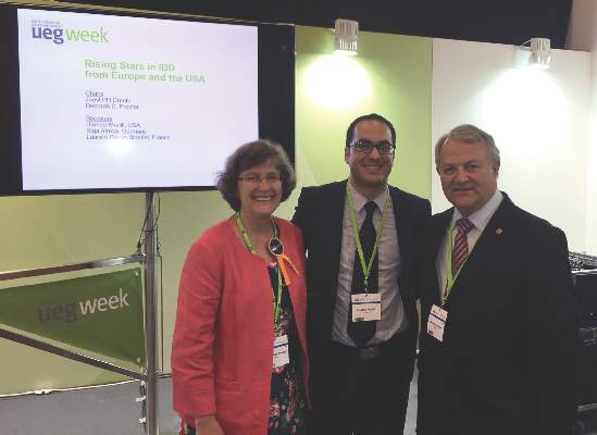
Leaders from AGA and United European Gastroenterology (UEG) gathered Tuesday, Oct. 27, 2015, in Barcelona, to celebrate the AGA Research Scholar/UEG Rising Star Scientific Exchange Program during UEG Week 2015 (http://live.ueg.eu/week). During the session, AGA honoree, Dr. Khalili, presented on oral contraceptive use in the etiopathogenesis of Crohn’s disease.
This recurring program fosters collaboration and goodwill with the international community and offers a unique opportunity for GI’s best and brightest to share research advances in a small group setting.
It is the ninth year of the program, which is held twice a year – once at Digestive Disease Week® (DDW) in May and once at UEG Week in October.

Lusutrombopag is effective for thrombocytopenia in liver disease
Lusutrombopag, an oral thrombopoietin receptor agonist, was found to reduce the need for platelet transfusion in patients with chronic liver disease with a planned invasive procedure, according to results in an abstract of a phase III trial that will be presented as a latebreaker at the annual meeting of the American Association for the Study of Liver Disease in San Francisco.
The novel therapy, which upregulates platelet production, was “efficacious and well tolerated,” producing a reduced risk of overall adverse events, including bleeding events, according to Dr. Namiki Izumi, Musashino Red Cross Hospital, Tokyo.

In this ongoing global phase III trial, called L-PLUS 2, 96 patients with chronic liver disease, a platelet count less than 50,000/microL, and a planned invasive procedure were randomized to receive a once-daily 3-mg tablet of lusutrombopag or a matching placebo for 7 days. The primary endpoint was the need for a preoperative platelet transfusion.
“The proportion of patients who required no preoperative platelet transfusion was significantly greater with lusutrombopag [29.2% vs. 12.5%; P less than .0001],” Dr. Izumi reported. The proportion of responders, defined by a platelet count greater than or equal to 50,000/microL and a greater than or equal to 20,000/microL-increase from baseline, was also significantly greater in the lusutrombopag arm (77.1% vs. 6.3%; P less than .0001).
In addition, the median time with a platelet count greater than or equal to 50,000/microL was 22.1 days in those who received lusutrombopag but no platelet transfusion versus 3.3 days in the placebo patients who did receive transfusion (P less than 0.0001).
Many adverse events occurred less frequently in the arm randomized to lusutrombopag. This included bleeding events (14.6% vs. 27.1%) and postoperative fever (39.6% vs. 56.3%). The rates of procedural hypertension (41.7% vs. 37.5%) and procedural pain (45.8% vs. 41.7%) were slightly greater in the group randomized to lusutrombopag, but elevations in liver enzymes, such as aspartate aminotransferase (22.9% vs. 31.3%) were somewhat lower.
“Protocol-required imaging revealed one thromboembolic event of the portal venous system in each study arm, neither of which was related to platelets,” according to Dr. Izumi, who reported that no patient discontinued therapy as a result of an adverse event.
Because of the frequency with which thrombocytopenia is observed in patients with chronic liver disease, platelet transfusion is considered a standard procedure when an invasive intervention is planned, according to Dr. Izumi. The data from this trial suggest that preoperative treatment with lusutrombopag may be an alternative.
Dr. Izumi reported financial relationships with Bayer, Daiichi Sankyo, Gilead, Merck, and Shionogi.
Lusutrombopag, an oral thrombopoietin receptor agonist, was found to reduce the need for platelet transfusion in patients with chronic liver disease with a planned invasive procedure, according to results in an abstract of a phase III trial that will be presented as a latebreaker at the annual meeting of the American Association for the Study of Liver Disease in San Francisco.
The novel therapy, which upregulates platelet production, was “efficacious and well tolerated,” producing a reduced risk of overall adverse events, including bleeding events, according to Dr. Namiki Izumi, Musashino Red Cross Hospital, Tokyo.
In this ongoing global phase III trial, called L-PLUS 2, 96 patients with chronic liver disease, a platelet count less than 50,000/microL, and a planned invasive procedure were randomized to receive a once-daily 3-mg tablet of lusutrombopag or a matching placebo for 7 days. The primary endpoint was the need for a preoperative platelet transfusion.
“The proportion of patients who required no preoperative platelet transfusion was significantly greater with lusutrombopag [29.2% vs. 12.5%; P less than .0001],” Dr. Izumi reported. The proportion of responders, defined by a platelet count greater than or equal to 50,000/microL and a greater than or equal to 20,000/microL-increase from baseline, was also significantly greater in the lusutrombopag arm (77.1% vs. 6.3%; P less than .0001).
In addition, the median time with a platelet count greater than or equal to 50,000/microL was 22.1 days in those who received lusutrombopag but no platelet transfusion versus 3.3 days in the placebo patients who did receive transfusion (P less than 0.0001).
Many adverse events occurred less frequently in the arm randomized to lusutrombopag. This included bleeding events (14.6% vs. 27.1%) and postoperative fever (39.6% vs. 56.3%). The rates of procedural hypertension (41.7% vs. 37.5%) and procedural pain (45.8% vs. 41.7%) were slightly greater in the group randomized to lusutrombopag, but elevations in liver enzymes, such as aspartate aminotransferase (22.9% vs. 31.3%) were somewhat lower.
“Protocol-required imaging revealed one thromboembolic event of the portal venous system in each study arm, neither of which was related to platelets,” according to Dr. Izumi, who reported that no patient discontinued therapy as a result of an adverse event.
Because of the frequency with which thrombocytopenia is observed in patients with chronic liver disease, platelet transfusion is considered a standard procedure when an invasive intervention is planned, according to Dr. Izumi. The data from this trial suggest that preoperative treatment with lusutrombopag may be an alternative.
Dr. Izumi reported financial relationships with Bayer, Daiichi Sankyo, Gilead, Merck, and Shionogi.
FROM THE LIVER MEETING 2015
Key clinical point: In a phase III trial, lusutrombopag was found to reduce the need for platelet transfusion in chronic liver disease patients requiring surgery.
Major finding: Prior to a planned invasive procedure, only 20.8% of lusutrombopag versus 87.5% of placebo patients (P less than .0001) required platelet transfusion.
Data source: A multicenter, double-blind phase III trial.
Disclosures: Dr. Izumi reported financial relationships with Bayer, Daiichi Sankyo, Gilead, Merck, and Shionogi.

Role of cyclins in malaria parasite development

Photo by James Gathany
Researchers say they have characterized the cyclin protein family in the rodent malaria parasite Plasmodium berghei.
The team found there are only 3 cyclins in this parasite, but one of these, the single P-type cyclin CYC3, plays a “vital role” in parasite development in the mosquito.
The researchers believe this work, published in PLoS Pathogens, could pave the way to a better understanding of malaria parasites and lead to potential new treatments.
“This first functional study of cyclin in the malaria parasite and its consequences in parasite development within pathogen-carrying mosquitoes will definitely further our understanding of parasite cell division, which I hope will lead to the elimination of this disease in the future,” said study author Magali Roques, PhD, of the University of Nottingham in the UK. ![]()