User login
SAA patients benefit from upfront eltrombopag combo
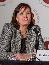
Photo courtesy of ASH
ORLANDO, FL—Investigators are pursuing an upfront approval for eltrombopag in combination with immunosuppressive therapy for the treatment of severe aplastic anemia (SAA).
Based on eltrombopag’s single-agent activity in refractory SAA, they hypothesized that its addition to standard immunosuppressive therapy of horse antithymocyte globulin (hATG) and cyclosporine (CsA) in the first-line setting could improve patient outcome.
And, in a phase 2 trial, it did.
“The addition of eltrombopag resulted in over 20% higher overall response rates and complete response rates for both 3 and 6 months,” said Danielle
Townsley, MD, who presented the data at the 2015 ASH Annual Meeting.
Dr Townsley, of the National Heart, Lung, and Blood Institute, National Institutes of Health, in Bethesda, Maryland, presented the findings as abstract LBA-2.*
The US Food and Drug Administration approved eltrombopag to treat refractory SAA in November 2014, and the European Commission approved it in 2015.
Investigators believed eltrombopag in the upfront, treatment-naïve setting could yield higher overall response rates (ORRs) than the 60% to 70% achieved with standard immunosuppressives worldwide.
“[It was] logical to consider treating patients early at the start of their disease,” Dr Townsley said.
So she and her colleagues conducted an investigator-initiated, phase 2, single-center trial of eltrombopag combined with immunosuppressive agents for first-line treatment of SAA.
Study design and patient population
Patients had to have confirmed treatment-naïve SAA, be a minimum of 2 years old, and weigh more than 12 kg. They were excluded if they had prior immunosuppressive therapy with ATG, alemtuzumab, or cyclophosphamide. They were also excluded if they had liver cirrhosis, AST/ALT more than 5 times normal, or Fanconi anemia.
Primary endpoints of the study were complete response (CR) at 6 months and toxicity. Secondary endpoints included ORR and partial response (PR) rate, survival, clonal evolution, and relapse.
Investigators defined CR as having an absolute neutrophil count (ANC) of 1000/μL or higher, a hemoglobin level of 10 g/dL or higher, and a platelet count of 100,000/μL or higher. They defined PR as blood counts no longer meeting criteria for SAA or CR.
All 92 patients received standard hATG (on days 1 to 4) and CsA (for 6 months). Patients in cohort 1 (n=30) also received eltrombopag at 150 mg daily, starting on day 14 for 6 months.
Patients in cohort 2 (n=31) received eltrombopag at 150 mg daily, starting on day 14 for 3 months. And the 31 patients in cohort 3 started 150 mg of daily eltrombopag simultaneously with the immunosuppressants and continued to receive the drug for 6 months.
Investigators assessed response at 3 and 6 months and planned to follow patients for at least 5 years.
Patients in all cohorts were a median of 32 years (range, 3–82), with 21% being younger than 18. About half were male, 66% had less than 1% of a paroxysmal nocturnal hemoglobinuria clone, 37% had a median neutrophil count less than 200/μL, a median reticulocyte count of 20,000/μL (range, 1600–60,400/μL), and a median platelet count of 9000/μL (range, 0–37,000/μL).
Results
At 3 months, the ORR for the entire population was 81%, and the CR rate was 28%. The ORR was 77% in cohorts 1 and 2 and 92% in cohort 3. The CR rate was 17%, 26%, and 44% in cohorts 1, 2, and 3, respectively.
At 6 months, the ORR for the entire population was 86%, and the CR rate was 37%. The ORR was 80%, 87%, and 95% in cohorts 1, 2, and 3, respectively. And the CR rate was 33%, 26%, and 60%, respectively.
Compared to historic rates for patients on hATG and CsA alone, “the addition of eltrombopag resulted in over 20% higher overall response rates and complete response rates for both 3 and 6 months,” Dr Townsley said.
“And for cohort 3, when eltrombopag is given on day 1, the rate of response in evaluable patients to date appears even higher, with 95% overall response rate at 6 months, of which 60% are complete.”
Dr Townsley also noted that, compared to historical experience, neutrophil recovery was more robust in responding patients treated with eltrombopag. Patients on eltrombopag had a mean ANC of 2253/μL, compared with an ANC of 1716/μL for the historic comparator.
“And likewise, more robust platelet recovery was observed with eltrombopag,” Dr Townsley said, with the eltrombopag-treated patients achieving a mean count of 115,262/μL, compared to a mean of 84,303/μL for the historic group.
She added that, among all eltrombopag-treated patients, the median time to neutrophil recovery was 29 days for an ANC greater than 200/μL and 47 days for an ANC greater than 500/μL. In cohort 3—in which eltrombopag was initiated on day 1—those endpoints were achieved in a median of 8 days and 38 days, respectively.
Patients became transfusion-independent for red cells in a median of 42 days and for platelets in a median of 32 days.
Eltrombopag-treated patients had a 99% overall survival at a median follow-up of 18 months (range 1 – 42) when censored for stem cell transplant. When not censored for transplant, their overall survival was 97%.
Adverse events
“The addition of eltrombopag to ATG and cyclosporine was, overall, well tolerated,” Dr Townsley said. “Few grade 3 to 4 events were attributed to eltrombopag.”
Severe cutaneous reactions in 2 patients caused eltrombopag to be stopped, and 10% of patients had grade 2–3 transaminase and bilirubin elevations.
Bone marrow biopsies revealed no increased fibrosis.
One patient with thymoma died while on study due to encephalopathy. And 2 deaths occurred after hematopoietic stem cell transplant, one with relapsed acute myeloid leukemia and the other from relapsed aplastic anemia.
Clonal evolution occurred in 7 patients, 2 who had achieved CR and evolved in 3 and 30 months. Neither patient had bone marrow dysplasia. One patient’s cytogenetics normalized, and the other had stable disease.
“In our protocol, we define any new cytogenetic abnormality as clonal evolution—we have always done this,” Dr Townsley said.
Of the other 5 patients who evolved, 1 achieved a CR and relapsed, 1 achieved a PR and relapsed, 2 achieved a PR, and 1 had no response. Three of these patients had stem cell transplants, 1 had stable disease, and 1 died of acute myeloid leukemia after stem cell transplant.
The investigators concluded that eltrombopag increases complete and overall hematologic response rates in treatment-naïve SAA patients. Immediate introduction of eltrombopag with immunosuppressant therapy may be optimal, and CR does not appear to prevent clonal evolution.
Investigators are currently in the process of conducting a long-term, serial genomic analysis. The study is open for accrual to an extension cohort.
Eltrombopag is marketed as Promacta in the US and Revolade in most countries outside the US.
Dr Townsley disclosed drug and research funding from GlaxoSmithKline and Novartis, developers of eltrombopag. ![]()
*Data in the abstract differ from the presentation.
Five-year data suggest ruxolitinib improves survival in MF

ASH Annual Meeting
Photo courtesy of ASH
ORLANDO, FL—Five-year results from the COMFORT-II trial appear to confirm that treatment with ruxolitinib can improve spleen size and survival in patients with myelofibrosis (MF).
“These results pave the way to use ruxolitinib earlier in the course of the disease,” said lead study author Claire Harrison, MD, a consultant hematologist at Guy’s and St. Thomas’ NHS Foundation Trust in London, UK.
Dr Harrison presented the results at the 2015 ASH Annual Meeting (abstract 59).
Ruxolitinib, a JAK1/JAK2 inhibitor, has demonstrated rapid, durable improvements in splenomegaly and MF symptoms, as well as improved survival in the phase 3 COMFORT-I and COMFORT-II studies.
In COMFORT-II, significantly more patients achieved the primary endpoint—a 35% or greater decrease in spleen volume from baseline at week 48—with ruxolitinib than with best available therapy (BAT).
The 3-year follow-up confirmed that spleen volume reductions were sustained, and ruxolitinib treatment remained tolerable with long-term use.
The randomized, open-label, multicenter study included 219 patients with primary MF, post-polycythemia vera MF, or post-essential thrombocythemia MF.
Two-thirds of patients received ruxolitinib twice daily, and one-third of patients received BAT, which was administered at doses and schedules determined by the investigator.
Almost two-thirds of the patients on the BAT arm crossed over to receive ruxolitinib upon protocol-defined progression following the primary analysis after week 48. All patients randomized to BAT have crossed over or discontinued, Dr Harrison said.
She presented the 5-year final study results, which showed that more than half of the patients (53.4%) experienced significant reductions in spleen size with ruxolitinib therapy and sustained this benefit over a median duration of 3.2 years.
“There was a 33% improvement in overall survival with ruxolitinib as compared to BAT,” she said.
Using a statistical model of survival if patients had not crossed-over to ruxolitinib, the survival benefit was 56% in favor of ruxolitinib.
“The plateau in spleen responses correlates well with the survival advantage,” Dr Harrison said.
She noted that the JAK allele burden was also reduced in the majority of patients who crossed over during the study. A recent bone marrow analysis shows a 20% improvement in fibrosis as well.
Nearly one-quarter of patients from both the ruxolitinib arm and those who crossed over from the BAT arm remained on treatment with ruxolitinib for 5 years.
All adverse events were consistent with previous analyses of treatment with ruxolitinib in MF, Dr Harrison said. The most common adverse events in ruxolitinib-treated patients were thrombocytopenia (52.4%), anemia (49.2%), diarrhea (35.6%), and peripheral edema (33%).
The most common grade 3/4 adverse events included anemia (22.5%), thrombocytopenia (15.2%), pneumonia (5.8%), general physical health deterioration (4.2%), and shortness of breath (4.2%).
“This long-term analysis after a vast number of patient-years shows the ongoing benefit, with no new safety signals and a strong survival message,” Dr Harrison said.
“Hematologists can be confident treating patients with ruxolitinib. It is safe, effective, and leads to significant long-term benefit. Myelofibrosis patients feel better, their spleens are smaller, and they may survive longer.”
COMFORT-II was sponsored by Novartis, which licensed ruxolitinib from Incyte Corporation for development and commercialization outside the US. COMFORT-I was sponsored by Incyte. ![]()
Regimen with intensified PEG-ASP feasible in young adults with ALL

Annual Meeting
Photo courtesy of ASH
ORLANDO, FL—Results of a DFCI ALL Consortium trial have shown that adults with acute lymphoblastic leukemia (ALL) can be successfully and safely treated with a pediatric regimen using intensified pegylated asparaginase (PEG-ASP).
Investigators recently reported that young adults treated with native E coli asparaginase as part of their regimen had improved 4-year disease-free survival and overall survival (OS) rates.
Now, the team has shown it is possible to use PEG-ASP to improve young adult outcomes as well.
The investigators also described the toxicities with PEG-ASP and compared them to the prior DFCI ALL Consortium trial with native E coli asparaginase.
“[Wendy] Stock, years ago, analyzed young adult patients 16 to 20 based on whether or not they were treated on Children’s Cancer Group trials or CALGB trials,” said Daniel J. DeAngelo, MD, PhD, of the Dana-Farber Cancer Institute in Boston, Massachusetts.
“And what she reported was that there was a dramatic improvement in the disease- and event-free survival . . . . And since that publication and presentation at ASH several years ago, there’ve been a large number of us who’ve tried to adapt pediatric trials or pediatric-inspired trials for the treatment of young adults with acute lymphoblastic leukemia.”
The PEG-ASP DFCI ALL trial is one such effort. Dr DeAngelo discussed the results of this trial at the 2015 ASH Annual Meeting (abstract 80).
Patient population
Investigators enrolled 110 patients on the trial.
Patients had to be between 18 and 50 years of age, with untreated ALL and no history of secondary ALL. Patients with Burkitt’s lymphoma were excluded.
The patients’ median age was 32 (range, 18–50), 62% were male, 80% were white, and 85% were non-Hispanic. Eleven percent had central nervous system (CNS) status 2 or 3 prior to the initiation of chemotherapy.
Most (87%) had a performance status of 0 or 1, 82% had the B-cell and 18% the T-cell phenotype, 19% were Ph-positive, 6% had an MLL translocation (11q23), and 18% had other translocations.
Study design
Induction chemotherapy consisted of doxorubicin, prednisone, vincristine, PEG-ASP, and intrathecal therapy.
The first consolidation consisted of high-dose methotrexate followed by a BFM-like intensification and high-dose cytarabine, etoposide, and dexamethasone.
CNS prophylaxis included intrathecal chemotherapy and cranial irradiation.
The second consolidation consisted of eight 3-week courses of doxorubicin, vincristine, dexamethasone, 6-mercaptopurine, and 30 weeks of PEG-ASP.
The PEG-ASP was initially dosed at the pediatric level of 2500 IU/m2 every 2 weeks.
“But due to some toxicity concerns in this treatment strategy—with specific emphasis on liver function abnormalities with hyperbilirubinemia, elevations of AST/ALT—we decided to amend the protocol and decrease the PEG dose from 2500 to 2000 and increase the interval from every 2 weeks to every 3 weeks,” Dr DeAngelo said.
Therefore, during this 30-week course, patients received 10 doses of PEG ASP as opposed to 15.
“We also went back and swapped out the PEG asparaginase during induction and reinserted native E coli to really ascertain comparative properties,” he said.
Maintenance therapy consisted of 3-week courses of vincristine, dexamethasone, methotrexate, and 6-mercaptopurine for 2 years from achievement of complete remission (CR).
During PEG-ASP therapy, patients received anticoagulation prophylaxis, preferably with low-molecular-weight heparin, as long as patients had a platelet count greater than 30,000/μL.
Results
Of the 110 patients enrolled, 65 received the higher dose of asparaginase, and 45 received the amended lower dose.
Ninety-one patients (89%) achieved a CR, 57 of whom received the higher dose of asparaginase and 34 the lower dose.
There were 2 induction deaths, both in the higher-dose group.
Twenty-one patients went on to transplant in CR1, 15 in the higher-dose group and 6 in the lower-dose group.
Twenty-three patients relapsed, 17 in the higher-dose group and 6 in the lower-dose group. Two of the relapses were CNS only.
Three patients died in remission, 2 in the higher-dose asparaginase group and 1 in the lower. And 3 patients died after stem cell transplant in CR, 2 in the higher-dose group and 1 in the lower.
At a median follow-up of 42.2 months, the 3-year disease-free survival for the entire cohort was 73%, and overall survival was 75%.
Subset analyses
The investigators performed subgroup analyses and came up with some “interesting observations,” Dr DeAngelo said.
Younger patients, ages 18 to 19 and 20 to 29, had a better OS than the older patients. The OS for younger patients is in the 80% to 85% range, “which is significantly better than the other patients in the 30 to 40 or 40 to 50 age groups,” Dr DeAngelo said.
Patients with T-cell ALL had a better OS than those with B-cell ALL and Ph-positive ALL, who had the worst OS of approximately 50%. The vast majority of Ph-negative patients were transplanted, and all received imatinib in addition to chemotherapy.
“[Patients with the T-cell phenotype] seemed to do particularly well on this strategy, which is something we showed in the last study as well, with an overall survival of 80%, compared to 70% for the B-cell Philadelphia-negative [patients],” Dr DeAngelo said.
He and his colleagues also found an association between OS and body mass index (BMI).
Obese or morbidly obese patients with a BMI of 30 or over had an OS of around 40%, while underweight or normal-weight patients had an OS of almost 90%, a “profound overall survival in the less-than-obese patients,” Dr DeAngelo said.
“And I think one of the things that is bringing down the curves is the obese,” he added, “which is a concern as the body mass index of the American population increases.”
Another discovery was that patients with minimal residual disease (MRD) of less than 10-4 had better OS than those with a high MRD level.
After a single dose of PEG ASP during induction on day 4, asparagine was depleted for a median of 3 weeks. With a single dose of E coli asparaginase, on the other hand, asparagine is depleted for about a week.
Asparaginase levels during consolidation were “extraordinarily elevated” with the 2500 dose level of PEG-ASP compared to the lower dose level. Toxicity was much more manageable, however, as the dose was reduced, Dr DeAngelo said. And asparagine was still depleted throughout the 30 weeks, as per protocol.
Toxicity
The higher asparaginase dose group “surprisingly, had a very low rate of clinical pancreatitis,” Dr DeAngelo said.
At the higher asparaginase dose—2500 IU/m2 every 2 weeks—66 patients experienced grade 3-5 adverse events: 30 (46%) febrile neutropenia, 1 (1%) pancreatitis, 19 (29%) AST, 34 (52%) ALT, 24 (36%) bilirubin, 19 (29%) thrombosis, 2 (3%) CNS hemorrhage, 3 (4%) hypersensitivity, and 4 (6%) osteonecrosis.
After the protocol amendment, “we saw a marked reduction in hyperbilirubinemia,” Dr DeAngelo said.
Grade 3–4 hyperbilirubinemia decreased from 36% to 7%, grade 3–4 ALT elevation decreased from 52% to 29%, and the rate of thrombosis decreased from 29% to 16%.
“Whether the latter decrease [thrombosis] was reflective of the decreased dose of asparaginase or the addition of anticoagulation, I can’t determine,” Dr DeAngelo said, “but it seemed to reflect other studies.”
The investigators concluded that a dose-intensified pediatric regimen in adults is feasible, with an acceptable toxicity profile.
The team said this approach may translate to better survival for adults with ALL, with the exception of older adults and patients with a high BMI. PEG-ASP has increased toxicity in these patients.
The investigators recommend addressing the challenges that remain—psychosocial issues, practice patterns, and biology—with a unified approach and more cooperative group trials in young adults. ![]()
Study reveals decrease in NIH-funded trials

Photo by Esther Dyson
A new study suggests that, in recent years, there has been a decrease in clinical trials funded by the National Institutes of Health (NIH) but an increase in trials with funding from other sources.
Researchers looked at trials newly registered on ClinicalTrials.gov and observed a substantial increase in trial listings from 2006 through 2014.
During that time period, the number of NIH-funded trials declined, but the number of trials funded by other US federal agencies, industry, and other groups (such as universities and organizations) increased.
Stephan Ehrhardt, MD, of Johns Hopkins Bloomberg School of Public Health in Baltimore, Maryland, and his colleagues conducted this study and recounted their findings in a letter to JAMA.
The researchers downloaded data from ClinicalTrials.gov, searched for “interventional study” and obtained counts of newly registered trials by funder type: “NIH,” “industry,” “other US federal agency,” or “all others (individuals, universities, organizations).”
According to the “first received” date (when trials were first registered with ClinicalTrials.gov), the number of newly registered trials increased from 9321 in 2006 to 18,400 in 2014 (97.4%).
During the same period, the number of industry-funded trials increased from 4585 to 6550 (42.9%), and the number of NIH-funded trials decreased from 1376 to 1048 (23.8%).
The number of trials funded by other US federal agencies increased from 263 to 339 (28.9%), and the number of trials funded by “all others” increased from 3240 to 10,597 (227.1%).
The researchers also examined the data according to the trial start date and observed similar patterns. They found the total number of trials increased from 9208 in 2006 to 14,618 in 2014 (58.8%).
The number of industry-funded trials increased from 4516 to 5274 (36.1%), and the number of NIH-funded trials decreased from 1189 to 873 (26.6%).
The number of trials funded by other US federal agencies increased from 229 to 292 (27.5%), and the number of trials funded by “all others” increased from 3397 to 8295 (144.2%).
Dr Ehrhardt said he believes the decline in NIH-funded studies can be traced to 2 things: flat NIH funding (the 2014 budget was 14% less than in 2006, after adjusting for inflation) and greater competition for these limited dollars from other, relatively new research areas such as genomic research or personalized medicine studies. ![]()
Press Ganey Executive Urges Physicians to Embrace Hospital Medicine Care Model
James Merlino, MD, president and CMO of Press Ganey's strategic consulting division, wants to convince physicians around the country that hospital medicine is good healthcare as a whole.
“[Hospitalists] are the holistic scorekeepers for a variety of medical conditions that a lot of physicians don’t understand and don’t treat very well,” says Dr. Merlino, whose company supports healthcare providers in improving the patient experience. “We know that when their model is allowed to foster, quality improves [and] safety improves. It’s a model that needs to be embraced so we can deliver better care for patients.”
Dr. Merlino recently talked with The Hospitalist:
Question: You say you’ve seen some specialists and primary care physicians disrespect hospitalists. Why do you believe that occurs?
Answer: It’s a relatively new model, and physicians who have patients in the hospital, nonhospitalists, don’t like to give up the autonomy and the control they feel they have or the responsibility they have to care for patients. The hospitalist model challenges that.
Q: How does healthcare develop a culture that prizes hospitalists and encourages teamwork?
A: Number one, people have to call it out and talk about it. What surprised me in one hospital I visited was [that] the hospitalists did not elevate the issue to leadership. The second thing that relates to changing physician culture is accountability of leadership. When medical staff leaders find out about this type of behavior, it must be addressed.
Q: Why does the challenge persist?
A: It’s leaders stepping up and holding people accountable for their actions. Leaders sometimes have a tendency to ignore behavior problems. When issues like lack of professionalism are identified, then medical leadership really needs to step in and deal with the individuals who are creating the problem. That is a gap in healthcare.
Q: What stops leaders from being accountable?
A: The problem is that physician leaders and other leaders tend to shy away from controversial problems. Pushing into a medical staff issue like this is complicated and difficult. Physicians are the engines of your organization. Leaders are working very hard to keep the medical staff in a steady state … and often there’s a reluctance to push into behavioral problems. TH
Visit our website for more information on multidisciplinary care.
Antidepressants may increase later onset of mania, bipolar
People diagnosed with unipolar depression have a higher chance of developing mania or bipolar disorder if they’ve previously been treated with antidepressants, a new study shows (BMJ Open. 2015 Dec 15. doi: 10.1136/bmjopen-2015-008341).
“Our findings demonstrate a significant association between antidepressant therapy in patients with unipolar depression and an increased incidence of mania,” Dr. Rashmi Patel of King’s College, London, and his associates reported in the study. Moreover, the association remains significant after adjusting for both age and gender, they wrote.

Dr. Patel and his associates conducted a retrospective cohort study on 21,012 individuals aged 16 to 65 years – all of whom were diagnosed with depression and had no previous diagnosis of mania or bipolar disorder between April 1, 2006, and March 31, 2013 – from the South London and Maudsley National Health Service Foundation Trust. Clinical data on subjects’ medical history, mental state examinations, diagnostic formulations, and management plans were collected. Subjects also were classified as having had “prior antidepressant therapy” if there was “documentation of antidepressant treatment prior to the date of diagnosis of depression.” Follow-ups occurred through March 31, 2014, and the primary outcome was a diagnosis of mania or bipolar disorder during that period.
Results showed an incidence rate of 10.9 per 1,000 person-years of mania or bipolar disorder across the entire study population. The lowest incidence, 8.3 per 1,000 person-years, was in the 56-65 years age cohort, while those in the 26-35 years age cohort had the highest incidence rate – 12.3 per 1,000 person-years (P = .004).
Subjects with prior antidepressant use saw significant increases in incidence rates of mania or bipolar disorder, depending on which antidepressant they were taking. Those on tricyclics (4.7% of subjects with previous antidepressant treatment) had a 13.1 per 1,000 person-years incidence rate, while those taking trazodone (0.8%) had a 19.1 per 1,000 person-years incidence rate (P = .09 and P = .03, respectively). The most commonly used antidepressants were selective serotonin reuptake inhibitors (35.5%), which yielded an incidence rate of 13.2 per 1,000 person-years.
“The association of antidepressant therapy with mania demonstrated in the present and previous studies highlights the importance of considering whether an individual who presents with depression could be at risk of future episodes of mania,” the authors concluded. They concluded that the findings reinforce the “ongoing need to develop better ways to predict future risk of mania in people with no prior history of bipolar disorder who present with an episode of depression.”
The study was supported by the U.K. Medical Research Council Clinical Research Training Fellowship. Neither Dr. Patel nor his associates reported relevant financial disclosures.
People diagnosed with unipolar depression have a higher chance of developing mania or bipolar disorder if they’ve previously been treated with antidepressants, a new study shows (BMJ Open. 2015 Dec 15. doi: 10.1136/bmjopen-2015-008341).
“Our findings demonstrate a significant association between antidepressant therapy in patients with unipolar depression and an increased incidence of mania,” Dr. Rashmi Patel of King’s College, London, and his associates reported in the study. Moreover, the association remains significant after adjusting for both age and gender, they wrote.
Dr. Patel and his associates conducted a retrospective cohort study on 21,012 individuals aged 16 to 65 years – all of whom were diagnosed with depression and had no previous diagnosis of mania or bipolar disorder between April 1, 2006, and March 31, 2013 – from the South London and Maudsley National Health Service Foundation Trust. Clinical data on subjects’ medical history, mental state examinations, diagnostic formulations, and management plans were collected. Subjects also were classified as having had “prior antidepressant therapy” if there was “documentation of antidepressant treatment prior to the date of diagnosis of depression.” Follow-ups occurred through March 31, 2014, and the primary outcome was a diagnosis of mania or bipolar disorder during that period.
Results showed an incidence rate of 10.9 per 1,000 person-years of mania or bipolar disorder across the entire study population. The lowest incidence, 8.3 per 1,000 person-years, was in the 56-65 years age cohort, while those in the 26-35 years age cohort had the highest incidence rate – 12.3 per 1,000 person-years (P = .004).
Subjects with prior antidepressant use saw significant increases in incidence rates of mania or bipolar disorder, depending on which antidepressant they were taking. Those on tricyclics (4.7% of subjects with previous antidepressant treatment) had a 13.1 per 1,000 person-years incidence rate, while those taking trazodone (0.8%) had a 19.1 per 1,000 person-years incidence rate (P = .09 and P = .03, respectively). The most commonly used antidepressants were selective serotonin reuptake inhibitors (35.5%), which yielded an incidence rate of 13.2 per 1,000 person-years.
“The association of antidepressant therapy with mania demonstrated in the present and previous studies highlights the importance of considering whether an individual who presents with depression could be at risk of future episodes of mania,” the authors concluded. They concluded that the findings reinforce the “ongoing need to develop better ways to predict future risk of mania in people with no prior history of bipolar disorder who present with an episode of depression.”
The study was supported by the U.K. Medical Research Council Clinical Research Training Fellowship. Neither Dr. Patel nor his associates reported relevant financial disclosures.
FROM BMJ OPEN
Key clinical point: Antidepressant use in patients can heighten the subsequent risk of developing mania or bipolar disorder.
Major finding: The overall incidence rate of mania/bipolar disorder was 10.9 per 1,000 person-years, but those numbers increased to 13.1-19.1 per 1,000 person-years when factoring in prior antidepressant treatment.
Data source: Retrospective cohort study of 21,012 adults with unipolar depression between April 1, 2006 and March 31, 2013.
Disclosures: The study was supported by the U.K. Medical Research Council Clinical Research Training Fellowship. Neither Dr. Patel nor his associates reported relevant financial disclosures.

The ‘easy’ but ‘not-so-easy’ brow lift
One of the most apparent and difficult-to-treat aspects of the aging eye is the descent of the eyebrow. The change in the orbital bone structure as well as fat loss and fat redistribution along the upper and lower eyelids and loss of skin elasticity contribute to the skeletonization of the periorbital area and a “drooping” of the eyebrow. Patients either have loss of volume across the entire brow, or primarily across the lateral and central brow creating what is known as an “a-frame” deformity.

Nonsurgical techniques that help lift the brow include a combination of relaxation of the orbicularis oculi muscle with neurotoxins, in addition to the injection of hyaluronic acid fillers along the brow margin and upper third of the face.
Small amounts of hyaluronic acid fillers injected with a 22- to 25-gauge cannula both above and below the eyebrow along the orbital rim provide an instantaneous lifting effect with long-lasting results. Hyaluronic acid and poly-L-lactic acid in dilute concentrations can also be injected with a cannula in the forehead, which creates a repletion of the volume in the upper face that is often lost with aging to create a lift of the eyebrows. Temple hollows can also be filled with calcium hydroxylapatite, poly-L-lactic acid and less often with hyaluronic acid to revolumize and create a lift of the lateral brow. Care should be taken as fillers used in these areas are off-label and need to be done by trained, expert injectors. The periorbital area is a danger zone with many vessels and nerves, and proper injection technique is crucial to avoid arterial blockage, nerve damage, and long-term complications.

Nonablative skin tightening with radiofrequency energy or ultrasound can be used for achieving a brow-lift. Although these techniques do provide collagen remodeling, multiple procedures are often necessary, and results are not always substantial. In a study of 36 patients undergoing ultrasound tightening of the face and neck, 86% showed a clinically significant brow-lift 90 days after treatment. The average brow elevation in this study was 1.7 mm.
In practice, however, patients are often more satisfied with the brow elevation they achieve with neurotoxins and fillers. Injectables provide a faster onset of results, fewer treatments, and minimal discomfort. Combination treatments provide the best overall results and although injectables in the periorbital area are technically difficult, patients are often very satisfied and return for repeat treatments.
References Aesthet Surg J. 2009;May-Jun;29(3):174-9.
J Am Acad Dermatol. 2010;Feb;62(2):262-9.
Dr. Wesley and Dr. Talakoub are cocontributors to this column. Dr. Talakoub is in private practice in McLean, Va. Dr. Wesley practices dermatology in Beverly Hills, Calif. This month’s column is by Dr. Talakoub.
One of the most apparent and difficult-to-treat aspects of the aging eye is the descent of the eyebrow. The change in the orbital bone structure as well as fat loss and fat redistribution along the upper and lower eyelids and loss of skin elasticity contribute to the skeletonization of the periorbital area and a “drooping” of the eyebrow. Patients either have loss of volume across the entire brow, or primarily across the lateral and central brow creating what is known as an “a-frame” deformity.
Nonsurgical techniques that help lift the brow include a combination of relaxation of the orbicularis oculi muscle with neurotoxins, in addition to the injection of hyaluronic acid fillers along the brow margin and upper third of the face.
Small amounts of hyaluronic acid fillers injected with a 22- to 25-gauge cannula both above and below the eyebrow along the orbital rim provide an instantaneous lifting effect with long-lasting results. Hyaluronic acid and poly-L-lactic acid in dilute concentrations can also be injected with a cannula in the forehead, which creates a repletion of the volume in the upper face that is often lost with aging to create a lift of the eyebrows. Temple hollows can also be filled with calcium hydroxylapatite, poly-L-lactic acid and less often with hyaluronic acid to revolumize and create a lift of the lateral brow. Care should be taken as fillers used in these areas are off-label and need to be done by trained, expert injectors. The periorbital area is a danger zone with many vessels and nerves, and proper injection technique is crucial to avoid arterial blockage, nerve damage, and long-term complications.
Nonablative skin tightening with radiofrequency energy or ultrasound can be used for achieving a brow-lift. Although these techniques do provide collagen remodeling, multiple procedures are often necessary, and results are not always substantial. In a study of 36 patients undergoing ultrasound tightening of the face and neck, 86% showed a clinically significant brow-lift 90 days after treatment. The average brow elevation in this study was 1.7 mm.
In practice, however, patients are often more satisfied with the brow elevation they achieve with neurotoxins and fillers. Injectables provide a faster onset of results, fewer treatments, and minimal discomfort. Combination treatments provide the best overall results and although injectables in the periorbital area are technically difficult, patients are often very satisfied and return for repeat treatments.
References Aesthet Surg J. 2009;May-Jun;29(3):174-9.
J Am Acad Dermatol. 2010;Feb;62(2):262-9.
Dr. Wesley and Dr. Talakoub are cocontributors to this column. Dr. Talakoub is in private practice in McLean, Va. Dr. Wesley practices dermatology in Beverly Hills, Calif. This month’s column is by Dr. Talakoub.

In hemodialysis, HDL cholesterol levels steady in men, fall in women
SAN DIEGO – Among incident hemodialysis patients, high-density lipoprotein cholesterol levels remained stable over time in men but mildly decreased over time in women, regardless of age.
In addition, larger deviations in HDL cholesterol in the first 6 months of incident hemodialysis were associated with a higher risk of 5-year all-cause mortality.
Those are key findings from a large analysis of patients who received hemodialysis care over a 4-year period, which was presented at the meeting sponsored by the American Society of Nephrology.
“Elevated high-density lipoprotein cholesterol levels, although protective in the general population, can be associated with higher mortality in hemodialysis patients,” Dr. Kamyar Kalantar-Zadeh and his colleagues wrote in an abstract of the study. “Association of HDL change over time with mortality has yet to be examined.”
Dr. Kalantar-Zadeh of the division of nephrology and hypertension at the University of California, Irvine, and his associates examined changes in HDL cholesterol levels over time in 24,400 incident hemodialysis patients who received care from facilities affiliated with DaVita HealthCare Partners between 2007 and 2011.
They examined the association of delta HDL (HDL cholesterol change between the first and second 91-day interval from dialysis start) and HDL cholesterol trajectory with all-cause mortality, using mixed effect and Cox regression models and adjusted for demographics, comorbidities, and baseline HDL cholesterol. Delta HDL was treated both as a continuous variable using restricted cubic splines and in five categories ranging from less than –6 mg/dL to 6 mg/dL or greater.
The mean age of patients was 65 years, 44% were female, 31% were black, and 66% had diabetes.
Mean baseline HDL cholesterol was 40.5 mg/dL, with an HDL cholesterol change of 1.7 mg/dL. The researchers found that while male patients had no significant change in HDL cholesterol over time, females had a significant decrease in HDL cholesterol (a mean of –0.6mg/dL per year). At the same time, a 6 mg/dL or greater increase or decrease in delta HDL was associated with a 7% and 37% increase in all-cause mortality, respectively, compared with the reference group. Delta HDL mortality associations did not differ across gender.
“Patients who have a greater than 6 mg/dL change in HDL between the first and second patient [tests] have a higher risk of 5-year all-cause mortality,” the researchers concluded. “Change in HDL exhibits a reverse J-shaped association with mortality in hemodialysis patients. Further studies are needed to examine the underlying causes of these HDL changes and pathophysiology leading to higher risk of all-cause mortality.”
The study was supported by funding from the National Institute of Diabetes and Digestive and Kidney Diseases. The researchers reported having no financial disclosures.
AT KIDNEY WEEK 2015
Key clinical point: Female incident hemodialysis patients have a mild decrease in HDL cholesterol levels over time, which is consistent across all age groups.
Major finding: While male patients had no significant change in HDL cholesterol over a 4-year time period, females had a significant decrease in HDL cholesterol (a mean of –0.6mg/dL per year).
Data source: The study population was 24,400 incident hemodialysis patients who received care from facilities affiliated with DaVita HealthCare Partners between 2007 and 2011.
Disclosures: The study was supported by funding from the National Institute of Diabetes and Digestive and Kidney Diseases. The researchers reported having no financial disclosures.
Six-year Norwood-RVPA results in matched patients outperformed BT-shunt
Concerns with delayed right ventricle dysfunction have offset the early survival advantages after Norwood procedure with right ventricle to pulmonary artery conduit (NW-RVPA) over the Norwood with Blalock-Taussig shunt (NW-BT) in newborns with left ventricular outflow tract obstruction, but a recent report provides evidence that RV function between the two procedures is comparable for up to six years.
Reporting in the January issue of the Journal of Thoracic and Cardiovascular Surgery (J Thorac Cardiovasc Surg. 2015 Dec;150:1440–52), investigators for the Congenital Heart Surgeons’ Society (CHSS) trial found that NW-RVPA has better overall six-year survival and superior right ventricle function in the short term after surgery than NW-BT. The study involved 454 newborns with critical left ventricular outflow tract obstruction (LVOTO) in the CHSS database who had Norwood stage-1 from 2005 to 2014. Propensity matching paired 169 NW-RVPA patients with the same number of NW-BT patients for comparison. CHSS along with the Hospital for Sick Children in Toronto provided funding for the study.

“For neonates with critical LVOTO and similar baseline characteristics undergoing a Norwood stage-1 operation, the six-year overall survival and transplant-free survival were significantly better after NW-RVPA vs. NW-BT,” said Dr. Travis J. Wilder and his colleagues from the Hospital for Sick Children in Toronto.
Key questions the study sought to answer involved the clinical implications of the small variations in RV function between the two procedures, as well as the association between Norwood procedures and tricuspid valve regurgitation (TR) and overall survival.
Overall six-year survival was 70% for the NW-RVPA group vs. 55% for the NW-BT group. Right ventricle dysfunction rates three months after the procedure were lower for the NW-RVPA group, 6% vs. 16%, but rates of late RV dysfunction were less than 5% for both groups. Likewise, rates of moderate or greater TR at two years were lower in the NW-RVPA group: 11% vs. 16%.
Rates of Fontan operation after six years were higher among the NW-RVPA group (54% vs. 49%), as were transplantation rates (6% vs. 2%). Overall, 2% converted to a biventricular repair, but only after NW-RVPA; and seven patients who had NW-RVPA underwent a tricuspid valve repair, compared with four in the NW-BT group.
“For all survivors not undergoing transplantation or biventricular repair, the prevalence of late moderate or greater RV dysfunction and TR were similar between NW-BT and NW-RVPA at six years, without evidence of increased RV dysfunction for patients who underwent NW-RVPA,” Dr. Wilder and his colleagues said.
Consistent with previous studies, the CHSS study showed an early risk of death after a Norwood stage-1 operation, which may be due to a greater prevalence of significant RV dysfunction as the operation transitions from stage 1 to stage 2, Dr. Wilder and his coauthors said. “Although causation between these two time-related events cannot directly be made, it suggests that poor RV function contributes to early hazard for death,” they said.
The authors acknowledge a number of limitations with their study: the variation in the quality of echocardiogram reports from the multiple institutions involved, the inability of propensity matching to account for unmeasured factors and the influence of center and surgeon volume among participating sites. They also said that the ventriculotomy the NW-RVPA involves can lead to late aneurysm, arrhythmias, and ventricular failure. The adverse effects of ventriculotomy on long-term RV function “may not become apparent for years,” Dr. Wilder and his coauthors said.
Dr. Wilder presented a report of the original results at the 2015 American Association for Thoracic Surgery Annual meeting.
The authors had no relationships to disclose.
“It is time to have the courage to confess that we need a more overarching quality improvement strategy,” Dr. James S. Tweddell of the Children’s Hospital of Wisconsin said of the results of the Congenital Heart Surgeons’ Society (CHSS) study in his invited commentary (J Thorac Cardiovasc Surg. 2015 Dec;150:1453–4).
Dr. Tweddell said the findings of the CHSS study and the earlier Single Ventricle Reconstruction (SVR) trial (N Engl J Med. 2010;362:1980-1992) are similar in terms of transplant-free survival in newborns. And while the dates of the studies’ enrollments overlap – 2005-2008 for SVR and 2005-2014 for CHSS – the more recent findings of the CHSS study would imply an advantage in terms of survival and right ventricle function. Nonetheless, the survival rates are similar, he said. “Only about 60% of patients remain alive.”

|
Dr. James S. Tweddell |
Dr. Tweddell pointed out the CHSS study is not a randomized, controlled trial, “and the shortcomings of the prospective observational study are well known.”
In calling for a “more overarching” quality improvement measure, Dr. Tweddell said that many programs use Norwood performance as a benchmark for outcomes. He proposed collaboration among high and low performing centers, imitating the adult cardiology model. He also suggested consolidation of programs performing the Norwood procedure to eliminate low-volume centers and develop centers of excellence. “The outcome of the Norwood procedure is dependent on both program and surgeon volume,” Dr. Tweddell said.
The CHSS study “is important and identifies a potentially durable benefit to the NW-RVPA,” Dr. Tweddell said, “but perhaps now is the time to focus on strategies between programs rather than solely within programs.”
“It is time to have the courage to confess that we need a more overarching quality improvement strategy,” Dr. James S. Tweddell of the Children’s Hospital of Wisconsin said of the results of the Congenital Heart Surgeons’ Society (CHSS) study in his invited commentary (J Thorac Cardiovasc Surg. 2015 Dec;150:1453–4).
Dr. Tweddell said the findings of the CHSS study and the earlier Single Ventricle Reconstruction (SVR) trial (N Engl J Med. 2010;362:1980-1992) are similar in terms of transplant-free survival in newborns. And while the dates of the studies’ enrollments overlap – 2005-2008 for SVR and 2005-2014 for CHSS – the more recent findings of the CHSS study would imply an advantage in terms of survival and right ventricle function. Nonetheless, the survival rates are similar, he said. “Only about 60% of patients remain alive.”
|
|
Dr. James S. Tweddell |
Dr. Tweddell pointed out the CHSS study is not a randomized, controlled trial, “and the shortcomings of the prospective observational study are well known.”
In calling for a “more overarching” quality improvement measure, Dr. Tweddell said that many programs use Norwood performance as a benchmark for outcomes. He proposed collaboration among high and low performing centers, imitating the adult cardiology model. He also suggested consolidation of programs performing the Norwood procedure to eliminate low-volume centers and develop centers of excellence. “The outcome of the Norwood procedure is dependent on both program and surgeon volume,” Dr. Tweddell said.
The CHSS study “is important and identifies a potentially durable benefit to the NW-RVPA,” Dr. Tweddell said, “but perhaps now is the time to focus on strategies between programs rather than solely within programs.”
Concerns with delayed right ventricle dysfunction have offset the early survival advantages after Norwood procedure with right ventricle to pulmonary artery conduit (NW-RVPA) over the Norwood with Blalock-Taussig shunt (NW-BT) in newborns with left ventricular outflow tract obstruction, but a recent report provides evidence that RV function between the two procedures is comparable for up to six years.
Reporting in the January issue of the Journal of Thoracic and Cardiovascular Surgery (J Thorac Cardiovasc Surg. 2015 Dec;150:1440–52), investigators for the Congenital Heart Surgeons’ Society (CHSS) trial found that NW-RVPA has better overall six-year survival and superior right ventricle function in the short term after surgery than NW-BT. The study involved 454 newborns with critical left ventricular outflow tract obstruction (LVOTO) in the CHSS database who had Norwood stage-1 from 2005 to 2014. Propensity matching paired 169 NW-RVPA patients with the same number of NW-BT patients for comparison. CHSS along with the Hospital for Sick Children in Toronto provided funding for the study.
“For neonates with critical LVOTO and similar baseline characteristics undergoing a Norwood stage-1 operation, the six-year overall survival and transplant-free survival were significantly better after NW-RVPA vs. NW-BT,” said Dr. Travis J. Wilder and his colleagues from the Hospital for Sick Children in Toronto.
Key questions the study sought to answer involved the clinical implications of the small variations in RV function between the two procedures, as well as the association between Norwood procedures and tricuspid valve regurgitation (TR) and overall survival.
Overall six-year survival was 70% for the NW-RVPA group vs. 55% for the NW-BT group. Right ventricle dysfunction rates three months after the procedure were lower for the NW-RVPA group, 6% vs. 16%, but rates of late RV dysfunction were less than 5% for both groups. Likewise, rates of moderate or greater TR at two years were lower in the NW-RVPA group: 11% vs. 16%.
Rates of Fontan operation after six years were higher among the NW-RVPA group (54% vs. 49%), as were transplantation rates (6% vs. 2%). Overall, 2% converted to a biventricular repair, but only after NW-RVPA; and seven patients who had NW-RVPA underwent a tricuspid valve repair, compared with four in the NW-BT group.
“For all survivors not undergoing transplantation or biventricular repair, the prevalence of late moderate or greater RV dysfunction and TR were similar between NW-BT and NW-RVPA at six years, without evidence of increased RV dysfunction for patients who underwent NW-RVPA,” Dr. Wilder and his colleagues said.
Consistent with previous studies, the CHSS study showed an early risk of death after a Norwood stage-1 operation, which may be due to a greater prevalence of significant RV dysfunction as the operation transitions from stage 1 to stage 2, Dr. Wilder and his coauthors said. “Although causation between these two time-related events cannot directly be made, it suggests that poor RV function contributes to early hazard for death,” they said.
The authors acknowledge a number of limitations with their study: the variation in the quality of echocardiogram reports from the multiple institutions involved, the inability of propensity matching to account for unmeasured factors and the influence of center and surgeon volume among participating sites. They also said that the ventriculotomy the NW-RVPA involves can lead to late aneurysm, arrhythmias, and ventricular failure. The adverse effects of ventriculotomy on long-term RV function “may not become apparent for years,” Dr. Wilder and his coauthors said.
Dr. Wilder presented a report of the original results at the 2015 American Association for Thoracic Surgery Annual meeting.
The authors had no relationships to disclose.
FROM THE JOURNAL OF THORACIC AND CARDIOTHORACIC SURGERY
Key clinical point: For newborns with critical left ventricular outflow tract obstruction (LVOTO) undergoing Norwood stage-1 procedures, Norwood with right ventricle to pulmonary artery conduit (NW-RVPA) resulted in better survival and less short-term right-ventricle dysfunction than matched patients who had the Norwood procedure with Blalock-Taussig shunt (NW-BT).
Major finding: Overall six-year survival was 70% for the NW-RVPA group vs. 55% for the NW-BT group, and RV dysfunction rates three months after the procedure were 6% for NW-RVPA vs. 16% for NW-BT.
Data source: Prospective group of 454 newborns with LVOTO in the Congenital Heart Surgeons’ Society database.
Disclosures: The Congenital Heart Surgeons’ Society and Hospital for Sick Children, Toronto, provided funding for the study. The authors had no relationships to disclose.

Treat high LDL cholesterol in CKD, even with inflammation
SAN DIEGO – Among patients with chronic kidney disease, LDL cholesterol level was positively associated with risk of atherosclerotic vascular events, regardless of baseline inflammation, according to a large, randomized study.
In addition, lowering LDL cholesterol with ezetimibe/simvastatin was similarly effective in the presence or absence of inflammation.
“There has been substantial interest in the relationship between LDL cholesterol and outcomes in the general population, and in particular among people with kidney disease,” study author Dr. Richard Haynes of the Nuffield Department of Population Health at the University of Oxford (England) said in an interview in advance of the meeting.
“Previous studies have found a paradoxical increased risk of death at low cholesterol levels, which may be due to another disease process – such as inflammation – both lowering cholesterol and increasing the risk of death, thereby creating a false association,” he explained.
Dr. Haynes and his associates set out to determine the relevance of LDL cholesterol and C-reactive protein (CRP) to cardiovascular risk among 9,270 patients with chronic kidney disease who were enrolled in the Study of Heart and Renal Protection (SHARP), in which they received either ezetimibe/simvastatin or placebo.
The researchers used Cox regression to analyze the hazard ratio for all atherosclerotic vascular events over a period of 4.9 years. The effect of ezetimibe/simvastatin on major atherosclerotic events was estimated in the presence and absence of inflammation, which was defined as a CRP of 3 mg/L. The mean age of SHARP participants was 62 years, and 63% were men.
Dr. Ben Storey, a colleague of Dr. Haynes at Oxford, reported the study results at the meeting sponsored by the American Society of Nephrology.
Among all patients, usual LDL was positively associated with the risk of atherosclerotic vascular events (hazard ratio, 1.34). Compared with patients who had low CRP, those with high CRP were at higher risk, but the relationship between LDL-C and atherosclerotic vascular event risk was similar in both groups (HR, 1.32 and 1.41, respectively).
Ezetimibe/simvastatin was similarly effective at reducing major atherosclerotic events in patients with low and high CRP (risk ratio, 0.84 and 0.83, respectively). Sensitivity analyses yielded similar results.
“The observational data from SHARP show that LDL cholesterol is positively associated with the risk of atherosclerotic vascular events, irrespective of baseline inflammation,” Dr. Storey concluded. “The randomized evidence showed that lowering LDL cholesterol was similarly effective in the presence or absence of inflammation, but CRP is associated with vascular risk in [chronic kidney disease].”
The findings’ clinical implications are that inflammation, “while it is relevant to patients’ outcomes, does not modify the beneficial effects of lowering LDL cholesterol,” according to Dr. Haynes, “So, clinicians should not be put off from prescribing statin-based, cholesterol-lowering therapy by the presence – or absence – of inflammation.”
Merck funded SHARP, but the University of Oxford conducted and analyzed the study independently. The Australian National Health and Medical Research Council, the British Heart Foundation, Cancer Research UK, and the UK Medical Research Council provided additional support.
AT KIDNEY WEEK 2015
Key clinical point: LDL cholesterol level is positively associated with the risk of atherosclerotic vascular events in patients with chronic kidney disease, regardless of baseline inflammation.
Major finding: Among all patients, usual LDL cholesterol was positively associated with the risk of atherosclerotic vascular events (HR, 1.34).
Data source: Investigators attempted to determine the relevance of LDL cholesterol and C-reactive protein to cardiovascular risk among 9,270 patients with chronic kidney disease who were enrolled in SHARP.
Disclosures: Merck funded SHARP, but the University of Oxford conducted and analyzed the study independently. The Australian National Health and Medical Research Council, the British Heart Foundation, Cancer Research UK, and the UK Medical Research Council provided additional support.
