User login
Hand-foot syndrome less with S-1 than capecitabine in mCRC
AMSTERDAM – For patients with metastatic colorectal cancer, S-1 was comparable in efficacy to capecitabine (Xeloda), and was associated with a lower incidence of all grades of hand-foot syndrome, reported investigators.
Among 161 patients with untreated metastatic colorectal cancer, the investigator-assessed incidence of all grades of hand-foot syndrome was 73% for patients randomly assigned to capecitabine, compared with 45% for patients randomized to S-1 (P = .0005).
Patient-assessed symptoms also were lower with S-1 than with capecitabine, reported Robert Jan Kwakman, MD, of the Academic Medical Center in Amsterdam.

“We conclude that treatment with S-1 is a useful alternative to capecitabine in the treatment of metastatic colorectal cancer,” he said at an annual congress sponsored by the European Cancer Organisation.
S-1 is an oral fluoropyrimidine consisting of tegafur, a prodrug of 5-fluorauracil (5-FU), combined with two 5-FU biochemical modulators. It is associated with a lower incidence of hand-foot syndrome than capecitabine, and has shown efficacy comparable to that of other fluoropyrimidines in Asian patients with gastrointestinal cancers, Dr. Kwakman noted.
Hand-foot syndrome can vary in severity from grade 1, marked by minimal skin changes or dermatitis without pain, to grade 3, characterized by severe skin changes with pain and significant limits to self care during activities of daily living.
In the randomized phase III SALTO trial (S1 Versus Capecitabine in the First Line Treatment of Metastatic Colorectal Cancer Patients), investigators in the Dutch Colorectal Cancer Group enrolled patients with untreated metastatic colorectal cancer with World Health Organization performance status 0-2 who were scheduled for treatment with fluoropyrimidine monotherapy. The patients were assigned to receive either capecitabine 1,250 mg/m2 for patients younger than 70, or 1,000 mg/m2 for those 70 and older, twice a day for days 1-14 of a 3-week cycle, or to S-1 30 mg/m2 twice daily on the same schedule.
In each arm, investigators could, at their discretion, also prescribe bevacizumab 7.5 mg/kg on day 1. In each arm, 59% of patients were scheduled to receive bevacizumab.
Patients were stratified by bevacizumab status, lactate dehydrogenase levels (normal vs. abnormal), performance status (0-1 vs. 2) and institution.
Patients were asked to keep diaries and record whether during the past 3 weeks they had experienced symptoms in their hands and/or feet such as tingling/numbness, pain, redness, swelling, and desquamation, and if so, whether the symptoms interfered with daily activities.
After a median follow-up of 16.1 months, investigators assessed hand-foot syndrome rates by grade were as follows:
• Grade 1: 21% for the capecitabine arm vs. 28% for the S-1 arm (not significant).
• Grade 2: 30% vs. 14% (P = .02).
• Grade 3: 21% vs. 4% (P = .003).
The incidence of patient-assessed hand-foot syndrome of all grades was 84% in the capecitabine arm and 58% in the S-1 arm (P = .004). Patient-reported grade 3 hand-foot syndrome occurred in 18% vs. 5%, respectively (P = .05).
The only other toxicity occurring more frequently among patients on S-1 was anorexia, which occurred in 29% of patients on capecitabine compared with 41% with S-1. Grade 3 or greater anorexia occurred in 3% vs. 13%, respectively (P = .03).
Significantly more dose reductions were required for patients on capecitabine (69% vs. 41%, P = .0008). The median relative dose intensity was higher for patients on S-1 (89% vs. 95%, P = .035).
Among all patients, median progression-free survival was 8.18 months with capecitabine vs. 8.39 months with S-1, a difference that was not significant. There was a trend toward better progression-free survival for patients in each arm who received bevacizumab (8.74 vs. 6.37 months without bevacizumab), but this difference was also not significant.
Overall survival rates at 12 months were 67% with capecitabine and 62% with S-1 (not significantly different), and respective rates at 18 months were 50% vs. 39%. The hazard ratio for mortality with S-1 was 1.28 (not significant).
The study was sponsored by the Dutch Colorectal Cancer Group, with research funds supplied by Nordic Pharma BV. Dr. Kwakman disclosed receiving an honorarium from the company.
AMSTERDAM – For patients with metastatic colorectal cancer, S-1 was comparable in efficacy to capecitabine (Xeloda), and was associated with a lower incidence of all grades of hand-foot syndrome, reported investigators.
Among 161 patients with untreated metastatic colorectal cancer, the investigator-assessed incidence of all grades of hand-foot syndrome was 73% for patients randomly assigned to capecitabine, compared with 45% for patients randomized to S-1 (P = .0005).
Patient-assessed symptoms also were lower with S-1 than with capecitabine, reported Robert Jan Kwakman, MD, of the Academic Medical Center in Amsterdam.
“We conclude that treatment with S-1 is a useful alternative to capecitabine in the treatment of metastatic colorectal cancer,” he said at an annual congress sponsored by the European Cancer Organisation.
S-1 is an oral fluoropyrimidine consisting of tegafur, a prodrug of 5-fluorauracil (5-FU), combined with two 5-FU biochemical modulators. It is associated with a lower incidence of hand-foot syndrome than capecitabine, and has shown efficacy comparable to that of other fluoropyrimidines in Asian patients with gastrointestinal cancers, Dr. Kwakman noted.
Hand-foot syndrome can vary in severity from grade 1, marked by minimal skin changes or dermatitis without pain, to grade 3, characterized by severe skin changes with pain and significant limits to self care during activities of daily living.
In the randomized phase III SALTO trial (S1 Versus Capecitabine in the First Line Treatment of Metastatic Colorectal Cancer Patients), investigators in the Dutch Colorectal Cancer Group enrolled patients with untreated metastatic colorectal cancer with World Health Organization performance status 0-2 who were scheduled for treatment with fluoropyrimidine monotherapy. The patients were assigned to receive either capecitabine 1,250 mg/m2 for patients younger than 70, or 1,000 mg/m2 for those 70 and older, twice a day for days 1-14 of a 3-week cycle, or to S-1 30 mg/m2 twice daily on the same schedule.
In each arm, investigators could, at their discretion, also prescribe bevacizumab 7.5 mg/kg on day 1. In each arm, 59% of patients were scheduled to receive bevacizumab.
Patients were stratified by bevacizumab status, lactate dehydrogenase levels (normal vs. abnormal), performance status (0-1 vs. 2) and institution.
Patients were asked to keep diaries and record whether during the past 3 weeks they had experienced symptoms in their hands and/or feet such as tingling/numbness, pain, redness, swelling, and desquamation, and if so, whether the symptoms interfered with daily activities.
After a median follow-up of 16.1 months, investigators assessed hand-foot syndrome rates by grade were as follows:
• Grade 1: 21% for the capecitabine arm vs. 28% for the S-1 arm (not significant).
• Grade 2: 30% vs. 14% (P = .02).
• Grade 3: 21% vs. 4% (P = .003).
The incidence of patient-assessed hand-foot syndrome of all grades was 84% in the capecitabine arm and 58% in the S-1 arm (P = .004). Patient-reported grade 3 hand-foot syndrome occurred in 18% vs. 5%, respectively (P = .05).
The only other toxicity occurring more frequently among patients on S-1 was anorexia, which occurred in 29% of patients on capecitabine compared with 41% with S-1. Grade 3 or greater anorexia occurred in 3% vs. 13%, respectively (P = .03).
Significantly more dose reductions were required for patients on capecitabine (69% vs. 41%, P = .0008). The median relative dose intensity was higher for patients on S-1 (89% vs. 95%, P = .035).
Among all patients, median progression-free survival was 8.18 months with capecitabine vs. 8.39 months with S-1, a difference that was not significant. There was a trend toward better progression-free survival for patients in each arm who received bevacizumab (8.74 vs. 6.37 months without bevacizumab), but this difference was also not significant.
Overall survival rates at 12 months were 67% with capecitabine and 62% with S-1 (not significantly different), and respective rates at 18 months were 50% vs. 39%. The hazard ratio for mortality with S-1 was 1.28 (not significant).
The study was sponsored by the Dutch Colorectal Cancer Group, with research funds supplied by Nordic Pharma BV. Dr. Kwakman disclosed receiving an honorarium from the company.
AT ECCO2017
Key clinical point: S-1 was associated with a lower incidence of hand-foot syndrome than was capecitabine in patients with metastatic colorectal cancer.
Major finding: Hand-foot syndrome of any grade occurred in 73% of patients on capecitabine vs. 45% on S-1 (P = .0005).
Data source: Randomized phase III trial of 161 patients with previously untreated metastatic colorectal cancer.
Disclosures: The study was sponsored by the Dutch Colorectal Cancer Group, with research funds supplied by Nordic Pharma BV. Dr. Kwakman disclosed receiving an honorarium from the company.
Postpartum Recovery Trends in Women with Hypertensive Disorders of Pregnancy
From the Department of Obstetrics and Gynecology, Kasturba Medical College, Manipal, Karnataka, India.
Abstracts
- Objective: To examine the association of the patient’s obstetric profile and time to normalization of blood pressure in the postnatal period among women with hypertensive disorders in pregnancy.
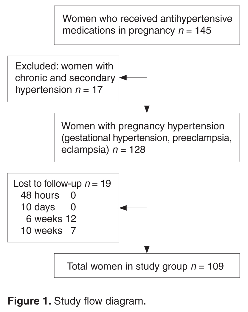
- Methods: We conducted a prospective cohort study at a tertiary level hospital between November 2014 and May 2015. Women with pregnancy hypertension who required antihypertensive treatment were recruited after delivery. The normalization trends in blood pressure were tested for associations with patient demographic data and details of pregnancy hypertension.
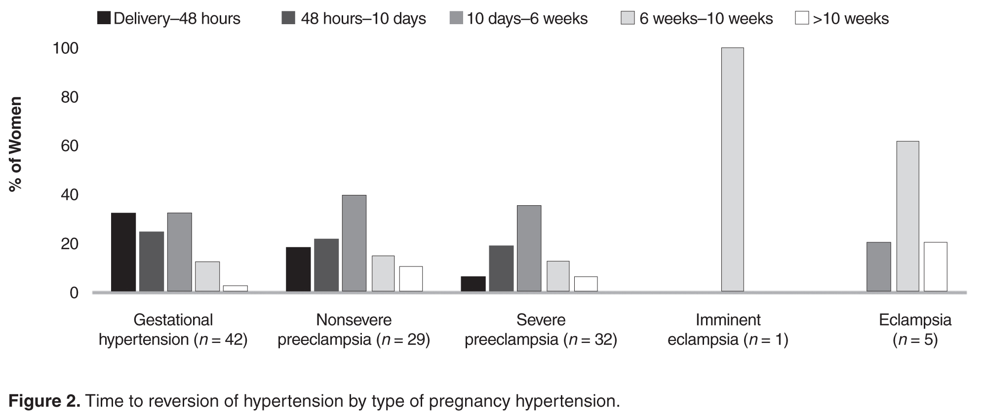
- Results: Among 109 women included in the study, earlier gestational age at onset of hypertension and earlier gestational age at delivery was correlated with slower resolution of hypertension. Time to resolution also was correlated with age, BMI, severity of hypertension, associated complications, and the number of antihypertensive medications received. There was no correlation with highest recorded systolic or diastolic blood pressures. Only 15% of women with gestational hypertension had persistent hypertension beyond 6 weeks. In the groups with nonsevere preeclampsia, severe preeclampsia, and eclampsia, blood pressure remained high after 6 weeks in 26%, 14%, and 50% of women, respectively.
- Conclusion: Women with advanced age, higher body mass index, early gestational age at the onset of hypertension, severe hypertension and who had complications of hypertension require prolonged monitoring and treatment when indicated for hypertension in postnatal period.
Key words: intensive care unit; communication; family meeting; critical illness; decision making; end of life care.
Hypertension is the most common medical problem encountered during pregnancy, complicating up to 10% of pregnancies worldwide [1]. The disorders of hypertension in pregnancy are generally classified as chronic hypertension, preeclampsia–eclampsia, preeclampsia superimposed on chronic hypertension, and gestational hypertension. The hypertensive disorders of pregnancy are a leading cause of mortality and morbidity in the perinatal period.
Women with hypertensive disorders in pregnancy show varying trends of blood pressure normalization, with the recovery period ranging from a few hours to several months after delivery. In one study, nearly one-fourth of women with preeclampsia/eclampsia had persistent high blood pressure after puerperium [2]. Identifying the obstetric risk factors for persistent hypertension will help in focusing care and research in this group of patients.
We undertook a prospective study to assess possible correlations of obstetric profile with time to normalization of blood pressure in the postnatal period among women with hypertensive disorders in pregnancy.
Methods
Setting
This prospective cohort study was conducted in the department of obstetrics and gynecology at Kasturba Hospital, Manipal, between November 2014 and May 2015. Permission for the study was obtained from the Institution Ethical Committee (IEC264/2015).
Patients
Women who had hypertension in pregnancy and required antihypertensive treatment were approached on the first postnatal day and invited to participate in the study. Women with chronic hypertension (women with known pre-pregnancy hypertension and with hypertension diagnosed before 20 weeks gestation) or secondary hypertension were excluded. After granting informed consent, enrolled women were followed until the time they no longer required antihypertensive medication (“reversion of hypertension”) or until 10 weeks postpartum, whichever came first.
During the hospital stay in the postnatal period, women had their blood pressure monitored and antihypertensives were adjusted as needed. After discharge from the hospital, blood pressure was monitored by the family physician who also made decisions regarding antihypertensive management. All women had a follow-up visit in the hospital in the 6th postnatal week as per the postnatal clinic protocol.
Definitions
Hypertension was defined as BP ≥ 140/90 mm Hg. The hypertension disorders of pregnancy were defined as follows:
- Gestational hypertension: hypertension after 20 weeks gestation on two occasions 4 hours apart without meeting criteria for preeclampsia.
- Preeclampsia: hypertension after 20 weeks gestation on two occasions 4 hours apart with proteinuria (≥ 300 mg/24 hour) or, in the absence of proteinuria, new onset of any of the following: thrombocytopenia, renal insufficiency, impaired liver function, pulmonary edema, or cerebral or visual symptoms [1]. Severe preeclampsia was defined as preeclampsia with any of the following: systolic blood pressure > 160 mm Hg diastolic BP > 110 mm Hg or more on 2 occasions 4 hours apart, thrombocytopenia (platelet count < 100,00/mL), renal insufficiency, impaired liver function, pulmonary edema, or cerebral or visual symptoms. Preeclampsia without any of these features was considered nonsevere preeclampsia.
- Eclampsia: Women with hypertension with epigastric pain, headache, vomiting, and blurring of vision were diagnosed with imminent eclampsia and those with hypertension-related convulsions were diagnosed with eclampsia.
- Complications of preeclampsia included eclampsia, placental abruption, pulmonary edema, thrombocytopenia, HELLP syndrome, disseminated intravascular coagulation, multiorgan failure, severe intrauterine growth restriction, and fetal demise.
Main Outcome Measure
Time to reversion of hypertension was the main outcome measure. We defined the reversion date as the day that hypertension medications were stopped. This information was obtained via in-person questioning on the 2nd postpartum day and at the 6-week postnatal visit and via telephonic survey on the 10th postnatal day and at 10 weeks postdelivery. Women who missed the 6-week postnatal visit were also followed up by telephone.
Data Collection
Demographic details (age, parity, BMI) as well as information regarding gestational age at onset of hypertension, severity, highest systolic and diastolic blood pressure recordings, treatment received, complications related to hypertension, pregnancy termination and delivery was obtained from the medical charts and/or via telephonic follow-up.
Analysis
We used Pearson’s chi-square test to assess the association between recovery trends in blood pressure and the patient’s demographic profile and details of pregnancy hypertension. Statistical analysis was done using SPSS16.
Results
In our study, earlier the gestational age at onset of hypertension and earlier gestation at delivery was associated with slower recovery from hypertension (Table 2). Time taken for recovery also was associated with age, BMI, severity of hypertension, associated complications, and the number of antihypertensive medications received (Table 2). Among women who received more than 3 antiphypertensives in pregnancy, nearly 50% continued to have hypertension beyond 6 weeks (Table 2). 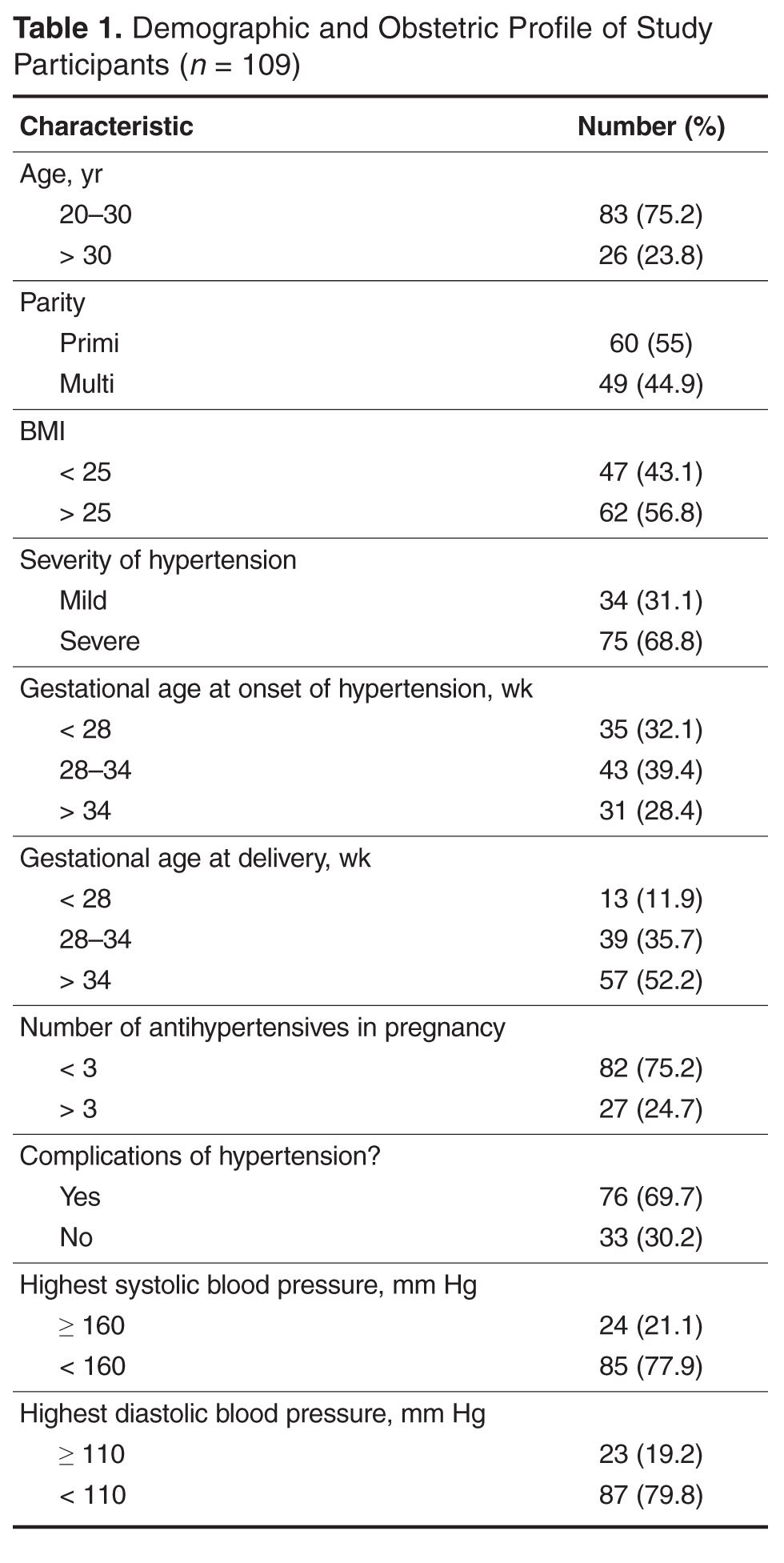
On testing for strength of correlation, it was found that body mass index and time to blood pressure normalization had a strong positive correlation (r = 0.8). The remaining parameters (ie, gestational age at onset, gestational age at delivery, severity and complications of hypertension and number of antihypertensive medications) and time to recovery were weakly correlated (r = 0.3 to 0.5 [+/–]).
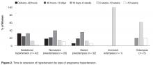
Women with gestational hypertension and mild preeclampsia had faster normalization of blood pressure compared to those with severe preeclampsia and eclampsia (Figure 2). Only 15% of women with gestational hypertension had persistent hypertension beyond 6 weeks, whereas in the groups with nonsevere preeclampsia, severe preeclampsia, and eclampsia, blood pressure 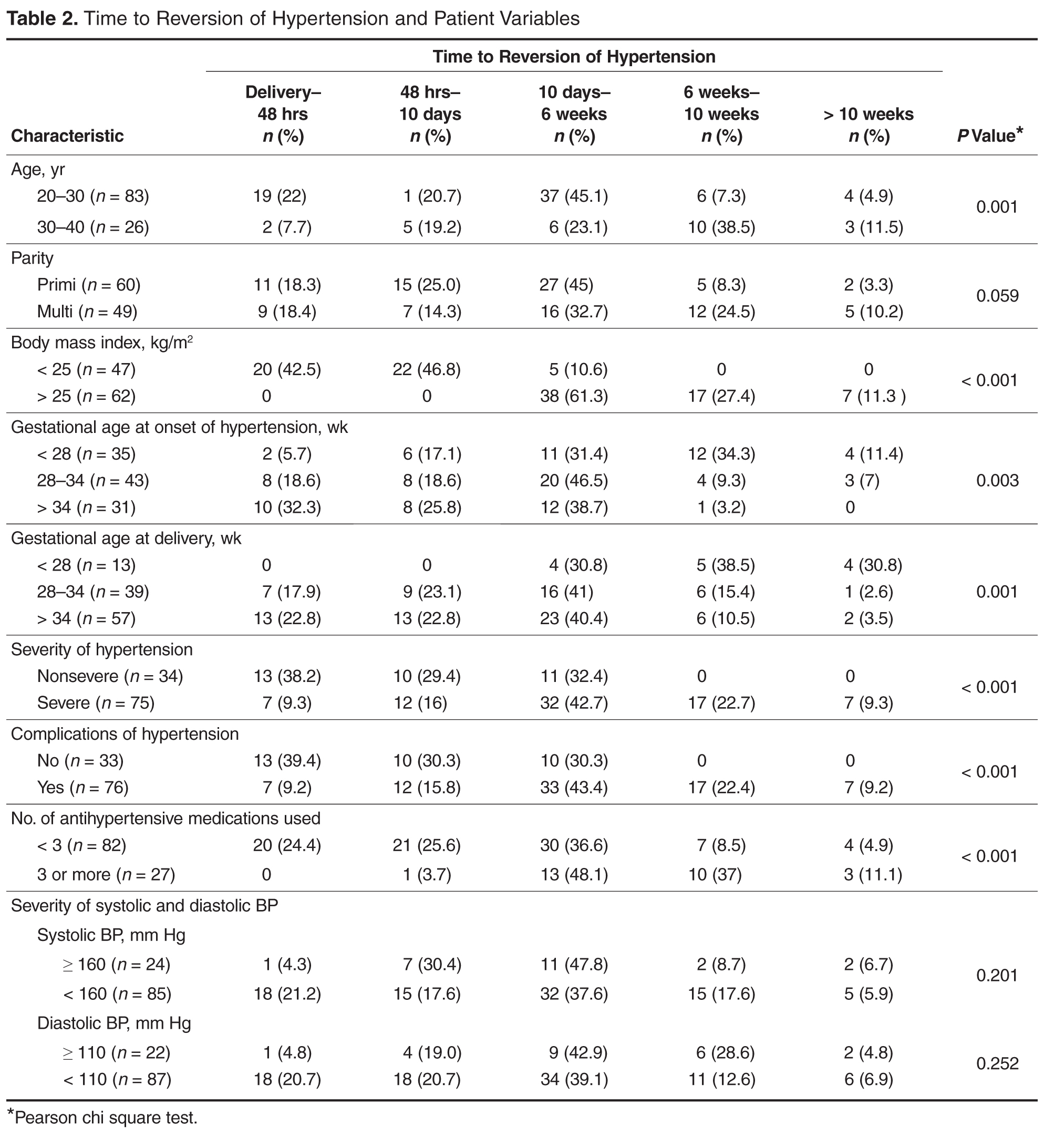
Eighteen women had additional medical problems: gestational diabetes (n = 5), anemia (n = 3), hypothyroidism (n = 4), rheumatic heart disease (n = 2), antiphospholipid antibody syndrome (n = 1) chronic kidney disease (n = 1), post atrial septal defect closure (n = 1), and tricuspid valve prolapse (TVP) with regurgitation and pulmonary arterial hypertension (n = 1). With the exception of the woman with chronic kidney disease, all reverted to normal blood pressure by 6 weeks; the woman with TVP reverted after corrective cardiac surgery in puerperium.
Discussion
In the present study we assessed possible correlations of obstetric profile with time to postpartum recovery of blood pressure in women with pregnancy hypertension. Women with advanced age, higher body mass index, early gestational age at the onset of hypertension, early gestational age at delivery, severe hypertension, and those with complications of hypertension took longer time in the postnatal period for normalization of blood pressure.
The strength of this study was its prospective design and high rate of follow-up. Those who missed a visit were followed up over telephone. However, 19 women were not available even by phone. A limitation of this study is that the information regarding when the antihypertensive was stopped was obtained by patient recall, raising the possibility of recall bias. However, as the range of recovery times was wide, an error of few days may not be significant.
In the study we noted that women with preeclampsia took a longer time to recovery compared with women with gestational hypertension. Earlier and more severe disease was associated with delay to recovery or persistence of hypertension beyond 10 weeks postpartum.
Similar to our observation, other authors have observed a consistent association of time to reversion of hypertension and early-onset hypertension in pregnancy [3–5]. Ferrazzani explained the longer time to normalization of blood pressure in preeclampsia compared to gestational hypertension as the recovery time of the endothelial damage in preeclampsia [4].
Berks et al [6] found a correlation of maximum diastolic blood pressure, maximum proteinuria in pregnancy, and diagnosis-to-delivery interval with time taken for resolution of hypertension; however, they did not find that time to resolution was correlated with gestational age at onset of preeclampsia. They opined that their observations reflected endothelial recovery after preeclampsia. They also suggested further research in the area of temporizing management of preeclampsia to determine if a conservative approach increases remote cardiovascular risk [6]. We did not study the diagnosis-to-delivery interval, but those with early delivery in our group had late postpartum recovery, indicating that they had severe/complicated preeclampsia that demanded early termination.
In conclusion, women with advanced age, higher body mass index, early gestational age at the onset of hypertension, severe and with complications of hypertension require prolonged monitoring and treatment when indicated for hypertension in the postnatal period. Women with a history of pregnancy hypertension have increased risk of stroke, cardiac ischemia, venous thrombosis within 10 to 20 years after pregnancy and higher risk of hypertension and type 2 diabetes mellitus [7–9]. Extended postnatal follow-up and regular monitoring is recommended to address the needs of these high-risk women.
Corresponding author: Dr. Shyamala Guruvare, 1-167 (C4), Lahari, Eshakripa Road, Parkala, Udupi District, Karnataka, India 576107, [email protected].
Financial disclosures: None reported.
1. American College of Obstetricians and Gynecologists; Task Force on Hypertension in Pregnancy. Hypertension in pregnancy. Report of the American College of Obstetricians and Gynecologists’ Task Force on Hypertension in Pregnancy. Obstet Gynecol 2013;122:1122–31.
2. Ndayambagye EB, Nakalembe M, Kaye DK. Factors associated with persistent hypertension after puerperium among women with preeclampsia/ eclampsia in Mulago Hospital, Uganda. BMC Pregnancy Childbirth 2010;10:12.
3. Mikami Y,Takagi K, Itaya Y, et al. Post-partum recovery course in patients with gestational hypertension and preeclampsia. J Obstet Gynaecol Res 2014;40:919–25.
4. Ferrazzini S, Carolis SD, Pomini F, et al. The duration of hypertension in the puerperium of preeclamptic women: Relationship with renal impairment and week of delivery. Am J Obstet Gynecol 1994;171:506–12.
5. Kaze FF, Njukeng FA , Kengne A, et al. Post-partum trend in blood pressure levels, renal function and proteinuria in women with severe preeclampsia and eclampsia in Sub-Saharan Africa: a 6-months cohort study.BMC Pregnancy Childbirth 2014;14:134
6. Berks D, Steegers EA, Molas M, Visser W. Resolution of hypertension and proteinuria after preeclampsia. Obstet Gynecol 2009;114:1307–14.
7. Gongora MC, Wenger NK. Cardiovascular complications of pregnancy. Int J Mol Sci 2015;16:23905–28.
8. Garovic VD, August P. Preeclampsia and the future risk of hypertension: the pregnant evidence. Curr Hypertens Rep 2013;114–21.
9. Zandstra M, Stekkinger E, van der Vlugt MJ, et al. Cardiac diastolic dysfunction and metabolic syndrome in young women after placental syndrome. Obstet Gynecol 2010;115:101–8.
From the Department of Obstetrics and Gynecology, Kasturba Medical College, Manipal, Karnataka, India.
Abstracts
- Objective: To examine the association of the patient’s obstetric profile and time to normalization of blood pressure in the postnatal period among women with hypertensive disorders in pregnancy.
- Methods: We conducted a prospective cohort study at a tertiary level hospital between November 2014 and May 2015. Women with pregnancy hypertension who required antihypertensive treatment were recruited after delivery. The normalization trends in blood pressure were tested for associations with patient demographic data and details of pregnancy hypertension.
- Results: Among 109 women included in the study, earlier gestational age at onset of hypertension and earlier gestational age at delivery was correlated with slower resolution of hypertension. Time to resolution also was correlated with age, BMI, severity of hypertension, associated complications, and the number of antihypertensive medications received. There was no correlation with highest recorded systolic or diastolic blood pressures. Only 15% of women with gestational hypertension had persistent hypertension beyond 6 weeks. In the groups with nonsevere preeclampsia, severe preeclampsia, and eclampsia, blood pressure remained high after 6 weeks in 26%, 14%, and 50% of women, respectively.
- Conclusion: Women with advanced age, higher body mass index, early gestational age at the onset of hypertension, severe hypertension and who had complications of hypertension require prolonged monitoring and treatment when indicated for hypertension in postnatal period.
Key words: intensive care unit; communication; family meeting; critical illness; decision making; end of life care.
Hypertension is the most common medical problem encountered during pregnancy, complicating up to 10% of pregnancies worldwide [1]. The disorders of hypertension in pregnancy are generally classified as chronic hypertension, preeclampsia–eclampsia, preeclampsia superimposed on chronic hypertension, and gestational hypertension. The hypertensive disorders of pregnancy are a leading cause of mortality and morbidity in the perinatal period.
Women with hypertensive disorders in pregnancy show varying trends of blood pressure normalization, with the recovery period ranging from a few hours to several months after delivery. In one study, nearly one-fourth of women with preeclampsia/eclampsia had persistent high blood pressure after puerperium [2]. Identifying the obstetric risk factors for persistent hypertension will help in focusing care and research in this group of patients.
We undertook a prospective study to assess possible correlations of obstetric profile with time to normalization of blood pressure in the postnatal period among women with hypertensive disorders in pregnancy.
Methods
Setting
This prospective cohort study was conducted in the department of obstetrics and gynecology at Kasturba Hospital, Manipal, between November 2014 and May 2015. Permission for the study was obtained from the Institution Ethical Committee (IEC264/2015).
Patients
Women who had hypertension in pregnancy and required antihypertensive treatment were approached on the first postnatal day and invited to participate in the study. Women with chronic hypertension (women with known pre-pregnancy hypertension and with hypertension diagnosed before 20 weeks gestation) or secondary hypertension were excluded. After granting informed consent, enrolled women were followed until the time they no longer required antihypertensive medication (“reversion of hypertension”) or until 10 weeks postpartum, whichever came first.
During the hospital stay in the postnatal period, women had their blood pressure monitored and antihypertensives were adjusted as needed. After discharge from the hospital, blood pressure was monitored by the family physician who also made decisions regarding antihypertensive management. All women had a follow-up visit in the hospital in the 6th postnatal week as per the postnatal clinic protocol.
Definitions
Hypertension was defined as BP ≥ 140/90 mm Hg. The hypertension disorders of pregnancy were defined as follows:
- Gestational hypertension: hypertension after 20 weeks gestation on two occasions 4 hours apart without meeting criteria for preeclampsia.
- Preeclampsia: hypertension after 20 weeks gestation on two occasions 4 hours apart with proteinuria (≥ 300 mg/24 hour) or, in the absence of proteinuria, new onset of any of the following: thrombocytopenia, renal insufficiency, impaired liver function, pulmonary edema, or cerebral or visual symptoms [1]. Severe preeclampsia was defined as preeclampsia with any of the following: systolic blood pressure > 160 mm Hg diastolic BP > 110 mm Hg or more on 2 occasions 4 hours apart, thrombocytopenia (platelet count < 100,00/mL), renal insufficiency, impaired liver function, pulmonary edema, or cerebral or visual symptoms. Preeclampsia without any of these features was considered nonsevere preeclampsia.
- Eclampsia: Women with hypertension with epigastric pain, headache, vomiting, and blurring of vision were diagnosed with imminent eclampsia and those with hypertension-related convulsions were diagnosed with eclampsia.
- Complications of preeclampsia included eclampsia, placental abruption, pulmonary edema, thrombocytopenia, HELLP syndrome, disseminated intravascular coagulation, multiorgan failure, severe intrauterine growth restriction, and fetal demise.
Main Outcome Measure
Time to reversion of hypertension was the main outcome measure. We defined the reversion date as the day that hypertension medications were stopped. This information was obtained via in-person questioning on the 2nd postpartum day and at the 6-week postnatal visit and via telephonic survey on the 10th postnatal day and at 10 weeks postdelivery. Women who missed the 6-week postnatal visit were also followed up by telephone.
Data Collection
Demographic details (age, parity, BMI) as well as information regarding gestational age at onset of hypertension, severity, highest systolic and diastolic blood pressure recordings, treatment received, complications related to hypertension, pregnancy termination and delivery was obtained from the medical charts and/or via telephonic follow-up.
Analysis
We used Pearson’s chi-square test to assess the association between recovery trends in blood pressure and the patient’s demographic profile and details of pregnancy hypertension. Statistical analysis was done using SPSS16.
Results
In our study, earlier the gestational age at onset of hypertension and earlier gestation at delivery was associated with slower recovery from hypertension (Table 2). Time taken for recovery also was associated with age, BMI, severity of hypertension, associated complications, and the number of antihypertensive medications received (Table 2). Among women who received more than 3 antiphypertensives in pregnancy, nearly 50% continued to have hypertension beyond 6 weeks (Table 2).
On testing for strength of correlation, it was found that body mass index and time to blood pressure normalization had a strong positive correlation (r = 0.8). The remaining parameters (ie, gestational age at onset, gestational age at delivery, severity and complications of hypertension and number of antihypertensive medications) and time to recovery were weakly correlated (r = 0.3 to 0.5 [+/–]).
Women with gestational hypertension and mild preeclampsia had faster normalization of blood pressure compared to those with severe preeclampsia and eclampsia (Figure 2). Only 15% of women with gestational hypertension had persistent hypertension beyond 6 weeks, whereas in the groups with nonsevere preeclampsia, severe preeclampsia, and eclampsia, blood pressure
Eighteen women had additional medical problems: gestational diabetes (n = 5), anemia (n = 3), hypothyroidism (n = 4), rheumatic heart disease (n = 2), antiphospholipid antibody syndrome (n = 1) chronic kidney disease (n = 1), post atrial septal defect closure (n = 1), and tricuspid valve prolapse (TVP) with regurgitation and pulmonary arterial hypertension (n = 1). With the exception of the woman with chronic kidney disease, all reverted to normal blood pressure by 6 weeks; the woman with TVP reverted after corrective cardiac surgery in puerperium.
Discussion
In the present study we assessed possible correlations of obstetric profile with time to postpartum recovery of blood pressure in women with pregnancy hypertension. Women with advanced age, higher body mass index, early gestational age at the onset of hypertension, early gestational age at delivery, severe hypertension, and those with complications of hypertension took longer time in the postnatal period for normalization of blood pressure.
The strength of this study was its prospective design and high rate of follow-up. Those who missed a visit were followed up over telephone. However, 19 women were not available even by phone. A limitation of this study is that the information regarding when the antihypertensive was stopped was obtained by patient recall, raising the possibility of recall bias. However, as the range of recovery times was wide, an error of few days may not be significant.
In the study we noted that women with preeclampsia took a longer time to recovery compared with women with gestational hypertension. Earlier and more severe disease was associated with delay to recovery or persistence of hypertension beyond 10 weeks postpartum.
Similar to our observation, other authors have observed a consistent association of time to reversion of hypertension and early-onset hypertension in pregnancy [3–5]. Ferrazzani explained the longer time to normalization of blood pressure in preeclampsia compared to gestational hypertension as the recovery time of the endothelial damage in preeclampsia [4].
Berks et al [6] found a correlation of maximum diastolic blood pressure, maximum proteinuria in pregnancy, and diagnosis-to-delivery interval with time taken for resolution of hypertension; however, they did not find that time to resolution was correlated with gestational age at onset of preeclampsia. They opined that their observations reflected endothelial recovery after preeclampsia. They also suggested further research in the area of temporizing management of preeclampsia to determine if a conservative approach increases remote cardiovascular risk [6]. We did not study the diagnosis-to-delivery interval, but those with early delivery in our group had late postpartum recovery, indicating that they had severe/complicated preeclampsia that demanded early termination.
In conclusion, women with advanced age, higher body mass index, early gestational age at the onset of hypertension, severe and with complications of hypertension require prolonged monitoring and treatment when indicated for hypertension in the postnatal period. Women with a history of pregnancy hypertension have increased risk of stroke, cardiac ischemia, venous thrombosis within 10 to 20 years after pregnancy and higher risk of hypertension and type 2 diabetes mellitus [7–9]. Extended postnatal follow-up and regular monitoring is recommended to address the needs of these high-risk women.
Corresponding author: Dr. Shyamala Guruvare, 1-167 (C4), Lahari, Eshakripa Road, Parkala, Udupi District, Karnataka, India 576107, [email protected].
Financial disclosures: None reported.
1. American College of Obstetricians and Gynecologists; Task Force on Hypertension in Pregnancy. Hypertension in pregnancy. Report of the American College of Obstetricians and Gynecologists’ Task Force on Hypertension in Pregnancy. Obstet Gynecol 2013;122:1122–31.
2. Ndayambagye EB, Nakalembe M, Kaye DK. Factors associated with persistent hypertension after puerperium among women with preeclampsia/ eclampsia in Mulago Hospital, Uganda. BMC Pregnancy Childbirth 2010;10:12.
3. Mikami Y,Takagi K, Itaya Y, et al. Post-partum recovery course in patients with gestational hypertension and preeclampsia. J Obstet Gynaecol Res 2014;40:919–25.
4. Ferrazzini S, Carolis SD, Pomini F, et al. The duration of hypertension in the puerperium of preeclamptic women: Relationship with renal impairment and week of delivery. Am J Obstet Gynecol 1994;171:506–12.
5. Kaze FF, Njukeng FA , Kengne A, et al. Post-partum trend in blood pressure levels, renal function and proteinuria in women with severe preeclampsia and eclampsia in Sub-Saharan Africa: a 6-months cohort study.BMC Pregnancy Childbirth 2014;14:134
6. Berks D, Steegers EA, Molas M, Visser W. Resolution of hypertension and proteinuria after preeclampsia. Obstet Gynecol 2009;114:1307–14.
7. Gongora MC, Wenger NK. Cardiovascular complications of pregnancy. Int J Mol Sci 2015;16:23905–28.
8. Garovic VD, August P. Preeclampsia and the future risk of hypertension: the pregnant evidence. Curr Hypertens Rep 2013;114–21.
9. Zandstra M, Stekkinger E, van der Vlugt MJ, et al. Cardiac diastolic dysfunction and metabolic syndrome in young women after placental syndrome. Obstet Gynecol 2010;115:101–8.
Are There Racial/Ethnic Differences in Weight-Related Care Encounters Reported by Patients?
Study Overview
Objective. To compare patients’ health care experiences related to their weight across racial and ethnic groups.
Design. Cross-sectional survey-based study.
Setting and participants. Between March and July 2015, 5400 individuals were randomly sampled from the Patient Outcomes to Advance Learning (PORTAL) obesity cohort, which includes over 5 million adults. The PORTAL network is a clinical data research network funded by the Patient Centered Outcomes Research Institute to promote collaboration across several large health systems with electronic medical records (EMRs), including all the Kaiser Permanente regions, Group Health Cooperative, Health Partners, and Denver Health. The selected 5400 cohort members were equally distributed across 3 geographically diverse Kaiser Permanente regions (Southwest, Northern and Southern California, Hawaii, Colorado, and Northwest) and Denver sites. Selected individuals were non-pregnant English or Spanish speakers with a body mass index (BMI) ≥ 25 kg/m2 (per their EMR) who were members of a participating health plan and had at least 1 outpatient visit in the last 12 months. Patients with BMI ≥ 40 kg/m2 were oversampled. Individuals were mailed a written 10-minute survey (offered in English or Spanish based on a patient’s written language preference noted in their EMR), consisting of 36 multiple-choice and fill-in-the-blank items. Telephone contact for verbal administration was attempted if a mailed response was not received within 4 weeks.
Main measures and analysis. The primary independent variable was a respondent’s racial/ethnic group, categorized as (1) non-Hispanic white (White), 2) non-Hispanic black (Black), 3) Hispanic, 4) Asian, or 5) Native Hawaiian/Other Pacific Islanders/American Indian/Native Alaskan (NA/PI).
Dependent variables focused on patients’ perceptions of the health care experience (based on services received at their usual place of care from their primary care providers) related to being overweight or obese using items based on the Rudd Center’s Patient Survey of Weight-Sensitive Healthcare Practices. Respondents described (1) whether and how often they avoid coming to their provider because they do not want to be weighed or have a discussion about their weight; (2) how often does their provider ask their permission before discussion their weight; (3) how often has their provider been supportive of their weight concerns and efforts to be healthy; (4) whether they think that their provider understands the physical and emotional challenges faced by individuals who are overweight or obese; (5) how often has their provider brought up their weight during a clinic visit; (6) whether their provider has ever given or discussed resources on healthy eating and weight loss; and (7) what types of weight loss resources were discussed with their provider and which types did they want more information about (ie, dietary changes, physical activity, classes, medications, meal replacements, and bariatric surgery). Covariate variables derived from EMR data included sex, age category, diabetes, hypertension, Charlson Index score (overall measures of morbidity), Medicaid enrollment, language preferences, site, and BMI. Survey-derived covariate variables included emotional well-being, perceived weight status, and educational attainment.
Descriptive statistics were generated and compared across racial/ethnic groups using Kruskal-Wallis and chi-square testing, as appropriate. To evaluate the association between a patient’s race/ethnicity and their perceived weight management experience, multinomial logistic regression adjusted for covariates was used to estimate odds ratios (OR).
Main results. From the original sample (n = 5400), 1569 individuals (29%) did not respond, 925 (17%) refused, and 114 (2%) were ineligible, leaving an eligible sample pool of 5286 individuals. The overall response rate was 53% (2197 written; 614 phone, n = 2811). Those with missing data were excluded (6 with missing race/ethnicity; 80 missing other covariates), leaving a final group of 2725 respondents for analysis. Mean age was 52.7 years (SD 15), almost 62% of participants were female, 51.7% identified as White, 21.1% identified as Black, 14.6% identified as Hispanic, 5.8% identified as Asian, and 6.7% identified as NA/PI. About a quarter (24.4%) had diabetes, less than half (43.5%) had hypertension, and most (86.2%) perceived themselves to be overweight. There were significant differences in measured baseline covariates by racial/ethnic groups including mean BMI, diabetes, and being a Medicaid beneficiary.
In response to the 7 key areas assessed regarding patients’ perceptions of the health care experience related to being overweight or obese:
- Black respondents were less likely than Whites to report that they frequently avoided care from their provider because they did not want to be weighed or discuss their weight (OR 0.49 [95% confidence interval, 0.26–0.90]), with a trend toward all groups being less likely to report frequent avoidance compared to Whites.
- While just over half of respondents (59.3%) indicated that their providers never asked for their permission before discussing their weight, Asians and NA/PI were more likely to report that their providers either frequently (Asians: OR 2.7 [1.3–5.6]; NA/PI: OR 2.3 [1.1–5.0]) or sometimes (Asians: OR 2.3 [1.2–4.3]; NA/PI: OR 2.1 [1.1–4.1]) asked their permission before discussing their weight compared to Whites.
- Over half (61.9%) indicated that their providers were sometimes or frequently supportive of their weight concerns, with no significant differences among racial/ethnic groups.
- Just over half (52.0%) indicated they felt their providers understood the physical and emotional challenges faced by people who are overweight/obese, with Blacks more likely to feel this way (OR 1.8 [1.2–2.8]) compared to Whites.
- Black patients were more likely than Whites (OR 2.0 [1.4–2.8]) to report that their providers discussed their weight with them at a clinic visit.
- While over half (59.7%) indicated that their providers had given or discussed resources with them on healthy eating and weight loss, Black and Asian respondents were more likely than Whites to recall these discussions (Black: OR 1.6 [1.2–2.1]; Asians: OR 1.8 [1.1–2.9]).
- Most weight loss resources or recommendations received were related to lifestyle changes, with very few resources given related to weight loss medications, meal replacement products, or bariatric surgery—few differences across racial/ethnic groups were identified. However, respondents from racial/ethnic minority groups were more likely than Whites to say that they wanted more information about lifestyle changes, classes, and meal replacements. Other than Blacks, all other racial/ethnic groups were also more likely than Whites to indicate that they wanted more information about bariatric surgery.
Conclusions. Most patients across racial/ethnic groups are having positive experiences with weight-related care. However, race/ethnicity correlates with patients’ perception of weight-related care and discussions in clinic encounters.
Commentary
The obesity epidemic in the United States is well-established [1], and recent data from 2014 show that over 37% of adults in the US are obese (defined as having a body mass index greater than 30 kg/m2) [2]. However, while obesity prevalence rates have increased over the past several decades across all genders, ethnicities, income levels, and education levels, important racial/ethnic disparities exist [2,3]. Primary care physicians (PCPs) are ideally situated to promote weight loss via effective obesity counseling since multiple clinic visits over time have the potential to enable rapport building and behavioral change management [4]. In fact, the US Preventive Services Task Force (USPTF) recommends that all patients be screened for obesity and offered intensive lifestyle counseling, as modest weight loss can have significant health benefits [5]. However, some studies have found racial/ethnic differences and disparities in weight-related diagnoses, counseling, and treatment by providers, but also patient perceptions of care and preferred interventions [6–10]. Other studies have described racial/ethnic differences in weight-related concerns and behaviors, body satisfaction, and body image [11–13]. Thus, research is needed to examine these differences.
This cross-sectional study contributes to the limited literature examining the potential for heterogeneity of care according to patient characteristics like race and ethnicity. Key strengths of the design include a large and both geographically and racially/ethnically diverse sample of patients (increased generalizability), the use of mailed brief surveys (reduces non-response rate and reporting bias) and telephone follow-up for verbal administration (reduces non-response rate, though it increases interviewer bias), oversampling of respondents with BMI ≥ 40 kg/m2, and the controlling of key covariates including sex, age, Medicaid enrollment, site, and BMI.
However, there are several important limitations, many of which are acknowledged by the authors. While respondents were overall representative of the targeted sample population, the final respondent population was comprised of mostly older females who received managed care, which may have contributed to selection bias and impacted generalizability of findings. Further, Whites were overrepresented, Hispanics were underrepresented, and the small combined sample of NA/PI may have masked important distinctions between these subpopulations. Importantly, this study only provided the survey in English and Spanish and did not include other language translations (eg, Chinese, Japanese, Tagalog), which likely contributed to underrepresented perspectives of immigrants and ESL patients who may struggle with receiving/discussing weight management counseling and resources. The use of a surveys collected subjective and self-reported data on patient encounters as opposed to objective observations. Lastly, the study did not adjust for individual provider factors or assess the potential impact of provider-level differences on care, such as provider-patient concordance on race, ethnicity, language, and/or weight. The incorporation of qualitative interviewers or focus groups with a subsample of each racial/ethnic may have also provided relevant context to understand differences in weight-related care experiences.
Applications for Clinical Practice
As the authors suggest, this study highlights several opportunities to continue improving weight-related care and weight management counseling. PCPs should engage all overweight/obese patients in weight management discussions, and in particular, high-risk minority patients who may desire these conversations and more weight loss advice and resources. However, these discussions require sensitivity and can benefit from the simple practice of asking permission of the patient to talk about their weight in order to reduce care avoidance and improve perceptions of care. Providers should also be mindful of patient priorities and assess patient preferences for all the different weight loss strategies, including lifestyle changes, meal replacements, medications, and surgery.
—Katrina F. Mateo, MPH
1. Mitchell NS, Catenacci VA, Wyatt HR, Hill JO. Obesity: overview of an epidemic. Psychiatr Clin North Am 2011;34:717–32.
2. Flegal KM, Kruszon-Moran D, Carroll MD, et al. Trends in obesity among adults in the United States, 2005 to 2014. JAMA 2016;315:2284.
3. Wong RJ, Chou C, Ahmed A. Long term trends and racial/ethnic disparities in the prevalence of obesity. J Community Health 2014;39:1150–60.
4. Schlair S, Moore S, Mcmacken M, Jay M. How to deliver high quality obesity counseling using the 5As framework. J Clin Outcomes Manag 2012;19:221–9.
5. Moyer VA. Screening for and management of obesity in adults: U.S. Preventive Services Task Force recommendation statement. Ann Intern Med 2012;157:373–8.
6. Davis NJ, Wildman RP, Forbes BF, Schechter CB. Trends and disparities in provider diagnosis of overweight analysis of NHANES 1999–2004. Obesity 2009;17:2110–3.
7. Wee CC, Huskey KW, Bolcic-Jankovic D, et al. Sex, race, and consideration of bariatric surgery among primary care patients with moderate to severe obesity. J Gen Intern Med 2014;29:68–75.
8. Johnson RL, Saha S, Arbelaez JJ, et al. Racial and ethnic differences in patient perceptions of bias and cultural competence in health care. J Gen Intern Med 2004;19:101–10.
9. Chugh M, Friedman AM, Clemow LP, Ferrante JM. Women weigh in: obese african american and white women’s perspectives on physicians’ roles in weight management. J Am Board Fam Med 2013;26:421–8.
10. Blixen CE, Singh A, Xu M, et al. What women want: understanding obesity and preferences for primary care weight reduction interventions among African-American and Caucasian women. J Natl Med Assoc 2006;98:1160–70.
11. Arcan C, Larson N, Bauer K, et al. Dietary and weight-related behaviors and body mass index among Hispanic, Hmong, Somali, and White adolescents. J Acad Nutr Diet 2014;114:375–83.
12. Kronenfeld LW, Reba-Harrelson L, Von Holle A, et al. Ethnic and racial differences in body size perception and satisfaction. Body Image 2010;7:131–6.
13. Gluck ME, Geliebter A. Racial/ethnic differences in body image and eating behaviors. Eat Behav 2002;3:143–51.
Study Overview
Objective. To compare patients’ health care experiences related to their weight across racial and ethnic groups.
Design. Cross-sectional survey-based study.
Setting and participants. Between March and July 2015, 5400 individuals were randomly sampled from the Patient Outcomes to Advance Learning (PORTAL) obesity cohort, which includes over 5 million adults. The PORTAL network is a clinical data research network funded by the Patient Centered Outcomes Research Institute to promote collaboration across several large health systems with electronic medical records (EMRs), including all the Kaiser Permanente regions, Group Health Cooperative, Health Partners, and Denver Health. The selected 5400 cohort members were equally distributed across 3 geographically diverse Kaiser Permanente regions (Southwest, Northern and Southern California, Hawaii, Colorado, and Northwest) and Denver sites. Selected individuals were non-pregnant English or Spanish speakers with a body mass index (BMI) ≥ 25 kg/m2 (per their EMR) who were members of a participating health plan and had at least 1 outpatient visit in the last 12 months. Patients with BMI ≥ 40 kg/m2 were oversampled. Individuals were mailed a written 10-minute survey (offered in English or Spanish based on a patient’s written language preference noted in their EMR), consisting of 36 multiple-choice and fill-in-the-blank items. Telephone contact for verbal administration was attempted if a mailed response was not received within 4 weeks.
Main measures and analysis. The primary independent variable was a respondent’s racial/ethnic group, categorized as (1) non-Hispanic white (White), 2) non-Hispanic black (Black), 3) Hispanic, 4) Asian, or 5) Native Hawaiian/Other Pacific Islanders/American Indian/Native Alaskan (NA/PI).
Dependent variables focused on patients’ perceptions of the health care experience (based on services received at their usual place of care from their primary care providers) related to being overweight or obese using items based on the Rudd Center’s Patient Survey of Weight-Sensitive Healthcare Practices. Respondents described (1) whether and how often they avoid coming to their provider because they do not want to be weighed or have a discussion about their weight; (2) how often does their provider ask their permission before discussion their weight; (3) how often has their provider been supportive of their weight concerns and efforts to be healthy; (4) whether they think that their provider understands the physical and emotional challenges faced by individuals who are overweight or obese; (5) how often has their provider brought up their weight during a clinic visit; (6) whether their provider has ever given or discussed resources on healthy eating and weight loss; and (7) what types of weight loss resources were discussed with their provider and which types did they want more information about (ie, dietary changes, physical activity, classes, medications, meal replacements, and bariatric surgery). Covariate variables derived from EMR data included sex, age category, diabetes, hypertension, Charlson Index score (overall measures of morbidity), Medicaid enrollment, language preferences, site, and BMI. Survey-derived covariate variables included emotional well-being, perceived weight status, and educational attainment.
Descriptive statistics were generated and compared across racial/ethnic groups using Kruskal-Wallis and chi-square testing, as appropriate. To evaluate the association between a patient’s race/ethnicity and their perceived weight management experience, multinomial logistic regression adjusted for covariates was used to estimate odds ratios (OR).
Main results. From the original sample (n = 5400), 1569 individuals (29%) did not respond, 925 (17%) refused, and 114 (2%) were ineligible, leaving an eligible sample pool of 5286 individuals. The overall response rate was 53% (2197 written; 614 phone, n = 2811). Those with missing data were excluded (6 with missing race/ethnicity; 80 missing other covariates), leaving a final group of 2725 respondents for analysis. Mean age was 52.7 years (SD 15), almost 62% of participants were female, 51.7% identified as White, 21.1% identified as Black, 14.6% identified as Hispanic, 5.8% identified as Asian, and 6.7% identified as NA/PI. About a quarter (24.4%) had diabetes, less than half (43.5%) had hypertension, and most (86.2%) perceived themselves to be overweight. There were significant differences in measured baseline covariates by racial/ethnic groups including mean BMI, diabetes, and being a Medicaid beneficiary.
In response to the 7 key areas assessed regarding patients’ perceptions of the health care experience related to being overweight or obese:
- Black respondents were less likely than Whites to report that they frequently avoided care from their provider because they did not want to be weighed or discuss their weight (OR 0.49 [95% confidence interval, 0.26–0.90]), with a trend toward all groups being less likely to report frequent avoidance compared to Whites.
- While just over half of respondents (59.3%) indicated that their providers never asked for their permission before discussing their weight, Asians and NA/PI were more likely to report that their providers either frequently (Asians: OR 2.7 [1.3–5.6]; NA/PI: OR 2.3 [1.1–5.0]) or sometimes (Asians: OR 2.3 [1.2–4.3]; NA/PI: OR 2.1 [1.1–4.1]) asked their permission before discussing their weight compared to Whites.
- Over half (61.9%) indicated that their providers were sometimes or frequently supportive of their weight concerns, with no significant differences among racial/ethnic groups.
- Just over half (52.0%) indicated they felt their providers understood the physical and emotional challenges faced by people who are overweight/obese, with Blacks more likely to feel this way (OR 1.8 [1.2–2.8]) compared to Whites.
- Black patients were more likely than Whites (OR 2.0 [1.4–2.8]) to report that their providers discussed their weight with them at a clinic visit.
- While over half (59.7%) indicated that their providers had given or discussed resources with them on healthy eating and weight loss, Black and Asian respondents were more likely than Whites to recall these discussions (Black: OR 1.6 [1.2–2.1]; Asians: OR 1.8 [1.1–2.9]).
- Most weight loss resources or recommendations received were related to lifestyle changes, with very few resources given related to weight loss medications, meal replacement products, or bariatric surgery—few differences across racial/ethnic groups were identified. However, respondents from racial/ethnic minority groups were more likely than Whites to say that they wanted more information about lifestyle changes, classes, and meal replacements. Other than Blacks, all other racial/ethnic groups were also more likely than Whites to indicate that they wanted more information about bariatric surgery.
Conclusions. Most patients across racial/ethnic groups are having positive experiences with weight-related care. However, race/ethnicity correlates with patients’ perception of weight-related care and discussions in clinic encounters.
Commentary
The obesity epidemic in the United States is well-established [1], and recent data from 2014 show that over 37% of adults in the US are obese (defined as having a body mass index greater than 30 kg/m2) [2]. However, while obesity prevalence rates have increased over the past several decades across all genders, ethnicities, income levels, and education levels, important racial/ethnic disparities exist [2,3]. Primary care physicians (PCPs) are ideally situated to promote weight loss via effective obesity counseling since multiple clinic visits over time have the potential to enable rapport building and behavioral change management [4]. In fact, the US Preventive Services Task Force (USPTF) recommends that all patients be screened for obesity and offered intensive lifestyle counseling, as modest weight loss can have significant health benefits [5]. However, some studies have found racial/ethnic differences and disparities in weight-related diagnoses, counseling, and treatment by providers, but also patient perceptions of care and preferred interventions [6–10]. Other studies have described racial/ethnic differences in weight-related concerns and behaviors, body satisfaction, and body image [11–13]. Thus, research is needed to examine these differences.
This cross-sectional study contributes to the limited literature examining the potential for heterogeneity of care according to patient characteristics like race and ethnicity. Key strengths of the design include a large and both geographically and racially/ethnically diverse sample of patients (increased generalizability), the use of mailed brief surveys (reduces non-response rate and reporting bias) and telephone follow-up for verbal administration (reduces non-response rate, though it increases interviewer bias), oversampling of respondents with BMI ≥ 40 kg/m2, and the controlling of key covariates including sex, age, Medicaid enrollment, site, and BMI.
However, there are several important limitations, many of which are acknowledged by the authors. While respondents were overall representative of the targeted sample population, the final respondent population was comprised of mostly older females who received managed care, which may have contributed to selection bias and impacted generalizability of findings. Further, Whites were overrepresented, Hispanics were underrepresented, and the small combined sample of NA/PI may have masked important distinctions between these subpopulations. Importantly, this study only provided the survey in English and Spanish and did not include other language translations (eg, Chinese, Japanese, Tagalog), which likely contributed to underrepresented perspectives of immigrants and ESL patients who may struggle with receiving/discussing weight management counseling and resources. The use of a surveys collected subjective and self-reported data on patient encounters as opposed to objective observations. Lastly, the study did not adjust for individual provider factors or assess the potential impact of provider-level differences on care, such as provider-patient concordance on race, ethnicity, language, and/or weight. The incorporation of qualitative interviewers or focus groups with a subsample of each racial/ethnic may have also provided relevant context to understand differences in weight-related care experiences.
Applications for Clinical Practice
As the authors suggest, this study highlights several opportunities to continue improving weight-related care and weight management counseling. PCPs should engage all overweight/obese patients in weight management discussions, and in particular, high-risk minority patients who may desire these conversations and more weight loss advice and resources. However, these discussions require sensitivity and can benefit from the simple practice of asking permission of the patient to talk about their weight in order to reduce care avoidance and improve perceptions of care. Providers should also be mindful of patient priorities and assess patient preferences for all the different weight loss strategies, including lifestyle changes, meal replacements, medications, and surgery.
—Katrina F. Mateo, MPH
1. Mitchell NS, Catenacci VA, Wyatt HR, Hill JO. Obesity: overview of an epidemic. Psychiatr Clin North Am 2011;34:717–32.
2. Flegal KM, Kruszon-Moran D, Carroll MD, et al. Trends in obesity among adults in the United States, 2005 to 2014. JAMA 2016;315:2284.
3. Wong RJ, Chou C, Ahmed A. Long term trends and racial/ethnic disparities in the prevalence of obesity. J Community Health 2014;39:1150–60.
4. Schlair S, Moore S, Mcmacken M, Jay M. How to deliver high quality obesity counseling using the 5As framework. J Clin Outcomes Manag 2012;19:221–9.
5. Moyer VA. Screening for and management of obesity in adults: U.S. Preventive Services Task Force recommendation statement. Ann Intern Med 2012;157:373–8.
6. Davis NJ, Wildman RP, Forbes BF, Schechter CB. Trends and disparities in provider diagnosis of overweight analysis of NHANES 1999–2004. Obesity 2009;17:2110–3.
7. Wee CC, Huskey KW, Bolcic-Jankovic D, et al. Sex, race, and consideration of bariatric surgery among primary care patients with moderate to severe obesity. J Gen Intern Med 2014;29:68–75.
8. Johnson RL, Saha S, Arbelaez JJ, et al. Racial and ethnic differences in patient perceptions of bias and cultural competence in health care. J Gen Intern Med 2004;19:101–10.
9. Chugh M, Friedman AM, Clemow LP, Ferrante JM. Women weigh in: obese african american and white women’s perspectives on physicians’ roles in weight management. J Am Board Fam Med 2013;26:421–8.
10. Blixen CE, Singh A, Xu M, et al. What women want: understanding obesity and preferences for primary care weight reduction interventions among African-American and Caucasian women. J Natl Med Assoc 2006;98:1160–70.
11. Arcan C, Larson N, Bauer K, et al. Dietary and weight-related behaviors and body mass index among Hispanic, Hmong, Somali, and White adolescents. J Acad Nutr Diet 2014;114:375–83.
12. Kronenfeld LW, Reba-Harrelson L, Von Holle A, et al. Ethnic and racial differences in body size perception and satisfaction. Body Image 2010;7:131–6.
13. Gluck ME, Geliebter A. Racial/ethnic differences in body image and eating behaviors. Eat Behav 2002;3:143–51.
It isn’t over until it’s over
Pediatricians take heart.
Yes, I know it is discouraging when families occasionally ignore our advice and refuse vaccines for their children. It is even worse when political leaders who ought to know better question the safety and value of vaccines.
But let’s not lose perspective. Let me share a quick reminder of why vaccines are (almost) universally considered one of the greatest public health achievements of the 20th century.
Not long ago, I reviewed a clinical case with students as part of a medical microbiology course. A 6-year-old girl presented with fever, headache, and flaccid paralysis of the right arm with areflexia. With little prompting, the students generated a short differential diagnosis. Enterovirus. West Nile virus. “I guess we should include polio,” one student offered. “But who gets that anymore?”
A mere 120 years changes everything. At the dawn of the 20th century, we didn’t even know with certainty what caused polio, although infection was suspected.

On Sept. 9, 1954, the Courier-Journal, a newspaper in my hometown of Louisville, Ky., carried a story about the annual number of polio cases in Jefferson County, noting that they had reached 198 and General Hospital had opened a polio ward usually reserved for epidemics. Concerns about the infection were rippling throughout the state, and the paper reported that at least one high school marching band had elected to withdraw from annual Kentucky State Fair competition because of concerns about infection.
My mom was 10 years old in the summer of 1954, and she recalls that it was a “scary” time. Swimming pools closed. Parents refused to allow their children to go to movie theaters or the local amusement park because of fear that they might come into contact with the virus. My mom said, “Then one of my friends was diagnosed with polio. We had played together the week before she got sick. We worried that we were going to get sick, too. And once you got sick, you didn’t necessarily get better.”

I probably don’t need to remind you that both Dr. Sabin and Dr. Salk did develop successful poliovirus vaccines. Dr. Enders, along with junior colleagues Fred C. Robbins, MD, and Thomas H. Weller, MD, developed the techniques to grow poliovirus and other viruses in culture, making the work of Dr. Sabin and Dr. Salk possible. For this, Dr. Enders, Dr. Robbins, and Dr. Weller received the Nobel Prize in 1954.
Regarding the prediction of long-term protection, I’d say we’re there. According to the Centers for Disease Control and Prevention, wild poliovirus cases have declined more than 99.9% since 1988. According to the Global Polio Eradication Initiative, that means that there are approximately 10 million people walking today who would have otherwise been paralyzed by the disease.
In 2015, there were only 74 cases identified in the world, and these were localized to two countries. Even better, a global commission announced that wild poliovirus type 2 had been eradicated from the world. Eradicated. The last known transmission occurred in India in 1999.
Type 3 poliovirus may not be far behind. The last known case of wildtype poliovirus 3 was detected in 2012.
The complete story of poliovirus eradication efforts could read like a suspense novel: There have been twists and turns, some missed deadlines, and now a bit of irony. Success, in large part, has hinged on the use of trivalent, live attenuated oral poliovirus vaccine (tOPV) throughout much of the world. Now eradication of all polio disease is going to require withdrawal of OPV in countries that still use it.
Rarely, the live attenuated vaccine viruses contained in OPV can cause polio, and since 2012, vaccine-derived cases have exceeded wild poliovirus cases. Vaccine-derived cases include vaccine-associated paralytic polio (VAPP) – paralysis occurs in a vaccine recipient or a close contact – as well as cases of circulating vaccine-derived polioviruses (cVDPVs). Remember that vaccine viruses are shed in the stool, and in communities with low immunization rates, they circulate and acquire mutations that confer the transmissibility and neurovirulence properties of wild viruses. Ultimately, cVDPVs lead to outbreaks.
In 2013, the Global Polio Eradication Initiative published a new “endgame plan” for polio that outlined a stepwise approach for removing OPV from immunization programs. First, it called on all countries to introduce at least one dose of inactivated poliovirus vaccine by the third quarter of 2015, immunizing infants at 14 weeks or at first contact thereafter. Second, it called for all countries to replace tOPV with a bivalent vaccine containing only types 1 and 3 by 2016. Given the eradication of wild poliovirus type 2, keeping type 2 in the oral vaccine just creates risk. An estimated 40% of VAPP cases and 98% of cVDPVs detected since 2012 were caused by poliovirus type 2. The type 2 component of tOPV also interferes with the immune response to the other types. Once poliovirus eradication has been achieved and certified, hopefully no later than 2019, all OPV will be withdrawn.

What’s the role of pediatricians in the United States in polio eradication? For now, our job is to continue to protect all children in the United States against all three types of poliovirus. Current Advisory Committee on Immunization Practices (ACIP) recommendations specify 4 doses of trivalent inactivated poliovirus vaccine (IPV) at ages 2 months, 4 months, 6-18 months, and 4-6 years. Children vaccinated outside the United States with bivalent vaccine, including immigrants and refugees, will need to be revaccinated. Those without appropriate documentation of vaccine (written, dated records that specify trivalent vaccine) also should be revaccinated.
Serologic testing for immunity is no longer recommended. In the past, children without documentation of vaccines could be tested for neutralizing antibodies to poliovirus types 1, 2, and 3. Moving forward, serologic testing for antibodies to poliovirus type 2 won’t be available because it requires live virus, and in accordance with World Health Organization recommendations, laboratories have been destroying supplies of poliovirus type 2.
We also need to make sure that our patients who are traveling internationally receive all recommended vaccines, including a dose of IPV when appropriate. Specific recommendations can be found on the CDC’s pages for travelers.
A 2015 statement from the American Academy of Pediatrics called on pediatricians to consider polio as a potential diagnosis of any child presenting with fever and acute flaccid paralysis (Pediatrics. 2015 Jan;135[1]:196-202). When polio is suspected, public health authorities should be notified and two stool samples collected 24 hours apart, and within 14 days of the onset of paralysis, sent for testing. According to lead author Walter A. Orenstein, MD, “because most polio infections are silent, a case of paralytic polio in the United States may have been acquired from an asymptomatic individual, so a history of travel to a polio-infected area may be absent in the case of paralysis.”
I’ll second what my mom said. Scary.
Dr. Bryant is a pediatrician specializing in infectious diseases at the University of Louisville (Ky.) and Kosair Children’s Hospital, also in Louisville. She said she had no relevant financial disclosures. Email her at [email protected].
Pediatricians take heart.
Yes, I know it is discouraging when families occasionally ignore our advice and refuse vaccines for their children. It is even worse when political leaders who ought to know better question the safety and value of vaccines.
But let’s not lose perspective. Let me share a quick reminder of why vaccines are (almost) universally considered one of the greatest public health achievements of the 20th century.
Not long ago, I reviewed a clinical case with students as part of a medical microbiology course. A 6-year-old girl presented with fever, headache, and flaccid paralysis of the right arm with areflexia. With little prompting, the students generated a short differential diagnosis. Enterovirus. West Nile virus. “I guess we should include polio,” one student offered. “But who gets that anymore?”
A mere 120 years changes everything. At the dawn of the 20th century, we didn’t even know with certainty what caused polio, although infection was suspected.
On Sept. 9, 1954, the Courier-Journal, a newspaper in my hometown of Louisville, Ky., carried a story about the annual number of polio cases in Jefferson County, noting that they had reached 198 and General Hospital had opened a polio ward usually reserved for epidemics. Concerns about the infection were rippling throughout the state, and the paper reported that at least one high school marching band had elected to withdraw from annual Kentucky State Fair competition because of concerns about infection.
My mom was 10 years old in the summer of 1954, and she recalls that it was a “scary” time. Swimming pools closed. Parents refused to allow their children to go to movie theaters or the local amusement park because of fear that they might come into contact with the virus. My mom said, “Then one of my friends was diagnosed with polio. We had played together the week before she got sick. We worried that we were going to get sick, too. And once you got sick, you didn’t necessarily get better.”
I probably don’t need to remind you that both Dr. Sabin and Dr. Salk did develop successful poliovirus vaccines. Dr. Enders, along with junior colleagues Fred C. Robbins, MD, and Thomas H. Weller, MD, developed the techniques to grow poliovirus and other viruses in culture, making the work of Dr. Sabin and Dr. Salk possible. For this, Dr. Enders, Dr. Robbins, and Dr. Weller received the Nobel Prize in 1954.
Regarding the prediction of long-term protection, I’d say we’re there. According to the Centers for Disease Control and Prevention, wild poliovirus cases have declined more than 99.9% since 1988. According to the Global Polio Eradication Initiative, that means that there are approximately 10 million people walking today who would have otherwise been paralyzed by the disease.
In 2015, there were only 74 cases identified in the world, and these were localized to two countries. Even better, a global commission announced that wild poliovirus type 2 had been eradicated from the world. Eradicated. The last known transmission occurred in India in 1999.
Type 3 poliovirus may not be far behind. The last known case of wildtype poliovirus 3 was detected in 2012.
The complete story of poliovirus eradication efforts could read like a suspense novel: There have been twists and turns, some missed deadlines, and now a bit of irony. Success, in large part, has hinged on the use of trivalent, live attenuated oral poliovirus vaccine (tOPV) throughout much of the world. Now eradication of all polio disease is going to require withdrawal of OPV in countries that still use it.
Rarely, the live attenuated vaccine viruses contained in OPV can cause polio, and since 2012, vaccine-derived cases have exceeded wild poliovirus cases. Vaccine-derived cases include vaccine-associated paralytic polio (VAPP) – paralysis occurs in a vaccine recipient or a close contact – as well as cases of circulating vaccine-derived polioviruses (cVDPVs). Remember that vaccine viruses are shed in the stool, and in communities with low immunization rates, they circulate and acquire mutations that confer the transmissibility and neurovirulence properties of wild viruses. Ultimately, cVDPVs lead to outbreaks.
In 2013, the Global Polio Eradication Initiative published a new “endgame plan” for polio that outlined a stepwise approach for removing OPV from immunization programs. First, it called on all countries to introduce at least one dose of inactivated poliovirus vaccine by the third quarter of 2015, immunizing infants at 14 weeks or at first contact thereafter. Second, it called for all countries to replace tOPV with a bivalent vaccine containing only types 1 and 3 by 2016. Given the eradication of wild poliovirus type 2, keeping type 2 in the oral vaccine just creates risk. An estimated 40% of VAPP cases and 98% of cVDPVs detected since 2012 were caused by poliovirus type 2. The type 2 component of tOPV also interferes with the immune response to the other types. Once poliovirus eradication has been achieved and certified, hopefully no later than 2019, all OPV will be withdrawn.
What’s the role of pediatricians in the United States in polio eradication? For now, our job is to continue to protect all children in the United States against all three types of poliovirus. Current Advisory Committee on Immunization Practices (ACIP) recommendations specify 4 doses of trivalent inactivated poliovirus vaccine (IPV) at ages 2 months, 4 months, 6-18 months, and 4-6 years. Children vaccinated outside the United States with bivalent vaccine, including immigrants and refugees, will need to be revaccinated. Those without appropriate documentation of vaccine (written, dated records that specify trivalent vaccine) also should be revaccinated.
Serologic testing for immunity is no longer recommended. In the past, children without documentation of vaccines could be tested for neutralizing antibodies to poliovirus types 1, 2, and 3. Moving forward, serologic testing for antibodies to poliovirus type 2 won’t be available because it requires live virus, and in accordance with World Health Organization recommendations, laboratories have been destroying supplies of poliovirus type 2.
We also need to make sure that our patients who are traveling internationally receive all recommended vaccines, including a dose of IPV when appropriate. Specific recommendations can be found on the CDC’s pages for travelers.
A 2015 statement from the American Academy of Pediatrics called on pediatricians to consider polio as a potential diagnosis of any child presenting with fever and acute flaccid paralysis (Pediatrics. 2015 Jan;135[1]:196-202). When polio is suspected, public health authorities should be notified and two stool samples collected 24 hours apart, and within 14 days of the onset of paralysis, sent for testing. According to lead author Walter A. Orenstein, MD, “because most polio infections are silent, a case of paralytic polio in the United States may have been acquired from an asymptomatic individual, so a history of travel to a polio-infected area may be absent in the case of paralysis.”
I’ll second what my mom said. Scary.
Dr. Bryant is a pediatrician specializing in infectious diseases at the University of Louisville (Ky.) and Kosair Children’s Hospital, also in Louisville. She said she had no relevant financial disclosures. Email her at [email protected].
Intermittent breaks from sunitinib feasible in metastatic RCC
Intermittent breaks from sunitinib therapy are feasible and don’t appear to compromise the agent’s clinical efficacy against metastatic renal cell carcinoma, according to investigators.
One of the greatest challenges in treating this cancer is balancing treatment-related toxicity against efficacy “in a setting ... in which patients are largely incurable and thus subjected to chronic therapy,” said Moshe C. Ornstein, MD, and his associates at the Cleveland Clinic Taussig Cancer Institute.
They performed a single-center phase II study that they described as the first prospective trial to assess treatment interruptions whenever tumor burden was reduced by 10% or more, followed by resumption of treatment if tumors then progressed 10% or more. This approach “may result in reduced toxicities, improved quality of life, cost savings, and potentially improved clinical outcomes.”
The study involved 37 adults (median age, 63 years) who were given 50 mg sunitinib once daily for the first 28 days of a 42-day cycle, with dose adjustments allowed to minimize toxicities. Twenty patients (54%) who showed a 10% or greater reduction in tumor burden after four cycles suspended treatment until scans showed a 10% or greater progression, at which point treatment was resumed. These 20 patients were able to take up to 11 treatment breaks (median, 3 breaks per patient), with each break having a median duration of more than 8 weeks (range, 4.7-192.1 weeks).
Seven patients were able to have extended drug holidays lasting 3-43 months, and three have never had to resume sunitinib therapy after their first break. “The overall sum of all the breaks was 1,296.6 weeks, which corresponds to 216 6-week cycles with a median of 9 saved cycles per patient,” Dr. Ornstein and his associates said (J Clin Oncol. 2017 Jan 23. doi: 10.1200/JCO.2016.71.1184).
Given that the average wholesale price of sunitinib is $17,811.83 per 28-day cycle, “the 9 cycles saved per patient translates into a median cost saving of $160,302 per patient and $3.85 million overall, not including the cost of toxicity management that may have been incurred without breaks,” they noted.
The median progression-free survival was 22.4 months for the entire study cohort and 37.6 months for the 20 patients who had intermittent therapy.
Patients who showed tumor progression during a treatment break were transitioned back to standard dosing without showing any adverse clinical effects. This suggests that “with close clinical and radiographic monitoring, patients can safely take a therapy break for extended periods of time,” the investigators said.
FROM THE JOURNAL OF CLINICAL ONCOLOGY
Key clinical point: Intermittent breaks from sunitinib therapy are feasible and don’t appear to compromise the agent’s clinical efficacy against metastatic renal cell carcinoma.
Major finding: Twenty patients were able to take up to 11 treatment breaks (median, 3 drug holidays per patient), with each break having a median duration of more than 8 weeks (range, 5-192 weeks).
Data source: A 2-year single-center prospective phase II trial involving 37 adults with metastatic renal cell carcinoma.
Disclosures: The Cleveland Clinic Taussig Cancer Institute supported the trial. Dr. Ornstein reported having no relevant financial disclosures; some of his associates reported ties to numerous industry sources.
What Are the Key Elements of an Initial Pediatric Concussion Evaluation?
VANCOUVER—Initial pediatric concussion evaluations provide an opportunity to recognize premorbid conditions that may be exacerbated by the injury; address problems with prior management, such as excessive rest or NSAID overuse; and identify treatment approaches, according to a seminar delivered at the 45th Annual Meeting of the Child Neurology Society.
Neurologists also may decide whether imaging is warranted and note risk factors for prolonged recovery, said Sharief Taraman, MD, Director of the Children’s Concussion Program at Children’s Hospital of Orange County in Orange, California, and Assistant Clinical Professor of Pediatrics at the University of California, Irvine.

Imaging Likely Not Needed
In most cases, a CT scan is not necessary, Dr. Taraman said. He encouraged neurologists to work with their emergency department colleagues to ensure that patients only undergo CT scans when appropriate. His department uses Pediatric Emergency Care Applied Research Network (PECARN) criteria to determine when a CT scan is warranted. Many patients do not have signs of altered mental status in the emergency department, and “there is really no good reason to scan many of these kids,” he said. Patients also typically do not undergo MRI unless “a significant neurologic finding … suggests that there might have been a stronger mechanism of action.”
History taking is a vital component of initial management, and identifying premorbid conditions is a key factor, Dr. Taraman said. “What we have seen is that concussion symptoms act as a magnifying glass. If I have migraine and I get a concussion, my migraine will likely become exacerbated,” he said. Patients may also present for evaluation of concussion but have an alternate diagnosis that better explains their symptoms. For example, Dr. Taraman described a patient who had persistent symptoms following a concussion. “Listening to the story, it was clearly sleep apnea,” he said. The child underwent polysomnography and received continuous positive airway pressure treatment because he had 40 apneas in an hour.
During the evaluation, neurologists can recognize poor initial management of the injury, such as excessive bed rest or removal from activities. NSAID overuse also is a big problem. Emergency departments may tell patients to take ibuprofen every eight hours for five weeks, which can lead to rebound headaches, Dr. Taraman said.
Facilitate Recovery
Recognition of certain symptoms can inform the patient’s prognosis and suggest ways to speed recovery. For example, neurologists should look for vestibular dysfunction or balance problems and decide whether to address these symptoms. Neurologists also should check for and address cervical strain and ocular dysfunction. Treating severe convergence insufficiency or excess may help patients recover faster.
Anxiety and mood disorders suggest a prolonged recovery. Some patients develop adjustment disorder after concussion. “Interestingly, we see that patients who have more severe traumatic brain injury … are unaware of their deficits,” whereas high-functioning patients who feel slightly off perceive their deficits, which “causes a lot of discomfort for them,” he said.
Symptoms from concussions that involve assaults and litigation tend to take longer to resolve. Some patients’ symptoms persist until litigation ends, although typically not due to malingering but rather due to increased psychological stress.
Poor headache control, sleep disturbances, prior concussions, and a history of prolonged concussion recovery are other risk factors for prolonged recovery.
The Sport Concussion Assessment Tool 3 (SCAT3) is a free, standardized way of assessing symptoms. Developed as a sideline assessment tool, the SCAT3 also works well as a symptoms form, Dr. Taraman said. The tool includes a quick cognitive assessment and balance exam, and online video tutorials explain how to perform the assessment. After assessing a patient’s symptoms, including cognition, concentration, balance, and convergence insufficiency, “then you can decide, how … to triage the patient and start managing them.”
—Jake Remaly
Suggested Reading
Bressan S, Romanato S, Mion T, et al. Implementation of adapted PECARN decision rule for children with minor head injury in the pediatric emergency department. Acad Emerg Med. 2012;19(7):801-807.
Kuppermann N, Holmes JF, Dayan PS, et al. Identification of children at very low risk of clinically-important brain injuries after head trauma: a prospective cohort study. Lancet. 2009;374(9696):1160-1170.
Yengo-Kahn AM, Hale AT, Zalneraitis BH, et al. The Sport Concussion Assessment Tool: a systematic review. Neurosurg Focus. 2016;40(4):E6.
VANCOUVER—Initial pediatric concussion evaluations provide an opportunity to recognize premorbid conditions that may be exacerbated by the injury; address problems with prior management, such as excessive rest or NSAID overuse; and identify treatment approaches, according to a seminar delivered at the 45th Annual Meeting of the Child Neurology Society.
Neurologists also may decide whether imaging is warranted and note risk factors for prolonged recovery, said Sharief Taraman, MD, Director of the Children’s Concussion Program at Children’s Hospital of Orange County in Orange, California, and Assistant Clinical Professor of Pediatrics at the University of California, Irvine.
Imaging Likely Not Needed
In most cases, a CT scan is not necessary, Dr. Taraman said. He encouraged neurologists to work with their emergency department colleagues to ensure that patients only undergo CT scans when appropriate. His department uses Pediatric Emergency Care Applied Research Network (PECARN) criteria to determine when a CT scan is warranted. Many patients do not have signs of altered mental status in the emergency department, and “there is really no good reason to scan many of these kids,” he said. Patients also typically do not undergo MRI unless “a significant neurologic finding … suggests that there might have been a stronger mechanism of action.”
History taking is a vital component of initial management, and identifying premorbid conditions is a key factor, Dr. Taraman said. “What we have seen is that concussion symptoms act as a magnifying glass. If I have migraine and I get a concussion, my migraine will likely become exacerbated,” he said. Patients may also present for evaluation of concussion but have an alternate diagnosis that better explains their symptoms. For example, Dr. Taraman described a patient who had persistent symptoms following a concussion. “Listening to the story, it was clearly sleep apnea,” he said. The child underwent polysomnography and received continuous positive airway pressure treatment because he had 40 apneas in an hour.
During the evaluation, neurologists can recognize poor initial management of the injury, such as excessive bed rest or removal from activities. NSAID overuse also is a big problem. Emergency departments may tell patients to take ibuprofen every eight hours for five weeks, which can lead to rebound headaches, Dr. Taraman said.
Facilitate Recovery
Recognition of certain symptoms can inform the patient’s prognosis and suggest ways to speed recovery. For example, neurologists should look for vestibular dysfunction or balance problems and decide whether to address these symptoms. Neurologists also should check for and address cervical strain and ocular dysfunction. Treating severe convergence insufficiency or excess may help patients recover faster.
Anxiety and mood disorders suggest a prolonged recovery. Some patients develop adjustment disorder after concussion. “Interestingly, we see that patients who have more severe traumatic brain injury … are unaware of their deficits,” whereas high-functioning patients who feel slightly off perceive their deficits, which “causes a lot of discomfort for them,” he said.
Symptoms from concussions that involve assaults and litigation tend to take longer to resolve. Some patients’ symptoms persist until litigation ends, although typically not due to malingering but rather due to increased psychological stress.
Poor headache control, sleep disturbances, prior concussions, and a history of prolonged concussion recovery are other risk factors for prolonged recovery.
The Sport Concussion Assessment Tool 3 (SCAT3) is a free, standardized way of assessing symptoms. Developed as a sideline assessment tool, the SCAT3 also works well as a symptoms form, Dr. Taraman said. The tool includes a quick cognitive assessment and balance exam, and online video tutorials explain how to perform the assessment. After assessing a patient’s symptoms, including cognition, concentration, balance, and convergence insufficiency, “then you can decide, how … to triage the patient and start managing them.”
—Jake Remaly
Suggested Reading
Bressan S, Romanato S, Mion T, et al. Implementation of adapted PECARN decision rule for children with minor head injury in the pediatric emergency department. Acad Emerg Med. 2012;19(7):801-807.
Kuppermann N, Holmes JF, Dayan PS, et al. Identification of children at very low risk of clinically-important brain injuries after head trauma: a prospective cohort study. Lancet. 2009;374(9696):1160-1170.
Yengo-Kahn AM, Hale AT, Zalneraitis BH, et al. The Sport Concussion Assessment Tool: a systematic review. Neurosurg Focus. 2016;40(4):E6.

Charging for medical records: For whom and at what cost?
Do you charge for medical records?
You probably do, and so do I, at times.
Generally, I’m willing to give a patient one copy of their records or transfer them to another doctor for continuation of care, at no charge. People move away. They change insurance or doctors. They have urgent hospital admissions. To me, charging to forward records in these cases is like withholding care.
That’s not to say I don’t lose money on them. It takes a few minutes (or more) of staff time to print them up and fax them. If they need to be mailed, postage costs money. And then there’s paper, printer ink, and so on. I’m sure it adds up to something over the course of the year, although I have no idea how much.

How much you can charge is a more complex issue, with each state setting its own rules. A recent article published in JAMA Internal Medicine noted that a patient in Georgia could pay up to $111.68 for a 100-page record. Hitting someone up for that amount, who’s already having health problems and may be relocating or trying to find a new doctor, seems like making an already difficult situation worse.
But we’re in the digital age now. So how much does it cost to send records? Most files (.doc, .pdf, .jpg, and so on) are interchangeable between Mac and Windows.
Things get iffy here. I mean, it’s easy to send a .pdf file by email, but that’s not particularly secure. And I hate having to sign up and create passwords for the many allegedly safer file-sharing services out there.
Burning records on a CD or DVD certainly saves postage, though takes about the same amount of computer time as printing them up. Not only that, but this seems to be a format that’s on its way out. The last three computers I’ve bought didn’t even have optical drives. CD/DVD’s are starting to resemble VHS tapes in the late 1990s.
Flash drives are the present and immediate future of transferred records. Small, lightweight, and capable of holding a lot. But they still need to be mailed, and are more expensive than paper. They also have security risks that concern me. When a patient hands me one and asks me to plug it in, I never do. There could be a virus or spyware that can compromise the security and privacy of my office, and cost a fortune to reverse the damage.
And so, at the end of that chain of thought, paper still appears to be king. It’s not going to carry ransomware into my office. It can be mailed or faxed, and is easily adaptable to any system (like mine) with a scanner. The paper world may hypothetically no longer exist, but for many things in medicine it still does, and is critical.
Some ultimate solutions, such as a universal database of health care data on all patients or a complete interchangeability between systems, sound great. No one would need to transfer records between doctors and all would have access to their own charts. But at this point in time, while technologically achievable, the privacy concerns and high-stakes security risks make such a thing impossible.
It’s easy to hope that the age of electronic medical records will lead to, as the article states, “easy, inexpensive” reproduction of medical records. But things never seem to be that simple, for some of the reasons I’ve mentioned above.
Dr. Block has a solo neurology practice in Scottsdale, Ariz.
Do you charge for medical records?
You probably do, and so do I, at times.
Generally, I’m willing to give a patient one copy of their records or transfer them to another doctor for continuation of care, at no charge. People move away. They change insurance or doctors. They have urgent hospital admissions. To me, charging to forward records in these cases is like withholding care.
That’s not to say I don’t lose money on them. It takes a few minutes (or more) of staff time to print them up and fax them. If they need to be mailed, postage costs money. And then there’s paper, printer ink, and so on. I’m sure it adds up to something over the course of the year, although I have no idea how much.
How much you can charge is a more complex issue, with each state setting its own rules. A recent article published in JAMA Internal Medicine noted that a patient in Georgia could pay up to $111.68 for a 100-page record. Hitting someone up for that amount, who’s already having health problems and may be relocating or trying to find a new doctor, seems like making an already difficult situation worse.
But we’re in the digital age now. So how much does it cost to send records? Most files (.doc, .pdf, .jpg, and so on) are interchangeable between Mac and Windows.
Things get iffy here. I mean, it’s easy to send a .pdf file by email, but that’s not particularly secure. And I hate having to sign up and create passwords for the many allegedly safer file-sharing services out there.
Burning records on a CD or DVD certainly saves postage, though takes about the same amount of computer time as printing them up. Not only that, but this seems to be a format that’s on its way out. The last three computers I’ve bought didn’t even have optical drives. CD/DVD’s are starting to resemble VHS tapes in the late 1990s.
Flash drives are the present and immediate future of transferred records. Small, lightweight, and capable of holding a lot. But they still need to be mailed, and are more expensive than paper. They also have security risks that concern me. When a patient hands me one and asks me to plug it in, I never do. There could be a virus or spyware that can compromise the security and privacy of my office, and cost a fortune to reverse the damage.
And so, at the end of that chain of thought, paper still appears to be king. It’s not going to carry ransomware into my office. It can be mailed or faxed, and is easily adaptable to any system (like mine) with a scanner. The paper world may hypothetically no longer exist, but for many things in medicine it still does, and is critical.
Some ultimate solutions, such as a universal database of health care data on all patients or a complete interchangeability between systems, sound great. No one would need to transfer records between doctors and all would have access to their own charts. But at this point in time, while technologically achievable, the privacy concerns and high-stakes security risks make such a thing impossible.
It’s easy to hope that the age of electronic medical records will lead to, as the article states, “easy, inexpensive” reproduction of medical records. But things never seem to be that simple, for some of the reasons I’ve mentioned above.
Dr. Block has a solo neurology practice in Scottsdale, Ariz.
Substantial long-term increase seen in multiple myeloma survival
The 5-year survival rate for multiple myeloma increased eightfold over an approximately 60-year span starting in the early 1950s, said Ali H. Mokdad, PhD, and his associates.
Patients with multiple myeloma had a 5-year relative survival rate of 6% in 1950-1954, compared with 49.8% in 2008-2013, according to data from the Surveillance, Epidemiology, and End Results Program (JAMA 2017;317[4]:388-406).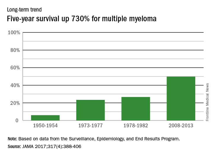
In 2014, there were about 13,000 deaths resulting from multiple myeloma, with 219,000 years of life lost, which ranked 17th among the 29 selected cancers, noted Dr. Mokdad and his associates at the Institute for Health Metrics and Evaluation at the University of Washington, Seattle.
FROM JAMA
AML leads percent gains in 5-year survival among leukemias
Over the 60-year span from the early 1950s to 2013, the 5-year survival rate for all leukemias increased by 500%, according to data from the Surveillance, Epidemiology, and End Results Program.
For 2008-2013, the 5-year relative survival rate for all leukemias was 60.1%, compared with 10% during 1950-1954, said Ali H. Mokdad, PhD, and his associates at the Institute for Health Metrics and Evaluation at the University of Washington, Seattle (JAMA 2017;317[4]:388-406).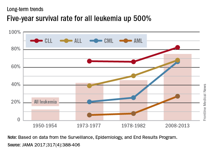
Long view shows doubling of survival in non-Hodgkin lymphoma
Five-year survival for patients with non-Hodgkin lymphoma has more than doubled since the early 1950s, according to Ali H. Mokdad, PhD, and his associates.
Data from the Surveillance, Epidemiology, and End Results Program show that the 5-year relative survival rate for non-Hodgkin lymphoma in the United States went from 33% in 1950-1954 to 71.2% in 2008-2013, an increase of 116%, Dr. Mokdad and his associates reported (JAMA 2017;317[4]:388-406).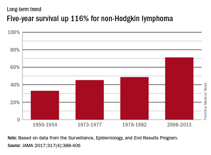
In 2014, mortality for non-Hodgkin lymphoma was the 7th highest among the 29 cancers included in the study, and more than 487,000 years of life were lost, which put it 6th among the 29 cancers, said Dr. Mokdad and his associates from the Institute for Health Metrics and Evaluation at the University of Washington, Seattle.
FROM JAMA