User login
Study reveals crazy quilt of laser laws across the United States
SAN DIEGO – Laser hair removal isn’t typically in an office cleaner’s job description. So it’s no wonder that Virginia legislators were spooked when they heard from a constituent who was treated by a spa worker who turned out to be a janitor.
Earlier this year, legislators in the Old Dominion passed a bill limiting laser hair removal procedures to a “properly trained” medical doctor, physician assistant, or nurse practitioner – or a “properly trained” person who is supervised by one of these professionals. Therefore, it’s still possible for a “properly trained” person without a degree of any kind to operate a laser in Virginia.
To the north in New Jersey, the rules are much stricter: Only physicians can perform laser procedures. But in New York, it appears that anyone can fire up a laser and go to work on unwanted hair. And in Florida, nonphysicians can perform laser procedures only if they’re physician assistants or nurse practitioners. But they’re only allowed to remove hair with lasers at a clinic that just performs laser hair removal.
Such is the chaotic state of laser law in the United States, a new study finds. The rules, which vary widely from state to state, are often vague and confusing. And, as Virginia’s new law shows, they’re still evolving. (The study is current as of March 2016.)

She and study coauthor Mathew M. Avram, MD, JD, director of the Laser and Cosmetic Center at Massachusetts General, analyzed regulations in the 50 states regarding the operation of lasers. They reported their findings at the annual meeting of the American Society for Laser Medicine and Surgery.
Dr. DiGiorgio said that laser operator laws address three issues:
1. Who can operate a laser?
At other clinics across the country, nonphysician employees — such as nurse practitioners and registered nurses – often operate lasers. Whether they can legally actually do so isn’t always obvious.
New Jersey is the only state that requires laser operators to be physicians. At the other extreme, 11 states, including Massachusetts, Colorado, Florida, Missouri, New York, and Pennsylvania, have “no” limits on who can perform laser procedures. (At Massachusetts General Hospital, physicians perform all laser procedures.)
So does that mean anyone can perform a laser procedure? It’s not clear. “The laws are a lot more vague than they should be,” Dr. DiGiorgio said in an interview.
Eighteen states allow people to perform laser procedures as part of the “practice of medicine,” although legislation can be vague on what that means. Those states include Illinois, Michigan, Minnesota, North Carolina, and Texas.
Another 19 states, including California, Ohio, Washington, Wisconsin, and now Virginia, have specific limits on who can perform laser procedures. In California, for example, physician assistants and registered nurses – but not licensed vocational nurses – are allowed to use lasers to remove hair, spider veins, and tattoos. Unlicensed medical assistants, cosmetologists, electrologists, and estheticians are not allowed to perform the procedures
2. Can someone delegate laser procedures to someone else?
In nine states, including Iowa and New Hampshire, there’s no oversight of delegation or nonphysicians can delegate procedures to someone else.
In another nine states, certain procedures can be delegated with no physician oversight, such as laser hair removal in Alaska and ablative procedures (to advanced practice registered nurses only) in Utah.
3. Is supervision required of nonphysicians?
Physicians don’t need to supervise certain laser procedures in 11 states, including Hawaii, Oregon, and Vermont, where they can be performed by a nonphysician with no supervision or under supervision by a non-physician.
In 17 states, supervision isn’t always required or it’s under the discretion of the supervising physician. These states include California, Michigan, Pennsylvania, and Wisconsin.
In 11 states, including Illinois and Massachusetts, only certain procedures require on-site supervision. Six states, including Connecticut and Maryland, require on-site physician supervision for all laser procedures, but Dr. DiGiorgio said the requirements can be vague about what “on site” actually means.
Idaho requires the physician to be on site or immediately available, and South Carolina allows registered nurses to perform laser hair and leg vein removal if a physician is on site and can respond within 5 minutes.
“We don’t know what the ideal regulation is,” Dr. DiGiorgio said. But she believes laser regulations are crucial to safety, especially as fields such as plastic surgery, ophthalmology, and gynecology embrace cosmetic laser procedures.
Information about state-by-state laser operator laws is available on the American Med Spa Association website.
Dr. DiGiorgio reported no relevant disclosures.
SAN DIEGO – Laser hair removal isn’t typically in an office cleaner’s job description. So it’s no wonder that Virginia legislators were spooked when they heard from a constituent who was treated by a spa worker who turned out to be a janitor.
Earlier this year, legislators in the Old Dominion passed a bill limiting laser hair removal procedures to a “properly trained” medical doctor, physician assistant, or nurse practitioner – or a “properly trained” person who is supervised by one of these professionals. Therefore, it’s still possible for a “properly trained” person without a degree of any kind to operate a laser in Virginia.
To the north in New Jersey, the rules are much stricter: Only physicians can perform laser procedures. But in New York, it appears that anyone can fire up a laser and go to work on unwanted hair. And in Florida, nonphysicians can perform laser procedures only if they’re physician assistants or nurse practitioners. But they’re only allowed to remove hair with lasers at a clinic that just performs laser hair removal.
Such is the chaotic state of laser law in the United States, a new study finds. The rules, which vary widely from state to state, are often vague and confusing. And, as Virginia’s new law shows, they’re still evolving. (The study is current as of March 2016.)
She and study coauthor Mathew M. Avram, MD, JD, director of the Laser and Cosmetic Center at Massachusetts General, analyzed regulations in the 50 states regarding the operation of lasers. They reported their findings at the annual meeting of the American Society for Laser Medicine and Surgery.
Dr. DiGiorgio said that laser operator laws address three issues:
1. Who can operate a laser?
At other clinics across the country, nonphysician employees — such as nurse practitioners and registered nurses – often operate lasers. Whether they can legally actually do so isn’t always obvious.
New Jersey is the only state that requires laser operators to be physicians. At the other extreme, 11 states, including Massachusetts, Colorado, Florida, Missouri, New York, and Pennsylvania, have “no” limits on who can perform laser procedures. (At Massachusetts General Hospital, physicians perform all laser procedures.)
So does that mean anyone can perform a laser procedure? It’s not clear. “The laws are a lot more vague than they should be,” Dr. DiGiorgio said in an interview.
Eighteen states allow people to perform laser procedures as part of the “practice of medicine,” although legislation can be vague on what that means. Those states include Illinois, Michigan, Minnesota, North Carolina, and Texas.
Another 19 states, including California, Ohio, Washington, Wisconsin, and now Virginia, have specific limits on who can perform laser procedures. In California, for example, physician assistants and registered nurses – but not licensed vocational nurses – are allowed to use lasers to remove hair, spider veins, and tattoos. Unlicensed medical assistants, cosmetologists, electrologists, and estheticians are not allowed to perform the procedures
2. Can someone delegate laser procedures to someone else?
In nine states, including Iowa and New Hampshire, there’s no oversight of delegation or nonphysicians can delegate procedures to someone else.
In another nine states, certain procedures can be delegated with no physician oversight, such as laser hair removal in Alaska and ablative procedures (to advanced practice registered nurses only) in Utah.
3. Is supervision required of nonphysicians?
Physicians don’t need to supervise certain laser procedures in 11 states, including Hawaii, Oregon, and Vermont, where they can be performed by a nonphysician with no supervision or under supervision by a non-physician.
In 17 states, supervision isn’t always required or it’s under the discretion of the supervising physician. These states include California, Michigan, Pennsylvania, and Wisconsin.
In 11 states, including Illinois and Massachusetts, only certain procedures require on-site supervision. Six states, including Connecticut and Maryland, require on-site physician supervision for all laser procedures, but Dr. DiGiorgio said the requirements can be vague about what “on site” actually means.
Idaho requires the physician to be on site or immediately available, and South Carolina allows registered nurses to perform laser hair and leg vein removal if a physician is on site and can respond within 5 minutes.
“We don’t know what the ideal regulation is,” Dr. DiGiorgio said. But she believes laser regulations are crucial to safety, especially as fields such as plastic surgery, ophthalmology, and gynecology embrace cosmetic laser procedures.
Information about state-by-state laser operator laws is available on the American Med Spa Association website.
Dr. DiGiorgio reported no relevant disclosures.
AT LASER 2017
Key clinical point:
Major finding: The study found wide variations in who can operate lasers, and in regulations regarding the delegation and supervision of laser treatments in the different states.
Data source: Analysis of regulations in the 50 states regarding the operation of lasers.
Disclosures: Dr. DiGiorgio reported no relevant disclosures
What, you never filled the prescription?!
How many times do you come out of the exam room after seeing a patient in follow-up and heave a sigh because the parents did not give their child the medicine as you prescribed it?
Without adherence to the medication plan, a lot of suboptimal outcomes can and do occur. A urinary tract infection may come back partially treated, requiring a more extensive work-up. A strep infection may spread to family members. Inflammatory bowel disease may require bowel resection. Asthma may simmer with long-term inflammation and pulmonary compromise as well as concurrent activity limitations. Often children with asthma are given less than 50% of prescribed controller medicines. In one pediatric study, medication adherence was not even asked about in 66% of cases. In adults, 20%-30% of prescriptions are never filled.
As physicians, we are carefully schooled in making complex diagnoses, sorting out and prioritizing the laboratory work-up, and memorizing the latest and most effective treatment regimens. What is rarely taught, however, is how to conduct the conversation needed to optimize subsequent adherence to the medication plan.
Problem-solving counseling is an evidence-based method to improve medication adherence. This is a semistructured form of cognitive-behavioral intervention designed to engage the responsible person (parent or child) in shared decision making about whether and how to take medication, and which one to take. After all, for good or for bad, it is really their choice!

1. Problem definition. This step involves developing a clear and specific definition of the problem. Educating families about a medical condition has to start with asking what they already know. This often includes sagas of bad outcomes in relatives. Ask: Who do you know with asthma? How was it for them? The family needs to know symptoms, simple pathophysiology (such as inflammation you can’t see or feel), course, and prognosis. They also need to know where their child’s condition falls on the continuum. And they need to understand the essential prevention aspect of controllers in what appears to be an asymptomatic child. Failure to communicate this is a common reason for nonadherence in asthma.
2. Generation of alternatives. This involves brainstorming to identify multiple and creative solutions. This step will reveal past experiences as well as things the family learned on the Internet that may be true and relevant, or true but irrelevant, or false. Ask: What have you heard about treatments for asthma? What do you think would be best for your child? Generic handouts with a sampling of medicines, advantages and disadvantages, side effects, and costs of the main choices have been shown to be helpful guides that enhance adherence through empowering the family in their choice and reassuring family members that you have been thorough. It can be a balancing act to describe possible side effects without scaring the family into shutting down and being unable to make any choice at all. However, failure to discuss common effects they may notice – such as a racing heart from rescue medications – but that you think of as trivial, may also lead to nonadherence. A way to communicate about perceived side effects and manage them has to be part of an effective plan. Planning a phone or email check-in can make a big difference.
3. Decision making. This step involves evaluating all the solutions to identify the most effective and feasible option. Once the family understands the problem and the alternatives, it is crucial for you, as the physician, to not only ask their preference but be ready to suggest what you think would be best. While not wanting to be patronized, families want your opinion. I like to have family members close their eyes and visualize carrying out the selected routine. This is a good hypnotic technique for future remembering, but you also may discover important facts by this simple exercise, such as that the child gets up alone for school, making morning dosing unreliable. Shared decision making is not a way to abdicate your expert opinion, just to incorporate family preferences and factors.
4. Solution implementation. This step involves carrying out the plan. There is no substitute for a real life trial! There may be surprising issues: Autistic children may be afraid of a nebulizer machine. Sensitive children may refuse the flavor of some inhalers.
5. Solution verification. Evaluate the effectiveness of the solution and modify the plan as necessary. Follow-up contact is crucial, especially at the start of a new chronic medication plan. When families know that the plan can be changed if things do not go well or they change their minds, they will be less fearful of giving it a try and more honest about barriers they perceive or encounter rather than simply showing up at the next visit with the child’s condition out of control.
Although using problem-solving counseling may sound complicated, it is intended to be focused and brief, and has been shown to be feasibly done in the clinic by primary care providers, without lengthening the visit. CHADIS even has teleprompter text specific to parent-reported barriers to help you.
Even when family members understand and agrees to a medication plan during the visit, there are a variety of reasons they may not adhere to it. They may forget to give the medicine, be unable to afford it once prescribed, experience unpleasant side effects, encounter resistance from the child, or get unanticipated push back from family members. All of these issues can be addressed if you know that they happen. You just have to ask! Recommending smartphone reminders or reminder apps (such as Medisafe), using GoodRx to find cheaper sources, suggesting candy as a chaser, recommending behavior strategies for feisty kids, and providing written materials (or a phone call) for reluctant relatives are strategies you can prepare in advance to have in your quiver and are well worth your time.
If it weren’t enough to address adherence to optimize outcomes, asthma management and control will likely be a Clinical Quality Measure, determining how we will be paid starting this year. Now you have a tool to do it!
Dr. Howard is assistant professor of pediatrics at Johns Hopkins University, Baltimore, and creator of CHADIS (www.CHADIS.com). She had no other relevant disclosures. Dr. Howard’s contribution to this publication was as a paid expert to Frontline Medical News.
How many times do you come out of the exam room after seeing a patient in follow-up and heave a sigh because the parents did not give their child the medicine as you prescribed it?
Without adherence to the medication plan, a lot of suboptimal outcomes can and do occur. A urinary tract infection may come back partially treated, requiring a more extensive work-up. A strep infection may spread to family members. Inflammatory bowel disease may require bowel resection. Asthma may simmer with long-term inflammation and pulmonary compromise as well as concurrent activity limitations. Often children with asthma are given less than 50% of prescribed controller medicines. In one pediatric study, medication adherence was not even asked about in 66% of cases. In adults, 20%-30% of prescriptions are never filled.
As physicians, we are carefully schooled in making complex diagnoses, sorting out and prioritizing the laboratory work-up, and memorizing the latest and most effective treatment regimens. What is rarely taught, however, is how to conduct the conversation needed to optimize subsequent adherence to the medication plan.
Problem-solving counseling is an evidence-based method to improve medication adherence. This is a semistructured form of cognitive-behavioral intervention designed to engage the responsible person (parent or child) in shared decision making about whether and how to take medication, and which one to take. After all, for good or for bad, it is really their choice!
1. Problem definition. This step involves developing a clear and specific definition of the problem. Educating families about a medical condition has to start with asking what they already know. This often includes sagas of bad outcomes in relatives. Ask: Who do you know with asthma? How was it for them? The family needs to know symptoms, simple pathophysiology (such as inflammation you can’t see or feel), course, and prognosis. They also need to know where their child’s condition falls on the continuum. And they need to understand the essential prevention aspect of controllers in what appears to be an asymptomatic child. Failure to communicate this is a common reason for nonadherence in asthma.
2. Generation of alternatives. This involves brainstorming to identify multiple and creative solutions. This step will reveal past experiences as well as things the family learned on the Internet that may be true and relevant, or true but irrelevant, or false. Ask: What have you heard about treatments for asthma? What do you think would be best for your child? Generic handouts with a sampling of medicines, advantages and disadvantages, side effects, and costs of the main choices have been shown to be helpful guides that enhance adherence through empowering the family in their choice and reassuring family members that you have been thorough. It can be a balancing act to describe possible side effects without scaring the family into shutting down and being unable to make any choice at all. However, failure to discuss common effects they may notice – such as a racing heart from rescue medications – but that you think of as trivial, may also lead to nonadherence. A way to communicate about perceived side effects and manage them has to be part of an effective plan. Planning a phone or email check-in can make a big difference.
3. Decision making. This step involves evaluating all the solutions to identify the most effective and feasible option. Once the family understands the problem and the alternatives, it is crucial for you, as the physician, to not only ask their preference but be ready to suggest what you think would be best. While not wanting to be patronized, families want your opinion. I like to have family members close their eyes and visualize carrying out the selected routine. This is a good hypnotic technique for future remembering, but you also may discover important facts by this simple exercise, such as that the child gets up alone for school, making morning dosing unreliable. Shared decision making is not a way to abdicate your expert opinion, just to incorporate family preferences and factors.
4. Solution implementation. This step involves carrying out the plan. There is no substitute for a real life trial! There may be surprising issues: Autistic children may be afraid of a nebulizer machine. Sensitive children may refuse the flavor of some inhalers.
5. Solution verification. Evaluate the effectiveness of the solution and modify the plan as necessary. Follow-up contact is crucial, especially at the start of a new chronic medication plan. When families know that the plan can be changed if things do not go well or they change their minds, they will be less fearful of giving it a try and more honest about barriers they perceive or encounter rather than simply showing up at the next visit with the child’s condition out of control.
Although using problem-solving counseling may sound complicated, it is intended to be focused and brief, and has been shown to be feasibly done in the clinic by primary care providers, without lengthening the visit. CHADIS even has teleprompter text specific to parent-reported barriers to help you.
Even when family members understand and agrees to a medication plan during the visit, there are a variety of reasons they may not adhere to it. They may forget to give the medicine, be unable to afford it once prescribed, experience unpleasant side effects, encounter resistance from the child, or get unanticipated push back from family members. All of these issues can be addressed if you know that they happen. You just have to ask! Recommending smartphone reminders or reminder apps (such as Medisafe), using GoodRx to find cheaper sources, suggesting candy as a chaser, recommending behavior strategies for feisty kids, and providing written materials (or a phone call) for reluctant relatives are strategies you can prepare in advance to have in your quiver and are well worth your time.
If it weren’t enough to address adherence to optimize outcomes, asthma management and control will likely be a Clinical Quality Measure, determining how we will be paid starting this year. Now you have a tool to do it!
Dr. Howard is assistant professor of pediatrics at Johns Hopkins University, Baltimore, and creator of CHADIS (www.CHADIS.com). She had no other relevant disclosures. Dr. Howard’s contribution to this publication was as a paid expert to Frontline Medical News.
Medicine’s revenge against traveler’s diarrhea
Traveler’s diarrhea (TD) is the most common illness in people traveling from resource-advantaged countries to resource-limited regions of the globe. Approximately 50% of these types of travelers will contract diarrhea.
I knew of a group of infectious disease experts traveling to India together – presumably well-versed in how to avoid such things – and one-half of the group developed it.
I have many patients sending me electronic messages asking me for their standard 3-day ciprofloxacin prescriptions, “just in case.” I am guilty of having provided this, along with warnings that we could make matters worse by giving them Clostridium difficile colitis. Antibiotics may also increase the risk for post-TD irritable bowel syndrome, which can occur in up to 15% of patients.

Mild TD is defined as passage of one or two unformed stools in 24 hours without nausea, vomiting, abdominal pain, fever, or blood in stools. What is the evidence for the effectiveness of antibiotics, compared with other interventions such as loperamide, for mild TD?
Tinja Lääveri, MD, of the University of Helsinki, and colleagues conducted a systematic review on the efficacy and safety of loperamide for TD (Travel Med Infect Dis. 2016 Jul-Aug;14[4]:299-312). Fifteen articles were retrieved.
Loperamide was observed to be noninferior to antibiotics for the treatment of TD. In one study, loperamide was observed to be superior to bismuth, which is commonly recommended to prevent TD. Adverse events with loperamide occurred predominantly among patients with bloody diarrhea.
The authors remind us that loperamide is different from antimotility agents such as diphenoxylate with atropine, as loperamide has an antisecretory effect at lower doses and decreases motility only at higher doses.
If you subscribe to the idea that diarrhea helps rid the body of toxins, be reminded that secretory diarrhea is exploited by the organism to spread the infestation to as many humans as possible.
The recommended regimen for loperamide is a 4-mg loading dose, followed by 2 mg after every episode of diarrhea. Tell your patients not to use loperamide if they are having fever or bloody diarrhea, or if you suspect they could have C. difficile colitis (that is, recent antibiotics or other risk factors).
Happy travels.
Dr. Ebbert is professor of medicine, a general internist at the Mayo Clinic in Rochester, Minn., and a diplomate of the American Board of Addiction Medicine. The opinions expressed are those of the author and do not necessarily represent the views and opinions of the Mayo Clinic. The opinions expressed in this article should not be used to diagnose or treat any medical condition, nor should they be used as a substitute for medical advice from a qualified, board-certified practicing clinician. Dr. Ebbert has no relevant financial disclosures about this article.
Traveler’s diarrhea (TD) is the most common illness in people traveling from resource-advantaged countries to resource-limited regions of the globe. Approximately 50% of these types of travelers will contract diarrhea.
I knew of a group of infectious disease experts traveling to India together – presumably well-versed in how to avoid such things – and one-half of the group developed it.
I have many patients sending me electronic messages asking me for their standard 3-day ciprofloxacin prescriptions, “just in case.” I am guilty of having provided this, along with warnings that we could make matters worse by giving them Clostridium difficile colitis. Antibiotics may also increase the risk for post-TD irritable bowel syndrome, which can occur in up to 15% of patients.
Mild TD is defined as passage of one or two unformed stools in 24 hours without nausea, vomiting, abdominal pain, fever, or blood in stools. What is the evidence for the effectiveness of antibiotics, compared with other interventions such as loperamide, for mild TD?
Tinja Lääveri, MD, of the University of Helsinki, and colleagues conducted a systematic review on the efficacy and safety of loperamide for TD (Travel Med Infect Dis. 2016 Jul-Aug;14[4]:299-312). Fifteen articles were retrieved.
Loperamide was observed to be noninferior to antibiotics for the treatment of TD. In one study, loperamide was observed to be superior to bismuth, which is commonly recommended to prevent TD. Adverse events with loperamide occurred predominantly among patients with bloody diarrhea.
The authors remind us that loperamide is different from antimotility agents such as diphenoxylate with atropine, as loperamide has an antisecretory effect at lower doses and decreases motility only at higher doses.
If you subscribe to the idea that diarrhea helps rid the body of toxins, be reminded that secretory diarrhea is exploited by the organism to spread the infestation to as many humans as possible.
The recommended regimen for loperamide is a 4-mg loading dose, followed by 2 mg after every episode of diarrhea. Tell your patients not to use loperamide if they are having fever or bloody diarrhea, or if you suspect they could have C. difficile colitis (that is, recent antibiotics or other risk factors).
Happy travels.
Dr. Ebbert is professor of medicine, a general internist at the Mayo Clinic in Rochester, Minn., and a diplomate of the American Board of Addiction Medicine. The opinions expressed are those of the author and do not necessarily represent the views and opinions of the Mayo Clinic. The opinions expressed in this article should not be used to diagnose or treat any medical condition, nor should they be used as a substitute for medical advice from a qualified, board-certified practicing clinician. Dr. Ebbert has no relevant financial disclosures about this article.
International travel vaccination updates
There are several things you should know about necessary vaccinations, and sometimes potential supply problems, if your families will be traveling internationally.
Yellow fever and vaccine supply
Yellow fever is caused by a Flavivirus transmitted by the bite of an infected mosquito. It occurs in sub-Saharan Africa and in tropical areas in South America. Multiple factors determine a traveler’s risk for acquisition, including destination, season, duration of potential exposure, activities, and the local transmission rate. The majority of those infected are asymptomatic or have minimal clinical symptoms. The incubation period is 3-6 days, which is then followed by an influenza-like illness. Approximately 15% of infected individuals develop more serious symptoms including jaundice, hemorrhagic symptoms, shock, and, ultimately, multiorgan system failure with a fatality rate of 90%. There is no specific treatment.

Previously, vaccine boosters were required every 10 years. However, the duration of immunity was extensively reviewed by the World Health Organization and effective July 11, 2016, boosters are no longer required. A single dose of vaccine is now valid for the lifetime of the individual. This includes those persons vaccinated prior to July 11, 2016. Since it is a live vaccine, administration is contraindicated in certain individuals. Exemption letters are provided for those who have a medical contraindication.
Caution is advised in persons receiving their initial dose of YF-VAX who are older than 60 years of age because they have an increased risk of serious side effects. This is not a concern for the pediatrician. The vaccine can only be administered at state approved facilities. It is one vaccine that is not only recommended, but may be required for entry into certain countries. Go to www.cdc.gov/yellowfever for a complete list.
Sanofi Pasteur is the only U.S. manufacturer of YF-VAX. Production has ceased until mid-2018, when a new manufacturing facility will open. Current supplies are anticipated to be depleted by mid-2017, and orders have been limited to 5 doses per month. Sanofi Pasteur, in conjunction with the Food and Drug Administration, will make Stamaril – a yellow fever vaccine manufactured by the company in France and licensed in over 70 countries – available to U.S. travelers through an Expanded Access Investigational New Drug Application. Details on how and when this program will be operational are forthcoming. What is known is that, nationwide, there will be a limited number of sites administering Stamaril. Once finalized, a list of locations will be posted on the CDC Yellow Fever site.
How does this affect your patients? If travel to a yellow fever risk area is anticipated, they should not delay in seeking pretravel advice and immunizations until the last minute. Individual clinic inventories will not be stable. Postponing a trip or changing destinations is preferred if the vaccine is not available. Yellow fever exemption letters are only provided for those persons who have a medical contraindication to receive YF-VAX.
Zika, dengue, and chikungunya
These three Flaviviruses all are transmitted by mosquitoes and can present with fever, rash, and headache. Their distribution is overlapping in several parts of the world. Most infected people are asymptomatic. If symptoms develop, they usually are self-limited. Disease prevention is by mosquito avoidance. There are no preventive vaccines.
Zika virus is the only one associated with a congenital syndrome. It is characterized by brain abnormalities with or without microcephaly, neural tube defects, and ocular abnormalities.
Guidelines for the evaluation and management of Zika virus–exposed infants were initially published in January, 2016, with the most recent update published in August 2016 (MMWR Morb Mortal Wkly Rep. 2016 Aug 26;65[33]:870-8).
Preliminary data from the U.S. Zika pregnancy registry of 442 completed pregnancies between Jan. 15 to Sept. 22, 2016, identified birth defects in 26 fetuses/ infants (6%). There were 21 infants with birth defects among 395 live births and 5 fetuses with birth defects among 47 pregnancy losses. Birth defects were reported for 16 of 271 (6%) asymptomatic and 10 of 167 (6%) symptomatic women. There were no birth defects in infants when exposure occurred after the first trimester. Of the 26 affected infants, 4 had microcephaly and no neuroimaging and 3 (12%) had no fetal or infant testing. Approximately 41% (82/442) of infants did not have Zika virus testing (JAMA. 2017 Jan 3;317[1]:59-68).
It is unclear why testing was not performed. One concern is that the pediatrician may not have been aware of the maternal Zika virus exposure or test results. It may behoove us to begin asking questions about parental international travel to provide optimal management for our patients. We also should be familiar with the current guidelines for evaluating any potentially exposed infants, which include postnatal neuroimaging, Zika virus testing, a comprehensive newborn examination including neurologic exam, and a standard newborn hearing screen prior to hospital discharge.
Regardless of maternal Zika virus test results, infants with any clinical findings suggestive of congenital Zika virus syndrome and possible maternal exposure based on epidemiologic link also should be tested. Zika virus travel alerts and the most up to date information can be found on the Centers for Disease Control and Prevention website (www. cdc.gov/Zika).

Measles
Although endemic measles was eliminated in the United States in 2000, it is still common in many countries in Europe, Africa, and the Pacific. Most cases in the United States occur in unvaccinated individuals, with 78 cases reported in 2016. As of March 25, 2017, 28 cases have been reported. At least 10 countries – including Belgium, France, Italy, Germany, Portugal, and Thailand – have reported outbreaks of measles since April 2017. As reminder, all children aged 6-11 months should receive one dose of MMR and those 12 months or older should receive two doses of MMR at least 28 days apart if international travel is planned. Adults born after 1956 also should have received two doses of MMR prior to international travel.
Dr. Word is a pediatric infectious disease specialist and director of the Houston Travel Medicine Clinic. She reported having no relevant financial disclosures.
There are several things you should know about necessary vaccinations, and sometimes potential supply problems, if your families will be traveling internationally.
Yellow fever and vaccine supply
Yellow fever is caused by a Flavivirus transmitted by the bite of an infected mosquito. It occurs in sub-Saharan Africa and in tropical areas in South America. Multiple factors determine a traveler’s risk for acquisition, including destination, season, duration of potential exposure, activities, and the local transmission rate. The majority of those infected are asymptomatic or have minimal clinical symptoms. The incubation period is 3-6 days, which is then followed by an influenza-like illness. Approximately 15% of infected individuals develop more serious symptoms including jaundice, hemorrhagic symptoms, shock, and, ultimately, multiorgan system failure with a fatality rate of 90%. There is no specific treatment.
Previously, vaccine boosters were required every 10 years. However, the duration of immunity was extensively reviewed by the World Health Organization and effective July 11, 2016, boosters are no longer required. A single dose of vaccine is now valid for the lifetime of the individual. This includes those persons vaccinated prior to July 11, 2016. Since it is a live vaccine, administration is contraindicated in certain individuals. Exemption letters are provided for those who have a medical contraindication.
Caution is advised in persons receiving their initial dose of YF-VAX who are older than 60 years of age because they have an increased risk of serious side effects. This is not a concern for the pediatrician. The vaccine can only be administered at state approved facilities. It is one vaccine that is not only recommended, but may be required for entry into certain countries. Go to www.cdc.gov/yellowfever for a complete list.
Sanofi Pasteur is the only U.S. manufacturer of YF-VAX. Production has ceased until mid-2018, when a new manufacturing facility will open. Current supplies are anticipated to be depleted by mid-2017, and orders have been limited to 5 doses per month. Sanofi Pasteur, in conjunction with the Food and Drug Administration, will make Stamaril – a yellow fever vaccine manufactured by the company in France and licensed in over 70 countries – available to U.S. travelers through an Expanded Access Investigational New Drug Application. Details on how and when this program will be operational are forthcoming. What is known is that, nationwide, there will be a limited number of sites administering Stamaril. Once finalized, a list of locations will be posted on the CDC Yellow Fever site.
How does this affect your patients? If travel to a yellow fever risk area is anticipated, they should not delay in seeking pretravel advice and immunizations until the last minute. Individual clinic inventories will not be stable. Postponing a trip or changing destinations is preferred if the vaccine is not available. Yellow fever exemption letters are only provided for those persons who have a medical contraindication to receive YF-VAX.
Zika, dengue, and chikungunya
These three Flaviviruses all are transmitted by mosquitoes and can present with fever, rash, and headache. Their distribution is overlapping in several parts of the world. Most infected people are asymptomatic. If symptoms develop, they usually are self-limited. Disease prevention is by mosquito avoidance. There are no preventive vaccines.
Zika virus is the only one associated with a congenital syndrome. It is characterized by brain abnormalities with or without microcephaly, neural tube defects, and ocular abnormalities.
Guidelines for the evaluation and management of Zika virus–exposed infants were initially published in January, 2016, with the most recent update published in August 2016 (MMWR Morb Mortal Wkly Rep. 2016 Aug 26;65[33]:870-8).
Preliminary data from the U.S. Zika pregnancy registry of 442 completed pregnancies between Jan. 15 to Sept. 22, 2016, identified birth defects in 26 fetuses/ infants (6%). There were 21 infants with birth defects among 395 live births and 5 fetuses with birth defects among 47 pregnancy losses. Birth defects were reported for 16 of 271 (6%) asymptomatic and 10 of 167 (6%) symptomatic women. There were no birth defects in infants when exposure occurred after the first trimester. Of the 26 affected infants, 4 had microcephaly and no neuroimaging and 3 (12%) had no fetal or infant testing. Approximately 41% (82/442) of infants did not have Zika virus testing (JAMA. 2017 Jan 3;317[1]:59-68).
It is unclear why testing was not performed. One concern is that the pediatrician may not have been aware of the maternal Zika virus exposure or test results. It may behoove us to begin asking questions about parental international travel to provide optimal management for our patients. We also should be familiar with the current guidelines for evaluating any potentially exposed infants, which include postnatal neuroimaging, Zika virus testing, a comprehensive newborn examination including neurologic exam, and a standard newborn hearing screen prior to hospital discharge.
Regardless of maternal Zika virus test results, infants with any clinical findings suggestive of congenital Zika virus syndrome and possible maternal exposure based on epidemiologic link also should be tested. Zika virus travel alerts and the most up to date information can be found on the Centers for Disease Control and Prevention website (www. cdc.gov/Zika).
Measles
Although endemic measles was eliminated in the United States in 2000, it is still common in many countries in Europe, Africa, and the Pacific. Most cases in the United States occur in unvaccinated individuals, with 78 cases reported in 2016. As of March 25, 2017, 28 cases have been reported. At least 10 countries – including Belgium, France, Italy, Germany, Portugal, and Thailand – have reported outbreaks of measles since April 2017. As reminder, all children aged 6-11 months should receive one dose of MMR and those 12 months or older should receive two doses of MMR at least 28 days apart if international travel is planned. Adults born after 1956 also should have received two doses of MMR prior to international travel.
Dr. Word is a pediatric infectious disease specialist and director of the Houston Travel Medicine Clinic. She reported having no relevant financial disclosures.
VIDEO: Big research trials at AAN bring up important cost decisions
BOSTON – Some of the most influential clinical research reports coming out of the annual meeting of the American Academy of Neurology raise questions on how neurologists will strike a balance between the improved efficacy and safety of drugs in new therapeutic classes and their affordability for patients.
Natalia Rost, MD, vice chair of the AAN Science Committee, discussed phase III clinical trials (ARISE and STRIVE) in episodic migraine with erenumab, an investigational humanized monoclonal antibody against calcitonin gene-related peptide receptor; phase III clinical trials (ENDEAR and CHERISH) of the antisense oligonucleotide drug nusinersen (Spinraza) that was approved by the Food and Drug Administration for spinal muscular atrophy in late 2016; as well as phase III trials of a pharmaceutical-grade extract of the cannabis-derived compound cannabidiol in patients with Dravet syndrome and Lennox-Gastaut syndrome.
Erenumab and nusinersen are “disease-specific targeted biologics” that have been developed over decades to target a specific disease pathway, and hence translate into high prices, Dr. Rost said in a video interview at the meeting.
“How you value the cost of a drug against improvement in a physiological outcome is very difficult to measure,” she noted, for relatively small gains in reducing migraine days per month and improvements in functional outcome and disability against placebo.
But this calculation is different with the potentially lifesaving effects of nusinersen for spinal muscular atrophy patients, in which “we’re not talking about days of improvement, we’re talking about days of life,” said Dr. Rost, director of acute stroke services at Massachusetts General Hospital, Boston. “And so that becomes an ethical dilemma in terms of the cost of administration, who is paying for the drug, and how this is covered. Whom do you offer treatment to?”
The development of cannabidiol as a potential adjunctive treatment for Dravet and Lennox-Gastaut syndromes is a welcome addition to the armamentarium against these conditions, Dr. Rost added, because it offers an alternative to the unregulated use of herbal medications and supplements – particularly cannabis in its various forms – that patients ask about but are difficult to dose consistently and to ensure a pharmaceutical-grade level of purity.
The video associated with this article is no longer available on this site. Please view all of our videos on the MDedge YouTube channel
AT AAN 2017

Will Targeting Alpha-Synuclein Lead to Effective Parkinson’s Disease Therapies?
MIAMI—The possibility that Parkinson’s disease is a prion disorder has led investigators to begin clinical trials of drugs that target misfolded α-synuclein, according to an overview provided at the First Pan American Parkinson’s Disease and Movement Disorders Congress. Other potential therapies targeting the protein are in preclinical studies.

Researchers are exploring four main treatment approaches: immunization, enhancing protein clearance, knocking down host α-synuclein, and inhibiting the prion conformer reaction, said C. Warren Olanow, MD, Professor and Chairman Emeritus of the Department of Neurology and Professor Emeritus of Neuroscience at the Mount Sinai School of Medicine in New York City.
Inhibiting the prion conformer reaction is the approach that Dr. Olanow believes is most likely to be effective. “But I also strongly believe every one of these approaches needs to be tested,” he said.
The Prion Hypothesis
A prion is an infectious particle composed solely of misfolded protein. It replicates by a process whereby the misfolded protein causes the native unfolded protein to misfold through a process called the prion conformer reaction. Misfolded proteins form beta-rich sheets that contain oligomers and rods, which are thought to be toxic, and polymerize to form aggregates. Prions can cause neurodegeneration and be transmitted from one species to another.
Researchers “have shown that α-synuclein can … replicate the prion biology,” and there are many reasons to suspect that α-synuclein “is integral to the pathology” of Parkinson’s disease, Dr. Olanow said. For instance, mutations as well as excess levels of wild type α-synuclein can cause Parkinson’s disease.
An α-synuclein monomer can misfold for various reasons, including a genetic mutation, a toxic or inflammatory event, or sporadically by chance. “As you get older, more and more proteins spontaneously misfold,” Dr. Olanow said.
The ubiquitin–proteasome and the autophagy–lysosomal systems normally clear misfolded proteins. If these systems cannot adequately clear the misfolded proteins—either because the clearance systems are not working enough or too much protein is being formed—the aggregates accumulate and can block the clearance systems, creating a vicious cycle that leads to further accumulation of misfolded α-synuclein, he said.
Removing Toxic Protein
One therapeutic approach based on the prion hypothesis involves targeting and removing toxic α-synuclein species by way of active or passive immunization.
Tran et al reported that α-synuclein immunotherapy blocks propagation of misfolded α-synuclein and neurodegeneration in an animal model. Several immune therapies are currently being tested in clinical trials. Trials by Prothena and Biogen are using passive immunization (ie, administering monoclonal antibodies targeted to a specific α-synuclein species), whereas trials by AFFiRiS are using active immunization (ie, stimulating the immune system to produce antibodies). “The good news so far is that there has been no major safety issue or tolerability concern,” Dr. Olanow said. “There have not, however, been long-term safety or efficacy data as yet.”
Challenges in immunization trials include uncertainty about whether antibodies reach the brain and which α-synuclein species should be targeted. Some α-synuclein species may be protective, said Dr. Olanow. “By no means are we assured that we are targeting exactly what we want.”
Enhancing Protein Clearance
Another approach entails facilitating protein clearance through the ubiquitin–proteasome system or the autophagy–lysosomal system, which are defective in Parkinson’s disease. Drugs that activate or enhance glucocerebrosidase (GCase) activity and promote lysosomal function have attracted particular attention.
Mutations in GBA, the gene that encodes for GCase, can cause Gaucher disease with features of parkinsonism, and as many as 10% of patients with Parkinson’s disease have GBA mutations. GCase levels are reduced by as much as 50% in patients with Parkinson’s disease.
When GCase is decreased, lysosomal function is reduced, and α-synuclein consequently increases. Likewise, overexpression of GCase can reduce α-synuclein, and overexpression of α-synuclein reduces GCase. Ambroxol, a molecular chaperone that has been shown experimentally to increase GCase levels, has entered clinical trials. More robust GCase-promoting agents are in development and will enter clinical trials in the near future, Dr. Olanow said.
Removing Host Protein
A third approach involves knockdown of host α-synuclein with, for example, iRNAs and antisense oligonucleotides directed at α-synuclein production or with chemicals that bind to α-synuclein. “If you knock down host α-synuclein, then you do not have any to participate in that prion conformer reaction,” he said.
Investigators are using high-throughput screening to identify chemicals that bind to α-synuclein. However, α-synuclein’s physiologic role is not completely known, nor is what happens if the protein is lowered in humans, or by how much α-synuclein would need to be reduced to provide benefit.
Blocking the Prion Conformer Reaction
If neurologists could block the prion conformer reaction, it might be the best approach. “We could stop the formation of misfolded protein, and we could preserve the host α-synuclein and its physiologic function,” Dr. Olanow said. “The problem is that we do not know the precise mechanism underlying the presumed templating in Parkinson’s disease.”
Any attempts to treat Parkinson’s disease with therapies aimed at α-synuclein likely should be initiated as early as possible in the disease process, and biomarkers will be crucial to initiating timely treatment, Dr. Olanow noted.
“This, to me, is one of the most exciting periods in Parkinson’s therapeutics,” he said. Over the next decade, neurologists may see “a revolution of therapies directed at α-synuclein that may ultimately prove to be disease modifying and fundamentally change the nature of the disease.”
—Jake Remaly
Suggested Reading
Dehay B, Bourdenx M, Gorry P, et al. Targeting α-synuclein for treating Parkinson’s disease: mechanistic and therapeutic considerations. Lancet Neurol. 2015;14(8):855-866.
Olanow CW, Kordower JH. Targeting α-synuclein as a therapy for Parkinson’s disease: The battle begins. Mov Disord. 2017;32(2):203-207.
Olanow CW, Brundin P. Parkinson’s disease and alpha synuclein: is Parkinson’s disease a prion-like disorder? Mov Disord. 2013;28(1):31-40.
Schenk DB, Koller M, Ness DK, et al. First-in-human assessment of PRX002, an anti-α-synuclein monoclonal antibody, in healthy volunteers. Mov Disord. 2017;32(2):211-218.
Tran HT, Chung CH, Iba M, et al. α-synuclein immunotherapy blocks uptake and templated propagation of misfolded α-synuclein and neurodegeneration. Cell Rep. 2014;7(6):2054-2065.
MIAMI—The possibility that Parkinson’s disease is a prion disorder has led investigators to begin clinical trials of drugs that target misfolded α-synuclein, according to an overview provided at the First Pan American Parkinson’s Disease and Movement Disorders Congress. Other potential therapies targeting the protein are in preclinical studies.
Researchers are exploring four main treatment approaches: immunization, enhancing protein clearance, knocking down host α-synuclein, and inhibiting the prion conformer reaction, said C. Warren Olanow, MD, Professor and Chairman Emeritus of the Department of Neurology and Professor Emeritus of Neuroscience at the Mount Sinai School of Medicine in New York City.
Inhibiting the prion conformer reaction is the approach that Dr. Olanow believes is most likely to be effective. “But I also strongly believe every one of these approaches needs to be tested,” he said.
The Prion Hypothesis
A prion is an infectious particle composed solely of misfolded protein. It replicates by a process whereby the misfolded protein causes the native unfolded protein to misfold through a process called the prion conformer reaction. Misfolded proteins form beta-rich sheets that contain oligomers and rods, which are thought to be toxic, and polymerize to form aggregates. Prions can cause neurodegeneration and be transmitted from one species to another.
Researchers “have shown that α-synuclein can … replicate the prion biology,” and there are many reasons to suspect that α-synuclein “is integral to the pathology” of Parkinson’s disease, Dr. Olanow said. For instance, mutations as well as excess levels of wild type α-synuclein can cause Parkinson’s disease.
An α-synuclein monomer can misfold for various reasons, including a genetic mutation, a toxic or inflammatory event, or sporadically by chance. “As you get older, more and more proteins spontaneously misfold,” Dr. Olanow said.
The ubiquitin–proteasome and the autophagy–lysosomal systems normally clear misfolded proteins. If these systems cannot adequately clear the misfolded proteins—either because the clearance systems are not working enough or too much protein is being formed—the aggregates accumulate and can block the clearance systems, creating a vicious cycle that leads to further accumulation of misfolded α-synuclein, he said.
Removing Toxic Protein
One therapeutic approach based on the prion hypothesis involves targeting and removing toxic α-synuclein species by way of active or passive immunization.
Tran et al reported that α-synuclein immunotherapy blocks propagation of misfolded α-synuclein and neurodegeneration in an animal model. Several immune therapies are currently being tested in clinical trials. Trials by Prothena and Biogen are using passive immunization (ie, administering monoclonal antibodies targeted to a specific α-synuclein species), whereas trials by AFFiRiS are using active immunization (ie, stimulating the immune system to produce antibodies). “The good news so far is that there has been no major safety issue or tolerability concern,” Dr. Olanow said. “There have not, however, been long-term safety or efficacy data as yet.”
Challenges in immunization trials include uncertainty about whether antibodies reach the brain and which α-synuclein species should be targeted. Some α-synuclein species may be protective, said Dr. Olanow. “By no means are we assured that we are targeting exactly what we want.”
Enhancing Protein Clearance
Another approach entails facilitating protein clearance through the ubiquitin–proteasome system or the autophagy–lysosomal system, which are defective in Parkinson’s disease. Drugs that activate or enhance glucocerebrosidase (GCase) activity and promote lysosomal function have attracted particular attention.
Mutations in GBA, the gene that encodes for GCase, can cause Gaucher disease with features of parkinsonism, and as many as 10% of patients with Parkinson’s disease have GBA mutations. GCase levels are reduced by as much as 50% in patients with Parkinson’s disease.
When GCase is decreased, lysosomal function is reduced, and α-synuclein consequently increases. Likewise, overexpression of GCase can reduce α-synuclein, and overexpression of α-synuclein reduces GCase. Ambroxol, a molecular chaperone that has been shown experimentally to increase GCase levels, has entered clinical trials. More robust GCase-promoting agents are in development and will enter clinical trials in the near future, Dr. Olanow said.
Removing Host Protein
A third approach involves knockdown of host α-synuclein with, for example, iRNAs and antisense oligonucleotides directed at α-synuclein production or with chemicals that bind to α-synuclein. “If you knock down host α-synuclein, then you do not have any to participate in that prion conformer reaction,” he said.
Investigators are using high-throughput screening to identify chemicals that bind to α-synuclein. However, α-synuclein’s physiologic role is not completely known, nor is what happens if the protein is lowered in humans, or by how much α-synuclein would need to be reduced to provide benefit.
Blocking the Prion Conformer Reaction
If neurologists could block the prion conformer reaction, it might be the best approach. “We could stop the formation of misfolded protein, and we could preserve the host α-synuclein and its physiologic function,” Dr. Olanow said. “The problem is that we do not know the precise mechanism underlying the presumed templating in Parkinson’s disease.”
Any attempts to treat Parkinson’s disease with therapies aimed at α-synuclein likely should be initiated as early as possible in the disease process, and biomarkers will be crucial to initiating timely treatment, Dr. Olanow noted.
“This, to me, is one of the most exciting periods in Parkinson’s therapeutics,” he said. Over the next decade, neurologists may see “a revolution of therapies directed at α-synuclein that may ultimately prove to be disease modifying and fundamentally change the nature of the disease.”
—Jake Remaly
Suggested Reading
Dehay B, Bourdenx M, Gorry P, et al. Targeting α-synuclein for treating Parkinson’s disease: mechanistic and therapeutic considerations. Lancet Neurol. 2015;14(8):855-866.
Olanow CW, Kordower JH. Targeting α-synuclein as a therapy for Parkinson’s disease: The battle begins. Mov Disord. 2017;32(2):203-207.
Olanow CW, Brundin P. Parkinson’s disease and alpha synuclein: is Parkinson’s disease a prion-like disorder? Mov Disord. 2013;28(1):31-40.
Schenk DB, Koller M, Ness DK, et al. First-in-human assessment of PRX002, an anti-α-synuclein monoclonal antibody, in healthy volunteers. Mov Disord. 2017;32(2):211-218.
Tran HT, Chung CH, Iba M, et al. α-synuclein immunotherapy blocks uptake and templated propagation of misfolded α-synuclein and neurodegeneration. Cell Rep. 2014;7(6):2054-2065.
What drives readmissions within 90 days after MI hospitalization
WASHINGTON – In a large population of Medicare patients hospitalized for acute MI, four factors stood out as predictors of increased likelihood of readmission within 90 days, Aaron D. Kugelmass, MD, reported at the annual meeting of the American College of Cardiology.
These four predictors of 90-day readmission were end-stage renal disease at the time of the initial admission for MI, which in a logistic regression model was independently associated with an 88% relative increase in readmission risk; no percutaneous coronary intervention (PCI) during the index hospitalization, which carried a 64% increase in risk; type 1 diabetes, with a 57% increased readmission rate; and heart failure at the initial hospitalization, with an associated 34% greater risk, according to Dr. Kugelmass, chief of cardiology and medical director of the heart and vascular center at Baystate Medical Center in Springfield, Mass.

“This is going to be a learning curve for everyone,” he said.
“The best way to deal with this change is to understand the factors driving costs and morbidity and mortality,” Dr. Kugelmass said, in explaining why he conducted a retrospective study of readmissions within 90 days in a population of 143,286 Medicare beneficiaries hospitalized for acute MI in 2014. The study focus was on readmissions because they add so much to total cost of care for a 90-day episode.
Twenty-eight percent of patients were readmitted at least once within 90 days of discharge following their acute MI. The Medicare bundled payment plan divides MI patients into two separate groups: those who undergo PCI during their initial hospitalization and those who receive medical management only. Thirty-one percent of the readmitted patients in Dr. Kugelmass’s study had undergone PCI during their index hospitalization, while the other 69% were managed medically.
Heart failure was the No. 1 reason for readmission within 90 days in patients who had PCI during the index hospitalization. It was the primary reason for 17.6% of readmissions. Next came recurrent angina or chest pain, which accounted for 6.6% of readmissions; chronic obstructive pulmonary disease or pneumonia, 6.3%; and GI bleeding with hemorrhage, which was the primary reason for 6.0% of readmissions. Together these four causes accounted for more than 36% of all readmissions in the PCI group.
“The GI bleeding data were really interesting,” the cardiologist said. “There’s a lot of talk now about reducing the duration of dual-antiplatelet therapy [DAPT] after PCI. This is an administrative data set that’s quite large, and it shows that GI bleeding in a post-PCI group early in the duration of DAPT is in fact a significant cause of readmission and poses significant hazard.”
Among patients who were medically managed during their index hospitalization, the top four reasons for readmission were heart failure, accounting for 20.6% of readmissions; cardiac surgery, 13.5%; sepsis, 7.8%; and chronic obstructive pulmonary disease/pneumonia, 6.3%. GI bleeding wasn’t a significant cause of readmission in this group.
“I think what we need to do next is dive deeper into the medically managed group. There is a cohort in there that’s incredibly sick and are likely to drive costs and be prone to readmission. And there’s another component of the medically managed group that had to be fairly healthy because they were able to undergo coronary artery bypass surgery within 90 days,” Dr. Kugelmass said.
He reported having no financial conflicts regarding his study.
WASHINGTON – In a large population of Medicare patients hospitalized for acute MI, four factors stood out as predictors of increased likelihood of readmission within 90 days, Aaron D. Kugelmass, MD, reported at the annual meeting of the American College of Cardiology.
These four predictors of 90-day readmission were end-stage renal disease at the time of the initial admission for MI, which in a logistic regression model was independently associated with an 88% relative increase in readmission risk; no percutaneous coronary intervention (PCI) during the index hospitalization, which carried a 64% increase in risk; type 1 diabetes, with a 57% increased readmission rate; and heart failure at the initial hospitalization, with an associated 34% greater risk, according to Dr. Kugelmass, chief of cardiology and medical director of the heart and vascular center at Baystate Medical Center in Springfield, Mass.
“This is going to be a learning curve for everyone,” he said.
“The best way to deal with this change is to understand the factors driving costs and morbidity and mortality,” Dr. Kugelmass said, in explaining why he conducted a retrospective study of readmissions within 90 days in a population of 143,286 Medicare beneficiaries hospitalized for acute MI in 2014. The study focus was on readmissions because they add so much to total cost of care for a 90-day episode.
Twenty-eight percent of patients were readmitted at least once within 90 days of discharge following their acute MI. The Medicare bundled payment plan divides MI patients into two separate groups: those who undergo PCI during their initial hospitalization and those who receive medical management only. Thirty-one percent of the readmitted patients in Dr. Kugelmass’s study had undergone PCI during their index hospitalization, while the other 69% were managed medically.
Heart failure was the No. 1 reason for readmission within 90 days in patients who had PCI during the index hospitalization. It was the primary reason for 17.6% of readmissions. Next came recurrent angina or chest pain, which accounted for 6.6% of readmissions; chronic obstructive pulmonary disease or pneumonia, 6.3%; and GI bleeding with hemorrhage, which was the primary reason for 6.0% of readmissions. Together these four causes accounted for more than 36% of all readmissions in the PCI group.
“The GI bleeding data were really interesting,” the cardiologist said. “There’s a lot of talk now about reducing the duration of dual-antiplatelet therapy [DAPT] after PCI. This is an administrative data set that’s quite large, and it shows that GI bleeding in a post-PCI group early in the duration of DAPT is in fact a significant cause of readmission and poses significant hazard.”
Among patients who were medically managed during their index hospitalization, the top four reasons for readmission were heart failure, accounting for 20.6% of readmissions; cardiac surgery, 13.5%; sepsis, 7.8%; and chronic obstructive pulmonary disease/pneumonia, 6.3%. GI bleeding wasn’t a significant cause of readmission in this group.
“I think what we need to do next is dive deeper into the medically managed group. There is a cohort in there that’s incredibly sick and are likely to drive costs and be prone to readmission. And there’s another component of the medically managed group that had to be fairly healthy because they were able to undergo coronary artery bypass surgery within 90 days,” Dr. Kugelmass said.
He reported having no financial conflicts regarding his study.
Key clinical point:
Major finding: Twenty-eight percent of Medicare patients hospitalized for acute MI were readmitted within 90 days.
Data source: A retrospective study of readmissions within 90 days among more than 143,000 Medicare beneficiaries hospitalized for acute MI in 2014.
Disclosures: The study presenter reported having no financial conflicts.
In PCOS, too much sitting means higher glucose levels
ORLANDO – Prolonged time spent sitting was associated with higher post–glucose tolerance test blood glucose levels among less-active women with polycystic ovary syndrome (PCOS).
The trend toward this effect persisted even after researchers controlled for age and body mass index, and was not seen in women who were more active.
The results showed a “compounded adverse metabolic effect of prolonged sitting time in women with PCOS who do not achieve exercise goals,” according to Eleni Greenwood, MD, and her colleagues at the University of California, San Francisco, department of obstetrics, gynecology, and reproductive sciences.
“In PCOS, insulin resistance is tissue specific and occurs in the skeletal muscle,” said Dr. Greenwood, a fellow in the reproductive endocrinology and infertility program at the University of California, San Francisco. Diet and exercise are primary interventions to help with these sequelae. In the general population, prolonged time spent being sedentary is associated with type 2 diabetes, cardiovascular disease, and even some cancers.
Because “the adverse effects of sitting are not reversed through exercise,” as Dr. Greenwood and her colleagues pointed out, the study sought to ascertain whether worse metabolic parameters would be seen in women with PCOS who had had more sedentary time, and whether the association would be independent of exercise status.
Accordingly, the investigators conducted a cross-sectional study of 324 women who met Rotterdam criteria for PCOS. The results were presented during a poster session at the annual meeting of the Endocrine Society.
Patients took the International Physical Activity Questionnaire, and responses were used to determine activity levels and amounts of sedentary times. Other measurements taken at the interdisciplinary clinic where the patients were seen included anthropometric measurements, as well as the results of serum lipid levels, fasting glucose and insulin levels, and the results of a 75-g, 2-hour oral glucose tolerance test (OGTT).
In their analysis, the investigators calculated homeostasis model assessment of insulin resistance (HOMA-IR), and used multivariable analysis to find correlations and eliminate potentially confounding variables. In a further attempt to eliminate confounders, Dr. Greenwood and her colleagues asked patients to stop hormonal contraceptive methods and insulin sensitizing medications 30 days before beginning the study.
The investigators looked at the women’s exercise status, meaning whether they had achieved the level of activity recommended by the U.S. Department of Health & Human Services. However, they also asked the women to report how sedentary they were, measured by the number of hours spent sitting in a day.
It would theoretically be possible for an individual to be very “active,” exercising vigorously for 2 hours each day, but also very “sedentary,” sitting for much of the rest of her waking hours.
Overall, two-thirds of the women (217, 67%) met the activity goals outlined by the HHS. That consisted of exercising enough to achieve 600 metabolic equivalents per week. If the women sat for more than 6 hours a day, they were judged to be sedentary. Of the inactive women, 35% (37) sat for 6 or fewer hours per day, compared with 44% (94) of the active women.
Though the results did not reach statistical significance, HOMA-IR and post-OGTT glucose levels tended to be lower among those who sat less (1.93 vs. 2.73, P = .10; 99 mg/dL vs. 107 mg/dL, P = .09).
Looking at just the inactive group, less sitting time was associated with significantly lower post-OGTT glucose levels (99.1 mg/dL vs. 117.6 mg/dL, P = .03). This difference was not seen among the group of women judged to be active.
“Our results indicate a compounded adverse metabolic effect of prolonged sitting time in women with PCOS who do not achieve exercise goals,” wrote Dr. Greenwood and her collaborators.
Because PCOS pathophysiology can be “disrupted” by an improvement in insulin sensitivity and overall metabolic health, “women with PCOS should be counseled regarding strategies for reducing sedentary time, in addition to improving exercise and diet, as a means of improving metabolic health,” they said.
Dr. Greenwood and her colleagues reported no relevant disclosures.
[email protected]
On Twitter @karioakes
AT ENDO 2017
Key clinical point:
Major finding: Less-active women who also had prolonged sitting time had significantly higher post–oral glucose tolerance test levels (99.1 mg/dL vs. 117.6 mg/dL, P = .03).
Data source: Cross-sectional study of 324 women who met Rotterdam criteria for PCOS.
Disclosures: None of the study authors reported relevant disclosures, and no external source of funding was reported.
Blepharoplasty Markers: Comparison of Ink Drying Time and Ink Spread
Blepharoplasty, or surgical manipulation of the upper and/or lower eyelids, is a commonly performed cosmetic procedure to improve the appearance and function of the eyelids by repositioning and/or removing excess skin and soft tissue from the eyelids, most often through external incisions that minimize scarring and maximize the aesthetic outcomes of the surgery. Therefore, the placement of the incisions is an important determinant of the surgical outcome, and the preoperative marking of the eyelids to indicate where the incisions should be placed is a crucial part of preparation for the surgery.
Preoperative marking has unique challenges due to the dynamicity of the eyelids and the delicate nature of the surgery. The mark must be narrow to minimize the risk of placing the incision higher or lower than intended. The mark also must dry quickly because the patient may blink and create multiple impressions of the marking on skinfolds in contact with the wet ink. Fast drying of the ink used to create the marks improves the efficiency and clarity of the presurgical planning.
We present data on the performance of the various blepharoplasty markers regarding drying time and ink spread width based on an evaluation of 13 surgical markers.
Methods
Eleven unique fine tip (FT) markers and 2 standard tip (ST) markers were obtained based on their accessibility at the researchers’ home institution and availability for direct purchase in small quantities from the distributors (Figure 1). Four markers were double tipped with one FT end and one ST end; for these markers, only the FT end was studied. The experiments were conducted on the bilateral upper eyelids and on hairless patches of skin of a single patient in a minor procedure room with surgical lighting and minimal draft of air. The sole experimenter (J.M.K.) conducting the study was not blinded.

The drying time of each marker was measured by marking 1-in lines on a patch of hairless skin that was first cleaned with an alcohol pad, then dried. Drying time for each marking was measured in increments of 5 seconds; at each time point, the markings were wiped with a single-ply, light-duty tissue under the weight of 10 US quarters to ensure that the same weight/pressure was applied when wiping the skin. Smudges observed with the naked eye on either the wipe or the patients’ skin were interpreted as nondry status of the marking. The first time point at which a marking was found to have no visible smudges either on the skin or the wipe was recorded as the drying time of the respective marker.
Ink spread was measured on clean eyelid skin by drawing curved lines along the natural crease as would be done for actual blepharoplasty planning. Each line was allowed to dry for 2 minutes. The greatest perpendicular spread width along the line observed with the naked eye was measured using a digital Vernier caliper with 0.01-mm graduations. Three measurements were obtained per marker and the values averaged to arrive at the final spread width.
Results
Drying time among the 13 total markers (11 FT and 2 ST) ranged from 5 to 70 seconds, with a mean of 20.8 seconds and median of 5 seconds (Table). The drying time for the DERMarker E-Z Removable Ink Mini Skin Marker (Delasco, LLC) with an ST was 5 seconds, while the drying time for the other ST marker, WriteSite Plus Surgical Skin Marker (Aspen Surgical, Inc), was 70 seconds. The FT markers spanned the entire range of drying times. The ink spread width among the markers ranged from 0.53 to 2.27 mm with a median of 0.9 mm and mean of 1.13 mm (Table). The 2 ST markers were found to make some of the widest marks measured, including the WriteSite Plus Surgical Skin Marker, a nonsterile ST marker that created the widest ink marks. The second widest mark was made by an FT marker (Sterile Mini Ultrafine Tip XL Prep Resistant Ink Marker [Viscot Medical, LLC]).
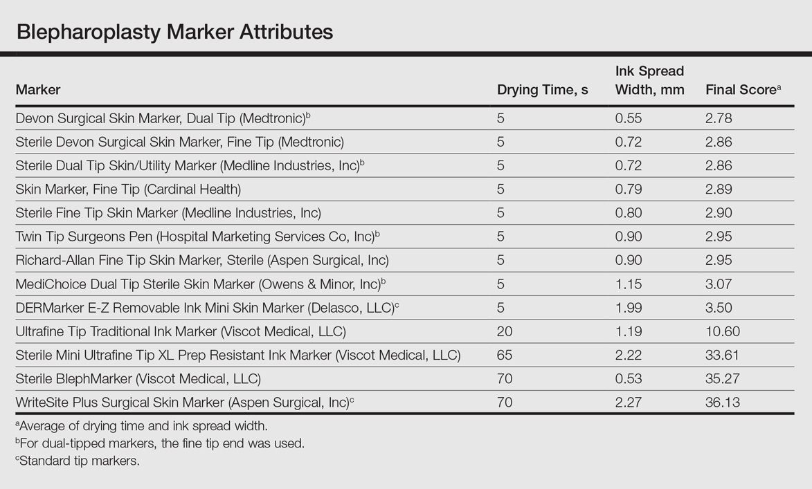
To prioritize short drying time coupled with minimal ink spread width, the values associated with each marker were averaged to arrive at the overall score for each marker. The smaller the overall score, the higher we ranked the marker. The Devon Surgical Skin Marker, Dual Tip (Medtronic) ranked the highest among the 13 markers with a final score of 2.78. Runner-up markers included the Sterile Devon Surgical Skin marker, Fine Tip (Medtronic)(final score, 2.86); the Sterile Dual Tip Skin/Utility Marker (Medline Industries, Inc)(final score, 2.86); and the Skin Marker, Fine Tip (Cardinal Health)(final score, 2.89). The 2 lowest-ranking markers were the WriteSite Plus Surgical Skin Marker, an ST marker (final score, 36.13), followed by the Sterile BlephMarker (Viscot Medical, LLC)(final score, 35.27).
Figure 2 shows the drying time and ink spread width for all 13 markers.
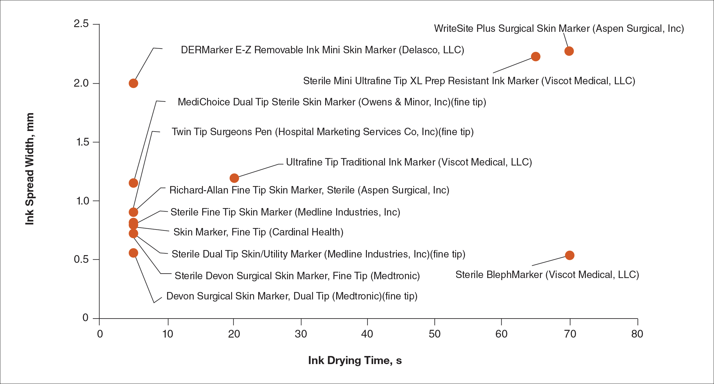
Comment
Blepharoplasty surgeons generally agree that meticulous presurgical planning with marking of the eyelids is critical for successful surgical outcomes.1,2 Fine tip markers have been recommended for this purpose due to the relative precision of the marks, but the prerequisite of these markers is that the marks must have minimal ink spread through skinfolds to allow for precision as well as short drying time to avoid unintentional duplication of the ink on overlapping skin, especially with the likely chance of reflexive blinking by the patient. The associated assumption is that FT markers automatically leave precise marks with minimal drying time. This study systemically compared these 2 qualities for 13 markers, and the results are notable for the unexpected wide range of performance. Although most of the FT markers had ink spread width of less than 1 mm, the Sterile Mini Ultrafine Tip XL Prep Resistant Ink Marker was an outlier among FT markers, with ink spread greater than 2 mm, making it too broad and imprecise for practical use. This result indicates that not every FT marker actually makes fine marks. The 2 ST markers in the study—DERMarker E-Z Removable Ink Mini Skin Marker and WriteSite Plus Surgical Skin Marker—left broad marks as anticipated.
The drying time of the markers also ranged from 5 to 70 seconds among both FT and ST markers. Indeed, most of the FT markers were dry at or before 5 seconds of marking, but 2 FT markers—Sterile Mini Ultrafine Tip XL Prep Resistant Ink Marker and Sterile BlephMarker—dried at 65 and 70 seconds, respectively. Such a long drying time would be considered impractical for use in blepharoplasty marking and also unexpected of FT markers, which usually are marketed for their precision and efficiency. Notable in the discussion of drying time is that one of the 2 ST markers in the study, the DERMarker E-Z Removable Ink Mini Skin Marker, had the shortest possible drying time of 5 seconds, while the other ST marker, WriteSite Plus Surgical Skin Marker, dried at 70 seconds. This observation coupled with the unexpected results of broad marks and long drying time for some of the FT markers indicates that a surgeon cannot simply assume that a FT marker would provide marks with precision and fast drying time, or that an ST marker would be the opposite.
Future directions for study include the addition of other markers and the extent of resistance to antiseptic routines that can fade the markings.
Conclusion
Among the 13 markers studied, FT markers typically had the shortest drying time and least ink
Acknowledgement
The authors would like to thank Laura B. Hall, MD (New Haven, Connecticut), for her participation as the volunteer in this study.
- Hartstein ME, Massry GG, Holds JB. Pearls and Pitfalls in Cosmetic Oculoplastic Surgery. New York, NY: Springer New York; 2015.
- Gladstone G, Black EH. Oculoplastic Surgery Atlas. New York, NY: Springer New York; 2005.
Blepharoplasty, or surgical manipulation of the upper and/or lower eyelids, is a commonly performed cosmetic procedure to improve the appearance and function of the eyelids by repositioning and/or removing excess skin and soft tissue from the eyelids, most often through external incisions that minimize scarring and maximize the aesthetic outcomes of the surgery. Therefore, the placement of the incisions is an important determinant of the surgical outcome, and the preoperative marking of the eyelids to indicate where the incisions should be placed is a crucial part of preparation for the surgery.
Preoperative marking has unique challenges due to the dynamicity of the eyelids and the delicate nature of the surgery. The mark must be narrow to minimize the risk of placing the incision higher or lower than intended. The mark also must dry quickly because the patient may blink and create multiple impressions of the marking on skinfolds in contact with the wet ink. Fast drying of the ink used to create the marks improves the efficiency and clarity of the presurgical planning.
We present data on the performance of the various blepharoplasty markers regarding drying time and ink spread width based on an evaluation of 13 surgical markers.
Methods
Eleven unique fine tip (FT) markers and 2 standard tip (ST) markers were obtained based on their accessibility at the researchers’ home institution and availability for direct purchase in small quantities from the distributors (Figure 1). Four markers were double tipped with one FT end and one ST end; for these markers, only the FT end was studied. The experiments were conducted on the bilateral upper eyelids and on hairless patches of skin of a single patient in a minor procedure room with surgical lighting and minimal draft of air. The sole experimenter (J.M.K.) conducting the study was not blinded.
The drying time of each marker was measured by marking 1-in lines on a patch of hairless skin that was first cleaned with an alcohol pad, then dried. Drying time for each marking was measured in increments of 5 seconds; at each time point, the markings were wiped with a single-ply, light-duty tissue under the weight of 10 US quarters to ensure that the same weight/pressure was applied when wiping the skin. Smudges observed with the naked eye on either the wipe or the patients’ skin were interpreted as nondry status of the marking. The first time point at which a marking was found to have no visible smudges either on the skin or the wipe was recorded as the drying time of the respective marker.
Ink spread was measured on clean eyelid skin by drawing curved lines along the natural crease as would be done for actual blepharoplasty planning. Each line was allowed to dry for 2 minutes. The greatest perpendicular spread width along the line observed with the naked eye was measured using a digital Vernier caliper with 0.01-mm graduations. Three measurements were obtained per marker and the values averaged to arrive at the final spread width.
Results
Drying time among the 13 total markers (11 FT and 2 ST) ranged from 5 to 70 seconds, with a mean of 20.8 seconds and median of 5 seconds (Table). The drying time for the DERMarker E-Z Removable Ink Mini Skin Marker (Delasco, LLC) with an ST was 5 seconds, while the drying time for the other ST marker, WriteSite Plus Surgical Skin Marker (Aspen Surgical, Inc), was 70 seconds. The FT markers spanned the entire range of drying times. The ink spread width among the markers ranged from 0.53 to 2.27 mm with a median of 0.9 mm and mean of 1.13 mm (Table). The 2 ST markers were found to make some of the widest marks measured, including the WriteSite Plus Surgical Skin Marker, a nonsterile ST marker that created the widest ink marks. The second widest mark was made by an FT marker (Sterile Mini Ultrafine Tip XL Prep Resistant Ink Marker [Viscot Medical, LLC]).
To prioritize short drying time coupled with minimal ink spread width, the values associated with each marker were averaged to arrive at the overall score for each marker. The smaller the overall score, the higher we ranked the marker. The Devon Surgical Skin Marker, Dual Tip (Medtronic) ranked the highest among the 13 markers with a final score of 2.78. Runner-up markers included the Sterile Devon Surgical Skin marker, Fine Tip (Medtronic)(final score, 2.86); the Sterile Dual Tip Skin/Utility Marker (Medline Industries, Inc)(final score, 2.86); and the Skin Marker, Fine Tip (Cardinal Health)(final score, 2.89). The 2 lowest-ranking markers were the WriteSite Plus Surgical Skin Marker, an ST marker (final score, 36.13), followed by the Sterile BlephMarker (Viscot Medical, LLC)(final score, 35.27).
Figure 2 shows the drying time and ink spread width for all 13 markers.
Comment
Blepharoplasty surgeons generally agree that meticulous presurgical planning with marking of the eyelids is critical for successful surgical outcomes.1,2 Fine tip markers have been recommended for this purpose due to the relative precision of the marks, but the prerequisite of these markers is that the marks must have minimal ink spread through skinfolds to allow for precision as well as short drying time to avoid unintentional duplication of the ink on overlapping skin, especially with the likely chance of reflexive blinking by the patient. The associated assumption is that FT markers automatically leave precise marks with minimal drying time. This study systemically compared these 2 qualities for 13 markers, and the results are notable for the unexpected wide range of performance. Although most of the FT markers had ink spread width of less than 1 mm, the Sterile Mini Ultrafine Tip XL Prep Resistant Ink Marker was an outlier among FT markers, with ink spread greater than 2 mm, making it too broad and imprecise for practical use. This result indicates that not every FT marker actually makes fine marks. The 2 ST markers in the study—DERMarker E-Z Removable Ink Mini Skin Marker and WriteSite Plus Surgical Skin Marker—left broad marks as anticipated.
The drying time of the markers also ranged from 5 to 70 seconds among both FT and ST markers. Indeed, most of the FT markers were dry at or before 5 seconds of marking, but 2 FT markers—Sterile Mini Ultrafine Tip XL Prep Resistant Ink Marker and Sterile BlephMarker—dried at 65 and 70 seconds, respectively. Such a long drying time would be considered impractical for use in blepharoplasty marking and also unexpected of FT markers, which usually are marketed for their precision and efficiency. Notable in the discussion of drying time is that one of the 2 ST markers in the study, the DERMarker E-Z Removable Ink Mini Skin Marker, had the shortest possible drying time of 5 seconds, while the other ST marker, WriteSite Plus Surgical Skin Marker, dried at 70 seconds. This observation coupled with the unexpected results of broad marks and long drying time for some of the FT markers indicates that a surgeon cannot simply assume that a FT marker would provide marks with precision and fast drying time, or that an ST marker would be the opposite.
Future directions for study include the addition of other markers and the extent of resistance to antiseptic routines that can fade the markings.
Conclusion
Among the 13 markers studied, FT markers typically had the shortest drying time and least ink
Acknowledgement
The authors would like to thank Laura B. Hall, MD (New Haven, Connecticut), for her participation as the volunteer in this study.
- Hartstein ME, Massry GG, Holds JB. Pearls and Pitfalls in Cosmetic Oculoplastic Surgery. New York, NY: Springer New York; 2015.
- Gladstone G, Black EH. Oculoplastic Surgery Atlas. New York, NY: Springer New York; 2005.
- Hartstein ME, Massry GG, Holds JB. Pearls and Pitfalls in Cosmetic Oculoplastic Surgery. New York, NY: Springer New York; 2015.
- Gladstone G, Black EH. Oculoplastic Surgery Atlas. New York, NY: Springer New York; 2005.
Resident Pearl
Based on the data presented in this study, blepharoplasty surgeons may choose to use the markers shown to have measurably short drying time and minimal ink spread to maximize efficiency of preincisional lid marking.

Risk of Migraine Is Increased Among the Obese and Underweight
Obesity and underweight status are associated with an increased risk of migraine, according to research published online ahead of print April 12 in Neurology. Age and sex are important covariates of this association.

“As obesity and being underweight are potentially modifiable risk factors for migraine, awareness of these risk factors is vital for both people with migraine and doctors,” said B. Lee Peterlin, DO, Director of Headache Research at Johns Hopkins University School of Medicine in Baltimore. “More research is needed to determine whether efforts to help people lose or gain weight could lower their risk for migraine.”
Previous studies have suggested an association between obesity and increased risk of migraine. These studies differed, however, in the populations they included, the way in which they categorized obesity status, and the way in which they were designed and conducted.
To further evaluate this possible association, Dr. Peterlin and colleagues searched the peer-reviewed literature for studies related to body composition status, as estimated by BMI and World Health Organization (WHO) physical status categories, and migraine. Their meta-analysis included 12 studies that encompassed data from 288,981 participants between ages 18 and 98.
After adjusting for age and sex, Dr. Peterlin and colleagues found that risk of migraine in people with obesity was increased by 27%, compared with people of normal weight. The risk remained increased after multivariate adjustments. Similarly, the age- and sex-adjusted risk of migraine was increased among overweight participants, but the result was not significant after multivariate adjustments were performed. The age- and sex-adjusted risk of migraine in underweight people was increased by 13%, compared with people of normal weight, and remained increased after multivariate adjustments.
“It is not clear how body composition could affect migraine,” said Dr. Peterlin. “Adipose tissue, or fatty tissue, secretes a wide range of molecules that could play a role in developing or triggering migraine. It is also possible that other factors such as changes in physical activity, medications, or other conditions such as depression play a role in the relationship between migraine and body composition.”
Half of the studies included in the meta-analysis relied on self-report of migraine and did not use International Classification of Headache Disorders criteria, thus introducing the potential for recall bias. Furthermore, eight of the studies used self-report of BMI. Previous data have indicated that people with migraine are more likely to underestimate their BMI.
One of the meta-analysis’s strengths, however, is its large sample size. Another is its use of uniform and consistent obesity status categories based on the WHO physical status categories for non-Asian populations.
Additional research could “advance our understanding of migraine and lead to the development of targeted therapeutic strategies based on obesity status,” Dr. Peterlin and colleagues concluded.
—Erik Greb
Suggested Reading
Gelaye B, Sacco S, Brown WJ, et al. Body composition status and the risk of migraine: a meta-analysis. Neurology. 2017 April 12 [Epub ahead of print].
Obesity and underweight status are associated with an increased risk of migraine, according to research published online ahead of print April 12 in Neurology. Age and sex are important covariates of this association.
“As obesity and being underweight are potentially modifiable risk factors for migraine, awareness of these risk factors is vital for both people with migraine and doctors,” said B. Lee Peterlin, DO, Director of Headache Research at Johns Hopkins University School of Medicine in Baltimore. “More research is needed to determine whether efforts to help people lose or gain weight could lower their risk for migraine.”
Previous studies have suggested an association between obesity and increased risk of migraine. These studies differed, however, in the populations they included, the way in which they categorized obesity status, and the way in which they were designed and conducted.
To further evaluate this possible association, Dr. Peterlin and colleagues searched the peer-reviewed literature for studies related to body composition status, as estimated by BMI and World Health Organization (WHO) physical status categories, and migraine. Their meta-analysis included 12 studies that encompassed data from 288,981 participants between ages 18 and 98.
After adjusting for age and sex, Dr. Peterlin and colleagues found that risk of migraine in people with obesity was increased by 27%, compared with people of normal weight. The risk remained increased after multivariate adjustments. Similarly, the age- and sex-adjusted risk of migraine was increased among overweight participants, but the result was not significant after multivariate adjustments were performed. The age- and sex-adjusted risk of migraine in underweight people was increased by 13%, compared with people of normal weight, and remained increased after multivariate adjustments.
“It is not clear how body composition could affect migraine,” said Dr. Peterlin. “Adipose tissue, or fatty tissue, secretes a wide range of molecules that could play a role in developing or triggering migraine. It is also possible that other factors such as changes in physical activity, medications, or other conditions such as depression play a role in the relationship between migraine and body composition.”
Half of the studies included in the meta-analysis relied on self-report of migraine and did not use International Classification of Headache Disorders criteria, thus introducing the potential for recall bias. Furthermore, eight of the studies used self-report of BMI. Previous data have indicated that people with migraine are more likely to underestimate their BMI.
One of the meta-analysis’s strengths, however, is its large sample size. Another is its use of uniform and consistent obesity status categories based on the WHO physical status categories for non-Asian populations.
Additional research could “advance our understanding of migraine and lead to the development of targeted therapeutic strategies based on obesity status,” Dr. Peterlin and colleagues concluded.
—Erik Greb
Suggested Reading
Gelaye B, Sacco S, Brown WJ, et al. Body composition status and the risk of migraine: a meta-analysis. Neurology. 2017 April 12 [Epub ahead of print].