User login
How to reduce the open hysterectomy rate
SAN ANTONIO – About a decade ago, the Northern California Permanente Medical Group realized it had a problem: There were too many open hysterectomies being performed.
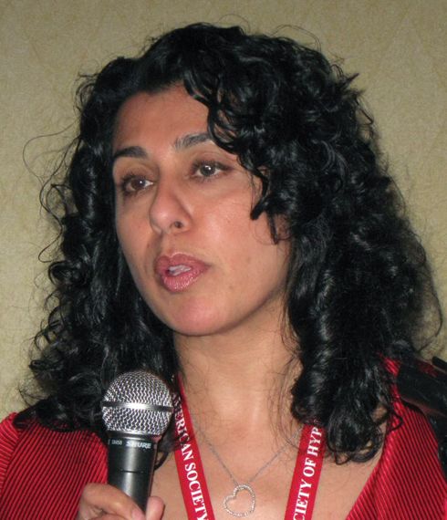
“We had a large number of low-volume surgeons doing a lot of open hysterectomies,” said Andrew Walter, MD, a gynecologic surgeon at the Permanente Medical Group (TPMG) campus in Roseville, Calif., part of Kaiser Permanente. Across 15 hospitals in the system, “our open rate was 64% in 2007.”
To address the problem, “our leadership basically set targets; we then provided surgical education and training in minimally invasive hysterectomy,” Dr. Walter said at the annual scientific meeting of the Society of Gynecologic Surgeons.

Kaiser Permanente is a capitated system, but even so, its experience in reducing open hysterectomy rates may be useful for other systems dealing with the same problem.
At first, Dr. Walter and his colleagues didn’t know how much of an improvement could be made. “In 2008, we said, ‘Okay, 60% seems doable’” as a minimally invasive target rate, Dr. Walter said. TPMG hit that target, and “the chiefs looked at the numbers and said we can do a little bit better.” With some more effort, the rate of minimally invasive hysterectomies hit 80%, and then 90%, about where it stands today, with a corresponding drop in open rates.
“It will be interesting to see if someone says, ‘Let’s try 95%,’ ” Dr. Walter said.
However, the process hasn’t been easy, and it hasn’t been entirely evidence based, he said. “We are working with hundreds of gynecologists, and very few of them have had advanced training. But ultimately, it worked.”
Surgeons trained with TPMG peers experienced in minimally invasive hysterectomy, and both surgeons were kept on full salary during the learning process. It took about 5-15 cases before learner surgeons were considered proficient. “Funded proctoring is the most important aspect of the program,” he said.
TPMG also reduced the number of physicians doing hysterectomies from 416 to 228 in 2015. “This was the hard part, deciding who is a surgeon and who is not,” Dr. Walter said. “I would like to tell you that these were Kumbaya moments, but there was consternation, and there remains consternation about the process.” A number of ob.gyns. voluntarily gave up their surgical privileges, saying, ‘Thank God I don’t have to operate anymore,’ ” he said.
For low-volume surgeons – those who performed 10 or fewer hysterectomies a year – who wanted stay in the operating room, “we either had to push them into this training or encourage them to give up their surgical practices.” The more than 3,600 hysterectomies performed annually at TPMG facilities are now mostly done by surgeons doing at least 11 of these procedures a year, and often more than 20.
TPMG also paid for training courses at outside institutions, and department chiefs were held accountable for performance.
“Obviously, there are unique processes within Kaiser Permanente that facilitated this, but some of them are not unique. Physician support by enhanced training – that’s something that can be done. The barriers are reimbursement, and deciding who is a surgeon,” he said.
The next target is vaginal hysterectomy. Rates have been stable lately at about 30%, but “we have found that many patients, when reviewed, are vaginal hysterectomy candidates. We’ve set a target of 40%.” The procedure needs to be incentivized, Dr. Walter said, but it’s unclear how to do that at this point.
Dr. Walter reported having no relevant financial disclosures.
* The meeting sponsor information was updated 6/9/2017.
SAN ANTONIO – About a decade ago, the Northern California Permanente Medical Group realized it had a problem: There were too many open hysterectomies being performed.
“We had a large number of low-volume surgeons doing a lot of open hysterectomies,” said Andrew Walter, MD, a gynecologic surgeon at the Permanente Medical Group (TPMG) campus in Roseville, Calif., part of Kaiser Permanente. Across 15 hospitals in the system, “our open rate was 64% in 2007.”
To address the problem, “our leadership basically set targets; we then provided surgical education and training in minimally invasive hysterectomy,” Dr. Walter said at the annual scientific meeting of the Society of Gynecologic Surgeons.
Kaiser Permanente is a capitated system, but even so, its experience in reducing open hysterectomy rates may be useful for other systems dealing with the same problem.
At first, Dr. Walter and his colleagues didn’t know how much of an improvement could be made. “In 2008, we said, ‘Okay, 60% seems doable’” as a minimally invasive target rate, Dr. Walter said. TPMG hit that target, and “the chiefs looked at the numbers and said we can do a little bit better.” With some more effort, the rate of minimally invasive hysterectomies hit 80%, and then 90%, about where it stands today, with a corresponding drop in open rates.
“It will be interesting to see if someone says, ‘Let’s try 95%,’ ” Dr. Walter said.
However, the process hasn’t been easy, and it hasn’t been entirely evidence based, he said. “We are working with hundreds of gynecologists, and very few of them have had advanced training. But ultimately, it worked.”
Surgeons trained with TPMG peers experienced in minimally invasive hysterectomy, and both surgeons were kept on full salary during the learning process. It took about 5-15 cases before learner surgeons were considered proficient. “Funded proctoring is the most important aspect of the program,” he said.
TPMG also reduced the number of physicians doing hysterectomies from 416 to 228 in 2015. “This was the hard part, deciding who is a surgeon and who is not,” Dr. Walter said. “I would like to tell you that these were Kumbaya moments, but there was consternation, and there remains consternation about the process.” A number of ob.gyns. voluntarily gave up their surgical privileges, saying, ‘Thank God I don’t have to operate anymore,’ ” he said.
For low-volume surgeons – those who performed 10 or fewer hysterectomies a year – who wanted stay in the operating room, “we either had to push them into this training or encourage them to give up their surgical practices.” The more than 3,600 hysterectomies performed annually at TPMG facilities are now mostly done by surgeons doing at least 11 of these procedures a year, and often more than 20.
TPMG also paid for training courses at outside institutions, and department chiefs were held accountable for performance.
“Obviously, there are unique processes within Kaiser Permanente that facilitated this, but some of them are not unique. Physician support by enhanced training – that’s something that can be done. The barriers are reimbursement, and deciding who is a surgeon,” he said.
The next target is vaginal hysterectomy. Rates have been stable lately at about 30%, but “we have found that many patients, when reviewed, are vaginal hysterectomy candidates. We’ve set a target of 40%.” The procedure needs to be incentivized, Dr. Walter said, but it’s unclear how to do that at this point.
Dr. Walter reported having no relevant financial disclosures.
* The meeting sponsor information was updated 6/9/2017.
EXPERT ANALYSIS FROM SGS 2017
WHO’s malaria pilot vaccine: No silver bullet, but a potential strike at malaria’s heart
EXPERT ANALYSIS FROM ECCMID 2017
VIENNA – The first malaria vaccine to enter a national pilot project is not a silver bullet against the disease that kills half a million every year, but it still might be powerful enough to significantly reduce global disease burden, and even impact transmission, according to infectious disease specialist Nick Beeching, MD.
The vaccine, RTS,S (Mosquirix; GlaxoSmithKline), will be tested in three African countries beginning next year, the World Health Organization announced on April 25. The pilot programs will target 720,000 children aged 5-17 months in high-risk areas of the three countries.
Even though it’s the first malaria vaccine to pass its pivotal phase III trial, RTS,S isn’t terribly effective by any standards, said Dr. Beeching of the Royal Liverpool (England) University.

April 25, 2017, is World Malaria Day, and Anthony S. Fauci, MD, and B. Fenton Hall, MD, PhD, of the U.S. National Institute of Allergy and Infectious Diseases, said in a statement, “Safe and effective vaccines are critical tools for future efforts to control, eliminate, and, ultimately, eradicate malaria. NIAID is supporting the development of numerous malaria vaccine candidates, 10 of which are in clinical trials. In 2015, an estimated 212 million new malaria cases and 429,000 deaths occurred. Nearly 90% of these cases were among children under the age of 5 years in Africa, where malaria claims the life of a child every 2 minutes.”
GSK has been working on this vaccine since 1985, according to the company’s RTS,S literature. It is a recombinant protein that targets the circumsporozoite protein of the Plasmodium falciparum parasite at an early stage, before it enters the liver and begins to embed in erythrocytes. The aim, Dr. Beeching said, was to develop an antigen that would mobilize the immune system from the moment a mosquito injected the sporozoites through a bite, “well before they have a chance to hide in the liver.”
The 2- and 3-year follow-up results of the phase III trial, conducted in 15,500 children, were published in the Lancet in 2015. RTS,S was administered as a three-dose series, plus a booster dose, beginning at 5 months of age. The primary immunizations were given with a minimum 4-week interval between doses, with the booster administered 18 months after the last dose.
The primary series reduced clinical cases by 26%. With the booster dose, cases were reduced by 39% overall. The vaccine averted 1,774 episodes of clinical malaria per 1,000 vaccinated children, and 983 cases per 1,000 vaccinated infants. But vaccine efficacy waned over time, disappearing completely in children who got only the three-dose series. The booster dose improved response stability somewhat; during the 12 months after the fourth dose, vaccine efficacy was about 25%.
Based on these results, GSK received approval from the European Medicines Agency in 2015, and the WHO recommended a large-scale implementation of the vaccine be carried out last year. GSK will provide the vaccine at no cost, and each country’s government will decide which regions to include in the pilot study.
This real-world use will put RTS,S to the ultimate test, Dr. Beeching said: “There is always the practical problem of how do you get four doses of vaccine into people. It’s easy to do in a clinical trial, but the operations and the logistics of getting it right on the ground are what really matter. We don’t know how good less than four doses would be, and we still don’t know how long the protective effect of the full series plus booster will last. I think there’s concern that it might wane with time.”
Still, he said, even a 39% reduction in disease burden is worth aggressively pursuing, not only because of the thousands of children’s lives that could be saved, but because unvaccinated children and adults could potentially be protected as well: “We could see a knock-on effect. By reducing the burden of malaria in children, it may also reduce transmission to other people who haven’t been vaccinated.”
The vaccine certainly won’t eradicate malaria, Dr. Beeching said. It needs to be viewed as an addition to WHO’s core vector control strategy, which includes insecticide-impregnated bed nets and mosquito eradication programs.
Cost is an unresolved issue. According to the Malaria Vaccine Initiative, which is partnering with GSK to launch RTS,S, the company won’t charge for the vaccine in the pilot project, and is committed to making sure the children who need it get it.
“In many African countries, childhood vaccines are provided at no cost to children or their families, thanks to existing international and national financing mechanisms,” the company said in a press release. “The RTS,S partnership anticipates that similar mechanisms would be implemented for a malaria vaccine. A shared goal is to have the cost of a malaria vaccine not be a barrier to access.
“GSK has previously stated that the price of RTS,S will cover the cost of manufacturing the vaccine together with a small return of around 5%, which will be reinvested in research and development for next-generation malaria vaccines or vaccines against other neglected tropical diseases.”
Finally, Dr. Beeching said, there’s no way to know to know how long any malaria vaccine would retain its effectiveness.
“Making a malaria vaccine has been a dream for years, and a tough one. The antigens change according to the stage of the parasite, and there is always continuous genetic variation. So there is a possibility of escape from vaccine coverage. These are very clever parasites,” he said.
Dr. Beeching has no financial interest in the vaccine.
[email protected]
On Twitter @Alz_gal
EXPERT ANALYSIS FROM ECCMID 2017
VIENNA – The first malaria vaccine to enter a national pilot project is not a silver bullet against the disease that kills half a million every year, but it still might be powerful enough to significantly reduce global disease burden, and even impact transmission, according to infectious disease specialist Nick Beeching, MD.
The vaccine, RTS,S (Mosquirix; GlaxoSmithKline), will be tested in three African countries beginning next year, the World Health Organization announced on April 25. The pilot programs will target 720,000 children aged 5-17 months in high-risk areas of the three countries.
Even though it’s the first malaria vaccine to pass its pivotal phase III trial, RTS,S isn’t terribly effective by any standards, said Dr. Beeching of the Royal Liverpool (England) University.
April 25, 2017, is World Malaria Day, and Anthony S. Fauci, MD, and B. Fenton Hall, MD, PhD, of the U.S. National Institute of Allergy and Infectious Diseases, said in a statement, “Safe and effective vaccines are critical tools for future efforts to control, eliminate, and, ultimately, eradicate malaria. NIAID is supporting the development of numerous malaria vaccine candidates, 10 of which are in clinical trials. In 2015, an estimated 212 million new malaria cases and 429,000 deaths occurred. Nearly 90% of these cases were among children under the age of 5 years in Africa, where malaria claims the life of a child every 2 minutes.”
GSK has been working on this vaccine since 1985, according to the company’s RTS,S literature. It is a recombinant protein that targets the circumsporozoite protein of the Plasmodium falciparum parasite at an early stage, before it enters the liver and begins to embed in erythrocytes. The aim, Dr. Beeching said, was to develop an antigen that would mobilize the immune system from the moment a mosquito injected the sporozoites through a bite, “well before they have a chance to hide in the liver.”
The 2- and 3-year follow-up results of the phase III trial, conducted in 15,500 children, were published in the Lancet in 2015. RTS,S was administered as a three-dose series, plus a booster dose, beginning at 5 months of age. The primary immunizations were given with a minimum 4-week interval between doses, with the booster administered 18 months after the last dose.
The primary series reduced clinical cases by 26%. With the booster dose, cases were reduced by 39% overall. The vaccine averted 1,774 episodes of clinical malaria per 1,000 vaccinated children, and 983 cases per 1,000 vaccinated infants. But vaccine efficacy waned over time, disappearing completely in children who got only the three-dose series. The booster dose improved response stability somewhat; during the 12 months after the fourth dose, vaccine efficacy was about 25%.
Based on these results, GSK received approval from the European Medicines Agency in 2015, and the WHO recommended a large-scale implementation of the vaccine be carried out last year. GSK will provide the vaccine at no cost, and each country’s government will decide which regions to include in the pilot study.
This real-world use will put RTS,S to the ultimate test, Dr. Beeching said: “There is always the practical problem of how do you get four doses of vaccine into people. It’s easy to do in a clinical trial, but the operations and the logistics of getting it right on the ground are what really matter. We don’t know how good less than four doses would be, and we still don’t know how long the protective effect of the full series plus booster will last. I think there’s concern that it might wane with time.”
Still, he said, even a 39% reduction in disease burden is worth aggressively pursuing, not only because of the thousands of children’s lives that could be saved, but because unvaccinated children and adults could potentially be protected as well: “We could see a knock-on effect. By reducing the burden of malaria in children, it may also reduce transmission to other people who haven’t been vaccinated.”
The vaccine certainly won’t eradicate malaria, Dr. Beeching said. It needs to be viewed as an addition to WHO’s core vector control strategy, which includes insecticide-impregnated bed nets and mosquito eradication programs.
Cost is an unresolved issue. According to the Malaria Vaccine Initiative, which is partnering with GSK to launch RTS,S, the company won’t charge for the vaccine in the pilot project, and is committed to making sure the children who need it get it.
“In many African countries, childhood vaccines are provided at no cost to children or their families, thanks to existing international and national financing mechanisms,” the company said in a press release. “The RTS,S partnership anticipates that similar mechanisms would be implemented for a malaria vaccine. A shared goal is to have the cost of a malaria vaccine not be a barrier to access.
“GSK has previously stated that the price of RTS,S will cover the cost of manufacturing the vaccine together with a small return of around 5%, which will be reinvested in research and development for next-generation malaria vaccines or vaccines against other neglected tropical diseases.”
Finally, Dr. Beeching said, there’s no way to know to know how long any malaria vaccine would retain its effectiveness.
“Making a malaria vaccine has been a dream for years, and a tough one. The antigens change according to the stage of the parasite, and there is always continuous genetic variation. So there is a possibility of escape from vaccine coverage. These are very clever parasites,” he said.
Dr. Beeching has no financial interest in the vaccine.
[email protected]
On Twitter @Alz_gal
Long-term albumin shows survival benefit in decompensated cirrhosis
AMSTERDAM – Long-term treatment with human albumin improved the overall survival of patients with decompensated liver cirrhosis, compared with standard medical care, in a randomized, controlled trial presented at the International Liver Congress.
The final results of the ANSWER study showed that a 38% reduction in the risk of death could be achieved at 18 months’ follow-up by giving patients human albumin, with an overall survival of 78% vs. 66% in the two groups, respectively (hazard ratio, 0.62; 95% confidence interval, 0.40-0.95; P = .028).
“Long-term albumin administration to patients with decompensated cirrhosis may be seen as a disease-modifying treatment,” said the presenting study author, Mauro Bernardi, MD, professor in the department of medical and surgical sciences at the University of Bologna (Italy).
Not only was the overall survival improved, but there was improvement in the management of ascites, a reduction in the incidence of severe complications (such as spontaneous bacterial peritonitis, renal dysfunction, and hepatic encephalopathy), a reduction in the number of hospitalizations and duration of in-hospital treatment, and a signal for improved quality of life, he said.
“We know that it is good to give albumin as an infusion in many, many circumstances,” said Frank Tacke, MD, PhD, who was not involved in the study, during a press briefing at the meeting, sponsored by the European Association for the Study of the Liver (EASL).
“What we did not know before was if there was a role for giving albumin – which is unfortunately quite expensive – for a longer period of time in patients with liver cirrhosis,” said Dr. Tacke, who is the EASL vice-secretary and professor of medicine in the department of gastroenterology, metabolic diseases and intensive care medicine at University Hospital Aachen (Germany).
Although long-term treatment with human albumin is a relatively expensive treatment, particularly because it requires weekly infusion, Dr. Bernardi noted that a cost analysis was being performed, and potentially the cost of treatment could be offset by the reduction in paracentesis, duration of hospitalization, and reduced need for treating patients with complications, compared with standard medical care alone.
“The idea of supplying albumin to patients with advanced cirrhosis is quite an old one, and there is long debate. The point is that a reliable study that could resolve this was simply lacking up to now,” Dr. Bernard said at the briefing.
The results could mean that patients with decompensated cirrhosis now have a much-needed therapeutic option. These patients have “a very poor prognosis,” Dr. Bernardi said. The 1-year probability of survival is about 20%, and the only curative therapy at present is liver transplantation.
A total of 440 patients, mostly male with an average age of 61 years, with cirrhosis and uncomplicated ascites were randomized at 33 Italian centers to receive standard medical treatment alone (n = 213) or with human albumin (n = 218) given at an infused dose of 40 g twice a week for the first 2 weeks, then 40 g every week. The albumin used in the trial was provided by five different pharmaceutical companies and sent to a central location for generic relabeling and distribution out to the participating trial centers.
Significantly fewer patients who were given human albumin than those who were not (66% vs. 38%, P less than .001) needed at least one paracentesis. The incidence rate for the removal of peritoneal fluid in the standard medical treatment arm was 3.5/person per year. There was a 54% reduction in this rate by the addition of human albumin (HR = 0.46; 95% CI, 0.40-0.53; P less than .0001). There was also a significant 46% reduction in the incidence of refractory ascites (48% vs. 25%, P less than .0001).
Patients who received standard medical treatment plus albumin needed fewer hospitalizations and fewer days of in-hospital care per person per year than those in the standard care–only arm. The use of human albumin reduced the number of hospital stays by 35% (HR = 0.65; 95% CI, 0.55-0.77; P less than .0001) and the duration of days in hospital by 45% (HR = 0.55; 95% CI, 0.52-0.58; P less than .0001).
Although not statistically significant, a trend for greater improvements and fewer decreases in quality of life, as measured using the EQ-5D visual assessment scale, at 3, 6, and 12 months, was seen with the use of human albumin.
Four patients had adverse drug reactions: two were mild allergic reactions, and two were potentially life-threatening septic shock that needed intensive care treatment. One of the latter cases might have been caused by pneumonia, and the other required study interruption. But in all cases the patients recovered, Dr. Bernardi reported.
The study was funded by the Italian Drug Agency. Dr. Bernardi had acted as a speaker for and consultant to CLS Behring and Baxter Healthcare, and as a speaker to the Plasma Protein Therapeutics Association’s Europe division, Grifols, Gilead Sciences, and AbbVie Italia. Dr. Tacke had nothing to disclose.
AT ILC 2017
Key clinical point: A weekly infusion of human albumin has a beneficial effect in patients with decompensated cirrhosis.
Major finding: Overall survival was 78% vs. 66% for standard medical care with albumin vs. no albumin (HR, 0.62; 95% CI, 0.40-0.95; P = .028).
Data source: The ANSWER study, a multicenter, open-label, randomized clinical trial of 440 patients with decompensated cirrhosis.
Disclosures: The study was funded by the Italian Drug Agency. Dr. Bernardi had acted as a speaker for and consultant to CLS Behring and Baxter Healthcare, and as a speaker to the Plasma Protein Therapeutics Association’s Europe division, Grifols, Gilead Sciences, and AbbVie Italia. Dr. Tacke had nothing to disclose.
USPSTF says check BP at each visit to screen for preeclampsia
Blood pressure should be checked at every prenatal visit to screen for preeclampsia, according to a new recommendation from the U.S. Preventive Services Task Force.
The USPSTF commissioned a review of the current literature to update its initial recommendation regarding preeclampsia screening, which was issued in 1996. The update was considered important “given changes to screening, diagnosis, and management practices, as well as population health, over the past two decades.”
However, the evidence regarding different screening approaches is still quite limited, and the USPSTF did not change its recommendation except to indicate blood pressure checks at every visit rather than “periodically.” The current advice is a “B” recommendation, meaning that the USPSTF recommends the service.
Blood pressure assessment remains the most reliable, least harmful strategy for identifying preeclampsia as early as possible, said Kirsten Bobbins-Domingo, MD, PhD, chair of the USPSTF and lead author of the recommendation statement, and her associates.
“The USPSTF concludes with moderate certainty that screening for preeclampsia in pregnant women with blood pressure measurements has a substantial net benefit,” the researchers wrote (JAMA. 2017;317[16]:1661-7).
The evidence report supporting this recommendation included 21 studies involving 13,982 pregnant women. But no studies directly compared the effectiveness of blood pressure screening between screened and unscreened populations, Jillian T. Henderson, PhD, of Kaiser Permanente Center for Health Research, Portland, Ore., and her associates said in the report.
Fourteen studies assessed tests for protein in the urine to identify preeclampsia, but the accuracy of such tests was highly variable. Sensitivity ranged from 22% to 100% and specificity ranged from 36% to 100%, so the USPSTF did not recommend urine testing for proteinuria as a screen for preeclampsia.
In addition, Dr. Henderson and her associates reviewed four studies (involving 7,123 women) assessing 16 different risk-prediction models. Again, the findings did not support the routine use of such tools in clinical practice: their positive predictive value was just 4% in the largest validation cohort, the investigators reported (JAMA. 2017;317[16]:1668-83).
The American College of Obstetricians and Gynecologists recommends monitoring blood pressure at every prenatal visit to screen for preeclampsia, as well as obtaining a detailed medical history to assess risk factors for the disorder. In contrast, the Society of Obstetricians and Gynaecologists of Canada and the National Institute for Health and Care Excellence in the United Kingdom recommend urinalysis for proteinuria in addition to blood pressure screening.
The full recommendation statement and evidence report are available at www.uspreventiveservicestaskforce.org.
The use of blood pressure measurement at every prenatal visit is a simple and effective way to identify and treat at-risk women without adding any substantial risks. In contrast, the risks related to preeclampsia are very high. Although risk assessment tools for preeclampsia are available, their low positive predictive value currently limits their usefulness for screening. Given the incidence of preeclampsia and the lack of predictability, it is important that all pregnant women have routine blood pressure assessments at every visit during the prenatal period.
The emerging data relating preeclampsia to the risk of future cardiovascular disease make this pregnancy-related issue important not just for obstetricians for the acute risks but also for internists and cardiologists for the long-term risks.
Martha Gulati, MD, is in the division of cardiology at the University of Arizona, Phoenix. She reported having no relevant financial disclosures. These remarks are excerpted from an editorial (JAMA Cardiol. 2017 Apr 25. doi: 10.1001/jamacardio.2017.1276).
Blood pressure should be checked at every prenatal visit to screen for preeclampsia, according to a new recommendation from the U.S. Preventive Services Task Force.
The USPSTF commissioned a review of the current literature to update its initial recommendation regarding preeclampsia screening, which was issued in 1996. The update was considered important “given changes to screening, diagnosis, and management practices, as well as population health, over the past two decades.”
However, the evidence regarding different screening approaches is still quite limited, and the USPSTF did not change its recommendation except to indicate blood pressure checks at every visit rather than “periodically.” The current advice is a “B” recommendation, meaning that the USPSTF recommends the service.
Blood pressure assessment remains the most reliable, least harmful strategy for identifying preeclampsia as early as possible, said Kirsten Bobbins-Domingo, MD, PhD, chair of the USPSTF and lead author of the recommendation statement, and her associates.
“The USPSTF concludes with moderate certainty that screening for preeclampsia in pregnant women with blood pressure measurements has a substantial net benefit,” the researchers wrote (JAMA. 2017;317[16]:1661-7).
The evidence report supporting this recommendation included 21 studies involving 13,982 pregnant women. But no studies directly compared the effectiveness of blood pressure screening between screened and unscreened populations, Jillian T. Henderson, PhD, of Kaiser Permanente Center for Health Research, Portland, Ore., and her associates said in the report.
Fourteen studies assessed tests for protein in the urine to identify preeclampsia, but the accuracy of such tests was highly variable. Sensitivity ranged from 22% to 100% and specificity ranged from 36% to 100%, so the USPSTF did not recommend urine testing for proteinuria as a screen for preeclampsia.
In addition, Dr. Henderson and her associates reviewed four studies (involving 7,123 women) assessing 16 different risk-prediction models. Again, the findings did not support the routine use of such tools in clinical practice: their positive predictive value was just 4% in the largest validation cohort, the investigators reported (JAMA. 2017;317[16]:1668-83).
The American College of Obstetricians and Gynecologists recommends monitoring blood pressure at every prenatal visit to screen for preeclampsia, as well as obtaining a detailed medical history to assess risk factors for the disorder. In contrast, the Society of Obstetricians and Gynaecologists of Canada and the National Institute for Health and Care Excellence in the United Kingdom recommend urinalysis for proteinuria in addition to blood pressure screening.
The full recommendation statement and evidence report are available at www.uspreventiveservicestaskforce.org.
Key clinical point:
Major finding: The 14 studies about testing for proteinuria and the 4 studies about 16 different risk prediction tools did not yield evidence to support either approach as a screen for preeclampsia.
Data source: A review of 21 studies involving 13,982 pregnant women, performed since the initial USPSTF recommendation was issued in 1996.
Disclosures: The USPSTF is an independent voluntary group supported by the Agency for Healthcare Research and Quality.
Does Gender Influence Pain Sensitivity?
STOWE, VT—Mounting evidence suggests that gender affects the modulation of pain to a greater extent than has been understood, according to an overview presented at the 27th Annual Headache Cooperative of New England Stowe Headache Symposium. Data indicate that the differences are biologic, and researchers are examining whether social and psychologic differences also may influence pain.

“If biomedicine ever comes up with a treatment, reproductive issues aside, that works and gets approval for one sex and not the other for treating the same underlying condition, it is going to happen in pain first,” said Jeffrey S. Mogil, PhD, Canada Research Chair in Genetics of Pain at McGill University in Montreal. “Maybe we are only 10 or 15 years away from seeing such a thing occur.”
A Clear Gender Difference
The first investigation of gender differences in pain was published in 1997, according to Dr. Mogil. It indicated that a greater number of chronic pain syndromes were more prevalent in women, compared with those that were more prevalent in men. Furthermore, the syndromes that were more prevalent in women were more common overall. The study also estimated that approximately 70% of patients with chronic pain are women. This result, however, could follow from reluctance among men to consult a physician, said Dr. Mogil.
When Dr. Mogil reviewed epidemiologic studies of pain, he found that women were between 5% and 10% more likely than men to endorse the symptoms of chronic pain. Although the studies used different definitions of chronic pain, each study used the same definition for men and women. A possible explanation for the result is that women are more susceptible to painful diseases.
But when he examined laboratory data from controlled experiments involving painful stimuli, Dr. Mogil found that “regardless of what type of pain you are looking at, and regardless of how it is measured, women are more sensitive to pain than men.” The difference in pain sensitivity is not great and depends on various factors, but it is clear and unmistakable, he added.
Biology Influences Pain Sensitivity
Biologic differences may explain gender differences in pain sensitivity. Research during the past 20 years has suggested that microglia play an important role in nociception. Newer data, however, indicate that microglial involvement in pain may be specific to males. Because the majority of animal research had been performed in male rodents, this observation had not been made previously, said Dr. Mogil.
He and his colleagues injured male and female mice to induce mechanical allodynia. The mice exhibited the same amount of mechanical allodynia, regardless of gender. The investigators next administered minocycline, a glial inhibitor, to the mice. The intervention reversed the allodynia in male mice, but not in female mice. Using fluorocitrate or propentofylline in place of minocycline produces the same result, said Dr. Mogil. Research into the biologic basis for pain modulation in females is ongoing.
Gender, Stress, and Analgesia
A patient’s sensitivity to pain may be influenced not only by his or her gender, but also by the gender of a person that the patient encounters. The results of certain mouse studies prompted researchers to hypothesize that the experimenters themselves may have produced analgesia, and Dr. Mogil decided to test this hypothesis.
When an investigator injected zymosan, an inflammatory agent, into a mouse’s ankle and left the room, the injection caused pain and grimacing in the mouse. When a male investigator administered zymosan to a mouse and remained in the room, the rate of grimacing decreased by approximately 40%. “In fact, experimenters are causing analgesia,” said Dr. Mogil. A female investigator did not produce the same effect, however.
Further research indicated that the effect has an olfactory origin. When injured mice are exposed to clothing previously worn by a male, or to bedding previously used by any male animal, they exhibit the same reduction in pain. “This is a stress phenomenon,” said Dr. Mogil. A mouse’s level of corticosterone, an equivalent to cortisol in humans, increases after exposure to male experimenters or their clothing.
The mice appear to be responding to axillary chemosignals (eg, androstadienone and androstenone) that males emit. The most important compound may be 3-Methyl-2-hexenoic acid, because it is “the only chemosignal that can produce this effect at reasonable concentrations in the nanomolar range,” said Dr. Mogil. He and his colleagues are studying whether these chemosignals produce the same responses in humans. “I would expect the effect to be much smaller and last for not quite as long,” he said. They also are studying the social modulation of pain in animals and humans.
—Erik Greb
Suggested Reading
Berkley KJ. Sex differences in pain. Behav Brain Sci. 1997;20(3):371-380; discussion 435-513.
Mogil JS. Sex differences in pain and pain inhibition: multiple explanations of a controversial phenomenon. Nat Rev Neurosci. 2012;13(12):859-866.
Sorge RE, Mapplebeck JC, Rosen S, et al. Different immune cells mediate mechanical pain hypersensitivity in male and female mice. Nat Neurosci. 2015;18(8):1081-1083.
Sorge RE, Martin LJ, Isbester KA, et al. Olfactory exposure to males, including men, causes stress and related analgesia in rodents. Nat Methods. 2014;11(6):629-632.
STOWE, VT—Mounting evidence suggests that gender affects the modulation of pain to a greater extent than has been understood, according to an overview presented at the 27th Annual Headache Cooperative of New England Stowe Headache Symposium. Data indicate that the differences are biologic, and researchers are examining whether social and psychologic differences also may influence pain.
“If biomedicine ever comes up with a treatment, reproductive issues aside, that works and gets approval for one sex and not the other for treating the same underlying condition, it is going to happen in pain first,” said Jeffrey S. Mogil, PhD, Canada Research Chair in Genetics of Pain at McGill University in Montreal. “Maybe we are only 10 or 15 years away from seeing such a thing occur.”
A Clear Gender Difference
The first investigation of gender differences in pain was published in 1997, according to Dr. Mogil. It indicated that a greater number of chronic pain syndromes were more prevalent in women, compared with those that were more prevalent in men. Furthermore, the syndromes that were more prevalent in women were more common overall. The study also estimated that approximately 70% of patients with chronic pain are women. This result, however, could follow from reluctance among men to consult a physician, said Dr. Mogil.
When Dr. Mogil reviewed epidemiologic studies of pain, he found that women were between 5% and 10% more likely than men to endorse the symptoms of chronic pain. Although the studies used different definitions of chronic pain, each study used the same definition for men and women. A possible explanation for the result is that women are more susceptible to painful diseases.
But when he examined laboratory data from controlled experiments involving painful stimuli, Dr. Mogil found that “regardless of what type of pain you are looking at, and regardless of how it is measured, women are more sensitive to pain than men.” The difference in pain sensitivity is not great and depends on various factors, but it is clear and unmistakable, he added.
Biology Influences Pain Sensitivity
Biologic differences may explain gender differences in pain sensitivity. Research during the past 20 years has suggested that microglia play an important role in nociception. Newer data, however, indicate that microglial involvement in pain may be specific to males. Because the majority of animal research had been performed in male rodents, this observation had not been made previously, said Dr. Mogil.
He and his colleagues injured male and female mice to induce mechanical allodynia. The mice exhibited the same amount of mechanical allodynia, regardless of gender. The investigators next administered minocycline, a glial inhibitor, to the mice. The intervention reversed the allodynia in male mice, but not in female mice. Using fluorocitrate or propentofylline in place of minocycline produces the same result, said Dr. Mogil. Research into the biologic basis for pain modulation in females is ongoing.
Gender, Stress, and Analgesia
A patient’s sensitivity to pain may be influenced not only by his or her gender, but also by the gender of a person that the patient encounters. The results of certain mouse studies prompted researchers to hypothesize that the experimenters themselves may have produced analgesia, and Dr. Mogil decided to test this hypothesis.
When an investigator injected zymosan, an inflammatory agent, into a mouse’s ankle and left the room, the injection caused pain and grimacing in the mouse. When a male investigator administered zymosan to a mouse and remained in the room, the rate of grimacing decreased by approximately 40%. “In fact, experimenters are causing analgesia,” said Dr. Mogil. A female investigator did not produce the same effect, however.
Further research indicated that the effect has an olfactory origin. When injured mice are exposed to clothing previously worn by a male, or to bedding previously used by any male animal, they exhibit the same reduction in pain. “This is a stress phenomenon,” said Dr. Mogil. A mouse’s level of corticosterone, an equivalent to cortisol in humans, increases after exposure to male experimenters or their clothing.
The mice appear to be responding to axillary chemosignals (eg, androstadienone and androstenone) that males emit. The most important compound may be 3-Methyl-2-hexenoic acid, because it is “the only chemosignal that can produce this effect at reasonable concentrations in the nanomolar range,” said Dr. Mogil. He and his colleagues are studying whether these chemosignals produce the same responses in humans. “I would expect the effect to be much smaller and last for not quite as long,” he said. They also are studying the social modulation of pain in animals and humans.
—Erik Greb
Suggested Reading
Berkley KJ. Sex differences in pain. Behav Brain Sci. 1997;20(3):371-380; discussion 435-513.
Mogil JS. Sex differences in pain and pain inhibition: multiple explanations of a controversial phenomenon. Nat Rev Neurosci. 2012;13(12):859-866.
Sorge RE, Mapplebeck JC, Rosen S, et al. Different immune cells mediate mechanical pain hypersensitivity in male and female mice. Nat Neurosci. 2015;18(8):1081-1083.
Sorge RE, Martin LJ, Isbester KA, et al. Olfactory exposure to males, including men, causes stress and related analgesia in rodents. Nat Methods. 2014;11(6):629-632.
VIDEO: Pilot stem cell trial for multiple system atrophy shows promising results
BOSTON – Intrathecal, autologous mesenchymal stem cell (MSC) treatment provided encouraging results for modifying the disease course of multiple system atrophy with relative safety and tolerability in a phase I/II trial.
The efficacy of MSCs on slowing multiple system atrophy (MSA) progression in a small trial of 24 patients appeared to be dependent on the dose, and, in the highest dose individuals, had a painful implantation response, trial investigator Wolfgang Singer, MD, reported at the annual meeting of the American Academy of Neurology.
Dr. Singer and his colleagues at the Mayo Clinic in Rochester, Minn., chose to investigate MSCs as a treatment because they are multipotent and capable of differentiating into different cell types, including glial cells, and they secrete neurotrophic factors, such as brain-derived neurotrophic factor and glial cell line-derived neurotrophic factor, which are thought to occur at pathologically low levels in individuals with MSA. The intrinsic immunomodulatory action of MSCs may also contribute to a potential benefit on neuroinflammatory aspects of MSA pathology.
MSCs have previously shown promising results on slowing disease progression in an open-label study of Korean patients with MSA, who received an intra-arterial infusion of MSCs into the internal carotids and dominant vertebral artery, followed by intravenous infusions (Clin Pharmacol Ther. 2008 May;83[5]:723-30). Those results were confirmed in a double-blind, placebo-controlled study (Ann Neurol. 2012 Jul;72[1]:32-40), but evidence of microemboli raised concerns with the Mayo Clinic team about stroke risk with the intra-arterial approach, Dr. Singer said.
The intrathecal route of administration also may be advantageous over an intra-arterial approach by reaching the targets of MSCs in the brain “in a more widespread fashion,” Dr. Singer said.
The relative safety and hint of efficacy with the different route of MSC administration in the Mayo Clinic study make it “a really groundbreaking direction to take,” session comoderator Christopher H. Gibbons, MD, said in an interview. “I think this a very good, small, but critical, step in demonstrating that ... you can do this, and maybe there’s a signal that it is, in fact, working at slowing down disease progression, which I think is incredibly important in this disease.”
In the current study, Dr. Singer and his associates intrathecally administered between 10 and 200 million autologous, fat-derived MSCs via lumbar puncture in predefined dose tiers and then followed patients over 1 year. Overall, 8 patients received a single dose of 10 million cells, 12 received a dose of 50 million cells followed by a second 50-million cell dose 4 weeks later, and 4 received a dose of 100 million cells followed by a second 100-million cell dose 4 weeks later.
The 24 patients in the study all met consensus criteria for probable MSA, had at least moderate laboratory evidence of autonomic failure, and were at an early stage of disease with a Unified MSA Rating Scale part 1 score of 18 or less.
In the primary outcome of safety, the investigators reported no treatment–attributable serious adverse events and said that the treatment was generally well-tolerated. All 16 patients who took either the high or medium doses had MRI abnormalities that showed thickening/enhancement of cauda equina nerve roots at the level of the puncture that were asymptomatic and did not lead to neurologic deficits.
The 12 medium-dose patients had variable elevation of cerebrospinal fluid protein and cell counts, and 5 had mild and transient low back pain. In the highest-dose tier, three of four patients developed low back pain, some of which was severe, and the same MRI findings, which signaled to the investigators that a dose-limiting toxicity had been reached.
Some patients also reported headaches after lumbar punctures, which were expected. Two patients also developed mild febrile reactions after administrations that were self-limited.
Treatment with MSCs led to a significantly lower rate of disease progression as measured by total score on the Unified MSA Rating Scale (0.43 vs. 1.22 points/month; P = .009), and this difference was even greater for the medium-dose tier than for the low-dose tier. The investigators used the placebo group from their recently completed rifampicin treatment trial in MSA to make the efficacy assessment.
There was no change between the treatment groups and the historical control group on the Composite Autonomic Symptom Scale or the Composite Autonomic Severity Score from baseline to 1 year.
Based on the promising findings, the Food and Drug Administration granted permission for a compassionate extension study for apparent responders to receive additional injections every 6 months, and, so far, 16 patients have been reinjected, according to Dr. Singer.
Dr. Singer and his associates have used these results as the basis for a multicenter, double-blind, placebo-controlled phase II/III study that is in the late planning stages. This kind of trial will be important for determining whether it’s possible to hold the quality of the MSC treatment steady and get the same sort of response across many centers, said Dr. Gibbons, a clinical neurophysiologist at Beth Israel Deaconess Medical Center, Boston, and past-president of the American Autonomic Society.
The trial was supported by grants from the National Institutes of Health, the Cure MSA Foundation, the Food and Drug Administration, the Autonomic Rare Disease Consortium, and Mayo Clinic funds. Dr. Singer and Dr. Gibbons reported having no relevant financial disclosures.
You can watch a video interview with Dr. Singer here.
AT AAN 2017
Key clinical point:
Major finding: Treatment with MSCs led to a significantly lower rate of disease progression as measured by total score on the Unified MSA Rating Scale (0.43 vs. 1.22 points/month in historical placebo group; P = .009).
Data source: A phase I/II trial of 24 patients with MSA treated intrathecally with autologous MSCs and compared against a historical control group.
Disclosures: The trial was supported by grants from the National Institutes of Health, the Cure MSA Foundation, the Food and Drug Administration, the Autonomic Rare Disease Consortium, and Mayo Clinic funds. Dr. Singer and Dr. Gibbons reported having no relevant financial disclosures.

Why state and school policies matter
Recently North Carolina proposed a bill (House Bill 780) that will ban same-sex marriage in the state, even though the U.S. Supreme Court ruled in 2015 that no states can ban same-sex marriage because doing so violates the 14th Amendment. Although many mainly will argue that H.B. 780 is unconstitutional, this bill also can be detrimental to the health of lesbian, gay, and bisexual (LGB) youth.
In March 2017, JAMA Pediatrics published a study on the association between same-sex marriage laws and the rates of suicide attempts.1 Using data from the Youth Risk Behavior Surveillance System, they analyzed the relationship between state policies that permitted same-sex marriage and self-report of suicide attempts within the last 12 months. Thirty percent of LGB youth (13% of the survey population) reported a suicide attempt in the past year prior to any state policies that permitted same-sex marriage, compared with about 9% of the general population. After states implemented pro–same-sex marriage policies, LGB youth suicide attempts dropped to about 26% – a 14% relative decline. This was not limited to LGB youth. The general youth suicide rates declined from 8.6% to 8% – a 7% relative decline. Although the change in suicide attempts was small, the authors determined that the likelihood it occurred by chance was very slim, and concluded that policies enabling same-sex marriage may be associated with an improvement in population health, especially for LGB youth.

This was not the first study examining the relationship between policies and the health of LGB youth. Mark Hatzenbuehler, PhD, of Columbia University, New York, and his associates have published multiple studies on this topic. Four years earlier, Hatzenbuehler et al. published a study on the relationship between antibullying policies and suicide among LGB youth. Using data from the Oregon Healthy Teen survey (2006-2008), he found that LGB youth who live in counties where there are fewer districts with antibullying policies are more than twice as likely to attempt suicide, compared with LGB youth who live in counties where more districts had antibullying policies. Furthermore, the type of antibullying policy mattered. If a district’s antibullying policy did not prohibit bullying based on sexual orientation, then it had no impact on the suicide attempt rates of LGB youth in that area.2 Similar results were found when the relationship between anti–homophobic bullying policies and LGB suicide attempts was examined in the larger Youth Risk Behavior Surveillance System.3
State and local policies also influence other health outcomes among LGB youth. Hatzenbuehler et al. did another study that examined community-level determinants of tobacco use among LGB youth. Again using the data from the Oregon Healthy Teen Survey, they found that LGB youth living in communities that were more supportive of LGB youth (i.e., communities with a high proportion of same-sex couples living in the area, a high proportion of gay-straight alliances at schools, and LGB-specific antibullying policies) were less likely to smoke cigarettes, compared with LGB youth living in communities that were less supportive.4 A similar study in Canada by Konishi et al. found that schools with gay-straight alliances and anti–homophobic bullying policies were less likely to have LGB youth engaging in risky alcohol or illicit drug use.5
Why do these policies matter? A common theme among these policies is that they can either cause or alleviate stress for LGB youth. States that restrict same-sex marriages or schools that do not have any gay-straight alliances may signal to LGB youth that they are not valued or welcomed, or at the very worse, are despised. This creates a hostile and stressful environment for LGB youth, which raises the risk for mental health problems, such as depression and anxiety, which in turn, raises the risk for substance use and suicide.6 Conversely, the presence of a gay-straight alliance at school or a state that allows same-sex marriage may indicate to LGB youth that they are welcomed, if not just tolerated, and may alleviate this risk. Furthermore, antibullying policies seem to reduce the stress associated with bullying among LGB youth because it may serve as a deterrent to bullying based on sexual orientation. Although passing pro-LGB policies will not solve all the health problems among LGB youth, these policies certainly have an impact.
There might be trepidation among some pediatricians about being vocal on a politically charged policy proposal such as H.B. 780. However, suicide and substance use are major concerns for all physicians – especially pediatricians. Policy makers considering passing laws that can affect their LGB constituents should read these studies to see what kind of influence these proposals can have on the health and well-being of LGB youth. Moreover, health care providers should use their expertise, influence, and standing in their community to support policies that encourage protection for LGB youth and oppose policies that can harm LGB youth. These leaders are responsible for ensuring the health of the people they serve.
Dr. Montano is clinical instructor of pediatrics at the University of Pittsburgh and an adolescent medicine physician at Children’s Hospital of Pittsburgh of UPMC.
References
1. JAMA Pediatr. 2017. doi: 10.1001/jamapediatrics.2016.4529.
2. J Adolesc Health. 2013:S21-6. doi: 10.1016/j.jadohealth.2012.08.010.
3. Am J Public Health. 2014. doi: 10.2105/AJPH.2013.301508.
4. Arch Pediatr Adolesc Med. 2011. doi: 10.1001/archpediatrics.2011.64.
5. Prev Med. 2013. doi: 10.1016/j.ypmed.2013.06.031.
6. Psychol Bull. 2003. doi: 10.1037/0033-2909.129.5.674.
Recently North Carolina proposed a bill (House Bill 780) that will ban same-sex marriage in the state, even though the U.S. Supreme Court ruled in 2015 that no states can ban same-sex marriage because doing so violates the 14th Amendment. Although many mainly will argue that H.B. 780 is unconstitutional, this bill also can be detrimental to the health of lesbian, gay, and bisexual (LGB) youth.
In March 2017, JAMA Pediatrics published a study on the association between same-sex marriage laws and the rates of suicide attempts.1 Using data from the Youth Risk Behavior Surveillance System, they analyzed the relationship between state policies that permitted same-sex marriage and self-report of suicide attempts within the last 12 months. Thirty percent of LGB youth (13% of the survey population) reported a suicide attempt in the past year prior to any state policies that permitted same-sex marriage, compared with about 9% of the general population. After states implemented pro–same-sex marriage policies, LGB youth suicide attempts dropped to about 26% – a 14% relative decline. This was not limited to LGB youth. The general youth suicide rates declined from 8.6% to 8% – a 7% relative decline. Although the change in suicide attempts was small, the authors determined that the likelihood it occurred by chance was very slim, and concluded that policies enabling same-sex marriage may be associated with an improvement in population health, especially for LGB youth.
This was not the first study examining the relationship between policies and the health of LGB youth. Mark Hatzenbuehler, PhD, of Columbia University, New York, and his associates have published multiple studies on this topic. Four years earlier, Hatzenbuehler et al. published a study on the relationship between antibullying policies and suicide among LGB youth. Using data from the Oregon Healthy Teen survey (2006-2008), he found that LGB youth who live in counties where there are fewer districts with antibullying policies are more than twice as likely to attempt suicide, compared with LGB youth who live in counties where more districts had antibullying policies. Furthermore, the type of antibullying policy mattered. If a district’s antibullying policy did not prohibit bullying based on sexual orientation, then it had no impact on the suicide attempt rates of LGB youth in that area.2 Similar results were found when the relationship between anti–homophobic bullying policies and LGB suicide attempts was examined in the larger Youth Risk Behavior Surveillance System.3
State and local policies also influence other health outcomes among LGB youth. Hatzenbuehler et al. did another study that examined community-level determinants of tobacco use among LGB youth. Again using the data from the Oregon Healthy Teen Survey, they found that LGB youth living in communities that were more supportive of LGB youth (i.e., communities with a high proportion of same-sex couples living in the area, a high proportion of gay-straight alliances at schools, and LGB-specific antibullying policies) were less likely to smoke cigarettes, compared with LGB youth living in communities that were less supportive.4 A similar study in Canada by Konishi et al. found that schools with gay-straight alliances and anti–homophobic bullying policies were less likely to have LGB youth engaging in risky alcohol or illicit drug use.5
Why do these policies matter? A common theme among these policies is that they can either cause or alleviate stress for LGB youth. States that restrict same-sex marriages or schools that do not have any gay-straight alliances may signal to LGB youth that they are not valued or welcomed, or at the very worse, are despised. This creates a hostile and stressful environment for LGB youth, which raises the risk for mental health problems, such as depression and anxiety, which in turn, raises the risk for substance use and suicide.6 Conversely, the presence of a gay-straight alliance at school or a state that allows same-sex marriage may indicate to LGB youth that they are welcomed, if not just tolerated, and may alleviate this risk. Furthermore, antibullying policies seem to reduce the stress associated with bullying among LGB youth because it may serve as a deterrent to bullying based on sexual orientation. Although passing pro-LGB policies will not solve all the health problems among LGB youth, these policies certainly have an impact.
There might be trepidation among some pediatricians about being vocal on a politically charged policy proposal such as H.B. 780. However, suicide and substance use are major concerns for all physicians – especially pediatricians. Policy makers considering passing laws that can affect their LGB constituents should read these studies to see what kind of influence these proposals can have on the health and well-being of LGB youth. Moreover, health care providers should use their expertise, influence, and standing in their community to support policies that encourage protection for LGB youth and oppose policies that can harm LGB youth. These leaders are responsible for ensuring the health of the people they serve.
Dr. Montano is clinical instructor of pediatrics at the University of Pittsburgh and an adolescent medicine physician at Children’s Hospital of Pittsburgh of UPMC.
References
1. JAMA Pediatr. 2017. doi: 10.1001/jamapediatrics.2016.4529.
2. J Adolesc Health. 2013:S21-6. doi: 10.1016/j.jadohealth.2012.08.010.
3. Am J Public Health. 2014. doi: 10.2105/AJPH.2013.301508.
4. Arch Pediatr Adolesc Med. 2011. doi: 10.1001/archpediatrics.2011.64.
5. Prev Med. 2013. doi: 10.1016/j.ypmed.2013.06.031.
6. Psychol Bull. 2003. doi: 10.1037/0033-2909.129.5.674.
Precision medicine taking shape in GI
BOSTON – Fundamental questions remain over how best to use personalized medicine to predict and treat a variety of GI cancers, according to a panel of expert speakers at the 2017 AGA Tech Summit, sponsored by the AGA Center for GI Innovation and Technology.
“There are no rules presently, but we’re hoping to define some of the problems,” Abigail T. Berman, MD, MSCE, associate clinical director of the Penn Center for Precision Medicine in Philadelphia and associate professor of radiation oncology, told the audience. “To know how we’re going to pay for it, we need to define it.”

Emerging technologies: Worth the cost?
Among the growing number of novel predictive tools for clinical use is the liquid biopsy. Mechanical engineer Shannon Stott, PhD, assistant professor of medicine at Harvard Medical School, Boston, described how she applies her knowledge of microfluidics, optics, tissue engineering, and cryopreservation to clinical medicine and cell biology. She is the coinventor of the Herringbone Circulating Tumor Cell Chip, a liquid biopsy device that captures cancer cells circulating in the bloodstream of localized and metastatic cancer patients. By observing the biology of these extremely rare cells, she and her collaborators are helping identify novel pathways for metastasis.

She and her colleagues have been working on this process using breast cancer cells, and while their work is still preclinical, Dr. Stott said, “It’s interesting that the cells metastasize to the same location where we derived them from.”
“The take-home message is that we and others are starting to see evidence that liquid biopsies can provide clinically actionable information, even in the absence of a tissue biopsy,” Erica L. Carpenter, PhD, director of the Circulating Tumor Material Laboratory at the University of Pennsylvania, Philadelphia, said in an interview.
Although such research is ongoing, liquid biopsies are not ready for routine use to detect early stage disease, or for monitoring patients with minimal residual disease or those at risk for cancer, according to Dr. Carpenter. She said she expects the number and type of clinically relevant liquid biopsy tests will expand over the next 5-10 years, which will improve options for physicians.

“We are also exploring whether the sensitivity of current approaches for mutation detection in plasma can be improved to a point where such tests can be used beyond patients with advanced or metastatic disease.”
Dr. Carpenter, who was also a panelist in the discussion, described for the audience one of her own studies, a prospective look at 102 lung cancer patients.
“We found that although a clinical plasma DNA test was successfully completed for all patients, we were only able to complete tissue sequencing for half the patients. Said another way, for this cohort of advanced lung cancer patients, liquid biopsy was the only way to effectively detect mutations to help selection of therapy, including targeted therapies,” she said in the interview.
Other emerging predictive technologies for colorectal cancer prediction seek to repair defective DNA mismatch and detect specific mutations in tumors, according to panelist C. Richard Boland, MD, AGAF, professor of medicine at the University of California, San Diego.

Blood tests for miRNAs or other noncoding RNAs also show “promising data,” he said, although none of these tests have yet been verified as clinically useful. “We can probably do all these tests efficiently with a new diagnostic platform. Germline testing has gone to multigene platforms, and interpretation is the current challenge.”
Cost will drive innovation
Because precision medicine is high tech, it’s commonly assumed that it will prove too expensive to benefit the health of a population. Seen from a prevention perspective, however, that assumption is not necessarily true. Still, skepticism about the cost of precision medicine remains, Dr. Berman, citing an in-submission article she coauthored that compared standard to targeted therapy, said that for every $1,000 spent on targeted therapy, patients lived an average of 0.129 months vs. about 0.126 per $1,000 spent on standard therapies.
“Maybe we’re not saving payers money, but the field is not necessarily spending more and the patients are living longer,” she said. “This might be difficult to think about in front of the patient, but on the societal level, as reimbursements are decreasing, these are value-based therapies.”
Perhaps the biggest hurdle to establishing how to incorporate precision medicine into practice is data collection. Payers are starting to request multiple data streams to determine when a test or procedure is medically necessary or investigational. Other questions are what should be done when a test or procedure does not fit current evidentiary standards and at what point should it become exploratory. “The bottom line is that there is no consensus as to what payers should routinely reimburse,” she said.
In terms of managing the cost of targeted therapies, current debate seems to focus on a few potential solutions, according to Dr. Berman. One example is to develop a special entity that will pay for precision medicines up front, then the patient pays back the amount over time, with interest. Another possible solution is to spread the cost over large pools of individuals, rather than over large employee pools, similar to the way catastrophic insurance is currently structured. An additional critical approach is to conduct research using novel clinical trial designs, such as “basket” trials, where different tumors with the same mutation are given a similar targeted therapy. And yet another important approach is to standardize testing as much as possible to cut the cost of deviations in screening and treatment pathways.
Regardless of the solution, Dr. Berman said that at the heart of precision medicine should be ensuring that every patient is being treated in the most effective and value-based way.
–Heidi Splete contributed to this article.
BOSTON – Fundamental questions remain over how best to use personalized medicine to predict and treat a variety of GI cancers, according to a panel of expert speakers at the 2017 AGA Tech Summit, sponsored by the AGA Center for GI Innovation and Technology.
“There are no rules presently, but we’re hoping to define some of the problems,” Abigail T. Berman, MD, MSCE, associate clinical director of the Penn Center for Precision Medicine in Philadelphia and associate professor of radiation oncology, told the audience. “To know how we’re going to pay for it, we need to define it.”
Emerging technologies: Worth the cost?
Among the growing number of novel predictive tools for clinical use is the liquid biopsy. Mechanical engineer Shannon Stott, PhD, assistant professor of medicine at Harvard Medical School, Boston, described how she applies her knowledge of microfluidics, optics, tissue engineering, and cryopreservation to clinical medicine and cell biology. She is the coinventor of the Herringbone Circulating Tumor Cell Chip, a liquid biopsy device that captures cancer cells circulating in the bloodstream of localized and metastatic cancer patients. By observing the biology of these extremely rare cells, she and her collaborators are helping identify novel pathways for metastasis.
She and her colleagues have been working on this process using breast cancer cells, and while their work is still preclinical, Dr. Stott said, “It’s interesting that the cells metastasize to the same location where we derived them from.”
“The take-home message is that we and others are starting to see evidence that liquid biopsies can provide clinically actionable information, even in the absence of a tissue biopsy,” Erica L. Carpenter, PhD, director of the Circulating Tumor Material Laboratory at the University of Pennsylvania, Philadelphia, said in an interview.
Although such research is ongoing, liquid biopsies are not ready for routine use to detect early stage disease, or for monitoring patients with minimal residual disease or those at risk for cancer, according to Dr. Carpenter. She said she expects the number and type of clinically relevant liquid biopsy tests will expand over the next 5-10 years, which will improve options for physicians.
“We are also exploring whether the sensitivity of current approaches for mutation detection in plasma can be improved to a point where such tests can be used beyond patients with advanced or metastatic disease.”
Dr. Carpenter, who was also a panelist in the discussion, described for the audience one of her own studies, a prospective look at 102 lung cancer patients.
“We found that although a clinical plasma DNA test was successfully completed for all patients, we were only able to complete tissue sequencing for half the patients. Said another way, for this cohort of advanced lung cancer patients, liquid biopsy was the only way to effectively detect mutations to help selection of therapy, including targeted therapies,” she said in the interview.
Other emerging predictive technologies for colorectal cancer prediction seek to repair defective DNA mismatch and detect specific mutations in tumors, according to panelist C. Richard Boland, MD, AGAF, professor of medicine at the University of California, San Diego.
Blood tests for miRNAs or other noncoding RNAs also show “promising data,” he said, although none of these tests have yet been verified as clinically useful. “We can probably do all these tests efficiently with a new diagnostic platform. Germline testing has gone to multigene platforms, and interpretation is the current challenge.”
Cost will drive innovation
Because precision medicine is high tech, it’s commonly assumed that it will prove too expensive to benefit the health of a population. Seen from a prevention perspective, however, that assumption is not necessarily true. Still, skepticism about the cost of precision medicine remains, Dr. Berman, citing an in-submission article she coauthored that compared standard to targeted therapy, said that for every $1,000 spent on targeted therapy, patients lived an average of 0.129 months vs. about 0.126 per $1,000 spent on standard therapies.
“Maybe we’re not saving payers money, but the field is not necessarily spending more and the patients are living longer,” she said. “This might be difficult to think about in front of the patient, but on the societal level, as reimbursements are decreasing, these are value-based therapies.”
Perhaps the biggest hurdle to establishing how to incorporate precision medicine into practice is data collection. Payers are starting to request multiple data streams to determine when a test or procedure is medically necessary or investigational. Other questions are what should be done when a test or procedure does not fit current evidentiary standards and at what point should it become exploratory. “The bottom line is that there is no consensus as to what payers should routinely reimburse,” she said.
In terms of managing the cost of targeted therapies, current debate seems to focus on a few potential solutions, according to Dr. Berman. One example is to develop a special entity that will pay for precision medicines up front, then the patient pays back the amount over time, with interest. Another possible solution is to spread the cost over large pools of individuals, rather than over large employee pools, similar to the way catastrophic insurance is currently structured. An additional critical approach is to conduct research using novel clinical trial designs, such as “basket” trials, where different tumors with the same mutation are given a similar targeted therapy. And yet another important approach is to standardize testing as much as possible to cut the cost of deviations in screening and treatment pathways.
Regardless of the solution, Dr. Berman said that at the heart of precision medicine should be ensuring that every patient is being treated in the most effective and value-based way.
–Heidi Splete contributed to this article.
AT THE 2017 AGA TECH SUMMIT
How Can Neurologists Help Patients With Medication Overuse Headache?
STOWE, VT—Medication overuse headache is frequent, treatable, and preventable, according to an overview provided at the Headache Cooperative of New England’s 27th Annual Stowe Headache Symposium.
The first step in treating medication overuse headache is to teach patients about the condition and how they can reduce their intake of acute medication. If patients still overuse medication, neurologists can prescribe a preventive therapy, such as topiramate or botulinum toxin. If preventive therapies fail, patients may require detoxification and integrated headache management, said Hans-Christoph Diener, MD, PhD, Senior Professor of Clinical Neurosciences and Director of the Headache Center at University Hospital Essen in Germany.

The diagnostic criteria for medication overuse headache include headache on 15 or more days per month, a pre-existing headache disorder, and regular overuse for more than three months of one or more acute or symptomatic headache drugs. The worldwide prevalence of medication overuse headache is about 1%, and about 10% of patients in headache centers have medication overuse headache.
Patients May Overuse Medication for Years
A meta-analysis of 29 studies with more than 2,600 patients found that medication overuse headache is more common in women than in men (3.5:1). About 65% of patients have a history of migraine, 27% have a history of tension-type headache, and 8% have a history of cluster headache. At diagnosis, patients’ average age is 40, average duration of primary headache is about 20 years, average duration of drug overuse is about 10 years, and average duration of medication overuse headache is about six years. “It is a slowly developing process,” Dr. Diener said. “Unfortunately, the patients present much too late.”
Risk factors include frequent headache, depression, anxiety, obesity, and other chronic pain conditions (eg, low back pain and fibromyalgia). Low socioeconomic status, being an immigrant, living alone, caffeine overuse, and opioid use also are associated with increased risk of medication overuse headache.
In an effort to treat patients with frequent episodic headache before they develop chronic migraine and medication overuse, Dr. Diener’s headache center evaluates high-risk patients who are referred by insurance companies. “If somebody has enough of these risk factors and frequent prescription of triptans, someone from the insurance company will call the patient and say, ‘Do you have problems with your headache?’” If so, the insurance company refers the patient. “In this way, we now get patients on the way to developing medication overuse, and
Overuse of triptans, ergots, barbiturates, and opioids may cause medication overuse headache, and a combination drug is more likely to cause medication overuse headache, compared with a drug with a single active ingredient. Limmroth et al conducted a prospective study of 98 patients with medication overuse headache to assess headache features associated with overuse of various drugs. With triptan overuse, migraine frequency increased in a majority of patients, and some patients developed a tension-type-like headache. With ergot overuse, patients tended to develop a tension-type-like daily headache. With analgesic overuse, the phenotype changed to a daily headache that resembled tension-type headache.
Start With Patient Education
When treating medication overuse headache, “simple advice is effective in a subgroup of patients,” Dr. Diener said. Grande et al taught 109 patients about medication overuse with the goal of reducing intake of acute medication. After 18 months, mean medication days per month significantly decreased from 22 at baseline to six. Education “worked in the majority of these patients,” Dr. Diener said.
Rossi et al conducted a prospective randomized trial in Italy that included 120 patients with medication overuse headache. The researchers randomized patients to advice, a structured outpatient program, or a one-week inpatient program. “There was basically no difference” between the three groups at four and eight weeks, Dr. Diener said. All three groups experienced a reduction in medication days per month of between 70% and 80%.
Do Preventive Therapies Work?
Neurologists had thought that medication overuse might render preventive medication ineffective, but studies have found that topiramate and onabotulinum toxinA are effective preventive agents in this population. The trials excluded patients who had overused barbiturates or opioids, however. In such cases, “it is not only overuse, it is addiction,” Dr. Diener said. “That is a totally different disease.”
Dr. Diener and colleagues conducted a randomized trial of topiramate in patients with chronic migraine. A majority of the patients overused triptans (ie, they took triptans on at least 10 days per four weeks). Patients had about 16 migraine days per month.
Compared with baseline, patients who received placebo had virtually no change in headache days per month, whereas patients who received topiramate had about five fewer headache days per month at 16 weeks. Two-thirds of patients in the active treatment group no longer overused medication at the end of the study. A parallel study in the United States had similar results. “We should offer this [treatment] to patients prior to drug withdrawal,” Dr. Diener said. “If patients refuse or cannot tolerate the drug,” then neurologists may consider other options, including botulinum toxin.
OnabotulinumtoxinA was evaluated in two trials. About 65% of patients overused medication in the active treatment and placebo groups of each trial, and treatment significantly reduced headache days in patients who did and did not overuse medication. In addition, compared with patients who received placebo, a greater proportion of patients who received onabotulinumtoxinA no longer overused medication for three and six consecutive months. Patients can choose between topiramate tablets and onabotulinumtoxinA injections as preventive therapy, Dr. Diener said.
Detoxification
If preventive medication fails to help patients, the next step is detoxification. Dr. Diener has patients stop acute medications cold turkey and initiates preventive therapy at that time. Patients at Dr. Diener’s center normally undergo detoxification on an outpatient basis.
Chiang et al assessed the evidence for withdrawal, withdrawal with preventive medications, and preventive medications alone. Withdrawal and withdrawal with preventive medications were found to be possibly effective, while the use of preventive medications alone was found to be likely effective. Withdrawal alone has a high failure rate and worsens symptoms in some patients. Withdrawal with preventive medication produced encouraging results in some studies, but the studies lacked control groups.
Studies suggest that the relapse rate for medication overuse headache is about 10% per year, Dr. Diener said.
One Center’s Experience
At Essen Headache Center, researchers prospectively studied 150 patients who had a diagnosis of migraine and medication overuse headache. A third achieved successful treatment with counseling and education. Another third benefited from topiramate or botulinum toxin. And the remaining third required detoxification. The majority of patients requiring detoxification had comorbidities (eg, chronic low back pain or depression). These patients require integrated headache care, including preventive therapy, exercise and physiotherapy, behavioral therapy (eg, stress management and relaxation), and education.
“The most important part is education,” Dr. Diener said. Headache center staff members tell patients that they will leave the center knowing more about their condition than their general practitioner does. “This is the goal that we want to achieve,” he said.
—Jake Remaly
Suggested Reading
Chiang CC, Schwedt TJ, Wang SJ, Dodick DW. Treatment of medication-overuse headache: A systematic review. Cephalalgia. 2016;36(4):371-386.
Diener HC, Dodick DW, Aurora SK, et al. OnabotulinumtoxinA for treatment of chronic migraine: results from the double-blind, randomized, placebo-controlled phase of the PREEMPT 2 trial. Cephalalgia. 2010;30(7):804-814.
Diener HC, Dodick DW, Goadsby PJ, et al. Utility of topiramate for the treatment of patients with chronic migraine in the presence or absence of acute medication overuse. Cephalalgia. 2009;29(10):1021-1027.
Diener HC, Holle D, Solbach K, Gaul C. Medication-overuse headache: risk factors, pathophysiology and management. Nat Rev Neurol. 2016;12(10):575-583.
Grande RB, Aaseth K, Benth JŠ, et al. Reduction in medication-overuse headache after short information. The Akershus study of chronic headache. Eur J Neurol. 2011;18(1):129-137.
Limmroth V, Katsarava Z, Fritsche G, et al. Features of medication overuse headache following overuse of different acute headache drugs. Neurology. 2002;59(7):1011-1014.
Pijpers JA, Louter MA, de Bruin ME, et al. Detoxification in medication-overuse headache, a retrospective controlled follow-up study: Does care by a headache nurse lead to cure? Cephalalgia. 2016;36(2):122-130.
Rossi P, Di Lorenzo C, Faroni J, et al. Advice alone vs. structured detoxification programmes for medication overuse headache: a prospective, randomized, open-label trial in transformed migraine patients with low medical needs. Cephalalgia. 2006;26(9):1097-1105.
STOWE, VT—Medication overuse headache is frequent, treatable, and preventable, according to an overview provided at the Headache Cooperative of New England’s 27th Annual Stowe Headache Symposium.
The first step in treating medication overuse headache is to teach patients about the condition and how they can reduce their intake of acute medication. If patients still overuse medication, neurologists can prescribe a preventive therapy, such as topiramate or botulinum toxin. If preventive therapies fail, patients may require detoxification and integrated headache management, said Hans-Christoph Diener, MD, PhD, Senior Professor of Clinical Neurosciences and Director of the Headache Center at University Hospital Essen in Germany.
The diagnostic criteria for medication overuse headache include headache on 15 or more days per month, a pre-existing headache disorder, and regular overuse for more than three months of one or more acute or symptomatic headache drugs. The worldwide prevalence of medication overuse headache is about 1%, and about 10% of patients in headache centers have medication overuse headache.
Patients May Overuse Medication for Years
A meta-analysis of 29 studies with more than 2,600 patients found that medication overuse headache is more common in women than in men (3.5:1). About 65% of patients have a history of migraine, 27% have a history of tension-type headache, and 8% have a history of cluster headache. At diagnosis, patients’ average age is 40, average duration of primary headache is about 20 years, average duration of drug overuse is about 10 years, and average duration of medication overuse headache is about six years. “It is a slowly developing process,” Dr. Diener said. “Unfortunately, the patients present much too late.”
Risk factors include frequent headache, depression, anxiety, obesity, and other chronic pain conditions (eg, low back pain and fibromyalgia). Low socioeconomic status, being an immigrant, living alone, caffeine overuse, and opioid use also are associated with increased risk of medication overuse headache.
In an effort to treat patients with frequent episodic headache before they develop chronic migraine and medication overuse, Dr. Diener’s headache center evaluates high-risk patients who are referred by insurance companies. “If somebody has enough of these risk factors and frequent prescription of triptans, someone from the insurance company will call the patient and say, ‘Do you have problems with your headache?’” If so, the insurance company refers the patient. “In this way, we now get patients on the way to developing medication overuse, and
Overuse of triptans, ergots, barbiturates, and opioids may cause medication overuse headache, and a combination drug is more likely to cause medication overuse headache, compared with a drug with a single active ingredient. Limmroth et al conducted a prospective study of 98 patients with medication overuse headache to assess headache features associated with overuse of various drugs. With triptan overuse, migraine frequency increased in a majority of patients, and some patients developed a tension-type-like headache. With ergot overuse, patients tended to develop a tension-type-like daily headache. With analgesic overuse, the phenotype changed to a daily headache that resembled tension-type headache.
Start With Patient Education
When treating medication overuse headache, “simple advice is effective in a subgroup of patients,” Dr. Diener said. Grande et al taught 109 patients about medication overuse with the goal of reducing intake of acute medication. After 18 months, mean medication days per month significantly decreased from 22 at baseline to six. Education “worked in the majority of these patients,” Dr. Diener said.
Rossi et al conducted a prospective randomized trial in Italy that included 120 patients with medication overuse headache. The researchers randomized patients to advice, a structured outpatient program, or a one-week inpatient program. “There was basically no difference” between the three groups at four and eight weeks, Dr. Diener said. All three groups experienced a reduction in medication days per month of between 70% and 80%.
Do Preventive Therapies Work?
Neurologists had thought that medication overuse might render preventive medication ineffective, but studies have found that topiramate and onabotulinum toxinA are effective preventive agents in this population. The trials excluded patients who had overused barbiturates or opioids, however. In such cases, “it is not only overuse, it is addiction,” Dr. Diener said. “That is a totally different disease.”
Dr. Diener and colleagues conducted a randomized trial of topiramate in patients with chronic migraine. A majority of the patients overused triptans (ie, they took triptans on at least 10 days per four weeks). Patients had about 16 migraine days per month.
Compared with baseline, patients who received placebo had virtually no change in headache days per month, whereas patients who received topiramate had about five fewer headache days per month at 16 weeks. Two-thirds of patients in the active treatment group no longer overused medication at the end of the study. A parallel study in the United States had similar results. “We should offer this [treatment] to patients prior to drug withdrawal,” Dr. Diener said. “If patients refuse or cannot tolerate the drug,” then neurologists may consider other options, including botulinum toxin.
OnabotulinumtoxinA was evaluated in two trials. About 65% of patients overused medication in the active treatment and placebo groups of each trial, and treatment significantly reduced headache days in patients who did and did not overuse medication. In addition, compared with patients who received placebo, a greater proportion of patients who received onabotulinumtoxinA no longer overused medication for three and six consecutive months. Patients can choose between topiramate tablets and onabotulinumtoxinA injections as preventive therapy, Dr. Diener said.
Detoxification
If preventive medication fails to help patients, the next step is detoxification. Dr. Diener has patients stop acute medications cold turkey and initiates preventive therapy at that time. Patients at Dr. Diener’s center normally undergo detoxification on an outpatient basis.
Chiang et al assessed the evidence for withdrawal, withdrawal with preventive medications, and preventive medications alone. Withdrawal and withdrawal with preventive medications were found to be possibly effective, while the use of preventive medications alone was found to be likely effective. Withdrawal alone has a high failure rate and worsens symptoms in some patients. Withdrawal with preventive medication produced encouraging results in some studies, but the studies lacked control groups.
Studies suggest that the relapse rate for medication overuse headache is about 10% per year, Dr. Diener said.
One Center’s Experience
At Essen Headache Center, researchers prospectively studied 150 patients who had a diagnosis of migraine and medication overuse headache. A third achieved successful treatment with counseling and education. Another third benefited from topiramate or botulinum toxin. And the remaining third required detoxification. The majority of patients requiring detoxification had comorbidities (eg, chronic low back pain or depression). These patients require integrated headache care, including preventive therapy, exercise and physiotherapy, behavioral therapy (eg, stress management and relaxation), and education.
“The most important part is education,” Dr. Diener said. Headache center staff members tell patients that they will leave the center knowing more about their condition than their general practitioner does. “This is the goal that we want to achieve,” he said.
—Jake Remaly
Suggested Reading
Chiang CC, Schwedt TJ, Wang SJ, Dodick DW. Treatment of medication-overuse headache: A systematic review. Cephalalgia. 2016;36(4):371-386.
Diener HC, Dodick DW, Aurora SK, et al. OnabotulinumtoxinA for treatment of chronic migraine: results from the double-blind, randomized, placebo-controlled phase of the PREEMPT 2 trial. Cephalalgia. 2010;30(7):804-814.
Diener HC, Dodick DW, Goadsby PJ, et al. Utility of topiramate for the treatment of patients with chronic migraine in the presence or absence of acute medication overuse. Cephalalgia. 2009;29(10):1021-1027.
Diener HC, Holle D, Solbach K, Gaul C. Medication-overuse headache: risk factors, pathophysiology and management. Nat Rev Neurol. 2016;12(10):575-583.
Grande RB, Aaseth K, Benth JŠ, et al. Reduction in medication-overuse headache after short information. The Akershus study of chronic headache. Eur J Neurol. 2011;18(1):129-137.
Limmroth V, Katsarava Z, Fritsche G, et al. Features of medication overuse headache following overuse of different acute headache drugs. Neurology. 2002;59(7):1011-1014.
Pijpers JA, Louter MA, de Bruin ME, et al. Detoxification in medication-overuse headache, a retrospective controlled follow-up study: Does care by a headache nurse lead to cure? Cephalalgia. 2016;36(2):122-130.
Rossi P, Di Lorenzo C, Faroni J, et al. Advice alone vs. structured detoxification programmes for medication overuse headache: a prospective, randomized, open-label trial in transformed migraine patients with low medical needs. Cephalalgia. 2006;26(9):1097-1105.
Later follow-up colonoscopy equals higher colorectal cancer risk
Patients who undergo follow-up colonoscopy 10 months or more after a positive fecal immunochemical test are at higher risk for colorectal cancer, and for having more advanced disease at diagnosis, than are those who undergo immediate (within 30 days) follow-up colonoscopy, according to a new report.
Clinical practice guidelines recommend follow-up colonoscopy after a positive fecal immunochemical test (FIT) result but differ about how quickly the procedure should be done, chiefly because there is little evidence on which to base any recommendation regarding timing, reported Douglas A. Corley, MD, PhD, of Kaiser Permanente Northern California, Oakland, and his associates. The study results were published in JAMA.

To examine whether the length of the interval between FIT testing and colonoscopy affected diagnosis, they analyzed clinical data for 70,124 adults aged 50-75 years who were members of two large health organizations and who completed FIT testing during a 3-year period. The investigators compared outcomes among the 27,176 patients who had follow-up colonoscopy within 30 days (the reference group) against those of patients who had follow-up colonoscopy after 2 months (24,644 patients), 3 months (8,666 patients), 4-6 months (5,251 patients), 7-9 months (1,335 patients), 10-12 months (748 patients), or more than 12 months (2,304 patients).
A total of 2,191 colorectal cancers were diagnosed at follow-up colonoscopy, including 601 cases of advanced disease, the investigators reported (JAMA. 2017;317[16]:1631-41).
There was no significant increase in risk for any colorectal cancer or for advanced colorectal cancer among patients who had follow-up colonoscopy within 9 months, compared with the reference group. However, the risks of any colorectal cancer and of advanced colorectal cancer increased significantly at 10 months, with odds ratios of 1.48 and 1.97, respectively.
These risks continued to rise as the interval between testing and colonoscopy lengthened, so that after 1 year, the odds ratios were 2.25 for any colorectal cancer and 3.22 for advanced-stage disease. After 1 year, the odds ratios were 1.32 for advanced adenomas, 2.94 for stage II, 3.07 for stage III, and 3.86 for stage IV colorectal cancer.
The National Cancer Institute supported the study. Dr. Corley reported receiving grant support from Wyeth/Pfizer.
The study by Corley et al. provides important reassurance for patients and physicians. The findings indicate that there is no immediate need to rush to have a colonoscopy after a positive FIT result, reflecting the clinical understanding that colorectal cancer is a disease that generally develops slowly. Longer time to follow-up will reduce the benefit of FIT, with detection of more cancers and more late-stage disease.
The study had one important limitation: The authors were unable to adjust for an important confounder – the indication for colonoscopy. The problem with not being able to account for indication is that patients who wish to avoid colonoscopy may delay the procedure until the onset of symptoms. Patients who return later could disproportionately represent symptomatic patients if colonoscopy-avoidant patients without symptoms are less likely to return for follow-up colonoscopy as they get further from the time of their positive FIT result.
Carolyn M. Rutter, PhD, is at the RAND Corporation, Santa Monica, Calif. John M. Inadomi, MD, is at the University of Washington, Seattle. Dr. Inadomi reported receiving grants from NinePoint Medical. Dr. Rutter reported having no disclosures. These comments are adapted from an accompanying editorial (JAMA 2107;317[16]:1627-8).
Patients who undergo follow-up colonoscopy 10 months or more after a positive fecal immunochemical test are at higher risk for colorectal cancer, and for having more advanced disease at diagnosis, than are those who undergo immediate (within 30 days) follow-up colonoscopy, according to a new report.
Clinical practice guidelines recommend follow-up colonoscopy after a positive fecal immunochemical test (FIT) result but differ about how quickly the procedure should be done, chiefly because there is little evidence on which to base any recommendation regarding timing, reported Douglas A. Corley, MD, PhD, of Kaiser Permanente Northern California, Oakland, and his associates. The study results were published in JAMA.
To examine whether the length of the interval between FIT testing and colonoscopy affected diagnosis, they analyzed clinical data for 70,124 adults aged 50-75 years who were members of two large health organizations and who completed FIT testing during a 3-year period. The investigators compared outcomes among the 27,176 patients who had follow-up colonoscopy within 30 days (the reference group) against those of patients who had follow-up colonoscopy after 2 months (24,644 patients), 3 months (8,666 patients), 4-6 months (5,251 patients), 7-9 months (1,335 patients), 10-12 months (748 patients), or more than 12 months (2,304 patients).
A total of 2,191 colorectal cancers were diagnosed at follow-up colonoscopy, including 601 cases of advanced disease, the investigators reported (JAMA. 2017;317[16]:1631-41).
There was no significant increase in risk for any colorectal cancer or for advanced colorectal cancer among patients who had follow-up colonoscopy within 9 months, compared with the reference group. However, the risks of any colorectal cancer and of advanced colorectal cancer increased significantly at 10 months, with odds ratios of 1.48 and 1.97, respectively.
These risks continued to rise as the interval between testing and colonoscopy lengthened, so that after 1 year, the odds ratios were 2.25 for any colorectal cancer and 3.22 for advanced-stage disease. After 1 year, the odds ratios were 1.32 for advanced adenomas, 2.94 for stage II, 3.07 for stage III, and 3.86 for stage IV colorectal cancer.
The National Cancer Institute supported the study. Dr. Corley reported receiving grant support from Wyeth/Pfizer.
Key clinical point:
Major finding: For patients who delayed follow-up colonoscopy for 10 months, the risks of any colorectal cancer (OR, 1.48) and of advanced colorectal cancer (OR, 1.97) increased significantly.
Data source: A retrospective cohort study involving 70,124 patients who had a positive FIT result and a follow-up colonoscopy during a 3-year period.
Disclosures: The National Cancer Institute supported the study. Dr. Corley reported receiving grant support from Wyeth/Pfizer.