User login
Guideline for reversal of antithrombotics in intracranial hemorrhage
Clinical Question: What is the current guideline for reversal of antithrombotics in intracranial hemorrhage (ICH)?
Background: Antithrombotics are used to treat or decrease the risk of thromboembolic events, and the use is expected to rise in the future because of an aging population and conditions such as atrial fibrillation. Patients on antithrombotics who experience spontaneous ICH have a higher risk of death or poor outcome, compared with those who are not. Rapid reversal of coagulopathy may help to improve outcomes.
Study design: A 13-person, multi-institutional, international committee with expertise in relevant medical fields reviewed a total of 488 articles to develop guidelines and treatment recommendations.
Synopsis: The committee developed guidelines for the reversal of antithrombotics after reviewing a total of 488 articles up through November 2015. The quality of evidence and treatment recommendations were drafted based on the GRADE system, as follows:
• Vitamin K antagonists: If international normalized ratio is greater than or equal to 1.4, administer vitamin K 10 mg IV, plus 3-4 factor prothrombin complex concentrate (PCC) or fresh frozen plasma.
• Direct factor Xa inhibitors: activated charcoal within 2 hr of ingestion, activated PCC or 4 factor PCC.
• Direct thrombin inhibitors – Dabigatran: activated charcoal within 2 hr of ingestion and Idarucizumab. Consider hemodialysis. Other DTIs: activated PCC or 4 factor PCC.
• Unfractionated heparin: protamine IV.
• Low-molecular-weight heparins – Enoxaparin: protamine IV, dose based on time of enoxaparin administration. Dalteparin/nadroparin/tinzaparin: protamine IV or recombinant factor (rF)VIIa.
• Danaparoid: rFVIIa.
• Pentasaccharides: activated PCC.
• Thrombolytic agents: cryoprecipitate 10 units or antifibrinolytics.
• Antiplatelet agents: desmopressin 0.4 mcg or platelet transfusion in neurosurgical procedure.
Bottom Line: This is a statement of the guideline for reversal of antithrombotics in intracranial hemorrhage from the Neurocritical Care Society and the Society of Critical Care Medicine.
Citation: Frontera J, Lewin JJ, Rabinstein AA, et al. “Guideline for reversal of antithrombotics in intracranial hemorrhage: a statement for healthcare professionals from the Neurocritical Care Society and Society of Critical Care Medicine.” Neurocrit Care. 2016 Feb;24(1):6-46.
Dr. Kim is clinical assistant professor in the division of hospital medicine, Loyola University Chicago, Maywood, Ill.
Genetic advances push personalization of cancer prevention
BOSTON – The avenues for identifying those individuals most likely to benefit from surveillance and chemoprevention of colorectal cancer (CRC) are multiplying, experts agreed at the 2017 AGA Tech Summit sponsored by the AGA Center for GI Innovation and Technology.
In a series of three consecutive presentations, the first updated efforts to improve germline genetic testing. The second identified biomarkers that appear likely to improve the risk-to-benefit ratio from aspirin in cancer prevention. The third cautioned about the potential pitfalls of genetic testing for cancer risk even though the overall effect was to highlight the value of this tool when performed appropriately.
Novel tools for germline testing

Dr. Syngal noted that the management of hereditary cancers differs from that of sporadic cancers in several ways – the surgical management of cancer, screening, and surveillance after the treatment of the primary cancer, surveillance for associated cancers, screening and surveillance of family members, and reproductive counseling.
In her talk, “Novel Tools to Accelerate Implementation of Germline Genetic Testing,” Dr. Syngal discussed the PREMM (Prediction Model for MLH1 and MSH2 gene mutations) risk assessment tool first developed in 2006. The web-based PREMM calculates a risk score based on personal and family histories, Dr. Syngal said. “If an individual scores above a certain threshold, they are referred to genetic counseling and possibly testing,” she noted.
A second-generation tool for identifying patients at increased genetic risk for CRC, called PREMM1,2,6, was based on a larger patient sample, and published in 2011. That model “is recommended by the National Comprehensive Cancer Network for testing for Lynch syndrome, so its use is already part of clinical practice in gastroenterology,” Dr. Syngal reported.
A third model, called PREMM5, was developed on the basis of an even larger patient sample, and it is now being tested in primary care practices with plans in the works to implement the model in general gastroenterology, oncology, and ob.gyn. settings, she added.
“There are probably about 1 million people in the United States who have Lynch syndrome and don’t know it,” according to Dr. Syngal, who noted that PREMM can be completed in only a minute or two by clinicians or patients once they collected information about CRC history among their blood relatives.
Ultimately, the same approaches can be applied to risk assessment for other GI cancers in which the goal is to first identify patients at an increased likelihood of having a genetic mutation that predicts cancer risk and then employing multigene panels to narrow down those who could benefit from specific surveillance strategies.
However, by itself, the work to develop better strategies to identify Lynch syndrome is important, according to Dr. Syngal. When a group of experts was recently convened under the Cancer Moonshot Program championed by former Vice President Joe Biden, “Lynch syndrome was identified as the top priority in terms of cancer prevention” over the coming 5-10 years.
Biomarkers for GI cancer chemoprevention

Citing a long list of studies and trials that have associated aspirin with protection against CRC, including a randomized controlled trial in Lynch syndrome, Dr. Chan characterized the evidence of a chemoprotective effect from ASA against GI cancer as “overwhelming.” However, not all individuals may derive a favorable risk-to-benefit ratio due to the antiplatelet effects of ASA that increase risk of bleeding events in the GI tract and elsewhere.
The best approach to providing a favorable risk-to-benefit ratio may involve identifying biomarkers that predict benefit. The progress in understanding how ASA inhibits pathways of tumor development has provided candidates, according to Dr. Chan, citing a series of studies, including those conducted at his center.
One proof of concept was derived from work in evaluating tumor expression of cyclooxygenase (COX), the enzyme that converts arachidonic acid to prostaglandins. Dr. Chan explained that prostaglandin are linked to many downstream cancer-promoting pathways. In a study that involved molecular analysis on tumor specimens from patients who participated in a cohort study, the presence of the COX-2 isoenzyme was a differentiator.
“When the evaluation was performed according to COX-2 status, we saw about a 30% reduction in risk for COX-2-positive tumors. In contrast, we did not see any appreciable reduction in risk in tumors that were COX-2-negative,” Dr. Chan reported.
Although Dr. Chan acknowledged that about 80% of CRC tumors are COX-2-positive, which may explain why CRC protection from ASA is observed in an unselected population, he reported that this study has supported pursuit of additional biomarkers, particularly those that might predict protection from ASA before or at the very earliest stages of the neoplastic process.
One such candidate is 15-prostaglandin dehydrogenase (15-PGDH) enzyme, which is known to catabolize or breakdown prostaglandin. When 15-PDGH levels were evaluated in normal tissue samples adjacent to CRC tumors, there was about a 50% reduction in risk of CRC in those with high 15-PGDH “but there was actually no reduction in risk among those with low 15-PGDH,” Dr. Chan reported.
Currently, ASA prophylaxis for preventing CRC is recommended by the U.S. Preventive Services Task Force in individuals who also have an increased risk of cardiovascular disease, but there is no accepted formula for weighing CRC risk against risk of bleeding. Biomarkers like 15-PGDH could be instrumental in guiding decisions for gastroenterologists.
“So you can imagine a strategy in which the endoscopist removes the polyp and also takes a biopsy of adjacent normal tissue to determine whether there are sufficient levels of 15-PGDH to potentially predict whether ASA will have a preventive effect,” Dr. Chan said.
There are other biomarkers also being pursued for their same potential to identify patients with a high likelihood to benefit from ASA, which Dr. Chan suggested could help clinicians determine when ASA is appropriate.
In clinical medicine, “we think a lot about biomarkers in respect to predicting who will benefit from cancer therapy, but we have thought less about whether we have the ability to use biomarkers to determine who would potentially be benefiting from a preventive intervention,” Dr. Chan said.
Genetic testing for GI cancer risk

“We have to be honest with ourselves about the limitations,” said Dr. Yurgelun, who expressed concern about how some of the commercially available multigene panels are being marketed and employed by both patients and physicians.
Despite the expectations of those without experience interpreting the results, “genetic testing often fails to give a black and white answer,” Dr. Yurgelun said in an interview. “Finding an inherited mutation in a cancer susceptibility gene carries plenty of uncertainty in many cases.”
One issue for interpretation of multigene panels involves variants of uncertain significance (VUS). These are germline genetic alterations that have been observed in individuals undergoing genetic testing but have not been confirmed as causative of inherited cancer risk.
“VUS are findings for which data are insufficient to classify them as either pathogenic or benign, and there’s significant concern about both patients and clinicians overinterpreting or misinterpreting their significance,” Dr. Yurgelun cautioned. In particular, aggressive intervention undertaken because of a VUS could introduce risk without benefit. He cited a recently published article that documented aggressive surgical management in the presence of VUS mutations “even though their genetic testing really should not have dictated that.”
Even when a mutation is present with a clear association with increased cancer risk, penetrance is another concept that may not be fully appreciated in gene panel interpretation. Penetrance expresses the proportion of patients with that mutation who will develop a cancer with which the gene mutation is associated.
“An inherited mutation in a cancer susceptibility gene influences their probability of developing cancer, but the odds are never zero and they are essentially never 100%,” Dr. Yurgelun explained. “Genetics is not necessarily destiny,” he added.
Genetic testing is important now, and its clinical value is improving, Dr. Yurgelun emphasized, but he cautioned that the “explosion in the availability of genetic testing” has the potential to cause unrealistic expectations and the potential for misuse of the information.
“Clinicians should expect to still encounter lots of uncertainty with genetic testing, which is why it’s so critically important that such testing be done with the guidance of professional genetic counselors who can help navigate many of these uncertainties. While our technology now allows for a tremendous breadth of genetic testing options, these options have hugely expanded the number of questions and the amount of uncertainty that can be generated,” he added.
Heidi Splete contributed to this report.
The video associated with this article is no longer available on this site. Please view all of our videos on the MDedge YouTube channel
BOSTON – The avenues for identifying those individuals most likely to benefit from surveillance and chemoprevention of colorectal cancer (CRC) are multiplying, experts agreed at the 2017 AGA Tech Summit sponsored by the AGA Center for GI Innovation and Technology.
In a series of three consecutive presentations, the first updated efforts to improve germline genetic testing. The second identified biomarkers that appear likely to improve the risk-to-benefit ratio from aspirin in cancer prevention. The third cautioned about the potential pitfalls of genetic testing for cancer risk even though the overall effect was to highlight the value of this tool when performed appropriately.
Novel tools for germline testing
Dr. Syngal noted that the management of hereditary cancers differs from that of sporadic cancers in several ways – the surgical management of cancer, screening, and surveillance after the treatment of the primary cancer, surveillance for associated cancers, screening and surveillance of family members, and reproductive counseling.
In her talk, “Novel Tools to Accelerate Implementation of Germline Genetic Testing,” Dr. Syngal discussed the PREMM (Prediction Model for MLH1 and MSH2 gene mutations) risk assessment tool first developed in 2006. The web-based PREMM calculates a risk score based on personal and family histories, Dr. Syngal said. “If an individual scores above a certain threshold, they are referred to genetic counseling and possibly testing,” she noted.
A second-generation tool for identifying patients at increased genetic risk for CRC, called PREMM1,2,6, was based on a larger patient sample, and published in 2011. That model “is recommended by the National Comprehensive Cancer Network for testing for Lynch syndrome, so its use is already part of clinical practice in gastroenterology,” Dr. Syngal reported.
A third model, called PREMM5, was developed on the basis of an even larger patient sample, and it is now being tested in primary care practices with plans in the works to implement the model in general gastroenterology, oncology, and ob.gyn. settings, she added.
“There are probably about 1 million people in the United States who have Lynch syndrome and don’t know it,” according to Dr. Syngal, who noted that PREMM can be completed in only a minute or two by clinicians or patients once they collected information about CRC history among their blood relatives.
Ultimately, the same approaches can be applied to risk assessment for other GI cancers in which the goal is to first identify patients at an increased likelihood of having a genetic mutation that predicts cancer risk and then employing multigene panels to narrow down those who could benefit from specific surveillance strategies.
However, by itself, the work to develop better strategies to identify Lynch syndrome is important, according to Dr. Syngal. When a group of experts was recently convened under the Cancer Moonshot Program championed by former Vice President Joe Biden, “Lynch syndrome was identified as the top priority in terms of cancer prevention” over the coming 5-10 years.
Biomarkers for GI cancer chemoprevention
Citing a long list of studies and trials that have associated aspirin with protection against CRC, including a randomized controlled trial in Lynch syndrome, Dr. Chan characterized the evidence of a chemoprotective effect from ASA against GI cancer as “overwhelming.” However, not all individuals may derive a favorable risk-to-benefit ratio due to the antiplatelet effects of ASA that increase risk of bleeding events in the GI tract and elsewhere.
The best approach to providing a favorable risk-to-benefit ratio may involve identifying biomarkers that predict benefit. The progress in understanding how ASA inhibits pathways of tumor development has provided candidates, according to Dr. Chan, citing a series of studies, including those conducted at his center.
One proof of concept was derived from work in evaluating tumor expression of cyclooxygenase (COX), the enzyme that converts arachidonic acid to prostaglandins. Dr. Chan explained that prostaglandin are linked to many downstream cancer-promoting pathways. In a study that involved molecular analysis on tumor specimens from patients who participated in a cohort study, the presence of the COX-2 isoenzyme was a differentiator.
“When the evaluation was performed according to COX-2 status, we saw about a 30% reduction in risk for COX-2-positive tumors. In contrast, we did not see any appreciable reduction in risk in tumors that were COX-2-negative,” Dr. Chan reported.
Although Dr. Chan acknowledged that about 80% of CRC tumors are COX-2-positive, which may explain why CRC protection from ASA is observed in an unselected population, he reported that this study has supported pursuit of additional biomarkers, particularly those that might predict protection from ASA before or at the very earliest stages of the neoplastic process.
One such candidate is 15-prostaglandin dehydrogenase (15-PGDH) enzyme, which is known to catabolize or breakdown prostaglandin. When 15-PDGH levels were evaluated in normal tissue samples adjacent to CRC tumors, there was about a 50% reduction in risk of CRC in those with high 15-PGDH “but there was actually no reduction in risk among those with low 15-PGDH,” Dr. Chan reported.
Currently, ASA prophylaxis for preventing CRC is recommended by the U.S. Preventive Services Task Force in individuals who also have an increased risk of cardiovascular disease, but there is no accepted formula for weighing CRC risk against risk of bleeding. Biomarkers like 15-PGDH could be instrumental in guiding decisions for gastroenterologists.
“So you can imagine a strategy in which the endoscopist removes the polyp and also takes a biopsy of adjacent normal tissue to determine whether there are sufficient levels of 15-PGDH to potentially predict whether ASA will have a preventive effect,” Dr. Chan said.
There are other biomarkers also being pursued for their same potential to identify patients with a high likelihood to benefit from ASA, which Dr. Chan suggested could help clinicians determine when ASA is appropriate.
In clinical medicine, “we think a lot about biomarkers in respect to predicting who will benefit from cancer therapy, but we have thought less about whether we have the ability to use biomarkers to determine who would potentially be benefiting from a preventive intervention,” Dr. Chan said.
Genetic testing for GI cancer risk
“We have to be honest with ourselves about the limitations,” said Dr. Yurgelun, who expressed concern about how some of the commercially available multigene panels are being marketed and employed by both patients and physicians.
Despite the expectations of those without experience interpreting the results, “genetic testing often fails to give a black and white answer,” Dr. Yurgelun said in an interview. “Finding an inherited mutation in a cancer susceptibility gene carries plenty of uncertainty in many cases.”
One issue for interpretation of multigene panels involves variants of uncertain significance (VUS). These are germline genetic alterations that have been observed in individuals undergoing genetic testing but have not been confirmed as causative of inherited cancer risk.
“VUS are findings for which data are insufficient to classify them as either pathogenic or benign, and there’s significant concern about both patients and clinicians overinterpreting or misinterpreting their significance,” Dr. Yurgelun cautioned. In particular, aggressive intervention undertaken because of a VUS could introduce risk without benefit. He cited a recently published article that documented aggressive surgical management in the presence of VUS mutations “even though their genetic testing really should not have dictated that.”
Even when a mutation is present with a clear association with increased cancer risk, penetrance is another concept that may not be fully appreciated in gene panel interpretation. Penetrance expresses the proportion of patients with that mutation who will develop a cancer with which the gene mutation is associated.
“An inherited mutation in a cancer susceptibility gene influences their probability of developing cancer, but the odds are never zero and they are essentially never 100%,” Dr. Yurgelun explained. “Genetics is not necessarily destiny,” he added.
Genetic testing is important now, and its clinical value is improving, Dr. Yurgelun emphasized, but he cautioned that the “explosion in the availability of genetic testing” has the potential to cause unrealistic expectations and the potential for misuse of the information.
“Clinicians should expect to still encounter lots of uncertainty with genetic testing, which is why it’s so critically important that such testing be done with the guidance of professional genetic counselors who can help navigate many of these uncertainties. While our technology now allows for a tremendous breadth of genetic testing options, these options have hugely expanded the number of questions and the amount of uncertainty that can be generated,” he added.
Heidi Splete contributed to this report.
The video associated with this article is no longer available on this site. Please view all of our videos on the MDedge YouTube channel
EXPERT ANALYSIS FROM THE 2017 AGA TECH SUMMIT
Can Metabolomic Profiling Predict Parkinson’s Disease Progression?
Metabolomic profiling of plasma strongly predicts Parkinson’s disease progression, according to a study published February 28 in Neurology. Metabolomic biomarkers may help researchers better understand Parkinson’s disease pathogenesis.

“Our findings offer novel biomarkers for studying Parkinson’s disease progression and, with them, several new directions for investigation of its pathogenesis,” said Peter A. LeWitt, MD, Professor of Neurology at Henry Ford Hospital and Wayne State University School of Medicine in Detroit. Diagnosing and measuring progression of Parkinson’s disease continues to present many challenges. How to identify biomarkers with high specificity and sensitivity also remains unclear. The latest methodologies of metabolomic analysis can measure a large fraction of low-molecular-weight compounds in biospecimens for characterizing the biochemical environment of the body.
Dr. LeWitt and colleagues sought to determine whether a Parkinson’s disease–specific biochemical signature might be found in plasma and CSF. They used ultra-high performance liquid chromatography linked to gas chromatography and tandem mass spectrometry to measure concentrations of small-molecule constituents of plasma and CSF of 49 unmedicated patients with mild parkinsonism. Participants were between ages 38 and 78, and the mean age was 62.9. Investigators collected specimens twice: at baseline and up to 24 months later. During the study, patients’ mean Unified Parkinson’s Disease Rating Scale (UPDRS) parts II and III scores increased by 47%.
The investigators performed unbiased univariate and multivariate analyses of the measured compounds to determine associations with Parkinson’s disease progression. The analyses included fitting data in multiple linear regressions with variable selection using the Least Absolute
—Erica Tricarico
Suggested Reading
LeWitt PA, Li J, Lu M, et al. Metabolomic biomarkers as strong correlates of Parkinson disease progression. Neurology. 2017;88(9):862-869.
Metabolomic profiling of plasma strongly predicts Parkinson’s disease progression, according to a study published February 28 in Neurology. Metabolomic biomarkers may help researchers better understand Parkinson’s disease pathogenesis.
“Our findings offer novel biomarkers for studying Parkinson’s disease progression and, with them, several new directions for investigation of its pathogenesis,” said Peter A. LeWitt, MD, Professor of Neurology at Henry Ford Hospital and Wayne State University School of Medicine in Detroit. Diagnosing and measuring progression of Parkinson’s disease continues to present many challenges. How to identify biomarkers with high specificity and sensitivity also remains unclear. The latest methodologies of metabolomic analysis can measure a large fraction of low-molecular-weight compounds in biospecimens for characterizing the biochemical environment of the body.
Dr. LeWitt and colleagues sought to determine whether a Parkinson’s disease–specific biochemical signature might be found in plasma and CSF. They used ultra-high performance liquid chromatography linked to gas chromatography and tandem mass spectrometry to measure concentrations of small-molecule constituents of plasma and CSF of 49 unmedicated patients with mild parkinsonism. Participants were between ages 38 and 78, and the mean age was 62.9. Investigators collected specimens twice: at baseline and up to 24 months later. During the study, patients’ mean Unified Parkinson’s Disease Rating Scale (UPDRS) parts II and III scores increased by 47%.
The investigators performed unbiased univariate and multivariate analyses of the measured compounds to determine associations with Parkinson’s disease progression. The analyses included fitting data in multiple linear regressions with variable selection using the Least Absolute
—Erica Tricarico
Suggested Reading
LeWitt PA, Li J, Lu M, et al. Metabolomic biomarkers as strong correlates of Parkinson disease progression. Neurology. 2017;88(9):862-869.
The future of GI: A choice between ‘care assembly’ or skilled risk management
BOSTON – Gastroenterology is becoming a game of risk: It’s either learn to leverage risk through the creation of advanced alternative payment methods (APM) under Medicare’s new Quality Payment Program or risk losing money through the commoditization of the field, according to an expert.
“Our culture right now is one where we get paid for making widgets,” said Lawrence Kosinski, MD, a practice councilor on the American Gastroenterological Association Governing Board and former chairman of its Practice Management and Economics Committee. He made his remarks in an interview in advance of his presentation at the 2017 AGA Tech Summit sponsored by the AGA Center for GI Innovation and Technology. “The more colonoscopies we do, the more money we make. The more we can charge for those colonoscopies and get collected, the better things are for us.”

“Over 80% of the cost of health care is for the management of chronic disease. We happen to have a very expensive set of chronic diseases that we take care of in GI, very complicated illnesses. We need to leverage the management of those patients, but we need to be able to show how our work decreases the overall cost of care so that we can get a piece of that risk premium,” he said.
In his own practice, Dr. Kosinski and his colleagues have created an APM – the first novel APM to be recommended to the Centers for Medicare & Medicaid Services for approval by the Physician-Focused Payment Model Technical Advisory Committee – that is based on better patient risk assessment, combined with earlier patient engagement.
After discovering in 2013 that one of his payers was spending $24,000 annually on patients with inflammatory bowel disease and that two-thirds of patients with inflammatory bowel disease who are admitted to the hospital had not had a CPT code issued in the 30 days before admission, Dr. Kosinski and his colleagues wanted to see if they could offer the insurer value by decreasing that hospitalization rate.
Using proprietary algorithms rooted in thorough patient risk assessment according to published guidelines for the management of patients with Crohn’s disease, they created a patient platform – coined ProjectSonar – that alerts their Crohn’s patients on their smart phones, engages them in a short survey, and provides them with instant feedback on their disease status and care needs based on their responses. Survey results are sent to the Web and to nurse case managers at the practice, who follow up with the patient accordingly.
A year-long pilot program of the patient portal with 50 people in the study population showed more than a 600% return on the cost of investment in the proprietary software, with an average of $6,000 in medical savings for test subjects who responded to texts, compared with controls who did not receive smart phone texts, for a total savings of more than half a million dollars. “All of the savings come from the patients who respond,” Dr. Kosinski said, noting that, in his practice, they now have a sustained patient response rate of more than 80% and that it helps to have the physician emphasize use of the platform to the patient.
“Patients love it. It is almost like chronic disease concierge medicine they don’t have to pay for,” Dr. Kosinski said during his presentation at the meeting, adding that the insurer likes it because it cuts costs, and physicians like it because they don’t have to take less reimbursement to help the insurer realize gains.
“We risk assess every patient, something the majority of doctors don’t do but [that] insurance companies do all the time,” Dr. Kosinski said in an interview after his presentation. “Then we apply the appropriate treatment using the scientific methods in the published guidelines. Then we analyze the data, which helps us refine our assessments and predict our costs of care in this population.”
Knowing the base cost of care for specific patient populations helps define the margin of financial risk he and his colleagues can tolerate. A gastroenterology practice that operates as a risk-bearing entity could theoretically offer to contract with affordable care organizations to manage IBD or other GI-type conditions, he said.
By learning to assess, measure, and leverage risk, gastroenterologists can become sought after for the value they provide rather than for the care they “assemble,” something Dr. Kosinski said is of rising concern as the Affordable Care Act has driven a lot of consolidation, with hospital systems buying up primary care physicians and specialists.
Otherwise, he said, “We’re just going to be commoditized proceduralists.”
Dr. Kosinski is president of SonarMDTM.
[email protected]
On Twitter @whitneymcknight
BOSTON – Gastroenterology is becoming a game of risk: It’s either learn to leverage risk through the creation of advanced alternative payment methods (APM) under Medicare’s new Quality Payment Program or risk losing money through the commoditization of the field, according to an expert.
“Our culture right now is one where we get paid for making widgets,” said Lawrence Kosinski, MD, a practice councilor on the American Gastroenterological Association Governing Board and former chairman of its Practice Management and Economics Committee. He made his remarks in an interview in advance of his presentation at the 2017 AGA Tech Summit sponsored by the AGA Center for GI Innovation and Technology. “The more colonoscopies we do, the more money we make. The more we can charge for those colonoscopies and get collected, the better things are for us.”
“Over 80% of the cost of health care is for the management of chronic disease. We happen to have a very expensive set of chronic diseases that we take care of in GI, very complicated illnesses. We need to leverage the management of those patients, but we need to be able to show how our work decreases the overall cost of care so that we can get a piece of that risk premium,” he said.
In his own practice, Dr. Kosinski and his colleagues have created an APM – the first novel APM to be recommended to the Centers for Medicare & Medicaid Services for approval by the Physician-Focused Payment Model Technical Advisory Committee – that is based on better patient risk assessment, combined with earlier patient engagement.
After discovering in 2013 that one of his payers was spending $24,000 annually on patients with inflammatory bowel disease and that two-thirds of patients with inflammatory bowel disease who are admitted to the hospital had not had a CPT code issued in the 30 days before admission, Dr. Kosinski and his colleagues wanted to see if they could offer the insurer value by decreasing that hospitalization rate.
Using proprietary algorithms rooted in thorough patient risk assessment according to published guidelines for the management of patients with Crohn’s disease, they created a patient platform – coined ProjectSonar – that alerts their Crohn’s patients on their smart phones, engages them in a short survey, and provides them with instant feedback on their disease status and care needs based on their responses. Survey results are sent to the Web and to nurse case managers at the practice, who follow up with the patient accordingly.
A year-long pilot program of the patient portal with 50 people in the study population showed more than a 600% return on the cost of investment in the proprietary software, with an average of $6,000 in medical savings for test subjects who responded to texts, compared with controls who did not receive smart phone texts, for a total savings of more than half a million dollars. “All of the savings come from the patients who respond,” Dr. Kosinski said, noting that, in his practice, they now have a sustained patient response rate of more than 80% and that it helps to have the physician emphasize use of the platform to the patient.
“Patients love it. It is almost like chronic disease concierge medicine they don’t have to pay for,” Dr. Kosinski said during his presentation at the meeting, adding that the insurer likes it because it cuts costs, and physicians like it because they don’t have to take less reimbursement to help the insurer realize gains.
“We risk assess every patient, something the majority of doctors don’t do but [that] insurance companies do all the time,” Dr. Kosinski said in an interview after his presentation. “Then we apply the appropriate treatment using the scientific methods in the published guidelines. Then we analyze the data, which helps us refine our assessments and predict our costs of care in this population.”
Knowing the base cost of care for specific patient populations helps define the margin of financial risk he and his colleagues can tolerate. A gastroenterology practice that operates as a risk-bearing entity could theoretically offer to contract with affordable care organizations to manage IBD or other GI-type conditions, he said.
By learning to assess, measure, and leverage risk, gastroenterologists can become sought after for the value they provide rather than for the care they “assemble,” something Dr. Kosinski said is of rising concern as the Affordable Care Act has driven a lot of consolidation, with hospital systems buying up primary care physicians and specialists.
Otherwise, he said, “We’re just going to be commoditized proceduralists.”
Dr. Kosinski is president of SonarMDTM.
[email protected]
On Twitter @whitneymcknight
FROM THE 2017 AGA TECH SUMMIT
VIDEO: Hidradenitis suppurativa in pediatrics similar to that in adults
WASHINGTON– Hidradenitis suppurativa (HS) can present early in adolescence, and the approach to treatment in pediatric patients is similar to treatment in adults, “with a few caveats,” A. Yasmine Kirkorian, MD, said in a video interview at an educational session held by George Washington University.
While the literature often states that HS starts in a person’s 20s, it primarily starts in adolescence, sometimes earlier, with a presentation that is similar to that seen in adults and appearing at the same sites, such as the armpits and groin, said Dr. Kirkorian a pediatric dermatologist at Children’s National Health System and George Washington University, Washington.
“As dermatologists, we know this. Kids are getting this disease, but we need to characterize that better in the literature so that we can start to apply the adult style therapeutics in clinical trials ... to children,” she added.
In the interview, Dr. Kirkorian discussed treatment strategies, as well as familial cases of HS and the links between HS and Down syndrome and inflammatory bowel disease in children.
The meeting was sponsored by AbbVie. Dr. Kirkorian had no financial conflicts to disclose.
The video associated with this article is no longer available on this site. Please view all of our videos on the MDedge YouTube channel
In mantle cell lymphoma, triple therapy proves too toxic
Combined idelalisib, lenalidomide, and rituximab proved excessively toxic for the treatment of relapsed and refractory mantle cell and follicular lymphoma in two phase I trials conducted by the Alliance for Clinical Trials in Oncology.
The unexpected outcome, which led to early study termination, underscores the need for caution as new treatment combinations are proposed, Sonali M. Smith, MD, of the University of Chicago and her colleagues said in The Lancet Haematology.
In four of the first eight patients enrolled in the mantle cell lymphoma (A051201) and follicular lymphoma (A051202) phase I trials between July 9, 2013, and Sept. 30, 2014, unexpected dose-limiting toxicities occurred, including grade 4 sepsis syndrome, grade 4 hypotension with grade 3 rash and fevers, grade 4 aspartate aminotransferase (AST) or alanine aminotransferase (ALT) elevation with fevers, and grade 3 pulmonary infection with grade 3 maculopapular rash.
The adverse events occurred between 9 and 20 days after treatment initiation and coincided with rituximab infusions, the researchers said. No treatment-related deaths occurred (Lancet Haematol. 2017 Apr;4:e176-82).
Overall, 8 of 11 patients were removed from treatment because of an adverse event, and 3 of those required intensive care unit level of care.
Although rituximab was removed in both trials, two of the remaining three patients in the studies, including three with mantle cell lymphoma and eight with follicular lymphoma, experienced grade 3 rashes, and one had grade 3 AST elevations. In those with mantle cell lymphoma, the most common grade 3-4 adverse events were ALT elevations and rash. In those with follicular lymphoma, the most common grade 3-4 adverse events were neutropenia and rash.
“Given the inability to deliver treatment due to toxicity, both studies were permanently closed,” the researchers wrote, noting that the primary endpoint of safety and tolerability was not met.
The trials had the overall goal of developing targeted regimens to replace cytotoxic therapy.
“Both ... trials were designed to capitalize on the clinical synergy of lenalidomide and rituximab observed in previous trials by adding the highly specific PI3K delta inhibitor, idelalisib, for patients with relapsed mantle cell lymphoma and follicular lymphoma,” they said.
Previously available data implied that lenalidomide plus rituximab would be a safe backbone for therapy, and there was clinical rationale for adding idelalisib to that combination, they explained.
“Overall, our brief experience underscores the limited knowledge regarding drug interactions and off-target effects and serves as a cautionary note in developing biological agents in combination and against ad-hoc combinations outside of carefully monitored clinical trials,” they said.
The researchers noted that the nature of the toxicities observed in these trials supports an immune-activated state characterized by excessive inflammation.
“A more detailed assessment of effect on cytokines, T-cell subsets, natural killer cells, and clinical features predictive of toxicity and response should be included in any further testing of these classes of agents, and they should never be combined outside of a carefully designed and diligently monitored clinical trial setting,” they concluded.
The study was funded by the National Cancer Institute. Dr. Smith received research funding and consulting fees from Gilead and Celgene.
The findings by Dr. Smith and her colleagues add to several other reported studies that involved unexpected toxicities with various combinations of targeted agents in lymphoid malignancies.
Combinations of B-cell receptor signaling inhibitors can lead to immune dysregulation, which can be acute and severe when combined with immunomodulatory agents.
While the study of rational targeted combinations continues to hold immense potential in both untreated and relapsed/refractory disease, the combination must be thoroughly studied in the context of carefully and conservatively designed clinical trials.
Given the unpredictable nature of adverse events, the use of novel combinations outside of a clinical trial should be strongly discouraged.
Patrick M. Reagan, MD , and Paul M. Barr, MD , are with the James P. Wilmot Cancer Institute, University of Rochester, New York. Dr. Reagan reported having no disclosures. Dr. Barr has consulted for Gilead, Pharmacyclics, AbbVie, and Celgene. They made their remarks in an editorial that accompanied the article.
Combined idelalisib, lenalidomide, and rituximab proved excessively toxic for the treatment of relapsed and refractory mantle cell and follicular lymphoma in two phase I trials conducted by the Alliance for Clinical Trials in Oncology.
The unexpected outcome, which led to early study termination, underscores the need for caution as new treatment combinations are proposed, Sonali M. Smith, MD, of the University of Chicago and her colleagues said in The Lancet Haematology.
In four of the first eight patients enrolled in the mantle cell lymphoma (A051201) and follicular lymphoma (A051202) phase I trials between July 9, 2013, and Sept. 30, 2014, unexpected dose-limiting toxicities occurred, including grade 4 sepsis syndrome, grade 4 hypotension with grade 3 rash and fevers, grade 4 aspartate aminotransferase (AST) or alanine aminotransferase (ALT) elevation with fevers, and grade 3 pulmonary infection with grade 3 maculopapular rash.
The adverse events occurred between 9 and 20 days after treatment initiation and coincided with rituximab infusions, the researchers said. No treatment-related deaths occurred (Lancet Haematol. 2017 Apr;4:e176-82).
Overall, 8 of 11 patients were removed from treatment because of an adverse event, and 3 of those required intensive care unit level of care.
Although rituximab was removed in both trials, two of the remaining three patients in the studies, including three with mantle cell lymphoma and eight with follicular lymphoma, experienced grade 3 rashes, and one had grade 3 AST elevations. In those with mantle cell lymphoma, the most common grade 3-4 adverse events were ALT elevations and rash. In those with follicular lymphoma, the most common grade 3-4 adverse events were neutropenia and rash.
“Given the inability to deliver treatment due to toxicity, both studies were permanently closed,” the researchers wrote, noting that the primary endpoint of safety and tolerability was not met.
The trials had the overall goal of developing targeted regimens to replace cytotoxic therapy.
“Both ... trials were designed to capitalize on the clinical synergy of lenalidomide and rituximab observed in previous trials by adding the highly specific PI3K delta inhibitor, idelalisib, for patients with relapsed mantle cell lymphoma and follicular lymphoma,” they said.
Previously available data implied that lenalidomide plus rituximab would be a safe backbone for therapy, and there was clinical rationale for adding idelalisib to that combination, they explained.
“Overall, our brief experience underscores the limited knowledge regarding drug interactions and off-target effects and serves as a cautionary note in developing biological agents in combination and against ad-hoc combinations outside of carefully monitored clinical trials,” they said.
The researchers noted that the nature of the toxicities observed in these trials supports an immune-activated state characterized by excessive inflammation.
“A more detailed assessment of effect on cytokines, T-cell subsets, natural killer cells, and clinical features predictive of toxicity and response should be included in any further testing of these classes of agents, and they should never be combined outside of a carefully designed and diligently monitored clinical trial setting,” they concluded.
The study was funded by the National Cancer Institute. Dr. Smith received research funding and consulting fees from Gilead and Celgene.
Key clinical point:
Major finding: Of 11 patients, 8 were removed from treatment because of an adverse event, and 3 of those required intensive care unit–level care.
Data source: Two phase I trials involving 11 patients.
Disclosures: The study was funded by the National Cancer Institute. Dr. Smith received research funding and consulting fees from Gilead and Celgene.
New guideline: Address GTCS frequency to reduce SUDEP risk
BOSTON – Generalized tonic-clonic seizures (GTCS) are a major risk factor for sudden unexpected death in epilepsy (SUDEP), which underscores the importance of advising people with epilepsy about controlling such seizures, according to a new practice guideline from the American Academy of Neurology and the American Epilepsy Society.
Though SUDEP is rare, with an incidence rate of 0.22/1,000 patient-years in children with epilepsy and 1.2/1,000 patient-years in adults with epilepsy, the guideline committee found that people with three or more GTCS per year are 15 times more likely to die suddenly than are those without this seizure type. The risk increases with increasing GTCS frequency. This translates to an absolute risk of up to 18 deaths per 1,000 patient-years for people with epilepsy who have frequent GTCS.
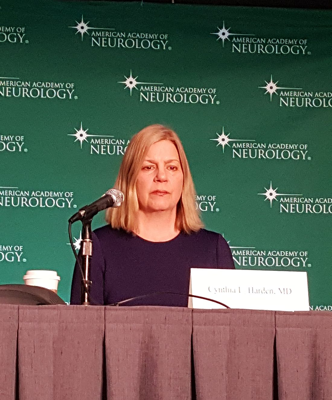
Specifically, the guideline recommends that , she said, adding that the guideline shows that “being free of seizures, particularly tonic-clonic seizures, is strongly associated with a decreased risk.”
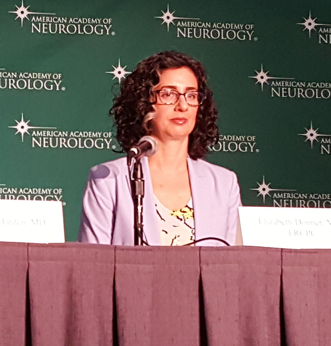
Guideline coauthor, Elizabeth Donner, MD, added that, for this reason, the guideline recommends “that health professionals work with people who continue to have, specifically, these kind of seizures to try and reduce them with medications or with epilepsy surgery, actively weighing the risks and benefits of any new approach to seizure management.”
The recommendations are based on moderate (Level B) evidence.
The team also looked at numerous other risk factors for SUDEP and found that the strength of the evidence was too weak to support additional recommendations, said Dr. Donner, director of the comprehensive epilepsy program at The Hospital for Sick Children in Toronto and chair of the American Epilepsy Society SUDEP Task Force.
“More research is now needed to identify other preventable risk factors for SUDEP so that we can focus future studies on finding ways to reduce how often SUDEP occurs,” she added.
While the message regarding the importance of reducing seizure frequency is not new, it is important that this message be reiterated in the context of SUDEP, Dr. Donner said.
“It’s very important for it to be clear that the risk of frequent generalized tonic-clonic seizures – and we’re not talking about really frequent here; we’re talking about significant increased risk of death with only three per year – is not related only to maintaining a driver’s license, maintaining work, or other outcomes like that. It’s actually related to risk of death,” she said, noting that she hopes this is a motivator for pursuing treatments beyond medication when medication isn’t successful for treating seizures.
The guideline, which is endorsed by the International Child Neurology Association, is available online and in print (Neurology. 2017;88:1674–80).
Dr. Harden receives royalties from Wiley and Up-to-Date. Dr. Donner has received research support from the Canadian Institutes of Health Research, Dravet Canada, and SUDEP Aware. Other guideline authors reported numerous disclosures, including many industry sources.
BOSTON – Generalized tonic-clonic seizures (GTCS) are a major risk factor for sudden unexpected death in epilepsy (SUDEP), which underscores the importance of advising people with epilepsy about controlling such seizures, according to a new practice guideline from the American Academy of Neurology and the American Epilepsy Society.
Though SUDEP is rare, with an incidence rate of 0.22/1,000 patient-years in children with epilepsy and 1.2/1,000 patient-years in adults with epilepsy, the guideline committee found that people with three or more GTCS per year are 15 times more likely to die suddenly than are those without this seizure type. The risk increases with increasing GTCS frequency. This translates to an absolute risk of up to 18 deaths per 1,000 patient-years for people with epilepsy who have frequent GTCS.
Specifically, the guideline recommends that , she said, adding that the guideline shows that “being free of seizures, particularly tonic-clonic seizures, is strongly associated with a decreased risk.”
Guideline coauthor, Elizabeth Donner, MD, added that, for this reason, the guideline recommends “that health professionals work with people who continue to have, specifically, these kind of seizures to try and reduce them with medications or with epilepsy surgery, actively weighing the risks and benefits of any new approach to seizure management.”
The recommendations are based on moderate (Level B) evidence.
The team also looked at numerous other risk factors for SUDEP and found that the strength of the evidence was too weak to support additional recommendations, said Dr. Donner, director of the comprehensive epilepsy program at The Hospital for Sick Children in Toronto and chair of the American Epilepsy Society SUDEP Task Force.
“More research is now needed to identify other preventable risk factors for SUDEP so that we can focus future studies on finding ways to reduce how often SUDEP occurs,” she added.
While the message regarding the importance of reducing seizure frequency is not new, it is important that this message be reiterated in the context of SUDEP, Dr. Donner said.
“It’s very important for it to be clear that the risk of frequent generalized tonic-clonic seizures – and we’re not talking about really frequent here; we’re talking about significant increased risk of death with only three per year – is not related only to maintaining a driver’s license, maintaining work, or other outcomes like that. It’s actually related to risk of death,” she said, noting that she hopes this is a motivator for pursuing treatments beyond medication when medication isn’t successful for treating seizures.
The guideline, which is endorsed by the International Child Neurology Association, is available online and in print (Neurology. 2017;88:1674–80).
Dr. Harden receives royalties from Wiley and Up-to-Date. Dr. Donner has received research support from the Canadian Institutes of Health Research, Dravet Canada, and SUDEP Aware. Other guideline authors reported numerous disclosures, including many industry sources.
AT AAN 2017
Apomorphine Reduces Off Time for People with Advanced Parkinson’s Disease
BOSTON—Apomorphine may provide relief from off time for people with advanced Parkinson’s disease, according to a study presented at the 69th Annual Meeting of the American Academy of Neurology.
“If a person with Parkinson’s disease can reduce their off times, that can have a great impact on their everyday life,” said study author Regina Katzenschlager, MD, of Danube Hospital, which is affiliated with the Medical University of Vienna. “In some patients in the trial, the insecurity of unpredictable periods of incapacity was completely alleviated.”
Open-Label Data Suggest Efficacy
Levodopa has long been the gold standard in the treatment of Parkinson’s disease, but the effects of the medication can partially wear off more quickly as the disease progresses. Patients consequently experience off time that includes symptoms such as slowness and muscle rigidity.
The drug apomorphine, which first was produced in 1865, began to be used to treat advanced Parkinson’s disease in the United States in 1950. Its use grew in the 1990s when European doctors started administering subcutaneous infusions of the drug to treat fluctuations in mobility that could not be controlled by levodopa.
Extensive data from open-label studies of apomorphine have demonstrated its efficacy in reducing off time, dyskinesias, and oral levodopa dose in patients with severe motor fluctuations that are poorly controlled by conventional therapy. Evidence from randomized, blinded studies of the drug has been lacking, however.
Comparing Apomorphine With Placebo
In a double-blind phase III study, researchers recruited 107 people with advanced Parkinson’s disease from 23 centers in seven countries.
Participants were randomized to either apomorphine subcutaneous infusion (≤ 8 mg/h) or a placebo saline infusion. The infusion was administered over a period of 14 to 18 hours each day through a small portable pump similar to the type used in the treatment of type 1 diabetes. The hourly flow rate of the infusion and dose of concomitant antiparkinsonian medication were adjusted during the first four weeks, based on efficacy and tolerability. The study’s primary end point was the absolute change in off time from baseline to Week 12 based on patient diaries.
Patients who received apomorphine had a significantly greater reduction of off time than those who received the placebo infusion. Active patients had, on average, 2.5 hours less off time per day, while participants who received the placebo infusion had an average of 35 minutes less off time per day. This improvement was apparent within the first week of treatment and was sustained over 12 weeks. At the same time, patients who received apomorphine had a significantly greater increase of on time without the dyskinesias that often are observed with levodopa, compared with placebo.
Participants were also asked to evaluate how well they thought the treatment worked. Those who received apomorphine gave their treatment higher scores at week 12 than those who received the placebo infusion. In the apomorphine group, 71% of patients felt improved, compared with 18% of patients who received placebo. Furthermore, 19% of patients worsened on apomorphine, compared with 45% on placebo. Apomorphine was generally well tolerated, and the researchers observed no serious side effects.
“It is our hope that these findings confirming the efficacy of apomorphine infusion will encourage doctors in the United States to offer this treatment to their patients and assess its efficacy in their own clinical practice,” Dr. Katzenschlager
concluded.
The study was supported by Britannia Pharmaceuticals, the maker of apomorphine.
May 2017 Quiz 2
Q2: Answer: B
Objective: Diagnose HELLP syndrome
Rationale: This patient’s presentation and laboratory findings are consistent with HELLP syndrome – the syndrome of hemolysis, elevated liver enzymes, and low platelets. HELLP is on the preeclampsia spectrum, which encompasses preeclampsia, eclampsia, and HELLP. Patients with HELLP will have hypertension (BP above 140/90), thrombocytopenia to less than 100,000/mm3 and aminotransferase levels above 70 U/L.
The diagnosis can be confirmed with an LDH (lactate dehydrogenase) greater than 600 U/L and microangiopathic hemolytic anemia on peripheral blood smear. On liver biopsy, HELLP is characterized by periportal or focal parenchyma necrosis with hyaline deposition of fibrin material in the sinusoids. However, liver biopsies are rarely performed in this setting as it likely will not change management (delivery of the fetus) and it exposes the mother and fetus to additional risks.
There is significant overlap between HELLP and acute fatty liver of pregnancy, although elevated prothrombin and partial thromboplastin time, severe hypoglycemia, and elevated creatinine are more common in acute fatty liver of pregnancy. Hypertension is more common in HELLP, and therefore this patient’s presentation is more consistent with HELLP.
Reference
1. Kia L, Rinella ME. Interpretation and management of hepatic abnormalities in pregnancy. Clin Gastroenterol Hepatol. 2013;11(11):1392-8.
Q2. A consult is requested for a 32-year-old woman who is 29 weeks pregnant and has presented to the emergency department with nausea, vomiting, and right upper quadrant abdominal pain. She is afebrile, pulse 89, BP 160/105. On exam, she has mild to moderate epigastric and right upper quadrant tenderness. Her labs are notable for WBCs 13.0 x 10⁹/L, Hgb 8.9 g/dL, platelets 55,000 x 109/L, AST 145 U/L, total bilirubin 2.1 mg/dL; PT and PTT are normal, blood glucose is 110 mg/dL.
After the epidemic, Ebola’s destructive power still haunts survivors
VIENNA – The Ebola crisis may be over in Sierra Leone, but the suffering is not.
The last patient from the epidemic was discharged in February 2016, but 78% of survivors now appear to have one or more sequelae of the infection. Some problems are mild, but some are so debilitating that life may never be the same.
Janet Scott, MD, of the University of Liverpool (England), heads a task force studying Ebola’s lingering aftereffects. These fall into four categories, Dr. Scott said at the European Society of Clinical Microbiology and Infectious Diseases annual congress: musculoskeletal pain, headache, eye problems, and psychological disorders.
They add up to an enormous risk of disability – survivors are more than 200 times more likely than controls to express at least moderate disability.

Dr. Scott and her team of researchers are partnering with clinicians and data managers in the Ebola treatment unit in the 34th Regimental Military Hospital (MH34) in Freetown, Sierra Leone. In Sierra Leone alone, she said, there were nearly 9,000 cases and 3,500 deaths from the virus; about 5,000 patients survived. So far, Dr. Scott and her team have collected data on about 500 patients for whom they also provide free health care.
The project has six arms, each headed by an expert: clinical care, data collection, disability, neurology, ophthalmology, and psychiatry.
The team sees patients in a large tent sectioned by a plywood wall*. Wireless Internet access, which she said is “enormously expensive” in Sierra Leone, has been donated by Omline Business Communications*. It’s the team’s lifeline, allowing them to transmit data between Freetown and participating units around the world. Members also Skype regularly, talking with patients and with each other.
All patients who come into the clinic have an initial visit that includes collection of demographics, their Ebola and clinical history (including an exploration of comorbidities), a maternal health screening for women, vital signs and symptom assessment, medication dispensing, and a treatment plan.
Then they visit the specialists, either onsite or through local referral*. These specialist modules include joints, eyes, headache, ears, neurology, cardiac, respiratory, gastrointestinal, renal and urologic, reproductive health for both genders, and psychiatry.

Last year, Dr. Scott published initial data on 44 patients. At ECCMID 2017, she expanded that report to include 203 survivors. They spanned all ages, but about 67% were in their most productive adult years, aged 20-39 years.
Her findings are striking: About 78% report musculoskeletal pain, with many saying they have trouble walking even short distances, climbing stairs, or picking up their children.
Headache was the next most common problem, reported by nearly 40%. About 15% report ocular problems, which include anterior uveitis, cataracts – even in very young children – and retinal lesions. Abdominal and chest pain affect about 10% of the survivors.
Although she didn’t present specific numbers, Dr. Scott also said that many of the survivors experience psychological sequelae, including insomnia, anxiety, and depression. Whether this is related to viral pathology isn’t clear; it could be a not-unexpected response to the trauma of living through the epidemic.
“Many of these people have lost their entire family, and those that are left now shun them,” she said in a live video interview on Facebook. “It’s almost like a post-traumatic stress reaction.”
The other symptoms probably are related to the disease pathology, she observed. “Unfortunately, we don’t have all the clinical details of the acute phase for everyone, but for those for whom we do have details, we are seeing correlation between some of the problems with viral loads at admission, and even episodes of becoming unconscious during the acute illness.”
Patrick Howlett, MD, of the King’s Sierra Leone Partnership, Freetown, leads the neurology study. So far, the researchers have collected data on 19 patients with severe neurological consequences. Of those, 12 (63%) experienced a period of unconsciousness during their acute Ebola episode. In a comparator group of 21 with nonsevere neurologic sequelae, 33% had experienced unconsciousness.
Headache was present in nine (47%) of the patients. Migraine was the most common diagnosis. “We don’t have money for migraine medications, but fortunately, most of our migraine patients seem to be doing well on beta blockers,” Dr. Scott said.
CT scans were performed on 17 patients: three showed cerebral or cerebellar atrophy and two had confirmed stroke.
The brain injuries were severe in two, including a 42-year-old with extensive gliosis in the left middle cerebral artery region and a dilated left ventricle secondary to loss of volume in that hemisphere. A 12-year-old girl showed extensive parietal and temporal lobe atrophy. She is now so disabled that her family can’t care for her at home.
Other neurological problems include peripheral neuropathy, brachial plexus neuropathy, and asymmetric lower limb muscular atrophy.
Paul Steptoe, MD, an ophthalmic registrar from St. Paul’s Eye Unit at the Royal Liverpool Hospital, heads the eye study. He has observed dense cataracts, even in children, and anterior uveitis that has blinded some patients. There is concern about live virus persisting in vitreal fluid, but two eye taps have been negative, Dr. Scott said.

The most exciting recent finding, however, was made possible by the donation of a digital retinal camera, which “enabled us to get dozens of amazing images,” Dr. Scott said. With it, Dr. Steptoe conducted a case-control study of 81 Ebola survivors and 106 community controls. The findings of this study are potentially very, very important, Dr. Scott said.
“The first thing we found out is that retinal scarring is pervasive in our control patients,” she said. “There is just a lot of it out here in the community. But more interesting is that Dr. Steptoe seems to have identified a characteristic retinal lesion seen only in our survivors. It could be evidence of neurotropic aspects of the Ebola virus.”
The lesions occurred in 12 (15%) of the survivors and none of the controls. They are of a striking and consistent shape: straight-edged and sharply angulated. The lesions are only on the surface of the retina and do not penetrate into deeper levels. Nor do they interfere with vision. Dr. Steptoe has proposed that they take their angular shape from the retina’s underlying structures. His paper documenting this finding has been accepted and will be published shortly in the journal Emerging Infectious Diseases.
All of the post-Ebola sequelae add up to general disability for survivors, Dr. Scott said. Soushieta Jagadesh, of the* Liverpool School of Tropical Medicine, is conducting a disability survey. The comparison between 27 survivors and 54 community controls employed the Washington Group extended disability questionnaire. “We noted major limitations 1 year after discharge in mobility, vision, cognition, and affect,” Dr. Scott said.
The hazard ratios for these issues are enormous: Overall, compared with controls, survivors were 23 times more likely to have some level of disability. They were 94 times more likely to have walking limitations and 65 times more likely to have problems with stairs. Survivors were over 200 times more likely to have moderate disability than were their unaffected neighbors.
If funding for the project is renewed – and Dr. Scott admitted this is an “if,” not a “when” – caring for and studying these survivors will continue. Just in this one city, she said, the need is huge.
According to data from the U.S. Centers for Disease Control and Prevention, more than 17,000 patients in Sierra Leone, Liberia, and Guinea survived the 2014 Ebola outbreak.
If the assessments of Freetown survivors hold true across this population, thousands of survivors face life-limiting sequelae of the disease.
“We still have patients walk in every day with musculoskeletal pain, headaches, and ocular issues,” Dr. Scott said. “At the beginning of the epidemic, we were just focusing on containing it and reducing transmission. Now, we are faced with the long-term consequences.”
The Wellcome Trust supported the study. The authors have been awarded a grant from the Enhancing Research Activity in Epidemic Situations (ERAES) program, funded by the Wellcome Trust to support further research into the sequelae of Ebola virus disease.
The video associated with this article is no longer available on this site. Please view all of our videos on the MDedge YouTube channel
[email protected]
On Twitter @Alz_gal
*This story was updated May 2, 2017.
VIENNA – The Ebola crisis may be over in Sierra Leone, but the suffering is not.
The last patient from the epidemic was discharged in February 2016, but 78% of survivors now appear to have one or more sequelae of the infection. Some problems are mild, but some are so debilitating that life may never be the same.
Janet Scott, MD, of the University of Liverpool (England), heads a task force studying Ebola’s lingering aftereffects. These fall into four categories, Dr. Scott said at the European Society of Clinical Microbiology and Infectious Diseases annual congress: musculoskeletal pain, headache, eye problems, and psychological disorders.
They add up to an enormous risk of disability – survivors are more than 200 times more likely than controls to express at least moderate disability.
Dr. Scott and her team of researchers are partnering with clinicians and data managers in the Ebola treatment unit in the 34th Regimental Military Hospital (MH34) in Freetown, Sierra Leone. In Sierra Leone alone, she said, there were nearly 9,000 cases and 3,500 deaths from the virus; about 5,000 patients survived. So far, Dr. Scott and her team have collected data on about 500 patients for whom they also provide free health care.
The project has six arms, each headed by an expert: clinical care, data collection, disability, neurology, ophthalmology, and psychiatry.
The team sees patients in a large tent sectioned by a plywood wall*. Wireless Internet access, which she said is “enormously expensive” in Sierra Leone, has been donated by Omline Business Communications*. It’s the team’s lifeline, allowing them to transmit data between Freetown and participating units around the world. Members also Skype regularly, talking with patients and with each other.
All patients who come into the clinic have an initial visit that includes collection of demographics, their Ebola and clinical history (including an exploration of comorbidities), a maternal health screening for women, vital signs and symptom assessment, medication dispensing, and a treatment plan.
Then they visit the specialists, either onsite or through local referral*. These specialist modules include joints, eyes, headache, ears, neurology, cardiac, respiratory, gastrointestinal, renal and urologic, reproductive health for both genders, and psychiatry.
Last year, Dr. Scott published initial data on 44 patients. At ECCMID 2017, she expanded that report to include 203 survivors. They spanned all ages, but about 67% were in their most productive adult years, aged 20-39 years.
Her findings are striking: About 78% report musculoskeletal pain, with many saying they have trouble walking even short distances, climbing stairs, or picking up their children.
Headache was the next most common problem, reported by nearly 40%. About 15% report ocular problems, which include anterior uveitis, cataracts – even in very young children – and retinal lesions. Abdominal and chest pain affect about 10% of the survivors.
Although she didn’t present specific numbers, Dr. Scott also said that many of the survivors experience psychological sequelae, including insomnia, anxiety, and depression. Whether this is related to viral pathology isn’t clear; it could be a not-unexpected response to the trauma of living through the epidemic.
“Many of these people have lost their entire family, and those that are left now shun them,” she said in a live video interview on Facebook. “It’s almost like a post-traumatic stress reaction.”
The other symptoms probably are related to the disease pathology, she observed. “Unfortunately, we don’t have all the clinical details of the acute phase for everyone, but for those for whom we do have details, we are seeing correlation between some of the problems with viral loads at admission, and even episodes of becoming unconscious during the acute illness.”
Patrick Howlett, MD, of the King’s Sierra Leone Partnership, Freetown, leads the neurology study. So far, the researchers have collected data on 19 patients with severe neurological consequences. Of those, 12 (63%) experienced a period of unconsciousness during their acute Ebola episode. In a comparator group of 21 with nonsevere neurologic sequelae, 33% had experienced unconsciousness.
Headache was present in nine (47%) of the patients. Migraine was the most common diagnosis. “We don’t have money for migraine medications, but fortunately, most of our migraine patients seem to be doing well on beta blockers,” Dr. Scott said.
CT scans were performed on 17 patients: three showed cerebral or cerebellar atrophy and two had confirmed stroke.
The brain injuries were severe in two, including a 42-year-old with extensive gliosis in the left middle cerebral artery region and a dilated left ventricle secondary to loss of volume in that hemisphere. A 12-year-old girl showed extensive parietal and temporal lobe atrophy. She is now so disabled that her family can’t care for her at home.
Other neurological problems include peripheral neuropathy, brachial plexus neuropathy, and asymmetric lower limb muscular atrophy.
Paul Steptoe, MD, an ophthalmic registrar from St. Paul’s Eye Unit at the Royal Liverpool Hospital, heads the eye study. He has observed dense cataracts, even in children, and anterior uveitis that has blinded some patients. There is concern about live virus persisting in vitreal fluid, but two eye taps have been negative, Dr. Scott said.
The most exciting recent finding, however, was made possible by the donation of a digital retinal camera, which “enabled us to get dozens of amazing images,” Dr. Scott said. With it, Dr. Steptoe conducted a case-control study of 81 Ebola survivors and 106 community controls. The findings of this study are potentially very, very important, Dr. Scott said.
“The first thing we found out is that retinal scarring is pervasive in our control patients,” she said. “There is just a lot of it out here in the community. But more interesting is that Dr. Steptoe seems to have identified a characteristic retinal lesion seen only in our survivors. It could be evidence of neurotropic aspects of the Ebola virus.”
The lesions occurred in 12 (15%) of the survivors and none of the controls. They are of a striking and consistent shape: straight-edged and sharply angulated. The lesions are only on the surface of the retina and do not penetrate into deeper levels. Nor do they interfere with vision. Dr. Steptoe has proposed that they take their angular shape from the retina’s underlying structures. His paper documenting this finding has been accepted and will be published shortly in the journal Emerging Infectious Diseases.
All of the post-Ebola sequelae add up to general disability for survivors, Dr. Scott said. Soushieta Jagadesh, of the* Liverpool School of Tropical Medicine, is conducting a disability survey. The comparison between 27 survivors and 54 community controls employed the Washington Group extended disability questionnaire. “We noted major limitations 1 year after discharge in mobility, vision, cognition, and affect,” Dr. Scott said.
The hazard ratios for these issues are enormous: Overall, compared with controls, survivors were 23 times more likely to have some level of disability. They were 94 times more likely to have walking limitations and 65 times more likely to have problems with stairs. Survivors were over 200 times more likely to have moderate disability than were their unaffected neighbors.
If funding for the project is renewed – and Dr. Scott admitted this is an “if,” not a “when” – caring for and studying these survivors will continue. Just in this one city, she said, the need is huge.
According to data from the U.S. Centers for Disease Control and Prevention, more than 17,000 patients in Sierra Leone, Liberia, and Guinea survived the 2014 Ebola outbreak.
If the assessments of Freetown survivors hold true across this population, thousands of survivors face life-limiting sequelae of the disease.
“We still have patients walk in every day with musculoskeletal pain, headaches, and ocular issues,” Dr. Scott said. “At the beginning of the epidemic, we were just focusing on containing it and reducing transmission. Now, we are faced with the long-term consequences.”
The Wellcome Trust supported the study. The authors have been awarded a grant from the Enhancing Research Activity in Epidemic Situations (ERAES) program, funded by the Wellcome Trust to support further research into the sequelae of Ebola virus disease.
The video associated with this article is no longer available on this site. Please view all of our videos on the MDedge YouTube channel
[email protected]
On Twitter @Alz_gal
*This story was updated May 2, 2017.
AT ECCMID 2017