User login
Midostaurin improves survival in new AML
Adding the multitargeted kinase inhibitor midostaurin to standard chemotherapy led to significantly longer overall and event-free survival, compared with placebo and standard chemotherapy in newly diagnosed acute myeloid leukemia (AML) patients with FLT3 gene mutations, according to phase III trial results published in the New England Journal of Medicine.*
About 30% of AML patients have mutations to the FLT3 gene – with three-quarters of those internal tandem duplication (ITD) mutations, which involves duplication of between 3 and 100 amino acids in the juxtamembrane region. These mutations are linked with a high relapse rate and poor prognosis, especially when there is a high ratio of these mutations to wild-type FLT3. About 8% of patients with newly diagnosed AML have an FLT3 point mutation in the tyrosine kinase domain (TKD), but the effect of these on prognosis isn’t clear.
In the trial, called RATIFY and conducted at 225 sites in 17 countries, 360 patients were randomized to the midostaurin group and 357* to placebo, and they were treated from 2008 to 2013. In all, 29.8% of patients were “ITD high,” meaning their ITD FLT3 mutation to wild-type FLT3 ratio was higher than 0.7, and 47.6% were “ITD low,” with a mutation-to-wild-type FLT3 ratio of 0.5 to 0.7. A total of 22.6% of patients had TKD mutations.
Patients received standard induction chemotherapy, with daunorubicine and cytarabine, and on days 8 through 21 either 50 mg of midostaurin or placebo orally twice a day. Patients were given an identical second cycle of induction therapy, with midostaurin or placebo, if they showed definitive clinically significant residual leukemia after the first induction treatment.
Those who achieved complete remission after induction were given 4, 28-day cycles of consolidation treatment, with midostaurin or placebo on days 8 through 21. If they stayed in remission after that, they were given maintenance of 12, 28-day cycles of midostaurin or placebo.
They were not required to receive hematopoetic stem cell transplantation (HSCT), but it was performed at investigator discretion.
Midostaurin improved survival but not rates of complete remission as defined in the trial protocol, researchers reported.
The hazard ratio for death in the midostaurin group was 0.78 (95% CI, 0.63 to 0.96; one-sided P = .0009). The 4-year overall survival rate was 51.4% for the midostaurin group and 44.3% for the placebo group. Midostaurin was shown to benefit all mutation subgroups, but with no greater benefit in one group than another.
Patients in the midostaurin group had a 21.6% lower likelihood of having an event, defined as failure to achieve protocol-defined complete remission, relapse or death without relapse.
There was no significant difference between the groups in complete remission, which under protocol had to occur by day 60.
HSCT was performed in 57% of patients – during the first complete remission in 28.1% of the midostaurin group and in 22.7% during the first complete remission in the placebo group. For those who were transplanted after the first complete remission, no treatment effect was seen.
Researchers noted that there was a therapeutic benefit even among patients with ITD mutations but with a low allelic burden, in whom the disease might be due largely to mutations other than FLT3.
“It is possible that the benefit of midostaurin, which is a multitargeted kinase inhibitor, might lie beyond its ability to inhibit FLT3,” possibly through inhibition of KIT, researchers said.
They also noted that as the trial went on, more and more investigators decided to treat patients with hematopoietic stem cell transplantation, based on newly reported data elsewhere. Since midostaurin was discontinued at the time of transplant, that could have limited exposure to the drug and limited its effect.
*CORRECTION 7/5/2017: An earlier version of this article misstated the number of patients in the placebo group as well as where the study originally appeared.
FROM NEJM
Key clinical point: Multitargeted kinase inhibitor midostaurin combined with standard chemotherapy improved survival in newly diagnosed acute myeloid leukemia patients.
Major finding: The 4-year overall survival rate was 51.4% for the midostaurin group and 44.3% for the placebo group. Midostaurin was shown to benefit all mutation subgroups — internal tandem mutations and point mutations in the tyrosine kinase domain – but with no greater benefit in one group than another.
Data source: A multicenter, multinational, randomized, double-blind, placebo-controlled trial.
Disclosures: The trial was funded by the National Cancer Institute and Novartis. Researchers reported receiving personal fees from Novartis and other companies.
Ibrutinib dons new anti-GVHD hat
MADRID – Talk about versatility: Ibrutinib (Imbruvica), a drug with marked activity against B-cell malignancies, also appears to be a safe and acceptable option for the treatment of patients with chronic graft vs. host disease (cGVHD) for whom frontline therapies have failed.
Among 42 patients in a phase II study with steroid-refractory cGVHD, the overall response rate with ibrutinib was 67%, with one-third of responders having a complete response, reported Iskra Pusic, MD, from Washington University School of Medicine in St. Louis.
Corticosteroids are the most commonly used therapy for cGVHD in the United States, but for those patients for whom corticosteroids are a bust, there is no established second-line therapy, and patients with refractory cGVHD are usually recommended for clinical trials, Dr. Pusic said.
The therapeutic rationale underpinning the use of ibrutinib in cGVHD, a condition marked by extensive immune dysregulation, is that the agent is an irreversible inhibitor of Bruton’s tyrosine kinase and interleukin-2 inducible T-cell kinase, and thus has wide-ranging immune-dampening activity, Dr. Pusic said.
She and colleagues in a multicenter study enrolled 42 patients with cGVHD that corticosteroids had failed to treat adequately, and treated them with oral ibrutinib 420 mg daily until cGVHD progression or unacceptable toxicity.
At a median follow-up of 13.9 months, a total of 28 patients (67%) had a response according to 2005 National Institutes of Health (NIH) criteria, including nine with a complete response, and 19 with partial responses.
Of the patients with responses, 79% had a response at the time of the first assessment for response, and 71% of responders had responses lasting at least 5 months.
Among patients with multiorgan involvement, responses were seen in two or more organs.
Grade 3 or greater adverse events included fatigue, diarrhea, muscles spasms, pneumonia, pyrexia, and headache. Two patients died on study, one from multilobular pneumonia and one from bronchopulmonary aspergillosis.
In general, the safety profile of ibrutinib was similar to that seen in studies of the drug in B-cell malignancies and to that seen with corticosteroid therapy for patients with cGVHD, Dr. Pusic said.
Investigators are currently enrolling patients in a double-blind clinical trial comparing ibrutinib or placebo in combination with corticosteroids in patients with newly diagnosed cGVHD, she noted.
The study was supported by Pharmacyclics. Dr. Pusic did not report disclosures.
AT EHA 2017
Key clinical point: The tyrosine kinase inhibitor ibrutinib was associated with complete and partial responses in two-thirds of patients with steroid-refractory chronic graft vs. host disease (cGVHD).
Major finding: A total of 28 patients (67%) had responses, including 9 complete responses.
Data source: Phase II clinical trial in 42 patients with cGVHD for whom corticosteroids had failed.
Disclosures: The study was supported by Pharmacyclics. Dr. Pusic did not report disclosures.
Apremilast eases PsA symptoms in patients who are naive to biologics
MADRID – Aprelimast rapidly improved symptoms for patients with active psoriatic arthritis who were naive to biologics, whether or not they were on background methotrexate.
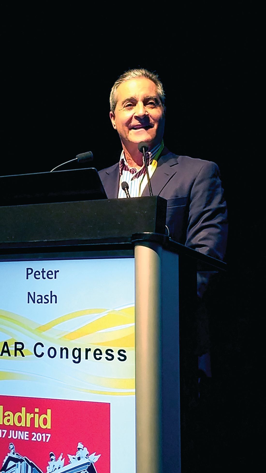
Response to the phosphodiesterase 4-inhibitor also increased with time, Peter Nash, MD, said at the European Congress of Rheumatology. By 2 weeks, 16% had achieved an ACR20 response. By 24 weeks, that had risen to 43%, and at 52 weeks, was 67%. Many patients responded even better, with 37% achieving an ACR50 response and 21% and ACR70 response by the end of the phase III placebo-controlled trial, said Dr. Nash of the University of Queensland, Brisbane, Australia.
ACTIVE randomized 229 patients with active psoriatic arthritis (PsA) to either placebo or 30 mg apremilast twice daily. The primary endpoint was ACR20 response at week 16. At that time, patients who had not improved by at least 10% were offered early escape into active therapy.
Patients were a mean of 49 years old, with mean disease duration of 4 years. This is considerably shorter than the duration seen in many PsA trials, Dr. Nash said. It was a reflection of the study requirement that patients have taken no more than one synthetic disease-modifying antirheumatic agent (sDMARD) and no biologics for their disease.
The active and placebo groups separated very early in this study, with 16% of the apremilast group achieving an ACR 20 response by 2 weeks compared to 6% taking placebo. The response curves remained significantly separated throughout the entire study. By the 16-week endpoint, 38% of the active group and 20% of the placebo group had achieved ACR20. At this point, 11 of those taking apremilast asked for early escape and went on open-label apremilast at the same dose; 35 taking placebo were switched to open-label apremilast.
By 24 weeks, the ACR20 responses were 44% in the apremilast group and 25% in the placebo group. That response rate continued to rise over the year of treatment. By week 52, 67% of patients who had been on the study drug since the beginning had an ACR20, 37% an ACR50, and 21% an ACR70 response. ACR response was just as good in patients who were taking background methotrexate as those who were not, Dr. Nash said.
Compared to placebo, apremilast was associated with significantly more improvement on the Disease Activity Score-28 (CRP). Again, the response curves separated significantly by 2 weeks (–0.59 apremilast vs. –0.31 placebo) and continued to separate at 16 weeks (–1.07 vs. –0.39), and 24 weeks (–1.26 vs. –0.76). At 52 weeks, among those who had been initially randomized to apremilast, the mean DAS-28 (CRP) score had changed –1.71 from baseline.
The drug also significantly improved enthesitis relative to placebo, with rapid onset of action that continued to improve by week 24. The adverse event profile was good, Dr. Nash said. There were no opportunistic infections and no reactivations of tuberculosis. Ten in the active group and five in the placebo group withdrew due to an adverse event. Diarrhea and headache were the most common issues. Diarrhea occurred in 11% of the placebo group and 15% of the apremilast group; headache in 4% of the placebo group and 7% of the apremilast group. These were more common in the beginning of the study and tended to resolve with time, Dr. Nash said.
Thirty patients in the study had a history of depression and of these, two had a depression flare that lead to withdrawal from the study. Weight loss of at least 10% occurred in 5% of those taking apremilast.
Celgene sponsored the study. Dr. Nash has received research support from the company.
[email protected]
On Twitter @Alz_gal
MADRID – Aprelimast rapidly improved symptoms for patients with active psoriatic arthritis who were naive to biologics, whether or not they were on background methotrexate.
Response to the phosphodiesterase 4-inhibitor also increased with time, Peter Nash, MD, said at the European Congress of Rheumatology. By 2 weeks, 16% had achieved an ACR20 response. By 24 weeks, that had risen to 43%, and at 52 weeks, was 67%. Many patients responded even better, with 37% achieving an ACR50 response and 21% and ACR70 response by the end of the phase III placebo-controlled trial, said Dr. Nash of the University of Queensland, Brisbane, Australia.
ACTIVE randomized 229 patients with active psoriatic arthritis (PsA) to either placebo or 30 mg apremilast twice daily. The primary endpoint was ACR20 response at week 16. At that time, patients who had not improved by at least 10% were offered early escape into active therapy.
Patients were a mean of 49 years old, with mean disease duration of 4 years. This is considerably shorter than the duration seen in many PsA trials, Dr. Nash said. It was a reflection of the study requirement that patients have taken no more than one synthetic disease-modifying antirheumatic agent (sDMARD) and no biologics for their disease.
The active and placebo groups separated very early in this study, with 16% of the apremilast group achieving an ACR 20 response by 2 weeks compared to 6% taking placebo. The response curves remained significantly separated throughout the entire study. By the 16-week endpoint, 38% of the active group and 20% of the placebo group had achieved ACR20. At this point, 11 of those taking apremilast asked for early escape and went on open-label apremilast at the same dose; 35 taking placebo were switched to open-label apremilast.
By 24 weeks, the ACR20 responses were 44% in the apremilast group and 25% in the placebo group. That response rate continued to rise over the year of treatment. By week 52, 67% of patients who had been on the study drug since the beginning had an ACR20, 37% an ACR50, and 21% an ACR70 response. ACR response was just as good in patients who were taking background methotrexate as those who were not, Dr. Nash said.
Compared to placebo, apremilast was associated with significantly more improvement on the Disease Activity Score-28 (CRP). Again, the response curves separated significantly by 2 weeks (–0.59 apremilast vs. –0.31 placebo) and continued to separate at 16 weeks (–1.07 vs. –0.39), and 24 weeks (–1.26 vs. –0.76). At 52 weeks, among those who had been initially randomized to apremilast, the mean DAS-28 (CRP) score had changed –1.71 from baseline.
The drug also significantly improved enthesitis relative to placebo, with rapid onset of action that continued to improve by week 24. The adverse event profile was good, Dr. Nash said. There were no opportunistic infections and no reactivations of tuberculosis. Ten in the active group and five in the placebo group withdrew due to an adverse event. Diarrhea and headache were the most common issues. Diarrhea occurred in 11% of the placebo group and 15% of the apremilast group; headache in 4% of the placebo group and 7% of the apremilast group. These were more common in the beginning of the study and tended to resolve with time, Dr. Nash said.
Thirty patients in the study had a history of depression and of these, two had a depression flare that lead to withdrawal from the study. Weight loss of at least 10% occurred in 5% of those taking apremilast.
Celgene sponsored the study. Dr. Nash has received research support from the company.
[email protected]
On Twitter @Alz_gal
AT EULAR 2017
Key clinical point: Apremilast improved symptoms of psoriatic arthritis in patients who were naive to biologics.
Major finding: By the 16-week endpoint, 38% of the active group and 20% of the placebo group had achieved ACR20.
Data source: The phase IIIb study randomized 229 patients to apremilast or placebo.
Disclosures: Celgene sponsored the trial. Dr. Nash has received research support from the company.
Consider intraperitoneal ropivacaine for colectomy ERAS
SEATTLE – Intraperitoneal ropivacaine decreases postoperative pain and improves functional recovery after laparoscopic colectomy, according to randomized, blinded trial from the Royal Adelaide (Australia) Hospital.
“We recommend routine inclusion of IPLA [intraperitoneal local anesthetic] in the multimodal analgesia component of ERAS [enhanced-recovery-after-surgery] programs for laparoscopic colectomy,” the investigators concluded.
Recovery was smoother in the ropivacaine group. On a 90-point surgical recovery scale assessing fatigue, mental function, and the ability to do normal daily activities, ropivacaine patients were a few points ahead on days 1 and 3; the gap widened to about 10 points on days 7, 30, and 45. Ropivacaine might have helped reduced inflammation, accounting for the extended benefit, Dr. Lewis said.
Pain control was better with ropivacaine, as well. Ropivacaine patients were about 15-20 points lower on 50-point scales assessing both visceral and abdominal pain at postop hours 3 and 24, and day 7. The findings were statistically significant.
Several trends also favored ropivacaine. Ropivacaine patients had their first bowel movement at around 70 hours postop, versus about 82 hours in the control group. They were also discharged almost a day sooner, and had less postop vomiting. Just one patient in the ropivacaine group was diagnosed with ileus, versus four in the control arm.
There was also a trend for less opioid use in the ropivacaine group, which Dr. Lewis suspected would have been statistically significant if the trial had more patients.
Ropivacaine patients were a mean of 67 years old, versus 62 years old in the control group. There were slightly more men than women in each arm. The mean body mass index in the ropivacaine group was 28.5 kg/m2, and in the control group 26.4 kg/m2. Ropivacaine was discontinued in two patients due to possible toxicity.
An audience member noted that intravenous lidocaine has shown similar benefits in abdominal surgery.
The investigators had no disclosures.
AT ASCRS 2017
Key clinical point:
Major finding: On a 90-point surgical recovery scale assessing fatigue, mental function, and the ability to do normal daily activities, ropivacaine patients were a few points ahead of saline controls on postop days 1 and 3; the gap widened to about 10 points on days 7, 30, and 45.
Data source: Randomized, blinded trial with 51 patients
Disclosures: The investigators had no disclosures.
Overall survival better in advanced Hodgkin lymphoma with shorter eBEACOPP
MADRID – Patients with advanced Hodgkin lymphoma who have a metabolic response after the first two cycles of extended-dose(e)BEACOPP can be spared from undergoing more than two additional cycles of the highly intensive and toxic regimen, investigators from the German Hodgkin Study Group (GHSG) contend.
Among 1,005 patients with Hodgkin lymphoma who had negative PET scans after the second cycle of eBEACOPP (bleomycin, etoposide, doxorubicin, cyclophosphamide, prednisone, procarbazine), progression-free survival (PFS) was virtually identical whether patients were randomized to undergo a total of six-to-eight cycles or only four cycles, reported Peter Borchmann, MD, from the University of Cologne, Germany.

“For patients with negative PET-2 after initial treatment with eBEACOPP. Therapy with only two additional cycles of eBEACOPP is very effective, obviously, very safe, very short – it just takes 12 weeks – and it’s affordable,” he said.
“When balancing efficacy and safety, results compare favorably with any other published treatment strategy so far. That’s why we recommend this treatment, PET-guided extended BEACOPP in patients with newly diagnosed, advanced-stage Hodgkin lymphoma,” he added.
Although most patients in the United States with newly diagnosed Hodgkin lymphoma receive ABVD (doxorubicin, bleomycin, vinblastine, and dacarbazine), BEACOPP is sometimes used for high-risk patients. BEACOPP is associated with considerable toxicities, however, including increased risk of secondary malignancies.
To see whether select patients could be cured with fewer cycles of therapy, the GHSG investigators designed the GHSG HD18 study in which patient with metabolic responses determined by fluorodeoxyglucose-PET after two eBEACOPP cycles were randomized to either two or six-to-eight additional cycles.
A total of 2,101 patients from the ages of 18-60 years with newly diagnosed advanced-stage Hodgkin lymphoma were enrolled from centers in Germany, Switzerland, Austria, the Czech Republic, and the Netherlands.
After the second cycle of therapy, patients underwent fluorodeoxyglucose-PET scans, and those with negative results were then randomized.
The trial was designed and powered for noninferiority of the shortened regimen, with a maximum allowable difference of 6%.
The trial met its primary endpoint and then some. After a median observation time of 53 months, the 5-year PFS rate for patients who received only four cycles was 91.2%, compared with 91.8% for patients who underwent six-to-eight cycles. As shown on a Kaplan-Meier curve, the two lines were superimposable and virtually impossible to tell apart.
Interestingly, 5-year overall survival was significantly better with the shorter, less toxic regimen, at 97.6% vs. 95.4%, respectively, translating into a hazard ratio favoring the shorter regimen of 0.36 (P = .006).
In addition, the four-cycle regimen was associated with fewer severe infections (8% vs 15%), and lower degrees of organ toxicity (8% vs. 18%). In addition, the rate of secondary acute myeloid leukemia or the myelodysplastic syndrome was 0.4% among the 501 patients treated with only four cycles, compared with 1.6% for the 504 patients who received six to eight cycles.
There were no treatment-related deaths among patients who underwent four cycles, compared with six deaths among patients treated with additional cycles.
In an interview, Dr. Borchmann said that the findings are likely to change the standard of care for those centers that use the BEACOPP regimen. He acknowledged that the regimen is highly toxic and requires intensive patient surveillance and management, which may be more practical in Europe where patients generally live closer to major cancer centers than in the more spacious United States.
The study was supported by German Cancer Aid, the Swiss State Secratariate for Education, Research and Innovation, and by Roche Pharma AG. Dr, Borchmann reported having no relevant disclosures.
MADRID – Patients with advanced Hodgkin lymphoma who have a metabolic response after the first two cycles of extended-dose(e)BEACOPP can be spared from undergoing more than two additional cycles of the highly intensive and toxic regimen, investigators from the German Hodgkin Study Group (GHSG) contend.
Among 1,005 patients with Hodgkin lymphoma who had negative PET scans after the second cycle of eBEACOPP (bleomycin, etoposide, doxorubicin, cyclophosphamide, prednisone, procarbazine), progression-free survival (PFS) was virtually identical whether patients were randomized to undergo a total of six-to-eight cycles or only four cycles, reported Peter Borchmann, MD, from the University of Cologne, Germany.
“For patients with negative PET-2 after initial treatment with eBEACOPP. Therapy with only two additional cycles of eBEACOPP is very effective, obviously, very safe, very short – it just takes 12 weeks – and it’s affordable,” he said.
“When balancing efficacy and safety, results compare favorably with any other published treatment strategy so far. That’s why we recommend this treatment, PET-guided extended BEACOPP in patients with newly diagnosed, advanced-stage Hodgkin lymphoma,” he added.
Although most patients in the United States with newly diagnosed Hodgkin lymphoma receive ABVD (doxorubicin, bleomycin, vinblastine, and dacarbazine), BEACOPP is sometimes used for high-risk patients. BEACOPP is associated with considerable toxicities, however, including increased risk of secondary malignancies.
To see whether select patients could be cured with fewer cycles of therapy, the GHSG investigators designed the GHSG HD18 study in which patient with metabolic responses determined by fluorodeoxyglucose-PET after two eBEACOPP cycles were randomized to either two or six-to-eight additional cycles.
A total of 2,101 patients from the ages of 18-60 years with newly diagnosed advanced-stage Hodgkin lymphoma were enrolled from centers in Germany, Switzerland, Austria, the Czech Republic, and the Netherlands.
After the second cycle of therapy, patients underwent fluorodeoxyglucose-PET scans, and those with negative results were then randomized.
The trial was designed and powered for noninferiority of the shortened regimen, with a maximum allowable difference of 6%.
The trial met its primary endpoint and then some. After a median observation time of 53 months, the 5-year PFS rate for patients who received only four cycles was 91.2%, compared with 91.8% for patients who underwent six-to-eight cycles. As shown on a Kaplan-Meier curve, the two lines were superimposable and virtually impossible to tell apart.
Interestingly, 5-year overall survival was significantly better with the shorter, less toxic regimen, at 97.6% vs. 95.4%, respectively, translating into a hazard ratio favoring the shorter regimen of 0.36 (P = .006).
In addition, the four-cycle regimen was associated with fewer severe infections (8% vs 15%), and lower degrees of organ toxicity (8% vs. 18%). In addition, the rate of secondary acute myeloid leukemia or the myelodysplastic syndrome was 0.4% among the 501 patients treated with only four cycles, compared with 1.6% for the 504 patients who received six to eight cycles.
There were no treatment-related deaths among patients who underwent four cycles, compared with six deaths among patients treated with additional cycles.
In an interview, Dr. Borchmann said that the findings are likely to change the standard of care for those centers that use the BEACOPP regimen. He acknowledged that the regimen is highly toxic and requires intensive patient surveillance and management, which may be more practical in Europe where patients generally live closer to major cancer centers than in the more spacious United States.
The study was supported by German Cancer Aid, the Swiss State Secratariate for Education, Research and Innovation, and by Roche Pharma AG. Dr, Borchmann reported having no relevant disclosures.
AT EHA 2017
Key clinical point: For patients with newly diagnosed advanced Hodgkin lymphoma with PET-confirmed metabolic responses, four cycles of BEACOPP were at least as good as six to eight cycles for progression-free and overall survival.
Major finding: 5-year overall survival with four cycles of extended-dose BEACOPP was 97.6%, compared with. 95.4% for six to eight cycles (P = .006).
Data source: Randomized trial in 1,005 patients with newly diagnosed Hodgkin lymphoma from five European nations.
Disclosures: The study was supported by German Cancer Aid, the Swiss State Secretariate for Education, Research and Innovation, and by Roche Pharma AG. Dr. Borchmann reported having no relevant disclosures.
Major bleeding deaths may outweigh VTE risk in older cancer patients
MADRID – Look before you leap into anticoagulation therapy for older cancer patients, results from a Canadian cohort study suggest.
Among patients 65 and older with cancer and a venous thromboembolic event (VTE) within 6 months of the cancer diagnosis, the 7-day mortality rate from VTE was 0.5%, compared with an 11% rate of death from a major bleeding episode, reported Alejandro Lazo-Langner MD, MSc from Western University in London, Canada.

If their findings are confirmed in further studies, “it would actually change what we do in terms of the treatment of thrombosis,” he said.
Risks for both VTE and for bleeding are known to be higher among patients with cancer than in the general population. Although a previously published systematic review suggested that mortality rates from recurrent VTE and major bleeding events were similar in the first 6 months of anticoagulation therapy, those results were limited by the heterogeneity of designs in the various studies included in the review, and by differences in outcome measures and the types of populations included, Dr. Lazo-Langner said.
To get a better idea of the case fatality rates of VTE recurrence and major bleeding and the case fatality rate ratio for each, the authors conducted a retrospective population-based cohort study in the Province of Ontario using de-identified linked administrative health care databases.
They assembled a cohort of patients 65 years of age and older who had a VTE event within 6 months of an initial cancer diagnosis. Recurrent VTE and major bleeding events were assessed within 180 days of the index date.
They found that from 2004 through 2014 there were 6,967 VTEs in cancer patients over 65 years of age (mean age 75) that were treated with an anticoagulant, either low-molecular-weight heparin (LMWH), LMWH plus warfarin, warfarin alone, or rivaroxaban (Xarelto).
Six months after the index VTE events, 235 patients (3%) had experienced a major bleeding event, and 1,184 (17%) had a recurrent VTE.
Within 7 days of the outcome event the mortality rate due to major bleeding was 11%, compared with 0.5% for recurrent VTEs. This translated into a mortality rate ratio for major bleeding vs. VTE of 21.8

Elizabeth Macintyre, MD, from the Hôpital Necker-Enfants Malades and University of Paris, France, who was not involved in the study, said in an interview that the results indicate that clinicians should not automatically assume that anticoagulation is a good idea for every older cancer patient.
“We now need to consider whether we should be reducing the time of anticoagulation, but that obviously depends on all sorts of other risk factors, so it has to be an individual patient decision. But the message is clearly don’t just anticoagulate to avoid the risk of thrombosis, and do in bear in mind that the other side of the coin could be just as serious,” she said.
The study was supported by the Institute for Clinical Evaluative Sciences, funded by an annual grant from the Ontario Ministry of Health and Long-Term Care. Dr. Lazo-Langner and Dr. Macintyre reported having no conflicts of interest to disclose.
MADRID – Look before you leap into anticoagulation therapy for older cancer patients, results from a Canadian cohort study suggest.
Among patients 65 and older with cancer and a venous thromboembolic event (VTE) within 6 months of the cancer diagnosis, the 7-day mortality rate from VTE was 0.5%, compared with an 11% rate of death from a major bleeding episode, reported Alejandro Lazo-Langner MD, MSc from Western University in London, Canada.
If their findings are confirmed in further studies, “it would actually change what we do in terms of the treatment of thrombosis,” he said.
Risks for both VTE and for bleeding are known to be higher among patients with cancer than in the general population. Although a previously published systematic review suggested that mortality rates from recurrent VTE and major bleeding events were similar in the first 6 months of anticoagulation therapy, those results were limited by the heterogeneity of designs in the various studies included in the review, and by differences in outcome measures and the types of populations included, Dr. Lazo-Langner said.
To get a better idea of the case fatality rates of VTE recurrence and major bleeding and the case fatality rate ratio for each, the authors conducted a retrospective population-based cohort study in the Province of Ontario using de-identified linked administrative health care databases.
They assembled a cohort of patients 65 years of age and older who had a VTE event within 6 months of an initial cancer diagnosis. Recurrent VTE and major bleeding events were assessed within 180 days of the index date.
They found that from 2004 through 2014 there were 6,967 VTEs in cancer patients over 65 years of age (mean age 75) that were treated with an anticoagulant, either low-molecular-weight heparin (LMWH), LMWH plus warfarin, warfarin alone, or rivaroxaban (Xarelto).
Six months after the index VTE events, 235 patients (3%) had experienced a major bleeding event, and 1,184 (17%) had a recurrent VTE.
Within 7 days of the outcome event the mortality rate due to major bleeding was 11%, compared with 0.5% for recurrent VTEs. This translated into a mortality rate ratio for major bleeding vs. VTE of 21.8
Elizabeth Macintyre, MD, from the Hôpital Necker-Enfants Malades and University of Paris, France, who was not involved in the study, said in an interview that the results indicate that clinicians should not automatically assume that anticoagulation is a good idea for every older cancer patient.
“We now need to consider whether we should be reducing the time of anticoagulation, but that obviously depends on all sorts of other risk factors, so it has to be an individual patient decision. But the message is clearly don’t just anticoagulate to avoid the risk of thrombosis, and do in bear in mind that the other side of the coin could be just as serious,” she said.
The study was supported by the Institute for Clinical Evaluative Sciences, funded by an annual grant from the Ontario Ministry of Health and Long-Term Care. Dr. Lazo-Langner and Dr. Macintyre reported having no conflicts of interest to disclose.
AT EHA 2017
Key clinical point: In cancer patients older than 65, the risk of major bleeding from anticoagulants may outweigh the risk of venous thromboembolism.
Major finding: 7-day mortality rates were 11% for major bleeding vs 0.5% for recurrent VTE.
Data source: Retrospective cohort study of 6,967 cancer patients age 65 and older from Ontario, Canada.
Disclosures: The study was supported by the Institute for Clinical Evaluative Sciences, funded by an annual grant from the Ontario Ministry of Health and Long-Term Care. Dr. Lazo-Langner and Dr. Macintyre reported having no conflicts of interest to disclose.
Gilteritinib shows safety, efficacy in relapsed/refractory AML
Gilteritinib, a tyrosine kinase inhibitor, had a generally favorable safety profile and inhibited FLT3 in a population enriched with relapsed/refractory acute myeloid leukemia (AML) patients who had the target mutations, based on results of a phase I/II trial.
The findings represent a step forward in treatment of AML with FLT3 inhibition, according to Alexander E. Perl, MD, of the University of Pennsylvania Abramson Comprehensive Cancer Center, Philadelphia, and his colleagues in the trial (NCT02014558), which is sponsored by Astellas Pharma Global Development.
Gilteritinib at 120 mg/day is being tested in phase III trials and in combination with chemotherapy regimens.
Initial entrants in the FLT3 inhibitor class had poor bioavailability, lacked potency and kinase specificity, and had low rates of response. While newer FLT3 inhibitors have had more potent effects, the proportions of patients who have responded have varied and their responses have often been transient, with resistance emerging within a few weeks of treatment.
Gilteritinib is attractive because it has in vitro activity against FLT3 internal tandem duplication mutations and tyrosine kinase domain mutations.
In the first-in-human, single-arm, open-label study — conducted at centers in the United States, Germany, France, and Italy — 252 patients were given one of seven gilteritinib doses, from 20 to 450 mg per day, either as part of a cohort to assess dose escalation or to expand a given dose.
FTL3 mutations were not required for study enrollment, but researchers did require 10 or more patients with confirmed FLT3 mutations to be enrolled in each of the dose expansion groups. Because they found that patients with the mutations were responding so much better than those with wild-type FLT3, they expanded the 120-mg and 200-mg dose cohorts to include only those with FLT3 mutations. In the end, 162 of 252 treated patients had internal tandem duplication mutations, 12 had codon D835 mutations, and 15 had both.
The most common grade 3 or 4 adverse events, regardless of relation to treatment, were neutropenia, seen in 39%, anemia (24%), thrombocytopenia (13%), sepsis (11%), and pneumonia (11%).
Commonly reported treatment-related adverse events were diarrhea (37%), anemia (34%), fatigue (33%), elevated aspartate aminotransferase (26%), and elevated alanine aminotransferase (19%).
Serious adverse events seen in at least 5% of patients included febrile neutropenia (39%; five cases of which were related to the treatment), progressive disease (17%), sepsis (14%; two of which were related to treatment), and pneumonia (11%), and acute renal failure (10%; five related to treatment), the researchers reported in The Lancet Oncology (doi: 10.1016/S1470-2045(17)30416-3).
Seven deaths were judged to be possibly or probably related to treatment, seen in the 20-mg, 80-mg, 120-mg, and 200-mg groups.
Of the 249 patients with data allowing a full analysis, 100 (40%) achieved a response, with 8% achieving a complete remission, 4% a complete remission with incomplete platelet recovery, 18% a complete remission with incomplete hematologic recovery, and 10% a partial remission.
At least 90% of the FLT3 inhibition was seen by the eighth day of treatment among those getting at least the 80-mg dose.
Median overall survival was 25 weeks, and leukemia-free survival will be reported in future data analyses, researchers said.
Only 19% of the patients with FLT3 mutations underwent a hematopoetic stem cell transplant after treatment, which was attributed in part to prior hematopioetic stem cell transplant and the advanced age of many of the patients. Among the patients who subsequently had transplants, the results did not have much effect. Median survival was 47 weeks for those with mutations who had an overall response to gilteritinib and had a transplant after treatment, compared to 42 weeks for those with mutations and an overall response but didn’t go on to transplant.
“Because gilteritinib as a single agent is likely to have limited curative capacity, even when used early in the disease course,” researchers wrote, “studies that integrate gilteritnib into frontline chemotherapy regimens are underway.”
Study authors reported receiving fees, grants, or nonfinancial support from Astellas, the sponsor of the trial, and other pharmaceutical companies.
FROM THE LANCET ONCOLOGY
Key clinical point: The highly selective tyrosine kinase inhibitor gilternitinib was generally safe and elicited responses in relapsed-refractory AML patients.
Major finding: Of the 249 patients with data allowing a full analysis, 100 (40%) achieved a response, with a median overall survival of 25 weeks.
Data source: Multicenter, single-arm, open-label study in Europe and the United States.
Disclosures: Astellas Pharma funded the study, and study authors reported receiving fees, grants or nonfinancial support from Astellas and other pharmaceutical companies.
New SC rituximab formulation approved, reduces administration time

The US Food and Drug Administration (FDA) approved a new, subcutaneous (SC) formulation of rituximab with hyaluronidase human (Rituxan Hycela™).
The new formulation includes the same monoclonal antibody as intravenous rituximab, but is combined with an enzyme that helps to deliver rituximab under the skin.
The new treatment reduces administration time from 1.5 hours or more for intravenous rituximab to 5 to 7 minutes for the subcutaneous injection.
It is approved for use in adults with previously untreated and relapsed or refractory follicular lymphoma (FL), previously untreated diffuse large B-cell lymphoma (DLBCL), and previously untreated and previously treated chronic lymphocytic leukemia (CLL).
“[P]eople with 3 of the most common blood cancers now have a new treatment option which provides efficacy comparable with intravenous Rituxan and can be delivered under the skin in minutes instead of hours through IV infusion,” said Sandra Horning, MD, chief medical officer of Genentech.
Rituxan Hycela is manufactured by Genentech, Inc, a member of the Roche Group, and jointly marketed by Biogen and Genentech USA, Inc.
“People who benefit from Rituxan may receive years of repeated treatments for their blood cancer, so an option that reduces the administration time can be important,” she noted.
The FDA based its decision on results from 4 clinical studies:
- SABRINA (NCT01200758): Phase 3 combination study with chemotherapy and maintenance study in previously untreated FL
- SAWYER (NCT01292603): Phase 1b study in previously untreated CLL
- MabEase (NCT01649856): Phase 3 study in previously untreated DLBCL
- PrefMab (NCT01724021): Phase 3 patient preference study in previously untreated FL and DLBCL
This last study showed that 77% of patients preferred subcutaneous over intravenous administration, primarily because it reduced administration time.
Together, these trials represented nearly 2,000 people and demonstrated that subcutaneous administration of rituximab/hyaluronidase resulted in non-inferior levels of rituximab in the blood compared to intravenous rituximab.
And the subcutaneous formulation also demonstrated comparable clinical efficacy outcomes to the intravenous formulation.
Patients must have had at least 1 full dose of intravenous rituximab without severe adverse reactions before receiving the subcutaneous injection. There is a higher risk of certain severe adverse reactions during the first infusion.
The safety profile of rituximab/hyaluronidase is also comparable to intravenous rituximab, except for cutaneous reactions.
The most common (≥20%) adverse reactions observed with rituximab/hyaluronidase were:
- In FL, infections, neutropenia, nausea, constipation, cough, and fatigue.
- In DLBCL, infections, neutropenia, alopecia, nausea, and anemia.
- In CLL, infections, neutropenia, nausea, thrombocytopenia, pyrexia, vomiting, and erythema at the injection site.
Rituxan Hycela will be available in the US within 1 to 2 weeks, according to the manufacturer. Intravenous rituximab will continue to be available.
A subcutaneous formulation of rituximab (MabThera) had previously been approved for use in European markets by the European Commission.
For further information on the new US formulation, see the full prescribing information. ![]()
Kids’ self-reports of symptoms, side effects reliable

A small study of 20 children aged 8 to 18 years with incurable or refractory cancers indicates children are reliable reporters of their symptoms and side effects.
The investigators collected reports from the children, who were enrolled on phase 1/2 clinical trials at 4 cancer centers and undergoing their first courses of chemotherapy.
The team assessed the feasibility and acceptability of collecting symptom, function, and quality of life (QOL) reports from the study participants.
The investigators also evaluated the measurement tool and interview questions at 2 time points.
They contend the youths’ self-reports potentially could be a new trial endpoint.
According to the investigators, only rarely do patient-reported outcomes (PROs) get incorporated into pediatric phase 1 or phase 2 trials.
And because these trials contribute to drug indications and labeling, the researchers decided to assess whether it was feasible to enroll young people and retain them in a PRO endeavor.
The researchers also assessed the reliability, validity, responsiveness, and range of the pediatric measures employed. They used the Patient-Reported Outcomes Measurement Information System (PROMIS) to capture statistically significant and clinically meaningful changes or minimally important differences (MIDs) in PROs.
Pamela S. Hinds, PhD, RN, of George Washington University in Washington, DC, reported the findings on behalf of the Children's National Health System researchers in Cancer, the journal of the American Cancer Society.
"When experimental cancer drugs are studied, researchers collect details about how these promising therapies affect children's organs, but rarely do they ask the children themselves about symptoms they feel or the side effects they experience," Dr Hinds said.
"Without this crucial information, the full impact of the experimental treatment on the pediatric patient is likely underreported and clinicians are hobbled in their ability to effectively manage side effects," she added.
The team recruited children and adolescents enrolled in phase 1 safety or phase 2 efficacy trials at Children's National, Seattle Children's Hospital, Children's Hospital of Philadelphia, and Boston Children's Hospital.
Findings
Sixty percent of the participants were male and 70% were white.
Median age of the participants was 13.6 years: 7 (35%) were age 8 to 12, and 13 (65%) were 13 to 17.
Thirteen participants (65%) had solid tumors, 5 (25%) had brain tumors, and 2 (10%) had lymphoma.
A total of 29 patients were eligible to participate in the trial during 20 months of screening. Five parents and 2 patients declined to participate.
The remaining 22 patients who agreed to participate accounted for a 75.9% enrollment rate. Twenty of them (90.9%) participated at the first data time point, which was at the time of enrollment, and 77.3% participated 3 weeks later at time point 2.
The authors noted that refusals to enroll were more likely to come from parents (17.2%) than the eligible patients (8.3%).
And refusals only occurred when the self-report measures were not embedded in the clinical trial.
Of the 10 protocols represented, 7 patients were enrolled on the same protocol in which the PRO measures were embedded.
The researchers administered the 6-item short-form measures for the scales of Mobility, Pain, Fatigue, Depressive Symptoms, Anxiety, and Peer Relationships.
They asked the 4 open-ended questions—concerning QOL while receiving therapy and acceptability of the patient reporting—at time point 2.
At time point 1, 3 patients did not complete 3 PROMIS measures, for a person-missing rate of 15% and a measure-missing rate of 3.3%.
At the second time point, 2 patients did not complete 1 measure each, for a person-missing rate of 11.8% and a measure-missing rate of 2%.
All but one measure at time point 1 met the reliability criterion and all measures did so at time point 2.
The research team believes their findings support the feasibility and acceptability of completing quantitative and qualitative measures regarding symptom, function, and QOL experiences among children and adolescents with incurable cancer.
The researchers note the small study size and the number of parent refusals are limitations of the trial.
Nevertheless, they recommend embedding PROs in future pediatric oncology phase 1/2 trials. ![]()
Transgender patients face mental health challenges
What I thought would be a typical morning at the major county hospital – if that were ever possible on a psychiatric emergency unit (EU) – quickly turned atypical when I encountered a patient whom I will call Olivia.
Olivia is a 51-year-old male-to-female transgender woman who has not yet undergone gender reassignment surgery. She was brought to the EU by police, after expressing suicidal ideation during a phone call with a tax accountant. This was Olivia’s third visit to the psychiatric emergency unit because of suicide threats within 2 months. Though she was chronically suicidal, her current visit was triggered by a tax audit, which led her to view herself as an unwanted burden on society.

As a male-to-female transgender woman, Olivia faced discrimination in various aspects of her life. Despite her professional competence as an accountant, she was unable to find an employer accepting of her transgender status. Her efforts to align her legal identity with her experienced gender by legally changing her name were met by a web of bureaucratic complications. In the face of numerous challenges, Olivia had minimal social support to rely on during her gender-affirming transition. In short, nothing – not work, family, or finances – was simple for Olivia.
Olivia’s case is not atypical. My purpose in describing Olivia’s circumstances is to highlight the issues faced by many transgender men and women in every aspect of their day-to-day lives. To protect her identity, I have been careful to change several specifics related to her case.
Historical perspective
Societal views of transgendered people vary. On one end of the spectrum is acceptance; in the middle is perhaps tolerance or mild discomfort at perceived abnormality; and at the other extreme are virulent hatred, discrimination, and invalidation of these individuals’ gender-affirming efforts.
Awareness of these hostile attitudes creates a vicious cycle of marginalization and mental illness among many transgender individuals.
Controversies surrounding sexual minorities are rooted in societal perceptions of gender delineations, and prevailing societal norms surrounding ethical and moral conduct. Most societies have a narrow perspective on gender, and seek to maintain a strict delineation of male and female identities, which contributes to the rejection of those with gender identity issues.1,2 This often results in the invalidation of or active opposition to transgender individuals’ transition efforts. The reasons behind the rejection of transgender men and women are multifactorial, and can include a lack of awareness, homophobia, religious dogma, social stigmatization, and perceived non-employability – all often stemming largely from the lack of awareness that sexual identity and orientation are not a choice, but rather are predetermined by biological mechanisms. The intensifying familial, societal, political, and financial pressures all contribute to increased mental health issues, including increased incidence of suicide among sexual minorities.
Globally, sexual minorities continue to be subjected to gross human rights violations. Against the backdrop of prevalent social discrimination, transgender people experience a roughly threefold increase in the risk of developing anxiety disorders, which impair self-esteem and interpersonal functioning.3 The lifetime risk of attempted suicide is four times higher among transgender men, and two times higher among transgender women, than it is among cisgender men and women, respectively. Institutional discrimination in the public and private sectors results from laws and policies that impose inequities, or fail to protect sexual minorities. Examples are current policies denying medical coverage for sex reassignment surgery, denying health care by a provider because of transgender identity and numerous obstacles to obtain health insurance coverage.4
Impact of low acceptance
Recall Olivia’s need to delay the vocal cord surgery that arguably would have had a positive impact on her self-esteem. The transgender population faces increased health risks and barriers to appropriate mental health evaluation and inclusive care, particularly individuals with low acceptance from family, friends, and partners.5
A century ago, changing one’s gender was considered both highly disreputable and an impossible feat.6 Today, sex reassignment is the focus of political debate, with activists seeking equal rights for transgender individuals, despite the high rate of mental disorders in the community. While there is some positive public perception of transgender people, most still hold religious or moral objections to sex reassignment. Olivia’s family, for example, is typical in their rejection of her gender-affirming efforts. One example of this rejection is forbidding her to wear women’s clothing, stating that it makes them feel ashamed and subjects them to social ridicule.
As a result, Olivia lacks the social support that could work to remedy, to some extent, her suicidal ideation. Efforts to alleviate financial burdens that result from workplace discrimination, impediments to the pursuit of health, security, and happiness and the bureaucratic hurdles to gender affirmation are needed.7,8 According to research by Kristen Clements-Nolle, PhD, MPH, and her associates, suicidality may be largely a reaction to the absence of legally secure equal rights for transgender men and women.9 In Olivia’s case, financial struggles with her mortgage, medical expenses for her autistic son, and anxiety about potentially losing the disability benefits on which she depends have added to her insecurity.
Financial insecurity resulting from her unemployment likely has exacerbated her feelings of inadequacy and depression. For example, because of her lack of financial resources, she had to delay the vocal cord surgery she desired. What would her prognosis be if her legal rights, including employment protection, were firmly in place? Likewise, the demands of parenting an autistic son posed another significant stressor that likely contributes to reciprocal stress for the child, resulting in the worsening of his autism symptoms.
In summary, transgender individuals face bias and discrimination in response to their gender-affirming efforts, which creates a vicious cycle of mental illness, suicidality, and societal marginalization. Addressing these endemic issues requires a multifaceted approach. Preventive strategies, including identification of mental health issues, and integration of mental health service with primary health care, are needed. Case registration, as a research measure to help understand the prevalence and severity of suicide among the LGBT population, would be beneficial.
Monitoring and follow-up of identified cases for supportive care (for example, gatekeeper training similar to the World Health Organization’s Suicide Prevention Initiative) also are needed to identify protective factors, in order to foster resilience in LGBT individuals facing negative reactions to disclosure of their sexual minority status. Legislation aimed at better facilitating a seamless integration of transgender men and women into mainstream society also is necessary. Supportive measures, particularly social supports promoting better mental health in trans individuals, could help reduce suicide rates. Finally, governmental initiatives to protect the human and constitutional rights of transgender people are key to minimizing the incidence of mental health issues and suicides among this vulnerable sexual minority group.
The path to addressing the issues faced by transgender individuals begins with education, which then leads to understanding. From understanding comes acceptance. Acceptance leads to equality – social, legal, and thereby, economic. Ensuring acceptance of all sexes as equal could mitigate the marginalization – in all its forms – experienced by gender-affirming individuals, with the end result being less mental illness and reduced rates of suicidality in this vulnerable population.7
Dr. Ahmed is a second-year resident in the department of psychiatry at the Nassau University Medical Center, East Meadow, New York. His interests include public social psychiatry, health care policy, health disparities, mental health stigma, and addiction psychiatry. Dr. Ahmed is a member of the American Psychiatric Association, the American Society of Clinical Psychopharmacology, and the American Association for Social Psychiatry.
References
1. LGBT Health. 2014 Jun;1(2):98-106.
2. Signs: Journal of Women in Culture and Society. 2006;32(1):83-111.
3. Int J Transgenderism. 2016:18(1):16-26.
4. Am J Public Health. 2014 Mar;104(3):e31-8.
5. Int J Transgenderism. 2017;18(1):104-18.
6. “Suicide attempts among transgender and gender non-conforming adults,” American Foundation for Suicide Prevention, 2014.
7. “Injustice at every turn: A report of the national transgender discrimination survey,” National Center for Transgender Equality, 2011.
8. J Gay Lesbian Soc Serv. 2014;26(2):186-206.
9. J Homosex. 2006;51(3):53-69.
What I thought would be a typical morning at the major county hospital – if that were ever possible on a psychiatric emergency unit (EU) – quickly turned atypical when I encountered a patient whom I will call Olivia.
Olivia is a 51-year-old male-to-female transgender woman who has not yet undergone gender reassignment surgery. She was brought to the EU by police, after expressing suicidal ideation during a phone call with a tax accountant. This was Olivia’s third visit to the psychiatric emergency unit because of suicide threats within 2 months. Though she was chronically suicidal, her current visit was triggered by a tax audit, which led her to view herself as an unwanted burden on society.
As a male-to-female transgender woman, Olivia faced discrimination in various aspects of her life. Despite her professional competence as an accountant, she was unable to find an employer accepting of her transgender status. Her efforts to align her legal identity with her experienced gender by legally changing her name were met by a web of bureaucratic complications. In the face of numerous challenges, Olivia had minimal social support to rely on during her gender-affirming transition. In short, nothing – not work, family, or finances – was simple for Olivia.
Olivia’s case is not atypical. My purpose in describing Olivia’s circumstances is to highlight the issues faced by many transgender men and women in every aspect of their day-to-day lives. To protect her identity, I have been careful to change several specifics related to her case.
Historical perspective
Societal views of transgendered people vary. On one end of the spectrum is acceptance; in the middle is perhaps tolerance or mild discomfort at perceived abnormality; and at the other extreme are virulent hatred, discrimination, and invalidation of these individuals’ gender-affirming efforts.
Awareness of these hostile attitudes creates a vicious cycle of marginalization and mental illness among many transgender individuals.
Controversies surrounding sexual minorities are rooted in societal perceptions of gender delineations, and prevailing societal norms surrounding ethical and moral conduct. Most societies have a narrow perspective on gender, and seek to maintain a strict delineation of male and female identities, which contributes to the rejection of those with gender identity issues.1,2 This often results in the invalidation of or active opposition to transgender individuals’ transition efforts. The reasons behind the rejection of transgender men and women are multifactorial, and can include a lack of awareness, homophobia, religious dogma, social stigmatization, and perceived non-employability – all often stemming largely from the lack of awareness that sexual identity and orientation are not a choice, but rather are predetermined by biological mechanisms. The intensifying familial, societal, political, and financial pressures all contribute to increased mental health issues, including increased incidence of suicide among sexual minorities.
Globally, sexual minorities continue to be subjected to gross human rights violations. Against the backdrop of prevalent social discrimination, transgender people experience a roughly threefold increase in the risk of developing anxiety disorders, which impair self-esteem and interpersonal functioning.3 The lifetime risk of attempted suicide is four times higher among transgender men, and two times higher among transgender women, than it is among cisgender men and women, respectively. Institutional discrimination in the public and private sectors results from laws and policies that impose inequities, or fail to protect sexual minorities. Examples are current policies denying medical coverage for sex reassignment surgery, denying health care by a provider because of transgender identity and numerous obstacles to obtain health insurance coverage.4
Impact of low acceptance
Recall Olivia’s need to delay the vocal cord surgery that arguably would have had a positive impact on her self-esteem. The transgender population faces increased health risks and barriers to appropriate mental health evaluation and inclusive care, particularly individuals with low acceptance from family, friends, and partners.5
A century ago, changing one’s gender was considered both highly disreputable and an impossible feat.6 Today, sex reassignment is the focus of political debate, with activists seeking equal rights for transgender individuals, despite the high rate of mental disorders in the community. While there is some positive public perception of transgender people, most still hold religious or moral objections to sex reassignment. Olivia’s family, for example, is typical in their rejection of her gender-affirming efforts. One example of this rejection is forbidding her to wear women’s clothing, stating that it makes them feel ashamed and subjects them to social ridicule.
As a result, Olivia lacks the social support that could work to remedy, to some extent, her suicidal ideation. Efforts to alleviate financial burdens that result from workplace discrimination, impediments to the pursuit of health, security, and happiness and the bureaucratic hurdles to gender affirmation are needed.7,8 According to research by Kristen Clements-Nolle, PhD, MPH, and her associates, suicidality may be largely a reaction to the absence of legally secure equal rights for transgender men and women.9 In Olivia’s case, financial struggles with her mortgage, medical expenses for her autistic son, and anxiety about potentially losing the disability benefits on which she depends have added to her insecurity.
Financial insecurity resulting from her unemployment likely has exacerbated her feelings of inadequacy and depression. For example, because of her lack of financial resources, she had to delay the vocal cord surgery she desired. What would her prognosis be if her legal rights, including employment protection, were firmly in place? Likewise, the demands of parenting an autistic son posed another significant stressor that likely contributes to reciprocal stress for the child, resulting in the worsening of his autism symptoms.
In summary, transgender individuals face bias and discrimination in response to their gender-affirming efforts, which creates a vicious cycle of mental illness, suicidality, and societal marginalization. Addressing these endemic issues requires a multifaceted approach. Preventive strategies, including identification of mental health issues, and integration of mental health service with primary health care, are needed. Case registration, as a research measure to help understand the prevalence and severity of suicide among the LGBT population, would be beneficial.
Monitoring and follow-up of identified cases for supportive care (for example, gatekeeper training similar to the World Health Organization’s Suicide Prevention Initiative) also are needed to identify protective factors, in order to foster resilience in LGBT individuals facing negative reactions to disclosure of their sexual minority status. Legislation aimed at better facilitating a seamless integration of transgender men and women into mainstream society also is necessary. Supportive measures, particularly social supports promoting better mental health in trans individuals, could help reduce suicide rates. Finally, governmental initiatives to protect the human and constitutional rights of transgender people are key to minimizing the incidence of mental health issues and suicides among this vulnerable sexual minority group.
The path to addressing the issues faced by transgender individuals begins with education, which then leads to understanding. From understanding comes acceptance. Acceptance leads to equality – social, legal, and thereby, economic. Ensuring acceptance of all sexes as equal could mitigate the marginalization – in all its forms – experienced by gender-affirming individuals, with the end result being less mental illness and reduced rates of suicidality in this vulnerable population.7
Dr. Ahmed is a second-year resident in the department of psychiatry at the Nassau University Medical Center, East Meadow, New York. His interests include public social psychiatry, health care policy, health disparities, mental health stigma, and addiction psychiatry. Dr. Ahmed is a member of the American Psychiatric Association, the American Society of Clinical Psychopharmacology, and the American Association for Social Psychiatry.
References
1. LGBT Health. 2014 Jun;1(2):98-106.
2. Signs: Journal of Women in Culture and Society. 2006;32(1):83-111.
3. Int J Transgenderism. 2016:18(1):16-26.
4. Am J Public Health. 2014 Mar;104(3):e31-8.
5. Int J Transgenderism. 2017;18(1):104-18.
6. “Suicide attempts among transgender and gender non-conforming adults,” American Foundation for Suicide Prevention, 2014.
7. “Injustice at every turn: A report of the national transgender discrimination survey,” National Center for Transgender Equality, 2011.
8. J Gay Lesbian Soc Serv. 2014;26(2):186-206.
9. J Homosex. 2006;51(3):53-69.