User login
FDA aims to improve access to clinical trial info

The US Food and Drug Administration (FDA) says it is taking new steps to improve public access to clinical trial information.
The agency is launching a pilot program to evaluate whether releasing portions of clinical study reports (CSRs) will improve public access to drug approval information.
The FDA is also planning to add NCT numbers to FDA materials to make it easier to match listings on ClinicalTrials.gov to FDA communications.
FDA Commissioner Scott Gottlieb, MD, noted that, when a drug is approved, the FDA releases certain information the agency used when reviewing the new drug application (NDA).
This includes summaries written by medical reviewers that capture their assessment of the data, the proposed labeling or other requirements, and other data supporting safe and effective use. This information is included in the drug approvals database, Drugs@FDA.
Dr Gottlieb said these summaries are packaged in a format that can sometimes make it difficult for external audiences to extract all of the detailed clinical evidence that supported the FDA’s approval decisions.
Therefore, the agency is launching a pilot program to evaluate whether disclosing certain information included in CSRs will provide stakeholders with more details on the clinical evidence supporting a drug application and more transparency into the FDA’s decision-making process.
The FDA plans to select up to 9 recently approved NDAs whose sponsors will provide portions of CSRs from trials that were submitted to the FDA.
The CSRs will be posted on a new web page on the FDA’s website that describes the pilot program, in addition to appearing on Drugs@FDA along with the drug’s approval information.
The pilot will begin this month. The FDA will soon begin contacting sponsors to see if they are interested in participating in the pilot.
The agency will provide participating sponsors with additional information to ensure their understanding of the process. The FDA also promises to protect patient privacy, trade secret, and confidential commercial information in the CSRs it releases.
Once the pilot program is complete, the FDA will seek public feedback through a Federal Register notice and docket for public comments.
To augment the CSR pilot, the FDA is adding NCT numbers to materials for future drug approvals.
The agency believes this will make it easier to associate listings on ClinicalTrials.gov with FDA communications about specific drugs, including product labeling and advisory committee meeting materials. ![]()
Drug’s label updated to include risk of allergic reactions
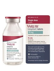
The US Food and Drug Administration (FDA) and Tesaro, Inc., have updated the prescribing information for Varubi® (rolapitant) injectable emulsion to include a new warning about the risk of allergic reactions.
Varubi injectable emulsion is a substance P/neurokinin receptor antagonist approved to prevent delayed nausea and vomiting associated with chemotherapy in adults.
Since Varubi injectable emulsion gained FDA approval, there have been reports of anaphylaxis, anaphylactic shock, and other serious hypersensitivity reactions to the drug, some of which required hospitalization.
Now, the labeling for Varubi injectable emulsion has been changed to include information about these events. The changes include modifications to the CONTRAINDICATIONS, WARNINGS and PRECAUTIONS, and ADVERSE REACTIONS sections of the label.
Since Varubi injectable emulsion was introduced to the US market in late November 2017, at least 7000 doses of the drug have been administered to patients receiving emetogenic chemotherapy in the US, according to Tesaro.
Anaphylaxis, anaphylactic shock, and other serious hypersensitivity reactions have occurred during or soon after the infusion of Varubi. Most reactions have occurred within the first few minutes of administration.
The FDA has advised that patients who are hypersensitive to any component of Varubi injectable emulsion (including soybean oil) do not receive the drug. And patients with known allergies to legumes or other related allergens should be monitored closely.
The FDA said healthcare professionals should be vigilant for signs of hypersensitivity or anaphylaxis in all patients receiving Varubi injectable emulsion, both during administration and afterward.
Symptoms of anaphylaxis can include wheezing, difficulty breathing, swelling of the face or throat, hives, flushing, itching, abdominal cramping, abdominal pain, vomiting, back pain, chest pain, hypotension, and shock.
If anaphylaxis or any other serious hypersensitivity/infusion reaction occurs, Varubi injectable emulsion should be stopped immediately and permanently. The patient should receive appropriate medical management, including epinephrine and/or antihistamines.
To ensure patients and healthcare professionals are aware of the label update to Varubi injectable emulsion, Tesaro has issued a Dear Healthcare Professional letter. In addition, the updated prescribing information has been posted on the Varubi website.
For any questions about the use of Varubi injectable emulsion or to report adverse events related to the drug, contact Tesaro’s medical information department at 1-844-4-TESARO (1-844-483-7276).
Adverse events related to Varubi should also be reported to the FDA’s MedWatch Safety Information and Adverse Event Reporting Program. ![]()
Nominations now open for the 2018 Surgical Volunteerism and Humanitarian Awards
The American College of Surgeons (ACS), in association with Pfizer, Inc., has begun accepting nominations for the 2018 Surgical Volunteerism and Humanitarian Awards. These annual awards recognize surgeons who have made significant contributions to communities in need of surgical aid, be that through organized volunteer activities or through the dedication of a significant portion of their surgical career to the underserved or a retirement characterized by surgical outreach.
Submit nominations today at www.facs.org/ogb/award-winners/nominations. All nominations must be received by February 28, 2018. For more information, contact [email protected] or visit the awards web page.
Apply now for the ACS Clinical Scholars in Residence positions
The American College of Surgeons (ACS) is now accepting applications for the 2019–2021 Clinical Scholar in Residence positions. Applications are due April 1.
The ACS Clinical Scholars in Residence Program is a two-year on-site fellowship in surgical outcomes research, health services research, and health care policy. It was initiated in 2005 to advance the College’s quality improvement initiatives and to offer opportunities for residents to work on ACS Quality Programs. More specifically, ACS Clinical Scholars in Residence perform research relevant to ongoing projects in the ACS Division of Research and Optimal Patient Care.
About the program
The primary objective of the fellowship is to address issues in health care quality, health policy, and patient safety, with the goal of helping the ACS Clinical Scholar in Residence prepare for a research career in academic surgery. The ACS Clinical Scholars in Residence have worked on projects and research using data from the ACS National Surgical Quality Improvement Program, the National Cancer Database, the National Trauma Data Bank®, the Surgeon Specific Registry, and the Metabolic and Bariatric Surgery Accreditation and Quality Improvement Program and have been involved in guideline development and accreditation programs. Scholars are assigned to the appropriate group within the ACS based on their interests and the College’s needs.
In addition, participants earn a master’s degree in health services and outcomes research or health care quality and patient safety during their two years at the ACS headquarters in Chicago, IL. The goal of this aspect of the program is to educate clinicians to become effective researchers in health care services and outcomes. The health services and outcomes research curriculum focuses on these issues within institutional and health care delivery systems, as well as in the external environment that shapes health policy centered on quality and safety issues.
The program takes approximately two years to complete. All coursework is done at Northwestern University’s downtown Chicago campus, one block from the ACS headquarters. The ACS also offers a variety of educational programs from which the Clinical Scholars may benefit, including the Outcomes Research Course and the Clinical Trials Course.
The ACS assigns internal mentors to meet regularly with each ACS Clinical Scholar in Residence. Scholars also have opportunities to interact with various surgeons who are affiliated with the ACS and the Division of Research and Optimal Patient Care. Mentorship is one of the most important aspects of this fellowship. Guidance and interaction with multiple individuals from diverse backgrounds will provide the best opportunity for success. In addition, a core of ACS staff statisticians and project analysts serve as invaluable resources to the ACS Clinical Scholars in Residence.
Past successes
Since its inception, surgical residents from throughout the U.S., including California, Colorado, Connecticut, Illinois, Kansas, Louisiana, Michigan, and Ohio, have participated in the ACS Clinical Scholars in Residence program. These individuals report excellent, productive experiences that have been useful in launching their careers in the field of academic surgery. In all, 16 scholars have completed the program and five scholars are currently participating. The ACS Clinical Scholars in Residence have demonstrated great dedication to outcomes research and the improvement of the quality of surgical care.
The ACS Clinical Scholars in Residence have presented their findings at national meetings and in high-impact, peer-reviewed publications, in addition to having contributed a great deal to the ACS Quality Programs. Furthermore, scholars have gone on to gain prestigious fellowships in several fields, including surgical oncology and pediatric surgery.
Apply now
The 2019–2021 scholars will begin their work July 1, 2019. Applications for these positions are due by April 1, 2018. Applicants are required to have funding from their institution or other grant mechanism.
For more information about the program and the application requirements, go to facs.org/clinicalscholars, or send an e-mail to [email protected].
ACSPA-SurgeonsPAC year-end update
The American College of Surgeons Professional Association (ACSPA) and members of its political action committee (ACSPA-SurgeonsPAC) provide nonpartisan financial support to federal lawmakers and individuals seeking office who share surgery’s perspective on health care policy issues and are well-positioned to advocate for surgery’s legislative goals. As of press time, the ACSPA-SurgeonsPAC raised more than $500,000 from more than 1,200 ACSPA members and staff, and disbursed more than $380,000 to more than 100 congressional candidates, leadership PACs, and political campaign committees.
Following are SurgeonsPAC fundraising and disbursement highlights:
• Willens Society ($25,000 or more over 10 years): 12
• Elite Donors ($2,500–$5,000): 23
• High Donors ($500–$2,499): 372
• General Donors ($100–$499): 669
• Top ACS leadership groups with 100 percent participation: Regents and Officers, Health Policy Advisory Council, Health Policy and Advocacy Group, and SurgeonsPAC Board of Directors
• Top five specialties: General, pediatric, vascular, colorectal, and plastic and reconstructive surgery
• Top states: Texas, California, New York, Ohio, and Pennsylvania
• Contributions disbursed in line with Congressional party ratios—58 percent to Republicans and 42 percent to Democrats
• Attended/hosted more than 200 health care industry events
• Organized eight physician community events for key surgical champions in Congress
• Facilitated more than 30 in-district check deliveries
• Supported 12 physician candidates
In addition, SurgeonsPAC co-hosted the eighth annual Specialty Physician and Dentist Candidate Workshop and 10th annual Medical and Dental PAC Forum.
To learn more about SurgeonsPAC fundraising and disbursement activities, contact SurgeonsPAC staff at [email protected] or 202-672-1520.
Note
Contributions to ACSPA-SurgeonsPAC are not deductible as charitable contributions for federal income tax purposes. Contributions are voluntary, and all members of the ACSPA have the right to refuse to contribute without reprisal. Federal Law prohibits ACSPA-SurgeonsPAC from accepting contributions from foreign nationals. By law, if your contributions are made using a personal check or credit card, ACSPA-SurgeonsPAC may only use your contribution to support candidates in federal elections. All corporate contributions to ACSPA-SurgeonsPAC will be used for educational and administrative fees of ACSPA, and other activities permissible under federal law. Federal law requires ACSPA-SurgeonsPAC to use its best efforts to collect and report the name, mailing address, occupation, and the name of the employer of individuals who contribute more than $200 in a calendar year. ACSPA-SurgeonsPAC is a program of the ACSPA which is exempt from federal income tax under section 501(c)(6) of the Internal Revenue Service.
Nominations for Board of Regents, Officers-Elect due February 23
The American College of Surgeons (ACS) 2018 Nominating Committee of the Fellows (NCF) and the Nominating Committee of the Board of Governors (NCBG) will be selecting nominees for leadership positions in the College.
The 2018 NCF will select nominees for the three Officer-Elect positions of the ACS:
• President-Elect
• First Vice-President-Elect
• Second Vice-President-Elect
The 2018 NCBG will select nominees for pending vacancies on the Board of Regents to be filled at Clinical Congress 2018. Nominations are open to surgeons of all specialties, but particular consideration will be given this nomination cycle to those in the following specialties:
• Burn and critical care surgery
• Gastrointestinal surgery
• General surgery
• Surgical oncology
• Transplantation
• Trauma
• Vascular surgery
For information only, the members of the Board of Regents who will be considered for reelection in 2018 are (all MD, FACS) John L. D. Atkinson, James C. Denneny III, Timothy J. Eberlein, Henri R. Ford, Enrique Hernandez, L. Scott Levin, Linda Phillips, Anton A. Sidawy, Beth H. Sutton, and Steven D. Wexner.
Visit the Bulletin website at http://bit.ly/2l69j2Y for a list of criteria for each nominating committee, as well as further details on how to submit a nomination and the nomination process. The deadline for submitting nominations is February 23, 2018.
Nominations may be submitted to [email protected]. If you have any questions, contact Emily Kalata, Staff Liaison for the NCF and NCBG, at 312-202-5360 or [email protected].
ACS and MacLean Center offer fellowships in surgical ethics
The American College of Surgeons (ACS) Division of Education is offering fellowships in surgical ethics with the MacLean Center for Clinical Medical Ethics, University of Chicago, IL. The MacLean Center will prepare two surgeons for careers that combine clinical surgery with scholarly studies in surgical ethics beginning with a five-week, full-time course in Chicago in July and August 2018. From September 2018 to June 2019, fellowship recipients will meet weekly for a structured ethics curriculum. In addition, fellows will participate in an ethics consultation service and complete a research project. For additional information about this fellowship, contact Patrice Gabler Blair, MPH, Associate Director, ACS Division of Education, at [email protected].
Application materials are due March 1, 2018.
16 cancer care facilities receive CoC Outstanding Achievement Award
The Commission on Cancer (CoC) of the American College of Surgeons (ACS) has granted its mid-year 2017 Outstanding Achievement Award to a select group of 16 accredited cancer programs throughout the U.S. Award criteria are based on qualitative and quantitative surveys conducted in the first half of 2017. A list of these award-winning cancer programs is available at www.facs.org/quality-programs/cancer/coc/info/outstanding/2017-part-1.
The purpose of the award is to raise the bar on quality cancer care, with the ultimate goal of increasing awareness about quality care choices among cancer patients and their loved ones. In addition, the award is intended to fulfill the following goals:
• Recognize those cancer programs that achieve excellence in providing quality care to cancer patients
• Motivate other cancer programs to work toward improving their level of care
• Facilitate dialogue between award recipients and health care professionals at other cancer facilities for the purpose of sharing best practices
• Encourage honorees to serve as quality care resources to other cancer programs
“More and more, we’re finding that patients and their families want to know how the health care institutions in their communities compare with one another,” said Lawrence N. Shulman, MD, FACP, Chair of the CoC. “They want access to information in terms of who’s providing the best quality of care, and they want to know about overall patient outcomes. Through this recognition program, I’d like to think we’re playing a small but vital role in helping them make informed decisions on their cancer care.”
The 16 award-winning cancer care programs represent approximately 6 percent of programs surveyed by the CoC January 1–June 30, 2017. “These cancer programs currently represent the best of the best when it comes to cancer care,” added Dr. Shulman.
Applications for Jacobson Award accepted through February 23
The American College of Surgeons (ACS) is accepting applications for the 2018 Joan L. and Julius H. Jacobson II Promising Investigator Award (JPIA) through February 23.
The JPIA was established to recognize outstanding surgeons who are engaging in research, advancing the art and science of surgery, and demonstrating early promise of significant contribution to the practice of surgery and the safety of surgical patients. The award is supported through a generous endowed fund established by the donors and administered by the ACS Surgical Research Committee.
Award criteria
The criteria for selection of the JPIA winner are as follows:
• Candidate must be a Fellow or an Associate Fellow of the ACS.
• Candidate must be board certified in a surgical specialty and must have completed surgical training, including fellowship, in the last six years, excluding military, medical, or family leave.
• Candidate must hold a faculty appointment at a research-based academic medical center or hold a military service position.
• Candidate must have received peer-reviewed funding, such as a K-series award from the National Institutes of Health (NIH), Veterans Administration, National Science Foundation, or U.S. Department of Defense merit review award to support their research effort. Surgeon-scientists who are well established (for example, recipients of NIH R01 and Veterans Affairs Merit grants or equivalent grants from other agencies) are ineligible.
• Only one application per surgical department will be accepted.
• Nomination documentation must include a one-page essay to the committee explaining why the candidate should be considered for the award and that describes the importance of their past and current research.
• Nomination documentation must include copies of the candidate’s three most significant publications from their current faculty position.
• Nomination documentation must include a letter of recommendation from the candidate’s department chair. Up to three additional letters of recommendation will be accepted.
• Nomination documentation includes an NIH-formatted biographical sketch though the electronic application system.
Special consideration will be given to surgeons who are at the “tipping point” of their research careers, with a track record indicative of early promise and potential (for example, a degree program in research or a K-award).
To be considered for the award in 2018, applications must be submitted to facs.org/jpia on or before February 23, 2018.
Additional details
For more information about the award, go to facs.org/jpia.
See a list of all past recipients at facs.org/quality-programs/about/cqi/jacobson/past-recipients.
Send comments and inquiries to Jorge Hernandez, Project Coordinator, Division of Research and Optimal Patient Care—Continuous Quality Improvement, at [email protected] or call 331-202-5319.
From the Editors: Finding joy
While basking in the fading glow of the holidays, I have been reflecting on the dynamic of combined with the negativity local and world events engender that can push us toward burnout.
The holiday season is a time when people tend to engage in activities that have been shown to improve mood and outlook: connecting with old friends, sharing memories of a happier time, spending time with children and grandchildren, sitting around a warm fire, enjoying the sights and smells of bright decorations and fragrant candles, attending traditional holiday plays, concerts, and ballets. The holiday traditions, no matter what your ethnic or religious background, create community and warm feelings. The workweek may be shortened, people take a little time out, smile a bit more. For many of us, these activities can be a tonic.
At the same time, the days become shorter, colder, and grayer in most of North America. The normal frustrations of our day-to-day professional lives may seem more profound during the winter, and some experience SAD (seasonal affective disorder) or even burnout.
If we are not affected by these events, we may be missing the compassion gene. But I would suggest that an acute awareness of a world of trouble around us compounded with our own heavy load as surgeons is a recipe for burnout. It may not be within the capacity of any of us to alter the reality of our present world, and the surgical profession is not going to become a low-key occupation any time soon. But we can control our response to all this and take steps to attend our own emotional health.
I have found that the single most effective measure to combat negative feelings is to connect with colleagues, friends, and family to share positive, enjoyable experiences: a potluck dinner, a concert, a hike (or snowshoe trip) in the woods. We should seek out optimistic, glass-half-full individuals. We all have some of these folks in our lives and they do us a world of good.
With regard to professional stressors, reaching out to colleagues to work together in identifying remedies for a dysfunctional workplace may not only address the problem, but also allow you to recognize that you are not alone in your distress. Joining forces as a team to forge a solution can be satisfying and empowering.
Nevertheless, surgical practice remains intense, stressful, and demanding. As surgeons, we tend to be perfectionists, wanting to dot every “i” and cross every “t,” no matter how trivial. It is critical to set realistic expectations for how much you can achieve. Identify and prioritize personal and professional goals, make the most important goals take front and center, and delegate (or just allow to disappear) items that are less important. This may be the single most important strategy to avoid burnout: Prioritize what is essential and let the rest go.
A great deal has been written recently about resilience and mindfulness – facile concepts that don’t address the struggles of individuals feeling helpless and overwhelmed by the onslaught of demands on his/her time. Even though clichés about mindfulness can ring hollow, I have found that taking small steps to build my own inner reserves can help.
Here is my advice: Take a moment several times a day to appreciate something beautiful around you: a textured sky, a peaceful field, city lights, a nearby river with the ripples of wind on the water. Smile and greet someone on the street or in the hallway at work. Say a good word to someone on a job nicely done. Reflect on how doing these things affect you. Do they make you feel calmer and happier? “Rest your brain” every 2 hours for just a minute or two; cognitive fatigue occurs after 60-90 minutes and drains your energy if the “pause button” isn’t pushed.
Many of us neglect our personal health. It goes without saying that we are all far more likely to avoid burnout if we have a balanced diet, adequate sleep, and some exercise. We should all have a primary care provider for regular checkups and preventive exams. We speak with great authority when we counsel our patients to do this, so what possible excuse do we have for neglecting our own health?
One of the most important habits that I cultivate to improve my own mood is to end each day reflecting on three positive things that happened that day. Amid all of the calamities that occur every day in the world, it should not be difficult for those of us who live a life of relative privilege and plenty to find positive things in our lives. A strong association has been demonstrated between a sense of thankfulness and individual happiness and contentment. As surgeons, we have a ready source of positive reinforcers – the gratitude of our patients. I have a “feel good drawer” for “thank yous.” I open that drawer and read some of those messages from grateful patients. Reflecting on how we have been able to help our patients can do us all good when we are having doubts about our professional lives.
I want to encourage all surgeons to take a little better care of themselves this year. Take some specific steps to attend to your physical and emotional health. Do some activities the only purpose of which is to rest, to reflect, and to find joy.
Dr. Deveney is professor of surgery and vice chair of education in the department of surgery, Oregon Health & Science University, Portland. She is the coeditor of ACS Surgery News.