User login
Malpractice and pain management
Question: A new patient with severe back pain sought treatment at the Ease-Your-Pain Clinic, a facility run by medical specialists. Following a history and physical, he was given a prescription for high-dose oxycodone. Shortly after filing the prescription, he became unresponsive, and toxicology studies postmortem revealed elevated blood levels.
In a wrongful death lawsuit against the doctor and clinic, which of the following statements is best?
A. A wrongful death lawsuit deals with a special type of malpractice in which criminal sanctions are likely.
B. The facts here fall within the domain of common knowledge, or res ipsa loquitur; so the case will not have to depend on an expert witness.
C. Substandard care is clearly evident from the fact that this opioid-naive patient was prescribed a much higher than usual starting dose of the drug.
D. The elevated blood levels indicate that the cause of death was an opioid overdose.
E. To prevail, the plaintiff has the burden of proving, by a preponderance of evidence and through expert testimony, that the defendant’s breach of duty of due care legally caused the patient’s death.
Answer: E. Criminal prosecution for homicide is quite different from a civil lawsuit for wrongful death, requiring state action with proof beyond reasonable doubt and a showing of intent. It is rare in medical malpractice cases, which require expert testimony to prove breach of duty and causation.

Choices C and D are incorrect, as the facts given are deliberately brief and vague. For example, the medical history may have revealed this patient to be a chronic user of opioid drugs rather than being opioid naive, thus the need for higher dosing. In addition, we cannot be certain of the actual cause of death; opioid levels obtained in body fluids postmortem may be difficult to interpret and are apt to be higher in chronic users (see Dallier v. Hsu, discussed below).
We are currently in the throes of an epidemic of opioid deaths involving both illicit street drugs such as heroin and synthetic fentanyl, as well as FDA-approved controlled substances such as morphine and codeine derivatives. Overdose now exceeds motor vehicle accidents as the leading cause of injury-related deaths, with nearly 100 Americans dying every day from an opioid overdose.
In an earlier column, we drew attention to physician and manufacturer criminality associated with opioid prescription deaths.1 In this article, we will focus on the civil liability facing doctors who treat patients afflicted with pain.
The onslaught of warnings, caution, and threats of criminal prosecution has prompted Medicare and pharmacy chains to impose restrictions on opioid prescriptions. Even more troubling, doctors are increasingly cutting back or stopping entirely their prescriptions. As a result, some patients may resort to desperate measures to obtain their drugs.
A recent article in the Washington Post draws attention to this situation.2 It tells the story of a 49-year-old trucker who had been taking large amounts of prescription opioids ever since hip surgery left him with nerve damage. Because no doctor nearby would write an opioid prescription, he had to drive 367 miles to his old pain clinic each month for a refill. According to the article, chronic pain patients may turn to unregulated alternatives such as kratom, and some have threatened suicide.
Citing data from the IQVIA Institute for Human Data Science, the article reported that the annual volume of prescription opioids shrank 29% between 2011 and 2017, even as the number of overdose deaths climbed ever higher. The drop in prescriptions was greatest for patients receiving high doses, most of whom had chronic pain.
Failure to treat pain can constitute substandard care. In Bergman v. Eden Medical Center, an Alameda County jury in California turned in a verdict against an internist charged with elder abuse and reckless negligence for failing to give enough pain medication to a patient dying of cancer.3 William Bergman was an 85-year-old retired railroad worker who complained of severe back pain. During his 6-day stay at the hospital, nurses consistently charted his pain in the 7-10 range, and on the day of discharge, his pain was at level 10. He died at home shortly thereafter.
After 4 days of deliberation, the jury, in a 9-3 vote, entered a guilty verdict, and awarded $1.5 million in general damages. That amount was subsequently reduced to $250,000 because of California’s cap on noneconomic damages. The Bergman case is notable for being the first of its kind, and it squarely put physicians on notice regarding their duty to adequately provide pain relief.
At the same time, prescribing opioid drugs that result in harm can be the basis of a successful claim – if the plaintiff can prove the tort elements of negligence. However, this may be harder than it looks.
For example, a Connecticut malpractice case alleging negligent opioid prescriptions resulted in a judgment in favor of the defendant.4 The patient had a congenital skeletal deformity called Madelung’s disease, and she suffered from many years of severe pain requiring chronic opioids such as oxycodone, methadone, morphine, fentanyl, and hydrocodone, in combination and with other types of medications for anxiety, sleep problems, and depression. She was not a compliant patient and had a history of inconsistent pill counts and urine tests, and a history of stockpiling and hoarding pills.
Following a recent fracture of her right arm and shoulder, she visited several doctors for narcotic prescriptions before consulting the defendant doctor, who concluded that hers was an emergency and urgent situation. He believed that if he did not prescribe medications to address her pain, she would engage in unsafe drug-seeking behaviors. Accordingly, the doctor prescribed the following: methadone, 40 mg, 4 pills per day; extended-release morphine sulfate, 60 mg, 2 per day; alprazolam, 1 mg, 3 per day. Within hours of filing her prescription, she began to stumble, developed slurred speech, and then became unresponsive and died.
The court was unpersuaded that the requisite standard of care was to contact the patient’s prior treaters or pharmacy, or to obtain her current records to determine her level of drug naiveté or tolerance, or that the defendant should have initiated treatment with starting doses of drugs. It held that that reflected “a narrow textbook approach to the practice of pain management and ignores the role of patient-physician interaction.”
Based on all the evidence, the patient’s tolerance for opiates had greatly escalated, and her level of pain remained at 10 on a scale of 10. The defendant had independently assessed the patient, determined her needs, and ruled out that she was opiate naive. Based on all the circumstances, he prescribed morphine, methadone, and alprazolam. The morphine prescription of 60 mg extended release every 12 hours was not lethal to her and was not her first opioid analgesic. The court ruled that the plaintiff had failed to sustain the burden of proving causation, there being no finding that the patient took more medications than prescribed or overdosed. It was unimpressed by the various calculations offered by the experts on postmortem drug levels and causation.
A recent report from the Doctor’s Company, a major malpractice carrier, reviewed 1,770 claims closed between 2007 and 2015 in which patient harm involved medication factors.5 In 272 of those claims (15%), the medications were narcotic analgesics, most often prescribed in the outpatient setting and involving methadone and oxycodone. The Centers for Disease Control and Prevention has recently published treatment guidelines noting that long-term opioid use among patients with chronic pain increased the likelihood of addiction and overdose and had uncertain benefits. They discouraged doses higher than the equivalent of 90 mg of morphine.6
While it is true that opioid-related malpractice lawsuits are not infrequent, stopping entirely the prescribing of controlled narcotic analgesics in the name of “first do no harm” is simplistic and misguided.
It is better to adopt widely recommended strategies in opioid prescribing, recognizing that, as Howard Marcus, MD, chair of the Texas Alliance for Patient Access, has said, “It is possible to prescribe opioids responsibly and safely for patients with chronic pain who do not obtain sufficient relief and reasonable function with nonopioid treatment. However, to do so, it is necessary to have adequate knowledge of the pharmacology of opioids, risk factors, and side effects. Safe opioid prescribing requires thorough patient evaluation, attention to detail, and familiarity with guidelines and regulations.”7
To this might be added the suggestion that, in tough cases, one should make a referral to a pain specialist skilled in the use of such drugs.
Dr. Tan is emeritus professor of medicine and former adjunct professor of law at the University of Hawaii, Honolulu. This article is meant to be educational and does not constitute medical, ethical, or legal advice. For additional information, readers may contact the author at [email protected].
References
1. “Physician liability in opioid deaths.” Internal Medicine News, July 13, 2017.
2. “Unintended consequences: Inside the fallout of America’s crackdown on opioids.” Washington Post, May 31, 2018.
3. Bergman v. Eden Medical Center, No. H205732-1 (Sup. Ct. Alameda Co., Cal., June 13, 2001).
4. Dallaire v. Hsu, CV 07-5004043 (Conn. Sup. Ct., May 18, 2010).
5. “Prescription opioid abuse epidemic: analysis of medication-related claims.” The Doctor’s Advocate, first quarter 2017.
6. N Engl J Med. 2016 Apr 21;374(16):1501-4.
7. “Prescribing opioids safely.” The Doctor’s Advocate, second quarter 2017.
Question: A new patient with severe back pain sought treatment at the Ease-Your-Pain Clinic, a facility run by medical specialists. Following a history and physical, he was given a prescription for high-dose oxycodone. Shortly after filing the prescription, he became unresponsive, and toxicology studies postmortem revealed elevated blood levels.
In a wrongful death lawsuit against the doctor and clinic, which of the following statements is best?
A. A wrongful death lawsuit deals with a special type of malpractice in which criminal sanctions are likely.
B. The facts here fall within the domain of common knowledge, or res ipsa loquitur; so the case will not have to depend on an expert witness.
C. Substandard care is clearly evident from the fact that this opioid-naive patient was prescribed a much higher than usual starting dose of the drug.
D. The elevated blood levels indicate that the cause of death was an opioid overdose.
E. To prevail, the plaintiff has the burden of proving, by a preponderance of evidence and through expert testimony, that the defendant’s breach of duty of due care legally caused the patient’s death.
Answer: E. Criminal prosecution for homicide is quite different from a civil lawsuit for wrongful death, requiring state action with proof beyond reasonable doubt and a showing of intent. It is rare in medical malpractice cases, which require expert testimony to prove breach of duty and causation.
Choices C and D are incorrect, as the facts given are deliberately brief and vague. For example, the medical history may have revealed this patient to be a chronic user of opioid drugs rather than being opioid naive, thus the need for higher dosing. In addition, we cannot be certain of the actual cause of death; opioid levels obtained in body fluids postmortem may be difficult to interpret and are apt to be higher in chronic users (see Dallier v. Hsu, discussed below).
We are currently in the throes of an epidemic of opioid deaths involving both illicit street drugs such as heroin and synthetic fentanyl, as well as FDA-approved controlled substances such as morphine and codeine derivatives. Overdose now exceeds motor vehicle accidents as the leading cause of injury-related deaths, with nearly 100 Americans dying every day from an opioid overdose.
In an earlier column, we drew attention to physician and manufacturer criminality associated with opioid prescription deaths.1 In this article, we will focus on the civil liability facing doctors who treat patients afflicted with pain.
The onslaught of warnings, caution, and threats of criminal prosecution has prompted Medicare and pharmacy chains to impose restrictions on opioid prescriptions. Even more troubling, doctors are increasingly cutting back or stopping entirely their prescriptions. As a result, some patients may resort to desperate measures to obtain their drugs.
A recent article in the Washington Post draws attention to this situation.2 It tells the story of a 49-year-old trucker who had been taking large amounts of prescription opioids ever since hip surgery left him with nerve damage. Because no doctor nearby would write an opioid prescription, he had to drive 367 miles to his old pain clinic each month for a refill. According to the article, chronic pain patients may turn to unregulated alternatives such as kratom, and some have threatened suicide.
Citing data from the IQVIA Institute for Human Data Science, the article reported that the annual volume of prescription opioids shrank 29% between 2011 and 2017, even as the number of overdose deaths climbed ever higher. The drop in prescriptions was greatest for patients receiving high doses, most of whom had chronic pain.
Failure to treat pain can constitute substandard care. In Bergman v. Eden Medical Center, an Alameda County jury in California turned in a verdict against an internist charged with elder abuse and reckless negligence for failing to give enough pain medication to a patient dying of cancer.3 William Bergman was an 85-year-old retired railroad worker who complained of severe back pain. During his 6-day stay at the hospital, nurses consistently charted his pain in the 7-10 range, and on the day of discharge, his pain was at level 10. He died at home shortly thereafter.
After 4 days of deliberation, the jury, in a 9-3 vote, entered a guilty verdict, and awarded $1.5 million in general damages. That amount was subsequently reduced to $250,000 because of California’s cap on noneconomic damages. The Bergman case is notable for being the first of its kind, and it squarely put physicians on notice regarding their duty to adequately provide pain relief.
At the same time, prescribing opioid drugs that result in harm can be the basis of a successful claim – if the plaintiff can prove the tort elements of negligence. However, this may be harder than it looks.
For example, a Connecticut malpractice case alleging negligent opioid prescriptions resulted in a judgment in favor of the defendant.4 The patient had a congenital skeletal deformity called Madelung’s disease, and she suffered from many years of severe pain requiring chronic opioids such as oxycodone, methadone, morphine, fentanyl, and hydrocodone, in combination and with other types of medications for anxiety, sleep problems, and depression. She was not a compliant patient and had a history of inconsistent pill counts and urine tests, and a history of stockpiling and hoarding pills.
Following a recent fracture of her right arm and shoulder, she visited several doctors for narcotic prescriptions before consulting the defendant doctor, who concluded that hers was an emergency and urgent situation. He believed that if he did not prescribe medications to address her pain, she would engage in unsafe drug-seeking behaviors. Accordingly, the doctor prescribed the following: methadone, 40 mg, 4 pills per day; extended-release morphine sulfate, 60 mg, 2 per day; alprazolam, 1 mg, 3 per day. Within hours of filing her prescription, she began to stumble, developed slurred speech, and then became unresponsive and died.
The court was unpersuaded that the requisite standard of care was to contact the patient’s prior treaters or pharmacy, or to obtain her current records to determine her level of drug naiveté or tolerance, or that the defendant should have initiated treatment with starting doses of drugs. It held that that reflected “a narrow textbook approach to the practice of pain management and ignores the role of patient-physician interaction.”
Based on all the evidence, the patient’s tolerance for opiates had greatly escalated, and her level of pain remained at 10 on a scale of 10. The defendant had independently assessed the patient, determined her needs, and ruled out that she was opiate naive. Based on all the circumstances, he prescribed morphine, methadone, and alprazolam. The morphine prescription of 60 mg extended release every 12 hours was not lethal to her and was not her first opioid analgesic. The court ruled that the plaintiff had failed to sustain the burden of proving causation, there being no finding that the patient took more medications than prescribed or overdosed. It was unimpressed by the various calculations offered by the experts on postmortem drug levels and causation.
A recent report from the Doctor’s Company, a major malpractice carrier, reviewed 1,770 claims closed between 2007 and 2015 in which patient harm involved medication factors.5 In 272 of those claims (15%), the medications were narcotic analgesics, most often prescribed in the outpatient setting and involving methadone and oxycodone. The Centers for Disease Control and Prevention has recently published treatment guidelines noting that long-term opioid use among patients with chronic pain increased the likelihood of addiction and overdose and had uncertain benefits. They discouraged doses higher than the equivalent of 90 mg of morphine.6
While it is true that opioid-related malpractice lawsuits are not infrequent, stopping entirely the prescribing of controlled narcotic analgesics in the name of “first do no harm” is simplistic and misguided.
It is better to adopt widely recommended strategies in opioid prescribing, recognizing that, as Howard Marcus, MD, chair of the Texas Alliance for Patient Access, has said, “It is possible to prescribe opioids responsibly and safely for patients with chronic pain who do not obtain sufficient relief and reasonable function with nonopioid treatment. However, to do so, it is necessary to have adequate knowledge of the pharmacology of opioids, risk factors, and side effects. Safe opioid prescribing requires thorough patient evaluation, attention to detail, and familiarity with guidelines and regulations.”7
To this might be added the suggestion that, in tough cases, one should make a referral to a pain specialist skilled in the use of such drugs.
Dr. Tan is emeritus professor of medicine and former adjunct professor of law at the University of Hawaii, Honolulu. This article is meant to be educational and does not constitute medical, ethical, or legal advice. For additional information, readers may contact the author at [email protected].
References
1. “Physician liability in opioid deaths.” Internal Medicine News, July 13, 2017.
2. “Unintended consequences: Inside the fallout of America’s crackdown on opioids.” Washington Post, May 31, 2018.
3. Bergman v. Eden Medical Center, No. H205732-1 (Sup. Ct. Alameda Co., Cal., June 13, 2001).
4. Dallaire v. Hsu, CV 07-5004043 (Conn. Sup. Ct., May 18, 2010).
5. “Prescription opioid abuse epidemic: analysis of medication-related claims.” The Doctor’s Advocate, first quarter 2017.
6. N Engl J Med. 2016 Apr 21;374(16):1501-4.
7. “Prescribing opioids safely.” The Doctor’s Advocate, second quarter 2017.
Social media use linked to 10% increase in risk of new ADHD symptoms among teens
according to results from a prospective, longitudinal cohort study published in JAMA. 
“Further research is needed to determine whether this association is causal,” cautioned the investigators.
Between fall 2014 (10th grade) and fall 2016 (12th grade), researchers studied 3,051 students from 10 different Los Angeles schools who were enrolled in the Happiness & Health Study and did not have significant ADHD symptoms; of these students, 2,587 adolescents (mean age 16 years, 54% girls) self-reported digital media use activities from 14 different types through surveys administered at baseline and again at 6-month, 12-month, 18-month and 24-month follow-up. Digital use activities included checking social media sites, engaging with social media content, texting, streaming music, and browsing or viewing images or videos, among others.
“Although some emerging research indicates that ADHD levels and use of certain forms of modern media may be concurrently associated, the role of modern digital media use in ADHD risk largely remains unclear from the prior literature due to limitations in exposure assessment and the application of designs incapable of supporting temporal or causal inferences,” wrote Chaelin K. Ra, MPH, of the University of Southern California, Los Angeles, and his colleagues. “The current study provides new longitudinal evidence on this topic using a 5-wave prospective design and comprehensive assessment across a wide continuum of digital media exposure, including numerous media platforms currently popular among youth.”
Students ranked their activities in a cumulative index and indicated whether they participated in those activities “0, 1-2 times per week, 1-2 times per day, or many times per day.” They also filled out the DSM-IV Current Symptoms Self-Report Form, which asked them to report whether they experienced any of nine different hyperactivity-impulsivity symptoms such as “difficulty organizing and completing tasks.”
Mr. Ra and his colleagues found that 81% of adolescents reported at least one digital media activity performed at a high-frequency rate, with 54% reporting they checked social media at a high-frequency rate. There was a mean of four baseline digital media activities performed at a high-frequency rate among students surveyed. Each additional digital media activity used at a high-frequency rate carried a statistically significant association of subsequent ADHD symptoms over the follow-up period (odds ratio 1.1; 95% confidence interval, 1.06-1.16), which remained after adjustment for covariates including age, sex, and subsidized lunch availability tied to family income (OR 1.1; 95% CI, 1.05-1.15). The researchers noted that there was a 5% mean prevalence of subsequent ADHD symptoms in follow-up among patients who reported no baseline high-frequency rate of digital media use, compared with 10% in students indicating 7 high-frequency activities and 11% in students indicating 14 high-frequency activities.
Limitations of the study included potential inaccuracies in self-reporting ADHD symptoms as opposed to students receiving a diagnosis through a clinical interview, the possibility of the association being influenced by ADHD symptoms not detected in the study, the fact that the media use measure in the study had not been validated, and use of a targeted age range in the sample of students that excluded students without surveys who had differing demographic data from the rest of the cohort, according to the researchers.
The authors reported having no relevant financial disclosures. The study was supported by a grant from the National Institutes of Health.
SOURCE: Ra CK et al. JAMA. 2018 Jul 17. doi: 10.1001/jama.2018.8931.
This study is among the first to capture current media use because technology tends to outpace researchers’ ability to publish data “within relevant time frames,” Jenny Radesky, MD of the University of Michigan in Ann Arbor, said in an editorial.
While previous studies linking ADHD symptoms and media use analyzed use of media such as television shows and video games, this study analyzes smartphone use and “always-on” media such as social media in addition to traditional media, she said.
It should be noted that “high-frequency media use was associated with only a 10% increased risk of later ADHD symptoms overall.
“Although the authors were able to adjust for income and mental health symptoms, they did not assess parent media use or whether the parents were involved in how the adolescents used media,” Dr. Radesky said. “Parent media use correlates with child media use, may interrupt parent-child activities, and is associated with child behavior difficulties in younger children.”
She said the study affirms guidelines from the American Academy of Pediatrics to “prioritize activities that promote adolescent executive functioning and well-being, including sleep, physical activity, distraction-free homework, and positive interactions with family and friends.
“Although not directly addressed by this study, the American Academy of Pediatrics recommendations for parent involvement in their adolescent’s media use – including discussions about prosocial uses of media, digital citizenship, misinformation, and persuasion awareness – are relevant to the cognitive and emotional reactions to digital media of adolescents,” she added.
Dr. Radesky noted that the potential for the association between media use and ADHD symptoms to be influenced by confounders not measured in the study is a “major limitation.”
Dr. Radesky is at the University of Michigan, Ann Arbor. She receives grant support from the Eunice Kennedy Shriver National Institute of Child Health and Human Development. These comments summarize her editorial accompanying the article by Ra et al. ( JAMA. 2018;320:237-39 ).
according to results from a prospective, longitudinal cohort study published in JAMA.
“Further research is needed to determine whether this association is causal,” cautioned the investigators.
Between fall 2014 (10th grade) and fall 2016 (12th grade), researchers studied 3,051 students from 10 different Los Angeles schools who were enrolled in the Happiness & Health Study and did not have significant ADHD symptoms; of these students, 2,587 adolescents (mean age 16 years, 54% girls) self-reported digital media use activities from 14 different types through surveys administered at baseline and again at 6-month, 12-month, 18-month and 24-month follow-up. Digital use activities included checking social media sites, engaging with social media content, texting, streaming music, and browsing or viewing images or videos, among others.
“Although some emerging research indicates that ADHD levels and use of certain forms of modern media may be concurrently associated, the role of modern digital media use in ADHD risk largely remains unclear from the prior literature due to limitations in exposure assessment and the application of designs incapable of supporting temporal or causal inferences,” wrote Chaelin K. Ra, MPH, of the University of Southern California, Los Angeles, and his colleagues. “The current study provides new longitudinal evidence on this topic using a 5-wave prospective design and comprehensive assessment across a wide continuum of digital media exposure, including numerous media platforms currently popular among youth.”
Students ranked their activities in a cumulative index and indicated whether they participated in those activities “0, 1-2 times per week, 1-2 times per day, or many times per day.” They also filled out the DSM-IV Current Symptoms Self-Report Form, which asked them to report whether they experienced any of nine different hyperactivity-impulsivity symptoms such as “difficulty organizing and completing tasks.”
Mr. Ra and his colleagues found that 81% of adolescents reported at least one digital media activity performed at a high-frequency rate, with 54% reporting they checked social media at a high-frequency rate. There was a mean of four baseline digital media activities performed at a high-frequency rate among students surveyed. Each additional digital media activity used at a high-frequency rate carried a statistically significant association of subsequent ADHD symptoms over the follow-up period (odds ratio 1.1; 95% confidence interval, 1.06-1.16), which remained after adjustment for covariates including age, sex, and subsidized lunch availability tied to family income (OR 1.1; 95% CI, 1.05-1.15). The researchers noted that there was a 5% mean prevalence of subsequent ADHD symptoms in follow-up among patients who reported no baseline high-frequency rate of digital media use, compared with 10% in students indicating 7 high-frequency activities and 11% in students indicating 14 high-frequency activities.
Limitations of the study included potential inaccuracies in self-reporting ADHD symptoms as opposed to students receiving a diagnosis through a clinical interview, the possibility of the association being influenced by ADHD symptoms not detected in the study, the fact that the media use measure in the study had not been validated, and use of a targeted age range in the sample of students that excluded students without surveys who had differing demographic data from the rest of the cohort, according to the researchers.
The authors reported having no relevant financial disclosures. The study was supported by a grant from the National Institutes of Health.
SOURCE: Ra CK et al. JAMA. 2018 Jul 17. doi: 10.1001/jama.2018.8931.
FROM JAMA
Key clinical point: High-frequency digital media use carries a statistically significant association with subsequent ADHD symptoms in adolescent students without previous significant ADHD symptoms at baseline.
Major finding: Of the adolescent students without significant ADHD symptoms,
Study details: A prospective, longitudinal study of 3,051 adolescent students (mean age, 16 years) in the Happiness & Health Study, followed for 24 months.
Disclosures: The authors reported having no relevant financial disclosures. The study was supported by a grant from the National Institutes of Health.
Source: Ra CK et al. JAMA. 2018 Jul 17. doi: 10.1001/jama.2018.8931.
Tabata training
I’m in really good shape. Well, more like really not bad shape. I eat healthy food (see my previous column on diet) and work out nearly every day. I have done so for years. I’ve learned that working out doesn’t make much difference with my weight, but it makes a huge difference with my mood, even more so than meditating. That’s why I’ll never give it up.

My approach is to vary my routine, typically by month. I’ve done “BUD/S qualification” months where I do only push-ups, sit-ups, pull-ups, and runs to meet the minimum requirements for the Navy Seal Training. (It’s not as hard as you might think, although I’m pretty lenient on form.)
When I have an hour to exercise and I’m deep into a podcast, then I’ll just keep going. If I’m trying to work out a piece I’m writing, like this one, then I’ll go for a run along the harbor here in San Diego. If I have to catch an early flight or drive to LA for the day, then I might have only 15 minutes. In that instance, I do high-intensity sprints, also known as high-intensity interval training (HIIT). Although it’s hard to break a good sweat, these workouts are both challenging and rewarding.
Recently, I participated in a wonderful physician wellness program at Kaiser Permanente, San Diego, where, over several weeks, we learned about nutrition, practiced meditation, and did Tabatas. What’s a Tabata you ask? It’s a kick in the butt.
. Yup, it’s a 4-minute workout that consists of 20 seconds of all-out, maximum effort, followed by 10 seconds of rest. The specific move you do for Tabatas is up to you, but it’s recommended that it be the same move for all 4 minutes. I like burpees which work your entire body – you jump, you drop into a push-up position, you pull your feet back in, and jump again. (Check out a video on YouTube.)
When we started the class, I thought Tabatas would be too easy for a gym rat like me. Plus, there were pediatricians, and even radiologists there, so how hard could they be? Let’s just say I couldn’t sit for 2 days after my first session: That’s how hard.
Tabatas are also a quick way to torch calories. A study published in the Journal of Sports Science and Medicine in 2013 found subjects who performed a 20-minute Tabata session experienced improved cardiorespiratory endurance and increased calorie burn (J Sports Sci Med. 2013 Sep;12[3]: 612-3).
Sometimes on a Monday, which is typically my difficult day, I’ll break out a few burpees in my office between patients. The energy jolt is real, and unlike caffeine, doesn’t leave me shaky. Because Tabatas require physical and mental focus, they’re an effective way to clear your mind after a grueling patient visit or if you’re feeling distracted. You simply can’t be thinking about that late patient or angry email when you’re jumping and lunging at full speed.
All the physicians in our program liked the Tabatas; many were even better than me. (Turns out we have pediatricians and radiologists who do things like run the Boston marathon and win Spartan races).
And if you start doing Tabatas, feel free to email me if you need a recommendation for a standing desk – you might not be able to sit as much afterward.
Dr. Benabio is director of Healthcare Transformation and chief of dermatology at Kaiser Permanente San Diego. The opinions expressed in this column are his own and do not represent those of Kaiser Permanente. Dr. Benabio is @Dermdoc on Twitter. Write to him at [email protected].
I’m in really good shape. Well, more like really not bad shape. I eat healthy food (see my previous column on diet) and work out nearly every day. I have done so for years. I’ve learned that working out doesn’t make much difference with my weight, but it makes a huge difference with my mood, even more so than meditating. That’s why I’ll never give it up.
My approach is to vary my routine, typically by month. I’ve done “BUD/S qualification” months where I do only push-ups, sit-ups, pull-ups, and runs to meet the minimum requirements for the Navy Seal Training. (It’s not as hard as you might think, although I’m pretty lenient on form.)
When I have an hour to exercise and I’m deep into a podcast, then I’ll just keep going. If I’m trying to work out a piece I’m writing, like this one, then I’ll go for a run along the harbor here in San Diego. If I have to catch an early flight or drive to LA for the day, then I might have only 15 minutes. In that instance, I do high-intensity sprints, also known as high-intensity interval training (HIIT). Although it’s hard to break a good sweat, these workouts are both challenging and rewarding.
Recently, I participated in a wonderful physician wellness program at Kaiser Permanente, San Diego, where, over several weeks, we learned about nutrition, practiced meditation, and did Tabatas. What’s a Tabata you ask? It’s a kick in the butt.
. Yup, it’s a 4-minute workout that consists of 20 seconds of all-out, maximum effort, followed by 10 seconds of rest. The specific move you do for Tabatas is up to you, but it’s recommended that it be the same move for all 4 minutes. I like burpees which work your entire body – you jump, you drop into a push-up position, you pull your feet back in, and jump again. (Check out a video on YouTube.)
When we started the class, I thought Tabatas would be too easy for a gym rat like me. Plus, there were pediatricians, and even radiologists there, so how hard could they be? Let’s just say I couldn’t sit for 2 days after my first session: That’s how hard.
Tabatas are also a quick way to torch calories. A study published in the Journal of Sports Science and Medicine in 2013 found subjects who performed a 20-minute Tabata session experienced improved cardiorespiratory endurance and increased calorie burn (J Sports Sci Med. 2013 Sep;12[3]: 612-3).
Sometimes on a Monday, which is typically my difficult day, I’ll break out a few burpees in my office between patients. The energy jolt is real, and unlike caffeine, doesn’t leave me shaky. Because Tabatas require physical and mental focus, they’re an effective way to clear your mind after a grueling patient visit or if you’re feeling distracted. You simply can’t be thinking about that late patient or angry email when you’re jumping and lunging at full speed.
All the physicians in our program liked the Tabatas; many were even better than me. (Turns out we have pediatricians and radiologists who do things like run the Boston marathon and win Spartan races).
And if you start doing Tabatas, feel free to email me if you need a recommendation for a standing desk – you might not be able to sit as much afterward.
Dr. Benabio is director of Healthcare Transformation and chief of dermatology at Kaiser Permanente San Diego. The opinions expressed in this column are his own and do not represent those of Kaiser Permanente. Dr. Benabio is @Dermdoc on Twitter. Write to him at [email protected].
New PET imaging agent shows loss of synaptic density in Alzheimer’s brains
Patients with mild Alzheimer’s disease dementia or amnestic mild cognitive impairment due to Alzheimer’s showed 41% less binding of an investigational PET imaging marker of synaptic density when compared with cognitively normal control patients.

“Synaptic loss in the association cortex and the limbic system is a robust and consistent pathology in AD [Alzheimer’s disease] and is closely correlated with cognitive impairment,” wrote Dr. Chen of Yale University, New Haven, Conn., and his associates. “However, until now, the assessment of synaptic density and the quantification of pre-synaptic proteins in AD could only be performed in postmortem brain tissues. The present study demonstrated, for the first time, that noninvasive PET imaging with 11C-UCB-J is capable of measuring reductions in synaptic density in vivo in the hippocampus of individuals with amnestic MCI [mild cognitive impairment] and AD dementia.”
The marker could potentially be used to identify early synaptic loss and track disease progression and therapeutic response, the authors suggested.
The team that created 11C-UCB-J, also known as ((R)-1-((3-(11C-methyl-11C)pyridin-4-yl)methyl)-4-(3,4,5-trifluorophenyl)pyrrolidin-2-one), conducted this study. The agent binds to synaptic vesicle glycoprotein 2A (SV2A), which is the only one of three isoforms that is expressed only in brain synapses. The team’s earlier work showed that SV2A is a marker of synaptic density and that 11C-UCB-J can effectively track changes in the glycoprotein’s presence.
The study comprised 5 patients with amnestic MCI due to AD, 5 with mild AD dementia, and 11 cognitively normal matched controls; they were a mean of 72 years old. All underwent amyloid PET scanning to confirm Alzheimer’s pathology, and then additional PET imaging with 11C-UCB-J.
In a model that corrected for age and brain atrophy, AD patients showed a 41% decrease in hippocampal binding with the agent. The decrease correlated positively with a composite score of episodic memory, the investigators noted.
Dr. Chen disclosed research support from Eli Lilly and Merck; two of his coauthors have a patent pending for a version of the agent.
SOURCE: Chen M-K et al. JAMA Neurol. 2018 July 16. doi: 10.1001/jamaneurol.2018.1836.
The work by Chen et al. could open a new avenue in the research of Alzheimer’s pathophysiology, Elizabeth C. Mormino, PhD, and William J. Jagust, MD, wrote in an editorial (JAMA Neurol. 2018 Jul 16. doi: 10.1001/jamaneurol.2018.1643).
“An examination of synaptic density in conjunction with spatial patterns of beta amyloid and tau (also measurable with PET) will improve our understanding of the long course of AD, beginning with the clinically asymptomatic stages of the disease,” they wrote. “Such studies could support or refute numerous proposed mechanisms of disease causation by these two protein aggregates, as well as many other pathological processes.”
Those lines of investigation might include when synaptic density changes in relation to AB and tau aggregation, and whether anti-amyloid and anti-tau drugs could rescue synapses.
“Given the strong associations between synaptic density and clinical symptoms, a reliable measurement of synaptic density in clinical trials could help establish mechanisms of drug efficacy and even potentially shorten the duration of trials,” Dr. Mormino and Dr. Jagust suggested. “... The addition of a PET ligand that measures synaptic density can provide a tool for disease monitoring and assessing treatment effects in people throughout the spectrum of AD. This approach could also be useful in examining synaptic loss as a disease mechanism in many other neurological disorders that involve brain degeneration,” including psychiatric illnesses that manifest synaptic alterations, such as autism and schizophrenia.
“These developments offer the potential for a much more complete understanding of AD, and potentially other neurodegenerative and psychiatric conditions.”
Dr. Mormino is with Stanford (Calif.) University and Dr. Jagust is with the University of California, Berkeley. They had no disclosures to report.
Patients with mild Alzheimer’s disease dementia or amnestic mild cognitive impairment due to Alzheimer’s showed 41% less binding of an investigational PET imaging marker of synaptic density when compared with cognitively normal control patients.
“Synaptic loss in the association cortex and the limbic system is a robust and consistent pathology in AD [Alzheimer’s disease] and is closely correlated with cognitive impairment,” wrote Dr. Chen of Yale University, New Haven, Conn., and his associates. “However, until now, the assessment of synaptic density and the quantification of pre-synaptic proteins in AD could only be performed in postmortem brain tissues. The present study demonstrated, for the first time, that noninvasive PET imaging with 11C-UCB-J is capable of measuring reductions in synaptic density in vivo in the hippocampus of individuals with amnestic MCI [mild cognitive impairment] and AD dementia.”
The marker could potentially be used to identify early synaptic loss and track disease progression and therapeutic response, the authors suggested.
The team that created 11C-UCB-J, also known as ((R)-1-((3-(11C-methyl-11C)pyridin-4-yl)methyl)-4-(3,4,5-trifluorophenyl)pyrrolidin-2-one), conducted this study. The agent binds to synaptic vesicle glycoprotein 2A (SV2A), which is the only one of three isoforms that is expressed only in brain synapses. The team’s earlier work showed that SV2A is a marker of synaptic density and that 11C-UCB-J can effectively track changes in the glycoprotein’s presence.
The study comprised 5 patients with amnestic MCI due to AD, 5 with mild AD dementia, and 11 cognitively normal matched controls; they were a mean of 72 years old. All underwent amyloid PET scanning to confirm Alzheimer’s pathology, and then additional PET imaging with 11C-UCB-J.
In a model that corrected for age and brain atrophy, AD patients showed a 41% decrease in hippocampal binding with the agent. The decrease correlated positively with a composite score of episodic memory, the investigators noted.
Dr. Chen disclosed research support from Eli Lilly and Merck; two of his coauthors have a patent pending for a version of the agent.
SOURCE: Chen M-K et al. JAMA Neurol. 2018 July 16. doi: 10.1001/jamaneurol.2018.1836.
FROM JAMA NEUROLOGY
Key clinical point:
Major finding: Compared to controls, patients with amnestic MCI due to AD or mild AD dementia had a 41% decrease in hippocampal binding with 11C-UCB-J.
Study details: The study comprised 5 patients with amnestic MCI due to AD, 5 with mild AD dementia, and 11 age-matched, cognitively normal controls.
Disclosures: Dr. Chen disclosed research support from Eli Lilly and Merck; two of his coauthors have a patent pending for a version of the agent.
Source: Chen M-K et al. JAMA Neurol. 2018 Jul 16. doi: 10.1001/jamaneurol.2018.1836.
About half of FDA expedited approvals lack double-blind trials
About half of the drugs approved under the Food and Drug Administrations’s Breakthrough Therapy designation have lacked the gold-standard evidence of a double-blind, randomized, placebo-controlled trial, according to a new JAMA report.
“This study of all FDA approvals granted Breakthrough Therapy designation from 2012 through 2017 suggests that pivotal trials supporting these approvals commonly lacked randomization, double-blinding, and control groups, used surrogate markers as primary end points, and enrolled small numbers of patients,” wrote Jeremy Puthumana and his coauthors. “Furthermore, more than half were based on a single, pivotal trial.”
The average premarket development time was about 5 years, but regulatory review of these agents took less than 7 months on average, the report found.
Mr. Puthumana, of Yale University, New Haven, Conn., and his coauthors, reviewed all 46 of the drugs and biologics approved by the FDA from 2012 to 2017 under the designation. The Breakthrough Therapy designation allows for the rapid review of drugs and biologics for serious or life-threatening conditions where there is preliminary evidence demonstrating a substantial improvement over existing therapies. The researchers identified all pivotal trials supporting approval, looking at randomization, blinding, comparator group, primary endpoint, and patient numbers.
Of these drugs, most (25) were oncologic agents. Other indications were infectious disease (8), genetic or metabolic disorders (5), and other unspecified purposes (8). The median number of patients enrolled among all pivotal trials supporting an indication approval was 222.
Most of the approvals (27) were based on randomized trials, 21 (45.7%) were based on double-blind randomization, 25 (54.3%) employed an active or placebo comparator group, and 10 (21.7%) used a clinical primary endpoint.
Compared with drugs without accelerated approval, drugs with accelerated approval status were less likely to be examined in randomized or double-blinded trials(24 vs. 3 and 20 vs. 1, respectively), and were less likely to include a control group (32 vs. 3).
However, all drugs with Accelerated Approval status underwent at least one clinical safety or efficacy-focused postmarketing requirement, as did 64.3% of those without that status.
“Patients and physicians may have misconceptions about the strength of evidence supporting breakthrough approvals,” the authors wrote. “FDA-required postmarketing studies will be critical to confirm the clinical benefit and safety of these promising, newly approved therapies.”
Mr. Puthumana reported having no financial disclosures.
SOURCE: Puthumana J et al. JAMA. 2018;320(3):301-3.
Expedited drug approvals raise concerns that important questions about safety and effectiveness might be insufficiently answered before an agent makes it to pharmacy shelves, Austin B. Frakt, PhD, wrote in an accompanying editorial.
Several key facts suggest that the Food and Drug Administration’s expedited review programs may invite greater risks than benefits, he wrote.
Most new drugs are approved with relatively little data about long-term outcomes.
More than two-thirds of approvals are based on studies lasting less than 6 months.
The FDA approves novel therapeutic agents more quickly than do similar regulatory bodies in Europe and Canada, with a median time of 6 months for cancer drug approval.
Expedited reviews have increased in the last 2 decades. The increase is driven by drugs that are not first in their class, implying that they aren’t addressing unmet needs.
“The idea that doing something more quickly means it is not done as well has considerable face validity,” Dr. Frakt wrote. Nevertheless, at least one study suggests that expedited FDA approvals do confer substantial gains in quality of life. “[The study suggests] that the FDA’s expedited drug review programs include drugs that provide greater benefits than those undergoing conventional review. Indeed, to the extent the expedited programs handle drugs for conditions for which there is unmet medical need, relatively larger QALY [quality-adjusted life-year] gains are to be expected.”
However, drugs subject to less FDA scrutiny are more likely to exhibit safety problems, be withdrawn from the market, or carry black box warnings. But in some cases, at least, the trade-off seems worth it.
“Because expedited review programs are intended for drugs that treat serious conditions and address unmet medical needs, accepting greater risk may be reasonable and more consistent with patients’ preferences,” he said. “However, because many of these drugs also come with high price tags, financed with public funds through Medicare, Medicaid, and other programs, the patients’ point of view is not the only one of relevance. A consideration of cost is also reasonable from the point of view of taxpayers.”
Dr. Frakt is director of the Partnered Evidence-based Policy Resource Center at the Boston Veterans Affairs Healthcare System. His remarks are adapted from an accompanying editorial (JAMA. 2018;320[3]:225-6).
About half of the drugs approved under the Food and Drug Administrations’s Breakthrough Therapy designation have lacked the gold-standard evidence of a double-blind, randomized, placebo-controlled trial, according to a new JAMA report.
“This study of all FDA approvals granted Breakthrough Therapy designation from 2012 through 2017 suggests that pivotal trials supporting these approvals commonly lacked randomization, double-blinding, and control groups, used surrogate markers as primary end points, and enrolled small numbers of patients,” wrote Jeremy Puthumana and his coauthors. “Furthermore, more than half were based on a single, pivotal trial.”
The average premarket development time was about 5 years, but regulatory review of these agents took less than 7 months on average, the report found.
Mr. Puthumana, of Yale University, New Haven, Conn., and his coauthors, reviewed all 46 of the drugs and biologics approved by the FDA from 2012 to 2017 under the designation. The Breakthrough Therapy designation allows for the rapid review of drugs and biologics for serious or life-threatening conditions where there is preliminary evidence demonstrating a substantial improvement over existing therapies. The researchers identified all pivotal trials supporting approval, looking at randomization, blinding, comparator group, primary endpoint, and patient numbers.
Of these drugs, most (25) were oncologic agents. Other indications were infectious disease (8), genetic or metabolic disorders (5), and other unspecified purposes (8). The median number of patients enrolled among all pivotal trials supporting an indication approval was 222.
Most of the approvals (27) were based on randomized trials, 21 (45.7%) were based on double-blind randomization, 25 (54.3%) employed an active or placebo comparator group, and 10 (21.7%) used a clinical primary endpoint.
Compared with drugs without accelerated approval, drugs with accelerated approval status were less likely to be examined in randomized or double-blinded trials(24 vs. 3 and 20 vs. 1, respectively), and were less likely to include a control group (32 vs. 3).
However, all drugs with Accelerated Approval status underwent at least one clinical safety or efficacy-focused postmarketing requirement, as did 64.3% of those without that status.
“Patients and physicians may have misconceptions about the strength of evidence supporting breakthrough approvals,” the authors wrote. “FDA-required postmarketing studies will be critical to confirm the clinical benefit and safety of these promising, newly approved therapies.”
Mr. Puthumana reported having no financial disclosures.
SOURCE: Puthumana J et al. JAMA. 2018;320(3):301-3.
FROM JAMA
Key clinical point:
Major finding: Just 45.7% of drugs granted Breakthrough Therapy approval by the Food and Drug Administration went through a double-blind, randomized study.
Study details: The review comprised 46 drugs granted Breakthrough status from 2012 to 2017.
Disclosures: Mr. Puthumana reported having no financial disclosures.
Source: Puthumana J et al. JAMA. 2018;320[3]:301-3.
CMS considers expanding telemedicine payments
Payment for more telemedicine services could be in store for physicians and other health providers if new proposals in the latest fee schedule from the Centers for Medicare & Medicaid Services are finalized.
Under the proposed physician fee schedule, announced July 12, the CMS would expand services that qualify for telemedicine payments and add reimbursement for virtual check-ins by phone or other technologies, such as Skype. Telemedicine clinicians would also be paid for time spent reviewing patient photos sent by text or e-mail under the suggested changes.
Such telehealth services would aid patients who have transportation difficulties by creating more opportunities for them to access personalized care, said CMS Administrator Seema Verma.
“CMS is committed to modernizing the Medicare program by leveraging technologies, such as audio/video applications or patient-facing health portals, that will help beneficiaries access high-quality services in a convenient manner,” Ms. Verma said in a statement.
Under the proposal, physicians could bill separately for brief, non–face-to-face patient check-ins with patients via communication technology beginning January 2019. In addition, the proposed rule carves out payments for the remote professional evaluation of patient-transmitted information conducted via prerecorded “store and forward” video or image technology. Doctors could use both services to determine whether an office visit or other service is warranted, according to the proposed rule.
The services would have limitations on when they could be separately billed. In cases where the brief communication technology–based service originated from a related evaluation and management (E/M) service provided within the previous 7 days by the same physician or other qualified health care professional, the service would be considered “bundled” into that previous E/M service and could not be billed separately. Similarly, a photo evaluation could not be separately billed if it stemmed from a related E/M service provided within the previous 7 days by the same physician, or if the evaluation results in an in-person E/M office visit with the same doctor.
Under the proposal, health providers could perform the newly covered telehealth services only with established patients, but the CMS is seeking comments as to whether in certain cases, such as dermatological or ophthalmological instances, it might be appropriate for a new patient to receive the services. Agency officials also want to know what types of communication technology are used by physicians in furnishing check-in services, including whether audio-only telephone interactions are sufficient, compared with interactions that are enhanced with video. The CMS is asking physicians whether it would be clinically appropriate to apply a frequency limitation on the use of the proposed telehealth services by the same physician.
Latoya Thomas, director of the American Telemedicine Association’s State Policy Resource Center, said the proposal is exciting because it acknowledges the pervasive growth, accessibility, and acceptance of technology advances.

“In expanding reimbursement to providers for more modality-neutral and site-neutral virtual care, such as store-and-forward and remote patient monitoring, [the rules] address longstanding barriers to broader dissemination of telehealth,” Ms. Thomas said in an interview. “By making available ‘virtual check ins’ to every Medicare beneficiary, it can improve patient engagement and reduce unnecessary trips back to their provider’s office.”
James P. Marcin, MD, a telemedicine physician and director of the Center for Health and Technology at UC Davis Children’s Hospital in Sacramento, Calif., said he was pleased with the proposed telehealth changes, but he noted that more work remains to address further telemedicine challenges.

“The needle is finally moving, albeit too slowly for some of us,” Dr. Marcin said in an interview. “There are still some areas users of telemedicine and organizations supporting the use of telemedicine want to address, including the need for verbal informed consent, the requirements for established relationships with patients, and of course, rate valuations for the remote patient monitoring and professional codes. But again, this is good news for patients.”
Public comments on the proposed rule are due by Sept. 10, 2018. Comments can be submitted to regulations.gov.
Payment for more telemedicine services could be in store for physicians and other health providers if new proposals in the latest fee schedule from the Centers for Medicare & Medicaid Services are finalized.
Under the proposed physician fee schedule, announced July 12, the CMS would expand services that qualify for telemedicine payments and add reimbursement for virtual check-ins by phone or other technologies, such as Skype. Telemedicine clinicians would also be paid for time spent reviewing patient photos sent by text or e-mail under the suggested changes.
Such telehealth services would aid patients who have transportation difficulties by creating more opportunities for them to access personalized care, said CMS Administrator Seema Verma.
“CMS is committed to modernizing the Medicare program by leveraging technologies, such as audio/video applications or patient-facing health portals, that will help beneficiaries access high-quality services in a convenient manner,” Ms. Verma said in a statement.
Under the proposal, physicians could bill separately for brief, non–face-to-face patient check-ins with patients via communication technology beginning January 2019. In addition, the proposed rule carves out payments for the remote professional evaluation of patient-transmitted information conducted via prerecorded “store and forward” video or image technology. Doctors could use both services to determine whether an office visit or other service is warranted, according to the proposed rule.
The services would have limitations on when they could be separately billed. In cases where the brief communication technology–based service originated from a related evaluation and management (E/M) service provided within the previous 7 days by the same physician or other qualified health care professional, the service would be considered “bundled” into that previous E/M service and could not be billed separately. Similarly, a photo evaluation could not be separately billed if it stemmed from a related E/M service provided within the previous 7 days by the same physician, or if the evaluation results in an in-person E/M office visit with the same doctor.
Under the proposal, health providers could perform the newly covered telehealth services only with established patients, but the CMS is seeking comments as to whether in certain cases, such as dermatological or ophthalmological instances, it might be appropriate for a new patient to receive the services. Agency officials also want to know what types of communication technology are used by physicians in furnishing check-in services, including whether audio-only telephone interactions are sufficient, compared with interactions that are enhanced with video. The CMS is asking physicians whether it would be clinically appropriate to apply a frequency limitation on the use of the proposed telehealth services by the same physician.
Latoya Thomas, director of the American Telemedicine Association’s State Policy Resource Center, said the proposal is exciting because it acknowledges the pervasive growth, accessibility, and acceptance of technology advances.
“In expanding reimbursement to providers for more modality-neutral and site-neutral virtual care, such as store-and-forward and remote patient monitoring, [the rules] address longstanding barriers to broader dissemination of telehealth,” Ms. Thomas said in an interview. “By making available ‘virtual check ins’ to every Medicare beneficiary, it can improve patient engagement and reduce unnecessary trips back to their provider’s office.”
James P. Marcin, MD, a telemedicine physician and director of the Center for Health and Technology at UC Davis Children’s Hospital in Sacramento, Calif., said he was pleased with the proposed telehealth changes, but he noted that more work remains to address further telemedicine challenges.
“The needle is finally moving, albeit too slowly for some of us,” Dr. Marcin said in an interview. “There are still some areas users of telemedicine and organizations supporting the use of telemedicine want to address, including the need for verbal informed consent, the requirements for established relationships with patients, and of course, rate valuations for the remote patient monitoring and professional codes. But again, this is good news for patients.”
Public comments on the proposed rule are due by Sept. 10, 2018. Comments can be submitted to regulations.gov.
ACA coverage mandate leads to more insurance for young women, early gynecologic cancer diagnoses
Young women are more likely to be insured and diagnosed with gynecologic cancers at an early stage after the Affordable Care Act’s dependent coverage mandate than before the mandate went into effect, according to a new study from Johns Hopkins University School of Medicine.
“We know if these women are identified early and treated early, they are much more likely to live longer and have their cancer go into remission,” Anna Jo Bodurtha Smith, MD, MPH, of Johns Hopkins University, Baltimore, stated in a press release.
Dr. Smith and her colleague, Amanda N. Fader, MD, evaluated 1,912 gynecologic cancer cases before the Affordable Care Act (ACA) and 2,059 cases after the ACA in women who were aged 21-26 years. They also analyzed 9,782 and 10,456 pre-ACA and post-ACA cases in women aged 27-35 years. The researchers obtained the pre-ACA data, which included cases of uterine, cervical, vaginal and vulvar cancer, from the 2006-2009 surveys in the National Cancer Database; post-ACA data were obtained from the 2011-2014 surveys in the same database. Using a difference-in-differences study design to compare both age groups, they assessed factors such as diagnosis stage, insurance status, and whether the patients received fertility-sparing treatment. The study results were published in Obstetrics & Gynecology.
The researchers found post-ACA insurance coverage increased (difference in differences, 2.2%; 95% confidence interval, −4.0 to 0.1; P = .04) in women aged 21-26 years, compared with women aged 27-35 years, with a significant increase in cases of gynecological cancer being detected early (difference in differences, 3.6%; 95% CI, 0.4-6.9; P = .03) in the women aged 21-26 years, compared with women aged 27-35 years. While both groups showed an increase in fertility-sparing treatments post ACA, there was no significant difference in differences between the two groups.
Insurance status affected whether women were diagnosed with gynecologic cancer early or received fertility-sparing treatment, as women who were privately insured had a greater likelihood of early diagnoses of gynecologic cancer and receiving fertility-sparing treatment, compared with women who were publicly insured or did not have insurance.
“As the debate on how we insure women goes on, reminding ourselves that these insurance gains have huge impacts on people’s lives is the big takeaway here,” Dr. Smith stated in the release.
Researchers noted limitations to the study included its 80% power to detect overall pre-post differences of 4% due to its design, lack of power due to the small sample size for women with gynecologic cancer in the younger cohort, and change in coverage status due to external economic factors.
The authors reported no relevant financial disclosures.
SOURCE: Smith AJB, Fader AN. Obstet Gynecol. 2018 Jun. doi: 10.1097/AOG.0000000000002592.
FROM OBSTETRICS & GYNECOLOGY
Key clinical point: Under the Affordable Care Act dependent coverage mandate, more young women are likely to be insured and receive early gynecological cancer diagnoses than before the law was passed.
Major finding: There was a 2.2% increase in insured young women and a 3.6% increase in early cancer diagnoses, according to a difference-in-differences model.
Data source: An analysis of 1,912 pre-ACA and 2,059 post-ACA gynecologic cancer cases in women aged 21-26 years and 9,782 pre-ACA and 10,456 post-ACA cases in women aged 27-35 years obtained from the National Cancer Center Database in a study with a difference-in-differences design.
Disclosures: The authors reported no relevant financial disclosures.
Source: Smith AJB, Fader AN. Obstet Gynecol. 2018 Jun. doi: 10.1097/AOG.0000000000002592.
Lab results may help predict complications in ALL treatment
(ALL) who were treated with four-drug induction therapy.
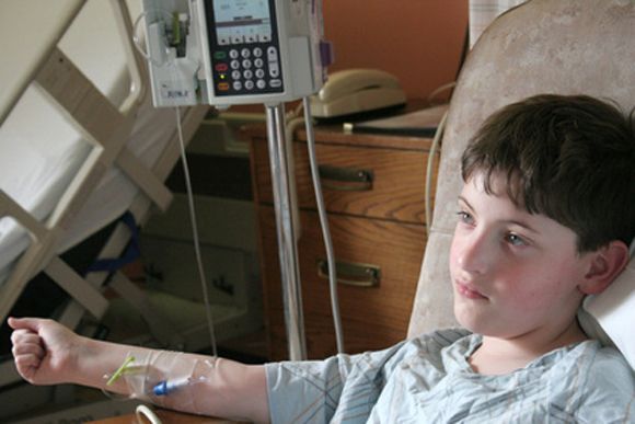
Kasper Warrick, MD, and his colleagues at Indiana University in Indianapolis reported findings from a retrospective study of 73 ALL patients at their hospital. They performed chart reviews comparing a cohort of 42 patients who were discharged on day 4 of their induction treatment with 31 similar patients who had a longer hospital stay or admission to the intensive care unit. The report was published in Leukemia Research.
Univariate analysis found that patients with a longer length of stay were more likely to have a fever, pretransfusion hemoglobin of less than 8 g/dL, lower serum bicarbonate values, abnormal serum calcium, and abnormal serum phosphate. Multivariate stepwise logistic regression found that low serum bicarbonate and a lower platelet count on day 4 of admission was predictive of a prolonged hospital stay. About a third of patients from each group had an unplanned readmission within 30 days.
The researchers concluded that early discharge is safe in only a subgroup of high-risk ALL patients undergoing induction chemotherapy. “Treating physicians could opt for a discharge only after normalization of electrolyte abnormalities and renal functions, and when no transfusion support is needed (stable hematocrit and platelet count),” they wrote. Even in those cases, they recommended “aggressive and close outpatient follow” since patients are vulnerable to complications and readmissions.
SOURCE: Warrick K et al. Leuk Res. 2018 Jun 30:71:36-42.
(ALL) who were treated with four-drug induction therapy.
Kasper Warrick, MD, and his colleagues at Indiana University in Indianapolis reported findings from a retrospective study of 73 ALL patients at their hospital. They performed chart reviews comparing a cohort of 42 patients who were discharged on day 4 of their induction treatment with 31 similar patients who had a longer hospital stay or admission to the intensive care unit. The report was published in Leukemia Research.
Univariate analysis found that patients with a longer length of stay were more likely to have a fever, pretransfusion hemoglobin of less than 8 g/dL, lower serum bicarbonate values, abnormal serum calcium, and abnormal serum phosphate. Multivariate stepwise logistic regression found that low serum bicarbonate and a lower platelet count on day 4 of admission was predictive of a prolonged hospital stay. About a third of patients from each group had an unplanned readmission within 30 days.
The researchers concluded that early discharge is safe in only a subgroup of high-risk ALL patients undergoing induction chemotherapy. “Treating physicians could opt for a discharge only after normalization of electrolyte abnormalities and renal functions, and when no transfusion support is needed (stable hematocrit and platelet count),” they wrote. Even in those cases, they recommended “aggressive and close outpatient follow” since patients are vulnerable to complications and readmissions.
SOURCE: Warrick K et al. Leuk Res. 2018 Jun 30:71:36-42.
FROM LEUKEMIA RESEARCH
Better ICU staff communication with family may improve end-of-life choices
A nurse-led support intervention for the families of critically ill patients did little to ease families’ psychological symptoms, but it did improve their perception of staff communication and family-centered care in the intensive care unit.

The length of ICU stay was also significantly shorter and the in-unit death rate higher among patients whose families received the intervention – a finding that suggests difficult end-of-life choices may have been eased, reported Douglas B. White, MD, and his colleagues (N Engl J Med. 2018;378:2365-75).
“The intervention resulted in significant improvements in markers of the quality of decision making, including the patient- and family-centeredness of care and the quality of clinician-family communication. Taken together, these findings suggest that the intervention allowed surrogates to transition a patient’s treatment to comfort-focused care when doing so aligned with the patient’s values,” wrote Dr. White of the University of Pittsburgh. “A previous study that was conducted in the context of advanced illness suggested that treatment that accords with the patient’s preferences may lead to shorter survival among those who prioritize comfort over longevity.”
The trial randomized 1,420 patients and their family surrogates in five ICUs to usual care, or to the multicomponent family-support intervention. The primary outcome was change in the surrogates’ scores on the Hospital Anxiety Depression Scale (HADS) at 6 months. The secondary outcomes were changes in Impact of Event Scale (IES; a measure of posttraumatic stress) the Quality of Communication (QOC) scale, quality of clinician-family communication measured by the Patient Perception of Patient Centeredness (PPPC) scale and the mean length of ICU stay.
The intervention was delivered by nurses who received special training on communication and other skills needed to support the families of critically ill patients. Nurses met with families every day and arranged regular meetings with ICU clinicians. A quality improvement specialist incorporated the family support into daily work flow.
In a fully adjusted model, there was no significant between-group difference in the 6-month HADS scores (11.7 vs. 12 points). Likewise, there was no significant difference between the groups in the mean IES score at 6 months.
Family members in the active group did rate the quality of clinician-family communication as significantly better, and they also gave significantly higher ratings to the quality of patient- and family-centered care during the ICU stay.
The shorter length of stay was reflected in the time to death among patients who died during the stay (4.4 days in the intervention group vs. 6.8 days in the control group), although there was no significant difference in length of stay among patients who survived to discharge. Significantly more patients in the intervention group died in the ICU as well (36% vs. 28.5%); however, there was no significant difference in 6-month mortality (60.4% vs. 55.4%).
The study was supported by an Innovation Award from the University of Pittsburgh Medical Center Health System and by the Greenwell Foundation. Dr. White reported having no financial disclosures
Although the results by White and colleagues “cannot be interpreted as clinically directive,” the study offers a glimpse of the path forward in improving the experience of families with critically ill loved ones, Daniela Lamas, MD, wrote in an accompanying editorial (N Engl J Med. 2018; 378:2431-2).
The study didn’t meet its primary endpoint of reducing surrogates’ psychological symptoms at 6 months, but it did lead to an improved ICU experience, with better clinician communication. There was another finding that deserves a close look: In the intervention group, ICU length of stay was shorter and in-hospital mortality greater, although mortality among those who survived to discharge was similar at 6 months.
These findings suggest that the intervention did not lead to the premature death of patients who would have otherwise done well, but rather was associated with a shorter dying process for those who faced a dismal prognosis, according to Dr. Lamas.
“As we increasingly look beyond mortality as the primary outcome that matters, seeking to maximize quality of life and minimize suffering, this work represents an ‘end of the beginning’ by suggesting the next steps in moving closer to achieving these goals.”
Dr. Lamas is a pulmonary and critical care doctor at Brigham & Women’s Hospital and on the faculty at Harvard Medical School, Boston.
A nurse-led support intervention for the families of critically ill patients did little to ease families’ psychological symptoms, but it did improve their perception of staff communication and family-centered care in the intensive care unit.
The length of ICU stay was also significantly shorter and the in-unit death rate higher among patients whose families received the intervention – a finding that suggests difficult end-of-life choices may have been eased, reported Douglas B. White, MD, and his colleagues (N Engl J Med. 2018;378:2365-75).
“The intervention resulted in significant improvements in markers of the quality of decision making, including the patient- and family-centeredness of care and the quality of clinician-family communication. Taken together, these findings suggest that the intervention allowed surrogates to transition a patient’s treatment to comfort-focused care when doing so aligned with the patient’s values,” wrote Dr. White of the University of Pittsburgh. “A previous study that was conducted in the context of advanced illness suggested that treatment that accords with the patient’s preferences may lead to shorter survival among those who prioritize comfort over longevity.”
The trial randomized 1,420 patients and their family surrogates in five ICUs to usual care, or to the multicomponent family-support intervention. The primary outcome was change in the surrogates’ scores on the Hospital Anxiety Depression Scale (HADS) at 6 months. The secondary outcomes were changes in Impact of Event Scale (IES; a measure of posttraumatic stress) the Quality of Communication (QOC) scale, quality of clinician-family communication measured by the Patient Perception of Patient Centeredness (PPPC) scale and the mean length of ICU stay.
The intervention was delivered by nurses who received special training on communication and other skills needed to support the families of critically ill patients. Nurses met with families every day and arranged regular meetings with ICU clinicians. A quality improvement specialist incorporated the family support into daily work flow.
In a fully adjusted model, there was no significant between-group difference in the 6-month HADS scores (11.7 vs. 12 points). Likewise, there was no significant difference between the groups in the mean IES score at 6 months.
Family members in the active group did rate the quality of clinician-family communication as significantly better, and they also gave significantly higher ratings to the quality of patient- and family-centered care during the ICU stay.
The shorter length of stay was reflected in the time to death among patients who died during the stay (4.4 days in the intervention group vs. 6.8 days in the control group), although there was no significant difference in length of stay among patients who survived to discharge. Significantly more patients in the intervention group died in the ICU as well (36% vs. 28.5%); however, there was no significant difference in 6-month mortality (60.4% vs. 55.4%).
The study was supported by an Innovation Award from the University of Pittsburgh Medical Center Health System and by the Greenwell Foundation. Dr. White reported having no financial disclosures
FROM NEW ENGLAND JOURNAL OF MEDICINE
Key clinical point: A family communication intervention didn’t improve 6-month psychological symptoms among those with loved ones in intensive care units.
Major finding: There was no significant difference on the Hospital Anxiety and Depression Scale at 6 months (11.7 vs. 12 points).
Study details: The study randomized 1,420 ICU patients and surrogates to the intervention or to usual care.
Disclosures: The study was supported by an Innovation Award from the University of Pittsburgh Medical Center Health System and by the Greenwell Foundation. Dr. White had no financial disclosures.
Source: White et al. N Engl J Med. 2018;378:2365-75.
Intranasal naloxone promising for type 1 hypoglycemia
ORLANDO –

“This has been a clinical problem for a very long time, and we see it all the time. A patient comes into my clinic, the nurses check their blood sugar, it’s 50 mg/dL, and they’re just sitting there without any symptoms,” said lead investigator Sandra Aleksic, MD, of the Albert Einstein College of Medicine, New York.
As blood glucose in the brain drops, people get confused, and their behavioral defenses are compromised. They might crash if they’re driving. “If you have HAAF, it makes you prone to more hypoglycemia, which blunts your response even more. It’s a vicious cycle,” she said at the annual scientific sessions of the American Diabetes Association.
Endogenous opioids are at least partly to blame. Hypoglycemia induces release of beta-endorphin, which in turn inhibits production of epinephrine. Einstein investigators have shown in previous small studies with healthy subjects that morphine blunts the response to induced hypoglycemia, and intravenous naloxone – an opioid blocker – prevents HAAF (Diabetes. 2017 Nov;66[11]:2764-73).
Intravenous naloxone, however, isn’t practical for outpatients, so the team wanted to see whether intranasal naloxone also prevented HAAF. The results “are very promising, but this is preliminary.” If it pans out, though, patients may one day carry intranasal naloxone along with their glucose pills and glucagon to treat hypoglycemia. “Any time they are getting low, they would take the spray,” Dr. Aleksic said.
The team used hypoglycemic, hyperinsulinemic clamps to drop blood glucose levels in seven healthy subjects down to 54 mg/dL for 2 hours twice in one day and gave them hourly sprays of either intranasal saline or 4 mg of intranasal naloxone; hypoglycemia was induced again for 2 hours the following day. The 2-day experiment was repeated 5 weeks later.
Overall, there was no difference in peak epinephrine levels between the first hypoglycemic episode on day 1 and the third episode on day 2 in subjects randomized to naloxone (942 pg/mL plus or minus 190 versus 857 pg/mL plus or minus 134; P = .4). The third episode, meanwhile, placed placebo subjects into HAAF (first hypoglycemic episode 1,375 pg/mL plus or minus 182 versus 858 pg/mL plus or minus 235; P = .004). There was also a trend toward higher hepatic glucose production in the naloxone group.
“These findings suggest that HAAF can be prevented by acute blockade of opioid receptors during hypoglycemia. ... Acute self-administration of intranasal naloxone could be an effective and feasible real-world approach to ameliorate HAAF in type 1 diabetes,” the investigators concluded. A trial in patients with T1DM is being considered.
Dr. Aleksic estimated that patients with T1DM drop blood glucose below 54 mg/dL maybe three or four times a month, on average, depending on how well they manage the condition. For now, it’s unknown how long protection from naloxone would last.
The study subjects were men, 43 years old, on average, with a mean body mass index of 26 kg/m2.
The investigators didn’t have any disclosures, and there was no industry funding for the work.
SOURCE: Aleksic S et al. ADA 2018, Abstract 10-LB.
ORLANDO –
“This has been a clinical problem for a very long time, and we see it all the time. A patient comes into my clinic, the nurses check their blood sugar, it’s 50 mg/dL, and they’re just sitting there without any symptoms,” said lead investigator Sandra Aleksic, MD, of the Albert Einstein College of Medicine, New York.
As blood glucose in the brain drops, people get confused, and their behavioral defenses are compromised. They might crash if they’re driving. “If you have HAAF, it makes you prone to more hypoglycemia, which blunts your response even more. It’s a vicious cycle,” she said at the annual scientific sessions of the American Diabetes Association.
Endogenous opioids are at least partly to blame. Hypoglycemia induces release of beta-endorphin, which in turn inhibits production of epinephrine. Einstein investigators have shown in previous small studies with healthy subjects that morphine blunts the response to induced hypoglycemia, and intravenous naloxone – an opioid blocker – prevents HAAF (Diabetes. 2017 Nov;66[11]:2764-73).
Intravenous naloxone, however, isn’t practical for outpatients, so the team wanted to see whether intranasal naloxone also prevented HAAF. The results “are very promising, but this is preliminary.” If it pans out, though, patients may one day carry intranasal naloxone along with their glucose pills and glucagon to treat hypoglycemia. “Any time they are getting low, they would take the spray,” Dr. Aleksic said.
The team used hypoglycemic, hyperinsulinemic clamps to drop blood glucose levels in seven healthy subjects down to 54 mg/dL for 2 hours twice in one day and gave them hourly sprays of either intranasal saline or 4 mg of intranasal naloxone; hypoglycemia was induced again for 2 hours the following day. The 2-day experiment was repeated 5 weeks later.
Overall, there was no difference in peak epinephrine levels between the first hypoglycemic episode on day 1 and the third episode on day 2 in subjects randomized to naloxone (942 pg/mL plus or minus 190 versus 857 pg/mL plus or minus 134; P = .4). The third episode, meanwhile, placed placebo subjects into HAAF (first hypoglycemic episode 1,375 pg/mL plus or minus 182 versus 858 pg/mL plus or minus 235; P = .004). There was also a trend toward higher hepatic glucose production in the naloxone group.
“These findings suggest that HAAF can be prevented by acute blockade of opioid receptors during hypoglycemia. ... Acute self-administration of intranasal naloxone could be an effective and feasible real-world approach to ameliorate HAAF in type 1 diabetes,” the investigators concluded. A trial in patients with T1DM is being considered.
Dr. Aleksic estimated that patients with T1DM drop blood glucose below 54 mg/dL maybe three or four times a month, on average, depending on how well they manage the condition. For now, it’s unknown how long protection from naloxone would last.
The study subjects were men, 43 years old, on average, with a mean body mass index of 26 kg/m2.
The investigators didn’t have any disclosures, and there was no industry funding for the work.
SOURCE: Aleksic S et al. ADA 2018, Abstract 10-LB.
REPORTING FROM ADA 2018
Key clinical point: Intranasal naloxone might be just the ticket to prevent hypoglycemia-associated autonomic failure in type 1 diabetes mellitus.
Major finding: Overall, there was no difference in peak epinephrine levels between the first day 1 hypoglycemic episode and the third episode on day 2 in subjects randomized to naloxone (942 plus or minus 190 pg/mL versus 857 plus or minus 134 pg/mL; P = 0.4).
Study details: Randomized trial with seven healthy volunteers
Disclosures: There was no industry funding for the work, and the investigators didn’t have any disclosures.
Source: Aleksic S et al. ADA 2018, Abstract 10-LB