User login
An integrated response to Surfside: Lessons learned
The catastrophic collapse of the Surfside, Fla., Champlain Towers South left ambiguous loss, trauma, grief, and other psychiatric and psychological sequelae in its wake.

Now that a few months have passed since the tragedy, which took the lives of 98 residents, it is helpful to examine the psychiatric and psychological support efforts that emerged.
We can think of those support efforts as operating on two tracks: one was pursued by mental health professionals representing numerous organizations; the other was pursued by local, regional, and international first responders – specifically, by Israeli Defense Force (IDF) members who came to our community at the request of Surfside families.
Those efforts were guided by existing frameworks for crisis response designed to provide containment amid the naturally disorganizing effects of the trauma and ambiguous loss. In retrospect, it was clear that the mechanisms by which those frameworks coalesced and functioned were more implicit and organically synchronous than explicitly coordinated and agreed upon. key themes emerged and revealed intrinsic links between the first-responder/search and rescue and psychological strategies.
In this article, we discuss relevant themes and parallels between the psychological intervention/strategies and the first-responder disaster response and the practical utility of implementing an integrated strategy. Our hope is that a better understanding of these strategies will help future therapists and responders who respond to crises.
Setting the frame
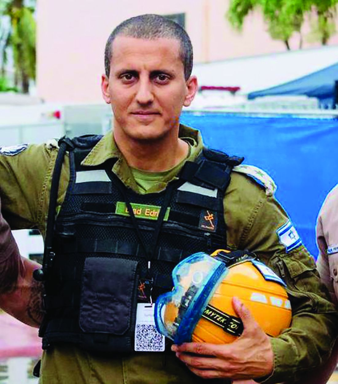
The importance of setting a psychotherapeutic frame is indisputable regardless of theoretical orientation or therapeutic modality. Predictable, consistent conditions under which therapy takes place support a patient’s capacity to tolerate the ambiguous and unpredictable aspects of the process. Those “rules of engagement” provide a structure where subjective experiences can be formulated, organized, understood, and integrated. Twice-daily briefs held in a centralized location (dubbed the Surfside “family center”) paralleled this frame and served that same containing function by offering structure, order, and predictability amid the palpable chaos of ambiguous loss and traumatic grief. Those briefs provided key information on the status of the operation and described the rescue strategy. These were led by the Miami-Dade assistant fire chief and IDF colonel (E.E.), who presented a unified front and consistent presence.
It is essential that briefings such as these be coordinated (and unified) with clear expectations about ground rules, much like what is involved in therapeutic informed consent. In this context, rules included permissions related to documentation of meetings, information sharing, and rules of communication with the media in an effort to protect the vulnerable.
The centralized meeting location served as an important center of gravity and unified place of waiting and information receipt. It provided a dedicated space to meet with humanitarian aid organizations and government officials, and symbolized continuity, consistency, ease of information transmission, and a place where practical needs could be addressed. Meals, toiletries, and other supplies were provided to simplify and maintain daily routines. Those are otherwise unremarkable practices that seemed impossible to manage amid a crisis, yet can be inherently grounding and emotionally organizing when facing deep psychological fragmentation.
Meeting in person allowed the IDF to offer operational visuals to allow those affected to feel less helpless and cultivate a sense of purpose by being part of the strategy/mission. Their strategy included “population intelligence,” which was aimed at both information gathering to practically facilitate the rescue/recovery process (for example, locating victims, property, and recreating a visual of how the building fell), and inspiring people to participate. This engagement helped many transition from a place of denial/repression to acknowledging loss/grief, and from a passive to active part of the effort, in a way that was safe and realistic – as opposed to going to the site and aiding themselves, as some had requested.
Naturally, a central location made it possible to offer immediate psychological assistance and support. Clinicians responding to crisis should be carefully selected in light of the immense suffering, emotional vulnerability, and heightened reactivity of those affected. People were overwhelmed by deep sorrow, fear, anger, and uncertainty, vacillating between hope and despair, and mobilized by a desire to help. Those providing support need to be interpersonally skilled and able to regulate their own emotions. They must be able to formulate – in real time – an understanding of what is needed, and implement a strategic plan. Like first responders, it is also key for providers to be easily accessible and identifiable in uniform so that people in the grip of a survival response can easily identify and elicit support.
The power of strategy
The Israeli delegation and mental health approaches were aligned with respect to cultivating a team identity and keeping the team spirit elevated. The delegation’s approach was to deemphasize rank during the mission in that everyone was responsible for anything that was needed and no task was below anyone’s rank. The same was true for the mental health support response: Early interventions were focused on addressing practical needs – providing blankets, water, chargers, food, and a calming presence to counter the initial chaos. No task was too small, regardless of title or role. As more structure and order ensued, it was possible to offer more traditional crisis-related interventions aimed at grounding those affected.

Both teams worked to ensure 24-hour coverage, which was crucial given the need for consistency and continuity. Our commitment was to support the victims’ families and survivors by fully embracing the chaos and the situational demands, offering attunement and support, and satisfying both basic and higher-level needs. We divided and conquered work, observed signals of need, offered immediate support where necessary, and coordinated longer-term care plans when possible. The importance of ongoing self-care, consultation, and debriefing while doing this work cannot be overstated. Time to address basic needs and the impact of vicarious trauma as a team must be built in.
Importance of flexibility
This tragedy came with unique complexities and sensitivities that needed to be identified expediently and addressed with a concrete, comprehensive plan. This was true for both the rescue and psychological support efforts, and flexibility was key. There was nothing traditional about our work from a therapeutic perspective – we found quiet corners and empty offices, went for walks, met in lobbies, and checked in by phone. The interventions were brief.
Roles shifted often between aiding in addressing practical needs, advocating for victims and connecting them to appropriate resources, supporting the police in making death notifications, providing support and space for processing during and after briefings, and more.
Similarly, the rescue team constantly reevaluated their strategy because of what they discovered as they dismantled the collapsed building, in addition to managing external impacting factors (heat, rain, lightning, and the threat of the remaining structure falling).
Language matters
The iteration of commitment to the families/victims/mission and to work speedily and efficiently was important for both rescuers and therapists. It was key during the briefings for the chief and colonel (E.E.) to share information in a manner that was professional, discreet, honest and explicit. Their willingness and ability to be vulnerable and to share their personal feelings as active rescuers humanized them. Their approach was matter of fact, yet warm, loving, and containing, all of which conveyed dignity and respect.
Word choice mattered, and the IDF’s intentional choice to refer to recovered victims as “souls,” rather than “bodies,” conveyed their sensitivity to the intensity of anguish, depth of loss, and gravity of the situation. From a psychological perspective, the transition between “rescue” efforts signifying the potential saving of lives to “recovery” of bodies or remains was significant and demarcated a dramatic shift. The weeks-long efforts, once painfully slow, then felt too abrupt to process.
One extraordinary moment was the chief’s response to the families’ discomfort at the news of the switch from rescue to recovery. The families were anxious about losing the structure that the briefings provided and were apprehensive about the handoff from fire to the police department. With great compassion and attunement, he assured them that he would stay with them, and they together, as a family, would decide when to conclude the in-person briefings. The colonel (E.E.), too, provided assurance that neither procedure nor the urgency of the recovery would change. It was both heart-warming and containing that information related to the operation was shared in a clear manner, and that the thought process and rationale behind major decisions (e.g., demolishing the remaining building, decision to pause operations, switch from rescue to recovery) was shared. It was useful for the clinicians to be aware of this rationale in helping individuals metabolize the information and process the associated trauma and grief.
Unification is key
Surfside has left an indelible impact on us. We saw and experienced unity in many respects – clinicians from various backgrounds collaborating, families bonding and caring for one another, community support and solidarity, and the cooperation and coordination of the search and rescue teams. The diverse groups providing support came to feel like a family, and the importance of inter- and intrateam integration cannot be overstated. We were transformed both by our professional collaborations and authentic connections with those affected, and will forever cherish the experience, one another, the families, and the souls lost.
Dr. Feldman is a licensed clinical psychologist in private practice in Miami. She is an adjunct professor in the college of psychology at Nova Southeastern University, Fort Lauderdale, Fla., where she teaches clinical psychology doctoral students. She also serves on the board of directors of The Southeast Florida Association for Psychoanalytic Psychology. Dr. Feldman has no disclosures. Col. Edri is the Israeli Defense Forces District Commander of the Home Front Command Haifa District. He served as the deputy commander for the Israeli Defense Forces Search and Rescue Delegation, which was brought in to provide international aid to the local and domestic forces responding to the Surfside, Fla., building collapse. Col. Edri has no disclosures. Dr. Davidtz is a licensed psychologist and associate professor in the College of Psychology at Nova Southeastern University, Fort Lauderdale, Fla., where she is director of internship training for the Psychology Services Center and director of psychological services for the emotionally distressed, a specialty clinic that serves people with serious mental illness and personality disorders. She also maintains a part-time private practice specializing in the treatment of complex posttraumatic conditions and personality disorders. Dr. Davidtz has no disclosures.
The catastrophic collapse of the Surfside, Fla., Champlain Towers South left ambiguous loss, trauma, grief, and other psychiatric and psychological sequelae in its wake.
Now that a few months have passed since the tragedy, which took the lives of 98 residents, it is helpful to examine the psychiatric and psychological support efforts that emerged.
We can think of those support efforts as operating on two tracks: one was pursued by mental health professionals representing numerous organizations; the other was pursued by local, regional, and international first responders – specifically, by Israeli Defense Force (IDF) members who came to our community at the request of Surfside families.
Those efforts were guided by existing frameworks for crisis response designed to provide containment amid the naturally disorganizing effects of the trauma and ambiguous loss. In retrospect, it was clear that the mechanisms by which those frameworks coalesced and functioned were more implicit and organically synchronous than explicitly coordinated and agreed upon. key themes emerged and revealed intrinsic links between the first-responder/search and rescue and psychological strategies.
In this article, we discuss relevant themes and parallels between the psychological intervention/strategies and the first-responder disaster response and the practical utility of implementing an integrated strategy. Our hope is that a better understanding of these strategies will help future therapists and responders who respond to crises.
Setting the frame
The importance of setting a psychotherapeutic frame is indisputable regardless of theoretical orientation or therapeutic modality. Predictable, consistent conditions under which therapy takes place support a patient’s capacity to tolerate the ambiguous and unpredictable aspects of the process. Those “rules of engagement” provide a structure where subjective experiences can be formulated, organized, understood, and integrated. Twice-daily briefs held in a centralized location (dubbed the Surfside “family center”) paralleled this frame and served that same containing function by offering structure, order, and predictability amid the palpable chaos of ambiguous loss and traumatic grief. Those briefs provided key information on the status of the operation and described the rescue strategy. These were led by the Miami-Dade assistant fire chief and IDF colonel (E.E.), who presented a unified front and consistent presence.
It is essential that briefings such as these be coordinated (and unified) with clear expectations about ground rules, much like what is involved in therapeutic informed consent. In this context, rules included permissions related to documentation of meetings, information sharing, and rules of communication with the media in an effort to protect the vulnerable.
The centralized meeting location served as an important center of gravity and unified place of waiting and information receipt. It provided a dedicated space to meet with humanitarian aid organizations and government officials, and symbolized continuity, consistency, ease of information transmission, and a place where practical needs could be addressed. Meals, toiletries, and other supplies were provided to simplify and maintain daily routines. Those are otherwise unremarkable practices that seemed impossible to manage amid a crisis, yet can be inherently grounding and emotionally organizing when facing deep psychological fragmentation.
Meeting in person allowed the IDF to offer operational visuals to allow those affected to feel less helpless and cultivate a sense of purpose by being part of the strategy/mission. Their strategy included “population intelligence,” which was aimed at both information gathering to practically facilitate the rescue/recovery process (for example, locating victims, property, and recreating a visual of how the building fell), and inspiring people to participate. This engagement helped many transition from a place of denial/repression to acknowledging loss/grief, and from a passive to active part of the effort, in a way that was safe and realistic – as opposed to going to the site and aiding themselves, as some had requested.
Naturally, a central location made it possible to offer immediate psychological assistance and support. Clinicians responding to crisis should be carefully selected in light of the immense suffering, emotional vulnerability, and heightened reactivity of those affected. People were overwhelmed by deep sorrow, fear, anger, and uncertainty, vacillating between hope and despair, and mobilized by a desire to help. Those providing support need to be interpersonally skilled and able to regulate their own emotions. They must be able to formulate – in real time – an understanding of what is needed, and implement a strategic plan. Like first responders, it is also key for providers to be easily accessible and identifiable in uniform so that people in the grip of a survival response can easily identify and elicit support.
The power of strategy
The Israeli delegation and mental health approaches were aligned with respect to cultivating a team identity and keeping the team spirit elevated. The delegation’s approach was to deemphasize rank during the mission in that everyone was responsible for anything that was needed and no task was below anyone’s rank. The same was true for the mental health support response: Early interventions were focused on addressing practical needs – providing blankets, water, chargers, food, and a calming presence to counter the initial chaos. No task was too small, regardless of title or role. As more structure and order ensued, it was possible to offer more traditional crisis-related interventions aimed at grounding those affected.
Both teams worked to ensure 24-hour coverage, which was crucial given the need for consistency and continuity. Our commitment was to support the victims’ families and survivors by fully embracing the chaos and the situational demands, offering attunement and support, and satisfying both basic and higher-level needs. We divided and conquered work, observed signals of need, offered immediate support where necessary, and coordinated longer-term care plans when possible. The importance of ongoing self-care, consultation, and debriefing while doing this work cannot be overstated. Time to address basic needs and the impact of vicarious trauma as a team must be built in.
Importance of flexibility
This tragedy came with unique complexities and sensitivities that needed to be identified expediently and addressed with a concrete, comprehensive plan. This was true for both the rescue and psychological support efforts, and flexibility was key. There was nothing traditional about our work from a therapeutic perspective – we found quiet corners and empty offices, went for walks, met in lobbies, and checked in by phone. The interventions were brief.
Roles shifted often between aiding in addressing practical needs, advocating for victims and connecting them to appropriate resources, supporting the police in making death notifications, providing support and space for processing during and after briefings, and more.
Similarly, the rescue team constantly reevaluated their strategy because of what they discovered as they dismantled the collapsed building, in addition to managing external impacting factors (heat, rain, lightning, and the threat of the remaining structure falling).
Language matters
The iteration of commitment to the families/victims/mission and to work speedily and efficiently was important for both rescuers and therapists. It was key during the briefings for the chief and colonel (E.E.) to share information in a manner that was professional, discreet, honest and explicit. Their willingness and ability to be vulnerable and to share their personal feelings as active rescuers humanized them. Their approach was matter of fact, yet warm, loving, and containing, all of which conveyed dignity and respect.
Word choice mattered, and the IDF’s intentional choice to refer to recovered victims as “souls,” rather than “bodies,” conveyed their sensitivity to the intensity of anguish, depth of loss, and gravity of the situation. From a psychological perspective, the transition between “rescue” efforts signifying the potential saving of lives to “recovery” of bodies or remains was significant and demarcated a dramatic shift. The weeks-long efforts, once painfully slow, then felt too abrupt to process.
One extraordinary moment was the chief’s response to the families’ discomfort at the news of the switch from rescue to recovery. The families were anxious about losing the structure that the briefings provided and were apprehensive about the handoff from fire to the police department. With great compassion and attunement, he assured them that he would stay with them, and they together, as a family, would decide when to conclude the in-person briefings. The colonel (E.E.), too, provided assurance that neither procedure nor the urgency of the recovery would change. It was both heart-warming and containing that information related to the operation was shared in a clear manner, and that the thought process and rationale behind major decisions (e.g., demolishing the remaining building, decision to pause operations, switch from rescue to recovery) was shared. It was useful for the clinicians to be aware of this rationale in helping individuals metabolize the information and process the associated trauma and grief.
Unification is key
Surfside has left an indelible impact on us. We saw and experienced unity in many respects – clinicians from various backgrounds collaborating, families bonding and caring for one another, community support and solidarity, and the cooperation and coordination of the search and rescue teams. The diverse groups providing support came to feel like a family, and the importance of inter- and intrateam integration cannot be overstated. We were transformed both by our professional collaborations and authentic connections with those affected, and will forever cherish the experience, one another, the families, and the souls lost.
Dr. Feldman is a licensed clinical psychologist in private practice in Miami. She is an adjunct professor in the college of psychology at Nova Southeastern University, Fort Lauderdale, Fla., where she teaches clinical psychology doctoral students. She also serves on the board of directors of The Southeast Florida Association for Psychoanalytic Psychology. Dr. Feldman has no disclosures. Col. Edri is the Israeli Defense Forces District Commander of the Home Front Command Haifa District. He served as the deputy commander for the Israeli Defense Forces Search and Rescue Delegation, which was brought in to provide international aid to the local and domestic forces responding to the Surfside, Fla., building collapse. Col. Edri has no disclosures. Dr. Davidtz is a licensed psychologist and associate professor in the College of Psychology at Nova Southeastern University, Fort Lauderdale, Fla., where she is director of internship training for the Psychology Services Center and director of psychological services for the emotionally distressed, a specialty clinic that serves people with serious mental illness and personality disorders. She also maintains a part-time private practice specializing in the treatment of complex posttraumatic conditions and personality disorders. Dr. Davidtz has no disclosures.
Neuroimaging may predict cognitive decline after chemotherapy for breast cancer
“Cognitive decline is frequently observed after chemotherapy,” according to Michiel B. de Ruiter, PhD, a research scientist with the Netherlands Cancer Institute in Amsterdam. He specializes in cognitive neuroscience and was the lead author of a study published online Sept. 30, 2021, in the Journal of Clinical Oncology. Dr. de Ruiter and colleagues found that fractional anisotropy may demonstrate a low brain white-matter reserve which could be a risk factor for cognitive decline after chemotherapy for breast cancer treatment.
Cognitive decline after chemotherapy has been reported in 20%-40% of patients with cancer affecting quality of life and daily living skills. Studies have suggested that genetic makeup, advanced age, fatigue, and premorbid intelligence quotient are risk factors for chemotherapy-associated cognitive decline. Changes in the microstructure of brain white matter, known as brain reserve, have been reported after exposure to chemotherapy, but its link to cognitive decline is understudied. Several studies outside of oncology have used MRI to derive fractional anisotropy as a measure for brain reserve.
In the new JCO study, researchers examined fractional anisotropy, as measured by MRI, before chemotherapy. The analysis included 49 patients who underwent neuropsychological tests before treatment with anthracycline-based chemotherapy, then again at 6 months and 2 years after chemotherapy.
The results were compared with those of patients with breast cancer who did not receive systemic therapy and then with a control group consisting of patients without cancer.
A low fractional anisotropy score suggested cognitive decline more than 3 years after receiving chemotherapy treatment. The finding was independent of age, premorbid intelligence quotient, baseline fatigue and baseline cognitive complaints. And, having low premorbid intelligence quotient was an independent risk factor for chemotherapy-associated cognitive decline, which the authors said is in line with previous findings.
Fractional anisotropy did not predict cognitive decline in patients who did not receive systemic therapy, as well as patients in the control group.
The findings could possibly lead to the development a pretreatment assessment to screen for patients who may at risk for cognitive decline, the authors wrote. “Clinically validated assessments of white-matter reserve as assessed with an MRI scan may be part of a pretreatment screening. This could also aid in early identification of cognitive decline after chemotherapy, allowing targeted and early interventions to improve cognitive problems,” such as psychoeducation and cognitive rehabilitation.
No potential conflicts of interest were reported.
FROM THE JOURNAL OF CLINICAL ONCOLOGY
Timeless stories
Let me tell you a story. In 5 billion years the sun will run out of hydrogen, the fuel it is currently burning to power my solar panels amongst other things. At that time, the sun will no longer be able to keep its core contracted and will expand into a fiery, red giant, engulfing earth and obliterating any sign that we ever existed. No buildings. No blog posts. No mausoleums. No stories. Nothing of us will remain.

Well, here for a moment anyway, I’ve gotten you to think about something other than COVID. You’re welcome.
Fascinatingly, the image in your mind’s eye right now of a barren scorched landscape was put there by me. Simply by placing a few words together I have caused new thoughts in your head. You might even share this story with someone else – I would have actually changed your behavior through the power of language. This miraculous phenomenon seems to be unique to us humans; we are the only ones who can create whole worlds in another individual’s head just by making a few sounds. We in medicine have the privilege of experiencing this miracle every day.
Last week, a 97-year-old pale, frail, white man saw me for a basal cell carcinoma on his cheek. While performing a simple electrodesiccation and curettage, I asked if he remembers getting a lot of sunburns when he was young. He certainly remembered one. On a blustery sunny day, he fell asleep for hours on the deck of the USS West Virginia while in the Philippines. As a radio man, he was exhausted from days of conflict and he recalled how warm breezes lulled him asleep. He was so sunburned that for days he forgot how afraid he was of the Japanese.
After listening to his story, I had an image in my mind of palm trees swaying in the tropical winds while hundreds of hulking gray castles sat hidden in the vast surrounding oceans awaiting one of the greatest naval conflicts in history. I got to hear it from surely one of the last remaining people in existence to be able to tell that story. Listening to a patient’s tales is one of the benefits of being a physician. Not only do they help bond us with our patients, but also help lessen our burden of having to make diagnosis after diagnosis and write note after note for hours on end. Somehow performing yet another biopsy that day is made just a bit easier if I’m also learning about what it was like at the Battle of Leyte Gulf.
Encouraging patients to talk more can be risky. No physician, not even allergists, can afford to be waylaid by a retiree with nothing else to do today. But meaningful encounters can not only be a vaccine against burnout, they also lead to better patient adherence and satisfaction. Sometimes, there is simply not time. But often there is a little window during a procedure or when you’re reasonably caught up and don’t expect delays ahead. And like every story, they literally transform us, the listener. In a true physical sense, their stories live on in me, and now that I’ve shared this one in writing, also with you for perpetuity. That is at least for the next 5 billion years when it, too, will be swallowed by the sun, leaving only a crispy, smoking rock where we once existed.
Dr. Benabio is director of Healthcare Transformation and chief of dermatology at Kaiser Permanente San Diego. The opinions expressed in this column are his own and do not represent those of Kaiser Permanente. Dr. Benabio is @Dermdoc on Twitter. Write to him at [email protected].
Let me tell you a story. In 5 billion years the sun will run out of hydrogen, the fuel it is currently burning to power my solar panels amongst other things. At that time, the sun will no longer be able to keep its core contracted and will expand into a fiery, red giant, engulfing earth and obliterating any sign that we ever existed. No buildings. No blog posts. No mausoleums. No stories. Nothing of us will remain.
Well, here for a moment anyway, I’ve gotten you to think about something other than COVID. You’re welcome.
Fascinatingly, the image in your mind’s eye right now of a barren scorched landscape was put there by me. Simply by placing a few words together I have caused new thoughts in your head. You might even share this story with someone else – I would have actually changed your behavior through the power of language. This miraculous phenomenon seems to be unique to us humans; we are the only ones who can create whole worlds in another individual’s head just by making a few sounds. We in medicine have the privilege of experiencing this miracle every day.
Last week, a 97-year-old pale, frail, white man saw me for a basal cell carcinoma on his cheek. While performing a simple electrodesiccation and curettage, I asked if he remembers getting a lot of sunburns when he was young. He certainly remembered one. On a blustery sunny day, he fell asleep for hours on the deck of the USS West Virginia while in the Philippines. As a radio man, he was exhausted from days of conflict and he recalled how warm breezes lulled him asleep. He was so sunburned that for days he forgot how afraid he was of the Japanese.
After listening to his story, I had an image in my mind of palm trees swaying in the tropical winds while hundreds of hulking gray castles sat hidden in the vast surrounding oceans awaiting one of the greatest naval conflicts in history. I got to hear it from surely one of the last remaining people in existence to be able to tell that story. Listening to a patient’s tales is one of the benefits of being a physician. Not only do they help bond us with our patients, but also help lessen our burden of having to make diagnosis after diagnosis and write note after note for hours on end. Somehow performing yet another biopsy that day is made just a bit easier if I’m also learning about what it was like at the Battle of Leyte Gulf.
Encouraging patients to talk more can be risky. No physician, not even allergists, can afford to be waylaid by a retiree with nothing else to do today. But meaningful encounters can not only be a vaccine against burnout, they also lead to better patient adherence and satisfaction. Sometimes, there is simply not time. But often there is a little window during a procedure or when you’re reasonably caught up and don’t expect delays ahead. And like every story, they literally transform us, the listener. In a true physical sense, their stories live on in me, and now that I’ve shared this one in writing, also with you for perpetuity. That is at least for the next 5 billion years when it, too, will be swallowed by the sun, leaving only a crispy, smoking rock where we once existed.
Dr. Benabio is director of Healthcare Transformation and chief of dermatology at Kaiser Permanente San Diego. The opinions expressed in this column are his own and do not represent those of Kaiser Permanente. Dr. Benabio is @Dermdoc on Twitter. Write to him at [email protected].
FDA expands use of HIV drug to young children
The new lower dose is approved for children weighing from at least 14 kg (30 pounds) to 25 kg (55 pounds) who are virologically suppressed or new to antiretroviral therapy.
“Children living with HIV are in need of effective and accessible formulations of antiretroviral therapy,” said Merdad Parsey, MD, PhD, chief medical officer of Gilead Sciences, the company that produces Biktarvy, in a press release. “The New Drug Application approval is an important step in fulfilling Gilead’s commitment to a goal of bringing pediatric formulations of Biktarvy to children living with HIV around the world,” he said.
Although advances in treatment for pregnant women with HIV have lowered the likelihood of perinatal HIV transmission, pediatric HIV remains a global public health challenge. In 2020, about 1.7 million children younger than 15 years were living with HIV worldwide; 850 children become infected every day.
The approval, announced October 18, expands the use of Biktarvy to younger children. The medication was originally approved in February 2018 for treatment-naive or virologically suppressed adults. In June 2019, the FDA approved updating of the label to include pediatric patients weighing at least 25 kg. This new lower dose of Biktarvy is for a three-drug combo containing bictegravir 30 mg, emtricitabine 120 mg, and tenofovir alafenamide 15 mg. It is given once a day in tablet form.
The most recent expanded indication was based on data from an open-label, single-arm study that included 22 virologically suppressed children living with HIV. After switching to Biktarvy, 91% of participants (20 of 22) remained virologically suppressed at 24 weeks. HIV-1 RNA was not collected for two patients because of «pandemic-related study disruption,» the press release said.
“As children living with HIV will be on therapy for the foreseeable future and from such a young age, there are a number of factors I weigh as a clinician when prescribing the right HIV treatment option to my pediatric patients,” said Carina Rodriguez, MD, the division chief of pediatric infectious diseases at the University of South Florida, who was one of the study investigators. “Finding an efficacious treatment option is paramount, but tolerability and safety are keys to ensuring treatment success. With this expanded approval, clinicians can add Biktarvy to their arsenal of options to help ensure these children maintain virologic suppression with a treatment option that makes sense for them.”
A version of this article first appeared on Medscape.com.
Prostate Cancer: Prostate-Specific Antigen Testing
Patient loses prostate after biopsy slide switched; more
It’s difficult enough when a patient’s prostate is removed because of cancer. But it’s another thing altogether when the prostate is removed because of a medical error, as a report on 3 CBS Philly, among other news outlets, makes clear.
The patient, Eric Spangs, lives in southeastern Pennsylvania. Testing indicated an elevation in prostate-specific antigen (PSA) level. He subsequently underwent biopsy of the prostate, which appeared to indicate cancer. In time, though, Mr. Spangs learned there had been an error: the tissue section used in the microscopic diagnosis had come from the biopsy specimen of a different patient. Mr. Spangs himself didn’t actually have cancer.
Ordinarily, such news would be cause for celebration. But this was far from a normal situ ation: Following his initial cancer diagnosis, Mr. Spangs underwent a radical laparoscopic prostatectomy at a local hospital.
“It’s devastated me emotionally and physically,” Mr. Spangs said. It has also been emotionally devastating for his wife, Melissa. (The couple has five children.)
Their attorney, Aaron Freiwald, has filed a suit against the health system to which the local hospital belongs and the area’s largest urologic practice.
The Spangs wish to caution other patients not to make the same mistake they did: they failed to get a second opinion from an oncology specialist, as recommended by the American Cancer Society. (Eric Spangs did receive a second opinion from someone at the urologic practice, but that practice doesn’t specialize in oncology.)
The Spangs also worry about the patient who received the false-negative biopsy result. They have been assured, however, that that patient will be properly notified of his actual cancer status.
Fertility specialist uses own sperm to impregnate patients
A suit claims that a Rochester, N.Y., gynecologist and fertility specialist used his own sperm to inseminate multiple patients, according to a story reported by the Associated Press and other news outlets.
The suit was filed last month by the daughter — call her “Harriet Jones” — of one of the women who received fertility services from the doctor during the 1980s. Ms. Jones’s suit alleges that at the time, the doctor told her mother that the sperm donor would be a medical student at the University of Rochester. In fact, the donor was the doctor himself. He kept that fact a secret for years, even after Ms. Jones — his own daughter — sought him out for gynecologic services.
The secret gradually began to come to light in 2016, when Ms. Jones’s nonbiological father — the man who had helped to raise her — died. Curious about her biological father, Ms. Jones sought to learn his identity from the Rochester gynecologist who had treated her mother and was now her own gynecologist. The doctor said he couldn’t be of help; he claimed he hadn’t kept the relevant records.
Ms. Jones then submitted a blood sample to a direct-to-consumer genetic testing company. The results surprised her: Not only did she learn of her ethnicity, but she also discovered the existence of two half siblings, who were donor-conceived in 1984 and 1985, respectively, the very period when her own mother was undergoing insemination procedures. Ms. Jones subsequently discovered the existence of additional half siblings, all born in the first half of the 1980s.
Initially elated by the discoveries, Ms. Jones soon grew despondent and anxious. She suffered from migraine headaches, among other symptoms. Her biological father, it seemed, had been “a serial sperm donor.”
Still, she continued to go to her Rochester doctor for treatment, having no reason to suspect anything untoward about him. Her visits, including those for prolonged menstrual bleeding, involved routine breast and pelvic exams, transvaginal ultrasounds, and intrauterine contraceptive placements under sedation.
Over this period, her doctor was friendly, asking her a variety of questions about her personal life. During one especially strange visit, however, he began chuckling and said, “You’re a really good kid, such a good kid.” During this visit, he invited his wife into the exam room, presumably to meet Ms. Jones.
It was at this moment that Ms. Jones had a revelation: Could her gynecologist actually be her biological father?
In May 2021, Ms. Jones and a half brother with whom she had been in touch contacted the gynecologist’s daughter from his first marriage. All three underwent genetic testing. The results showed a 99.99% chance of a genetic link.
Ms. Jones has said in her suit that “no reasonable woman” would have submitted to pelvic examinations and other examinations by a doctor whom she knew to be her father.
Besides fraud, her suit alleges medical malpractice, battery, infliction of mental distress, and lack of informed consent. She is seeking compensation for all harm caused to her, including past and future economic damages, past unreimbursed medical expenses, and future expenses related to her mental health treatment and care.
The story included no further details about the civil litigation. As for criminal charges, it’s unlikely Ms. Jones’s biological father — her gynecologist — will face criminal charges for his alleged crimes because they fall outside of the state’s statute of limitations.
Parents say daughter’s stroke wasn’t identified
The Georgia parents of a young woman who died from a stroke following a series of alleged misdiagnoses are suing multiple practitioners, reports Legal Newswire and other news outlets
In June 2019, Michaela Smith was training for her job as a detention officer when she began experiencing a variety of symptoms, including headache, shortness of breath, throat swelling, and slurred speech. She was taken to the emergency department (ED) at Hamilton Medical Center, in Dalton, Ga.
There, she was examined by an attending ED doctor, who ordered a CT scan. The results were read by radiologist Michael J. Cooney, MD. In his reading, the Smith family’s lawsuit alleges, Dr. Cooney failed to identify the basilar artery sign, which is a key indicator of a vessel occlusion in stroke patients. Dr. Cooney concluded that Ms. Smith’s scan showed no acute intracranial abnormality. He sent her home without further discharge instructions.
At home, Ms. Smith fell asleep but awoke in an altered mental state, one of several classic stroke symptoms that she had been experiencing. She returned to the ED. This time, she was examined by David F. Hawkins, MD, an ED physician. Although his differential diagnosis identified Ms. Smith’s symptoms as most likely stroke related, Dr. Hawkins allegedly failed to immediately corroborate his findings with additional vascular imaging. Later in the day, Ms. Smith did undergo an MRI, which a second radiologist, Kevin F. Johnson, MD, misread as showing no signs of ischemia in her basilar artery, according to the lawsuit.
That same day, Dr. Hawkins conferred with a second neurologist, Jeffrey T. Glass, MD, who recommended that Ms. Smith be admitted to the hospital because of her deteriorating condition. The Smiths’ suit claims that Dr. Glass also failed to diagnosis their daughter’s underlying condition, although he did sign off on her transfer to Baroness Erlanger Hospital, in Chattanooga, Tenn.
There, Ms. Smith’s condition continued to worsen. She soon required mechanical ventilation and tube feeding. On July 3, 2019, she was pronounced dead.
“This is an egregious case of negligence,” said the attorney representing the Smiths, who are suing the physicians involved and their practices, as well as Hamilton Medical Center and several unnamed defendants.
“Although two radiology studies and her clinical presentation indicated that Michaela was having a catastrophic stroke, her doctors repeatedly misread the studies as normal, failed to diagnose the stroke, and failed to treat her deficits as a neurological emergency,” the family’s lawyer stated.
At press time, there had been no response from any of the defendants or their attorneys.
A version of this article first appeared on Medscape.com.
‘Multimorbidity’ more commonly seen in people with lupus
People with systemic lupus erythematosus (SLE) have a threefold greater likelihood of having up to five or more comorbidities in comparison with people in the general population, according to the results of two separate U.S. population-based studies.
The higher rate of comorbidities seen included many of those commonly reported before, such as cardiovascular and renal disease, but also some that may be less frequently associated with SLE, notably chronic obstructive pulmonary disease (COPD) and cardiac arrhythmias.
“In the past, the characterization of SLE comorbidities has relied on individual comorbidity assessment,” Alí Duarte García, MD, said at the 14th International Congress on Systemic Lupus Erythematosus, held together will the 6th International Congress on Controversies in Rheumatology and Autoimmunity.
“However, a patient-centric approach where a patient as a whole is seen and how many comorbidities they accrue has not been performed.” added Duarte García, who is a rheumatologist at the Mayo Clinic in Rochester, Minn.
Multiple conditions “overrepresented” in SLE patients
Dr. Duarte García reported the findings of one of the studies, both of which used data from the Rochester Epidemiology Project, a record-linkage system that collates clinical and hospital data from individuals who live in 19 counties in southeast Minnesota and eight counties in western Wisconsin; these patients have agreed to share their medical records for research.
The study population included 479 individuals diagnosed with SLE according to joint 2019 European Alliance of Associations for Rheumatology and American College of Rheumatology criteria. These were matched by age, sex, race, and county to 479 individuals without SLE.
The mean age of the study population was 53 years, 82% were women, and 86% were White.
“We defined multimorbidity as those patients who have two or more comorbidities and substantial multimorbidity as those patients who have five or more comorbidities,” Dr. Duarte García explained.
A previously published list of 44 categories of comorbidities was used to classify the multimorbidity seen, and 27 of these were “overrepresented” in patients with SLE.
Patients with SLE averaged 5.3 comorbidities, whereas control study subjects had 2.9. Comparing SLE with non-SLE individuals, the odds ratio for having two or more comorbid conditions was 2.96, and for five or more comorbidities it was 3.06.
The highest odds ratio comparing SLE with non-SLE individuals was seen for pulmonary disorders (39.0).
Dr. Duarte García highlighted four comorbidities that occurred in SLE patients that were perhaps more unusual: congestive heart failure (OR, 13.3), valvular heart disease (OR, 4.2), cardiac arrhythmias (OR, 2.85), and COPD (OR, 2.7).
“Given the association of multimorbidity with poor outcomes, care delivery strategies to manage multimorbidity are needed in SLE,” Dr. Duarte García concluded.
Similar findings seen in cutaneous lupus
There is also an excess of comorbid conditions in people with cutaneous lupus erythematosus (CLE), Mehmet Hocaoglu, MD, said in reporting the findings of the second study.
Dr. Hocaoglu, an internal medicine resident at the University of Maryland Medical Center in Baltimore, and part of the same team of researchers as Dr. Duarte García, noted that in skin-related lupus the risk of multimorbidity was about doubled.
For this separate analysis, a total of 303 patients with cutaneous lupus had been matched to 303 controls from the general population. Odds ratios for having two or more or five or more comorbidities were a respective 2.27 and 1.65.
Among the comorbidities seen that were higher in those with cutaneous lupus than in the general population subjects were fibromyalgia, liver disease, hypertension, anemia, hypothyroidism, and COPD.
“Further research is definitely needed to identify if the driver of this multimorbidity in CLE patients is the disease itself or the treatments CLE patients are receiving or a multifactorial cause that is driving the disease association,” Dr. Hocaoglu said.
Comment and perspective
“Comorbidities that are not appropriate to the general population, compared to SLE,” seem to have been included in the overall SLE and the cutaneous lupus analyses, Raquel Faria, MD, suggested.
Dr. Faria, an internal medicine consultant at Unidade de Imunologia Clínica – Centro Hospitalar Universitário Porto (Portugal), chaired the poster discussion session in which the two studies had been presented.
She wondered if the researchers had analyzed the data while accounting for “the comorbidities that you knew are due to activity in lupus, like anemia?”
The number of patients with SLE who had pulmonary circulation disorders – 7.5% vs. 0.2% of the general population – also caught Dr. Faria’s attention.
That’s “a really huge number,” Dr. Faria pointed out, “I think it is pretty overrepresented.”
Dr. Duarte García acknowledged that they “took a very broad approach” in using a “very large comorbidity index.”
“What we were observing initially is precisely what you’re mentioning,” he responded to Dr. Faria.
“We were pulling patients who were having disease manifestation rather than a comorbidity,” Dr. Duarte-García said.
These are initial and very exploratory data, he stressed. “We have now moved on to modify the index.” Some of the changes that they have made were to incorporate the SLICC Damage Index Score and tighten up the list of ICD codes used.
No outside funding was received for either of the studies. Dr. Duarte García and Dr. Hocaoglu individually stated that they had no actual or potential conflicts of interest in relation to their presentations.
A version of this article first appeared on Medscape.com.
Injectable patch shows promise for heart attack recovery
After a heart attack, the damaged area of the heart often becomes scar tissue that can’t receive electrical messages to contract and pump blood to the body. The result is a weakened heart that could get an irregular beat, known as an arrhythmia, or go into failure.
Right now, doctors have two imperfect options for repairing this damaged tissue. One is to surgically implant a scaffold that conducts electrically and bridges the heart’s signaling system past the dead tissue. But these implants require open-chest surgery, which is risky and can lead to other heart problems.
Clinicians can use an approach that avoids opening the chest, but the patch used for these procedures may not hold its shape when grafted to damaged tissue.
Now,
The patch hasn’t been tested in humans -- any such trials are still a long way off -- but early results in animals show potential.
This experimental patch can be rolled up, threaded into a catheter or a syringe, and injected into damaged heart tissue, where it unfurls and attaches to the muscle. Once in place, the patch supports normal heart function, according to results from studies using rats and pigs. The findings were published in Nature Biomedical Engineering.
When researchers placed the patch on damaged heart muscle in rats, they found this fix resulted in a return to mostly normal heart function within 4 weeks. Results were similar when scientists tested the patch in a small number of pigs, which are considered to resemble humans more closely than rodents.
The patched hearts did a better job in pumping oxygen-rich blood to the body, and the amount of heart tissue that wasn’t getting enough oxygen also declined.
A version of this article first appeared on WebMD.com.
National Academies issue guidance for childhood COVID-19 vaccines
While the U.S. Food and Drug Administration has yet to give the green light to COVID-19 vaccination for children who are under age 12, it is expected that approval will be granted. In anticipation of the FDA’s go-ahead, which is expected in the coming weeks, a new “rapid expert consultation” has identified “actionable guidance” that state and local decision-makers can use to communicate with the public. The goal is to build confidence in and promote the uptake of COVID-19 vaccines, especially for parents who are contemplating vaccinating their children.
They note that key factors in decision-making concern vaccine side effects, the efficacy of the vaccine in children, availability of research in their child’s age group, research conducted by the parents themselves, and recommendations by the child’s health care provider.
“One of the reasons that the COVID vaccine only became available for children 12 and over months after it was approved for adults is that it takes time and many, many trial participants who are closely monitored before the vaccine ever reaches the general public,” said Nusheen Ameenuddin, MD, MPH, MPA, an assistant professor of pediatrics at the Mayo Clinic, Rochester, Minn. “We continue to talk to parents about the fact that the vaccines have been very safe and effective in this group, and even though people are concerned about side effects, they are much milder and less frequent than the effects of the disease itself.”
Dr. Ameenuddin noted that the lack of data in this age group can be concerning for parents. “It’s not like other vaccines which have been available for a long time, and the clinical trial data are still limited for this age group,” she said. “But I think the main point that practitioners need to emphasize is that, even though the vaccine is new, the science for this vaccine has been around for about a decade.”
The unique circumstances of a pandemic, she pointed out, allowed for important information about effectiveness, safety, and side effects to be obtained more quickly from clinical trial data.
“We have really good evidence for kids 12 and over, about safety and effectiveness, and even though children are not small adults and have their own unique physiology, this has provided a good starting point to suggest that kids slightly younger will also respond well to the vaccines,” said Dr. Ameenuddin, who is also chair of the American Academy of Pediatrics Council on Communications and Media. “As we learn more, we can start gathering more information about even younger kids to ensure that the right dosage and spacing of vaccines can provide maximum vaccine effectiveness and protection from disease.”
The guidance was published Oct. 13 by the National Academies of Sciences, Engineering, and Medicine.
The rapid expert consultation was produced through the Societal Experts Action Network, an activity of the National Academies that is sponsored by the NASEM and the Alfred P. Sloan Foundation. The goal of SEAN is to connect researchers in the social, behavioral, and economic sciences with decision-makers to respond to policy questions related to the COVID-19 pandemic.
In their expert consultation, the authors emphasize that vaccination is critical for decreasing transmission and controlling infection, as well as limiting the emergence of future serious variants. As of Oct. 3, 2021, about 65% of the U.S. population had received at least one dose of the vaccine, and the rate has begun to lag in many areas of the country. There are a variety of reasons for vaccine hesitancy, they note, including perception of low risks from COVID-19 or of high risks from COVID-19 vaccines, exposure to media, political agendas, lack of confidence in science, and distrust of the medical establishment. The Pfizer/BioNTech vaccine is currently authorized for emergency use for individuals 12 years of age and older and fully approved for those aged 16 and older, while the Moderna and the Johnson & Johnson vaccines are authorized for emergency use for those 18 years of age and older.
Many children between the ages of 12 and 17 have not been vaccinated, and the major concerns reported by parents include not knowing enough about the long-term effects of the COVID-19 vaccine in children (88%), concerns about children experiencing serious side effects (79%), and concerns that the COVID-19 vaccine might negatively affect future fertility (73%).
The National Academies have previously released two other “rapid expert consultations” which have addressed building vaccine confidence, and both reports provide key strategies for communicating information about COVID-19 vaccines. In this paper, the focus was on communicating with parents to gain confidence in the vaccine and address concerns.
Key points
The key strategies highlighted for communicating with parents include the following:
- Emphasizing safety and efficacy: Parents should be informed about the ongoing research and clinical trials that will answer more questions about the vaccine and that there is continued monitoring for any safety risks. Pointing to the safety data from the clinical trials for 12- to 17-year-olds, and the lack of serious adverse events from the vaccine in this age group may help alleviate concerns.
- CalibriEncouraging parents to talk with a primary care provider: Research shows that parents trust family physicians and other health care practitioners to provide them with accurate information about vaccines. Local, state, and national leaders can provide messaging templates and other resources to health care professionals who are engaged in these conversations.
- Leveraging social networks to influence parents’ vaccination decisions: Parents are influenced by their social network connections. It is important to engage these networks, especially with members of their community who are considered trustworthy and influential. Social networks may also be very diverse, and include family members, friends, coworkers, social media, and members of their religious community.
While the guidance states that different groups of parents will require different messaging, they suggest that communication can begin with a focus on the things that vaccination can accomplish. In addition to preventing infection with COVID-19, it will allow children to attend school in person and participate in extracurricular activities such as sports, without risking their health. “One thing I’ve learned over several years of working with vaccine-hesitant parents is that you have to tailor each approach to the individual,” said Dr. Ameenuddin. “Different people have different concerns, and first and foremost, it’s important to listen.”
For some parents, emphasizing that the more people that can be vaccinated and the sooner it can be done, the sooner everyone can return to a normal life is a good approach, she added. “I think it’s important to emphasize both the individual and communal benefits of vaccines, but that won’t necessarily reach every person with concerns. I think it’s important to find out what is most important to individuals and work from there to find a way to connect with that family to encourage vaccination.”
Dr. Ameenuddin has no disclosures.
PA defends against license suspension for COVID treatment
The suspension stemmed from allegations against Scott C. Miller, PA-C, by at least six COVID patients, including some who weren’t his patients or whom he never examined and a few who later died from the virus, according to the Washington Medical Commission.
“Miller’s treatment of COVID-19 patients fell below the standard of care,” the suspension report states. “Miller began a public campaign promoting ivermectin as a curative for COVID-19, and prescribing it without adequate examination to at least one person, with no reliable clinical studies that establish its efficacy in preventing or treating COVID-19.”
Mr. Miller has until early November to respond to the allegations. On his clinic’s website, Mr. Miller stated, “In response to the charges, I want to reassure all of you that the initial attacks against me have been brought on by a small handful of people that have no ties to our medical practice, and by pharmacies and hospitals that have a zero tolerance policy on family members asking that I help them advocate for loved ones that have been admitted and written off in our current system of dismissiveness and neglect.”
Mr. Miller also expressed gratitude for the support he has received recently. A GoFundMe campaign to raise money for Mr. Miller’s legal fund had raised more than $59,000 at press time. His GoFundMe page had been shared 2,400 times. He has more than 550 followers and more than 400 donors.
“I don’t know that I have the words to adequately describe the deep sense of love and connection I have received from you, the families I serve, and those that have reached out to me in this deeply challenging time,” he wrote on the clinic website.
Mr. Miller has spoken publicly about his anti-mask views and his support for ivermectin, according to the commission report. As part of the suspension, he was charged with making “misleading representations regarding the efficacy of non-FDA approved treatment and mask use.”
In one case that was cited in the report, a 39-year-old patient contacted the pediatric clinic, and Mr. Miller spoke with the patient by phone. The patient reported that he had tested positive for COVID. Mr. Miller advised the patient to take supplements, including vitamin D and C, zinc, and melatonin, and he prescribed ivermectin, dexamethasone, and azithromycin. He did not perform an exam, verify the information that the patient had provided, advise the patient regarding interactions, or order follow-up testing, the report states.
Other charges against Mr. Miller include harassing hospital staff by making threatening statements about hospitals and doctors who treat COVID-19 patients and misrepresenting his original 2013 license application. He denied on the application that he was being investigated by another licensing board. At the time, the California Physician Assistant Board was investigating him for providing medical care and prescribing without a supervising doctor’s authorization and without conducting physical exams, among other charges.
A version of this article first appeared on Medscape.com.