User login
New CDC COVID-19 isolation guidelines still up for debate among experts
It’s a true Goldilocks debate: , with some calling them suitable, some saying they’re “reckless,” and at least one expert saying they’re “right in the middle.”
The controversy may lead to more updates. On Jan. 2, Anthony S. Fauci, MD, President Joe Biden’s chief medical adviser, said on CNN’s State of the Union that he anticipates further clarification of the guidelines soon.
Sparking the most debate: Infected people are not told to test before leaving isolation, the vaccinated and unvaccinated who are exposed are given some of the same advice, and the mask advice is not specific enough.
As issued on Dec. 27, the guidelines for the general public recommend:
- Anyone who tests positive should stay home and isolate for 5 days (instead of 10) and if the person has no symptoms or the symptoms resolve after 5 days, leaving the house is okay. A mask should be worn around others for 5 more days. In the event of a fever, the person must stay home until it resolves.
- If people are exposed to someone infected with COVID-19 and they have been boosted, finished the primary series of either the Pfizer or Moderna vaccine within the past 6 months, or finished the primary series of the Johnson & Johnson vaccine within the past 2 months, they should wear a mask around others for 10 days and, if possible, test on day 5. However, if symptoms develop, they should get a test and stay home.
- If people are exposed to someone infected with COVID-19 and they are unvaccinated or are more than 6 months out from their second dose of the Pfizer or Moderna vaccine (or more than 2 months after the J&J vaccine) and not boosted, they should quarantine for 5 days and then wear a mask for 5 more days. If quarantine is impossible, a mask should be worn for 10 days. A test on day 5 is suggested if possible. If symptoms occur, they should quarantine and test.
On social media and in interviews with this news organization, public health experts expressed an array of opinions.
A tweet from Eric Topol, MD, editor-in-chief of Medscape, posted the day after the new guidelines came out, had an empty box and this: “The data that support the new @CDCgov 5 day isolation period without a negative test.”
In a tweet on Jan. 2, Ashish K. Jha, MD, MPH, dean of the Brown University School of Public Health, said: “Hearing that CDC considering adding testing to isolation guidelines. That would be great. I’ve been arguing for a while that serial negative antigen tests provide a lot of confidence that someone is not contagious.”
Michael Mina, MD, PhD, chief science officer of eMed, a digital point-of-care platform enabling at-home diagnostic testing, tweeted: “CDC’s new guidance to drop isolation of positives to 5 days without a negative test is reckless. Some [people] stay infectious 3 days, some 12. I absolutely don’t want to sit next to someone who turned [positive] 5 days ago and hasn’t tested Neg. Test Neg to leave isolation early is just smart.”
Paul Offit, MD, director of the Vaccine Education Center at the Children’s Hospital of Philadelphia and an infectious disease specialist, disagrees. Typically, he said, an infected person sheds virus for 7 days.
“If you are asymptomatic, the chances that you are shedding a significant amount of virus is very, very small,” he said in an interview.
Under debate
Testing: While many public health experts say a recommendation to test before leaving isolation is needed, CDC Director Rochelle Walensky, MD, explained testing was not recommended before leaving isolation because PCR testing can stay positive up to 12 weeks after a person is first infected with COVID-19.
Asked why there was not a recommendation for a rapid antigen test before leaving isolation, Dr. Walensky told CNN that it is not known how these tests perform at the end of infection and that the tests are not Food and Drug Administration–authorized for that purpose.
And while the guidelines suggest that those exposed – whether they are boosted, vaccinated, or not – should test on day 5 if possible, that recommendation should be stronger, some said. “At the very least recommend a test in those who can get it done,” said Dr. Topol.
However, making that recommendation is difficult when experts know how difficult it is for people to obtain tests now, William Schaffner, MD, professor of preventive medicine and an infectious disease specialist at Vanderbilt University, Nashville, Tenn., said in an interview.
“I am sure this was intensely debated,” Dr. Schaffner said of the recommendation on testing.
Vaccination status categories: Amesh Adalja, MD, senior scholar at the Johns Hopkins Center for Health Security, Baltimore, questioned the scientific basis behind treating the fully vaccinated (with two mRNA or one J&J vaccine) who are exposed ‘’as the equivalent of the unvaccinated when it comes to the quarantine requirement since the fully vaccinated are protected against what matters.”
Dr. Topol agreed: Guidelines “should be different for vaccinated versus unvaccinated.”
The recommendations for the exposed should definitely be simpler, Dr. Offit said. “I think it would be much simpler to just say, ‘If you are exposed, mask for 10 days,’ “ regardless of vaccination status.
Masks: The guidelines should also be more specific about the type of masks, Dr. Topol said. They should spell out that the masks need to be N95 or KN95, he said.
Science-driven or economy-driven? Was the guidance changed due more to concerns about the economy than to scientific information about infection and transmission? “It was,” Dr. Topol said.
Dr. Adalja sees it differently. “While it is true that this updated guidance will help the economy, it is based on a scientific foundation and should have been issued much earlier than it was.”
Tough decisions
The agency is walking a tightrope, Dr. Schaffner said, adding that he is in general agreement with what the CDC is trying to do. “The tightrope is between the public health ideal and trying to determine what will be acceptable,’’ he said.
The revised guidelines are more practical than before, others said. “The goal is harm reduction and many people just don’t do any isolation if they are faced with a 10-day period,” Dr. Adalja said.
Before issuing the new guidance, the CDC looked at the accumulating science and also took into account stresses on the health care system and other factors, Dr. Schaffner said. “Is it perfect?” Dr. Schaffner said of the new guideline. “No. Is it carefree? No. It’s right in the middle.”
Dr. Schaffner does think the messages about the new recommendations and how they were decided upon could have been communicated better, and in a more understandable manner. Some experts, for instance, led with the economy and the need for people to return to work and school when explaining the guidelines and then brought up the science behind the revisions.
That order should have been reversed, Dr. Schaffner said.
A version of this article first appeared on Medscape.com.
It’s a true Goldilocks debate: , with some calling them suitable, some saying they’re “reckless,” and at least one expert saying they’re “right in the middle.”
The controversy may lead to more updates. On Jan. 2, Anthony S. Fauci, MD, President Joe Biden’s chief medical adviser, said on CNN’s State of the Union that he anticipates further clarification of the guidelines soon.
Sparking the most debate: Infected people are not told to test before leaving isolation, the vaccinated and unvaccinated who are exposed are given some of the same advice, and the mask advice is not specific enough.
As issued on Dec. 27, the guidelines for the general public recommend:
- Anyone who tests positive should stay home and isolate for 5 days (instead of 10) and if the person has no symptoms or the symptoms resolve after 5 days, leaving the house is okay. A mask should be worn around others for 5 more days. In the event of a fever, the person must stay home until it resolves.
- If people are exposed to someone infected with COVID-19 and they have been boosted, finished the primary series of either the Pfizer or Moderna vaccine within the past 6 months, or finished the primary series of the Johnson & Johnson vaccine within the past 2 months, they should wear a mask around others for 10 days and, if possible, test on day 5. However, if symptoms develop, they should get a test and stay home.
- If people are exposed to someone infected with COVID-19 and they are unvaccinated or are more than 6 months out from their second dose of the Pfizer or Moderna vaccine (or more than 2 months after the J&J vaccine) and not boosted, they should quarantine for 5 days and then wear a mask for 5 more days. If quarantine is impossible, a mask should be worn for 10 days. A test on day 5 is suggested if possible. If symptoms occur, they should quarantine and test.
On social media and in interviews with this news organization, public health experts expressed an array of opinions.
A tweet from Eric Topol, MD, editor-in-chief of Medscape, posted the day after the new guidelines came out, had an empty box and this: “The data that support the new @CDCgov 5 day isolation period without a negative test.”
In a tweet on Jan. 2, Ashish K. Jha, MD, MPH, dean of the Brown University School of Public Health, said: “Hearing that CDC considering adding testing to isolation guidelines. That would be great. I’ve been arguing for a while that serial negative antigen tests provide a lot of confidence that someone is not contagious.”
Michael Mina, MD, PhD, chief science officer of eMed, a digital point-of-care platform enabling at-home diagnostic testing, tweeted: “CDC’s new guidance to drop isolation of positives to 5 days without a negative test is reckless. Some [people] stay infectious 3 days, some 12. I absolutely don’t want to sit next to someone who turned [positive] 5 days ago and hasn’t tested Neg. Test Neg to leave isolation early is just smart.”
Paul Offit, MD, director of the Vaccine Education Center at the Children’s Hospital of Philadelphia and an infectious disease specialist, disagrees. Typically, he said, an infected person sheds virus for 7 days.
“If you are asymptomatic, the chances that you are shedding a significant amount of virus is very, very small,” he said in an interview.
Under debate
Testing: While many public health experts say a recommendation to test before leaving isolation is needed, CDC Director Rochelle Walensky, MD, explained testing was not recommended before leaving isolation because PCR testing can stay positive up to 12 weeks after a person is first infected with COVID-19.
Asked why there was not a recommendation for a rapid antigen test before leaving isolation, Dr. Walensky told CNN that it is not known how these tests perform at the end of infection and that the tests are not Food and Drug Administration–authorized for that purpose.
And while the guidelines suggest that those exposed – whether they are boosted, vaccinated, or not – should test on day 5 if possible, that recommendation should be stronger, some said. “At the very least recommend a test in those who can get it done,” said Dr. Topol.
However, making that recommendation is difficult when experts know how difficult it is for people to obtain tests now, William Schaffner, MD, professor of preventive medicine and an infectious disease specialist at Vanderbilt University, Nashville, Tenn., said in an interview.
“I am sure this was intensely debated,” Dr. Schaffner said of the recommendation on testing.
Vaccination status categories: Amesh Adalja, MD, senior scholar at the Johns Hopkins Center for Health Security, Baltimore, questioned the scientific basis behind treating the fully vaccinated (with two mRNA or one J&J vaccine) who are exposed ‘’as the equivalent of the unvaccinated when it comes to the quarantine requirement since the fully vaccinated are protected against what matters.”
Dr. Topol agreed: Guidelines “should be different for vaccinated versus unvaccinated.”
The recommendations for the exposed should definitely be simpler, Dr. Offit said. “I think it would be much simpler to just say, ‘If you are exposed, mask for 10 days,’ “ regardless of vaccination status.
Masks: The guidelines should also be more specific about the type of masks, Dr. Topol said. They should spell out that the masks need to be N95 or KN95, he said.
Science-driven or economy-driven? Was the guidance changed due more to concerns about the economy than to scientific information about infection and transmission? “It was,” Dr. Topol said.
Dr. Adalja sees it differently. “While it is true that this updated guidance will help the economy, it is based on a scientific foundation and should have been issued much earlier than it was.”
Tough decisions
The agency is walking a tightrope, Dr. Schaffner said, adding that he is in general agreement with what the CDC is trying to do. “The tightrope is between the public health ideal and trying to determine what will be acceptable,’’ he said.
The revised guidelines are more practical than before, others said. “The goal is harm reduction and many people just don’t do any isolation if they are faced with a 10-day period,” Dr. Adalja said.
Before issuing the new guidance, the CDC looked at the accumulating science and also took into account stresses on the health care system and other factors, Dr. Schaffner said. “Is it perfect?” Dr. Schaffner said of the new guideline. “No. Is it carefree? No. It’s right in the middle.”
Dr. Schaffner does think the messages about the new recommendations and how they were decided upon could have been communicated better, and in a more understandable manner. Some experts, for instance, led with the economy and the need for people to return to work and school when explaining the guidelines and then brought up the science behind the revisions.
That order should have been reversed, Dr. Schaffner said.
A version of this article first appeared on Medscape.com.
It’s a true Goldilocks debate: , with some calling them suitable, some saying they’re “reckless,” and at least one expert saying they’re “right in the middle.”
The controversy may lead to more updates. On Jan. 2, Anthony S. Fauci, MD, President Joe Biden’s chief medical adviser, said on CNN’s State of the Union that he anticipates further clarification of the guidelines soon.
Sparking the most debate: Infected people are not told to test before leaving isolation, the vaccinated and unvaccinated who are exposed are given some of the same advice, and the mask advice is not specific enough.
As issued on Dec. 27, the guidelines for the general public recommend:
- Anyone who tests positive should stay home and isolate for 5 days (instead of 10) and if the person has no symptoms or the symptoms resolve after 5 days, leaving the house is okay. A mask should be worn around others for 5 more days. In the event of a fever, the person must stay home until it resolves.
- If people are exposed to someone infected with COVID-19 and they have been boosted, finished the primary series of either the Pfizer or Moderna vaccine within the past 6 months, or finished the primary series of the Johnson & Johnson vaccine within the past 2 months, they should wear a mask around others for 10 days and, if possible, test on day 5. However, if symptoms develop, they should get a test and stay home.
- If people are exposed to someone infected with COVID-19 and they are unvaccinated or are more than 6 months out from their second dose of the Pfizer or Moderna vaccine (or more than 2 months after the J&J vaccine) and not boosted, they should quarantine for 5 days and then wear a mask for 5 more days. If quarantine is impossible, a mask should be worn for 10 days. A test on day 5 is suggested if possible. If symptoms occur, they should quarantine and test.
On social media and in interviews with this news organization, public health experts expressed an array of opinions.
A tweet from Eric Topol, MD, editor-in-chief of Medscape, posted the day after the new guidelines came out, had an empty box and this: “The data that support the new @CDCgov 5 day isolation period without a negative test.”
In a tweet on Jan. 2, Ashish K. Jha, MD, MPH, dean of the Brown University School of Public Health, said: “Hearing that CDC considering adding testing to isolation guidelines. That would be great. I’ve been arguing for a while that serial negative antigen tests provide a lot of confidence that someone is not contagious.”
Michael Mina, MD, PhD, chief science officer of eMed, a digital point-of-care platform enabling at-home diagnostic testing, tweeted: “CDC’s new guidance to drop isolation of positives to 5 days without a negative test is reckless. Some [people] stay infectious 3 days, some 12. I absolutely don’t want to sit next to someone who turned [positive] 5 days ago and hasn’t tested Neg. Test Neg to leave isolation early is just smart.”
Paul Offit, MD, director of the Vaccine Education Center at the Children’s Hospital of Philadelphia and an infectious disease specialist, disagrees. Typically, he said, an infected person sheds virus for 7 days.
“If you are asymptomatic, the chances that you are shedding a significant amount of virus is very, very small,” he said in an interview.
Under debate
Testing: While many public health experts say a recommendation to test before leaving isolation is needed, CDC Director Rochelle Walensky, MD, explained testing was not recommended before leaving isolation because PCR testing can stay positive up to 12 weeks after a person is first infected with COVID-19.
Asked why there was not a recommendation for a rapid antigen test before leaving isolation, Dr. Walensky told CNN that it is not known how these tests perform at the end of infection and that the tests are not Food and Drug Administration–authorized for that purpose.
And while the guidelines suggest that those exposed – whether they are boosted, vaccinated, or not – should test on day 5 if possible, that recommendation should be stronger, some said. “At the very least recommend a test in those who can get it done,” said Dr. Topol.
However, making that recommendation is difficult when experts know how difficult it is for people to obtain tests now, William Schaffner, MD, professor of preventive medicine and an infectious disease specialist at Vanderbilt University, Nashville, Tenn., said in an interview.
“I am sure this was intensely debated,” Dr. Schaffner said of the recommendation on testing.
Vaccination status categories: Amesh Adalja, MD, senior scholar at the Johns Hopkins Center for Health Security, Baltimore, questioned the scientific basis behind treating the fully vaccinated (with two mRNA or one J&J vaccine) who are exposed ‘’as the equivalent of the unvaccinated when it comes to the quarantine requirement since the fully vaccinated are protected against what matters.”
Dr. Topol agreed: Guidelines “should be different for vaccinated versus unvaccinated.”
The recommendations for the exposed should definitely be simpler, Dr. Offit said. “I think it would be much simpler to just say, ‘If you are exposed, mask for 10 days,’ “ regardless of vaccination status.
Masks: The guidelines should also be more specific about the type of masks, Dr. Topol said. They should spell out that the masks need to be N95 or KN95, he said.
Science-driven or economy-driven? Was the guidance changed due more to concerns about the economy than to scientific information about infection and transmission? “It was,” Dr. Topol said.
Dr. Adalja sees it differently. “While it is true that this updated guidance will help the economy, it is based on a scientific foundation and should have been issued much earlier than it was.”
Tough decisions
The agency is walking a tightrope, Dr. Schaffner said, adding that he is in general agreement with what the CDC is trying to do. “The tightrope is between the public health ideal and trying to determine what will be acceptable,’’ he said.
The revised guidelines are more practical than before, others said. “The goal is harm reduction and many people just don’t do any isolation if they are faced with a 10-day period,” Dr. Adalja said.
Before issuing the new guidance, the CDC looked at the accumulating science and also took into account stresses on the health care system and other factors, Dr. Schaffner said. “Is it perfect?” Dr. Schaffner said of the new guideline. “No. Is it carefree? No. It’s right in the middle.”
Dr. Schaffner does think the messages about the new recommendations and how they were decided upon could have been communicated better, and in a more understandable manner. Some experts, for instance, led with the economy and the need for people to return to work and school when explaining the guidelines and then brought up the science behind the revisions.
That order should have been reversed, Dr. Schaffner said.
A version of this article first appeared on Medscape.com.
Tech can help teens connect with docs about sexual health
Maria Trent, MD, MPH, was studying ways clinicians can leverage technology to care for adolescents years before COVID-19 exposed the challenges and advantages of telehealth.
Dr. Trent, a pediatrician and adolescent medicine specialist and professor of pediatrics at Johns Hopkins University, Baltimore, has long believed that the phones in her patients’ pockets have the potential to improve the sexual health of youth. The pandemic has only made that view stronger.
“They’re a generation that’s really wired and online,” Dr. Trent told this news organization. “I think that we can meet them in that space.”
Her research has incorporated texting, apps, and videos. Out of necessity, technology increasingly became part of patient care during the pandemic. “We had to stretch our ability to do some basic triage and assessments of patients online,” Dr. Trent said.
Even when clinics are closed, doctors might be able to provide initial care remotely, such as writing prescriptions to manage symptoms or directing patients to a lab for testing.
Telemedicine could allow a clinician to guide a teenager who thinks they might be pregnant to take a store-bought test and avoid possible exposure to COVID-19 in the ED, for instance.
But doctors have concerns about the legal and practical limits of privacy and confidentiality. Who else is at home listening to a phone conversation? Are parents accessing the patient’s online portal? Will parents receive an explanation of benefits that lists testing for a sexually transmitted infection, or see a testing kit that is delivered to their home?
When a young patient needs in-person care, transportation can be a barrier. And then there’s the matter of clinicians being able to bill for telehealth services.
Practices are learning how to navigate these issues, and relevant laws vary by state.
“I think this is going to become part of standard practice,” Dr. Trent said. “I think we have to do the hard work to make sure that it’s safe, that it’s accessible, and that it is actually improving care.”
Texts, apps, videos
In one early study, Dr. Trent and colleagues found that showing adolescents with pelvic inflammatory disease a 6-minute video may improve treatment rates for their sexual partners.
Another study provided preliminary evidence that text messaging support might improve clinic attendance for moderately long-acting reversible contraception.
A third trial showed that adolescents and young adults with pelvic inflammatory disease who were randomly assigned to receive text-message prompts to take their medications and provide information about the doses they consumed had greater decreases in Neisseria gonorrhoeae and Chlamydia trachomatis infections, compared with patients who received standard care.
Dr. Trent and coinvestigators are assessing a technology-based intervention for youth with HIV, in which patients can use an app to submit videos of themselves taking antiretroviral therapy and report any side effects. The technology provides a way to monitor patients remotely and support them between visits, she said.
Will pandemic-driven options remain?
In 2020, Laura D. Lindberg, PhD, principal research scientist at the Guttmacher Institute in New York, and coauthors discussed the possible ramifications of the pandemic on the sexual and reproductive health of adolescents and young adults.
If telemedicine options driven by COVID-19 are here to stay, adolescents and young adults could be “the age group most likely to continue that approach rather than returning to traditional in-person visits,” the researchers wrote. “Innovations in health care service provision, such as use of telemedicine and obtaining contraceptives and STI testing by mail, will help expand access to [sexual and reproductive health] care for young people.”
At the 2021 annual conference of the American Academy of Pediatrics, Dr. Trent described telehealth as a viable way to provide sexual and reproductive health care to adolescents and young adults, including anticipatory guidance, contraception counseling, coordination of follow-up care and testing, and connecting patients to resources.
Her presentation cited several websites that can help patients receive testing for STIs, including Yes Means Test, the Centers for Disease Control and Prevention’s GetTested page, and I Want the Kit. Planned Parenthood has telehealth options, and the Kaiser Family Foundation compiled information about 26 online platforms that were providing contraception or STI services.
Who else is in the room?
“There’s only so much time in the day and so many patients you can see, regardless of whether you have telehealth or not,” said David L. Bell, MD, MPH, president of the Society for Adolescent Health and Medicine and a coauthor of the Perspectives on Sexual and Reproductive Health paper. In addition, “you never know who else is in the room” with the patient on the other end, added Dr. Bell, a professor of population and family health and pediatrics at the Columbia University Medical Center and medical director of the Young Men’s Clinic, both in New York.
In some respects, young patients may not be able to participate in telehealth visits the same way they would in a medical office, Dr. Trent acknowledged. Encouraging the use of headphones is one way to help protect confidentiality when talking with patients who are at home and might not be alone.
But if patients are able to find a private space for remote visits, they might be more open than usual. In that way, telemedicine could provide additional opportunities to address issues like substance use disorders and mental health, as well, she said.
“Then, if they need something, we have to problem solve,” Dr. Trent said. Next steps may involve engaging a parent or getting the patient to a lab or the clinic.
Sex ed may be lacking
The Perspectives article also raised concerns that the pandemic might exacerbate shortcomings in sex education, which already may have been lacking.
“Before the pandemic, schools were a key source of formal sex education for young people,” the authors wrote. “Sex education, which was already limited in many areas of the country, has likely not been included in the national shift to online learning. Even when in-person schooling resumes, missed sex education instruction is unlikely to be made up, given the modest attention it received prior to the pandemic.”
A recently published study in the Journal of Adolescent Health indicates that American teenagers currently receive less formal sex education than they did 25 years ago, with “troubling” inequities by race.
Researchers surveyed adolescents about what they had learned about topics such as how to say no to sex, methods of birth control and where to get them, and STIs.
Dr. Lindberg and Leslie M. Kantor, PhD, MPH, professor and chair of the department of urban-global public health at Rutgers University, Newark, N.J., conducted the analysis.
“Pediatricians and other health care providers that work with children and adolescents have a critical role to play in providing information about sexuality to both the patients and to the parents,” said Dr. Kantor, who also coauthored the Perspectives article with Dr. Lindberg and Dr. Bell. The new research “shows that doctors play an even more critical role, because they can’t assume that their patients are going to get the information that they need in a timely way from schools.”
By age 15, 21% of girls and 20% of boys have had sexual intercourse at least once, according to data from the 2015-2017 National Survey of Family Growth. By age 17, the percentages were 53% of girls and 48% of boys. By age 20, the percentages were 79% of women and 77% of men. The CDC’s 2021 guidelines on treatment and screening for STIs note that prevalence rates of certain infections – such as chlamydia and gonorrhea in females – are highest among adolescents and young adults.
Those trends underscore the importance of counseling on sexual health that clinicians can provide, but time constraints may limit how much they can discuss in a single session with a patient. To cover all topics that are important to parents and patients, doctors may need to discuss sexual and reproductive health sooner and more frequently.
Young people are getting more and more explicit information from their phones and media, yet educators are giving them less information to navigate these topics and learn what’s real, Dr. Kantor said. That mismatch can be toxic. In a December 2021 interview with Howard Stern, the pop star Billie Eilish said she started watching pornography at about age 11 and frequently watched videos that were violent. “I think it really destroyed my brain and I feel incredibly devastated that I was exposed to so much porn,” Ms. Eilish told Mr. Stern.
Researchers and a psychologist told CNN that the singer’s story may be typical. It also highlights a need to be aware of kids’ online activities and to have conversations about how pornography may not depict healthy interactions, they said.
Beyond discussing a plan for preventing pregnancy and STIs, Dr. Kantor encouraged discussions about what constitutes healthy relationships, as well as check-ins about intimate partner violence and how romantic relationships are going.
“I think for pediatricians and for parents, it’s a muscle,” she said. “As you bring up these topics more, listen, and respond, you get more comfortable with it.”
Dr. Trent has served as an advisory board member on a sexual health council for Trojan (Church & Dwight Company) and has received research funding from Hologic and research supplies from SpeeDx. Dr. Bell has received funds from the Merck Foundation, Merck, and Gilead. Dr. Kantor had no disclosures.
A version of this article first appeared on Medscape.com.
Maria Trent, MD, MPH, was studying ways clinicians can leverage technology to care for adolescents years before COVID-19 exposed the challenges and advantages of telehealth.
Dr. Trent, a pediatrician and adolescent medicine specialist and professor of pediatrics at Johns Hopkins University, Baltimore, has long believed that the phones in her patients’ pockets have the potential to improve the sexual health of youth. The pandemic has only made that view stronger.
“They’re a generation that’s really wired and online,” Dr. Trent told this news organization. “I think that we can meet them in that space.”
Her research has incorporated texting, apps, and videos. Out of necessity, technology increasingly became part of patient care during the pandemic. “We had to stretch our ability to do some basic triage and assessments of patients online,” Dr. Trent said.
Even when clinics are closed, doctors might be able to provide initial care remotely, such as writing prescriptions to manage symptoms or directing patients to a lab for testing.
Telemedicine could allow a clinician to guide a teenager who thinks they might be pregnant to take a store-bought test and avoid possible exposure to COVID-19 in the ED, for instance.
But doctors have concerns about the legal and practical limits of privacy and confidentiality. Who else is at home listening to a phone conversation? Are parents accessing the patient’s online portal? Will parents receive an explanation of benefits that lists testing for a sexually transmitted infection, or see a testing kit that is delivered to their home?
When a young patient needs in-person care, transportation can be a barrier. And then there’s the matter of clinicians being able to bill for telehealth services.
Practices are learning how to navigate these issues, and relevant laws vary by state.
“I think this is going to become part of standard practice,” Dr. Trent said. “I think we have to do the hard work to make sure that it’s safe, that it’s accessible, and that it is actually improving care.”
Texts, apps, videos
In one early study, Dr. Trent and colleagues found that showing adolescents with pelvic inflammatory disease a 6-minute video may improve treatment rates for their sexual partners.
Another study provided preliminary evidence that text messaging support might improve clinic attendance for moderately long-acting reversible contraception.
A third trial showed that adolescents and young adults with pelvic inflammatory disease who were randomly assigned to receive text-message prompts to take their medications and provide information about the doses they consumed had greater decreases in Neisseria gonorrhoeae and Chlamydia trachomatis infections, compared with patients who received standard care.
Dr. Trent and coinvestigators are assessing a technology-based intervention for youth with HIV, in which patients can use an app to submit videos of themselves taking antiretroviral therapy and report any side effects. The technology provides a way to monitor patients remotely and support them between visits, she said.
Will pandemic-driven options remain?
In 2020, Laura D. Lindberg, PhD, principal research scientist at the Guttmacher Institute in New York, and coauthors discussed the possible ramifications of the pandemic on the sexual and reproductive health of adolescents and young adults.
If telemedicine options driven by COVID-19 are here to stay, adolescents and young adults could be “the age group most likely to continue that approach rather than returning to traditional in-person visits,” the researchers wrote. “Innovations in health care service provision, such as use of telemedicine and obtaining contraceptives and STI testing by mail, will help expand access to [sexual and reproductive health] care for young people.”
At the 2021 annual conference of the American Academy of Pediatrics, Dr. Trent described telehealth as a viable way to provide sexual and reproductive health care to adolescents and young adults, including anticipatory guidance, contraception counseling, coordination of follow-up care and testing, and connecting patients to resources.
Her presentation cited several websites that can help patients receive testing for STIs, including Yes Means Test, the Centers for Disease Control and Prevention’s GetTested page, and I Want the Kit. Planned Parenthood has telehealth options, and the Kaiser Family Foundation compiled information about 26 online platforms that were providing contraception or STI services.
Who else is in the room?
“There’s only so much time in the day and so many patients you can see, regardless of whether you have telehealth or not,” said David L. Bell, MD, MPH, president of the Society for Adolescent Health and Medicine and a coauthor of the Perspectives on Sexual and Reproductive Health paper. In addition, “you never know who else is in the room” with the patient on the other end, added Dr. Bell, a professor of population and family health and pediatrics at the Columbia University Medical Center and medical director of the Young Men’s Clinic, both in New York.
In some respects, young patients may not be able to participate in telehealth visits the same way they would in a medical office, Dr. Trent acknowledged. Encouraging the use of headphones is one way to help protect confidentiality when talking with patients who are at home and might not be alone.
But if patients are able to find a private space for remote visits, they might be more open than usual. In that way, telemedicine could provide additional opportunities to address issues like substance use disorders and mental health, as well, she said.
“Then, if they need something, we have to problem solve,” Dr. Trent said. Next steps may involve engaging a parent or getting the patient to a lab or the clinic.
Sex ed may be lacking
The Perspectives article also raised concerns that the pandemic might exacerbate shortcomings in sex education, which already may have been lacking.
“Before the pandemic, schools were a key source of formal sex education for young people,” the authors wrote. “Sex education, which was already limited in many areas of the country, has likely not been included in the national shift to online learning. Even when in-person schooling resumes, missed sex education instruction is unlikely to be made up, given the modest attention it received prior to the pandemic.”
A recently published study in the Journal of Adolescent Health indicates that American teenagers currently receive less formal sex education than they did 25 years ago, with “troubling” inequities by race.
Researchers surveyed adolescents about what they had learned about topics such as how to say no to sex, methods of birth control and where to get them, and STIs.
Dr. Lindberg and Leslie M. Kantor, PhD, MPH, professor and chair of the department of urban-global public health at Rutgers University, Newark, N.J., conducted the analysis.
“Pediatricians and other health care providers that work with children and adolescents have a critical role to play in providing information about sexuality to both the patients and to the parents,” said Dr. Kantor, who also coauthored the Perspectives article with Dr. Lindberg and Dr. Bell. The new research “shows that doctors play an even more critical role, because they can’t assume that their patients are going to get the information that they need in a timely way from schools.”
By age 15, 21% of girls and 20% of boys have had sexual intercourse at least once, according to data from the 2015-2017 National Survey of Family Growth. By age 17, the percentages were 53% of girls and 48% of boys. By age 20, the percentages were 79% of women and 77% of men. The CDC’s 2021 guidelines on treatment and screening for STIs note that prevalence rates of certain infections – such as chlamydia and gonorrhea in females – are highest among adolescents and young adults.
Those trends underscore the importance of counseling on sexual health that clinicians can provide, but time constraints may limit how much they can discuss in a single session with a patient. To cover all topics that are important to parents and patients, doctors may need to discuss sexual and reproductive health sooner and more frequently.
Young people are getting more and more explicit information from their phones and media, yet educators are giving them less information to navigate these topics and learn what’s real, Dr. Kantor said. That mismatch can be toxic. In a December 2021 interview with Howard Stern, the pop star Billie Eilish said she started watching pornography at about age 11 and frequently watched videos that were violent. “I think it really destroyed my brain and I feel incredibly devastated that I was exposed to so much porn,” Ms. Eilish told Mr. Stern.
Researchers and a psychologist told CNN that the singer’s story may be typical. It also highlights a need to be aware of kids’ online activities and to have conversations about how pornography may not depict healthy interactions, they said.
Beyond discussing a plan for preventing pregnancy and STIs, Dr. Kantor encouraged discussions about what constitutes healthy relationships, as well as check-ins about intimate partner violence and how romantic relationships are going.
“I think for pediatricians and for parents, it’s a muscle,” she said. “As you bring up these topics more, listen, and respond, you get more comfortable with it.”
Dr. Trent has served as an advisory board member on a sexual health council for Trojan (Church & Dwight Company) and has received research funding from Hologic and research supplies from SpeeDx. Dr. Bell has received funds from the Merck Foundation, Merck, and Gilead. Dr. Kantor had no disclosures.
A version of this article first appeared on Medscape.com.
Maria Trent, MD, MPH, was studying ways clinicians can leverage technology to care for adolescents years before COVID-19 exposed the challenges and advantages of telehealth.
Dr. Trent, a pediatrician and adolescent medicine specialist and professor of pediatrics at Johns Hopkins University, Baltimore, has long believed that the phones in her patients’ pockets have the potential to improve the sexual health of youth. The pandemic has only made that view stronger.
“They’re a generation that’s really wired and online,” Dr. Trent told this news organization. “I think that we can meet them in that space.”
Her research has incorporated texting, apps, and videos. Out of necessity, technology increasingly became part of patient care during the pandemic. “We had to stretch our ability to do some basic triage and assessments of patients online,” Dr. Trent said.
Even when clinics are closed, doctors might be able to provide initial care remotely, such as writing prescriptions to manage symptoms or directing patients to a lab for testing.
Telemedicine could allow a clinician to guide a teenager who thinks they might be pregnant to take a store-bought test and avoid possible exposure to COVID-19 in the ED, for instance.
But doctors have concerns about the legal and practical limits of privacy and confidentiality. Who else is at home listening to a phone conversation? Are parents accessing the patient’s online portal? Will parents receive an explanation of benefits that lists testing for a sexually transmitted infection, or see a testing kit that is delivered to their home?
When a young patient needs in-person care, transportation can be a barrier. And then there’s the matter of clinicians being able to bill for telehealth services.
Practices are learning how to navigate these issues, and relevant laws vary by state.
“I think this is going to become part of standard practice,” Dr. Trent said. “I think we have to do the hard work to make sure that it’s safe, that it’s accessible, and that it is actually improving care.”
Texts, apps, videos
In one early study, Dr. Trent and colleagues found that showing adolescents with pelvic inflammatory disease a 6-minute video may improve treatment rates for their sexual partners.
Another study provided preliminary evidence that text messaging support might improve clinic attendance for moderately long-acting reversible contraception.
A third trial showed that adolescents and young adults with pelvic inflammatory disease who were randomly assigned to receive text-message prompts to take their medications and provide information about the doses they consumed had greater decreases in Neisseria gonorrhoeae and Chlamydia trachomatis infections, compared with patients who received standard care.
Dr. Trent and coinvestigators are assessing a technology-based intervention for youth with HIV, in which patients can use an app to submit videos of themselves taking antiretroviral therapy and report any side effects. The technology provides a way to monitor patients remotely and support them between visits, she said.
Will pandemic-driven options remain?
In 2020, Laura D. Lindberg, PhD, principal research scientist at the Guttmacher Institute in New York, and coauthors discussed the possible ramifications of the pandemic on the sexual and reproductive health of adolescents and young adults.
If telemedicine options driven by COVID-19 are here to stay, adolescents and young adults could be “the age group most likely to continue that approach rather than returning to traditional in-person visits,” the researchers wrote. “Innovations in health care service provision, such as use of telemedicine and obtaining contraceptives and STI testing by mail, will help expand access to [sexual and reproductive health] care for young people.”
At the 2021 annual conference of the American Academy of Pediatrics, Dr. Trent described telehealth as a viable way to provide sexual and reproductive health care to adolescents and young adults, including anticipatory guidance, contraception counseling, coordination of follow-up care and testing, and connecting patients to resources.
Her presentation cited several websites that can help patients receive testing for STIs, including Yes Means Test, the Centers for Disease Control and Prevention’s GetTested page, and I Want the Kit. Planned Parenthood has telehealth options, and the Kaiser Family Foundation compiled information about 26 online platforms that were providing contraception or STI services.
Who else is in the room?
“There’s only so much time in the day and so many patients you can see, regardless of whether you have telehealth or not,” said David L. Bell, MD, MPH, president of the Society for Adolescent Health and Medicine and a coauthor of the Perspectives on Sexual and Reproductive Health paper. In addition, “you never know who else is in the room” with the patient on the other end, added Dr. Bell, a professor of population and family health and pediatrics at the Columbia University Medical Center and medical director of the Young Men’s Clinic, both in New York.
In some respects, young patients may not be able to participate in telehealth visits the same way they would in a medical office, Dr. Trent acknowledged. Encouraging the use of headphones is one way to help protect confidentiality when talking with patients who are at home and might not be alone.
But if patients are able to find a private space for remote visits, they might be more open than usual. In that way, telemedicine could provide additional opportunities to address issues like substance use disorders and mental health, as well, she said.
“Then, if they need something, we have to problem solve,” Dr. Trent said. Next steps may involve engaging a parent or getting the patient to a lab or the clinic.
Sex ed may be lacking
The Perspectives article also raised concerns that the pandemic might exacerbate shortcomings in sex education, which already may have been lacking.
“Before the pandemic, schools were a key source of formal sex education for young people,” the authors wrote. “Sex education, which was already limited in many areas of the country, has likely not been included in the national shift to online learning. Even when in-person schooling resumes, missed sex education instruction is unlikely to be made up, given the modest attention it received prior to the pandemic.”
A recently published study in the Journal of Adolescent Health indicates that American teenagers currently receive less formal sex education than they did 25 years ago, with “troubling” inequities by race.
Researchers surveyed adolescents about what they had learned about topics such as how to say no to sex, methods of birth control and where to get them, and STIs.
Dr. Lindberg and Leslie M. Kantor, PhD, MPH, professor and chair of the department of urban-global public health at Rutgers University, Newark, N.J., conducted the analysis.
“Pediatricians and other health care providers that work with children and adolescents have a critical role to play in providing information about sexuality to both the patients and to the parents,” said Dr. Kantor, who also coauthored the Perspectives article with Dr. Lindberg and Dr. Bell. The new research “shows that doctors play an even more critical role, because they can’t assume that their patients are going to get the information that they need in a timely way from schools.”
By age 15, 21% of girls and 20% of boys have had sexual intercourse at least once, according to data from the 2015-2017 National Survey of Family Growth. By age 17, the percentages were 53% of girls and 48% of boys. By age 20, the percentages were 79% of women and 77% of men. The CDC’s 2021 guidelines on treatment and screening for STIs note that prevalence rates of certain infections – such as chlamydia and gonorrhea in females – are highest among adolescents and young adults.
Those trends underscore the importance of counseling on sexual health that clinicians can provide, but time constraints may limit how much they can discuss in a single session with a patient. To cover all topics that are important to parents and patients, doctors may need to discuss sexual and reproductive health sooner and more frequently.
Young people are getting more and more explicit information from their phones and media, yet educators are giving them less information to navigate these topics and learn what’s real, Dr. Kantor said. That mismatch can be toxic. In a December 2021 interview with Howard Stern, the pop star Billie Eilish said she started watching pornography at about age 11 and frequently watched videos that were violent. “I think it really destroyed my brain and I feel incredibly devastated that I was exposed to so much porn,” Ms. Eilish told Mr. Stern.
Researchers and a psychologist told CNN that the singer’s story may be typical. It also highlights a need to be aware of kids’ online activities and to have conversations about how pornography may not depict healthy interactions, they said.
Beyond discussing a plan for preventing pregnancy and STIs, Dr. Kantor encouraged discussions about what constitutes healthy relationships, as well as check-ins about intimate partner violence and how romantic relationships are going.
“I think for pediatricians and for parents, it’s a muscle,” she said. “As you bring up these topics more, listen, and respond, you get more comfortable with it.”
Dr. Trent has served as an advisory board member on a sexual health council for Trojan (Church & Dwight Company) and has received research funding from Hologic and research supplies from SpeeDx. Dr. Bell has received funds from the Merck Foundation, Merck, and Gilead. Dr. Kantor had no disclosures.
A version of this article first appeared on Medscape.com.
Low BMI, weight loss predict mortality risk in ILD
A low body mass index (BMI) indicative of being underweight as well as a weight loss of 2 kg or more over the course of 1 year were both independently associated with a higher mortality risk in the following year in patients with fibrotic interstitial lung disease (ILD). In contrast, being both overweight and obese appeared to be protective against mortality at the same 1-year endpoint, according to the results of an observational, retrospective cohort study.
Compared with patients with a normal BMI, patients who were underweight at a BMI of less than 18.5 kg/m2 were over three times more likely to die at 1 year, at a hazard ratio of 3.19 (P < .001), senior author Christopher Ryerson, MD, University of British Columbia, Vancouver, and colleagues reported in the journal Chest.
In contrast, patients who were overweight with a BMI of 25-29 had roughly half the mortality risk as those who were underweight, at an HR of 0.52 (P < .001). Results were roughly similar among the patients with obesity with a BMI in excess of 30, among whom the HR for mortality at 1 year was 0.55 (P < .001), compared with those who were underweight.
“All patients with fibrotic ILD should still engage in exercise and eat an appropriate diet and it is still okay if you are obese and lose weight as a consequence of these lifestyle choices,” Dr. Ryerson told this news organization. “But physicians should be concerned about patients who have severe ILD and who start to lose weight unintentionally since this often represents end-stage fibrosis or some other major comorbidity such as cancer.”
Two large cohorts
Patients from two large cohorts, including the six-center Canadian Registry for Pulmonary Fibrosis (CARE-PF) and the ILD registry at the University of California, San Francisco, were enrolled in the study. A total of 1,786 patients were included from the CARE-PF registry, which served as the derivation cohort, while another 1,779 patients from the UCSF registry served as the validation cohort. In the CARE-PF cohort, 21% of all ILD patients experienced a weight loss of at least 1 kg in the first year of follow-up, including 31% of patients with idiopathic pulmonary fibrosis (IPF).
“Fewer patients experienced a weight loss of at least 1 kg during the first year of the study period in the UCSF cohort,” the authors noted, at only 12% of all ILD patients, some 14% of those with IPF losing at least 1 kg of weight over the course of the year. At 2 years’ follow-up, 35% of all ILD patients had lost at least 1 kg, as had 46% of all IPF patients. Looking at BMI, “a higher value was associated with decreased 1-year mortality in both cohorts on unadjusted analysis,” the investigators observed.
In the CARE-PF cohort, the HR for 1-year mortality was 0.96 per unit difference in BMI (P < .001), while in the UCSF cohort, the HR for 1-year mortality was exactly the same, at 0.96 per unit difference in BMI (P < .001). The authors then adjusted findings for the ILD-GAP index, which included gender, age, and physiology index. After adjusting for this index, the HR for 1-year mortality in the CARE-PF cohort was 0.93 per unit change in BMI (95% CI, 0.90-0.967; P < .001), while in the UCSF cohort, the HR was 0.96 per unit change in BMI (95% CI, 0.94-0.98; P = .001).
Indeed, each 1-kg change above a BMI of 30, adjusted for the ILD-GAP index, was associated with a reduced risk of mortality at 1 year in both cohorts, at an HR of 0.98 (P = .001) in the CARE-PF cohort and an HR of 0.98 (P < .001) in the UCSF cohort. In contrast, patients who experienced a BMI weight loss of 2 kg or more within 1 year had a 41% increased risk of death in the subsequent year after adjusting for the ILD-GAP index and baseline BMI category, at an HR of 1.41 (P = .04). “The absolute change in mortality is much smaller than this,” Dr. Ryerson acknowledged.
“However, the magnitude [in mortality risk] did impress us and this illustrates how weight loss is a frequent consequence of end-stage disease which is something that we have all observed clinically as well,” he added.
Mortality risk plateaued in patients with a greater weight loss, the investigators observed, and there was no association between weight and subsequent 1-year mortality in either cohort on unadjusted analysis.
On the other hand, being underweight was associated with between a 13% and 16% higher mortality risk at 1 year after adjusting for the ILD-GAP, at an HR of 0.84 per 10 kg (P = .001) in the CARE-PF cohort and an HR of 0.87 per 10 kg (P < .001) in the UCSF cohort. “Results were similar in the two studied cohorts, suggesting a robust and generalizable association of both low BMI and weight loss with mortality,” the authors emphasized.
“Together these studies highlight the potential link between obesity and ILD pathogenesis and further suggest the possibility that nutritional support may have a more specific and important role in the management of fibrotic ILD,” the authors wrote. Dr. Ryerson in turn noted that being able to determine mortality risk more accurately than current mortality risk prediction models are able to do is very helpful when dealing with what are sometimes life-and-death decisions.
He also said that having more insight into a patient’s prognosis can change how physicians manage patients with respect to either transplantation or palliation and potentially the need to be more aggressive with pharmacotherapy as well.
Addressing weight loss
Asked to comment on the findings, Elizabeth Volkmann, MD, associate professor of medicine, University of California, Los Angeles, said that this was a very important study and something that she feels does not get adequate attention in clinical practice.
“Weight loss and malnutrition occur in many patients with ILD due to various factors such as gastrointestinal side effects from antifibrotic therapies, decreased oral intake due to psychosocial issues including depression, and increased caloric requirements due to increased work of breathing,” she said in an interview. That said, weight loss and malnutrition are still often underaddressed during clinical encounters for patients with ILD where the focus is on lung health.
“This study illuminates the importance of addressing weight loss in all patients with ILD as it can contribute to heightened risk of mortality,” Dr. Volkmann reemphasized. Dr. Volkmann and colleagues themselves recently reported that radiographic progression of scleroderma lung disease over the course of 1-2 years is associated with an increased risk of long-term mortality, based on two independent studies of systemic sclerosis–interstitial lung disease with extensive follow-up.
Over 8 years of follow-up, patients in the Scleroderma Lung Study II who exhibited an increase of 2% or more in the QILD score – a score that reflects the sum of all abnormally classified scores, including those for fibrosis, ground glass opacity, and honeycombing – for the whole lung at 24 months had an almost fourfold increased risk in mortality, which was significant (P = .014).
The association of an increase in the QILD of at least 2% at 12 months was suggestive in its association with mortality in the SLS I cohort at 12 years of follow-up, a finding that suggests that radiographic progression measured at 2 years is a better predictor of long-term mortality than at 1 year, as the authors concluded.
The CARR-PF is funded by Boehringer Ingelheim. Dr. Ryerson reported receiving personal fees from Boehringer Ingelheim. Dr. Volkmann consults or has received speaker fees from Boehringer Ingelheim and has received grant support from Kadmon and Horizon Therapeutics.
A version of this article first appeared on Medscape.com.
A low body mass index (BMI) indicative of being underweight as well as a weight loss of 2 kg or more over the course of 1 year were both independently associated with a higher mortality risk in the following year in patients with fibrotic interstitial lung disease (ILD). In contrast, being both overweight and obese appeared to be protective against mortality at the same 1-year endpoint, according to the results of an observational, retrospective cohort study.
Compared with patients with a normal BMI, patients who were underweight at a BMI of less than 18.5 kg/m2 were over three times more likely to die at 1 year, at a hazard ratio of 3.19 (P < .001), senior author Christopher Ryerson, MD, University of British Columbia, Vancouver, and colleagues reported in the journal Chest.
In contrast, patients who were overweight with a BMI of 25-29 had roughly half the mortality risk as those who were underweight, at an HR of 0.52 (P < .001). Results were roughly similar among the patients with obesity with a BMI in excess of 30, among whom the HR for mortality at 1 year was 0.55 (P < .001), compared with those who were underweight.
“All patients with fibrotic ILD should still engage in exercise and eat an appropriate diet and it is still okay if you are obese and lose weight as a consequence of these lifestyle choices,” Dr. Ryerson told this news organization. “But physicians should be concerned about patients who have severe ILD and who start to lose weight unintentionally since this often represents end-stage fibrosis or some other major comorbidity such as cancer.”
Two large cohorts
Patients from two large cohorts, including the six-center Canadian Registry for Pulmonary Fibrosis (CARE-PF) and the ILD registry at the University of California, San Francisco, were enrolled in the study. A total of 1,786 patients were included from the CARE-PF registry, which served as the derivation cohort, while another 1,779 patients from the UCSF registry served as the validation cohort. In the CARE-PF cohort, 21% of all ILD patients experienced a weight loss of at least 1 kg in the first year of follow-up, including 31% of patients with idiopathic pulmonary fibrosis (IPF).
“Fewer patients experienced a weight loss of at least 1 kg during the first year of the study period in the UCSF cohort,” the authors noted, at only 12% of all ILD patients, some 14% of those with IPF losing at least 1 kg of weight over the course of the year. At 2 years’ follow-up, 35% of all ILD patients had lost at least 1 kg, as had 46% of all IPF patients. Looking at BMI, “a higher value was associated with decreased 1-year mortality in both cohorts on unadjusted analysis,” the investigators observed.
In the CARE-PF cohort, the HR for 1-year mortality was 0.96 per unit difference in BMI (P < .001), while in the UCSF cohort, the HR for 1-year mortality was exactly the same, at 0.96 per unit difference in BMI (P < .001). The authors then adjusted findings for the ILD-GAP index, which included gender, age, and physiology index. After adjusting for this index, the HR for 1-year mortality in the CARE-PF cohort was 0.93 per unit change in BMI (95% CI, 0.90-0.967; P < .001), while in the UCSF cohort, the HR was 0.96 per unit change in BMI (95% CI, 0.94-0.98; P = .001).
Indeed, each 1-kg change above a BMI of 30, adjusted for the ILD-GAP index, was associated with a reduced risk of mortality at 1 year in both cohorts, at an HR of 0.98 (P = .001) in the CARE-PF cohort and an HR of 0.98 (P < .001) in the UCSF cohort. In contrast, patients who experienced a BMI weight loss of 2 kg or more within 1 year had a 41% increased risk of death in the subsequent year after adjusting for the ILD-GAP index and baseline BMI category, at an HR of 1.41 (P = .04). “The absolute change in mortality is much smaller than this,” Dr. Ryerson acknowledged.
“However, the magnitude [in mortality risk] did impress us and this illustrates how weight loss is a frequent consequence of end-stage disease which is something that we have all observed clinically as well,” he added.
Mortality risk plateaued in patients with a greater weight loss, the investigators observed, and there was no association between weight and subsequent 1-year mortality in either cohort on unadjusted analysis.
On the other hand, being underweight was associated with between a 13% and 16% higher mortality risk at 1 year after adjusting for the ILD-GAP, at an HR of 0.84 per 10 kg (P = .001) in the CARE-PF cohort and an HR of 0.87 per 10 kg (P < .001) in the UCSF cohort. “Results were similar in the two studied cohorts, suggesting a robust and generalizable association of both low BMI and weight loss with mortality,” the authors emphasized.
“Together these studies highlight the potential link between obesity and ILD pathogenesis and further suggest the possibility that nutritional support may have a more specific and important role in the management of fibrotic ILD,” the authors wrote. Dr. Ryerson in turn noted that being able to determine mortality risk more accurately than current mortality risk prediction models are able to do is very helpful when dealing with what are sometimes life-and-death decisions.
He also said that having more insight into a patient’s prognosis can change how physicians manage patients with respect to either transplantation or palliation and potentially the need to be more aggressive with pharmacotherapy as well.
Addressing weight loss
Asked to comment on the findings, Elizabeth Volkmann, MD, associate professor of medicine, University of California, Los Angeles, said that this was a very important study and something that she feels does not get adequate attention in clinical practice.
“Weight loss and malnutrition occur in many patients with ILD due to various factors such as gastrointestinal side effects from antifibrotic therapies, decreased oral intake due to psychosocial issues including depression, and increased caloric requirements due to increased work of breathing,” she said in an interview. That said, weight loss and malnutrition are still often underaddressed during clinical encounters for patients with ILD where the focus is on lung health.
“This study illuminates the importance of addressing weight loss in all patients with ILD as it can contribute to heightened risk of mortality,” Dr. Volkmann reemphasized. Dr. Volkmann and colleagues themselves recently reported that radiographic progression of scleroderma lung disease over the course of 1-2 years is associated with an increased risk of long-term mortality, based on two independent studies of systemic sclerosis–interstitial lung disease with extensive follow-up.
Over 8 years of follow-up, patients in the Scleroderma Lung Study II who exhibited an increase of 2% or more in the QILD score – a score that reflects the sum of all abnormally classified scores, including those for fibrosis, ground glass opacity, and honeycombing – for the whole lung at 24 months had an almost fourfold increased risk in mortality, which was significant (P = .014).
The association of an increase in the QILD of at least 2% at 12 months was suggestive in its association with mortality in the SLS I cohort at 12 years of follow-up, a finding that suggests that radiographic progression measured at 2 years is a better predictor of long-term mortality than at 1 year, as the authors concluded.
The CARR-PF is funded by Boehringer Ingelheim. Dr. Ryerson reported receiving personal fees from Boehringer Ingelheim. Dr. Volkmann consults or has received speaker fees from Boehringer Ingelheim and has received grant support from Kadmon and Horizon Therapeutics.
A version of this article first appeared on Medscape.com.
A low body mass index (BMI) indicative of being underweight as well as a weight loss of 2 kg or more over the course of 1 year were both independently associated with a higher mortality risk in the following year in patients with fibrotic interstitial lung disease (ILD). In contrast, being both overweight and obese appeared to be protective against mortality at the same 1-year endpoint, according to the results of an observational, retrospective cohort study.
Compared with patients with a normal BMI, patients who were underweight at a BMI of less than 18.5 kg/m2 were over three times more likely to die at 1 year, at a hazard ratio of 3.19 (P < .001), senior author Christopher Ryerson, MD, University of British Columbia, Vancouver, and colleagues reported in the journal Chest.
In contrast, patients who were overweight with a BMI of 25-29 had roughly half the mortality risk as those who were underweight, at an HR of 0.52 (P < .001). Results were roughly similar among the patients with obesity with a BMI in excess of 30, among whom the HR for mortality at 1 year was 0.55 (P < .001), compared with those who were underweight.
“All patients with fibrotic ILD should still engage in exercise and eat an appropriate diet and it is still okay if you are obese and lose weight as a consequence of these lifestyle choices,” Dr. Ryerson told this news organization. “But physicians should be concerned about patients who have severe ILD and who start to lose weight unintentionally since this often represents end-stage fibrosis or some other major comorbidity such as cancer.”
Two large cohorts
Patients from two large cohorts, including the six-center Canadian Registry for Pulmonary Fibrosis (CARE-PF) and the ILD registry at the University of California, San Francisco, were enrolled in the study. A total of 1,786 patients were included from the CARE-PF registry, which served as the derivation cohort, while another 1,779 patients from the UCSF registry served as the validation cohort. In the CARE-PF cohort, 21% of all ILD patients experienced a weight loss of at least 1 kg in the first year of follow-up, including 31% of patients with idiopathic pulmonary fibrosis (IPF).
“Fewer patients experienced a weight loss of at least 1 kg during the first year of the study period in the UCSF cohort,” the authors noted, at only 12% of all ILD patients, some 14% of those with IPF losing at least 1 kg of weight over the course of the year. At 2 years’ follow-up, 35% of all ILD patients had lost at least 1 kg, as had 46% of all IPF patients. Looking at BMI, “a higher value was associated with decreased 1-year mortality in both cohorts on unadjusted analysis,” the investigators observed.
In the CARE-PF cohort, the HR for 1-year mortality was 0.96 per unit difference in BMI (P < .001), while in the UCSF cohort, the HR for 1-year mortality was exactly the same, at 0.96 per unit difference in BMI (P < .001). The authors then adjusted findings for the ILD-GAP index, which included gender, age, and physiology index. After adjusting for this index, the HR for 1-year mortality in the CARE-PF cohort was 0.93 per unit change in BMI (95% CI, 0.90-0.967; P < .001), while in the UCSF cohort, the HR was 0.96 per unit change in BMI (95% CI, 0.94-0.98; P = .001).
Indeed, each 1-kg change above a BMI of 30, adjusted for the ILD-GAP index, was associated with a reduced risk of mortality at 1 year in both cohorts, at an HR of 0.98 (P = .001) in the CARE-PF cohort and an HR of 0.98 (P < .001) in the UCSF cohort. In contrast, patients who experienced a BMI weight loss of 2 kg or more within 1 year had a 41% increased risk of death in the subsequent year after adjusting for the ILD-GAP index and baseline BMI category, at an HR of 1.41 (P = .04). “The absolute change in mortality is much smaller than this,” Dr. Ryerson acknowledged.
“However, the magnitude [in mortality risk] did impress us and this illustrates how weight loss is a frequent consequence of end-stage disease which is something that we have all observed clinically as well,” he added.
Mortality risk plateaued in patients with a greater weight loss, the investigators observed, and there was no association between weight and subsequent 1-year mortality in either cohort on unadjusted analysis.
On the other hand, being underweight was associated with between a 13% and 16% higher mortality risk at 1 year after adjusting for the ILD-GAP, at an HR of 0.84 per 10 kg (P = .001) in the CARE-PF cohort and an HR of 0.87 per 10 kg (P < .001) in the UCSF cohort. “Results were similar in the two studied cohorts, suggesting a robust and generalizable association of both low BMI and weight loss with mortality,” the authors emphasized.
“Together these studies highlight the potential link between obesity and ILD pathogenesis and further suggest the possibility that nutritional support may have a more specific and important role in the management of fibrotic ILD,” the authors wrote. Dr. Ryerson in turn noted that being able to determine mortality risk more accurately than current mortality risk prediction models are able to do is very helpful when dealing with what are sometimes life-and-death decisions.
He also said that having more insight into a patient’s prognosis can change how physicians manage patients with respect to either transplantation or palliation and potentially the need to be more aggressive with pharmacotherapy as well.
Addressing weight loss
Asked to comment on the findings, Elizabeth Volkmann, MD, associate professor of medicine, University of California, Los Angeles, said that this was a very important study and something that she feels does not get adequate attention in clinical practice.
“Weight loss and malnutrition occur in many patients with ILD due to various factors such as gastrointestinal side effects from antifibrotic therapies, decreased oral intake due to psychosocial issues including depression, and increased caloric requirements due to increased work of breathing,” she said in an interview. That said, weight loss and malnutrition are still often underaddressed during clinical encounters for patients with ILD where the focus is on lung health.
“This study illuminates the importance of addressing weight loss in all patients with ILD as it can contribute to heightened risk of mortality,” Dr. Volkmann reemphasized. Dr. Volkmann and colleagues themselves recently reported that radiographic progression of scleroderma lung disease over the course of 1-2 years is associated with an increased risk of long-term mortality, based on two independent studies of systemic sclerosis–interstitial lung disease with extensive follow-up.
Over 8 years of follow-up, patients in the Scleroderma Lung Study II who exhibited an increase of 2% or more in the QILD score – a score that reflects the sum of all abnormally classified scores, including those for fibrosis, ground glass opacity, and honeycombing – for the whole lung at 24 months had an almost fourfold increased risk in mortality, which was significant (P = .014).
The association of an increase in the QILD of at least 2% at 12 months was suggestive in its association with mortality in the SLS I cohort at 12 years of follow-up, a finding that suggests that radiographic progression measured at 2 years is a better predictor of long-term mortality than at 1 year, as the authors concluded.
The CARR-PF is funded by Boehringer Ingelheim. Dr. Ryerson reported receiving personal fees from Boehringer Ingelheim. Dr. Volkmann consults or has received speaker fees from Boehringer Ingelheim and has received grant support from Kadmon and Horizon Therapeutics.
A version of this article first appeared on Medscape.com.
FROM CHEST
Case series show no consensus on treatment for palmoplantar pustulosis, generalized pustular psoriasis
that evaluated the characteristics and course of the disease in patients diagnosed with PPP or GPP.
“These case series confirm the rarity of both generalized pustular psoriasis and palmoplantar pustulosis (PPP) and highlight the persistence of symptoms over time and the lack of effective treatment options available to patients,” Megan H. Noe, MD, MPH, MSCE, first author of both case series and assistant professor of dermatology, Harvard Medical School, and a dermatologist at Brigham and Women’s Hospital, both in Boston, said in an interview. In both studies, she added, “more than 20 different therapies were utilized, demonstrating a lack of consensus regarding effective treatment.”
The two case series were published in JAMA Dermatology.
Palmoplantar pustulosis
In the case series of 197 patients with PPP , data were obtained from a retrospective review at 20 academic dermatology practices in the United States between January 2007 and December 2018. The patients were mostly women (73.6%) who were White (60.9%), with a mean age of 53 years; 38.1% were current smokers, and 27.4% were former smokers, and the mean follow-up time was 22.1 months. About half (48.2%) of patients who presented to their respective centers had skin pain, 19.8% had problems using their hands and feet, 12.7% had arthralgias, and 2% had myalgias. Clinicians who examined these patients found pustules on the palms (80.2%), soles (76.7%), and both palms and soles (59.9%); some nail unit involvement was reported in 10.2%.
Patients were treated with a variety of topical therapies, systemic steroids, systemic anti-infectives, and systemic psoriasis therapies, Dr. Noe and colleagues said. The most common initial treatments included a topical steroid (84.8%), with the vast majority of clinicians using a high-potency topical steroid (153 of 167 patients; 91.6%), or topical therapy only (64.5%).
Other initial treatments used were other types of topical medications in 34 of the patients in the series (17.3%), such as a vitamin D analogue in 27 patients (79.4%); oral systemic treatments such as acitretin in 27 patients (13.7%) or methotrexate in 22 patients (11.2%); narrowband UVB phototherapy in 15 patients (7.7%); systemic steroids in 10 patients (5.1%); or systemic antibiotics in 9 patients (4.6%). Less commonly used were biologic agents like adalimumab, used in 6 patients (3.1%).
The researchers also examined health care utilization in 128 patients and found that 82% had at least one follow-up visit, 31.3% required two to three follow-up visits, and 18.8% had five or more follow-up visits. When adjusted to account for age and sex, there was a decreased risk of requiring five or more healthcare visits per year for women (odds ratio, 0.49; 95% confidence interval, 0.25-0.95)
Generalized pustular psoriasis
Dr. Noe and colleagues also evaluated 95 patients with GPP in a retrospective longitudinal case series of patients treated at 20 academic dermatology practices in the United States between January 2007 and December 2018. As in the PPP group, most patients in the GPP case series were women (70.5%), and over half were White (53.7%); the mean age was 50.3 years old, and the mean follow-up time was 19.8 months. A majority of patients with GPP were never-smokers (52.6%) or former smokers (20%). When patients with GPP initially presented to the study sites, 36.8% were admitted as inpatients, 9.5% presented in the emergency department, and 53.7% presented in an outpatient or ambulatory dermatology setting.
GPP commonly appeared on the trunk and extremities, but was “also reported on the scalp, face, genitals, nail unit, and mucous membranes in a minority of patients,” the researchers said. Overall, 62.1% of patients had skin pain, 26.2% had joint pain, 16.8% reported tachycardia, and 9.5% reported fever. Hypertension, depression, diabetes, chronic kidney disease, and hypothyroidism were common comorbidities of GPP, the researchers noted.
Clinicians reported treating GPP with topical steroids (86.3%) and topical treatments alone (32.3%). Oral systemic treatments such as acitretin (24.2%), cyclosporine (22.1%), and methotrexate (13.7%) were also used, as well as systemic steroids (20%). Other treatments used were narrowband UVB phototherapy (5.3%) and biologic agents like adalimumab (4.2%) and infliximab (4.2%).
For 53 patients with follow-up data of at least 6 months, 19 (35.8%) had been hospitalized because of their symptoms, and 8 patients were hospitalized for further GPP-specific concerns. Patients with GPP had a median 3.2 dermatology visits per year and a maximum of 18 visits. A model that was adjusted for age and sex showed women were at a decreased risk for being admitted to the hospital or emergency department in the follow-up period (odds ratio, 0.19; 95% confidence interval, 0.04-0.83).
PPP and GPP in practice
Sylvia Hsu, MD, professor and chair of the department of dermatology at Temple University, Philadelphia, who was not involved with the research, noted that most dermatologists will see few, if any, cases of PPP and GPP in a year. At her center, she estimated that she sees about one PPP case per week, and one or two cases of GPP a year. In general, she said that her clinical experience matched what was found by the authors of both case series.

For patients with PPP, “I would say the average dermatologist would probably start out with a superpotent topical steroid like clobetasol or halobetasol ointment,” Dr. Hsu said.
If they are not of childbearing age, she added, she would also prescribe acitretin, which she avoids giving to patients of childbearing age because of its teratogenicity. “Acitretin has the reputation that it doesn’t work well or fast for psoriasis. It doesn’t work well or fast for plaque-type psoriasis, but it works well and fast for pustular psoriasis,” she said.
In place of acitretin, Dr. Hsu recommended cyclosporine for a patient of childbearing age as a short-term solution to resolve symptoms before transitioning them to another therapy. “A woman of childbearing age, you put on cyclosporine, you’ve got to transition to something else,” she said. “And so many times you wean them off, the pustular psoriasis comes back because the topical steroid doesn’t work that well.”
One possible option is the interluekin-23 inhibitor guselkumab (approved by the Food and Drug Administration for treating moderate to severe plaque psoriasis and psoriatic arthritis) but cost and effectiveness can be a factor. Although studies have shown efficacy, biologics as treatments for PPP are “hit or miss,” Dr. Hsu said.
Regarding use of systemic therapies, Dr. Hsu cautioned against using them to treat plaque-type psoriasis. “We always learn, don’t use a systemic steroid like prednisone to treat psoriasis because it helps, but it comes back with a vengeance,” she said. “Sometimes when you treat plaque-type psoriasis with prednisone, it could come back with a vengeance, and it can come back as generalized pustular psoriasis.”
For patients with GPP, “you need a quick fix” because of the painful symptoms associated with the disease, Dr. Hsu said. In this case, she recommended cyclosporine and said she would avoid prescribing topical medications. “You’re going to have to give an oral drug because usually when we’re seeing somebody with GPP, they’re either a hospital consult or they just walked in the door,” she said. After prescribing cyclosporine, you would transition to another treatment like a biologic “as quickly as you can” with the knowledge that the biologic “may or may not work.”
New treatment options needed
Commenting on both case series in a related editorial, Edward W. Cowen, MD, MHSc, senior clinician and head of the dermatology consultation service in the dermatology branch of the National Institute of Arthritis and Musculoskeletal and Skin Diseases, Bethesda, Md., said that “much of the clinical presentation of pustular disease remains a mystery,” including why tobacco use is a risk factor for developing pustular psoriasis, and why tumor necrosis factor inhibitors “induce pustular disease in a small number of patients” with psoriasis vulgaris.

“Most importantly, we still do not know if localized and generalized pustular psoriasis all truly represent different variants of the same disease process, and if not, which biologic treatment represents the best option for a given clinical variant,” he wrote.
Dr. Cowen noted that the multi-institutional approach to collecting the retrospective data in these case series could be used as a “basic framework to build on for future clinical trials for rare skin diseases such as pustular psoriasis.”
In the interview, Dr. Noe said that she hoped that the “Pustular Psoriasis in the US Research Group” she and her coauthors created for the case series could help with the development of prospective clinical trials. “For pustular psoriasis and other rare diseases in dermatology, multi-institutional collaborations are necessary to conduct prospective research,” she said.
“While not directly studied in our research, I think it is important to consider the negative impact on quality of life, experienced by patients with pustular psoriasis. In our study, many patients experienced exacerbations of their disease over time, and it is important to consider the impact this has on patients,” she said in the interview. “Continued research on pustular psoriasis is necessary to decrease the negative impact of these diseases on the lives of our patients.”
The case series were funded in part by an institutional grant from Boehringer Ingelheim. The authors report relationships with various pharmaceutical and biopharmaceutical companies, technology companies, medical publishing companies, medical journals, and medical societies with connections to the topic area in the form of serving in roles as a chief medical editor, consultant, data safety monitoring board member, deputy editor, principal investigator, research investigator, scientific adviser, or speaker; or having received grants, honoraria, personal fees, or research funding. Dr. Cowen has no disclosures. Dr. Hsu reports serving on a Boehringer Ingelheim advisory board for a product being evaluated as a potential treatment for GPP.
that evaluated the characteristics and course of the disease in patients diagnosed with PPP or GPP.
“These case series confirm the rarity of both generalized pustular psoriasis and palmoplantar pustulosis (PPP) and highlight the persistence of symptoms over time and the lack of effective treatment options available to patients,” Megan H. Noe, MD, MPH, MSCE, first author of both case series and assistant professor of dermatology, Harvard Medical School, and a dermatologist at Brigham and Women’s Hospital, both in Boston, said in an interview. In both studies, she added, “more than 20 different therapies were utilized, demonstrating a lack of consensus regarding effective treatment.”
The two case series were published in JAMA Dermatology.
Palmoplantar pustulosis
In the case series of 197 patients with PPP , data were obtained from a retrospective review at 20 academic dermatology practices in the United States between January 2007 and December 2018. The patients were mostly women (73.6%) who were White (60.9%), with a mean age of 53 years; 38.1% were current smokers, and 27.4% were former smokers, and the mean follow-up time was 22.1 months. About half (48.2%) of patients who presented to their respective centers had skin pain, 19.8% had problems using their hands and feet, 12.7% had arthralgias, and 2% had myalgias. Clinicians who examined these patients found pustules on the palms (80.2%), soles (76.7%), and both palms and soles (59.9%); some nail unit involvement was reported in 10.2%.
Patients were treated with a variety of topical therapies, systemic steroids, systemic anti-infectives, and systemic psoriasis therapies, Dr. Noe and colleagues said. The most common initial treatments included a topical steroid (84.8%), with the vast majority of clinicians using a high-potency topical steroid (153 of 167 patients; 91.6%), or topical therapy only (64.5%).
Other initial treatments used were other types of topical medications in 34 of the patients in the series (17.3%), such as a vitamin D analogue in 27 patients (79.4%); oral systemic treatments such as acitretin in 27 patients (13.7%) or methotrexate in 22 patients (11.2%); narrowband UVB phototherapy in 15 patients (7.7%); systemic steroids in 10 patients (5.1%); or systemic antibiotics in 9 patients (4.6%). Less commonly used were biologic agents like adalimumab, used in 6 patients (3.1%).
The researchers also examined health care utilization in 128 patients and found that 82% had at least one follow-up visit, 31.3% required two to three follow-up visits, and 18.8% had five or more follow-up visits. When adjusted to account for age and sex, there was a decreased risk of requiring five or more healthcare visits per year for women (odds ratio, 0.49; 95% confidence interval, 0.25-0.95)
Generalized pustular psoriasis
Dr. Noe and colleagues also evaluated 95 patients with GPP in a retrospective longitudinal case series of patients treated at 20 academic dermatology practices in the United States between January 2007 and December 2018. As in the PPP group, most patients in the GPP case series were women (70.5%), and over half were White (53.7%); the mean age was 50.3 years old, and the mean follow-up time was 19.8 months. A majority of patients with GPP were never-smokers (52.6%) or former smokers (20%). When patients with GPP initially presented to the study sites, 36.8% were admitted as inpatients, 9.5% presented in the emergency department, and 53.7% presented in an outpatient or ambulatory dermatology setting.
GPP commonly appeared on the trunk and extremities, but was “also reported on the scalp, face, genitals, nail unit, and mucous membranes in a minority of patients,” the researchers said. Overall, 62.1% of patients had skin pain, 26.2% had joint pain, 16.8% reported tachycardia, and 9.5% reported fever. Hypertension, depression, diabetes, chronic kidney disease, and hypothyroidism were common comorbidities of GPP, the researchers noted.
Clinicians reported treating GPP with topical steroids (86.3%) and topical treatments alone (32.3%). Oral systemic treatments such as acitretin (24.2%), cyclosporine (22.1%), and methotrexate (13.7%) were also used, as well as systemic steroids (20%). Other treatments used were narrowband UVB phototherapy (5.3%) and biologic agents like adalimumab (4.2%) and infliximab (4.2%).
For 53 patients with follow-up data of at least 6 months, 19 (35.8%) had been hospitalized because of their symptoms, and 8 patients were hospitalized for further GPP-specific concerns. Patients with GPP had a median 3.2 dermatology visits per year and a maximum of 18 visits. A model that was adjusted for age and sex showed women were at a decreased risk for being admitted to the hospital or emergency department in the follow-up period (odds ratio, 0.19; 95% confidence interval, 0.04-0.83).
PPP and GPP in practice
Sylvia Hsu, MD, professor and chair of the department of dermatology at Temple University, Philadelphia, who was not involved with the research, noted that most dermatologists will see few, if any, cases of PPP and GPP in a year. At her center, she estimated that she sees about one PPP case per week, and one or two cases of GPP a year. In general, she said that her clinical experience matched what was found by the authors of both case series.
For patients with PPP, “I would say the average dermatologist would probably start out with a superpotent topical steroid like clobetasol or halobetasol ointment,” Dr. Hsu said.
If they are not of childbearing age, she added, she would also prescribe acitretin, which she avoids giving to patients of childbearing age because of its teratogenicity. “Acitretin has the reputation that it doesn’t work well or fast for psoriasis. It doesn’t work well or fast for plaque-type psoriasis, but it works well and fast for pustular psoriasis,” she said.
In place of acitretin, Dr. Hsu recommended cyclosporine for a patient of childbearing age as a short-term solution to resolve symptoms before transitioning them to another therapy. “A woman of childbearing age, you put on cyclosporine, you’ve got to transition to something else,” she said. “And so many times you wean them off, the pustular psoriasis comes back because the topical steroid doesn’t work that well.”
One possible option is the interluekin-23 inhibitor guselkumab (approved by the Food and Drug Administration for treating moderate to severe plaque psoriasis and psoriatic arthritis) but cost and effectiveness can be a factor. Although studies have shown efficacy, biologics as treatments for PPP are “hit or miss,” Dr. Hsu said.
Regarding use of systemic therapies, Dr. Hsu cautioned against using them to treat plaque-type psoriasis. “We always learn, don’t use a systemic steroid like prednisone to treat psoriasis because it helps, but it comes back with a vengeance,” she said. “Sometimes when you treat plaque-type psoriasis with prednisone, it could come back with a vengeance, and it can come back as generalized pustular psoriasis.”
For patients with GPP, “you need a quick fix” because of the painful symptoms associated with the disease, Dr. Hsu said. In this case, she recommended cyclosporine and said she would avoid prescribing topical medications. “You’re going to have to give an oral drug because usually when we’re seeing somebody with GPP, they’re either a hospital consult or they just walked in the door,” she said. After prescribing cyclosporine, you would transition to another treatment like a biologic “as quickly as you can” with the knowledge that the biologic “may or may not work.”
New treatment options needed
Commenting on both case series in a related editorial, Edward W. Cowen, MD, MHSc, senior clinician and head of the dermatology consultation service in the dermatology branch of the National Institute of Arthritis and Musculoskeletal and Skin Diseases, Bethesda, Md., said that “much of the clinical presentation of pustular disease remains a mystery,” including why tobacco use is a risk factor for developing pustular psoriasis, and why tumor necrosis factor inhibitors “induce pustular disease in a small number of patients” with psoriasis vulgaris.
“Most importantly, we still do not know if localized and generalized pustular psoriasis all truly represent different variants of the same disease process, and if not, which biologic treatment represents the best option for a given clinical variant,” he wrote.
Dr. Cowen noted that the multi-institutional approach to collecting the retrospective data in these case series could be used as a “basic framework to build on for future clinical trials for rare skin diseases such as pustular psoriasis.”
In the interview, Dr. Noe said that she hoped that the “Pustular Psoriasis in the US Research Group” she and her coauthors created for the case series could help with the development of prospective clinical trials. “For pustular psoriasis and other rare diseases in dermatology, multi-institutional collaborations are necessary to conduct prospective research,” she said.
“While not directly studied in our research, I think it is important to consider the negative impact on quality of life, experienced by patients with pustular psoriasis. In our study, many patients experienced exacerbations of their disease over time, and it is important to consider the impact this has on patients,” she said in the interview. “Continued research on pustular psoriasis is necessary to decrease the negative impact of these diseases on the lives of our patients.”
The case series were funded in part by an institutional grant from Boehringer Ingelheim. The authors report relationships with various pharmaceutical and biopharmaceutical companies, technology companies, medical publishing companies, medical journals, and medical societies with connections to the topic area in the form of serving in roles as a chief medical editor, consultant, data safety monitoring board member, deputy editor, principal investigator, research investigator, scientific adviser, or speaker; or having received grants, honoraria, personal fees, or research funding. Dr. Cowen has no disclosures. Dr. Hsu reports serving on a Boehringer Ingelheim advisory board for a product being evaluated as a potential treatment for GPP.
that evaluated the characteristics and course of the disease in patients diagnosed with PPP or GPP.
“These case series confirm the rarity of both generalized pustular psoriasis and palmoplantar pustulosis (PPP) and highlight the persistence of symptoms over time and the lack of effective treatment options available to patients,” Megan H. Noe, MD, MPH, MSCE, first author of both case series and assistant professor of dermatology, Harvard Medical School, and a dermatologist at Brigham and Women’s Hospital, both in Boston, said in an interview. In both studies, she added, “more than 20 different therapies were utilized, demonstrating a lack of consensus regarding effective treatment.”
The two case series were published in JAMA Dermatology.
Palmoplantar pustulosis
In the case series of 197 patients with PPP , data were obtained from a retrospective review at 20 academic dermatology practices in the United States between January 2007 and December 2018. The patients were mostly women (73.6%) who were White (60.9%), with a mean age of 53 years; 38.1% were current smokers, and 27.4% were former smokers, and the mean follow-up time was 22.1 months. About half (48.2%) of patients who presented to their respective centers had skin pain, 19.8% had problems using their hands and feet, 12.7% had arthralgias, and 2% had myalgias. Clinicians who examined these patients found pustules on the palms (80.2%), soles (76.7%), and both palms and soles (59.9%); some nail unit involvement was reported in 10.2%.
Patients were treated with a variety of topical therapies, systemic steroids, systemic anti-infectives, and systemic psoriasis therapies, Dr. Noe and colleagues said. The most common initial treatments included a topical steroid (84.8%), with the vast majority of clinicians using a high-potency topical steroid (153 of 167 patients; 91.6%), or topical therapy only (64.5%).
Other initial treatments used were other types of topical medications in 34 of the patients in the series (17.3%), such as a vitamin D analogue in 27 patients (79.4%); oral systemic treatments such as acitretin in 27 patients (13.7%) or methotrexate in 22 patients (11.2%); narrowband UVB phototherapy in 15 patients (7.7%); systemic steroids in 10 patients (5.1%); or systemic antibiotics in 9 patients (4.6%). Less commonly used were biologic agents like adalimumab, used in 6 patients (3.1%).
The researchers also examined health care utilization in 128 patients and found that 82% had at least one follow-up visit, 31.3% required two to three follow-up visits, and 18.8% had five or more follow-up visits. When adjusted to account for age and sex, there was a decreased risk of requiring five or more healthcare visits per year for women (odds ratio, 0.49; 95% confidence interval, 0.25-0.95)
Generalized pustular psoriasis
Dr. Noe and colleagues also evaluated 95 patients with GPP in a retrospective longitudinal case series of patients treated at 20 academic dermatology practices in the United States between January 2007 and December 2018. As in the PPP group, most patients in the GPP case series were women (70.5%), and over half were White (53.7%); the mean age was 50.3 years old, and the mean follow-up time was 19.8 months. A majority of patients with GPP were never-smokers (52.6%) or former smokers (20%). When patients with GPP initially presented to the study sites, 36.8% were admitted as inpatients, 9.5% presented in the emergency department, and 53.7% presented in an outpatient or ambulatory dermatology setting.
GPP commonly appeared on the trunk and extremities, but was “also reported on the scalp, face, genitals, nail unit, and mucous membranes in a minority of patients,” the researchers said. Overall, 62.1% of patients had skin pain, 26.2% had joint pain, 16.8% reported tachycardia, and 9.5% reported fever. Hypertension, depression, diabetes, chronic kidney disease, and hypothyroidism were common comorbidities of GPP, the researchers noted.
Clinicians reported treating GPP with topical steroids (86.3%) and topical treatments alone (32.3%). Oral systemic treatments such as acitretin (24.2%), cyclosporine (22.1%), and methotrexate (13.7%) were also used, as well as systemic steroids (20%). Other treatments used were narrowband UVB phototherapy (5.3%) and biologic agents like adalimumab (4.2%) and infliximab (4.2%).
For 53 patients with follow-up data of at least 6 months, 19 (35.8%) had been hospitalized because of their symptoms, and 8 patients were hospitalized for further GPP-specific concerns. Patients with GPP had a median 3.2 dermatology visits per year and a maximum of 18 visits. A model that was adjusted for age and sex showed women were at a decreased risk for being admitted to the hospital or emergency department in the follow-up period (odds ratio, 0.19; 95% confidence interval, 0.04-0.83).
PPP and GPP in practice
Sylvia Hsu, MD, professor and chair of the department of dermatology at Temple University, Philadelphia, who was not involved with the research, noted that most dermatologists will see few, if any, cases of PPP and GPP in a year. At her center, she estimated that she sees about one PPP case per week, and one or two cases of GPP a year. In general, she said that her clinical experience matched what was found by the authors of both case series.
For patients with PPP, “I would say the average dermatologist would probably start out with a superpotent topical steroid like clobetasol or halobetasol ointment,” Dr. Hsu said.
If they are not of childbearing age, she added, she would also prescribe acitretin, which she avoids giving to patients of childbearing age because of its teratogenicity. “Acitretin has the reputation that it doesn’t work well or fast for psoriasis. It doesn’t work well or fast for plaque-type psoriasis, but it works well and fast for pustular psoriasis,” she said.
In place of acitretin, Dr. Hsu recommended cyclosporine for a patient of childbearing age as a short-term solution to resolve symptoms before transitioning them to another therapy. “A woman of childbearing age, you put on cyclosporine, you’ve got to transition to something else,” she said. “And so many times you wean them off, the pustular psoriasis comes back because the topical steroid doesn’t work that well.”
One possible option is the interluekin-23 inhibitor guselkumab (approved by the Food and Drug Administration for treating moderate to severe plaque psoriasis and psoriatic arthritis) but cost and effectiveness can be a factor. Although studies have shown efficacy, biologics as treatments for PPP are “hit or miss,” Dr. Hsu said.
Regarding use of systemic therapies, Dr. Hsu cautioned against using them to treat plaque-type psoriasis. “We always learn, don’t use a systemic steroid like prednisone to treat psoriasis because it helps, but it comes back with a vengeance,” she said. “Sometimes when you treat plaque-type psoriasis with prednisone, it could come back with a vengeance, and it can come back as generalized pustular psoriasis.”
For patients with GPP, “you need a quick fix” because of the painful symptoms associated with the disease, Dr. Hsu said. In this case, she recommended cyclosporine and said she would avoid prescribing topical medications. “You’re going to have to give an oral drug because usually when we’re seeing somebody with GPP, they’re either a hospital consult or they just walked in the door,” she said. After prescribing cyclosporine, you would transition to another treatment like a biologic “as quickly as you can” with the knowledge that the biologic “may or may not work.”
New treatment options needed
Commenting on both case series in a related editorial, Edward W. Cowen, MD, MHSc, senior clinician and head of the dermatology consultation service in the dermatology branch of the National Institute of Arthritis and Musculoskeletal and Skin Diseases, Bethesda, Md., said that “much of the clinical presentation of pustular disease remains a mystery,” including why tobacco use is a risk factor for developing pustular psoriasis, and why tumor necrosis factor inhibitors “induce pustular disease in a small number of patients” with psoriasis vulgaris.
“Most importantly, we still do not know if localized and generalized pustular psoriasis all truly represent different variants of the same disease process, and if not, which biologic treatment represents the best option for a given clinical variant,” he wrote.
Dr. Cowen noted that the multi-institutional approach to collecting the retrospective data in these case series could be used as a “basic framework to build on for future clinical trials for rare skin diseases such as pustular psoriasis.”
In the interview, Dr. Noe said that she hoped that the “Pustular Psoriasis in the US Research Group” she and her coauthors created for the case series could help with the development of prospective clinical trials. “For pustular psoriasis and other rare diseases in dermatology, multi-institutional collaborations are necessary to conduct prospective research,” she said.
“While not directly studied in our research, I think it is important to consider the negative impact on quality of life, experienced by patients with pustular psoriasis. In our study, many patients experienced exacerbations of their disease over time, and it is important to consider the impact this has on patients,” she said in the interview. “Continued research on pustular psoriasis is necessary to decrease the negative impact of these diseases on the lives of our patients.”
The case series were funded in part by an institutional grant from Boehringer Ingelheim. The authors report relationships with various pharmaceutical and biopharmaceutical companies, technology companies, medical publishing companies, medical journals, and medical societies with connections to the topic area in the form of serving in roles as a chief medical editor, consultant, data safety monitoring board member, deputy editor, principal investigator, research investigator, scientific adviser, or speaker; or having received grants, honoraria, personal fees, or research funding. Dr. Cowen has no disclosures. Dr. Hsu reports serving on a Boehringer Ingelheim advisory board for a product being evaluated as a potential treatment for GPP.
FROM JAMA DERMATOLOGY
COVID-19 outbreak hits research station in Antarctica
Two-thirds of the 25 workers have tested positive at the station, despite all of them being fully vaccinated and going through several testing stages before being allowed entrance, the Belgium publication Le Soir reported.
So far, all the cases are mild at the station, which is owned by Belgium and operated by a private group: the International Polar Foundation.
The first case was discovered Dec. 14 among a group that arrived a week earlier in Antarctica, Le Soir reported. The first three people to test positive evacuated Dec. 23, Le Soir said, but the virus continued to spread among the remaining workers at the base.
Le Soir, citing a virologist, said the Omicron variant probably caused the outbreak, because the crew made its last stop in South Africa before arriving in Antarctica.
New arrivals to the station have been put on hold until the outbreak is brought under control, and one of the missions planned for the base has been postponed, Le Soir said.
“The situation isn’t dramatic,” Joseph Cheek, a project manager for the International Polar Foundation, told the BBC. “While it has been an inconvenience to have to quarantine certain members of the staff who caught the virus, it hasn’t significantly affected our work at the station overall.”
The BBC said there was another COVID outbreak in Antarctica about a year ago at the Bernardo O’Higgins research station operated by Chile.
A version of this article first appeared on WebMD.com.
Two-thirds of the 25 workers have tested positive at the station, despite all of them being fully vaccinated and going through several testing stages before being allowed entrance, the Belgium publication Le Soir reported.
So far, all the cases are mild at the station, which is owned by Belgium and operated by a private group: the International Polar Foundation.
The first case was discovered Dec. 14 among a group that arrived a week earlier in Antarctica, Le Soir reported. The first three people to test positive evacuated Dec. 23, Le Soir said, but the virus continued to spread among the remaining workers at the base.
Le Soir, citing a virologist, said the Omicron variant probably caused the outbreak, because the crew made its last stop in South Africa before arriving in Antarctica.
New arrivals to the station have been put on hold until the outbreak is brought under control, and one of the missions planned for the base has been postponed, Le Soir said.
“The situation isn’t dramatic,” Joseph Cheek, a project manager for the International Polar Foundation, told the BBC. “While it has been an inconvenience to have to quarantine certain members of the staff who caught the virus, it hasn’t significantly affected our work at the station overall.”
The BBC said there was another COVID outbreak in Antarctica about a year ago at the Bernardo O’Higgins research station operated by Chile.
A version of this article first appeared on WebMD.com.
Two-thirds of the 25 workers have tested positive at the station, despite all of them being fully vaccinated and going through several testing stages before being allowed entrance, the Belgium publication Le Soir reported.
So far, all the cases are mild at the station, which is owned by Belgium and operated by a private group: the International Polar Foundation.
The first case was discovered Dec. 14 among a group that arrived a week earlier in Antarctica, Le Soir reported. The first three people to test positive evacuated Dec. 23, Le Soir said, but the virus continued to spread among the remaining workers at the base.
Le Soir, citing a virologist, said the Omicron variant probably caused the outbreak, because the crew made its last stop in South Africa before arriving in Antarctica.
New arrivals to the station have been put on hold until the outbreak is brought under control, and one of the missions planned for the base has been postponed, Le Soir said.
“The situation isn’t dramatic,” Joseph Cheek, a project manager for the International Polar Foundation, told the BBC. “While it has been an inconvenience to have to quarantine certain members of the staff who caught the virus, it hasn’t significantly affected our work at the station overall.”
The BBC said there was another COVID outbreak in Antarctica about a year ago at the Bernardo O’Higgins research station operated by Chile.
A version of this article first appeared on WebMD.com.
New data support a causal role for depression in Alzheimer’s
Researchers have known for some time that depression is associated with Alzheimer’s disease (AD), but a causal link has been elusive. Now, using newly available data, they have uncovered genetic evidence of a causal role for depression in AD.
As depression typically affects those in early or midlife and dementia often occurs in later life, “it’s fascinating to see a connection between the two brain illnesses that manifest in different time windows,” coinvestigator Aliza P. Wingo, MD, associate professor of psychiatry and behavioral science, Emory University, Atlanta, said in an interview.

“If we can treat the depression early on, we may help reduce risk for dementia for our patients later in life,” Dr. Wingo said.
The findings were published online Dec. 16, 2021, in Biological Psychiatry.
Postmortem data
The investigators, who are all from the Emory University Center for Neurodegenerative Disease, wanted to clarify the genetic basis underlying the association between the established link between depression and dementia risk.
They used data from the largest and most recent genomewide association studies (GWAS). These included a 2019 analysis of depression among 807,553 individuals and a 2019 study of AD among 455,258 individuals, all of European ancestry. For sensitivity analyses, they used results from two additional AD GWAS.
The researchers also accessed postmortem brain samples from participants in the Religious Orders Study (ROS) and the Rush Memory and Aging Project (MAP). These participants were cognitively normal at enrollment, underwent annual clinical evaluations, and agreed to donate their brains.
They also assessed brain samples donated by participants in the Banner Sun Health Research Institute longitudinal study of healthy aging, Alzheimer’s, and Parkinson’s disease.
The brain samples allowed researchers to use deep brain proteomic data to help determine molecular links between depression and AD.
After quality control, the analysis included 8,356 proteins in 391 ROS/MAP participants and 7,854 proteins in 196 Banner participants.
suggesting the two conditions have a shared genetic basis.
The investigators also applied a framework called “Mendelian randomization” to determine causality between depression and AD.
After assessing the effect of 115 independent single-nucleotide polymorphisms (SNPs) from the GWAS of depression, they uncovered significant evidence “that the SNPs cause depression, which in turn cause AD,” said Dr. Wingo.
One-way relationship
The researchers conducted the same analysis on 61 significant SNPs from the GWAS of AD but did not find evidence to conclude AD causes depression.
“We found genetic evidence supporting a causal role of depression in AD but not vice versa,” Dr. Wingo said.
In addition, the investigators identified 75 brain transcripts (messenger RNA) and 28 brain proteins regulated by the depression-predisposing genetic variants. Of these, 46 brain transcripts and seven proteins were significantly associated with at least one AD feature – for example, beta-amyloid, tau tangles, and cognitive trajectory.
“These findings support the notion that the depression risk variants contribute to AD via regulating expression of their corresponding transcripts in the brain,” the investigators wrote.

It is only recently that large enough studies have allowed researchers sufficient power to reach these conclusions, coinvestigator Thomas Wingo, MD, said in an interview.
These additional “insights” into the relationship between depression and AD might “motivate” clinicians more to screen for and treat depressive symptoms, Dr. Aliza Wingo noted.
The new results also have implications for developing therapeutics to treat depression, she said. “If we target the genes, the brain proteins, that are shared risk between depression and AD, the medications that target that gene might mitigate risk for AD later on.”
However, the investigators advised caution. “A lot of this is still unknown,” said Dr. Thomas Wingo.
For example, it is not clear whether successfully treating depression mitigates the eventual risk of dementia, which is “a very important topic of inquiry and one we continue to work on,” he said, adding that a significant number of patients do not respond well to existing antidepressants such as SSRIs.
Need for further research
Commenting on the findings, Claire Sexton, DPhil, director of scientific programs and outreach, Alzheimer’s Association, said the study contributes to the debate about whether depression increases risk for AD, whether AD increases risk for depression, or both.
“These newly published findings strengthen our understanding of the role of depression as a risk factor for Alzheimer’s dementia,” said Dr. Sexton, who was not involved with the research.
While experts do not yet fully understand the impact of treating depression on dementia risk, “the findings emphasize the importance of assessing mental health status, particularly depression, and getting it properly diagnosed and treated in a timely manner,” she said.
However, she agreed more research in this area is needed. “Importantly, these findings need replication in broader, more diverse study populations,” Dr. Sexton said.
A study funded by the Alzheimer’s Association may provide more information on the link between depression and AD. It will investigate whether machine learning, an advanced computer science technique, can better predict cognitive decline, compared with traditional methods.
Over a period of 6 months, researchers will collect smartphone conversations from 225 older adults with dementia, mild cognitive impairment, or no cognitive impairment. They will also have data from cognitive tests, brain scans, and biomarkers such as cerebrospinal fluid samples to study brain changes associated with AD.
The novel method of analysis should be able to identify subtle differences in speech quality to indicate which depressive symptoms an individual might be experiencing.
“The study could help us further understand the potential impact of depression in the risk of developing dementia,” said Dr. Sexton.
Dr. Aliza Wingo and Dr. Thomas Wingo reported no relevant financial relationships.
A version of this article first appeared on Medscape.com.
Researchers have known for some time that depression is associated with Alzheimer’s disease (AD), but a causal link has been elusive. Now, using newly available data, they have uncovered genetic evidence of a causal role for depression in AD.
As depression typically affects those in early or midlife and dementia often occurs in later life, “it’s fascinating to see a connection between the two brain illnesses that manifest in different time windows,” coinvestigator Aliza P. Wingo, MD, associate professor of psychiatry and behavioral science, Emory University, Atlanta, said in an interview.
“If we can treat the depression early on, we may help reduce risk for dementia for our patients later in life,” Dr. Wingo said.
The findings were published online Dec. 16, 2021, in Biological Psychiatry.
Postmortem data
The investigators, who are all from the Emory University Center for Neurodegenerative Disease, wanted to clarify the genetic basis underlying the association between the established link between depression and dementia risk.
They used data from the largest and most recent genomewide association studies (GWAS). These included a 2019 analysis of depression among 807,553 individuals and a 2019 study of AD among 455,258 individuals, all of European ancestry. For sensitivity analyses, they used results from two additional AD GWAS.
The researchers also accessed postmortem brain samples from participants in the Religious Orders Study (ROS) and the Rush Memory and Aging Project (MAP). These participants were cognitively normal at enrollment, underwent annual clinical evaluations, and agreed to donate their brains.
They also assessed brain samples donated by participants in the Banner Sun Health Research Institute longitudinal study of healthy aging, Alzheimer’s, and Parkinson’s disease.
The brain samples allowed researchers to use deep brain proteomic data to help determine molecular links between depression and AD.
After quality control, the analysis included 8,356 proteins in 391 ROS/MAP participants and 7,854 proteins in 196 Banner participants.
suggesting the two conditions have a shared genetic basis.
The investigators also applied a framework called “Mendelian randomization” to determine causality between depression and AD.
After assessing the effect of 115 independent single-nucleotide polymorphisms (SNPs) from the GWAS of depression, they uncovered significant evidence “that the SNPs cause depression, which in turn cause AD,” said Dr. Wingo.
One-way relationship
The researchers conducted the same analysis on 61 significant SNPs from the GWAS of AD but did not find evidence to conclude AD causes depression.
“We found genetic evidence supporting a causal role of depression in AD but not vice versa,” Dr. Wingo said.
In addition, the investigators identified 75 brain transcripts (messenger RNA) and 28 brain proteins regulated by the depression-predisposing genetic variants. Of these, 46 brain transcripts and seven proteins were significantly associated with at least one AD feature – for example, beta-amyloid, tau tangles, and cognitive trajectory.
“These findings support the notion that the depression risk variants contribute to AD via regulating expression of their corresponding transcripts in the brain,” the investigators wrote.
It is only recently that large enough studies have allowed researchers sufficient power to reach these conclusions, coinvestigator Thomas Wingo, MD, said in an interview.
These additional “insights” into the relationship between depression and AD might “motivate” clinicians more to screen for and treat depressive symptoms, Dr. Aliza Wingo noted.
The new results also have implications for developing therapeutics to treat depression, she said. “If we target the genes, the brain proteins, that are shared risk between depression and AD, the medications that target that gene might mitigate risk for AD later on.”
However, the investigators advised caution. “A lot of this is still unknown,” said Dr. Thomas Wingo.
For example, it is not clear whether successfully treating depression mitigates the eventual risk of dementia, which is “a very important topic of inquiry and one we continue to work on,” he said, adding that a significant number of patients do not respond well to existing antidepressants such as SSRIs.
Need for further research
Commenting on the findings, Claire Sexton, DPhil, director of scientific programs and outreach, Alzheimer’s Association, said the study contributes to the debate about whether depression increases risk for AD, whether AD increases risk for depression, or both.
“These newly published findings strengthen our understanding of the role of depression as a risk factor for Alzheimer’s dementia,” said Dr. Sexton, who was not involved with the research.
While experts do not yet fully understand the impact of treating depression on dementia risk, “the findings emphasize the importance of assessing mental health status, particularly depression, and getting it properly diagnosed and treated in a timely manner,” she said.
However, she agreed more research in this area is needed. “Importantly, these findings need replication in broader, more diverse study populations,” Dr. Sexton said.
A study funded by the Alzheimer’s Association may provide more information on the link between depression and AD. It will investigate whether machine learning, an advanced computer science technique, can better predict cognitive decline, compared with traditional methods.
Over a period of 6 months, researchers will collect smartphone conversations from 225 older adults with dementia, mild cognitive impairment, or no cognitive impairment. They will also have data from cognitive tests, brain scans, and biomarkers such as cerebrospinal fluid samples to study brain changes associated with AD.
The novel method of analysis should be able to identify subtle differences in speech quality to indicate which depressive symptoms an individual might be experiencing.
“The study could help us further understand the potential impact of depression in the risk of developing dementia,” said Dr. Sexton.
Dr. Aliza Wingo and Dr. Thomas Wingo reported no relevant financial relationships.
A version of this article first appeared on Medscape.com.
Researchers have known for some time that depression is associated with Alzheimer’s disease (AD), but a causal link has been elusive. Now, using newly available data, they have uncovered genetic evidence of a causal role for depression in AD.
As depression typically affects those in early or midlife and dementia often occurs in later life, “it’s fascinating to see a connection between the two brain illnesses that manifest in different time windows,” coinvestigator Aliza P. Wingo, MD, associate professor of psychiatry and behavioral science, Emory University, Atlanta, said in an interview.
“If we can treat the depression early on, we may help reduce risk for dementia for our patients later in life,” Dr. Wingo said.
The findings were published online Dec. 16, 2021, in Biological Psychiatry.
Postmortem data
The investigators, who are all from the Emory University Center for Neurodegenerative Disease, wanted to clarify the genetic basis underlying the association between the established link between depression and dementia risk.
They used data from the largest and most recent genomewide association studies (GWAS). These included a 2019 analysis of depression among 807,553 individuals and a 2019 study of AD among 455,258 individuals, all of European ancestry. For sensitivity analyses, they used results from two additional AD GWAS.
The researchers also accessed postmortem brain samples from participants in the Religious Orders Study (ROS) and the Rush Memory and Aging Project (MAP). These participants were cognitively normal at enrollment, underwent annual clinical evaluations, and agreed to donate their brains.
They also assessed brain samples donated by participants in the Banner Sun Health Research Institute longitudinal study of healthy aging, Alzheimer’s, and Parkinson’s disease.
The brain samples allowed researchers to use deep brain proteomic data to help determine molecular links between depression and AD.
After quality control, the analysis included 8,356 proteins in 391 ROS/MAP participants and 7,854 proteins in 196 Banner participants.
suggesting the two conditions have a shared genetic basis.
The investigators also applied a framework called “Mendelian randomization” to determine causality between depression and AD.
After assessing the effect of 115 independent single-nucleotide polymorphisms (SNPs) from the GWAS of depression, they uncovered significant evidence “that the SNPs cause depression, which in turn cause AD,” said Dr. Wingo.
One-way relationship
The researchers conducted the same analysis on 61 significant SNPs from the GWAS of AD but did not find evidence to conclude AD causes depression.
“We found genetic evidence supporting a causal role of depression in AD but not vice versa,” Dr. Wingo said.
In addition, the investigators identified 75 brain transcripts (messenger RNA) and 28 brain proteins regulated by the depression-predisposing genetic variants. Of these, 46 brain transcripts and seven proteins were significantly associated with at least one AD feature – for example, beta-amyloid, tau tangles, and cognitive trajectory.
“These findings support the notion that the depression risk variants contribute to AD via regulating expression of their corresponding transcripts in the brain,” the investigators wrote.
It is only recently that large enough studies have allowed researchers sufficient power to reach these conclusions, coinvestigator Thomas Wingo, MD, said in an interview.
These additional “insights” into the relationship between depression and AD might “motivate” clinicians more to screen for and treat depressive symptoms, Dr. Aliza Wingo noted.
The new results also have implications for developing therapeutics to treat depression, she said. “If we target the genes, the brain proteins, that are shared risk between depression and AD, the medications that target that gene might mitigate risk for AD later on.”
However, the investigators advised caution. “A lot of this is still unknown,” said Dr. Thomas Wingo.
For example, it is not clear whether successfully treating depression mitigates the eventual risk of dementia, which is “a very important topic of inquiry and one we continue to work on,” he said, adding that a significant number of patients do not respond well to existing antidepressants such as SSRIs.
Need for further research
Commenting on the findings, Claire Sexton, DPhil, director of scientific programs and outreach, Alzheimer’s Association, said the study contributes to the debate about whether depression increases risk for AD, whether AD increases risk for depression, or both.
“These newly published findings strengthen our understanding of the role of depression as a risk factor for Alzheimer’s dementia,” said Dr. Sexton, who was not involved with the research.
While experts do not yet fully understand the impact of treating depression on dementia risk, “the findings emphasize the importance of assessing mental health status, particularly depression, and getting it properly diagnosed and treated in a timely manner,” she said.
However, she agreed more research in this area is needed. “Importantly, these findings need replication in broader, more diverse study populations,” Dr. Sexton said.
A study funded by the Alzheimer’s Association may provide more information on the link between depression and AD. It will investigate whether machine learning, an advanced computer science technique, can better predict cognitive decline, compared with traditional methods.
Over a period of 6 months, researchers will collect smartphone conversations from 225 older adults with dementia, mild cognitive impairment, or no cognitive impairment. They will also have data from cognitive tests, brain scans, and biomarkers such as cerebrospinal fluid samples to study brain changes associated with AD.
The novel method of analysis should be able to identify subtle differences in speech quality to indicate which depressive symptoms an individual might be experiencing.
“The study could help us further understand the potential impact of depression in the risk of developing dementia,” said Dr. Sexton.
Dr. Aliza Wingo and Dr. Thomas Wingo reported no relevant financial relationships.
A version of this article first appeared on Medscape.com.
FROM BIOLOGICAL PSYCHIATRY
Wisdom from an unexpected source
“I am capable and ready to begin.”
Sounds trite, doesn’t it? What slush pile did that come from?

Actually, it was the closing sentence of the 1988 “personal statement” I wrote for my medical school applications. (I applied to something like 25 schools, maybe more.) Come to think of it, I suspect my father came up with that line.
Have you read your personal statement since you became an attending? It’s like a letter from an alternate universe, where you weren’t a doctor, weren’t sure you’d ever be one, and were trying very hard to sound confident in the face of an uncertain future.
Mine began in a melodramatic way, emphasizing what I’d seen as an emergency department volunteer. When I wrote it I thought I’d be an ED doc, and never imagined that years later I’d be doing something entirely different – and loving it.
Having the opportunity to go back and talk to our younger selves is a common trope in movies, but in real life reading something like this is as close as it gets. But it’s still neat. It brings back not who you are, but who you were. Reminds you why you wanted to be a doctor, when you were younger, probably more naive, and felt medicine was a calling, not a job.
Do you still feel that way, after years of paperwork, insurance games, a mortgage, a family, defensive medicine, your own health changes, and all the other things life and the often-jaded medical field bring?
I hope the answer is still yes.
On my first day at Creighton Medical School, our dean – the late William L. Pancoe, PhD – gave us a “go get ‘em!” speech. His main theme was that we should “wear sneakers and hit the ground running” on day 1, because otherwise we’d never catch up. But he also told us to remember and hold on to the feeling we had when we got our first medical school acceptance letter. That feeling of relief, joy, the realization that we’d been given a chance to make our dream come true. He told us that feeling might be all that would get us through the long nights of studying, the occasional failures, the self-doubts, and all the other things in the 4 years to come.
Dean Pancoe, you were absolutely right. Today I’m older than you were when you gave us that speech. My only additions would be:
1. Don’t just hold onto that feeling for medical school, but for life.
2. Always keep one copy of your personal statement (even if in your picture you were wearing hideous 1980s-style glasses, like mine). Keep it in your work desk, not in the bottom of a filing cabinet or scrapbook. Read it at least once a year. It’ll take maybe 2 minutes. You have that much time to spare.
Because
Dr. Block has a solo neurology practice in Scottsdale, Ariz.
“I am capable and ready to begin.”
Sounds trite, doesn’t it? What slush pile did that come from?
Actually, it was the closing sentence of the 1988 “personal statement” I wrote for my medical school applications. (I applied to something like 25 schools, maybe more.) Come to think of it, I suspect my father came up with that line.
Have you read your personal statement since you became an attending? It’s like a letter from an alternate universe, where you weren’t a doctor, weren’t sure you’d ever be one, and were trying very hard to sound confident in the face of an uncertain future.
Mine began in a melodramatic way, emphasizing what I’d seen as an emergency department volunteer. When I wrote it I thought I’d be an ED doc, and never imagined that years later I’d be doing something entirely different – and loving it.
Having the opportunity to go back and talk to our younger selves is a common trope in movies, but in real life reading something like this is as close as it gets. But it’s still neat. It brings back not who you are, but who you were. Reminds you why you wanted to be a doctor, when you were younger, probably more naive, and felt medicine was a calling, not a job.
Do you still feel that way, after years of paperwork, insurance games, a mortgage, a family, defensive medicine, your own health changes, and all the other things life and the often-jaded medical field bring?
I hope the answer is still yes.
On my first day at Creighton Medical School, our dean – the late William L. Pancoe, PhD – gave us a “go get ‘em!” speech. His main theme was that we should “wear sneakers and hit the ground running” on day 1, because otherwise we’d never catch up. But he also told us to remember and hold on to the feeling we had when we got our first medical school acceptance letter. That feeling of relief, joy, the realization that we’d been given a chance to make our dream come true. He told us that feeling might be all that would get us through the long nights of studying, the occasional failures, the self-doubts, and all the other things in the 4 years to come.
Dean Pancoe, you were absolutely right. Today I’m older than you were when you gave us that speech. My only additions would be:
1. Don’t just hold onto that feeling for medical school, but for life.
2. Always keep one copy of your personal statement (even if in your picture you were wearing hideous 1980s-style glasses, like mine). Keep it in your work desk, not in the bottom of a filing cabinet or scrapbook. Read it at least once a year. It’ll take maybe 2 minutes. You have that much time to spare.
Because
Dr. Block has a solo neurology practice in Scottsdale, Ariz.
“I am capable and ready to begin.”
Sounds trite, doesn’t it? What slush pile did that come from?
Actually, it was the closing sentence of the 1988 “personal statement” I wrote for my medical school applications. (I applied to something like 25 schools, maybe more.) Come to think of it, I suspect my father came up with that line.
Have you read your personal statement since you became an attending? It’s like a letter from an alternate universe, where you weren’t a doctor, weren’t sure you’d ever be one, and were trying very hard to sound confident in the face of an uncertain future.
Mine began in a melodramatic way, emphasizing what I’d seen as an emergency department volunteer. When I wrote it I thought I’d be an ED doc, and never imagined that years later I’d be doing something entirely different – and loving it.
Having the opportunity to go back and talk to our younger selves is a common trope in movies, but in real life reading something like this is as close as it gets. But it’s still neat. It brings back not who you are, but who you were. Reminds you why you wanted to be a doctor, when you were younger, probably more naive, and felt medicine was a calling, not a job.
Do you still feel that way, after years of paperwork, insurance games, a mortgage, a family, defensive medicine, your own health changes, and all the other things life and the often-jaded medical field bring?
I hope the answer is still yes.
On my first day at Creighton Medical School, our dean – the late William L. Pancoe, PhD – gave us a “go get ‘em!” speech. His main theme was that we should “wear sneakers and hit the ground running” on day 1, because otherwise we’d never catch up. But he also told us to remember and hold on to the feeling we had when we got our first medical school acceptance letter. That feeling of relief, joy, the realization that we’d been given a chance to make our dream come true. He told us that feeling might be all that would get us through the long nights of studying, the occasional failures, the self-doubts, and all the other things in the 4 years to come.
Dean Pancoe, you were absolutely right. Today I’m older than you were when you gave us that speech. My only additions would be:
1. Don’t just hold onto that feeling for medical school, but for life.
2. Always keep one copy of your personal statement (even if in your picture you were wearing hideous 1980s-style glasses, like mine). Keep it in your work desk, not in the bottom of a filing cabinet or scrapbook. Read it at least once a year. It’ll take maybe 2 minutes. You have that much time to spare.
Because
Dr. Block has a solo neurology practice in Scottsdale, Ariz.
Indurated Mass on the Right Central Back
The Diagnosis: Actinomycetoma
Histopathology revealed evidence of an actinomycete organism within the suppuration, consistent with actinomycosis (quiz image [inset]). Given the clinical presentation and histopathologic findings, our patient was diagnosed with actinomycetoma.
Actinomycetoma is an indolent, progressive, subcutaneous infection characterized by a well-known clinical triad of tumefaction/subcutaneous mass, draining sinuses, and an exudate containing grains on microscopy. The sinus tracts are formed from the chronic infectious process that destroys tissue, creating tunnels. This infectious disease of soft tissue is a clinical subset of mycetoma, which is categorized as eumycetoma (fungal) and actinomycetoma (bacterial). Actinomycetoma resembles the behavior of insidious and chronic fungal infections; however, most mycetoma infections are bacterial.1,2 Actinomycetoma may be confused with actinomycosis, which is caused by Actinomycoses species, commensal organisms commonly located on the teeth and oral mucosa in association with other microorganisms that may pathogenically cause cervicofacial actinomycosis.3,4 Actinomycetoma can be caused by Nocardia, Streptomyces, and Actinomadura. 2,5 The foot is the most common location of involvement followed by the thoracic region. It is more common in tropical or equatorial locations and may be contracted through exposure to soil or wood.5 Mycetoma is considered a neglected tropical disease by the World Health Organization.1 In tropical countries, this disease may go undiagnosed or untreated for so long that surgical amputation may be the only effective treatment.
Actinomycetoma commonly is identifiable by direct microscopy, Gram stain, or bacterial culture, with Gram stain being more sensitive than bacterial culture.3 It is important to indicate the suspected organism to the microbiology laboratory because common bacterial pathogens are detected within 24 to 48 hours, but the causative microorganism in actinomycetoma may require up to 4 weeks for culture,2 leading to possible false negatives due to inadequate culture time.3 Histopathology of actinomycotic infections will demonstrate granulomatous inflammation, focal suppuration, and the presence of grains (ie, a colony of filamentous bacteria in a stellate shaped mass)(quiz image [inset]).
The gold standard of treatment is trimethoprim-sulfamethoxazole for up to several years.4,5 Amoxicillin–clavulanic acid, dapsone, amikacin, streptomycin, and beta-lactams have been used successfully.2,5 The treatment course is dependent on clinical severity and location of the disease. The cure rate with appropriate antibiotics can be as high as 90%,2,5 and thus surgical intervention can be avoided.
In the differential, cutaneous tuberculosis would show tuberculoid granulomas with epithelioid histiocytes with possible caseation on histopathology, typically alongside positive tuberculosis screening. Botryomycosis has a similar clinical presentation of a swollen or indurated lesion with draining sinus tracts, but it less commonly occurs on the trunk. Histopathology also is a close mimic of actinomycetoma with a small grain inside a suppurative infiltrate; however, it has no filamentous bacteria. A foreign body reaction would not histologically present with suppuration or grains, and draining sinuses typically would not be seen on clinical presentation. Sarcoma is a neoplastic process and most commonly would show a proliferation of cells with soft tissue or bone origin on histopathology and not primarily an inflammatory cell process.6
- Verma P, Jha A. Mycetoma: reviewing a neglected disease. Clin Exp Dermatol. 2019;44:123-129.
- Valour F, Sénéchal A, Dupieux C, et al. Actinomycosis: etiology, clinical features, diagnosis, treatment, and management. Infect Drug Resist. 2014;7:183-197.
- Bennhoff DF. Actinomycosis: diagnostic and therapeutic considerations and a review of 32 cases. Laryngoscope. 1984;94:1198-1217.
- Welsh O, Vera-Cabrera L, Welsh E, et al. Actinomycetoma and advances in its treatment. Clin Dermatol. 2012;30:372-381.
- Arenas R, Fernandez Martinez RF, Torres-Guerrero E, et al. Actinomycetoma: an update on diagnosis and treatment. Cutis. 2017;99:E11-E15.
- Weedon D. Weedon’s Skin Pathology. 3rd ed. Churchill Livingstone Elsevier; 2010.
The Diagnosis: Actinomycetoma
Histopathology revealed evidence of an actinomycete organism within the suppuration, consistent with actinomycosis (quiz image [inset]). Given the clinical presentation and histopathologic findings, our patient was diagnosed with actinomycetoma.
Actinomycetoma is an indolent, progressive, subcutaneous infection characterized by a well-known clinical triad of tumefaction/subcutaneous mass, draining sinuses, and an exudate containing grains on microscopy. The sinus tracts are formed from the chronic infectious process that destroys tissue, creating tunnels. This infectious disease of soft tissue is a clinical subset of mycetoma, which is categorized as eumycetoma (fungal) and actinomycetoma (bacterial). Actinomycetoma resembles the behavior of insidious and chronic fungal infections; however, most mycetoma infections are bacterial.1,2 Actinomycetoma may be confused with actinomycosis, which is caused by Actinomycoses species, commensal organisms commonly located on the teeth and oral mucosa in association with other microorganisms that may pathogenically cause cervicofacial actinomycosis.3,4 Actinomycetoma can be caused by Nocardia, Streptomyces, and Actinomadura. 2,5 The foot is the most common location of involvement followed by the thoracic region. It is more common in tropical or equatorial locations and may be contracted through exposure to soil or wood.5 Mycetoma is considered a neglected tropical disease by the World Health Organization.1 In tropical countries, this disease may go undiagnosed or untreated for so long that surgical amputation may be the only effective treatment.
Actinomycetoma commonly is identifiable by direct microscopy, Gram stain, or bacterial culture, with Gram stain being more sensitive than bacterial culture.3 It is important to indicate the suspected organism to the microbiology laboratory because common bacterial pathogens are detected within 24 to 48 hours, but the causative microorganism in actinomycetoma may require up to 4 weeks for culture,2 leading to possible false negatives due to inadequate culture time.3 Histopathology of actinomycotic infections will demonstrate granulomatous inflammation, focal suppuration, and the presence of grains (ie, a colony of filamentous bacteria in a stellate shaped mass)(quiz image [inset]).
The gold standard of treatment is trimethoprim-sulfamethoxazole for up to several years.4,5 Amoxicillin–clavulanic acid, dapsone, amikacin, streptomycin, and beta-lactams have been used successfully.2,5 The treatment course is dependent on clinical severity and location of the disease. The cure rate with appropriate antibiotics can be as high as 90%,2,5 and thus surgical intervention can be avoided.
In the differential, cutaneous tuberculosis would show tuberculoid granulomas with epithelioid histiocytes with possible caseation on histopathology, typically alongside positive tuberculosis screening. Botryomycosis has a similar clinical presentation of a swollen or indurated lesion with draining sinus tracts, but it less commonly occurs on the trunk. Histopathology also is a close mimic of actinomycetoma with a small grain inside a suppurative infiltrate; however, it has no filamentous bacteria. A foreign body reaction would not histologically present with suppuration or grains, and draining sinuses typically would not be seen on clinical presentation. Sarcoma is a neoplastic process and most commonly would show a proliferation of cells with soft tissue or bone origin on histopathology and not primarily an inflammatory cell process.6
The Diagnosis: Actinomycetoma
Histopathology revealed evidence of an actinomycete organism within the suppuration, consistent with actinomycosis (quiz image [inset]). Given the clinical presentation and histopathologic findings, our patient was diagnosed with actinomycetoma.
Actinomycetoma is an indolent, progressive, subcutaneous infection characterized by a well-known clinical triad of tumefaction/subcutaneous mass, draining sinuses, and an exudate containing grains on microscopy. The sinus tracts are formed from the chronic infectious process that destroys tissue, creating tunnels. This infectious disease of soft tissue is a clinical subset of mycetoma, which is categorized as eumycetoma (fungal) and actinomycetoma (bacterial). Actinomycetoma resembles the behavior of insidious and chronic fungal infections; however, most mycetoma infections are bacterial.1,2 Actinomycetoma may be confused with actinomycosis, which is caused by Actinomycoses species, commensal organisms commonly located on the teeth and oral mucosa in association with other microorganisms that may pathogenically cause cervicofacial actinomycosis.3,4 Actinomycetoma can be caused by Nocardia, Streptomyces, and Actinomadura. 2,5 The foot is the most common location of involvement followed by the thoracic region. It is more common in tropical or equatorial locations and may be contracted through exposure to soil or wood.5 Mycetoma is considered a neglected tropical disease by the World Health Organization.1 In tropical countries, this disease may go undiagnosed or untreated for so long that surgical amputation may be the only effective treatment.
Actinomycetoma commonly is identifiable by direct microscopy, Gram stain, or bacterial culture, with Gram stain being more sensitive than bacterial culture.3 It is important to indicate the suspected organism to the microbiology laboratory because common bacterial pathogens are detected within 24 to 48 hours, but the causative microorganism in actinomycetoma may require up to 4 weeks for culture,2 leading to possible false negatives due to inadequate culture time.3 Histopathology of actinomycotic infections will demonstrate granulomatous inflammation, focal suppuration, and the presence of grains (ie, a colony of filamentous bacteria in a stellate shaped mass)(quiz image [inset]).
The gold standard of treatment is trimethoprim-sulfamethoxazole for up to several years.4,5 Amoxicillin–clavulanic acid, dapsone, amikacin, streptomycin, and beta-lactams have been used successfully.2,5 The treatment course is dependent on clinical severity and location of the disease. The cure rate with appropriate antibiotics can be as high as 90%,2,5 and thus surgical intervention can be avoided.
In the differential, cutaneous tuberculosis would show tuberculoid granulomas with epithelioid histiocytes with possible caseation on histopathology, typically alongside positive tuberculosis screening. Botryomycosis has a similar clinical presentation of a swollen or indurated lesion with draining sinus tracts, but it less commonly occurs on the trunk. Histopathology also is a close mimic of actinomycetoma with a small grain inside a suppurative infiltrate; however, it has no filamentous bacteria. A foreign body reaction would not histologically present with suppuration or grains, and draining sinuses typically would not be seen on clinical presentation. Sarcoma is a neoplastic process and most commonly would show a proliferation of cells with soft tissue or bone origin on histopathology and not primarily an inflammatory cell process.6
- Verma P, Jha A. Mycetoma: reviewing a neglected disease. Clin Exp Dermatol. 2019;44:123-129.
- Valour F, Sénéchal A, Dupieux C, et al. Actinomycosis: etiology, clinical features, diagnosis, treatment, and management. Infect Drug Resist. 2014;7:183-197.
- Bennhoff DF. Actinomycosis: diagnostic and therapeutic considerations and a review of 32 cases. Laryngoscope. 1984;94:1198-1217.
- Welsh O, Vera-Cabrera L, Welsh E, et al. Actinomycetoma and advances in its treatment. Clin Dermatol. 2012;30:372-381.
- Arenas R, Fernandez Martinez RF, Torres-Guerrero E, et al. Actinomycetoma: an update on diagnosis and treatment. Cutis. 2017;99:E11-E15.
- Weedon D. Weedon’s Skin Pathology. 3rd ed. Churchill Livingstone Elsevier; 2010.
- Verma P, Jha A. Mycetoma: reviewing a neglected disease. Clin Exp Dermatol. 2019;44:123-129.
- Valour F, Sénéchal A, Dupieux C, et al. Actinomycosis: etiology, clinical features, diagnosis, treatment, and management. Infect Drug Resist. 2014;7:183-197.
- Bennhoff DF. Actinomycosis: diagnostic and therapeutic considerations and a review of 32 cases. Laryngoscope. 1984;94:1198-1217.
- Welsh O, Vera-Cabrera L, Welsh E, et al. Actinomycetoma and advances in its treatment. Clin Dermatol. 2012;30:372-381.
- Arenas R, Fernandez Martinez RF, Torres-Guerrero E, et al. Actinomycetoma: an update on diagnosis and treatment. Cutis. 2017;99:E11-E15.
- Weedon D. Weedon’s Skin Pathology. 3rd ed. Churchill Livingstone Elsevier; 2010.
A 26-year-old Guatemalan man who was a former carpenter presented with an indurated, nontender, nonpruritic, subcutaneous mass on the right central back with multiple draining sinus tracts on the surface and several depressed circular atrophic scars on the periphery of the mass. He noticed that the lesion began as a pustule 1.5 years prior and gradually enlarged. He denied any trauma, insect bites, fever, chills, headaches, weight loss, or travel history (he relocated to the United States 3.5 years ago) prior to the skin eruption. A biopsy was performed by an outside dermatologist 1 year prior to the current presentation, with a diagnosis of Pityrosporum folliculitis. Throughout his clinical course, treatment with oral antifungals, oral doxycycline, and topical clindamycin all failed. The mass was removed by plastic surgery 1 year prior.
A tissue biopsy for histology and culture was obtained at presentation to our institution. Laboratory findings showed that the basic metabolic panel was within reference range. Chest radiography indicated no active disease. A tuberculosis screening was negative. A bacterial culture of the lesion identified no growth after 48 hours. Our tissue biopsy revealed fibrosing granulation tissue, but the surgical pathology from a prior mass excision revealed sinus tracts with suppuration, evidence of scarring, foreign body giant cell reaction, and a characteristic finding (inset: H&E, original magnification ×200).
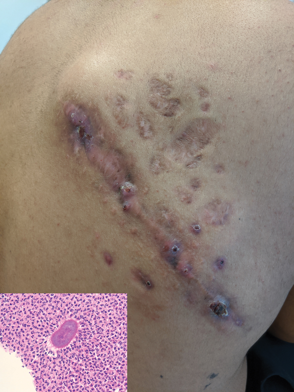
Even healthy Black and Hispanic women have more cesareans than White women
New research offers more insight into potentially dangerous racial disparities in cesarean deliveries: In first-time live births, healthy African-American and Hispanic mothers were 21% and 26% more likely than White mothers, respectively, to deliver by cesarean section despite being low risk. The higher number of cesareans appeared to boost their risk of morbidity.
“A 20% increased odds of cesarean among otherwise healthy, low-risk, nulliparous individuals at term – with limited medical or obstetric explanation – is a significant concern, especially when considering that cesarean is the most common surgical procedure in the U.S.,” said study author Michelle P. Debbink, MD, PhD, an assistant professor with the department of obstetrics and gynecology at the University of Utah, in an interview.
Dr. Debbink and colleagues launched the study, published in the Jan. 2022 issue of Obstetrics & Gynecology, to better understand the racial gap in cesarean sections, which are considered riskier than vaginal deliveries. “Several studies have shown that Black women undergo cesarean more frequently than non-Hispanic White women. Numerous studies also show that Hispanic/Latina women undergo cesarean more frequently than White women, although these data are a bit more mixed,” she said. “What we don’t know, however, is why these differences occur and whether there are clues in the data which can point us toward interventions to close the gap.”
One theory, she said, is that Black and Hispanic women have more comorbidities and therefore more cesareans. To test that idea, the researchers found a cohort of healthy women in a randomized trial that studied the induction of labor.
For the study, they focused on 5,759 women (24.3% Black, 30% Hispanic, 46.6% White). Major differences between the groups included maternal age (average = 21 for Black, 22 for Hispanic, and 26 for White, P < .001), private insurance (17% for Black and Hispanic, 75% for White; P < .001), and full or part-time employment (37% for Black, 31% for Hispanic, and 71% for White; P < .001).
A total of 1,158 of the women (20.1%) underwent cesarean deliveries, accounting for 23% of deliveries by Black women, 22.8% of those by Hispanic women, and 17.6% of those by White women (P < .001). Black women were 21% more likely than White women to give birth via cesarean (adjusted relative risk = 1.21, 95% CI: 1.03-1.42) and Hispanic women were 26% more likely (aRR = 1.26, 95% CI: 1.08-1.46).
The study doesn’t explore why Black and Hispanic women have more cesarean deliveries. However, Dr. Debbink said, “we hypothesize that the difference likely stems more from differing treatment of Black or Hispanic individuals during labor.” It’s unlikely, she said, that these women are more likely to prefer cesarean sections. For one thing, she said, other research hasn’t shown a difference in preferences by race.
The researchers also analyzed maternal morbidity, defined as “transfusion of 4 or more units of red blood cells, any transfusion of other products, postpartum infection, intensive care unit admission, hysterectomy, venous thromboembolism, or maternal death.”
The study found that while few women (2.3%) suffered from morbidity, Black (aRR = 2.05, 95% CI: 1.21-3.47) and Hispanic (aRR = 1.92, 95% CI: 1.17-3.14) women were more likely to suffer from it than White women.
The researchers report that “cesarean birth accounted for an estimated 15.8% (95% CI: 2.1%-48.7%) and 16.5% (95% CI: 4.0%-44.0%) of excess maternal morbidity among non-Hispanic Black and Hispanic people, respectively.”
“Both endometritis and wound complications are much more common among individuals with cesarean, and blood clots, hysterectomy, and ICU admission are also more common after cesarean compared with vaginal delivery,” Dr. Debbink said.
The message of the study, she said, is that the health care system “perpetuates gaps in cesarean delivery for Black and Hispanic individuals compared to White individuals” even in low-risk, first-time live births. “We do not yet know exactly what the right levers are to address this gap, but it is important that we ob-gyns examine our practice patterns and our hospitals’ practice patterns to ensure equity for all our patients.”
Rebecca Delafield, PhD, an assistant professor of Native Hawaiian Health at the University of Hawaii, praised the study as “well-conducted” in an interview. “I agree with the assessment that while the cesarean delivery accounts for a modest proportion of excess morbidity in this study, the impact at the population level is significant,” said Dr. Delafield, who studies health disparities and didn’t take part in the study. “Delivery is complex and the causes of disparities observed are likely multifactorial, therefore research such as this is necessary and compelling.”
She added: “It is becoming increasingly evident that studies investigating racial/ethnic disparities in cesarean delivery and other maternal health outcomes must look beyond maternal behavioral or medical risk factors – e.g., obesity or hypertension – and consider the contribution of a broader set of factors, including societal prejudices.”
The study is funded by the Eunice Kennedy Shriver National Institute of Child Health and Human Development and the National Center for Advancing Translational Sciences. One study author reports funding from GlaxoSmithKline, Pfizer, Moderna, and UpToDate (contributor) and from the Centers for Disease Control and Prevention (via her institution). Dr. Debbink, the other authors, and Dr. Delafield report no disclosures.
New research offers more insight into potentially dangerous racial disparities in cesarean deliveries: In first-time live births, healthy African-American and Hispanic mothers were 21% and 26% more likely than White mothers, respectively, to deliver by cesarean section despite being low risk. The higher number of cesareans appeared to boost their risk of morbidity.
“A 20% increased odds of cesarean among otherwise healthy, low-risk, nulliparous individuals at term – with limited medical or obstetric explanation – is a significant concern, especially when considering that cesarean is the most common surgical procedure in the U.S.,” said study author Michelle P. Debbink, MD, PhD, an assistant professor with the department of obstetrics and gynecology at the University of Utah, in an interview.
Dr. Debbink and colleagues launched the study, published in the Jan. 2022 issue of Obstetrics & Gynecology, to better understand the racial gap in cesarean sections, which are considered riskier than vaginal deliveries. “Several studies have shown that Black women undergo cesarean more frequently than non-Hispanic White women. Numerous studies also show that Hispanic/Latina women undergo cesarean more frequently than White women, although these data are a bit more mixed,” she said. “What we don’t know, however, is why these differences occur and whether there are clues in the data which can point us toward interventions to close the gap.”
One theory, she said, is that Black and Hispanic women have more comorbidities and therefore more cesareans. To test that idea, the researchers found a cohort of healthy women in a randomized trial that studied the induction of labor.
For the study, they focused on 5,759 women (24.3% Black, 30% Hispanic, 46.6% White). Major differences between the groups included maternal age (average = 21 for Black, 22 for Hispanic, and 26 for White, P < .001), private insurance (17% for Black and Hispanic, 75% for White; P < .001), and full or part-time employment (37% for Black, 31% for Hispanic, and 71% for White; P < .001).
A total of 1,158 of the women (20.1%) underwent cesarean deliveries, accounting for 23% of deliveries by Black women, 22.8% of those by Hispanic women, and 17.6% of those by White women (P < .001). Black women were 21% more likely than White women to give birth via cesarean (adjusted relative risk = 1.21, 95% CI: 1.03-1.42) and Hispanic women were 26% more likely (aRR = 1.26, 95% CI: 1.08-1.46).
The study doesn’t explore why Black and Hispanic women have more cesarean deliveries. However, Dr. Debbink said, “we hypothesize that the difference likely stems more from differing treatment of Black or Hispanic individuals during labor.” It’s unlikely, she said, that these women are more likely to prefer cesarean sections. For one thing, she said, other research hasn’t shown a difference in preferences by race.
The researchers also analyzed maternal morbidity, defined as “transfusion of 4 or more units of red blood cells, any transfusion of other products, postpartum infection, intensive care unit admission, hysterectomy, venous thromboembolism, or maternal death.”
The study found that while few women (2.3%) suffered from morbidity, Black (aRR = 2.05, 95% CI: 1.21-3.47) and Hispanic (aRR = 1.92, 95% CI: 1.17-3.14) women were more likely to suffer from it than White women.
The researchers report that “cesarean birth accounted for an estimated 15.8% (95% CI: 2.1%-48.7%) and 16.5% (95% CI: 4.0%-44.0%) of excess maternal morbidity among non-Hispanic Black and Hispanic people, respectively.”
“Both endometritis and wound complications are much more common among individuals with cesarean, and blood clots, hysterectomy, and ICU admission are also more common after cesarean compared with vaginal delivery,” Dr. Debbink said.
The message of the study, she said, is that the health care system “perpetuates gaps in cesarean delivery for Black and Hispanic individuals compared to White individuals” even in low-risk, first-time live births. “We do not yet know exactly what the right levers are to address this gap, but it is important that we ob-gyns examine our practice patterns and our hospitals’ practice patterns to ensure equity for all our patients.”
Rebecca Delafield, PhD, an assistant professor of Native Hawaiian Health at the University of Hawaii, praised the study as “well-conducted” in an interview. “I agree with the assessment that while the cesarean delivery accounts for a modest proportion of excess morbidity in this study, the impact at the population level is significant,” said Dr. Delafield, who studies health disparities and didn’t take part in the study. “Delivery is complex and the causes of disparities observed are likely multifactorial, therefore research such as this is necessary and compelling.”
She added: “It is becoming increasingly evident that studies investigating racial/ethnic disparities in cesarean delivery and other maternal health outcomes must look beyond maternal behavioral or medical risk factors – e.g., obesity or hypertension – and consider the contribution of a broader set of factors, including societal prejudices.”
The study is funded by the Eunice Kennedy Shriver National Institute of Child Health and Human Development and the National Center for Advancing Translational Sciences. One study author reports funding from GlaxoSmithKline, Pfizer, Moderna, and UpToDate (contributor) and from the Centers for Disease Control and Prevention (via her institution). Dr. Debbink, the other authors, and Dr. Delafield report no disclosures.
New research offers more insight into potentially dangerous racial disparities in cesarean deliveries: In first-time live births, healthy African-American and Hispanic mothers were 21% and 26% more likely than White mothers, respectively, to deliver by cesarean section despite being low risk. The higher number of cesareans appeared to boost their risk of morbidity.
“A 20% increased odds of cesarean among otherwise healthy, low-risk, nulliparous individuals at term – with limited medical or obstetric explanation – is a significant concern, especially when considering that cesarean is the most common surgical procedure in the U.S.,” said study author Michelle P. Debbink, MD, PhD, an assistant professor with the department of obstetrics and gynecology at the University of Utah, in an interview.
Dr. Debbink and colleagues launched the study, published in the Jan. 2022 issue of Obstetrics & Gynecology, to better understand the racial gap in cesarean sections, which are considered riskier than vaginal deliveries. “Several studies have shown that Black women undergo cesarean more frequently than non-Hispanic White women. Numerous studies also show that Hispanic/Latina women undergo cesarean more frequently than White women, although these data are a bit more mixed,” she said. “What we don’t know, however, is why these differences occur and whether there are clues in the data which can point us toward interventions to close the gap.”
One theory, she said, is that Black and Hispanic women have more comorbidities and therefore more cesareans. To test that idea, the researchers found a cohort of healthy women in a randomized trial that studied the induction of labor.
For the study, they focused on 5,759 women (24.3% Black, 30% Hispanic, 46.6% White). Major differences between the groups included maternal age (average = 21 for Black, 22 for Hispanic, and 26 for White, P < .001), private insurance (17% for Black and Hispanic, 75% for White; P < .001), and full or part-time employment (37% for Black, 31% for Hispanic, and 71% for White; P < .001).
A total of 1,158 of the women (20.1%) underwent cesarean deliveries, accounting for 23% of deliveries by Black women, 22.8% of those by Hispanic women, and 17.6% of those by White women (P < .001). Black women were 21% more likely than White women to give birth via cesarean (adjusted relative risk = 1.21, 95% CI: 1.03-1.42) and Hispanic women were 26% more likely (aRR = 1.26, 95% CI: 1.08-1.46).
The study doesn’t explore why Black and Hispanic women have more cesarean deliveries. However, Dr. Debbink said, “we hypothesize that the difference likely stems more from differing treatment of Black or Hispanic individuals during labor.” It’s unlikely, she said, that these women are more likely to prefer cesarean sections. For one thing, she said, other research hasn’t shown a difference in preferences by race.
The researchers also analyzed maternal morbidity, defined as “transfusion of 4 or more units of red blood cells, any transfusion of other products, postpartum infection, intensive care unit admission, hysterectomy, venous thromboembolism, or maternal death.”
The study found that while few women (2.3%) suffered from morbidity, Black (aRR = 2.05, 95% CI: 1.21-3.47) and Hispanic (aRR = 1.92, 95% CI: 1.17-3.14) women were more likely to suffer from it than White women.
The researchers report that “cesarean birth accounted for an estimated 15.8% (95% CI: 2.1%-48.7%) and 16.5% (95% CI: 4.0%-44.0%) of excess maternal morbidity among non-Hispanic Black and Hispanic people, respectively.”
“Both endometritis and wound complications are much more common among individuals with cesarean, and blood clots, hysterectomy, and ICU admission are also more common after cesarean compared with vaginal delivery,” Dr. Debbink said.
The message of the study, she said, is that the health care system “perpetuates gaps in cesarean delivery for Black and Hispanic individuals compared to White individuals” even in low-risk, first-time live births. “We do not yet know exactly what the right levers are to address this gap, but it is important that we ob-gyns examine our practice patterns and our hospitals’ practice patterns to ensure equity for all our patients.”
Rebecca Delafield, PhD, an assistant professor of Native Hawaiian Health at the University of Hawaii, praised the study as “well-conducted” in an interview. “I agree with the assessment that while the cesarean delivery accounts for a modest proportion of excess morbidity in this study, the impact at the population level is significant,” said Dr. Delafield, who studies health disparities and didn’t take part in the study. “Delivery is complex and the causes of disparities observed are likely multifactorial, therefore research such as this is necessary and compelling.”
She added: “It is becoming increasingly evident that studies investigating racial/ethnic disparities in cesarean delivery and other maternal health outcomes must look beyond maternal behavioral or medical risk factors – e.g., obesity or hypertension – and consider the contribution of a broader set of factors, including societal prejudices.”
The study is funded by the Eunice Kennedy Shriver National Institute of Child Health and Human Development and the National Center for Advancing Translational Sciences. One study author reports funding from GlaxoSmithKline, Pfizer, Moderna, and UpToDate (contributor) and from the Centers for Disease Control and Prevention (via her institution). Dr. Debbink, the other authors, and Dr. Delafield report no disclosures.
FROM OBSTETRICS & GYNECOLOGY
Atopic dermatitis can be especially burdensome in the elderly
During the Revolutionizing Atopic Dermatitis virtual symposium, Katrina Abuabara, MD, highlighted the epidemiology and burden of AD among older adults. She began by noting that the disease peaks in infancy and older adulthood. In an analysis that she and her colleagues made of physician-diagnosed AD among more than 8.6 million patients in the United Kingdom between 1994 and 2013, the mean prevalence in a given year was 12.3% among those aged 0-17 years, 5.1% among those age 18-74 years, and 8.7% among those age 75 and older.

“We saw what we expected in early infancy with very high rates of active disease,” said Dr. Abuabara, associate professor of dermatology and epidemiology at the University of California, San Francisco. “We also saw a second peak in older adulthood. This was more surprising to us because the disease hadn’t been as well studied in this population.” Researchers who analyzed data from the Global Burden of Disease Study, which evaluates disease-related morbidity and mortality worldwide, found a somewhat attenuated peak but a similar trend around the world. Its authors ranked AD as 15th among all nonfatal diseases.
In a separate analysis, Dr. Abuabara and colleagues evaluated records of more than 9.1 million primary care patients in the United Kingdom between 1994 and 2013, and who were followed for an average of 6 years. They examined AD activity and found that, based on doctor visits and prescriptions, AD appeared to be active in 48% of those aged 0-17 years, compared with 42% of those aged 18-74 years, and 60% of those aged 75 years and older. “Also, when we looked at the distribution of active disease in older adults, we saw that those who were older had more severe disease,” she said. When they evaluated the prevalence of AD by sociodemographic factors, AD increased with age among older adults (adjusted odd ratio, 1.06), while it decreased by 14% annually among children. In addition, female older adults had about three-fourths the odds of prevalent disease as their male counterparts (aOR, 0.73).
“We also looked at rural and urban differences and found that across ages it was more common in urban as compared to rural populations,” she said. “As for socioeconomic status, it tends to be more common among those of higher socioeconomic status in children and in the older adult group.”
In a study that drew from medical records of 3.85 million primary care patients in the United Kingdom, AD was more common in Asian and Black ethnic groups than in people of White ethnicity. In addition, higher socioeconomic status was associated with a greater incidence of eczema in infants aged younger than 2 years, but the reverse was seen for all other age groups.
To identify subtypes of atopic eczema based on patterns of disease activity through mid-adulthood, Dr. Abuabara and colleagues evaluated members of two population-based birth cohorts: the 1958 National Childhood Development Study and the 1970 British Cohort Study. The patients were classified into one of four patters of disease activity followed to age 50: rare/none, increasing, decreasing, and high. “We found that there was the early-onset decreasing subgroup, which tend to have a lower probability of AD over time,” Dr. Abuabara said. “We also found that there was a small subgroup that had a constant high probability of AD over time. But we were surprised to find a subgroup with increasing probability over time. This was a fairly sizable subgroup.”
In an earlier study, she and her colleagues examined whether there were differences based on whether people had adult-onset or childhood-onset disease in the same two cohorts of U.K. patients. Those with childhood-onset disease had stronger associations with known genetic risk factors and they tended to be of higher socioeconomic status. “They also tended to have more asthma and other allergic comorbidities,” Dr. Abuabara said. “On the other hand, the adult-onset group [after age 23] were more likely to be female, more likely to be smokers, and tended to have lower childhood socioeconomic status.”
According to the best available evidence, she continued, there is good data on higher relative risk of osteoporosis/fractures and dementia specifically among older adults with AD, and good data on associations with cardiometabolic disease and atopic disease among adults overall, as well as data showing that AD does not seem to be associated with cancer overall. In a study conducted by Jonathan I. Silverberg, MD, PhD, MPH, and Mohammed S. Shaheen, JD, the researchers used physician-diagnosed AD to investigate the associations of osteopenia and osteoporosis in two large U.S. databases: the 2006-2012 Nationwide Emergency Department Sample (NEDS) database and 2002-2012 National Inpatient Sample (NIS). Among patients aged 50 years and older, AD was associated with a higher odds of osteoporosis in NEDS (aOR, 1.31) and NIS (aOR, 1.25) and osteopenia in NEDS (aOR, 1.86).
In a separate matched cohort study, Dr. Abuabara and colleagues used U.K. primary care patient data to evaluate the association between AD and fracture and whether fracture risk varies with AD severity. Overall, they observed a 10% increase in fracture risk among people with AD, compared with those without, especially those of the hip, spine, pelvis, and wrist. “We found that there was a dose-response effect,” she said. “Those with more severe eczema had a much higher risk of fractures. When we looked at different age groups, we found a similar increased risk in the oldest adults as in younger adults.”
In a longitudinal cohort study of primary care medical records from more than 1.1 million individuals in the United Kingdom, AD was associated with an increased risk of vascular dementia (hazard ratio, 1.88), Alzheimer’s disease (HR, 1.69, and other/unspecified dementia (HR, 1.48; .269). “We found a nice dose response, where people with more severe AD had higher rates of dementia,” Dr. Abuabara said. Results from a more recent, smaller study of patients in Taiwan also found an increased risk between AD and the risk of dementia, but not a dose-response effect, likely because of a much smaller sample size.
Mounting research suggests that the risk for cardiovascular disease is also elevated in patients with AD. “There is some variability in the literature, but I think it’s important that when we’re talking about atopic dermatitis to think about the heterogeneity of the disease,” Dr. Abuabara said. In a meta-analysis and systematic review of 19 studies on the topic, she and her colleagues found that AD was associated with an increased risk of myocardial infarction (relative risk, 1.12), stroke (RR, 1.10), ischemic stroke (RR, 1.17), angina (RR, 1.18), and heart failure (RR, 1.26). “For all the different [cardiovascular disease] outcomes there was increasing risk with increasing disease severity,” she said.
She reported that UCSF receives research funding from Pfizer and Cosmetique Active International. She also receives consulting fees from Target RWE.
During the Revolutionizing Atopic Dermatitis virtual symposium, Katrina Abuabara, MD, highlighted the epidemiology and burden of AD among older adults. She began by noting that the disease peaks in infancy and older adulthood. In an analysis that she and her colleagues made of physician-diagnosed AD among more than 8.6 million patients in the United Kingdom between 1994 and 2013, the mean prevalence in a given year was 12.3% among those aged 0-17 years, 5.1% among those age 18-74 years, and 8.7% among those age 75 and older.
“We saw what we expected in early infancy with very high rates of active disease,” said Dr. Abuabara, associate professor of dermatology and epidemiology at the University of California, San Francisco. “We also saw a second peak in older adulthood. This was more surprising to us because the disease hadn’t been as well studied in this population.” Researchers who analyzed data from the Global Burden of Disease Study, which evaluates disease-related morbidity and mortality worldwide, found a somewhat attenuated peak but a similar trend around the world. Its authors ranked AD as 15th among all nonfatal diseases.
In a separate analysis, Dr. Abuabara and colleagues evaluated records of more than 9.1 million primary care patients in the United Kingdom between 1994 and 2013, and who were followed for an average of 6 years. They examined AD activity and found that, based on doctor visits and prescriptions, AD appeared to be active in 48% of those aged 0-17 years, compared with 42% of those aged 18-74 years, and 60% of those aged 75 years and older. “Also, when we looked at the distribution of active disease in older adults, we saw that those who were older had more severe disease,” she said. When they evaluated the prevalence of AD by sociodemographic factors, AD increased with age among older adults (adjusted odd ratio, 1.06), while it decreased by 14% annually among children. In addition, female older adults had about three-fourths the odds of prevalent disease as their male counterparts (aOR, 0.73).
“We also looked at rural and urban differences and found that across ages it was more common in urban as compared to rural populations,” she said. “As for socioeconomic status, it tends to be more common among those of higher socioeconomic status in children and in the older adult group.”
In a study that drew from medical records of 3.85 million primary care patients in the United Kingdom, AD was more common in Asian and Black ethnic groups than in people of White ethnicity. In addition, higher socioeconomic status was associated with a greater incidence of eczema in infants aged younger than 2 years, but the reverse was seen for all other age groups.
To identify subtypes of atopic eczema based on patterns of disease activity through mid-adulthood, Dr. Abuabara and colleagues evaluated members of two population-based birth cohorts: the 1958 National Childhood Development Study and the 1970 British Cohort Study. The patients were classified into one of four patters of disease activity followed to age 50: rare/none, increasing, decreasing, and high. “We found that there was the early-onset decreasing subgroup, which tend to have a lower probability of AD over time,” Dr. Abuabara said. “We also found that there was a small subgroup that had a constant high probability of AD over time. But we were surprised to find a subgroup with increasing probability over time. This was a fairly sizable subgroup.”
In an earlier study, she and her colleagues examined whether there were differences based on whether people had adult-onset or childhood-onset disease in the same two cohorts of U.K. patients. Those with childhood-onset disease had stronger associations with known genetic risk factors and they tended to be of higher socioeconomic status. “They also tended to have more asthma and other allergic comorbidities,” Dr. Abuabara said. “On the other hand, the adult-onset group [after age 23] were more likely to be female, more likely to be smokers, and tended to have lower childhood socioeconomic status.”
According to the best available evidence, she continued, there is good data on higher relative risk of osteoporosis/fractures and dementia specifically among older adults with AD, and good data on associations with cardiometabolic disease and atopic disease among adults overall, as well as data showing that AD does not seem to be associated with cancer overall. In a study conducted by Jonathan I. Silverberg, MD, PhD, MPH, and Mohammed S. Shaheen, JD, the researchers used physician-diagnosed AD to investigate the associations of osteopenia and osteoporosis in two large U.S. databases: the 2006-2012 Nationwide Emergency Department Sample (NEDS) database and 2002-2012 National Inpatient Sample (NIS). Among patients aged 50 years and older, AD was associated with a higher odds of osteoporosis in NEDS (aOR, 1.31) and NIS (aOR, 1.25) and osteopenia in NEDS (aOR, 1.86).
In a separate matched cohort study, Dr. Abuabara and colleagues used U.K. primary care patient data to evaluate the association between AD and fracture and whether fracture risk varies with AD severity. Overall, they observed a 10% increase in fracture risk among people with AD, compared with those without, especially those of the hip, spine, pelvis, and wrist. “We found that there was a dose-response effect,” she said. “Those with more severe eczema had a much higher risk of fractures. When we looked at different age groups, we found a similar increased risk in the oldest adults as in younger adults.”
In a longitudinal cohort study of primary care medical records from more than 1.1 million individuals in the United Kingdom, AD was associated with an increased risk of vascular dementia (hazard ratio, 1.88), Alzheimer’s disease (HR, 1.69, and other/unspecified dementia (HR, 1.48; .269). “We found a nice dose response, where people with more severe AD had higher rates of dementia,” Dr. Abuabara said. Results from a more recent, smaller study of patients in Taiwan also found an increased risk between AD and the risk of dementia, but not a dose-response effect, likely because of a much smaller sample size.
Mounting research suggests that the risk for cardiovascular disease is also elevated in patients with AD. “There is some variability in the literature, but I think it’s important that when we’re talking about atopic dermatitis to think about the heterogeneity of the disease,” Dr. Abuabara said. In a meta-analysis and systematic review of 19 studies on the topic, she and her colleagues found that AD was associated with an increased risk of myocardial infarction (relative risk, 1.12), stroke (RR, 1.10), ischemic stroke (RR, 1.17), angina (RR, 1.18), and heart failure (RR, 1.26). “For all the different [cardiovascular disease] outcomes there was increasing risk with increasing disease severity,” she said.
She reported that UCSF receives research funding from Pfizer and Cosmetique Active International. She also receives consulting fees from Target RWE.
During the Revolutionizing Atopic Dermatitis virtual symposium, Katrina Abuabara, MD, highlighted the epidemiology and burden of AD among older adults. She began by noting that the disease peaks in infancy and older adulthood. In an analysis that she and her colleagues made of physician-diagnosed AD among more than 8.6 million patients in the United Kingdom between 1994 and 2013, the mean prevalence in a given year was 12.3% among those aged 0-17 years, 5.1% among those age 18-74 years, and 8.7% among those age 75 and older.
“We saw what we expected in early infancy with very high rates of active disease,” said Dr. Abuabara, associate professor of dermatology and epidemiology at the University of California, San Francisco. “We also saw a second peak in older adulthood. This was more surprising to us because the disease hadn’t been as well studied in this population.” Researchers who analyzed data from the Global Burden of Disease Study, which evaluates disease-related morbidity and mortality worldwide, found a somewhat attenuated peak but a similar trend around the world. Its authors ranked AD as 15th among all nonfatal diseases.
In a separate analysis, Dr. Abuabara and colleagues evaluated records of more than 9.1 million primary care patients in the United Kingdom between 1994 and 2013, and who were followed for an average of 6 years. They examined AD activity and found that, based on doctor visits and prescriptions, AD appeared to be active in 48% of those aged 0-17 years, compared with 42% of those aged 18-74 years, and 60% of those aged 75 years and older. “Also, when we looked at the distribution of active disease in older adults, we saw that those who were older had more severe disease,” she said. When they evaluated the prevalence of AD by sociodemographic factors, AD increased with age among older adults (adjusted odd ratio, 1.06), while it decreased by 14% annually among children. In addition, female older adults had about three-fourths the odds of prevalent disease as their male counterparts (aOR, 0.73).
“We also looked at rural and urban differences and found that across ages it was more common in urban as compared to rural populations,” she said. “As for socioeconomic status, it tends to be more common among those of higher socioeconomic status in children and in the older adult group.”
In a study that drew from medical records of 3.85 million primary care patients in the United Kingdom, AD was more common in Asian and Black ethnic groups than in people of White ethnicity. In addition, higher socioeconomic status was associated with a greater incidence of eczema in infants aged younger than 2 years, but the reverse was seen for all other age groups.
To identify subtypes of atopic eczema based on patterns of disease activity through mid-adulthood, Dr. Abuabara and colleagues evaluated members of two population-based birth cohorts: the 1958 National Childhood Development Study and the 1970 British Cohort Study. The patients were classified into one of four patters of disease activity followed to age 50: rare/none, increasing, decreasing, and high. “We found that there was the early-onset decreasing subgroup, which tend to have a lower probability of AD over time,” Dr. Abuabara said. “We also found that there was a small subgroup that had a constant high probability of AD over time. But we were surprised to find a subgroup with increasing probability over time. This was a fairly sizable subgroup.”
In an earlier study, she and her colleagues examined whether there were differences based on whether people had adult-onset or childhood-onset disease in the same two cohorts of U.K. patients. Those with childhood-onset disease had stronger associations with known genetic risk factors and they tended to be of higher socioeconomic status. “They also tended to have more asthma and other allergic comorbidities,” Dr. Abuabara said. “On the other hand, the adult-onset group [after age 23] were more likely to be female, more likely to be smokers, and tended to have lower childhood socioeconomic status.”
According to the best available evidence, she continued, there is good data on higher relative risk of osteoporosis/fractures and dementia specifically among older adults with AD, and good data on associations with cardiometabolic disease and atopic disease among adults overall, as well as data showing that AD does not seem to be associated with cancer overall. In a study conducted by Jonathan I. Silverberg, MD, PhD, MPH, and Mohammed S. Shaheen, JD, the researchers used physician-diagnosed AD to investigate the associations of osteopenia and osteoporosis in two large U.S. databases: the 2006-2012 Nationwide Emergency Department Sample (NEDS) database and 2002-2012 National Inpatient Sample (NIS). Among patients aged 50 years and older, AD was associated with a higher odds of osteoporosis in NEDS (aOR, 1.31) and NIS (aOR, 1.25) and osteopenia in NEDS (aOR, 1.86).
In a separate matched cohort study, Dr. Abuabara and colleagues used U.K. primary care patient data to evaluate the association between AD and fracture and whether fracture risk varies with AD severity. Overall, they observed a 10% increase in fracture risk among people with AD, compared with those without, especially those of the hip, spine, pelvis, and wrist. “We found that there was a dose-response effect,” she said. “Those with more severe eczema had a much higher risk of fractures. When we looked at different age groups, we found a similar increased risk in the oldest adults as in younger adults.”
In a longitudinal cohort study of primary care medical records from more than 1.1 million individuals in the United Kingdom, AD was associated with an increased risk of vascular dementia (hazard ratio, 1.88), Alzheimer’s disease (HR, 1.69, and other/unspecified dementia (HR, 1.48; .269). “We found a nice dose response, where people with more severe AD had higher rates of dementia,” Dr. Abuabara said. Results from a more recent, smaller study of patients in Taiwan also found an increased risk between AD and the risk of dementia, but not a dose-response effect, likely because of a much smaller sample size.
Mounting research suggests that the risk for cardiovascular disease is also elevated in patients with AD. “There is some variability in the literature, but I think it’s important that when we’re talking about atopic dermatitis to think about the heterogeneity of the disease,” Dr. Abuabara said. In a meta-analysis and systematic review of 19 studies on the topic, she and her colleagues found that AD was associated with an increased risk of myocardial infarction (relative risk, 1.12), stroke (RR, 1.10), ischemic stroke (RR, 1.17), angina (RR, 1.18), and heart failure (RR, 1.26). “For all the different [cardiovascular disease] outcomes there was increasing risk with increasing disease severity,” she said.
She reported that UCSF receives research funding from Pfizer and Cosmetique Active International. She also receives consulting fees from Target RWE.
FROM REVOLUTIONIZING AD 2021