User login
Lens-free microscope a ‘milestone’

Credit: Максим Кукушкин
Researchers say they’ve developed a lens-free microscope that can detect the presence of cell-level abnormalities with the same accuracy as larger and more expensive optical microscopes.
The invention could lead to less expensive, more portable technology for performing examinations of tissue, blood, and other biomedical specimens, according to the researchers.
It may prove especially useful in remote areas and when large numbers of samples need to be examined quickly.
Aydogan Ozcan, PhD, of the University of California, Los Angeles, and his colleagues described their work with the microscope in Science Translational Medicine.
“This is a milestone in the work we’ve been doing,” Dr Ozcan said. “This is the first time tissue samples have been imaged in 3D using a lens-free, on-chip microscope.”
The device works by using a laser or light-emitting-diode to illuminate a tissue or blood sample that has been placed on a slide and inserted into the device. A sensor array on a microchip captures and records the pattern of shadows created by the sample.
The device processes these patterns as a series of holograms, forming 3-D images of the specimen and giving medical personnel a virtual depth-of-field view. An algorithm color codes the reconstructed images, making the contrasts in the samples more apparent than they would be in the holograms and making any abnormalities easier to detect.
Dr Ozcan’s team tested the device using blood samples containing sickle cell anemia, Pap smears that indicated cervical cancer, and tissue specimens containing cancerous breast cells.
In a blind test, a board-certified pathologist analyzed sets of specimen images that had been created by the lens-free technology and by conventional microscopes. The pathologist’s diagnoses using the lens-free microscopic images proved accurate 99% of the time.
Another benefit of the lens-free device, according to the researchers, is that it produces images that are several hundred times larger in area, or field of view, than those captured by conventional bright-field optical microscopes. This makes it possible to process specimens more quickly.
“While mobile healthcare has expanded rapidly with the growth of consumer electronics—cellphones in particular—pathology is still, by and large, constrained to advanced clinical laboratory settings,” Dr Ozcan said. “Accompanied by advances in its graphical user interface, this platform could scale up for use in clinical, biomedical, scientific, educational, and citizen-science applications, among others.” ![]()
Study shows higher risk of MDS, leukemia after breast cancer

chemotherapy
Credit: Rhoda Baer
The risk of developing myelodysplastic syndromes (MDS) or leukemias after treatment for early stage breast cancer is higher than previously reported, according to a study published in the Journal of Clinical Oncology.
Data from earlier studies showed that about 0.25% of breast cancer patients develop MDS or leukemia as a late effect of chemotherapy, said Judith Karp, MD, of the Johns Hopkins University School of Medicine in Baltimore, Maryland.
But Dr Karp and her colleagues found the 10-year incidence of MDS and leukemia among breast cancer patients to be about 0.5%.
“[T]he cumulative risk over a decade is now shown to be twice as high as we thought it was, and that risk doesn’t seem to slow down 5 years after treatment,” Dr Karp said. “Most medical oncologists have come to think that the risk is early and short-lived. So this was a little bit of a wake-up call that we are not seeing any plateau of that risk, and it is higher.”
Dr Karp and her colleagues reviewed data on 20,063 breast cancer patients treated at 8 US cancer centers between 1998 and 2007 whose cancer recurrence and secondary cancer rates were recorded in a database kept by the National Comprehensive Cancer Network.
At a median follow-up of 5.1 years, 50 patients had developed a marrow neoplasm, including acute myeloid leukemia (n=24), MDS/acute myeloid leukemia (n=15), chronic lymphocytic leukemia/small lymphocytic lymphoma (n=5), chronic myeloid leukemia (n=3), or acute lymphoblastic leukemia (n=3).
The risk of developing MDS/leukemia was about 7 times higher for patients who underwent surgery and received chemotherapy, compared to patients who did not receive chemotherapy. For patients who underwent surgery and received both chemotherapy and radiation, the risk was about 8 times higher.
The MDS/leukemia rates per 1000 person-years were 0.16 for surgery, 0.43 for surgery plus radiation, 0.46 for surgery plus chemotherapy, and 0.54 for all 3 modalities.
The cumulative incidence of MDS/leukemia doubled between years 5 and 10, rising from 0.24% to 0.48%. And only 9% of patients were alive at 10 years.
Antonio Wolff, MD, of the Johns Hopkins University School of Medicine, said this study could help early stage breast cancer patients and their physicians think more carefully about the use of preventive or adjuvant chemotherapy, especially when patients have a low risk of cancer recurrence.
“Our study provides useful information for physicians and patients to consider a potential downside of preventive or adjuvant chemotherapy in patients with very low risk of breast cancer recurrence,” he said.
“It could be a false and dangerous security blanket to some patients by exposing them to a small risk of serious late effects with little or no real benefit from the treatment.”
The researchers included a hypothetical case to put the risks of early stage breast cancer and its treatment in perspective. They described a 60-year-old woman in average health who was diagnosed with stage 1 breast cancer that was rapidly growing and estrogen receptor-positive.
The patient had an estimated 12.3% risk of dying of breast cancer after 10 years. She could improve her 10-year survival rate by 1.8% with 4 cycles of chemotherapy, but she would also increase her risk of MDS/leukemia over that same time by 0.5%.
Dr Wolff noted that it’s unclear whether the increased risk of MDS/leukemia after postsurgical chemotherapy applies to patients with other kinds of solid tumors, as the drug regimens used in breast cancer differ from those used for other cancers. ![]()
Care as a Continuum
Patients who are hospitalized for an acute event often have a range of prior outpatient experiences within the healthcare system, both before and after a hospitalization. In particular, continuity with a primary care provider can influence health outcomes.[1] In this issue of the Journal of Hospital Medicine, Boonyasai et al. found several characteristics of primary care physicians that were associated with whether their hospitalized patients were cared for by hospitalists.[2] Using Medicare claims data from the state of Texas during years 2001 to 2009, the authors calculated the percent of primary care physicians' hospitalized patients who were cared for by hospitalists. Hospitalist use increased overall during the time period, but primary care physicians differed in the rate and extent of hospitalist use. A minority of physicians were early adopters, with the majority of their hospitalized patients cared for by hospitalists during the entire time period. A sizeable group of primary care physicians mostly avoided using hospitalists. Moreover, there was a significant cluster of primary care physicians who, at some point during the study period, rapidly began using hospitalists within a relatively short time.
Several physician characteristics were associated with a greater adoption of the hospitalist model, including being female, in a family practice specialty, or in a rural practice setting. What this study lacks is the ability to explain why some physicians used hospitalists and others did not. It is probable that adoption (or not) of hospitalists is less an individual physician decision and instead reflects a choice of their clinical practice group. If an outpatient practice group or provider can influence whether or not their patients are cared for by hospitalists, it is also conceivable that they can affect hospital‐based outcomes as well. This finding reinforces the importance of examining the care and outcomes of patient care across the continuum of care, rather than focusing on the inpatient or outpatient setting.
As a result of the Affordable Care Act and rising healthcare costs, provider groups are beginning to form accountable care organizations (ACOs). An ACO is partnership between payers and providers to care for a population of patients across the continuum of care. In these arrangements, the providers often take on financial risk for the total cost of care for a population as well as for providing high‐quality care as monitored by specific metrics.[3] The population of patients for which ACOs take risks often include predominantly patients who receive primary care from the group.[4] For overall cost management, given that acute hospitalizations are disproportionately high cost, a primary focus of a majority of ACOs is to reduce unnecessary hospital days. Overall, ACOs that have been successful in the short term in managing costs have done so primarily by reducing overall hospital days.[5] ACOs have started to do so by creating intensive outpatient care management programs for high‐risk patients, by focusing on transitions of care to help decrease readmissions, by working with primary care clinics to transform into patient‐centered medical homes, where same‐day access to care is a priority, and developing other disease‐management tools to keep patients healthy.
To manage hospital utilization, many ACOs have developed plans to transform primary care and shift hospital care to outpatient care through enhanced outpatient case management for complex cases. As the way primary care is delivered changes, it will be very important to understand how this will modify the utilization and impact of hospitalist care on patients. The hope is that these modifications will work synergistically with hospitalist programs.
As the lines between outpatient and inpatient care become increasingly blurred, it may not be fair to attribute hospitalization outcome measures to hospitalists alone, particularly as ACOs are likely to move only the sickest or most difficult to manage patients to the inpatient setting. This may affect hospital‐based quality metrics such as readmissions and mortality. Seamless communication and transfer of information between outpatient and inpatient care will be vital to the success of ACOs.[6] In addition to improved communication, however, some systems may look to hospitalists to staff postdischarge clinics or act as extensivists or ambulatory intensivists to help manage the sickest in the population.[7]
Boonyasi et al. show that primary care physician characteristics as associated with whether or not patients' receive care from hospitalists.[2] As such, it reinforces the concept that providers in part of the continuum of care are integrally tied to care received by patients in different treatment settings. As our healthcare system rapidly transforms over the next few years, it will become more important to understand how outpatient and inpatient providers influence one another's care patterns and how these relationships influence care and cost‐related outcomes for patients.
ACKNOWLEDGMENTS
Disclosure: Nothing to report.
- , . Does continuity of care improve patient outcomes? J Fam Pract. 2004;53(12):974–980.
- et al. Characteristics of primary care providers who adopted the hospitalist model 2001–2009. J Hosp Med.
- . Understanding the new vocabulary of healthcare reform. J Hosp Med. 2010;5:197–199.
- Centers for Medicare 8:472–477.
- Agency for Healthcare Research and Quality. Medical “extensivists” care for high‐acuity patients across settings, leading to reduced hospital use. AHRQ Service Delivery Innovation Profile. Available at: https://innovations.ahrq.gov/profiles/medical‐extensivists‐care‐high‐acuity‐patients‐across‐settings‐leading‐reduced‐hospital‐use. Accessed December 17, 2014.
Patients who are hospitalized for an acute event often have a range of prior outpatient experiences within the healthcare system, both before and after a hospitalization. In particular, continuity with a primary care provider can influence health outcomes.[1] In this issue of the Journal of Hospital Medicine, Boonyasai et al. found several characteristics of primary care physicians that were associated with whether their hospitalized patients were cared for by hospitalists.[2] Using Medicare claims data from the state of Texas during years 2001 to 2009, the authors calculated the percent of primary care physicians' hospitalized patients who were cared for by hospitalists. Hospitalist use increased overall during the time period, but primary care physicians differed in the rate and extent of hospitalist use. A minority of physicians were early adopters, with the majority of their hospitalized patients cared for by hospitalists during the entire time period. A sizeable group of primary care physicians mostly avoided using hospitalists. Moreover, there was a significant cluster of primary care physicians who, at some point during the study period, rapidly began using hospitalists within a relatively short time.
Several physician characteristics were associated with a greater adoption of the hospitalist model, including being female, in a family practice specialty, or in a rural practice setting. What this study lacks is the ability to explain why some physicians used hospitalists and others did not. It is probable that adoption (or not) of hospitalists is less an individual physician decision and instead reflects a choice of their clinical practice group. If an outpatient practice group or provider can influence whether or not their patients are cared for by hospitalists, it is also conceivable that they can affect hospital‐based outcomes as well. This finding reinforces the importance of examining the care and outcomes of patient care across the continuum of care, rather than focusing on the inpatient or outpatient setting.
As a result of the Affordable Care Act and rising healthcare costs, provider groups are beginning to form accountable care organizations (ACOs). An ACO is partnership between payers and providers to care for a population of patients across the continuum of care. In these arrangements, the providers often take on financial risk for the total cost of care for a population as well as for providing high‐quality care as monitored by specific metrics.[3] The population of patients for which ACOs take risks often include predominantly patients who receive primary care from the group.[4] For overall cost management, given that acute hospitalizations are disproportionately high cost, a primary focus of a majority of ACOs is to reduce unnecessary hospital days. Overall, ACOs that have been successful in the short term in managing costs have done so primarily by reducing overall hospital days.[5] ACOs have started to do so by creating intensive outpatient care management programs for high‐risk patients, by focusing on transitions of care to help decrease readmissions, by working with primary care clinics to transform into patient‐centered medical homes, where same‐day access to care is a priority, and developing other disease‐management tools to keep patients healthy.
To manage hospital utilization, many ACOs have developed plans to transform primary care and shift hospital care to outpatient care through enhanced outpatient case management for complex cases. As the way primary care is delivered changes, it will be very important to understand how this will modify the utilization and impact of hospitalist care on patients. The hope is that these modifications will work synergistically with hospitalist programs.
As the lines between outpatient and inpatient care become increasingly blurred, it may not be fair to attribute hospitalization outcome measures to hospitalists alone, particularly as ACOs are likely to move only the sickest or most difficult to manage patients to the inpatient setting. This may affect hospital‐based quality metrics such as readmissions and mortality. Seamless communication and transfer of information between outpatient and inpatient care will be vital to the success of ACOs.[6] In addition to improved communication, however, some systems may look to hospitalists to staff postdischarge clinics or act as extensivists or ambulatory intensivists to help manage the sickest in the population.[7]
Boonyasi et al. show that primary care physician characteristics as associated with whether or not patients' receive care from hospitalists.[2] As such, it reinforces the concept that providers in part of the continuum of care are integrally tied to care received by patients in different treatment settings. As our healthcare system rapidly transforms over the next few years, it will become more important to understand how outpatient and inpatient providers influence one another's care patterns and how these relationships influence care and cost‐related outcomes for patients.
ACKNOWLEDGMENTS
Disclosure: Nothing to report.
- , . Does continuity of care improve patient outcomes? J Fam Pract. 2004;53(12):974–980.
- et al. Characteristics of primary care providers who adopted the hospitalist model 2001–2009. J Hosp Med.
- . Understanding the new vocabulary of healthcare reform. J Hosp Med. 2010;5:197–199.
- Centers for Medicare 8:472–477.
- Agency for Healthcare Research and Quality. Medical “extensivists” care for high‐acuity patients across settings, leading to reduced hospital use. AHRQ Service Delivery Innovation Profile. Available at: https://innovations.ahrq.gov/profiles/medical‐extensivists‐care‐high‐acuity‐patients‐across‐settings‐leading‐reduced‐hospital‐use. Accessed December 17, 2014.
Dashboards and P4P in VTE Prophylaxis
The Affordable Care Act explicitly outlines improving the value of healthcare by increasing quality and decreasing costs. It emphasizes value‐based purchasing, the transparency of performance metrics, and the use of payment incentives to reward quality.[1, 2] Venous thromboembolism (VTE) prophylaxis is one of these publicly reported performance measures. The National Quality Forum recommends that each patient be evaluated on hospital admission and during their hospitalization for VTE risk level and for appropriate thromboprophylaxis to be used, if required.[3] Similarly, the Joint Commission includes appropriate VTE prophylaxis in its Core Measures.[4] Patient experience and performance metrics, including VTE prophylaxis, constitute the hospital value‐based purchasing (VBP) component of healthcare reform.[5] For a hypothetical 327‐bed hospital, an estimated $1.7 million of a hospital's inpatient payments from Medicare will be at risk from VBP alone.[2]
VTE prophylaxis is a common target of quality improvement projects. Effective, safe, and cost‐effective measures to prevent VTE exist, including pharmacologic and mechanical prophylaxis.[6, 7] Despite these measures, compliance rates are often below 50%.[8] Different interventions have been pursued to ensure appropriate VTE prophylaxis, including computerized provider order entry (CPOE), electronic alerts, mandatory VTE risk assessment and prophylaxis, and provider education campaigns.[9] Recent studies show that CPOE systems with mandatory fields can increase VTE prophylaxis rates to above 80%, yet the goal of a high reliability health system is for 100% of patients to receive recommended therapy.[10, 11, 12, 13, 14, 15] Interventions to improve prophylaxis rates that have included multiple strategies, such as computerized order sets, feedback, and education, have been the most effective, increasing compliance to above 90%.[9, 11, 16] These systems can be enhanced with additional interventions such as providing individualized provider education and feedback, understanding of work flow, and ensuring patients receive the prescribed therapies.[12] For example, a physician dashboard could be employed to provide a snapshot and historical trend of key performance indicators using graphical displays and indicators.[17]
Dashboards and pay‐for‐performance programs have been increasingly used to increase the visibility of these metrics, provide feedback, visually display benchmarks and goals, and proactively monitor for achievements and setbacks.[18] Although these strategies are often addressed at departmental (or greater) levels, applying them at the level of the individual provider may assist hospitals in reducing preventable harm and achieving safety and quality goals, especially at higher benchmarks. With their expanding role, hospitalists provide a key opportunity to lead improvement efforts and to study the impact of dashboards and pay‐for performance at the provider level to achieve VTE prophylaxis performance targets. Hospitalists are often the front‐line provider for inpatients and deliver up to 70% of inpatient general medical services.[19] The objective of our study was to evaluate the impact of providing individual provider feedback and employing a pay‐for‐performance program on baseline performance of VTE prophylaxis among hospitalists. We hypothesized that performance feedback through the use of a dashboard would increase appropriate VTE prophylaxis, and this effect would be further augmented by incorporation of a pay‐for‐performance program.
METHODS
Hospitalist Dashboard
In 2010, hospitalist program leaders met with hospital administrators to create a hospitalist dashboard that would provide regularly updated summaries of performance measures for individual hospitalists. The final set of metrics identified included appropriate VTE prophylaxis, length of stay, patients discharged per day, discharges before 3 pm, depth of coding, patient satisfaction, readmissions, communication with the primary care provider, and time to signature for discharge summaries (Figure 1A). The dashboard was introduced at a general hospitalist meeting during which its purpose, methodology, and accessibility were described; it was subsequently implemented in January 2011.

Benchmarks were established for each metric, standardized to establish a scale ranging from 1 through 9, and incorporated into the dashboard (Figure 1A). Higher scores (creating a larger geometric shape) were desirable. For the VTE prophylaxis measure, scores of 1 through 9 corresponded to <60%, 60% to 64.9%, 65% to 69.9%, 70% to 74.9%, 75% to 79.9%, 80% to 84.9%, 85% to 89.9%, 90% to 94.9%, and 95% American College of Chest Physicians (ACCP)‐compliant VTE prophylaxis, respectively.[12, 20] Each provider was able to access the aggregated dashboard (showing the group mean) and his/her individualized dashboard using an individualized login and password for the institutional portal. This portal is used during the provider's workflow, including medical record review and order entry. Both a polygonal summary graphic (Figure 1A) and trend (Figure 1B) view of the dashboard were available to the provider. A comparison of the individual provider to the hospitalist group average was displayed (Figure 1A). At monthly program meetings, the dashboard, group results, and trends were discussed.
Venous Thromboembolism Prophylaxis Compliance
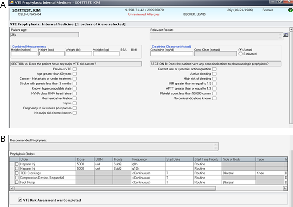
Our study was performed in a tertiary academic medical center with an approximately 20‐member hospitalist group (the precise membership varied over time), whose responsibilities include, among other clinical duties, staffing a 17‐bed general medicine unit with telemetry. The scope of diagnoses and acuity of patients admitted to the hospitalist service is similar to the housestaff services. Some hospitalist faculty serve both as hospitalist and nonhospitalist general medicine service team attendings, but the comparison groups were staffed by hospitalists for <20% of the time. For admissions, all hospitalists use a standardized general medicine admission order set that is integrated into the CPOE system (Sunrise Clinical Manager; Allscripts, Chicago, IL) and completed for all admitted patients. A mandatory VTE risk screen, which includes an assessment of VTE risk factors and pharmacological prophylaxis contraindications, must be completed by the ordering physician as part of this order set (Figure 2A). The system then prompts the provider with a risk‐appropriate VTE prophylaxis recommendation that the provider may subsequently order, including mechanical prophylaxis (Figure 2B). Based on ACCP VTE prevention guidelines, risk‐appropriate prophylaxis was determined using an electronic algorithm that categorized patients into risk categories based on the presence of major VTE risk factors (Figure 2A).[12, 15, 20] If none of these were present, the provider selected No major risk factors known. Both an assessment of current use of anticoagulation and a clinically high risk of bleeding were also included (Figure 2A). If none of these were present, the provider selected No contraindications known. This algorithm is published in detail elsewhere and has been shown to not increase major bleeding episodes.[12, 15] The VTE risk assessment, but not the VTE order itself, was a mandatory field. This allowed the physician discretion to choose among various pharmacological agents and mechanical mechanisms based on patient and physician preferences.
Compliance of risk‐appropriate VTE prophylaxis was determined 24 hours after the admission order set was completed using an automated electronic query of the CPOE system. Low molecular‐weight heparin prescription was included in the compliance algorithm as acceptable prophylaxis. Prescription of pharmacological VTE prophylaxis when a contraindication was present was considered noncompliant. The metric was assigned to the attending physician who billed for the first inpatient encounter.
Pay‐for‐Performance Program
In July 2011, a pay‐for‐performance program was added to the dashboard. All full‐time and part‐time hospitalists were eligible. The financial incentive was determined according to hospital priority and funds available. The VTE prophylaxis metric was prorated by clinical effort, with a maximum of $0.50 per work relative value unit (RVU). To optimize performance, a threshold of 80% compliance had to be surpassed before any payment was made. Progressively increasing percentages of the incentive were earned as compliance increased from 80% to 100%, corresponding to dashboard scores of 6, 7, 8, and 9: <80% (scores 1 to 5)=no payment; 80% to 84.9% (score 6)=$0.125 per RVU; 85% to 89.9% (score 7)=$0.25 per RVU; 90% to 94.9% (score 8)=$0.375 per RVU; and 95% (score 9)=$0.50 per RVU (maximum incentive). Payments were accrued quarterly and paid at the end of the fiscal year as a cumulative, separate performance supplement.
Individualized physician feedback through the dashboard was continued during the pay‐for‐performance period. Average hospitalist group compliance continued to be displayed on the electronic dashboard and was explicitly reviewed at monthly hospitalist meetings.
The VTE prophylaxis order set and data collection and analyses were approved by the Johns Hopkins Medicine Institutional Review Board. The dashboard and pay‐for‐performance program were initiated by the institution as part of a proof of concept quality improvement project.
Analysis
We examined all inpatient admissions to the hospitalist unit from 2008 to 2012. We included patients admitted to and discharged from the hospitalist unit and excluded patients transferred into/out of the unit and encounters with a length of stay <24 hours. VTE prophylaxis orders were queried from the CPOE system 24 hours after the patient was admitted to determine compliance.
After allowing for a run‐in period (2008), we analyzed the change in percent compliance for 3 periods: (1) CPOE‐based VTE order set alone (baseline [BASE], January 2009 to December 2010); (2) group and individual physician feedback using the dashboard (dashboard only [DASH], January to June 2011); and (3) dashboard tied to the pay‐for‐performance program (dashboard with pay‐for‐performance [P4P], July 2011 to December 2012). The CPOE‐based VTE order set was used during all 3 periods. We used the other medical services as a control to ensure that there were no temporal trends toward improved prophylaxis on a service without the intervention. VTE prophylaxis compliance was examined by calculating percent compliance using the same algorithm for the 4 resident‐staffed general medicine service teams at our institution, which utilized the same CPOE system but did not receive the dashboard or pay‐for‐performance interventions. We used locally weighted scatterplot smoothing, a locally weighted regression of percent compliance over time, to graphically display changes in group compliance over time.[21, 22]
We also performed linear regression to assess the rate of change in group compliance and included spline terms that allowed slope to vary for each of the 3 time periods.[23, 24] Clustered analysis accounted for potentially correlated serial measurements of compliance for an individual provider. A separate analysis examined the effect of provider turnover and individual provider improvement during each of the 3 periods. Tests of significance were 2‐sided, with an level of 0.05. Statistical analysis was performed using Stata 12.1 (StataCorp LP, College Station, TX).
RESULTS
Venous Thromboembolism Prophylaxis Compliance
We analyzed 3144 inpatient admissions by 38 hospitalists from 2009 to 2012. The 5 most frequent coded diagnoses were heart failure, acute kidney failure, syncope, pneumonia, and chest pain. Patients had a median length of stay of 3 days [interquartile range: 26]. During the dashboard‐only period, on average, providers improved in compliance by 4% (95% confidence interval [CI]: 35; P<0.001). With the addition of the pay‐for‐performance program, providers improved by an additional 4% (95% CI: 35; P<0.001). Group compliance significantly improved from 86% (95% CI: 8588) during the BASE period of the CPOE‐based VTE order set to 90% (95% CI: 8893) during the DASH period (P=0.01) and 94% (95% CI: 9396) during the subsequent P4P program (P=0.01) (Figure 3). Both inappropriate prophylaxis and lack of prophylaxis, when indicated, resulted in a non‐compliance rating. During the 3 periods, inappropriate prophylaxis decreased from 7.9% to 6.2% to 2.6% during the BASE, DASH, and subsequent P4P periods, respectively. Similarly, lack of prophylaxis when indicated decreased from 6.1% to 3.2% to 3.1% during the BASE, DASH, and subsequent P4P periods, respectively.
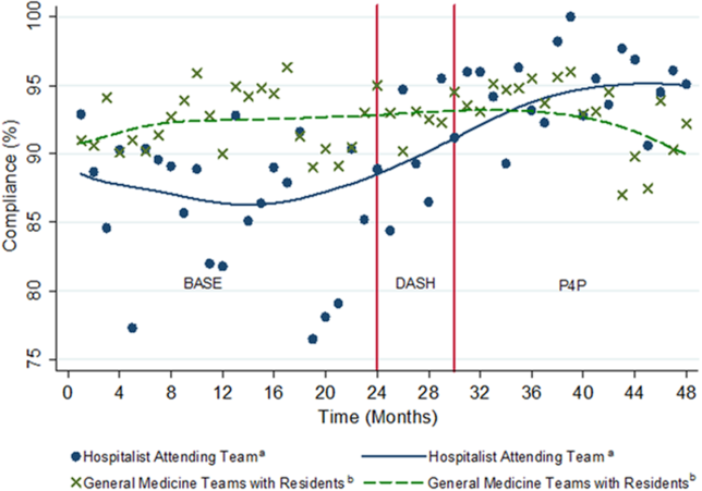
The average compliance of the 4 non‐hospitalist general medicine service teams was initially higher than that of the hospitalist service during the CPOE‐based VTE order set (90%) and DASH (92%) periods, but subsequently plateaued and was exceeded by the hospitalist service during the combined P4P (92%) period (Figure 3). However, there was no statistically significant difference between the general medicine service teams and hospitalist service during the DASH (P=0.15) and subsequent P4P (P=0.76) periods.
We also analyzed the rate of VTE prophylaxis compliance improvement (slope) with cut points at each time period transition (Figure 3). Risk‐appropriate VTE prophylaxis during the BASE period did not exhibit significant improvement as indicated by the slope (P=0.23) (Figure 3). In contrast, during the DASH period, VTE prophylaxis compliance significantly increased by 1.58% per month (95% CI: 0.41‐2.76; P=0.01). The addition of the P4P program, however, did not further significantly increase the rate of compliance (P=0.78).
A subgroup analysis restricted to the 19 providers present during all 3 periods was performed to assess for potential confounding from physician turnover. The percent compliance increased in a similar fashion: BASE period of CPOE‐based VTE order set, 85% (95% CI: 8386); DASH, 90% (95% CI: 8893); and P4P, 94% (95% CI: 9296).
Pay‐for‐Performance Program
Nineteen providers met the threshold for pay‐for‐performance (80% appropriate VTE prophylaxis), with 9 providers in the intermediate categories (80%94.9%) and 10 in the full incentive category (95%). The mean individual payout for the incentive was $633 (standard deviation 350), with a total disbursement of $12,029. The majority of payments (17 of 19) were under $1000.
DISCUSSION
A key component of healthcare reform has been value‐based purchasing, which emphasizes extrinsic motivation through the transparency of performance metrics and use of payment incentives to reward quality. Our study evaluates the impact of both extrinsic (payments) and intrinsic (professionalism and peer norms) motivation. It specifically attributed an individual performance metric, VTE prophylaxis, to an attending physician, provided both individualized and group feedback using an electronic dashboard, and incorporated a pay‐for‐performance program. Prescription of risk‐appropriate VTE prophylaxis significantly increased with the implementation of the dashboard and subsequent pay‐for performance program. The fastest rate of improvement occurred after the addition of the dashboard. Sensitivity analyses for provider turnover and comparisons to the general medicine services showed our results to be independent of a general trend of improvement, both at the provider and institutional levels.
Our prior studies demonstrated that order sets significantly improve performance, from a baseline compliance of risk‐appropriate VTE prophylaxis of 66% to 84%.[13, 15, 25] In the current study, compliance was relatively flat during the BASE period, which included these order sets. The greatest rate of continued improvement in compliance occurred during the DASH period, emphasizing both the importance of provider feedback and receptivity and adaptability in the prescribing behavior of hospitalists. Because the goal of a high‐reliability health system is for 100% of patients to receive recommended therapy, multiple approaches are necessary for success.
Nationally, benchmarks for performance measures continue to be raised, with the highest performers achieving above 95%.[26] Additional interventions, such as dashboards and pay‐for‐performance programs, supplement CPOE systems to achieve high reliability. In our study, the compliance rate during the baseline period, which included a CPOE‐based, clinical support‐enabled VTE order set, was 86%. Initially the compliance of the general medicine teams with residents exceeded that of the hospitalist attending teams, which may reflect a greater willingness of resident teams to comply with order sets and automated recommendations. This emphasizes the importance of continuous individual feedback and provider education at the attending physician level to enhance both guideline compliance and decrease provider care variation. Ultimately, with the addition of the dashboard and subsequent pay‐for‐performance program, compliance was increased to 90% and 94%, respectively. Although the major mechanism used by policymakers to improve quality of care is extrinsic motivation, this study demonstrates that intrinsic motivation through peer norms can enhance extrinsic efforts and may be more influential. Both of these programs, dashboards and pay‐for‐performance, may ultimately assist institutions in changing provider behavior and achieving these harder‐to‐achieve higher benchmarks.
We recognize that there are several limitations to our study. First, this is a single‐site program limited to an attending‐physician‐only service. There was strong data support and a defined CPOE algorithm for this initiative. Multi‐site studies will need to overcome the additional challenges of varying service structures and electronic medical record and provider order entry systems. Second, it is difficult to show actual changes in VTE events over time with appropriate prophylaxis. Although VTE prophylaxis is recommended for patients with VTE risk factors, there are conflicting findings about whether prophylaxis prevents VTE events in lower‐risk patients, and current studies suggest that most patients with VTE events are severely ill and develop VTE despite receiving prophylaxis.[27, 28, 29] Our study was underpowered to detect these potential differences in VTE rates, and although the algorithm has been shown to not increase bleeding rates, we did not measure bleeding rates during this study.[12, 15] Our institutional experience suggests that the majority of VTE events occur despite appropriate prophylaxis.[30] Also, VTE prophylaxis may be ordered, but intervening events, such as procedures and changes in risk status or patient refusal, may prevent patients from receiving appropriate prophylaxis.[31, 32] Similarly, hospitals with higher quality scores have higher VTE prophylaxis rates but worse risk‐adjusted VTE rates, which may result from increased surveillance for VTE, suggesting surveillance bias limits the usefulness of the VTE quality measure.[33, 34] Nevertheless, VTE prophylaxis remains a publicly reported Core Measure tied to financial incentives.[4, 5] Third, there may be an unmeasured factor specific to the hospitalist program, which could potentially account for an overall improvement in quality of care. Although the rate of increase in appropriate prophylaxis was not statistically significant during the baseline period, there did appear to be some improvement in prophylaxis toward the end of the period. However, there were no other VTE‐related provider feedback programs being simultaneously pursued during this study. VTE prophylaxis for the non‐hospitalist services showed a relatively stable, non‐increasing compliance rate for the general medical services. Although it was possible for successful residents to age into the hospitalist service, thereby improving rates of prophylaxis based on changes in group makeup, our subgroup analysis of the providers present throughout all phases of the study showed our results to be robust. Similarly, there may have been a cross‐contamination effect of hospitalist faculty who attended on both hospitalist and non‐hospitalist general medicine service teams. This, however, would attenuate any impact of the programs, and thus the effects may in fact be greater than reported. Fourth, establishment of both the dashboard and pay‐for‐performance program required significant institutional and program leadership and resources. To be successful, the dashboard must be in the provider's workflow, transparent, minimize reporter burden, use existing systems, and be actively fed back to providers, ideally those directly entering orders. Our greatest rate of improvement occurred during the feedback‐only phase of this study, emphasizing the importance of physician feedback, provider‐level accountability, and engagement. We suspect that the relatively modest pay‐for‐performance incentive served mainly as a means of engaging providers in self‐monitoring, rather than as a means to change behavior through true incentivization. Although we did not track individual physician views of the dashboard, we reinforced trends, deviations, and expectations at regularly scheduled meetings and provided feedback and patient‐level data to individual providers. Fifth, the design of the pay‐for‐performance program may have also influenced its effectiveness. These types of programs may be more effective when they provide frequent visible, small payments rather than one large payment, and when the payment is framed as a loss rather than a gain.[35] Finally, physician champions and consistent feedback through departmental meetings or visual displays may be required for program success. The initial resources to create the dashboard, continued maintenance and monitoring of performance, and payment of financial incentives all require institutional commitment. A partnership of physicians, program leaders, and institutional administrators is necessary for both initial and continued success.
To achieve performance goals and benchmarks, multiple strategies that combine extrinsic and intrinsic motivation are necessary. As shown by our study, the use of a dashboard and pay‐for‐performance can be tailored to an institution's goals, in line with national standards. The specific goal (risk‐appropriate VTE prophylaxis) and benchmarks (80%, 85%, 90%, 95%) can be individualized to a particular institution. For example, if readmission rates are above target, readmissions could be added as a dashboard metric. The specific benchmark would be determined by historical trends and administrative targets. Similarly, the overall financial incentives could be adjusted based on the financial resources available. Other process measures, such as influenza vaccination screening and administration, could also be targeted. For all of these objectives, continued provider feedback and engagement are critical for progressive success, especially to decrease variability in care at the attending physician level. Incorporating the value‐based purchasing philosophy from the Affordable Care Act, our study suggests that the combination of standardized order sets, real‐time dashboards, and physician‐level incentives may assist hospitals in achieving quality and safety benchmarks, especially at higher targets.
Acknowledgements
The authors thank Meir Gottlieb, BS, from Salar Inc. for data support; Murali Padmanaban, BS, from Johns Hopkins University for his assistance in linking the administrative billing data with real‐time physician orders; and Hsin‐Chieh Yeh, PhD, from the Bloomberg School of Public Health for her statistical advice and additional review. We also thank Mr. Ronald R. Peterson, President, Johns Hopkins Health System and Johns Hopkins Hospital, for providing funding support for the physician incentive payments.
Disclosures: Drs. Michtalik and Brotman had full access to all of the data in the study and take responsibility for the integrity of the data and accuracy of the data analysis. Study concept and design: Drs. Michtalik, Streiff, Finkelstein, Pronovost, and Brotman. Acquisition of data: Drs. Michtalik, Streiff, Brotman and Mr. Carolan, Mr. Lau, Mrs. Durkin. Analysis and interpretation of data: Drs. Michtalik, Haut, Streiff, Brotman and Mr. Carolan, Mr. Lau. Drafting of the manuscript: Drs. Michtalik and Brotman. Critical revision of the manuscript for important intellectual content: Drs. Michtalik, Haut, Streiff, Finkelstein, Pronovost, Brotman and Mr. Carolan, Mr. Lau, Mrs. Durkin. Statistical analysis and supervision: Drs. Michtalik and Brotman. Obtaining funding: Drs. Streiff and Brotman. Technical support: Dr. Streiff and Mr. Carolan, Mr. Lau, Mrs. Durkin
This study was supported by a National Institutes of Health grant T32 HP10025‐17‐00 (Dr. Michtalik), the National Institutes of Health/Johns Hopkins Institute for Clinical and Translational Research KL2 Award 5KL2RR025006 (Dr. Michtalik), the Agency for Healthcare Research and Quality Mentored Clinical Scientist Development K08 Awards 1K08HS017952‐01 (Dr. Haut) and 1K08HS022331‐01A1 (Dr. Michtalik), and the Johns Hopkins Hospitalist Scholars Fund. The funders had no role in the design and conduct of the study; collection, management, analysis, and interpretation of the data; preparation, review, or approval of the manuscript; and decision to submit the manuscript for publication.
Dr. Haut receives royalties from Lippincott, Williams & Wilkins. Dr. Streiff has received research funding from Portola and Bristol Myers Squibb, honoraria for CME lectures from Sanofi‐Aventis and Ortho‐McNeil, consulted for Eisai, Daiichi‐Sankyo, Boerhinger‐Ingelheim, Janssen Healthcare, and Pfizer. Mr. Lau, Drs. Haut, Streiff, and Pronovost are supported by a contract from the Patient‐Centered Outcomes Research Institute (PCORI) titled Preventing Venous Thromboembolism: Empowering Patients and Enabling Patient‐Centered Care via Health Information Technology (CE‐12‐11‐4489). Dr. Brotman has received research support from Siemens Healthcare Diagnostics, Bristol‐Myers Squibb, the Agency for Healthcare Research and Quality, Centers for Medicare & Medicaid Services, the Amerigroup Corporation, and the Guerrieri Family Foundation. He has received honoraria from the Gerson Lehrman Group, the Dunn Group, and from Quantia Communications, and received royalties from McGraw‐Hill.
- Medicare Program, Centers for Medicare 76(88):26490–26547.
- . Quality meets finance: payments at risk with value‐based purchasing, readmission, and hospital‐acquired conditions force hospitalists to focus. Hospitalist. 2013;17(1):31.
- National Quality Forum. March 2009. Safe practices for better healthcare—2009 update. Available at: http://www.qualityforum.org/Publications/2009/03/Safe_Practices_for_Better_Healthcare%E2%80%932009_Update.aspx. Accessed November 1, 2014.
- Joint Commission on Accreditation of Healthcare Organizations. Approved: more options for hospital core measures. Jt Comm Perspect. 2009;29(4):1–6.
- Centers for Medicare 208(2):227–240.
- , . Thromboprophylaxis in nonsurgical patients. Hematology Am Soc Hematol Educ Program. 2012;2012:631–637.
- , , , et al. Venous thromboembolism risk and prophylaxis in the acute hospital care setting (ENDORSE study): a multinational cross‐sectional study. Lancet. 2008;371(9610):387–394.
- , . Practices to prevent venous thromboembolism: a brief review. BMJ Qual Saf. 2014;23(3):187–195.
- , , , et al. Improving hospital venous thromboembolism prophylaxis with electronic decision support. J Hosp Med. 2013;8(3):115–120.
- , , . Innovative approaches to increase deep vein thrombosis prophylaxis rate resulting in a decrease in hospital‐acquired deep vein thrombosis at a tertiary‐care teaching hospital. J Hosp Med. 2008;3(2):148–155.
- , , , et al. Lessons from the Johns Hopkins Multi‐Disciplinary Venous Thromboembolism (VTE) Prevention Collaborative. BMJ. 2012;344:e3935.
- , , , et al. Improved prophylaxis and decreased rates of preventable harm with the use of a mandatory computerized clinical decision support tool for prophylaxis for venous thromboembolism in trauma. Arch Surg. 2012;147(10):901–907.
- , . Designing and implementing effective venous thromboembolism prevention protocols: lessons from collaborative efforts. J Thromb Thrombolysis. 2010;29(2):159–166.
- , , , et al. Impact of a venous thromboembolism prophylaxis "smart order set": improved compliance, fewer events. Am J Hematol. 2013;88(7):545–549.
- , . Improving adherence to venous thromoembolism prophylaxis using multiple interventions. BMJ. 2012;344:e3935.
- Health Resources and Services Administration of the U.S. Department of Health and Human Services. Managing data for performance improvement. Available at: http://www.hrsa.gov/quality/toolbox/methodology/performanceimprovement/part2.html. Accessed December 18, 2014.
- , . Improving patient safety by taking systems seriously. JAMA. 2008;299(4):445–447.
- , , , . Growth in the care of older patients by hospitalists in the United States. N Engl J Med. 2009;360(11):1102–1112.
- , , , et al. Prevention of venous thromboembolism: American College of Chest Physicians evidence‐based clinical practice guidelines (8th edition). Chest. 2008;133(6 suppl):381S–453S.
- . Robust locally weighted regression and smoothing scatterplots. J Am Stat Assoc. 1979;74(368):829–836.
- , . Locally weighted regression: An approach to regression analysis by local fitting. J Am Stat Assoc. 1988;83(403):596–610.
- , , , . Regression Methods in Biostatistics: Linear, Logistic, Survival, and Repeated Measures Models. 2nd ed. New York, NY: Springer; 2012.
- . Regression Modeling Strategies: With Applications to Linear Models, Logistic Regression, and Survival Analysis. New York, NY: Springer‐Verlag; 2001.
- , , , et al. Eliminating healthcare disparities via mandatory clinical decision support: the venous thromboembolism (VTE) example [published online ahead of print November 4, 2014]. Med Care. doi: 10.1097/MLR.0000000000000251.
- Joint Commission. Improving America's hospitals: the Joint Commission's annual report on quality and safety. 2012. Available at: http://www.jointcommission.org/assets/1/18/TJC_Annual_Report_2012.pdf. Accessed September 8, 2013.
- , , , et al. Hospital performance for pharmacologic venous thromboembolism prophylaxis and rate of venous thromboembolism: a cohort study. JAMA Intern Med. 2014;174(10):1577–1584.
- , , , et al. Incidence of hospital‐acquired venous thromboembolic codes in medical patients hospitalized in academic medical centers. J Hosp Med. 2014;9(4):221–225.
- , , , , . No association between hospital‐reported perioperative venous thromboembolism prophylaxis and outcome rates in publicly reported data. JAMA Surg. 2014;149(4):400–401.
- , , , , . Linking processes and outcomes: a key strategy to prevent and report harm from venous thromboembolism in surgical patients. JAMA Surg. 2013;148(3):299–300.
- , , , et al. Patterns of non‐administration of ordered doses of venous thromboembolism prophylaxis: implications for novel intervention strategies. PLoS One. 2013;8(6):e66311.
- , , , et al. Nonadministration of thromboprophylaxis in hospitalized patients with HIV: a missed opportunity for prevention? J Hosp Med. 2014;9(4):215–220.
- , , , et al. Evaluation of surveillance bias and the validity of the venous thromboembolism quality measure. JAMA. 2013;310(14):1482–1489.
- , . Surveillance bias in outcomes reporting. JAMA. 2011;305(23):2462–2463.
- . Pay for performance in health care: an international overview of initiatives. Med Care Res Rev. 2012;69(3):251–276.
The Affordable Care Act explicitly outlines improving the value of healthcare by increasing quality and decreasing costs. It emphasizes value‐based purchasing, the transparency of performance metrics, and the use of payment incentives to reward quality.[1, 2] Venous thromboembolism (VTE) prophylaxis is one of these publicly reported performance measures. The National Quality Forum recommends that each patient be evaluated on hospital admission and during their hospitalization for VTE risk level and for appropriate thromboprophylaxis to be used, if required.[3] Similarly, the Joint Commission includes appropriate VTE prophylaxis in its Core Measures.[4] Patient experience and performance metrics, including VTE prophylaxis, constitute the hospital value‐based purchasing (VBP) component of healthcare reform.[5] For a hypothetical 327‐bed hospital, an estimated $1.7 million of a hospital's inpatient payments from Medicare will be at risk from VBP alone.[2]
VTE prophylaxis is a common target of quality improvement projects. Effective, safe, and cost‐effective measures to prevent VTE exist, including pharmacologic and mechanical prophylaxis.[6, 7] Despite these measures, compliance rates are often below 50%.[8] Different interventions have been pursued to ensure appropriate VTE prophylaxis, including computerized provider order entry (CPOE), electronic alerts, mandatory VTE risk assessment and prophylaxis, and provider education campaigns.[9] Recent studies show that CPOE systems with mandatory fields can increase VTE prophylaxis rates to above 80%, yet the goal of a high reliability health system is for 100% of patients to receive recommended therapy.[10, 11, 12, 13, 14, 15] Interventions to improve prophylaxis rates that have included multiple strategies, such as computerized order sets, feedback, and education, have been the most effective, increasing compliance to above 90%.[9, 11, 16] These systems can be enhanced with additional interventions such as providing individualized provider education and feedback, understanding of work flow, and ensuring patients receive the prescribed therapies.[12] For example, a physician dashboard could be employed to provide a snapshot and historical trend of key performance indicators using graphical displays and indicators.[17]
Dashboards and pay‐for‐performance programs have been increasingly used to increase the visibility of these metrics, provide feedback, visually display benchmarks and goals, and proactively monitor for achievements and setbacks.[18] Although these strategies are often addressed at departmental (or greater) levels, applying them at the level of the individual provider may assist hospitals in reducing preventable harm and achieving safety and quality goals, especially at higher benchmarks. With their expanding role, hospitalists provide a key opportunity to lead improvement efforts and to study the impact of dashboards and pay‐for performance at the provider level to achieve VTE prophylaxis performance targets. Hospitalists are often the front‐line provider for inpatients and deliver up to 70% of inpatient general medical services.[19] The objective of our study was to evaluate the impact of providing individual provider feedback and employing a pay‐for‐performance program on baseline performance of VTE prophylaxis among hospitalists. We hypothesized that performance feedback through the use of a dashboard would increase appropriate VTE prophylaxis, and this effect would be further augmented by incorporation of a pay‐for‐performance program.
METHODS
Hospitalist Dashboard
In 2010, hospitalist program leaders met with hospital administrators to create a hospitalist dashboard that would provide regularly updated summaries of performance measures for individual hospitalists. The final set of metrics identified included appropriate VTE prophylaxis, length of stay, patients discharged per day, discharges before 3 pm, depth of coding, patient satisfaction, readmissions, communication with the primary care provider, and time to signature for discharge summaries (Figure 1A). The dashboard was introduced at a general hospitalist meeting during which its purpose, methodology, and accessibility were described; it was subsequently implemented in January 2011.
Benchmarks were established for each metric, standardized to establish a scale ranging from 1 through 9, and incorporated into the dashboard (Figure 1A). Higher scores (creating a larger geometric shape) were desirable. For the VTE prophylaxis measure, scores of 1 through 9 corresponded to <60%, 60% to 64.9%, 65% to 69.9%, 70% to 74.9%, 75% to 79.9%, 80% to 84.9%, 85% to 89.9%, 90% to 94.9%, and 95% American College of Chest Physicians (ACCP)‐compliant VTE prophylaxis, respectively.[12, 20] Each provider was able to access the aggregated dashboard (showing the group mean) and his/her individualized dashboard using an individualized login and password for the institutional portal. This portal is used during the provider's workflow, including medical record review and order entry. Both a polygonal summary graphic (Figure 1A) and trend (Figure 1B) view of the dashboard were available to the provider. A comparison of the individual provider to the hospitalist group average was displayed (Figure 1A). At monthly program meetings, the dashboard, group results, and trends were discussed.
Venous Thromboembolism Prophylaxis Compliance
Our study was performed in a tertiary academic medical center with an approximately 20‐member hospitalist group (the precise membership varied over time), whose responsibilities include, among other clinical duties, staffing a 17‐bed general medicine unit with telemetry. The scope of diagnoses and acuity of patients admitted to the hospitalist service is similar to the housestaff services. Some hospitalist faculty serve both as hospitalist and nonhospitalist general medicine service team attendings, but the comparison groups were staffed by hospitalists for <20% of the time. For admissions, all hospitalists use a standardized general medicine admission order set that is integrated into the CPOE system (Sunrise Clinical Manager; Allscripts, Chicago, IL) and completed for all admitted patients. A mandatory VTE risk screen, which includes an assessment of VTE risk factors and pharmacological prophylaxis contraindications, must be completed by the ordering physician as part of this order set (Figure 2A). The system then prompts the provider with a risk‐appropriate VTE prophylaxis recommendation that the provider may subsequently order, including mechanical prophylaxis (Figure 2B). Based on ACCP VTE prevention guidelines, risk‐appropriate prophylaxis was determined using an electronic algorithm that categorized patients into risk categories based on the presence of major VTE risk factors (Figure 2A).[12, 15, 20] If none of these were present, the provider selected No major risk factors known. Both an assessment of current use of anticoagulation and a clinically high risk of bleeding were also included (Figure 2A). If none of these were present, the provider selected No contraindications known. This algorithm is published in detail elsewhere and has been shown to not increase major bleeding episodes.[12, 15] The VTE risk assessment, but not the VTE order itself, was a mandatory field. This allowed the physician discretion to choose among various pharmacological agents and mechanical mechanisms based on patient and physician preferences.
Compliance of risk‐appropriate VTE prophylaxis was determined 24 hours after the admission order set was completed using an automated electronic query of the CPOE system. Low molecular‐weight heparin prescription was included in the compliance algorithm as acceptable prophylaxis. Prescription of pharmacological VTE prophylaxis when a contraindication was present was considered noncompliant. The metric was assigned to the attending physician who billed for the first inpatient encounter.
Pay‐for‐Performance Program
In July 2011, a pay‐for‐performance program was added to the dashboard. All full‐time and part‐time hospitalists were eligible. The financial incentive was determined according to hospital priority and funds available. The VTE prophylaxis metric was prorated by clinical effort, with a maximum of $0.50 per work relative value unit (RVU). To optimize performance, a threshold of 80% compliance had to be surpassed before any payment was made. Progressively increasing percentages of the incentive were earned as compliance increased from 80% to 100%, corresponding to dashboard scores of 6, 7, 8, and 9: <80% (scores 1 to 5)=no payment; 80% to 84.9% (score 6)=$0.125 per RVU; 85% to 89.9% (score 7)=$0.25 per RVU; 90% to 94.9% (score 8)=$0.375 per RVU; and 95% (score 9)=$0.50 per RVU (maximum incentive). Payments were accrued quarterly and paid at the end of the fiscal year as a cumulative, separate performance supplement.
Individualized physician feedback through the dashboard was continued during the pay‐for‐performance period. Average hospitalist group compliance continued to be displayed on the electronic dashboard and was explicitly reviewed at monthly hospitalist meetings.
The VTE prophylaxis order set and data collection and analyses were approved by the Johns Hopkins Medicine Institutional Review Board. The dashboard and pay‐for‐performance program were initiated by the institution as part of a proof of concept quality improvement project.
Analysis
We examined all inpatient admissions to the hospitalist unit from 2008 to 2012. We included patients admitted to and discharged from the hospitalist unit and excluded patients transferred into/out of the unit and encounters with a length of stay <24 hours. VTE prophylaxis orders were queried from the CPOE system 24 hours after the patient was admitted to determine compliance.
After allowing for a run‐in period (2008), we analyzed the change in percent compliance for 3 periods: (1) CPOE‐based VTE order set alone (baseline [BASE], January 2009 to December 2010); (2) group and individual physician feedback using the dashboard (dashboard only [DASH], January to June 2011); and (3) dashboard tied to the pay‐for‐performance program (dashboard with pay‐for‐performance [P4P], July 2011 to December 2012). The CPOE‐based VTE order set was used during all 3 periods. We used the other medical services as a control to ensure that there were no temporal trends toward improved prophylaxis on a service without the intervention. VTE prophylaxis compliance was examined by calculating percent compliance using the same algorithm for the 4 resident‐staffed general medicine service teams at our institution, which utilized the same CPOE system but did not receive the dashboard or pay‐for‐performance interventions. We used locally weighted scatterplot smoothing, a locally weighted regression of percent compliance over time, to graphically display changes in group compliance over time.[21, 22]
We also performed linear regression to assess the rate of change in group compliance and included spline terms that allowed slope to vary for each of the 3 time periods.[23, 24] Clustered analysis accounted for potentially correlated serial measurements of compliance for an individual provider. A separate analysis examined the effect of provider turnover and individual provider improvement during each of the 3 periods. Tests of significance were 2‐sided, with an level of 0.05. Statistical analysis was performed using Stata 12.1 (StataCorp LP, College Station, TX).
RESULTS
Venous Thromboembolism Prophylaxis Compliance
We analyzed 3144 inpatient admissions by 38 hospitalists from 2009 to 2012. The 5 most frequent coded diagnoses were heart failure, acute kidney failure, syncope, pneumonia, and chest pain. Patients had a median length of stay of 3 days [interquartile range: 26]. During the dashboard‐only period, on average, providers improved in compliance by 4% (95% confidence interval [CI]: 35; P<0.001). With the addition of the pay‐for‐performance program, providers improved by an additional 4% (95% CI: 35; P<0.001). Group compliance significantly improved from 86% (95% CI: 8588) during the BASE period of the CPOE‐based VTE order set to 90% (95% CI: 8893) during the DASH period (P=0.01) and 94% (95% CI: 9396) during the subsequent P4P program (P=0.01) (Figure 3). Both inappropriate prophylaxis and lack of prophylaxis, when indicated, resulted in a non‐compliance rating. During the 3 periods, inappropriate prophylaxis decreased from 7.9% to 6.2% to 2.6% during the BASE, DASH, and subsequent P4P periods, respectively. Similarly, lack of prophylaxis when indicated decreased from 6.1% to 3.2% to 3.1% during the BASE, DASH, and subsequent P4P periods, respectively.
The average compliance of the 4 non‐hospitalist general medicine service teams was initially higher than that of the hospitalist service during the CPOE‐based VTE order set (90%) and DASH (92%) periods, but subsequently plateaued and was exceeded by the hospitalist service during the combined P4P (92%) period (Figure 3). However, there was no statistically significant difference between the general medicine service teams and hospitalist service during the DASH (P=0.15) and subsequent P4P (P=0.76) periods.
We also analyzed the rate of VTE prophylaxis compliance improvement (slope) with cut points at each time period transition (Figure 3). Risk‐appropriate VTE prophylaxis during the BASE period did not exhibit significant improvement as indicated by the slope (P=0.23) (Figure 3). In contrast, during the DASH period, VTE prophylaxis compliance significantly increased by 1.58% per month (95% CI: 0.41‐2.76; P=0.01). The addition of the P4P program, however, did not further significantly increase the rate of compliance (P=0.78).
A subgroup analysis restricted to the 19 providers present during all 3 periods was performed to assess for potential confounding from physician turnover. The percent compliance increased in a similar fashion: BASE period of CPOE‐based VTE order set, 85% (95% CI: 8386); DASH, 90% (95% CI: 8893); and P4P, 94% (95% CI: 9296).
Pay‐for‐Performance Program
Nineteen providers met the threshold for pay‐for‐performance (80% appropriate VTE prophylaxis), with 9 providers in the intermediate categories (80%94.9%) and 10 in the full incentive category (95%). The mean individual payout for the incentive was $633 (standard deviation 350), with a total disbursement of $12,029. The majority of payments (17 of 19) were under $1000.
DISCUSSION
A key component of healthcare reform has been value‐based purchasing, which emphasizes extrinsic motivation through the transparency of performance metrics and use of payment incentives to reward quality. Our study evaluates the impact of both extrinsic (payments) and intrinsic (professionalism and peer norms) motivation. It specifically attributed an individual performance metric, VTE prophylaxis, to an attending physician, provided both individualized and group feedback using an electronic dashboard, and incorporated a pay‐for‐performance program. Prescription of risk‐appropriate VTE prophylaxis significantly increased with the implementation of the dashboard and subsequent pay‐for performance program. The fastest rate of improvement occurred after the addition of the dashboard. Sensitivity analyses for provider turnover and comparisons to the general medicine services showed our results to be independent of a general trend of improvement, both at the provider and institutional levels.
Our prior studies demonstrated that order sets significantly improve performance, from a baseline compliance of risk‐appropriate VTE prophylaxis of 66% to 84%.[13, 15, 25] In the current study, compliance was relatively flat during the BASE period, which included these order sets. The greatest rate of continued improvement in compliance occurred during the DASH period, emphasizing both the importance of provider feedback and receptivity and adaptability in the prescribing behavior of hospitalists. Because the goal of a high‐reliability health system is for 100% of patients to receive recommended therapy, multiple approaches are necessary for success.
Nationally, benchmarks for performance measures continue to be raised, with the highest performers achieving above 95%.[26] Additional interventions, such as dashboards and pay‐for‐performance programs, supplement CPOE systems to achieve high reliability. In our study, the compliance rate during the baseline period, which included a CPOE‐based, clinical support‐enabled VTE order set, was 86%. Initially the compliance of the general medicine teams with residents exceeded that of the hospitalist attending teams, which may reflect a greater willingness of resident teams to comply with order sets and automated recommendations. This emphasizes the importance of continuous individual feedback and provider education at the attending physician level to enhance both guideline compliance and decrease provider care variation. Ultimately, with the addition of the dashboard and subsequent pay‐for‐performance program, compliance was increased to 90% and 94%, respectively. Although the major mechanism used by policymakers to improve quality of care is extrinsic motivation, this study demonstrates that intrinsic motivation through peer norms can enhance extrinsic efforts and may be more influential. Both of these programs, dashboards and pay‐for‐performance, may ultimately assist institutions in changing provider behavior and achieving these harder‐to‐achieve higher benchmarks.
We recognize that there are several limitations to our study. First, this is a single‐site program limited to an attending‐physician‐only service. There was strong data support and a defined CPOE algorithm for this initiative. Multi‐site studies will need to overcome the additional challenges of varying service structures and electronic medical record and provider order entry systems. Second, it is difficult to show actual changes in VTE events over time with appropriate prophylaxis. Although VTE prophylaxis is recommended for patients with VTE risk factors, there are conflicting findings about whether prophylaxis prevents VTE events in lower‐risk patients, and current studies suggest that most patients with VTE events are severely ill and develop VTE despite receiving prophylaxis.[27, 28, 29] Our study was underpowered to detect these potential differences in VTE rates, and although the algorithm has been shown to not increase bleeding rates, we did not measure bleeding rates during this study.[12, 15] Our institutional experience suggests that the majority of VTE events occur despite appropriate prophylaxis.[30] Also, VTE prophylaxis may be ordered, but intervening events, such as procedures and changes in risk status or patient refusal, may prevent patients from receiving appropriate prophylaxis.[31, 32] Similarly, hospitals with higher quality scores have higher VTE prophylaxis rates but worse risk‐adjusted VTE rates, which may result from increased surveillance for VTE, suggesting surveillance bias limits the usefulness of the VTE quality measure.[33, 34] Nevertheless, VTE prophylaxis remains a publicly reported Core Measure tied to financial incentives.[4, 5] Third, there may be an unmeasured factor specific to the hospitalist program, which could potentially account for an overall improvement in quality of care. Although the rate of increase in appropriate prophylaxis was not statistically significant during the baseline period, there did appear to be some improvement in prophylaxis toward the end of the period. However, there were no other VTE‐related provider feedback programs being simultaneously pursued during this study. VTE prophylaxis for the non‐hospitalist services showed a relatively stable, non‐increasing compliance rate for the general medical services. Although it was possible for successful residents to age into the hospitalist service, thereby improving rates of prophylaxis based on changes in group makeup, our subgroup analysis of the providers present throughout all phases of the study showed our results to be robust. Similarly, there may have been a cross‐contamination effect of hospitalist faculty who attended on both hospitalist and non‐hospitalist general medicine service teams. This, however, would attenuate any impact of the programs, and thus the effects may in fact be greater than reported. Fourth, establishment of both the dashboard and pay‐for‐performance program required significant institutional and program leadership and resources. To be successful, the dashboard must be in the provider's workflow, transparent, minimize reporter burden, use existing systems, and be actively fed back to providers, ideally those directly entering orders. Our greatest rate of improvement occurred during the feedback‐only phase of this study, emphasizing the importance of physician feedback, provider‐level accountability, and engagement. We suspect that the relatively modest pay‐for‐performance incentive served mainly as a means of engaging providers in self‐monitoring, rather than as a means to change behavior through true incentivization. Although we did not track individual physician views of the dashboard, we reinforced trends, deviations, and expectations at regularly scheduled meetings and provided feedback and patient‐level data to individual providers. Fifth, the design of the pay‐for‐performance program may have also influenced its effectiveness. These types of programs may be more effective when they provide frequent visible, small payments rather than one large payment, and when the payment is framed as a loss rather than a gain.[35] Finally, physician champions and consistent feedback through departmental meetings or visual displays may be required for program success. The initial resources to create the dashboard, continued maintenance and monitoring of performance, and payment of financial incentives all require institutional commitment. A partnership of physicians, program leaders, and institutional administrators is necessary for both initial and continued success.
To achieve performance goals and benchmarks, multiple strategies that combine extrinsic and intrinsic motivation are necessary. As shown by our study, the use of a dashboard and pay‐for‐performance can be tailored to an institution's goals, in line with national standards. The specific goal (risk‐appropriate VTE prophylaxis) and benchmarks (80%, 85%, 90%, 95%) can be individualized to a particular institution. For example, if readmission rates are above target, readmissions could be added as a dashboard metric. The specific benchmark would be determined by historical trends and administrative targets. Similarly, the overall financial incentives could be adjusted based on the financial resources available. Other process measures, such as influenza vaccination screening and administration, could also be targeted. For all of these objectives, continued provider feedback and engagement are critical for progressive success, especially to decrease variability in care at the attending physician level. Incorporating the value‐based purchasing philosophy from the Affordable Care Act, our study suggests that the combination of standardized order sets, real‐time dashboards, and physician‐level incentives may assist hospitals in achieving quality and safety benchmarks, especially at higher targets.
Acknowledgements
The authors thank Meir Gottlieb, BS, from Salar Inc. for data support; Murali Padmanaban, BS, from Johns Hopkins University for his assistance in linking the administrative billing data with real‐time physician orders; and Hsin‐Chieh Yeh, PhD, from the Bloomberg School of Public Health for her statistical advice and additional review. We also thank Mr. Ronald R. Peterson, President, Johns Hopkins Health System and Johns Hopkins Hospital, for providing funding support for the physician incentive payments.
Disclosures: Drs. Michtalik and Brotman had full access to all of the data in the study and take responsibility for the integrity of the data and accuracy of the data analysis. Study concept and design: Drs. Michtalik, Streiff, Finkelstein, Pronovost, and Brotman. Acquisition of data: Drs. Michtalik, Streiff, Brotman and Mr. Carolan, Mr. Lau, Mrs. Durkin. Analysis and interpretation of data: Drs. Michtalik, Haut, Streiff, Brotman and Mr. Carolan, Mr. Lau. Drafting of the manuscript: Drs. Michtalik and Brotman. Critical revision of the manuscript for important intellectual content: Drs. Michtalik, Haut, Streiff, Finkelstein, Pronovost, Brotman and Mr. Carolan, Mr. Lau, Mrs. Durkin. Statistical analysis and supervision: Drs. Michtalik and Brotman. Obtaining funding: Drs. Streiff and Brotman. Technical support: Dr. Streiff and Mr. Carolan, Mr. Lau, Mrs. Durkin
This study was supported by a National Institutes of Health grant T32 HP10025‐17‐00 (Dr. Michtalik), the National Institutes of Health/Johns Hopkins Institute for Clinical and Translational Research KL2 Award 5KL2RR025006 (Dr. Michtalik), the Agency for Healthcare Research and Quality Mentored Clinical Scientist Development K08 Awards 1K08HS017952‐01 (Dr. Haut) and 1K08HS022331‐01A1 (Dr. Michtalik), and the Johns Hopkins Hospitalist Scholars Fund. The funders had no role in the design and conduct of the study; collection, management, analysis, and interpretation of the data; preparation, review, or approval of the manuscript; and decision to submit the manuscript for publication.
Dr. Haut receives royalties from Lippincott, Williams & Wilkins. Dr. Streiff has received research funding from Portola and Bristol Myers Squibb, honoraria for CME lectures from Sanofi‐Aventis and Ortho‐McNeil, consulted for Eisai, Daiichi‐Sankyo, Boerhinger‐Ingelheim, Janssen Healthcare, and Pfizer. Mr. Lau, Drs. Haut, Streiff, and Pronovost are supported by a contract from the Patient‐Centered Outcomes Research Institute (PCORI) titled Preventing Venous Thromboembolism: Empowering Patients and Enabling Patient‐Centered Care via Health Information Technology (CE‐12‐11‐4489). Dr. Brotman has received research support from Siemens Healthcare Diagnostics, Bristol‐Myers Squibb, the Agency for Healthcare Research and Quality, Centers for Medicare & Medicaid Services, the Amerigroup Corporation, and the Guerrieri Family Foundation. He has received honoraria from the Gerson Lehrman Group, the Dunn Group, and from Quantia Communications, and received royalties from McGraw‐Hill.
- Medicare Program, Centers for Medicare 76(88):26490–26547.
- . Quality meets finance: payments at risk with value‐based purchasing, readmission, and hospital‐acquired conditions force hospitalists to focus. Hospitalist. 2013;17(1):31.
- National Quality Forum. March 2009. Safe practices for better healthcare—2009 update. Available at: http://www.qualityforum.org/Publications/2009/03/Safe_Practices_for_Better_Healthcare%E2%80%932009_Update.aspx. Accessed November 1, 2014.
- Joint Commission on Accreditation of Healthcare Organizations. Approved: more options for hospital core measures. Jt Comm Perspect. 2009;29(4):1–6.
- Centers for Medicare 208(2):227–240.
- , . Thromboprophylaxis in nonsurgical patients. Hematology Am Soc Hematol Educ Program. 2012;2012:631–637.
- , , , et al. Venous thromboembolism risk and prophylaxis in the acute hospital care setting (ENDORSE study): a multinational cross‐sectional study. Lancet. 2008;371(9610):387–394.
- , . Practices to prevent venous thromboembolism: a brief review. BMJ Qual Saf. 2014;23(3):187–195.
- , , , et al. Improving hospital venous thromboembolism prophylaxis with electronic decision support. J Hosp Med. 2013;8(3):115–120.
- , , . Innovative approaches to increase deep vein thrombosis prophylaxis rate resulting in a decrease in hospital‐acquired deep vein thrombosis at a tertiary‐care teaching hospital. J Hosp Med. 2008;3(2):148–155.
- , , , et al. Lessons from the Johns Hopkins Multi‐Disciplinary Venous Thromboembolism (VTE) Prevention Collaborative. BMJ. 2012;344:e3935.
- , , , et al. Improved prophylaxis and decreased rates of preventable harm with the use of a mandatory computerized clinical decision support tool for prophylaxis for venous thromboembolism in trauma. Arch Surg. 2012;147(10):901–907.
- , . Designing and implementing effective venous thromboembolism prevention protocols: lessons from collaborative efforts. J Thromb Thrombolysis. 2010;29(2):159–166.
- , , , et al. Impact of a venous thromboembolism prophylaxis "smart order set": improved compliance, fewer events. Am J Hematol. 2013;88(7):545–549.
- , . Improving adherence to venous thromoembolism prophylaxis using multiple interventions. BMJ. 2012;344:e3935.
- Health Resources and Services Administration of the U.S. Department of Health and Human Services. Managing data for performance improvement. Available at: http://www.hrsa.gov/quality/toolbox/methodology/performanceimprovement/part2.html. Accessed December 18, 2014.
- , . Improving patient safety by taking systems seriously. JAMA. 2008;299(4):445–447.
- , , , . Growth in the care of older patients by hospitalists in the United States. N Engl J Med. 2009;360(11):1102–1112.
- , , , et al. Prevention of venous thromboembolism: American College of Chest Physicians evidence‐based clinical practice guidelines (8th edition). Chest. 2008;133(6 suppl):381S–453S.
- . Robust locally weighted regression and smoothing scatterplots. J Am Stat Assoc. 1979;74(368):829–836.
- , . Locally weighted regression: An approach to regression analysis by local fitting. J Am Stat Assoc. 1988;83(403):596–610.
- , , , . Regression Methods in Biostatistics: Linear, Logistic, Survival, and Repeated Measures Models. 2nd ed. New York, NY: Springer; 2012.
- . Regression Modeling Strategies: With Applications to Linear Models, Logistic Regression, and Survival Analysis. New York, NY: Springer‐Verlag; 2001.
- , , , et al. Eliminating healthcare disparities via mandatory clinical decision support: the venous thromboembolism (VTE) example [published online ahead of print November 4, 2014]. Med Care. doi: 10.1097/MLR.0000000000000251.
- Joint Commission. Improving America's hospitals: the Joint Commission's annual report on quality and safety. 2012. Available at: http://www.jointcommission.org/assets/1/18/TJC_Annual_Report_2012.pdf. Accessed September 8, 2013.
- , , , et al. Hospital performance for pharmacologic venous thromboembolism prophylaxis and rate of venous thromboembolism: a cohort study. JAMA Intern Med. 2014;174(10):1577–1584.
- , , , et al. Incidence of hospital‐acquired venous thromboembolic codes in medical patients hospitalized in academic medical centers. J Hosp Med. 2014;9(4):221–225.
- , , , , . No association between hospital‐reported perioperative venous thromboembolism prophylaxis and outcome rates in publicly reported data. JAMA Surg. 2014;149(4):400–401.
- , , , , . Linking processes and outcomes: a key strategy to prevent and report harm from venous thromboembolism in surgical patients. JAMA Surg. 2013;148(3):299–300.
- , , , et al. Patterns of non‐administration of ordered doses of venous thromboembolism prophylaxis: implications for novel intervention strategies. PLoS One. 2013;8(6):e66311.
- , , , et al. Nonadministration of thromboprophylaxis in hospitalized patients with HIV: a missed opportunity for prevention? J Hosp Med. 2014;9(4):215–220.
- , , , et al. Evaluation of surveillance bias and the validity of the venous thromboembolism quality measure. JAMA. 2013;310(14):1482–1489.
- , . Surveillance bias in outcomes reporting. JAMA. 2011;305(23):2462–2463.
- . Pay for performance in health care: an international overview of initiatives. Med Care Res Rev. 2012;69(3):251–276.
© 2014 Society of Hospital Medicine
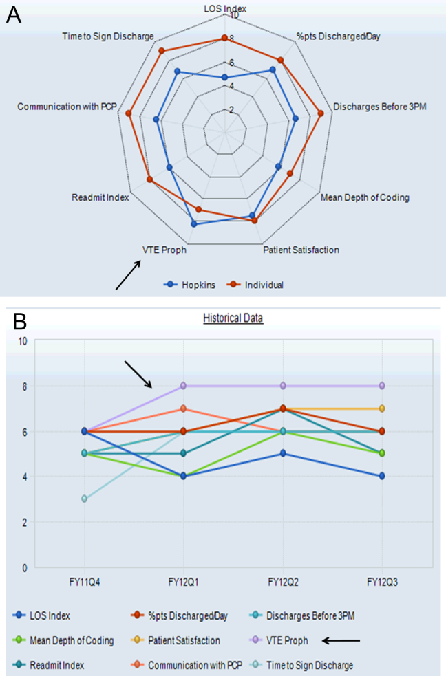
How malaria parasites evade the immune system

Credit: St Jude
Children’s Research Hospital
A new study has shown that malaria parasites can rapidly change proteins on the surface of human red blood cells (RBCs) during the course of a single infection, which helps the parasites evade the immune system.
The findings, which overturn previous thinking about the Plasmodium falciparum parasite’s lifecycle, could explain why so many attempts to create an effective malaria vaccine have failed and how the parasites are able to survive in the human body for such long periods of time.
Investigators described this research in PLOS Genetics.
The team kept P falciparum parasites dividing in human blood in the lab for over a year and sequenced the full parasite genome regularly. This provided snapshots of the parasite’s genome at multiple time points, allowing them to track evolution as it unfolded in the lab.
They found that the 60 or so genes that control proteins on the surface of infected human RBCs, known as var genes, swapped genetic information regularly, creating around a million new and unrecognizable surface proteins in every infected human every 2 days.
“These genes are like decks of cards constantly being shuffled,” explained study author William Hamilton, a graduate student at the Wellcome Trust Sanger Institute in Cambridge, UK.
“The use of whole-genome sequencing and the sheer number of samples we collected gave us a detailed picture of how the var gene repertoire changes continuously within red blood cells.”
The results showed, for the first time, that recombination does not occur when the malaria parasite is inside the mosquito, as previously thought. Instead, it occurs during the asexual stage of the parasite’s lifecycle inside human RBCs. This finding may help explain how chronic asymptomatic infection, a crucial problem for malaria elimination, is possible.
“It’s very likely that mosquitos are re-infected with Plasmodium falciparum parasites at the beginning of each wet season by biting humans who have carried the parasites, often asymptomatically, for up to 8 months during the dry season,” said study author Antoine Claessens, PhD, of the Wellcome Trust Sanger Institute.
“During those months, the parasite’s var genes are busy recombining to create millions of different versions—cunning disguises that mean they remain safe from the immune system and ready for the new malarial season.”
While further work will be required to fully understand the mechanism driving the recombination of P falciparum’s var genes, the investigators were able to calculate the rate at which it happens. They found that var gene recombination takes place in about 0.2% of parasites after each 48-hour life cycle in the RBC.
With about a billion parasites living inside a typical infected human, there is huge potential for the parasite to create new, recombined var genes inside each person with malaria. This pace of change far exceeds that of genes in any other region of the parasite’s genome.
“When you consider that 200 million people across the world are infected with malaria, and each of them is harboring parasites that are continually generating millions of antigenic variants, it becomes apparent why our fight against malaria is so challenging,” said study author Dominic Kwiatkowski, MBBS, of the Wellcome Trust Sanger Institute.
“This study is a great example of how genome sequence analysis is enriching our understanding of malaria biology. By learning the genetic tricks that the parasite uses to evade the human immune system, we will be in a much better position to eliminate this deadly disease.” ![]()
FDA recommends changing blood donor policy for MSM

Photo by Elisa Amendola
The US Food and Drug Administration (FDA) is recommending a change to the policy that prevents men who have sex with men (MSM) from donating blood, according to FDA Commissioner Margaret A. Hamburg.
The FDA would like to allow MSM to donate blood if they have abstained from sexual contact for 1 year.
The agency intends to issue a draft guidance recommending this policy change in 2015. The guidance will be open for public comment.
In a prepared statement, Hamburg said that, over the past few years, the FDA and other government agencies have carefully considered the scientific evidence relevant to the blood donor deferral policy for MSM.
This review, as well as the recommendations of advisory committees to the US Department of Health and Human Services (HHS) and the FDA, has prompted the FDA to recommend the change.
“This recommended change is consistent with the recommendation of an independent expert advisory panel, the HHS Advisory Committee on Blood and Tissue Safety and Availability, and will better align the deferral period with that of other men and women at increased risk for HIV infection,” Hamburg said.
“Additionally, in collaboration with the NIH’s National Heart Lung and Blood Institute (NHLBI), the FDA has already taken steps to implement a national blood surveillance system that will help the agency monitor the effect of a policy change and further help to ensure the continued safety of the blood supply.” ![]()
Classic HL vulnerable to PD-1 blockade therapy

SAN FRANCISCO—Two monoclonal antibodies that block the programmed death-1 (PD-1) pathway are showing promise in early phase trials in relapsed/refractory classic Hodgkin lymphoma (cHL).
Nivolumab prompted an 87% overall response rate (ORR) in heavily pretreated patients, and pembrolizumab elicited a 66% ORR in patients who had failed prior treatment with brentuximab vedotin.
These results were presented in 2 abstracts at the 2014 ASH Annual Meeting.
The rationale for using PD-1 blockade in cHL is that these patients frequently have an alteration in chromosome 9p24.1, which leads to increased expression of the PD-1 ligands, PD-L1 and PD-L2. The ligands engage the PD-1 receptors on activated T cells, inducing T-cell exhaustion. More than 85% of cHL tumors overexpress PD-L1.
Craig H. Moskowitz, MD, who presented the data on pembrolizumab at the meeting, sees nivolumab and pembrolizumab as being very similar.
“My gut feeling is that, at the end of the day, the response rates will be very similar,” he said. “The complete response rates will be similar. I think the toxicity profiles may be slightly dissimilar, and we’ll have to see what happens when these studies are both peer-reviewed.”
Nivolumab
Philippe Armand, MD, of Dana-Farber Cancer Institute in Boston, presented data on nivolumab in cHL (abstract 289), which was an independent expansion cohort of a phase 1b study in hematologic malignancies.
The 23 cHL patients received nivolumab at 3 mg/kg on weeks 1 and 4, then every 2 weeks.
Patients were a median age of 35 years (range, 20 to 54), and about two-thirds had received 4 or more prior systemic therapies. Seventy-eight percent had prior autologous stem cell transplant, and 78% had prior treatment with brentuximab.
“These were extensively pretreated patients” Dr Armand said, “with few options available.”
Twenty patients responded, for an ORR of 87%. Four patients (17%) achieved a complete response (CR), 16 (70%) had a partial response, and 3 (13%) had stable disease.
There were no progressions. And, at 24 weeks, the progression-free survival was 86%.
There were no life-threatening adverse events (AEs), no drug-related deaths, and no drug-related grade 4 AEs. Twenty-two patients (96%) experienced an AE, 18 (78%) had a drug-related AE, 5 (22%) had a grade 3 drug-related AE, and 2 (9%) patients discontinued treatment due to a drug-related AE.
The 2 events leading to discontinuation were myelodysplastic syndromes with grade 3 thrombocytopenia and grade 3 pancreatitis. The other grade 3 drug-related AEs were lymphopenia, increased lipase, GI inflammation, pneumonitis, colitis, and stomatitis.
“Overall, nivolumab has been used in thousands of patients already on clinical trials in solid tumors,” Dr Armand said. “And, overall, this safety profile mirrors that from what we expected in solid tumors.”
“But the interesting thing about that, from our standpoint, is that there was no apparent increase in the incidence of lung toxicity, which is something we worry about for those patients because many of them had had radiation or other drugs that can cause lung injury.”
This study was recently published in NEJM. It was funded by Bristol-Myers Squibb, the company developing nivolumab, and others.
Based on results of this study, the US Food and Drug Administration (FDA) granted nivolumab breakthrough therapy designation to treat HL. The drug recently gained FDA approval to treat advanced melanoma.
Pembrolizumab
Dr Moskowitz, of Memorial Sloan Kettering Cancer Center in New York, presented data on pembrolizumab as abstract 290.*
Investigators enrolled 31 patients onto the cHL cohort of the Keynote 013 trial. Patients were a median age of 32 years (range, 20 to 67).
All patients had failed therapy with brentuximab vedotin, 69% failed prior stem cell transplant, and 28% were transplant ineligible. Patients had to have an ECOG performance status of 0 or 1 and could not have autoimmune disease or interstitial lung disease.
Patients received 10 mg/kg of pembrolizumab intravenously every 2 weeks for up to 24 months or until progression.
Twenty-nine patients were evaluable for efficacy. The ORR was 66%, with a CR rate of 21% and a partial response rate of 45%. Twenty-one percent of patients had stable disease, and 14% had progressive disease. So the clinical benefit rate was 86%.
The median time to response was 12 weeks, and the median duration of response ranged from 1 to 185 days, but the median had not yet been reached.
Nine patients (31%) discontinued therapy, 1 (3%) due to an AE, 7 (24%) due to disease progression, and 1 (3%) after achieving a CR. Twenty patients (69%) were still on therapy at the time of the presentation, and 1 patient went on to transplant.
Sixteen patients (55%) experienced 1 or more treatment-related AE of any grade. Those occurring in 2 or more patients included hypothyroidism (10%), pneumonitis (10%), constipation (7%), diarrhea (7%), nausea (7%), hypercholesterolemia (7%), hypertriglyceridemia (7%), and hematuria (7%).
Treatment-related AEs of grade 3 or higher included axillary pain (3%), hypoxia (3%), joint swelling (3%), and pneumonitis (3%). Three patients experienced 4 grade 3 or higher AEs. There were no grade 4 treatment-related AEs or treatment-related deaths.
“In my opinion,” Dr Moskowitz concluded, “these results support continued development of pembrolizumab in Hodgkin lymphoma.”
“I think that these drugs are here to stay. Where we are going to put them in the armamentarium in Hodgkin lymphoma remains to be seen.”
This study was funded by Merck Sharp & Dohme Corp., the company developing pembrolizumab. ![]()
*Information in the abstract differs from that presented at the meeting.
Mitoxantrone lots recalled worldwide

Credit: Bill Branson
Hospira, Inc. has initiated a worldwide, user-level recall of 10 lots of Mitoxantrone (both human and veterinary) due to confirmed subpotency and elevated impurity levels.
Drugs in the affected lots may exhibit decreased effectiveness, require additional dosing, or prompt cumulative impurity toxicity requiring medical intervention.
However, Hospira has not received reports of any adverse events associated with subpotency and impurities for these lots to date.
The lots were distributed to hospitals and veterinary clinics worldwide from February 2013 through November 2014.
The following lots are affected by the recall. (To ensure this list displays properly, click the “Hide” icon on the right side of this page to hide the “In this Section” column.)
United States
Product NDC Number Lot Expiration Date
MitoXANTRONE Injection, USP, 61703-343-18 Z054636AA December 2014
(concentrate) 20 mg/10 mL, A014636AA April 2015
2 mg/mL in 10 mL, 10 mL Vial, A024636AB July 2015
Multi Dose Vial
MitoXANTRONE Injection, USP, 61703-343-65 A014643AA April 2015
(concentrate) 25 mg/12.5 mL,
2 mg/mL in 12.5 mL, 12.5 mL Vial,
Multi Dose Vial
MitoXANTRONE Injection, USP, 61703-343-66 A014645AA November 2015
(concentrate) 30 mg/15 mL,
2 mg/mL in 15 mL, 15 mL Vial,
Multi Dose Vial
Australia and New Zealand
Product Product Code Batch Number Expiration Date
DBL™ MitoXANTRONE M4636A A024636AA July 2015
Hydrochloride Injection
(concentrate) 20mg/10mL
Injection Vial
United Kingdom, Ireland, Cyprus, Saudi Arabia, Qatar, Oman and Bahrain
Product List Number Lot Expiration Date
MitoXANTRONE 2 mg/mL; M4636AGB1 A014636AB April 2015
Concentrate for Infusion A024636AD July 2015
Z054636AB Dec 2014
Canada
Product List Number DIN Lot Expiration Date
MitoXANTRONE for
Injection 20mg /10mL USP 4636A001 02244614 A024636AC July 2015
Anyone with an existing inventory of the recalled lots should stop use and distribution, and quarantine the product immediately. This recall is being carried out to the user level (both human and veterinary).
Hospira has notified its direct customers via a recall letter and is arranging for impacted product to be returned to Stericycle in the US. For additional assistance in the US, call Stericycle at 1-844-265-7407 between the hours of 8 am and 5 pm ET, Monday through Friday. Customers outside the US should work with their local Hospira offices to return the product per local recall notifications.
For medical inquiries, contact Hospira Medical Communications at 1-800-615-0187 or [email protected] (Available 24 hours a day/7 days per week).
To report adverse events or for product complaints, contact Hospira Global Complaint Management at 1-800-441-4100 (M-F, 8 am to 5 pm CT).
Adverse events or quality problems associated with Mitoxantrone can also be reported to the FDA’s MedWatch Adverse Event Reporting Program. ![]()
Similar Outcomes From Weekend Discharge
Hospitals typically reduce staffing levels and the availability of diagnostic, laboratory, and treatment services on weekends, and patients admitted on weekends exhibit poorer in‐hospital outcomes for several medical conditions.[1, 2, 3, 4, 5, 6, 7, 8, 9] Whether or not patients discharged on weekends have worse clinical outcomes has been less well studied.[10, 11, 12] Discharge rates on Saturday and Sunday are lower than for the other 5 days of the week,[12] but bed shortages and hospital overcrowding have increased the demand for maximizing 24/7 week‐round discharge efficiency. Given that the number of patients discharged on weekends is likely to continue to increase, it is important to assess the risk of weekend discharge on outcomes monitored as performance indicators by organizations such as the Centers for Medicare and Medicaid Services, the American Medical Association Physicians Consortium for Performance Improvement, the National Quality Forum, and the Joint Commission.
Thus, we designed this study to evaluate baseline characteristics, length of stay (LOS), and postdischarge outcomes for general internal medicine (GIM) patients in teaching hospitals discharged on weekends compared to weekdays. Our objective was to determine whether postdischarge outcomes differed for patients discharged on weekends versus weekdays.
METHODS
Study Setting
The Canadian province of Alberta has a single vertically integrated healthcare system that is government‐funded and provides universal access to hospitals, emergency departments (EDs), and outpatient physician services for all 4.1 million Albertans as well as all prescription medications for the poor, socially disadvantaged, disabled, or those age 65 years and older. This study received approval from the University of Alberta Health Research Ethics Board with waiver of informed consent.
Data Sources
This study used deidentified linked data from 3 Alberta Health administrative databases that capture vital status and all hospital or ED visits and have previously been shown to have high accuracy for medical diagnoses.[13] The Alberta Health Care Insurance Plan Registry tracks date of death or emigration from the province. The Discharge Abstract Database includes the most responsible diagnosis identified by the hospital attending physician, up to 25 other diagnoses coded by nosologists in each hospital, the admission and discharge dates, and the admission category (elective or urgent/emergent) for all acute care hospitalizations. Of note, unlike US studies, the hospital databases are able to distinguish in‐hospital (eg, adverse events) versus premorbid diagnoses (eg, preexisting comorbidities). The Ambulatory Care Database captures all patient visits to EDs with coding for up to 10 conditions per encounter.
Study Cohort
We identified all adults with an acute care hospitalization on the GIM services at all 7 Alberta teaching hospitals (ie, defined as those with Royal College of Physicians and Surgeons of Canadaapproved residency training programs in internal medicine, the equivalent of the Association of American Medical Colleges certification in the United States) between October 1, 2009 and September 30, 2010 and between April 1, 2011 and December 1, 2011 (these 20 months covered most of the pre/post intervals for a recently reported quality improvement initiative at 1 of the teaching hospitals that had no significant impact on postdischarge outcomes).[14] Patients from out of the province or transferred from/to another inpatient service (eg, the intensive care unit, a different service in the same hospital [such as surgery], another acute care hospital, or rehabilitation hospital) or with lengths of stay greater than 30 days were excluded. We only included the first hospitalization for any patient in our study timeframe and thus excluded repeat discharges of the same patient.
Explanatory Variable of Interest
The independent variable of interest was calendar day of discharge, stratified according to weekday (Monday thru Friday) versus weekend (Saturday and Sunday). Only 1.4% of weekday discharges occurred on a statutory holiday, and for the purposes of this study, these discharges were also considered weekend discharges. At the 7 teaching hospitals in Alberta, nursing staffing ratios do not differ between weekend and weekday, but availability of all other members of the healthcare team does. Physician census decreases from 4 to 5 per ward to 1 to 2, and ward‐based social workers, occupational therapists, physiotherapists, and pharmacist educators are generally not available on weekends.
Outcomes
Our primary outcome of interest was the composite outcome of death or all‐cause nonelective readmission within 30 days of discharge (ie, not including in‐hospital events prior to discharge or elective readmissions after discharge for planned procedures such as chemotherapy); hereafter we refer to this as death or readmission. This is a patient‐relevant outcome that is highlighted in the Affordable Care Act and for which there are several validated risk adjustment models.[15] We chose a composite outcome to deal with the issue of competing risks; if weekend discharges were more likely to die then we could observe a spurious association between weekend discharge and reduced readmissions if we focused on only that outcome.
Other Measures
Comorbidities for each patient were identified using International Classification of Diseases, Ninth Revision and Tenth Revision codes from the Discharge Abstract Database for the index hospitalization and any hospitalizations in the 12 months prior to their index admission, a method previously validated in Alberta databases.[13] We also recorded health resource use during their index hospitalization and calculated each patient's LACE score at the time of discharge, which is an index for predicting unplanned readmission or early death postdischarge previously validated in Canadian administrative databases.[15] The LACE index includes length of hospital stay (L), acuity of admission (A, based on the admission category variable described earlier), comorbidity burden quantified using the Charlson Comorbidity Index (C), and emergency department visits in the 6 months prior to admission (E); patients with discharge LACE scores >10 (total possible score is 19) are defined as being at high risk of death/readmission within 30 days.[16] As detailed below, to deal with potential concerns that LOS may be a mediator in the causal pathway, we ran 2 sensitivity analyses, 1 in which we excluded LOS from the analyses and 1 in which we included expected LOS rather than the actual LOS. Expected LOS is a data‐driven estimate based on the most current 2 years of patient LOS information available in the Canadian Institute for Health Information discharge abstract database (
Statistical Analysis
Baseline patient characteristics between weekend and weekday discharges were compared with t tests for continuous variables and [2] tests for binary or categorical variables. Logistic regression was used for comparison of death or readmission for weekend versus weekday discharges. Multivariable models were adjusted for age, sex, hospital, and LACE scores (as a continuous variable) at time of discharge; in sensitivity analyses we adjusted for (1) LACE score without including LOS and (2) LACE score using expected LOS rather than actual LOS. In further sensitivity analyses we (1) restricted the analysis to only those patients deemed to be at high risk for events due to LACE scores of 10 or greater and (2) included ED visits as part of the composite endpoint (ie, death, unplanned readmission, or unplanned ED visit within 30 days of discharge). Day of admission (weekend vs weekday) was also considered for the multivariable models, but was not found to be significant and thus was omitted from final models. We do not have any physician identifying variables in our dataset and thus could not investigate the potential correlation among patients discharged by the same physician. We did explore the hospital intraclass correlation coefficient, and as it was very small (0.001), we did not utilize models to account for the hierarchical nature of the data, but did include hospital as a fixed effect in the logistic models. The results were virtually identical whether we did or did not include hospital in the models. Adjusted odds ratios (aORs) are displayed with 95% confidence intervals (CI) and P values. Average LOS was calculated for weekend and weekday discharges with 95% CIs. P values for adjusted length of stay were calculated using multivariable linear regression adjusting for age, sex, day of admission, and Charlson score. All statistical analyses were done using SAS for Windows version 9.4 (SAS Institute, Inc., Cary, NC).
RESULTS
Patient Characteristics
Of the 7991 patients discharged during our study interval, 1146 (14.3%) were discharged on weekend or holiday days (Table 1). In contrast, 2180 of our cohort were admitted on a weekend (27.3%). The mean age of our study population was 62.1 years, 51.9% were men, mean Charlson score was 2.56, and 4591 (57.5%) had LACE scores of at least 10 at discharge.
| Characteristic | Weekend Discharge | Weekday Discharge | P Value |
|---|---|---|---|
| |||
| No. of patients | 1,146 | 6,845 | |
| Age, y, mean (SD) | 57.97 (19.70) | 62.77 (19.37) | <0.0001 |
| Male | 601 (52.4) | 3,548 (51.8) | 0.70 |
| Top 5 most responsible diagnoses | |||
| COPD | 74 (6.5) | 507 (7.4) | |
| Pneumonia | 64 (5.6) | 326 (4.8) | |
| Heart failure | 31 (2.7) | 375 (5.5) | |
| Urinary tract infection | 39 (3.4) | 254 (3.7) | |
| Venous thromboembolism | 31 (2.7) | 259 (3.8) | |
| Charlson score, mean (SD) | 2.17 (3.29) | 2.63 (3.30) | <0.0001 |
| Comorbidities (based on index hospitalization and prior 12 months) | |||
| Hypertension | 485 (42.3) | 3,265 (47.7) | 0.00 |
| Diabetes mellitus | 326 (28.4) | 2,106 (30.8) | 0.11 |
| Fluid imbalance | 332 (29.0) | 1,969 (28.8) | 0.89 |
| COPD | 255 (22.3) | 1,790 (26.2) | 0.01 |
| Psychiatric disorder | 179 (15.6) | 1,459 (21.3) | <0.0001 |
| Pneumonia | 242 (21.1) | 1,427 (20.8) | 0.84 |
| Anemia | 167 (14.6) | 1,233 (18.0) | 0.00 |
| Trauma | 169 (14.7) | 1,209 (17.7) | 0.02 |
| Atrial fibrillation | 141 (12.3) | 1,069 (15.6) | 0.00 |
| Heart failure | 101 (8.8) | 946 (13.8) | <0.0001 |
| Drug abuse | 188 (16.4) | 966 (14.1) | 0.04 |
| Cancer | 124 (10.8) | 867 (12.7) | 0.08 |
| Renal disease | 93 (8.1) | 689 (10.1) | 0.04 |
| Dementia | 49 (4.3) | 564 (8.2) | <0.0001 |
| Mild liver disease | 99 (8.6) | 587 (8.6) | 0.94 |
| Cerebrovascular disease | 59 (5.1) | 492 (7.2) | 0.01 |
| Gastrointestinal bleed | 84 (7.3) | 496 (7.2) | 0.92 |
| Asthma | 83 (7.2) | 426 (6.2) | 0.19 |
| Stroke | 42 (3.7) | 332 (4.9) | 0.08 |
| Prior myocardial infarction | 47 (4.1) | 329 (4.8) | 0.30 |
| Arthritis | 42 (3.7) | 309 (4.5) | 0.19 |
| Peripheral vascular disease | 42 (3.7) | 259 (3.8) | 0.84 |
| Severe liver disease | 44 (3.8) | 261 (3.8) | 0.97 |
| Valve disease | 24 (2.1) | 188 (2.7) | 0.20 |
| Paralysis | 31 (2.7) | 201 (2.9) | 0.67 |
| Skin ulcer | 17 (1.5) | 137 (2.0) | 0.24 |
| Shock | 19 (1.7) | 99 (1.4) | 0.58 |
| HIV | 15 (1.3) | 109 (1.6) | 0.47 |
| Protein calorie malnutrition | 0 (0.0) | 9 (0.1) | 0.21 |
| Features of index hospitalization | |||
| Resource intensity weight, mean (SD) | 1.10 (0.82) | 1.38 (1.24) | <0.0001 |
| LACE score, mean (SD) | 9.45 (2.85) | 10.51 (3.03) | <0.0001 |
| Expected LOS, mean (SD) | 6.20 (4.08) | 7.12 (4.89) | <0.0001 |
| Acute LOS, mean (SD) | 5.64 (4.99) | 7.86 (6.13) | <0.0001 |
| Weekend admission | 244 (21.3) | 1,936 (28.3) | <0.0001 |
| Discharge disposition | <0.0001 | ||
| Transferred to another inpatient hospital | 14 (1.2) | 189 (2.8) | |
| Transferred to long‐term care facility | 36 (3.1) | 532 (7.8) | |
| Transferred to other (except hospice) | 5 (0.4) | 24 (0.4) | |
| Discharged to home setting with support services | 125 (10.9) | 1,318 (19.3) | |
| Discharged home | 926 (80.8) | 4,646 (67.9) | |
| Left against medical advice | 40 (3.5) | 136 (2.0) | |
Weekday Versus Weekend Discharge
Although patients admitted on weekdays and weekends were very similar (data available upon request), patients discharged on weekends (compared to those discharged on weekdays) were younger, more likely to be discharged home without additional support, and had fewer comorbidities (Table 1, Figure 1). Patients discharged on weekends had shorter lengths of stay than those discharged on weekdays (5.6 days vs 7.9 days, P<0.0001). In adjusted linear regression analyses, this 2.3‐day difference remained statistically significant (adjusted P value <0.0001).

Patients discharged on a weekend exhibited lower unadjusted 30‐day rates of death or readmission than those discharged on a weekday (10.6% vs 13.2%), but these differences disappeared after multivariable adjustment that accounted for differences in risk profile (aOR: 0.94, 95% CI: 0.771.16 (Table 2). Results were similar in sensitivity analyses adjusting for LACE scores without LOS included (aOR: 0.88, 95% CI: 0.711.08) or adjusting for LACE scores using expected LOS rather than actual LOS (aOR: 0.90, 95% CI: 0.731.10). Restricting the analysis to only those patients deemed to be at high risk for events due to LACE scores of 10 or greater confirmed that weekend and weekday discharges had similar outcomes in the first 30 days after discharge (aOR: 1.09, 95% CI: 0.851.41, Table 2). Similar patterns were seen when we included ED visits as part of the composite endpoint (ie, death, unplanned readmission, or unplanned ED visit within 30 days of discharge) (Table 2).
| Weekend Discharge, n/N (%) | Weekday Discharge, n/N (%) | Unadjusted P Value | aOR* (95% CI) | Adjusted P Value | |
|---|---|---|---|---|---|
| |||||
| Death/readmission within 30 days | |||||
| All 7 teaching hospitals, all patients | 121/1146 (10.6) | 901/6845 (13.2) | 0.01 | 0.94 (0.77‐1.16) | 0.58 |
| All 7 teaching hospitals, but only patients with LACE <10 | 37/647 (5.7) | 225/2753 (8.2) | 0.04 | 0.72 (0.50, 1.03) | 0.07 |
| All 7 teaching hospitals, but only patients with LACE 10 | 84/499 (16.8) | 676/4092 (16.5) | 0.86 | 1.09 (0.85‐1.41) | 0.49 |
| Death/readmission/ED visit within 30 days | |||||
| All 7 teaching hospitals, all patients | 218/1146 (19.0) | 1445/6845 (21.1) | 0.11 | 0.98 (0.83‐1.15) | 0.79 |
| All 7 teaching hospitals, but only patients with LACE <10 | 90/647 (13.9) | 460/2753 (16.7) | 0.08 | 0.83 (0.64‐1.06) | 0.13 |
| All 7 teaching hospitals, but only patients with LACE 10 | 128/499 (25.7) | 985/4092 (24.1) | 0.44 | 1.12 (0.90‐1.39) | 0.31 |
| Death within 30 days | |||||
| All 7 teaching hospitals, all patients | 24/1146 (2.1) | 215/6845 (3.1) | 0.05 | 0.97 (0.63‐1.51) | 0.89 |
| All 7 teaching hospitals, but only patients with LACE <10 | 4/647 (0.6) | 23/2753 (0.8) | 0.58 | 0.89 (0.30, 2.62) | 0.83 |
| All 7 teaching hospitals, but only patients with LACE 10 | 20/499 (4.0) | 192/4092 (4.7) | 0.49 | 0.99 (0.61‐1.61) | 0.98 |
| Readmission within 30 days | |||||
| All 7 teaching hospitals, all patients | 105/1146 (9.2) | 751/6845 (11.0) | 0.07 | 0.94 (0.76‐1.17) | 0.59 |
| All 7 teaching hospitals, but only patients with LACE <10 | 33/647 (5.1) | 211/2753 (7.7) | 0.02 | 0.68 (0.46‐0.99) | 0.04 |
| All 7 teaching hospitals, but only patients with LACE 10 | 72/499 (14.4) | 540/4092 (13.2) | 0.44 | 1.14 (0.87‐1.49) | 0.34 |
| ED visit within 30 days | |||||
| All 7 teaching hospitals, all patients | 182/1146 (15.9) | 1118/6845 (16.3) | 0.70 | 1.00 (0.84‐1.19) | 0.99 |
| All 7 teaching hospitals, but only patients with LACE <10 | 83/647 (12.8) | 412/2753 (15.0) | 0.17 | 0.84 (0.65, 1.09) | 0.20 |
| All 7 teaching hospitals, but only patients with LACE 10 | 99/499 (19.8) | 706/4092 (17.3) | 0.15 | 1.17 (0.92‐1.48) | 0.20 |
DISCUSSION
Our data suggest that patients discharged from the GIM teaching wards we studied on weekends were appropriately triaged, as they did not exhibit a higher risk of adverse events postdischarge. Although patients discharged on weekends tended to be younger and had less comorbidities than those discharged during the week, we adjusted for baseline covariates in analyses, and we did not find an association between weekend discharge and increased postdischarge events even among the subset of patients deemed to be at high risk for postdischarge adverse events (based on high LACE scores). To our knowledge, although we previously examined this issue in patients with a most‐responsible diagnosis of heart failure,[10] examining weekend versus weekday discharges in the full gamut of general medical patients admitted to teaching hospitals has not previously been examined.
In our previous study[10] of over 24,000 heart failure patients discharged over 10 years (up to June 2009, therefore no overlap with any patients in this study), we also found that patients discharged on the weekends were younger, had fewer comorbidities, and shorter lengths of stay. Although postdischarge death/readmission rates were higher for weekend discharged patients in our earlier study (21.1% vs 19.5%, adjusted hazard ratio: 1.15, 95% CI: 1.061.25), it is worth noting that this was almost entirely driven by data from nonteaching hospitals and cardiology wards. Thus, it is important to reiterate that the findings in our current study are for GIM wards in teaching hospitals and may not be generalizable to less‐structured nonteaching settings.
Although we did not study physician decision making, our results suggest that physicians are incorporating discharge day into their discharge decision making. They may be selecting younger patients with less comorbidities for weekend discharges, or they may be delaying the discharges of older patients with more comorbidities for weekday discharges. Either is not surprising given the realities of weekend inpatient care: reduced staffing and frequent cross‐coverage (of physicians, nurses, physiotherapists, pharmacists, and occupational therapists), limited support services (such as laboratory services or diagnostic imaging), and decreased availability of community services (including home care and social support services).[17] For example, in 1 large US heart failure registry, patients discharged on a weekend received less complete discharge instructions than those discharged on weekdays.[11] Given that early follow‐up postdischarge is associated with better outcomes,[18, 19] future studies should also explore whether patterns of patient follow‐up differ after weekend versus weekday discharges.
Although we were able to capture all interactions with the healthcare system in a single payer system with universal access, there are some limitations to our study. First, we used administrative data, which preclude fully adjusting for severity of diagnoses or functional status, although we used proxies such as admission from/discharge to a long‐term care facility.[20, 21] Second, we did not have access to process of care measures such as diagnostic testing or prescribing data, and thus cannot determine whether quality of care or patient adherence differed by the day of the week they were discharged on, although this seems unlikely. Third, although postdischarge follow‐up may be associated with better outcomes,[18, 19] we were unable to adjust for patterns of outpatient follow‐up in this study. Fourth, we acknowledge that death or readmission soon after discharge does not necessarily mean that the quality of care during the preceding hospitalization was suboptimal or that these deaths or readmissions were even potentially preventable. Many factors influence postdischarge mortality and/or readmission, and quality of inpatient care is only one.[22, 23, 24, 25] Fifth, although some may express concern that LOS may be a mediator in the causal pathway between discharge decision and postdischarge events, and that adjusting for LOS in analyses could thus spuriously obscure a true association, it is worth pointing out that our 2 sensitivity analyses to explore this (the 1 in which we excluded LOS from the analyses and the 1 in which we included expected LOS rather than the actual LOS) revealed nearly identical point estimates and 95% CI as our main analysis. Finally, as our study is observational, we cannot definitively conclude causality, nor can we exclude an 18% excess risk for patients discharged on weekends (or a 22% lower risk either), given our 95% CI for postdischarge adverse outcomes.
CONCLUSION
We found that the proportion of patients discharged on weekends is lower than the proportion admitted on weekends. We also found that lower risk/less severely ill patients appear to be preferentially discharged on weekends, and as a result, postdischarge outcomes are similar between weekend and weekday discharges despite shorter LOS and less availability of outpatient resources for patients discharged on a weekend. The reasons why more complicated patients are not discharged on weekends deserves further study, as safely increasing weekend discharge rates would improve efficiency and safety (by reducing unnecessary exposure to in‐hospital adverse events such as falls, unnecessary urinary catheterizations, and healthcare‐acquired infections). Although hospital admission has become a 24/7 business, we believe that hospital discharge processes should strive for the same level of efficiency.
ACKNOWLEDGMENTS
Disclosures: This study is based in part on data provided by Alberta Health. The interpretation and conclusions contained herein are those of the researchers and do not necessarily represent the views of the government of Alberta. Neither the government of Alberta nor Alberta Health express any opinion in relation to this study. F.A.M. and S.R.M. are supported by salary awards from Alberta Innovates‐Health Solutions (AIHS). F.A.M. holds the Capital Health Chair in Cardiology Outcomes Research. S.R.M. holds the Endowed Chair in Patient Health Management. This project was funded by AIHS through an investigator‐initiated peer reviewed operating grant. The funding agencies did not have input into study design, data collection, interpretation of results, or write up/approval for submission. The authors report no conflicts of interest.
- , . Mortality among patients admitted to hospitals on weekends as compared with weekdays. N Engl J Med. 2001;345:663–668.
- , , , et al. Relationship between time of day, day of week, timeliness of reperfusion, and in‐hospital mortality for patients with acute ST‐segment elevation myocardial infarction. JAMA. 2005;294:803–812.
- , . Waiting for urgent procedures on the weekend among emergently hospitalized patients. Am J Med. 2004;117:175–181.
- . Do hospitals provide lower quality care on weekends? Health Serv Res. 2007;42:1589–1612.
- , , , et al. Day of admission and clinical outcomes for patients hospitalized for heart failure: findings from the organized program to initiate lifesaving treatment in hospitalized patients with heart failure (OPTIMIZE‐HF). Circ Heart Fail. 2008;1:50–57.
- , , , et al. Weekend hospitalization and additional risk of death: an analysis of inpatient data. J R Soc Med. 2012;105:74–84.
- , , , . Weekends: a dangerous time for having a stroke? Stroke. 2007;38:1211–1215.
- , , , . Day of the week of intensive care admission and patient outcomes: a multisite regional evaluation. Med Care. 2002;40:530–539.
- , , , . Effects of weekend admission and hospital teaching status on in‐hospital mortality. Am J Med. 2004;117:151–157.
- , , , , . Postdischarge outcomes in heart failure are better for teaching hospitals and weekday discharges. Circ Heart Fail. 2013;6:922–929.
- , , , et al. Weekend hospital admission and discharge for heart failure: association with quality of care and clinical outcomes. Am Heart J. 2009;158:451–458.
- , . Risk of death or readmission among people discharged from hospital on Fridays. CMAJ. 2002;166:1672–1673.
- , , , , et al.; IMECCHI Investigators. Assessing validity of ICD‐9‐CM and ICD‐10 administrative data in recording clinical conditions in a unique dually coded database. Health Serv Res. 2008;43:1424–1441.
- , , , et al. Safely and effectively reducing inpatient length of stay: a controlled study of the General Internal Medicine Care Transformation Initiative. BMJ Qual Saf. 2014;23:446–456.
- , , , et al. Derivation and validation of an index to predict early death or unplanned readmission after discharge from hospital to the community. CMAJ. 2010;182:551–557.
- , , , et al. Unplanned readmissions after hospital discharge among patients identified as being at high risk for readmission using a validated predictive algorithm. Open Med. 2011;5(2):e104–e111.
- , . Excellent hospital care for all: open and operating 24/7. J Gen Intern Med. 2011;26:1050–1052.
- , , , et al. Relationship between early physician follow‐up and 30‐day readmission among Medicare beneficiaries hospitalized for heart failure. JAMA. 2010;303:1716–1722.
- , , , , , . Impact of physician continuity on death or urgent readmission after discharge among patients with heart failure. CMAJ. 2013;185:e681–e689.
- , , , , , . Discordance of databases designed for claims payment versus clinical information systems. Implications for outcomes research. Ann Intern Med. 1993;119:844–850.
- , , , . Predictions of hospital mortality rates: a comparison of data sources. Ann Intern Med. 1997;126:347–354.
- , , , et al. Impact of social factors on risk of readmission or mortality in pneumonia and heart failure: systematic review. J Gen Intern Med. 2013;28(2):269–282.
- , . Investigating early readmission as an indicator for quality of care studies. Med Care. 1991;29(4):377–394.
- , , , et al. Risk prediction models for hospital readmission: a systematic review. JAMA. 2011;306(15):1688–1698.
- , , , , . Proportion of hospital readmissions deemed avoidable: a systematic review. CMAJ. 2011;183(7):E391–E402.
Hospitals typically reduce staffing levels and the availability of diagnostic, laboratory, and treatment services on weekends, and patients admitted on weekends exhibit poorer in‐hospital outcomes for several medical conditions.[1, 2, 3, 4, 5, 6, 7, 8, 9] Whether or not patients discharged on weekends have worse clinical outcomes has been less well studied.[10, 11, 12] Discharge rates on Saturday and Sunday are lower than for the other 5 days of the week,[12] but bed shortages and hospital overcrowding have increased the demand for maximizing 24/7 week‐round discharge efficiency. Given that the number of patients discharged on weekends is likely to continue to increase, it is important to assess the risk of weekend discharge on outcomes monitored as performance indicators by organizations such as the Centers for Medicare and Medicaid Services, the American Medical Association Physicians Consortium for Performance Improvement, the National Quality Forum, and the Joint Commission.
Thus, we designed this study to evaluate baseline characteristics, length of stay (LOS), and postdischarge outcomes for general internal medicine (GIM) patients in teaching hospitals discharged on weekends compared to weekdays. Our objective was to determine whether postdischarge outcomes differed for patients discharged on weekends versus weekdays.
METHODS
Study Setting
The Canadian province of Alberta has a single vertically integrated healthcare system that is government‐funded and provides universal access to hospitals, emergency departments (EDs), and outpatient physician services for all 4.1 million Albertans as well as all prescription medications for the poor, socially disadvantaged, disabled, or those age 65 years and older. This study received approval from the University of Alberta Health Research Ethics Board with waiver of informed consent.
Data Sources
This study used deidentified linked data from 3 Alberta Health administrative databases that capture vital status and all hospital or ED visits and have previously been shown to have high accuracy for medical diagnoses.[13] The Alberta Health Care Insurance Plan Registry tracks date of death or emigration from the province. The Discharge Abstract Database includes the most responsible diagnosis identified by the hospital attending physician, up to 25 other diagnoses coded by nosologists in each hospital, the admission and discharge dates, and the admission category (elective or urgent/emergent) for all acute care hospitalizations. Of note, unlike US studies, the hospital databases are able to distinguish in‐hospital (eg, adverse events) versus premorbid diagnoses (eg, preexisting comorbidities). The Ambulatory Care Database captures all patient visits to EDs with coding for up to 10 conditions per encounter.
Study Cohort
We identified all adults with an acute care hospitalization on the GIM services at all 7 Alberta teaching hospitals (ie, defined as those with Royal College of Physicians and Surgeons of Canadaapproved residency training programs in internal medicine, the equivalent of the Association of American Medical Colleges certification in the United States) between October 1, 2009 and September 30, 2010 and between April 1, 2011 and December 1, 2011 (these 20 months covered most of the pre/post intervals for a recently reported quality improvement initiative at 1 of the teaching hospitals that had no significant impact on postdischarge outcomes).[14] Patients from out of the province or transferred from/to another inpatient service (eg, the intensive care unit, a different service in the same hospital [such as surgery], another acute care hospital, or rehabilitation hospital) or with lengths of stay greater than 30 days were excluded. We only included the first hospitalization for any patient in our study timeframe and thus excluded repeat discharges of the same patient.
Explanatory Variable of Interest
The independent variable of interest was calendar day of discharge, stratified according to weekday (Monday thru Friday) versus weekend (Saturday and Sunday). Only 1.4% of weekday discharges occurred on a statutory holiday, and for the purposes of this study, these discharges were also considered weekend discharges. At the 7 teaching hospitals in Alberta, nursing staffing ratios do not differ between weekend and weekday, but availability of all other members of the healthcare team does. Physician census decreases from 4 to 5 per ward to 1 to 2, and ward‐based social workers, occupational therapists, physiotherapists, and pharmacist educators are generally not available on weekends.
Outcomes
Our primary outcome of interest was the composite outcome of death or all‐cause nonelective readmission within 30 days of discharge (ie, not including in‐hospital events prior to discharge or elective readmissions after discharge for planned procedures such as chemotherapy); hereafter we refer to this as death or readmission. This is a patient‐relevant outcome that is highlighted in the Affordable Care Act and for which there are several validated risk adjustment models.[15] We chose a composite outcome to deal with the issue of competing risks; if weekend discharges were more likely to die then we could observe a spurious association between weekend discharge and reduced readmissions if we focused on only that outcome.
Other Measures
Comorbidities for each patient were identified using International Classification of Diseases, Ninth Revision and Tenth Revision codes from the Discharge Abstract Database for the index hospitalization and any hospitalizations in the 12 months prior to their index admission, a method previously validated in Alberta databases.[13] We also recorded health resource use during their index hospitalization and calculated each patient's LACE score at the time of discharge, which is an index for predicting unplanned readmission or early death postdischarge previously validated in Canadian administrative databases.[15] The LACE index includes length of hospital stay (L), acuity of admission (A, based on the admission category variable described earlier), comorbidity burden quantified using the Charlson Comorbidity Index (C), and emergency department visits in the 6 months prior to admission (E); patients with discharge LACE scores >10 (total possible score is 19) are defined as being at high risk of death/readmission within 30 days.[16] As detailed below, to deal with potential concerns that LOS may be a mediator in the causal pathway, we ran 2 sensitivity analyses, 1 in which we excluded LOS from the analyses and 1 in which we included expected LOS rather than the actual LOS. Expected LOS is a data‐driven estimate based on the most current 2 years of patient LOS information available in the Canadian Institute for Health Information discharge abstract database (
Statistical Analysis
Baseline patient characteristics between weekend and weekday discharges were compared with t tests for continuous variables and [2] tests for binary or categorical variables. Logistic regression was used for comparison of death or readmission for weekend versus weekday discharges. Multivariable models were adjusted for age, sex, hospital, and LACE scores (as a continuous variable) at time of discharge; in sensitivity analyses we adjusted for (1) LACE score without including LOS and (2) LACE score using expected LOS rather than actual LOS. In further sensitivity analyses we (1) restricted the analysis to only those patients deemed to be at high risk for events due to LACE scores of 10 or greater and (2) included ED visits as part of the composite endpoint (ie, death, unplanned readmission, or unplanned ED visit within 30 days of discharge). Day of admission (weekend vs weekday) was also considered for the multivariable models, but was not found to be significant and thus was omitted from final models. We do not have any physician identifying variables in our dataset and thus could not investigate the potential correlation among patients discharged by the same physician. We did explore the hospital intraclass correlation coefficient, and as it was very small (0.001), we did not utilize models to account for the hierarchical nature of the data, but did include hospital as a fixed effect in the logistic models. The results were virtually identical whether we did or did not include hospital in the models. Adjusted odds ratios (aORs) are displayed with 95% confidence intervals (CI) and P values. Average LOS was calculated for weekend and weekday discharges with 95% CIs. P values for adjusted length of stay were calculated using multivariable linear regression adjusting for age, sex, day of admission, and Charlson score. All statistical analyses were done using SAS for Windows version 9.4 (SAS Institute, Inc., Cary, NC).
RESULTS
Patient Characteristics
Of the 7991 patients discharged during our study interval, 1146 (14.3%) were discharged on weekend or holiday days (Table 1). In contrast, 2180 of our cohort were admitted on a weekend (27.3%). The mean age of our study population was 62.1 years, 51.9% were men, mean Charlson score was 2.56, and 4591 (57.5%) had LACE scores of at least 10 at discharge.
| Characteristic | Weekend Discharge | Weekday Discharge | P Value |
|---|---|---|---|
| |||
| No. of patients | 1,146 | 6,845 | |
| Age, y, mean (SD) | 57.97 (19.70) | 62.77 (19.37) | <0.0001 |
| Male | 601 (52.4) | 3,548 (51.8) | 0.70 |
| Top 5 most responsible diagnoses | |||
| COPD | 74 (6.5) | 507 (7.4) | |
| Pneumonia | 64 (5.6) | 326 (4.8) | |
| Heart failure | 31 (2.7) | 375 (5.5) | |
| Urinary tract infection | 39 (3.4) | 254 (3.7) | |
| Venous thromboembolism | 31 (2.7) | 259 (3.8) | |
| Charlson score, mean (SD) | 2.17 (3.29) | 2.63 (3.30) | <0.0001 |
| Comorbidities (based on index hospitalization and prior 12 months) | |||
| Hypertension | 485 (42.3) | 3,265 (47.7) | 0.00 |
| Diabetes mellitus | 326 (28.4) | 2,106 (30.8) | 0.11 |
| Fluid imbalance | 332 (29.0) | 1,969 (28.8) | 0.89 |
| COPD | 255 (22.3) | 1,790 (26.2) | 0.01 |
| Psychiatric disorder | 179 (15.6) | 1,459 (21.3) | <0.0001 |
| Pneumonia | 242 (21.1) | 1,427 (20.8) | 0.84 |
| Anemia | 167 (14.6) | 1,233 (18.0) | 0.00 |
| Trauma | 169 (14.7) | 1,209 (17.7) | 0.02 |
| Atrial fibrillation | 141 (12.3) | 1,069 (15.6) | 0.00 |
| Heart failure | 101 (8.8) | 946 (13.8) | <0.0001 |
| Drug abuse | 188 (16.4) | 966 (14.1) | 0.04 |
| Cancer | 124 (10.8) | 867 (12.7) | 0.08 |
| Renal disease | 93 (8.1) | 689 (10.1) | 0.04 |
| Dementia | 49 (4.3) | 564 (8.2) | <0.0001 |
| Mild liver disease | 99 (8.6) | 587 (8.6) | 0.94 |
| Cerebrovascular disease | 59 (5.1) | 492 (7.2) | 0.01 |
| Gastrointestinal bleed | 84 (7.3) | 496 (7.2) | 0.92 |
| Asthma | 83 (7.2) | 426 (6.2) | 0.19 |
| Stroke | 42 (3.7) | 332 (4.9) | 0.08 |
| Prior myocardial infarction | 47 (4.1) | 329 (4.8) | 0.30 |
| Arthritis | 42 (3.7) | 309 (4.5) | 0.19 |
| Peripheral vascular disease | 42 (3.7) | 259 (3.8) | 0.84 |
| Severe liver disease | 44 (3.8) | 261 (3.8) | 0.97 |
| Valve disease | 24 (2.1) | 188 (2.7) | 0.20 |
| Paralysis | 31 (2.7) | 201 (2.9) | 0.67 |
| Skin ulcer | 17 (1.5) | 137 (2.0) | 0.24 |
| Shock | 19 (1.7) | 99 (1.4) | 0.58 |
| HIV | 15 (1.3) | 109 (1.6) | 0.47 |
| Protein calorie malnutrition | 0 (0.0) | 9 (0.1) | 0.21 |
| Features of index hospitalization | |||
| Resource intensity weight, mean (SD) | 1.10 (0.82) | 1.38 (1.24) | <0.0001 |
| LACE score, mean (SD) | 9.45 (2.85) | 10.51 (3.03) | <0.0001 |
| Expected LOS, mean (SD) | 6.20 (4.08) | 7.12 (4.89) | <0.0001 |
| Acute LOS, mean (SD) | 5.64 (4.99) | 7.86 (6.13) | <0.0001 |
| Weekend admission | 244 (21.3) | 1,936 (28.3) | <0.0001 |
| Discharge disposition | <0.0001 | ||
| Transferred to another inpatient hospital | 14 (1.2) | 189 (2.8) | |
| Transferred to long‐term care facility | 36 (3.1) | 532 (7.8) | |
| Transferred to other (except hospice) | 5 (0.4) | 24 (0.4) | |
| Discharged to home setting with support services | 125 (10.9) | 1,318 (19.3) | |
| Discharged home | 926 (80.8) | 4,646 (67.9) | |
| Left against medical advice | 40 (3.5) | 136 (2.0) | |
Weekday Versus Weekend Discharge
Although patients admitted on weekdays and weekends were very similar (data available upon request), patients discharged on weekends (compared to those discharged on weekdays) were younger, more likely to be discharged home without additional support, and had fewer comorbidities (Table 1, Figure 1). Patients discharged on weekends had shorter lengths of stay than those discharged on weekdays (5.6 days vs 7.9 days, P<0.0001). In adjusted linear regression analyses, this 2.3‐day difference remained statistically significant (adjusted P value <0.0001).
Patients discharged on a weekend exhibited lower unadjusted 30‐day rates of death or readmission than those discharged on a weekday (10.6% vs 13.2%), but these differences disappeared after multivariable adjustment that accounted for differences in risk profile (aOR: 0.94, 95% CI: 0.771.16 (Table 2). Results were similar in sensitivity analyses adjusting for LACE scores without LOS included (aOR: 0.88, 95% CI: 0.711.08) or adjusting for LACE scores using expected LOS rather than actual LOS (aOR: 0.90, 95% CI: 0.731.10). Restricting the analysis to only those patients deemed to be at high risk for events due to LACE scores of 10 or greater confirmed that weekend and weekday discharges had similar outcomes in the first 30 days after discharge (aOR: 1.09, 95% CI: 0.851.41, Table 2). Similar patterns were seen when we included ED visits as part of the composite endpoint (ie, death, unplanned readmission, or unplanned ED visit within 30 days of discharge) (Table 2).
| Weekend Discharge, n/N (%) | Weekday Discharge, n/N (%) | Unadjusted P Value | aOR* (95% CI) | Adjusted P Value | |
|---|---|---|---|---|---|
| |||||
| Death/readmission within 30 days | |||||
| All 7 teaching hospitals, all patients | 121/1146 (10.6) | 901/6845 (13.2) | 0.01 | 0.94 (0.77‐1.16) | 0.58 |
| All 7 teaching hospitals, but only patients with LACE <10 | 37/647 (5.7) | 225/2753 (8.2) | 0.04 | 0.72 (0.50, 1.03) | 0.07 |
| All 7 teaching hospitals, but only patients with LACE 10 | 84/499 (16.8) | 676/4092 (16.5) | 0.86 | 1.09 (0.85‐1.41) | 0.49 |
| Death/readmission/ED visit within 30 days | |||||
| All 7 teaching hospitals, all patients | 218/1146 (19.0) | 1445/6845 (21.1) | 0.11 | 0.98 (0.83‐1.15) | 0.79 |
| All 7 teaching hospitals, but only patients with LACE <10 | 90/647 (13.9) | 460/2753 (16.7) | 0.08 | 0.83 (0.64‐1.06) | 0.13 |
| All 7 teaching hospitals, but only patients with LACE 10 | 128/499 (25.7) | 985/4092 (24.1) | 0.44 | 1.12 (0.90‐1.39) | 0.31 |
| Death within 30 days | |||||
| All 7 teaching hospitals, all patients | 24/1146 (2.1) | 215/6845 (3.1) | 0.05 | 0.97 (0.63‐1.51) | 0.89 |
| All 7 teaching hospitals, but only patients with LACE <10 | 4/647 (0.6) | 23/2753 (0.8) | 0.58 | 0.89 (0.30, 2.62) | 0.83 |
| All 7 teaching hospitals, but only patients with LACE 10 | 20/499 (4.0) | 192/4092 (4.7) | 0.49 | 0.99 (0.61‐1.61) | 0.98 |
| Readmission within 30 days | |||||
| All 7 teaching hospitals, all patients | 105/1146 (9.2) | 751/6845 (11.0) | 0.07 | 0.94 (0.76‐1.17) | 0.59 |
| All 7 teaching hospitals, but only patients with LACE <10 | 33/647 (5.1) | 211/2753 (7.7) | 0.02 | 0.68 (0.46‐0.99) | 0.04 |
| All 7 teaching hospitals, but only patients with LACE 10 | 72/499 (14.4) | 540/4092 (13.2) | 0.44 | 1.14 (0.87‐1.49) | 0.34 |
| ED visit within 30 days | |||||
| All 7 teaching hospitals, all patients | 182/1146 (15.9) | 1118/6845 (16.3) | 0.70 | 1.00 (0.84‐1.19) | 0.99 |
| All 7 teaching hospitals, but only patients with LACE <10 | 83/647 (12.8) | 412/2753 (15.0) | 0.17 | 0.84 (0.65, 1.09) | 0.20 |
| All 7 teaching hospitals, but only patients with LACE 10 | 99/499 (19.8) | 706/4092 (17.3) | 0.15 | 1.17 (0.92‐1.48) | 0.20 |
DISCUSSION
Our data suggest that patients discharged from the GIM teaching wards we studied on weekends were appropriately triaged, as they did not exhibit a higher risk of adverse events postdischarge. Although patients discharged on weekends tended to be younger and had less comorbidities than those discharged during the week, we adjusted for baseline covariates in analyses, and we did not find an association between weekend discharge and increased postdischarge events even among the subset of patients deemed to be at high risk for postdischarge adverse events (based on high LACE scores). To our knowledge, although we previously examined this issue in patients with a most‐responsible diagnosis of heart failure,[10] examining weekend versus weekday discharges in the full gamut of general medical patients admitted to teaching hospitals has not previously been examined.
In our previous study[10] of over 24,000 heart failure patients discharged over 10 years (up to June 2009, therefore no overlap with any patients in this study), we also found that patients discharged on the weekends were younger, had fewer comorbidities, and shorter lengths of stay. Although postdischarge death/readmission rates were higher for weekend discharged patients in our earlier study (21.1% vs 19.5%, adjusted hazard ratio: 1.15, 95% CI: 1.061.25), it is worth noting that this was almost entirely driven by data from nonteaching hospitals and cardiology wards. Thus, it is important to reiterate that the findings in our current study are for GIM wards in teaching hospitals and may not be generalizable to less‐structured nonteaching settings.
Although we did not study physician decision making, our results suggest that physicians are incorporating discharge day into their discharge decision making. They may be selecting younger patients with less comorbidities for weekend discharges, or they may be delaying the discharges of older patients with more comorbidities for weekday discharges. Either is not surprising given the realities of weekend inpatient care: reduced staffing and frequent cross‐coverage (of physicians, nurses, physiotherapists, pharmacists, and occupational therapists), limited support services (such as laboratory services or diagnostic imaging), and decreased availability of community services (including home care and social support services).[17] For example, in 1 large US heart failure registry, patients discharged on a weekend received less complete discharge instructions than those discharged on weekdays.[11] Given that early follow‐up postdischarge is associated with better outcomes,[18, 19] future studies should also explore whether patterns of patient follow‐up differ after weekend versus weekday discharges.
Although we were able to capture all interactions with the healthcare system in a single payer system with universal access, there are some limitations to our study. First, we used administrative data, which preclude fully adjusting for severity of diagnoses or functional status, although we used proxies such as admission from/discharge to a long‐term care facility.[20, 21] Second, we did not have access to process of care measures such as diagnostic testing or prescribing data, and thus cannot determine whether quality of care or patient adherence differed by the day of the week they were discharged on, although this seems unlikely. Third, although postdischarge follow‐up may be associated with better outcomes,[18, 19] we were unable to adjust for patterns of outpatient follow‐up in this study. Fourth, we acknowledge that death or readmission soon after discharge does not necessarily mean that the quality of care during the preceding hospitalization was suboptimal or that these deaths or readmissions were even potentially preventable. Many factors influence postdischarge mortality and/or readmission, and quality of inpatient care is only one.[22, 23, 24, 25] Fifth, although some may express concern that LOS may be a mediator in the causal pathway between discharge decision and postdischarge events, and that adjusting for LOS in analyses could thus spuriously obscure a true association, it is worth pointing out that our 2 sensitivity analyses to explore this (the 1 in which we excluded LOS from the analyses and the 1 in which we included expected LOS rather than the actual LOS) revealed nearly identical point estimates and 95% CI as our main analysis. Finally, as our study is observational, we cannot definitively conclude causality, nor can we exclude an 18% excess risk for patients discharged on weekends (or a 22% lower risk either), given our 95% CI for postdischarge adverse outcomes.
CONCLUSION
We found that the proportion of patients discharged on weekends is lower than the proportion admitted on weekends. We also found that lower risk/less severely ill patients appear to be preferentially discharged on weekends, and as a result, postdischarge outcomes are similar between weekend and weekday discharges despite shorter LOS and less availability of outpatient resources for patients discharged on a weekend. The reasons why more complicated patients are not discharged on weekends deserves further study, as safely increasing weekend discharge rates would improve efficiency and safety (by reducing unnecessary exposure to in‐hospital adverse events such as falls, unnecessary urinary catheterizations, and healthcare‐acquired infections). Although hospital admission has become a 24/7 business, we believe that hospital discharge processes should strive for the same level of efficiency.
ACKNOWLEDGMENTS
Disclosures: This study is based in part on data provided by Alberta Health. The interpretation and conclusions contained herein are those of the researchers and do not necessarily represent the views of the government of Alberta. Neither the government of Alberta nor Alberta Health express any opinion in relation to this study. F.A.M. and S.R.M. are supported by salary awards from Alberta Innovates‐Health Solutions (AIHS). F.A.M. holds the Capital Health Chair in Cardiology Outcomes Research. S.R.M. holds the Endowed Chair in Patient Health Management. This project was funded by AIHS through an investigator‐initiated peer reviewed operating grant. The funding agencies did not have input into study design, data collection, interpretation of results, or write up/approval for submission. The authors report no conflicts of interest.
- , . Mortality among patients admitted to hospitals on weekends as compared with weekdays. N Engl J Med. 2001;345:663–668.
- , , , et al. Relationship between time of day, day of week, timeliness of reperfusion, and in‐hospital mortality for patients with acute ST‐segment elevation myocardial infarction. JAMA. 2005;294:803–812.
- , . Waiting for urgent procedures on the weekend among emergently hospitalized patients. Am J Med. 2004;117:175–181.
- . Do hospitals provide lower quality care on weekends? Health Serv Res. 2007;42:1589–1612.
- , , , et al. Day of admission and clinical outcomes for patients hospitalized for heart failure: findings from the organized program to initiate lifesaving treatment in hospitalized patients with heart failure (OPTIMIZE‐HF). Circ Heart Fail. 2008;1:50–57.
- , , , et al. Weekend hospitalization and additional risk of death: an analysis of inpatient data. J R Soc Med. 2012;105:74–84.
- , , , . Weekends: a dangerous time for having a stroke? Stroke. 2007;38:1211–1215.
- , , , . Day of the week of intensive care admission and patient outcomes: a multisite regional evaluation. Med Care. 2002;40:530–539.
- , , , . Effects of weekend admission and hospital teaching status on in‐hospital mortality. Am J Med. 2004;117:151–157.
- , , , , . Postdischarge outcomes in heart failure are better for teaching hospitals and weekday discharges. Circ Heart Fail. 2013;6:922–929.
- , , , et al. Weekend hospital admission and discharge for heart failure: association with quality of care and clinical outcomes. Am Heart J. 2009;158:451–458.
- , . Risk of death or readmission among people discharged from hospital on Fridays. CMAJ. 2002;166:1672–1673.
- , , , , et al.; IMECCHI Investigators. Assessing validity of ICD‐9‐CM and ICD‐10 administrative data in recording clinical conditions in a unique dually coded database. Health Serv Res. 2008;43:1424–1441.
- , , , et al. Safely and effectively reducing inpatient length of stay: a controlled study of the General Internal Medicine Care Transformation Initiative. BMJ Qual Saf. 2014;23:446–456.
- , , , et al. Derivation and validation of an index to predict early death or unplanned readmission after discharge from hospital to the community. CMAJ. 2010;182:551–557.
- , , , et al. Unplanned readmissions after hospital discharge among patients identified as being at high risk for readmission using a validated predictive algorithm. Open Med. 2011;5(2):e104–e111.
- , . Excellent hospital care for all: open and operating 24/7. J Gen Intern Med. 2011;26:1050–1052.
- , , , et al. Relationship between early physician follow‐up and 30‐day readmission among Medicare beneficiaries hospitalized for heart failure. JAMA. 2010;303:1716–1722.
- , , , , , . Impact of physician continuity on death or urgent readmission after discharge among patients with heart failure. CMAJ. 2013;185:e681–e689.
- , , , , , . Discordance of databases designed for claims payment versus clinical information systems. Implications for outcomes research. Ann Intern Med. 1993;119:844–850.
- , , , . Predictions of hospital mortality rates: a comparison of data sources. Ann Intern Med. 1997;126:347–354.
- , , , et al. Impact of social factors on risk of readmission or mortality in pneumonia and heart failure: systematic review. J Gen Intern Med. 2013;28(2):269–282.
- , . Investigating early readmission as an indicator for quality of care studies. Med Care. 1991;29(4):377–394.
- , , , et al. Risk prediction models for hospital readmission: a systematic review. JAMA. 2011;306(15):1688–1698.
- , , , , . Proportion of hospital readmissions deemed avoidable: a systematic review. CMAJ. 2011;183(7):E391–E402.
© 2014 Society of Hospital Medicine
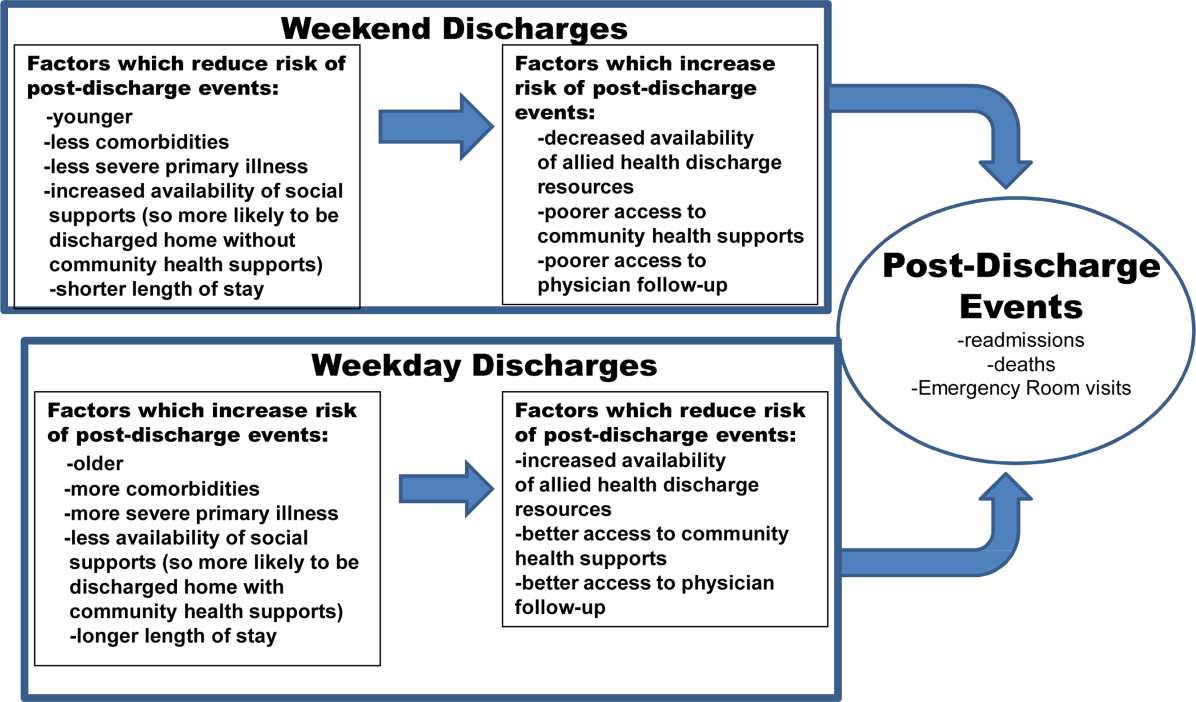
The $64,000 Question/
A 40‐year‐old Sudanese man was admitted due to worsening abdominal pain with recurrent ascites. He had a history of hepatitis B (HBV) infection and diabetes. He previously drank 3 beers per day on the weekends, but he had not consumed alcohol in over a year. He was born in Sudan but lived in Egypt most of his adult life; he immigrated to the United States 6 years previously. He was hospitalized out of state 9 months ago for a swollen abdomen and underwent an exploratory laparotomy that reportedly was unremarkable except for ascites.
Portal hypertension due to liver disease is the most common cause of ascites. This patient has a known risk factor for liver disease (history of HBV infection). Although his reported alcohol consumption is low, there is a synergistic effect on liver injury in the setting of chronic hepatitis. Abdominal pain in the setting of ascites needs to be urgently evaluated to exclude spontaneous bacterial peritonitis (SBP). Also, because chronic HBV infection is the major risk factor for hepatocellular carcinoma in the world, malignant ascites is in the differential. Hepatic vascular thrombosis and tuberculous peritonitis (given the patient's country of origin and travel history) also should be considered. The most appropriate initial test would be a diagnostic paracentesis to support or exclude the presence of SBP and direct the evaluation toward liver disease or other less‐common causes of ascites.
The patient was seen as an outpatient 5 months prior to admission with transient fever and joint pains. Laboratory studies at that visit were notable for a serum albumin of 3.2 g/dL (normal 3.55), 2.4 g of predicted 24‐hour protein on urinalysis (normal 30 mg per 24 hours), creatinine of 0.5 mg/dL (normal 0.81.3), and a positive hepatitis B surface antibody. The working diagnosis was a nonspecific viral syndrome and his symptoms resolved without treatment. One month later, he developed ascites and mild lower extremity edema. Additional laboratory studies at that time showed a normocytic anemia with hemoglobin 11.7 g/dL (normal 13.517.5) and leukopenia with white blood cell count of 2.4 109/L (normal 3.510.5), neutrophil count of 1.45 109/L (normal 1.77.0), and lymphocyte count of 0.58 109/L (normal 0.902.90). Transaminases, serum bilirubin, prothrombin time, alpha fetoprotein, and peripheral blood smear were normal. Human immunodeficiency virus antibody screen and QuantiFERON‐TB assay were negative. Hemoglobin A1c was 6.2% (normal 4.06.0). Repeat urinalysis demonstrated 883 mg of predicted 24‐hour protein. Computed tomography (CT) of the abdomen showed a large amount of intra‐abdominal ascites; the liver and spleen were normal, and there were no varices or other evidence of portal hypertension. Echocardiogram was normal except for a small inferior vena cava (IVC) and a mildly increased right ventricular systolic pressure of 32 mm Hg (systolic blood pressure 98 mm Hg). Due to the indeterminate cause for the patient's ascites, referral was made for gastroenterology evaluation with consideration for a paracentesis.
Cirrhotic ascites seems less likely. Postsinusoidal causes of portal hypertension (eg, cardiomyopathy) are also less likely given the absence of suggestive findings on echocardiography. Malignant ascites also appears less probable in the absence of suggestive findings such as mass lesions, lymphadenopathy, or peritoneal carcinomatosis on CT imaging. The suspicion for tuberculous peritonitis is lower with the negative QuantiFERON‐TB test. Hypoalbuminemia, normocytic anemia, leukopenia, and proteinuria all suggest a systemic inflammatory condition (eg, systemic lupus erythematosus [SLE]) with inflammatory serositis causing ascites). Nephrotic syndrome can cause hypoalbuminemia, edema, and ascites, but his total urine protein losses of 3.5 grams per 24 hours are not in keeping with this diagnosis. Other uncommon causes of ascites such as chylous ascites have not yet been excluded. The most appropriate next step remains ascitic fluid analysis.
A paracentesis yielded 7.8 L of clear‐yellow fluid and improvement in his abdominal discomfort. Analysis showed 224 total nucleated cells/L with 2% neutrophils, 57% lymphocytes, and 37% monocytes. Ascites total protein was 3.8 g/dL and glucose was 55 mg/dL. Gram stain and culture were negative, and cytology was negative for malignancy but showed lymphocytes, plasma cells, monocytes, and reactive mesothelial cells interpreted as consistent with chronic inflammation. The serum‐ascites albumin gradient (SAAG) was not obtained.
With a low leukocyte count and a paucity of neutrophils, this is not SBP. The ascites fluid did not have a chylous appearance. The SAAG, which can distinguish between portal hypertensive and nonportal hypertensive causes for ascites using a cutoff of 1.1 g/dL, was not done. The total protein was high, arguing against cirrhosis. High protein ascites with a high SAAG would suggest a posthepatic source of portal hypertension (eg, Budd‐Chiari syndrome, constrictive pericarditis). High protein ascites with a low SAAG would suggest an inflammatory or malignant source of ascites. The relative lymphocytosis in the ascites fluid suggests an inflammatory process, but is a nonspecific finding. The negative cytology does not completely exclude a malignancy, but given the absence of findings on the CT, malignant ascites is less likely.
Three months before admission, the patient underwent a repeat large‐volume paracentesis and a liver biopsy. The biopsy showed ectopic portal vein branches consistent with hepatoportal sclerosis, but no actual sclerosis was identified. The pathologist concluded that the findings suggested noncirrhotic portal hypertension due to a vascular in‐flow abnormality. Abdominal ultrasound with Doppler was unremarkable other than slightly increased echogenicity of the liver. Magnetic resonance (MR) angiogram showed narrowing of the intra‐abdominal IVC at the level of the diaphragm. Because of concern that hepatic congestion from high pressures in the narrowed IVC was leading to poor vascular inflow as suggested by the biopsy findings, an inferior vena cavagram was performed. This study was normal, although no transhepatic pressure measurements were obtained. Three stool specimens and 2 urine specimens were negative for parasites. The patient required repeat large‐volume paracenteses monthly. SBP was again ruled out, but no other diagnostic labs were obtained. He had anorexia with poor oral intake each time his abdomen became distended.
The patient was started on furosemide 1 month prior to admission to the hospital but had only a slight improvement in the ascites. His other medications included insulin, tamsulosin, and hydrocodone‐acetaminophen. Five days prior to admission, he underwent a diagnostic laparoscopy, which showed only ascites and small adhesions to the anterior abdominal wall. There was no visual evidence of malignancy, and the surgeon commented that the liver was normal. No additional biopsies were obtained.
The liver biopsy findings could be seen in noncirrhotic portal hypertension, although this diagnosis would be unlikely without splenomegaly, varices, or other signs of portal hypertension. However, 2 possible etiologies for noncirrhotic portal hypertension in this patient would be hepatic congestion from the narrowed IVC (although the normal IVC study argues against this) and hepatic schistosomiasis. Schistosomiasis is an important cause of noncirrhotic portal hypertension in endemic areas like this patient's country of origin, but the negative stool and urine studies, combined with the lack of granulomas or fibrosis seen on biopsy, make this condition unlikely.
Systemic amyloidosis (primary or secondary) could also be a cause of ascites and could present with multiorgan involvement (diarrhea and nephrotic syndrome). Amyloid deposits would have probably been seen in the liver biopsy, if present, but may not have been apparent unless specific stains (Congo red) were performed.
Evaluation for systemic, inflammatory autoimmune processes is indicated. Serum autoantibodies (anti‐nuclear antibody [ANA] and extractable nuclear antigens), and a serum and 24‐hour urine protein electrophoresis would be appropriate diagnostic tests. Peritoneal biopsies would have been helpful to assess for serosal diseases.
The patient subsequently developed acute right‐sided abdominal pain requiring urgent evaluation and admission to the hospital. He was initially assessed by a general surgeon, who found no evidence of postoperative complications. His temperature was 36.7C, blood pressure 105/64, heart rate 82, respiratory rate 16, and oxygen saturation 97% on room air. He appeared chronically ill, but he was in no distress and he had a normal mental status. Cardiac exam was normal except for mild jugular venous distension. He had mild bibasilar lung crackles. His abdomen was distended with superficial abdominal tenderness and a fluid wave, but he had normal bowel sounds and no peritoneal signs. He had mild scrotal edema but no peripheral edema. Joint exam did not suggest synovitis and there were no rashes or oral ulcers. Lactate was 0.9 mmol/L (normal 0.62.3), albumin was 2.6 g/dL, and prealbumin was 9 mg/dL (normal 1938). Erythrocyte sedimentation rate and C‐reactive protein were 46 mm/hour (normal 22) and 33.1 mg/L (normal 8), respectively. He had a normocytic anemia and leukopenia. Liver tests and routine chemistries were normal. Serum protein electrophoresis indicated no monoclonal protein. Complete 24‐hour urine collection showed 1.2 g of protein (normal 102 mg). Paracentesis of 3.4 L demonstrated 227 total nucleated cells/L with 2% neutrophils. Following the fluid removal, he had improvement in his pain, which he felt was related to the ascites rather than the recent surgery. Ascites total protein was 3.9 g/dL and ascites albumin was 1.7 g/dL. Ascites culture was negative for infection. Serum Schistosoma immunoglobulin G (IgG) antibody was positive at 3.53 (normal 1.00).
Further history revealed prior episodes of polyarticular joint pain and swelling in his hands and knees 5 years before admission. At that time, he reported a diffuse, pruritic, papular body rash. In addition, he noticed that his fingertips and toes turned white with cold exposure.
Importantly, surgical and infectious complications have been excluded. High protein ascites with a low SAAG of 0.9 suggests an inflammatory source of ascites. The follow‐up clinical data (arthritis, normocytic anemia, leukopenia, rash, Raynaud's phenomenon) suggest a systemic inflammatory syndrome such as SLE, with accompanying serositis. Serologic testing for autoantibodies would be recommended. Peritoneal biopsies, if obtained, may have demonstrated chronic, inflammatory infiltrate (nonspecific) or leukocytoclastic vasculitis (strongly supportive).
ANA enzyme immunoassay was >12 U (normal 1.0 U). Extractable nuclear antigens revealed positive autoantibodies for anti‐SSA, anti‐SSB, and anti‐ribosomal P. Moreover, double‐stranded DNA IgG antibody was 120 IU/mL (normal 30 IU/mL) and C3, C4, and total complement levels were low.
The clinical data support a diagnosis of SLE with serositis. Treatment of the underlying connective tissue disease will typically result in resolution of the ascites; diuretic therapy is generally ineffective.
In consultation with rheumatology and gastroenterology specialists, the diagnosis of SLE was made based on criteria of serositis, persistent leukopenia, arthritis, renal disease (proteinuria), positive ANA, elevated ds‐DNA antibodies, and hypocomplementemia. MR imaging of the abdominal vasculature demonstrated no evidence of vasculitis. The patient was given intravenous methylprednisolone 1 g daily for 3 days followed by high‐dose oral corticosteroids with a gradual taper. He was also started on mycophenolate mofetil as a steroid‐sparing medication (which was later changed to leflunomide due to persistent leukopenia) and hydroxychloroquine. His isolated positive Schistosoma IgG antibody in the absence of other findings was consistent with past exposure or infection. The infectious disease specialist felt there was no evidence of active schistosomiasis, but recommended treatment with a single dose of praziquantel due to the potential benefit with low risk of side effects. The patient had ongoing improvement following dismissal. He had 1 additional paracentesis of 4.1 L, 10 days after his hospitalization, and his ascites and proteinuria resolved. At the 5‐year follow‐up visit, there had been no recurrence of abdominal ascites or abdominal pain. He remains on low‐dose prednisone at 5 mg daily, leflunomide, and hydroxychloroquine.
COMMENTARY
This patient had recurrent ascites with 29.6 L removed over the 4 months prior to admission and an additional 3.4 L during his hospitalization. His outpatient providers initially considered a portal hypertensive etiology of his ascites due to his history of HBV and prior alcohol use. They also appropriately investigated for a possible infectious process. They next directed their evaluation toward the liver biopsy findings, which raised concern for a vascular inflow abnormality. However, the evaluation could have been performed more rapidly and far more cost‐efficiently had a diagnostic paracentesis with calculation of the SAAG been performed early in the evaluation.
The SAAG, which was first described in 1983 by Par and colleagues, is a parameter reflecting the oncotic pressure gradient between the vascular bed and the interstitial splanchnic or ascitic fluid. [1] In the classic study by Runyon and colleagues, a SAAG difference of 1.1 g/dL correctly differentiated causes of ascites due to portal hypertension from those that were not due to portal hypertension 96.7% of the time. [2] Conditions such as nephrotic syndrome, peritoneal carcinomatosis, and serositis (lupus peritonitis) can cause ascites in patients without portal hypertension.
Serositis in the form of pleuritis and/or pericarditis is a common feature of SLE, and ascites has been described in 8% to 11% of SLE patients.[3] However, massive ascites due to lupus peritonitis as a presenting symptom is rare.[4] More common causes of ascites in the setting of SLE include nephrotic syndrome, heart failure, protein‐losing enteropathy, constrictive pericarditis, Budd‐Chiari syndrome, indolent infections such as tuberculosis, and chylous ascites.[5, 6, 7] Of note, lupus peritonitis may be chronic or acute. Chronic ascites develops insidiously with few manifestations of active lupus and may be painless, whereas ascites from acute lupus peritonitis typically develops rapidly and presents with acute abdominal pain and other signs of increased lupus activity.[3, 5, 6, 8, 9]
Ascites from lupus peritonitis may be due to marked serosal exudative accumulation with reduced absorptive capacity in the peritoneum.[3, 4, 10] Other possible causes include peritoneal inflammation from deposition of immune complexes or vasculitis of peritoneal vessels and visceral serous membranes.[4, 9, 11] Although subserosal and submucosal vasculitis have been found in acute ascites, chronic ascites may be related to scarring from vasculitis and serosal inflammation leading to poor venous and lymph drainage.[9] Ascitic fluid characteristics from lupus peritonitis include a SAAG 1.1, presence of white blood cells anywhere in a broad range from 10 to 1630/L, and a range of fluid protein from 3.4 to 4.7 mg/dL.[3] Although not tested in this patient, findings of low complement levels, positive ANA, and elevated anti‐DNA antibody in the ascitic fluid would be supportive of lupus peritonitis, but not specific.[5, 9, 12] Lupus erythematosus cells are occasionally found in the ascitic fluid, but do not rule out other causes of ascites.[9] On retrospective analysis, lupus erythematosus cells were not seen in this patient's pathology specimens.
Treatment of lupus peritonitis and ascites is with high‐dose glucocorticoid therapy, but many patients may need a second immunosuppressant, possibly because of impaired peritoneal circulation from chronic inflammation leading to decreased drug delivery.[13, 14] Chronic ascites may be recalcitrant to systemic glucocorticoids,[3] so a possible alternative therapy is intraperitoneal injection of triamcinolone, which successfully treated massive ascites in a patient who did not respond to oral glucocorticoid treatment.[13] Although ascites may be refractory in some patients, those with chronic lupus peritonitis can generally achieve remission, yet the overall prognosis depends on the presence and severity of multiorgan involvement from SLE. As with any SLE patient, there are also risks of infection from immunosuppression and increased cardiovascular risks.
This patient's evaluation and treatment could have been expedited if he had undergone a paracenteses with determination of the SAAG early in his workup. It is not known why the SAAG was not obtained despite multiple outpatient visits and paracenteses, his history of HBV, and prior alcohol use. This may have been simply an unfortunate oversight. Alternatively, it may have been that his outpatient providers focused on tantalizing clues such as his country of origin, which led to concern for schistosomiasis, and the biopsy findings suggestive of a vascular inflow abnormality that led to further extensive testing. In so doing, the clinicians committed several diagnostic errors, including multiple alternatives bias, anchoring, and confirmation bias.[15] As a result, the patient accrued excess charges of $64,000 from multiple tests, laparoscopic surgery, and 2 hospitalizations. This case highlights how cognitive errors introduce costly variability into patient care, especially when a simple and accurate test is at the beginning of the decision tree.
CLINICAL TEACHING POINTS
- Diagnostic paracentesis, with calculation of the serum‐ascites albumin gradient, should be the first test in the workup for ascites and can distinguish portal hypertensive causes from nonportal hypertensive causes.
- Ascites related to SLE can be acute or chronic and caused by bowel infarction, perforation, pancreatitis, mesenteric vasculitis, nephrotic syndrome, heart failure, protein‐losing enteropathy, constrictive pericarditis, lupus peritonitis, Budd‐Chiari syndrome, or serositis (lupus peritonitis).
- Ascites caused by lupus peritonitis is rare. Once treated, management should be directed toward keeping the SLE in remission.
ACKNOWLEDGMENTS
Disclosure: Nothing to report.
- , , . Serum‐ascites albumin concentration gradient: a physiologic approach to the differential diagnosis of ascites. Gastroenterology. 1983;85(2):240–244.
- , , , et al. The serum‐ascites albumin gradient is superior to the exudate‐transudate concept in the differential diagnosis of ascites. Ann Intern Med. 1992;117:215–220.
- , , . Insidious onset of massive painless ascites as initial manifestation of systemic lupus erythematosus. Lupus. 2011;20:754–757.
- , . Rapid onset of massive ascites as the initial presentation of systemic lupus erythematosus. Am J Gastroenterol. 2000;95:302–303.
- , . Gastrointestinal and hepatic manifestations of systemic lupus erythematosus. J Clin Gastroenterol. 2011;45:436–441.
- , , , . Massive ascites as a presenting feature of lupus. Int J Rheum Dis. 2012;15:e15–e16.
- , , , et al. Concurrent occurrence of chylothorax, chylous ascites, and protein‐losing enteropathy in systemic lupus erythematosus. J Rheumatol. 2002;29:1330–1333.
- , , , et al. Abdominal manifestations in childhood‐onset systemic lupus erythematosus. Ann Rheum Dis. 2007;66:174–178.
- , , . Chronic lupus peritonitis with ascites: review of the literature with a case report. Semin Arthritis Rheum. 1988;18:121–126.
- , . Nonhepatic Gastrointestinal Manifestations of Systemic Lupus Erythematosus. London, United Kingdom: Churchill Livingstone; 1987:747–760.
- , , , . Ascites due to lupus peritonitis: a rare form of onset of systemic lupus erythematosus. Rev Bras Reumatol. 2012;52(1):113–119.
- , , , . New‐onset lupus presenting as serositis in an 80‐year‐old woman: does a high‐titer ANA in pleural, pericardial, or peritoneal fluid help confirm the diagnosis? J Clin Rheum.2005:11(5):292–293.
- , , , . Successful treatment of massive ascites with intraperitoneal administration of a steroid in a case of systemic lupus erythematosus. Lupus. 2009;18:740–742.
- , , , et al. Chronic lupus peritonitis with massive ascites at elderly onset: case report and review of the literature. Intern Med. 2002;41:1056–1061.
- . The Importance of cognitive errors in diagnosis and strategies to minimize them. Acad Med. 2003;78:775–780.
A 40‐year‐old Sudanese man was admitted due to worsening abdominal pain with recurrent ascites. He had a history of hepatitis B (HBV) infection and diabetes. He previously drank 3 beers per day on the weekends, but he had not consumed alcohol in over a year. He was born in Sudan but lived in Egypt most of his adult life; he immigrated to the United States 6 years previously. He was hospitalized out of state 9 months ago for a swollen abdomen and underwent an exploratory laparotomy that reportedly was unremarkable except for ascites.
Portal hypertension due to liver disease is the most common cause of ascites. This patient has a known risk factor for liver disease (history of HBV infection). Although his reported alcohol consumption is low, there is a synergistic effect on liver injury in the setting of chronic hepatitis. Abdominal pain in the setting of ascites needs to be urgently evaluated to exclude spontaneous bacterial peritonitis (SBP). Also, because chronic HBV infection is the major risk factor for hepatocellular carcinoma in the world, malignant ascites is in the differential. Hepatic vascular thrombosis and tuberculous peritonitis (given the patient's country of origin and travel history) also should be considered. The most appropriate initial test would be a diagnostic paracentesis to support or exclude the presence of SBP and direct the evaluation toward liver disease or other less‐common causes of ascites.
The patient was seen as an outpatient 5 months prior to admission with transient fever and joint pains. Laboratory studies at that visit were notable for a serum albumin of 3.2 g/dL (normal 3.55), 2.4 g of predicted 24‐hour protein on urinalysis (normal 30 mg per 24 hours), creatinine of 0.5 mg/dL (normal 0.81.3), and a positive hepatitis B surface antibody. The working diagnosis was a nonspecific viral syndrome and his symptoms resolved without treatment. One month later, he developed ascites and mild lower extremity edema. Additional laboratory studies at that time showed a normocytic anemia with hemoglobin 11.7 g/dL (normal 13.517.5) and leukopenia with white blood cell count of 2.4 109/L (normal 3.510.5), neutrophil count of 1.45 109/L (normal 1.77.0), and lymphocyte count of 0.58 109/L (normal 0.902.90). Transaminases, serum bilirubin, prothrombin time, alpha fetoprotein, and peripheral blood smear were normal. Human immunodeficiency virus antibody screen and QuantiFERON‐TB assay were negative. Hemoglobin A1c was 6.2% (normal 4.06.0). Repeat urinalysis demonstrated 883 mg of predicted 24‐hour protein. Computed tomography (CT) of the abdomen showed a large amount of intra‐abdominal ascites; the liver and spleen were normal, and there were no varices or other evidence of portal hypertension. Echocardiogram was normal except for a small inferior vena cava (IVC) and a mildly increased right ventricular systolic pressure of 32 mm Hg (systolic blood pressure 98 mm Hg). Due to the indeterminate cause for the patient's ascites, referral was made for gastroenterology evaluation with consideration for a paracentesis.
Cirrhotic ascites seems less likely. Postsinusoidal causes of portal hypertension (eg, cardiomyopathy) are also less likely given the absence of suggestive findings on echocardiography. Malignant ascites also appears less probable in the absence of suggestive findings such as mass lesions, lymphadenopathy, or peritoneal carcinomatosis on CT imaging. The suspicion for tuberculous peritonitis is lower with the negative QuantiFERON‐TB test. Hypoalbuminemia, normocytic anemia, leukopenia, and proteinuria all suggest a systemic inflammatory condition (eg, systemic lupus erythematosus [SLE]) with inflammatory serositis causing ascites). Nephrotic syndrome can cause hypoalbuminemia, edema, and ascites, but his total urine protein losses of 3.5 grams per 24 hours are not in keeping with this diagnosis. Other uncommon causes of ascites such as chylous ascites have not yet been excluded. The most appropriate next step remains ascitic fluid analysis.
A paracentesis yielded 7.8 L of clear‐yellow fluid and improvement in his abdominal discomfort. Analysis showed 224 total nucleated cells/L with 2% neutrophils, 57% lymphocytes, and 37% monocytes. Ascites total protein was 3.8 g/dL and glucose was 55 mg/dL. Gram stain and culture were negative, and cytology was negative for malignancy but showed lymphocytes, plasma cells, monocytes, and reactive mesothelial cells interpreted as consistent with chronic inflammation. The serum‐ascites albumin gradient (SAAG) was not obtained.
With a low leukocyte count and a paucity of neutrophils, this is not SBP. The ascites fluid did not have a chylous appearance. The SAAG, which can distinguish between portal hypertensive and nonportal hypertensive causes for ascites using a cutoff of 1.1 g/dL, was not done. The total protein was high, arguing against cirrhosis. High protein ascites with a high SAAG would suggest a posthepatic source of portal hypertension (eg, Budd‐Chiari syndrome, constrictive pericarditis). High protein ascites with a low SAAG would suggest an inflammatory or malignant source of ascites. The relative lymphocytosis in the ascites fluid suggests an inflammatory process, but is a nonspecific finding. The negative cytology does not completely exclude a malignancy, but given the absence of findings on the CT, malignant ascites is less likely.
Three months before admission, the patient underwent a repeat large‐volume paracentesis and a liver biopsy. The biopsy showed ectopic portal vein branches consistent with hepatoportal sclerosis, but no actual sclerosis was identified. The pathologist concluded that the findings suggested noncirrhotic portal hypertension due to a vascular in‐flow abnormality. Abdominal ultrasound with Doppler was unremarkable other than slightly increased echogenicity of the liver. Magnetic resonance (MR) angiogram showed narrowing of the intra‐abdominal IVC at the level of the diaphragm. Because of concern that hepatic congestion from high pressures in the narrowed IVC was leading to poor vascular inflow as suggested by the biopsy findings, an inferior vena cavagram was performed. This study was normal, although no transhepatic pressure measurements were obtained. Three stool specimens and 2 urine specimens were negative for parasites. The patient required repeat large‐volume paracenteses monthly. SBP was again ruled out, but no other diagnostic labs were obtained. He had anorexia with poor oral intake each time his abdomen became distended.
The patient was started on furosemide 1 month prior to admission to the hospital but had only a slight improvement in the ascites. His other medications included insulin, tamsulosin, and hydrocodone‐acetaminophen. Five days prior to admission, he underwent a diagnostic laparoscopy, which showed only ascites and small adhesions to the anterior abdominal wall. There was no visual evidence of malignancy, and the surgeon commented that the liver was normal. No additional biopsies were obtained.
The liver biopsy findings could be seen in noncirrhotic portal hypertension, although this diagnosis would be unlikely without splenomegaly, varices, or other signs of portal hypertension. However, 2 possible etiologies for noncirrhotic portal hypertension in this patient would be hepatic congestion from the narrowed IVC (although the normal IVC study argues against this) and hepatic schistosomiasis. Schistosomiasis is an important cause of noncirrhotic portal hypertension in endemic areas like this patient's country of origin, but the negative stool and urine studies, combined with the lack of granulomas or fibrosis seen on biopsy, make this condition unlikely.
Systemic amyloidosis (primary or secondary) could also be a cause of ascites and could present with multiorgan involvement (diarrhea and nephrotic syndrome). Amyloid deposits would have probably been seen in the liver biopsy, if present, but may not have been apparent unless specific stains (Congo red) were performed.
Evaluation for systemic, inflammatory autoimmune processes is indicated. Serum autoantibodies (anti‐nuclear antibody [ANA] and extractable nuclear antigens), and a serum and 24‐hour urine protein electrophoresis would be appropriate diagnostic tests. Peritoneal biopsies would have been helpful to assess for serosal diseases.
The patient subsequently developed acute right‐sided abdominal pain requiring urgent evaluation and admission to the hospital. He was initially assessed by a general surgeon, who found no evidence of postoperative complications. His temperature was 36.7C, blood pressure 105/64, heart rate 82, respiratory rate 16, and oxygen saturation 97% on room air. He appeared chronically ill, but he was in no distress and he had a normal mental status. Cardiac exam was normal except for mild jugular venous distension. He had mild bibasilar lung crackles. His abdomen was distended with superficial abdominal tenderness and a fluid wave, but he had normal bowel sounds and no peritoneal signs. He had mild scrotal edema but no peripheral edema. Joint exam did not suggest synovitis and there were no rashes or oral ulcers. Lactate was 0.9 mmol/L (normal 0.62.3), albumin was 2.6 g/dL, and prealbumin was 9 mg/dL (normal 1938). Erythrocyte sedimentation rate and C‐reactive protein were 46 mm/hour (normal 22) and 33.1 mg/L (normal 8), respectively. He had a normocytic anemia and leukopenia. Liver tests and routine chemistries were normal. Serum protein electrophoresis indicated no monoclonal protein. Complete 24‐hour urine collection showed 1.2 g of protein (normal 102 mg). Paracentesis of 3.4 L demonstrated 227 total nucleated cells/L with 2% neutrophils. Following the fluid removal, he had improvement in his pain, which he felt was related to the ascites rather than the recent surgery. Ascites total protein was 3.9 g/dL and ascites albumin was 1.7 g/dL. Ascites culture was negative for infection. Serum Schistosoma immunoglobulin G (IgG) antibody was positive at 3.53 (normal 1.00).
Further history revealed prior episodes of polyarticular joint pain and swelling in his hands and knees 5 years before admission. At that time, he reported a diffuse, pruritic, papular body rash. In addition, he noticed that his fingertips and toes turned white with cold exposure.
Importantly, surgical and infectious complications have been excluded. High protein ascites with a low SAAG of 0.9 suggests an inflammatory source of ascites. The follow‐up clinical data (arthritis, normocytic anemia, leukopenia, rash, Raynaud's phenomenon) suggest a systemic inflammatory syndrome such as SLE, with accompanying serositis. Serologic testing for autoantibodies would be recommended. Peritoneal biopsies, if obtained, may have demonstrated chronic, inflammatory infiltrate (nonspecific) or leukocytoclastic vasculitis (strongly supportive).
ANA enzyme immunoassay was >12 U (normal 1.0 U). Extractable nuclear antigens revealed positive autoantibodies for anti‐SSA, anti‐SSB, and anti‐ribosomal P. Moreover, double‐stranded DNA IgG antibody was 120 IU/mL (normal 30 IU/mL) and C3, C4, and total complement levels were low.
The clinical data support a diagnosis of SLE with serositis. Treatment of the underlying connective tissue disease will typically result in resolution of the ascites; diuretic therapy is generally ineffective.
In consultation with rheumatology and gastroenterology specialists, the diagnosis of SLE was made based on criteria of serositis, persistent leukopenia, arthritis, renal disease (proteinuria), positive ANA, elevated ds‐DNA antibodies, and hypocomplementemia. MR imaging of the abdominal vasculature demonstrated no evidence of vasculitis. The patient was given intravenous methylprednisolone 1 g daily for 3 days followed by high‐dose oral corticosteroids with a gradual taper. He was also started on mycophenolate mofetil as a steroid‐sparing medication (which was later changed to leflunomide due to persistent leukopenia) and hydroxychloroquine. His isolated positive Schistosoma IgG antibody in the absence of other findings was consistent with past exposure or infection. The infectious disease specialist felt there was no evidence of active schistosomiasis, but recommended treatment with a single dose of praziquantel due to the potential benefit with low risk of side effects. The patient had ongoing improvement following dismissal. He had 1 additional paracentesis of 4.1 L, 10 days after his hospitalization, and his ascites and proteinuria resolved. At the 5‐year follow‐up visit, there had been no recurrence of abdominal ascites or abdominal pain. He remains on low‐dose prednisone at 5 mg daily, leflunomide, and hydroxychloroquine.
COMMENTARY
This patient had recurrent ascites with 29.6 L removed over the 4 months prior to admission and an additional 3.4 L during his hospitalization. His outpatient providers initially considered a portal hypertensive etiology of his ascites due to his history of HBV and prior alcohol use. They also appropriately investigated for a possible infectious process. They next directed their evaluation toward the liver biopsy findings, which raised concern for a vascular inflow abnormality. However, the evaluation could have been performed more rapidly and far more cost‐efficiently had a diagnostic paracentesis with calculation of the SAAG been performed early in the evaluation.
The SAAG, which was first described in 1983 by Par and colleagues, is a parameter reflecting the oncotic pressure gradient between the vascular bed and the interstitial splanchnic or ascitic fluid. [1] In the classic study by Runyon and colleagues, a SAAG difference of 1.1 g/dL correctly differentiated causes of ascites due to portal hypertension from those that were not due to portal hypertension 96.7% of the time. [2] Conditions such as nephrotic syndrome, peritoneal carcinomatosis, and serositis (lupus peritonitis) can cause ascites in patients without portal hypertension.
Serositis in the form of pleuritis and/or pericarditis is a common feature of SLE, and ascites has been described in 8% to 11% of SLE patients.[3] However, massive ascites due to lupus peritonitis as a presenting symptom is rare.[4] More common causes of ascites in the setting of SLE include nephrotic syndrome, heart failure, protein‐losing enteropathy, constrictive pericarditis, Budd‐Chiari syndrome, indolent infections such as tuberculosis, and chylous ascites.[5, 6, 7] Of note, lupus peritonitis may be chronic or acute. Chronic ascites develops insidiously with few manifestations of active lupus and may be painless, whereas ascites from acute lupus peritonitis typically develops rapidly and presents with acute abdominal pain and other signs of increased lupus activity.[3, 5, 6, 8, 9]
Ascites from lupus peritonitis may be due to marked serosal exudative accumulation with reduced absorptive capacity in the peritoneum.[3, 4, 10] Other possible causes include peritoneal inflammation from deposition of immune complexes or vasculitis of peritoneal vessels and visceral serous membranes.[4, 9, 11] Although subserosal and submucosal vasculitis have been found in acute ascites, chronic ascites may be related to scarring from vasculitis and serosal inflammation leading to poor venous and lymph drainage.[9] Ascitic fluid characteristics from lupus peritonitis include a SAAG 1.1, presence of white blood cells anywhere in a broad range from 10 to 1630/L, and a range of fluid protein from 3.4 to 4.7 mg/dL.[3] Although not tested in this patient, findings of low complement levels, positive ANA, and elevated anti‐DNA antibody in the ascitic fluid would be supportive of lupus peritonitis, but not specific.[5, 9, 12] Lupus erythematosus cells are occasionally found in the ascitic fluid, but do not rule out other causes of ascites.[9] On retrospective analysis, lupus erythematosus cells were not seen in this patient's pathology specimens.
Treatment of lupus peritonitis and ascites is with high‐dose glucocorticoid therapy, but many patients may need a second immunosuppressant, possibly because of impaired peritoneal circulation from chronic inflammation leading to decreased drug delivery.[13, 14] Chronic ascites may be recalcitrant to systemic glucocorticoids,[3] so a possible alternative therapy is intraperitoneal injection of triamcinolone, which successfully treated massive ascites in a patient who did not respond to oral glucocorticoid treatment.[13] Although ascites may be refractory in some patients, those with chronic lupus peritonitis can generally achieve remission, yet the overall prognosis depends on the presence and severity of multiorgan involvement from SLE. As with any SLE patient, there are also risks of infection from immunosuppression and increased cardiovascular risks.
This patient's evaluation and treatment could have been expedited if he had undergone a paracenteses with determination of the SAAG early in his workup. It is not known why the SAAG was not obtained despite multiple outpatient visits and paracenteses, his history of HBV, and prior alcohol use. This may have been simply an unfortunate oversight. Alternatively, it may have been that his outpatient providers focused on tantalizing clues such as his country of origin, which led to concern for schistosomiasis, and the biopsy findings suggestive of a vascular inflow abnormality that led to further extensive testing. In so doing, the clinicians committed several diagnostic errors, including multiple alternatives bias, anchoring, and confirmation bias.[15] As a result, the patient accrued excess charges of $64,000 from multiple tests, laparoscopic surgery, and 2 hospitalizations. This case highlights how cognitive errors introduce costly variability into patient care, especially when a simple and accurate test is at the beginning of the decision tree.
CLINICAL TEACHING POINTS
- Diagnostic paracentesis, with calculation of the serum‐ascites albumin gradient, should be the first test in the workup for ascites and can distinguish portal hypertensive causes from nonportal hypertensive causes.
- Ascites related to SLE can be acute or chronic and caused by bowel infarction, perforation, pancreatitis, mesenteric vasculitis, nephrotic syndrome, heart failure, protein‐losing enteropathy, constrictive pericarditis, lupus peritonitis, Budd‐Chiari syndrome, or serositis (lupus peritonitis).
- Ascites caused by lupus peritonitis is rare. Once treated, management should be directed toward keeping the SLE in remission.
ACKNOWLEDGMENTS
Disclosure: Nothing to report.
- , , . Serum‐ascites albumin concentration gradient: a physiologic approach to the differential diagnosis of ascites. Gastroenterology. 1983;85(2):240–244.
- , , , et al. The serum‐ascites albumin gradient is superior to the exudate‐transudate concept in the differential diagnosis of ascites. Ann Intern Med. 1992;117:215–220.
- , , . Insidious onset of massive painless ascites as initial manifestation of systemic lupus erythematosus. Lupus. 2011;20:754–757.
- , . Rapid onset of massive ascites as the initial presentation of systemic lupus erythematosus. Am J Gastroenterol. 2000;95:302–303.
- , . Gastrointestinal and hepatic manifestations of systemic lupus erythematosus. J Clin Gastroenterol. 2011;45:436–441.
- , , , . Massive ascites as a presenting feature of lupus. Int J Rheum Dis. 2012;15:e15–e16.
- , , , et al. Concurrent occurrence of chylothorax, chylous ascites, and protein‐losing enteropathy in systemic lupus erythematosus. J Rheumatol. 2002;29:1330–1333.
- , , , et al. Abdominal manifestations in childhood‐onset systemic lupus erythematosus. Ann Rheum Dis. 2007;66:174–178.
- , , . Chronic lupus peritonitis with ascites: review of the literature with a case report. Semin Arthritis Rheum. 1988;18:121–126.
- , . Nonhepatic Gastrointestinal Manifestations of Systemic Lupus Erythematosus. London, United Kingdom: Churchill Livingstone; 1987:747–760.
- , , , . Ascites due to lupus peritonitis: a rare form of onset of systemic lupus erythematosus. Rev Bras Reumatol. 2012;52(1):113–119.
- , , , . New‐onset lupus presenting as serositis in an 80‐year‐old woman: does a high‐titer ANA in pleural, pericardial, or peritoneal fluid help confirm the diagnosis? J Clin Rheum.2005:11(5):292–293.
- , , , . Successful treatment of massive ascites with intraperitoneal administration of a steroid in a case of systemic lupus erythematosus. Lupus. 2009;18:740–742.
- , , , et al. Chronic lupus peritonitis with massive ascites at elderly onset: case report and review of the literature. Intern Med. 2002;41:1056–1061.
- . The Importance of cognitive errors in diagnosis and strategies to minimize them. Acad Med. 2003;78:775–780.