User login
Promise of Genetics-Based Medicine on Display at Annual Meeting
Neither the threat of a government shutdown nor a hurricane could dampen spirits at the recently concluded 2015 AVAHO annual meeting. More than 400 physicians, nurses, pharmacists, and tumor registrars focused on hematology and oncology care convened in Washington, DC, for the meeting. Mary Thomas, MS, CNS, AOCN,was named president elect, and Anita Aggarwal, DO, took over as president from Joao Ascensao, MD.
The meeting opened with a look at the progress over the past 25 years since the launch of the Human Genome Project. Eric D. Green, MD, PhD, director of the National Human Genome Research Institute at the National Institutes of Health, delivered a keynote address, kicking off the meeting, followed by more in-depth discussions of incorporating value into care by Robert Nussbaum, MD, clinical professor at the University of California San Francisco.
The ability to screen for specific genetic mutations within a patient and to better identify specific mutations within a cancer cell is already transforming the ability of providers to personalize care. Even more promising, however, is the increasing awareness of the role of inheritance in individual variation in drug-response phenotypes, explained Richard Weinshilboum, MD, of the division of clinical pharmacology at Mayo Clinic’s department of molecular pharmacology and experimental therapeutics.
If the meeting opened with a focus on the promise of ’nomics-based medicine, it closed with a focus on the importance of compassionate care at the VA. Betty Ferrell, PhD, MA, FAAN, FPCN, director and professor, nursing research and education associate director for nursing research at California-based City of Hope, delivered a second keynote on the science and research behind quality of life measures and the essential role of empathy to personalized medicine. The meeting concluded with a dynamic discussion of difficult conversations with patients, from diagnosis to end of life planning.
Hospitalists’ Research Analyzes Links between Hyperglycemia, Sleep Deprivation
An RIV poster presented at HM15 highlights a common problem hospitalists face: morning hyperglycemia in hospitalized patients, including patients not previously diagnosed with diabetes.1 Lead author Regina Heyl DePietro, BA, now a medical student at Stony Brook (N.Y.) School of Medicine, working with colleagues including David O. Meltzer, MD, PhD, MHM, and Vineet Arora, MD, MAPP, FHM, at the University of Chicago, gathered data to analyze the connections among sleep deprivation, diabetes, and hyperglycemia of hospitalization.
Prior epidemiologic and laboratory research has shown a correlation between hyperglycemia and impaired sleep, DePietro says, but she is not aware of any inpatient cohort study done on this subject. Although diabetic patients have worse morning fasting glucose measures, the correlation between poor quality and quantity of sleep and higher blood glucose levels is also present in patients not previously diagnosed with diabetes.
In her study, participating patients reported their sleep quality prior to hospitalization, while wrist actigraphy measured the duration and efficiency of their sleep in the hospital. Every hour of inpatient sleep loss raised the odds of elevated subsequent morning blood glucose rates by 17%.
by 17%.
“Sleep helps healing,” DePietro says. “Sleep deprivation is a preventable patient quality metric that we have shown affects a health measure.”
Based on additional research, hospitals could take behavioral and/or design measures to help ameliorate this problem.
Reference
- DePietro RH, Spampinato LM, Knutson KL, Cauter EV, Meltzer DO, Arora VM. Hyperglycemia of hospitalization: side effect of sleep deprivation? [abstract] Society of Hospital Medicine Annual Meeting 2015. Accessed September 12, 2015.
Empathy, Patients, and Caregivers
Empathy—the feeling that you understand and share another person’s experiences and emotions: the ability to share someone else’s feelings.
—Merriam-Webster
By the time I became a third-year medical resident, I had mastered the repertoire of “don’t tread on me” behaviors that seemed essential to survive as a senior level trainee. I emulated my supervisors, mostly residents, as they advocated for themselves in the face of an onslaught of demand from other departments and from patients. I remember one occasion when, in front of my intern, I firmly “told off” a patient who was obviously poor and possibly homeless and who I thought was faking pain in order to get admitted to the hospital and receive analgesics. I was pleased with myself when I informed the ED staff that I would not accept the patient onto the medical service.
In retrospect, I wonder how and why I had become a “tough guy”? What had happened to my desire to “be there” for patients in their hour of need? Had I lost my aspiration to care for others, fueled by role models like my father, an internist and pillar in the community?
Does Empathy Decrease over Time?
A number of studies support my personal observation that physician empathy decreases during the training years and later persists at lower levels.1
Yet, perhaps ironically, increased empathy is associated with fewer medical errors, increased patient satisfaction, fewer malpractice claims, and improved clinical outcomes.1
Can We Increase Empathy?
In a 2012 study, Helen Reiss and colleagues randomized residents from several specialties into two groups, one receiving standard post-graduate education and a second whose education included three 60-minute empathy training modules. The empathy training consisted of the following elements:
- Neurobiology of empathy;
- Approaches to increase awareness of the physiology of emotions during patient encounters;
- Skills involved in interpreting the meaning of facial expressions; and
- Breathing exercises and mindfulness practices to enhance empathic responses to patients.
Using a validated instrument to measure empathy as rated by patients, the study reported increased empathy scores for the residents who participated in the empathy training program. An important skill the residents learned in the training was the ability to read/decode the facial expressions of patients and use that information to alter their behavior, thereby increasing patient-reported empathy.1
The authors point to the need for more studies to learn if, and to what extent, empathy training can improve performance in key areas like patient outcomes, malpractice claims, physician well-being, and patient satisfaction. Furthermore, they concluded that “long-lasting improvements in empathic clinical care cannot be sustained without organizational changes at all levels of healthcare. Such cultural changes require a commitment from clinical and administrative leaders to place empathic care at the forefront of institutional missions.”
Committing to Enhancing Physician Empathy
The Cleveland Clinic has addressed empathy as an important element of its institutional mission. Consider the following initiatives and interventions:
- The health system’s CEO publicly prioritizes empathy as a path to better patient experience and caregiver well-being.
- There is a chief experience officer position.
- All employees receive specialized H.E.A.R.T. (Hear, Empathize, Apologize, Respond, Thank) training; embedded approaches and practices support ongoing prioritization of empathy.
- All employees are trained to see themselves as caregivers.
- Physicians and trainees receive training in communication with patients.
- The health system holds an annual national summit on empathy and patient experience.
If you haven’t seen the Cleveland Clinic video that has gone viral, Google “Empathy: The Human Connection to Patient Care.” The video takes advantage of a universal human trait: When we truly know what another person is experiencing and feeling, we can experience and feel the same thing.
Can Hospitalists Retain an Empathic Approach over the Long Term?
I believe hospitalists can retain or regain the empathy that led to our choice of medicine as a career. To do this, we should consider a few critical practices, some of which occur at work and some at home. These include the following strategies:
- Find ways to be fully present in your human encounters with patients and co-workers. This includes minimizing interruptions whenever possible, sitting with people, making eye contact, and putting your phone away.
- Reward yourself for hard work. Make rewards, which needn’t always be expensive, a regular part of your life.
- Take measures to avoid overwork. Know when to say “no” to added responsibilities. Find time to add a wellness practice to your life, such as exercise, art, literature, spending time with your spouse/children, or community service.
- Express the gratitude you are feeling to those you work and live with.
Resources for Empathy Training
Empathetics.com offers CME and nursing continuing education credits for training in “how to detect and manage the emotional states of patients and how to respond with empathy and compassion, even in difficult interactions.”
PaulEkman.com has a series of training modules geared to detecting the “unspoken feelings” of others by recognizing the meaning of facial expressions.

Reference
- Reiss H, Kelley JM, Bailey RW, Dunn EJ, Phillips M. Empathy training for resident physicians: a randomized controlled trial of a neuroscience-informed curriculum. J Gen Intern Med. 2012;27(10):1280-1286.
Empathy—the feeling that you understand and share another person’s experiences and emotions: the ability to share someone else’s feelings.
—Merriam-Webster
By the time I became a third-year medical resident, I had mastered the repertoire of “don’t tread on me” behaviors that seemed essential to survive as a senior level trainee. I emulated my supervisors, mostly residents, as they advocated for themselves in the face of an onslaught of demand from other departments and from patients. I remember one occasion when, in front of my intern, I firmly “told off” a patient who was obviously poor and possibly homeless and who I thought was faking pain in order to get admitted to the hospital and receive analgesics. I was pleased with myself when I informed the ED staff that I would not accept the patient onto the medical service.
In retrospect, I wonder how and why I had become a “tough guy”? What had happened to my desire to “be there” for patients in their hour of need? Had I lost my aspiration to care for others, fueled by role models like my father, an internist and pillar in the community?
Does Empathy Decrease over Time?
A number of studies support my personal observation that physician empathy decreases during the training years and later persists at lower levels.1
Yet, perhaps ironically, increased empathy is associated with fewer medical errors, increased patient satisfaction, fewer malpractice claims, and improved clinical outcomes.1
Can We Increase Empathy?
In a 2012 study, Helen Reiss and colleagues randomized residents from several specialties into two groups, one receiving standard post-graduate education and a second whose education included three 60-minute empathy training modules. The empathy training consisted of the following elements:
- Neurobiology of empathy;
- Approaches to increase awareness of the physiology of emotions during patient encounters;
- Skills involved in interpreting the meaning of facial expressions; and
- Breathing exercises and mindfulness practices to enhance empathic responses to patients.
Using a validated instrument to measure empathy as rated by patients, the study reported increased empathy scores for the residents who participated in the empathy training program. An important skill the residents learned in the training was the ability to read/decode the facial expressions of patients and use that information to alter their behavior, thereby increasing patient-reported empathy.1
The authors point to the need for more studies to learn if, and to what extent, empathy training can improve performance in key areas like patient outcomes, malpractice claims, physician well-being, and patient satisfaction. Furthermore, they concluded that “long-lasting improvements in empathic clinical care cannot be sustained without organizational changes at all levels of healthcare. Such cultural changes require a commitment from clinical and administrative leaders to place empathic care at the forefront of institutional missions.”
Committing to Enhancing Physician Empathy
The Cleveland Clinic has addressed empathy as an important element of its institutional mission. Consider the following initiatives and interventions:
- The health system’s CEO publicly prioritizes empathy as a path to better patient experience and caregiver well-being.
- There is a chief experience officer position.
- All employees receive specialized H.E.A.R.T. (Hear, Empathize, Apologize, Respond, Thank) training; embedded approaches and practices support ongoing prioritization of empathy.
- All employees are trained to see themselves as caregivers.
- Physicians and trainees receive training in communication with patients.
- The health system holds an annual national summit on empathy and patient experience.
If you haven’t seen the Cleveland Clinic video that has gone viral, Google “Empathy: The Human Connection to Patient Care.” The video takes advantage of a universal human trait: When we truly know what another person is experiencing and feeling, we can experience and feel the same thing.
Can Hospitalists Retain an Empathic Approach over the Long Term?
I believe hospitalists can retain or regain the empathy that led to our choice of medicine as a career. To do this, we should consider a few critical practices, some of which occur at work and some at home. These include the following strategies:
- Find ways to be fully present in your human encounters with patients and co-workers. This includes minimizing interruptions whenever possible, sitting with people, making eye contact, and putting your phone away.
- Reward yourself for hard work. Make rewards, which needn’t always be expensive, a regular part of your life.
- Take measures to avoid overwork. Know when to say “no” to added responsibilities. Find time to add a wellness practice to your life, such as exercise, art, literature, spending time with your spouse/children, or community service.
- Express the gratitude you are feeling to those you work and live with.
Resources for Empathy Training
Empathetics.com offers CME and nursing continuing education credits for training in “how to detect and manage the emotional states of patients and how to respond with empathy and compassion, even in difficult interactions.”
PaulEkman.com has a series of training modules geared to detecting the “unspoken feelings” of others by recognizing the meaning of facial expressions.
Reference
- Reiss H, Kelley JM, Bailey RW, Dunn EJ, Phillips M. Empathy training for resident physicians: a randomized controlled trial of a neuroscience-informed curriculum. J Gen Intern Med. 2012;27(10):1280-1286.
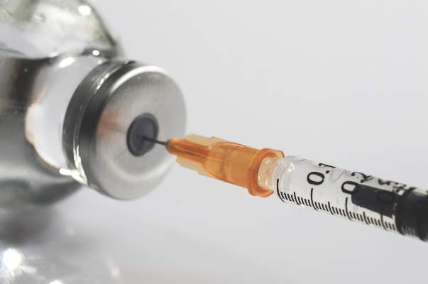
Flu shot linked to lower risk of hospitalization for influenza pneumonia
Patients hospitalized with laboratory-confirmed, influenza-associated pneumonia had a 57% lower odds of having received the influenza vaccine than controls whose pneumonia was due to other causes, investigators reported Oct. 5 in JAMA.
The findings could be used in future studies to estimate the number of hospitalizations prevented by influenza vaccination, according to Dr. Carlos Grijalva of Vanderbilt University, Nashville, Tenn., and his associates.
Seasonal influenza causes about 226,000 hospitalizations and 3,000 to 49,000 deaths every year in the United States. Observational studies show that influenza vaccination helps prevent hospitalizations for acute respiratory illness, but whether it also cuts the odds of hospitalization for community-acquired pneumonia is unknown, the investigators wrote.
To explore this question, they conducted an observational, multicenter study of 2,767 patients who had been hospitalized with community-acquired pneumonia over 3 consecutive influenza seasons at four sites in the United States. Patients were at least 6 months old, were not severely immunosuppressed, and had not been recently hospitalized or resided in a long-term care facility (JAMA. 2015 Oct 5, doi:10.1001/jama.2015.12160.).
In all, 162 (6%) patients had laboratory-confirmed influenza, including 17% who had received the influenza vaccine, the researchers wrote. In contrast, 29% of controls had received the vaccine, for an adjusted odds ratio of 0.43 (95% confidence interval, 0.28 to 0.68) after controlling for demographic characteristics, comorbidities, influenza season, study site, and time of disease onset. The estimated vaccine effectiveness was 57%.
The test-positive case, test-negative control design is widely used to study vaccine effectiveness and is better than comparing hospitalized cases with population controls, because it “implicitly” accounts for the risk of hospitalization, the researchers wrote. But “despite enrollment over 3 consecutive seasons, a relatively small number of influenza-associated pneumonia cases met eligibility criteria, resulting in limited precision for some subgroup analyses,” they added. “Thus, the association between influenza vaccines and pneumonia among older adults remains controversial, and additional studies in this group are needed.”
Dr. Grijalva reported having served as a consultant to Pfizer. Several coauthors reported having received grant and other support from the National Institutes of Health, the Agency for Healthcare Research and Quality, Medscape, MedImmune, Roche, Abbvie, and a number of pharmaceutical companies.
FROM JAMA
Key clinical point: Hospitalized patients with laboratory-confirmed, influenza-associated pneumonia were less likely to have been vaccinated against influenza than hospitalized controls with non-influenza pneumonia.
Major finding: Influenza-associated pneumonia patients had a 57% lower odds of having been vaccinated against influenza than controls (adjusted odds ratio, 0.43).
Data source: An observational, multicenter study of 2,767 hospitalizations for community-acquired pneumonia at four sites in the United States.
Disclosures: The Centers for Disease Control and Prevention funded the study. Dr. Grijalva reported having served as a consultant to Pfizer. Several coauthors reported having received grant and other support from the National Institutes of Health, the Agency for Healthcare Research and Quality, Medscape, MedImmune, Roche, Abbvie, and a number of pharmaceutical companies.

Expert Witness Primer Offers Tips for Hospitalists
Editor’s note: Second in a two-part series on hospitalists as expert witnesses.
You have officially decided to take the plunge and become an expert witness, but you have never seen the inside of a courtroom, sat for a deposition, or prepared an expert report. This article serves as a primer for all of those things, as well as testifying at trial.
Given the tremendous advantage to be gained by having the expert available to advise the attorney in preparing discovery and responding to the opposing attorney’s discovery, hopefully you have been actively involved in the litigation process and are not trying to get up to speed just weeks or even days before your deposition or the deadline for your expert report.
Steps you can take to become an indispensable expert witness, above and beyond your expert report, deposition, and trial testimony, include:
- Familiarizing yourself with all relevant aspects of the case so that you understand where your opinion fits in;
- Advising the attorney of both favorable and unfavorable facts;
- Identifying key documents that must be obtained;
- Spotting false or weak assumptions and inadequate work by the opposing expert; and/or
- Providing peer-reviewed journal articles and other literature, which decipher complex subjects for the attorney.
Expert Reports
Now that you have become an indispensable expert, what needs to be included in your expert report? If the matter is in state court, the content of the expert report will depend on state court rules that vary by jurisdiction and the judge’s own preferences. In federal court, the mandatory signed expert report must contain at least the following six things:
- A complete statement of all opinions the witness will express and the basis and reasons for these opinions;
- The facts or data considered by the witness in forming them;
- Any exhibits that will be used to summarize or support them;
- The witness’s qualifications, including a list of all publications authored in the previous 10 years;
- A list of all other cases in which, during the previous four years, the witness testified as an expert at trial or by deposition; and
- A statement of the compensation to be paid for the study and testimony in the case.
The report is due at least 90 days before the case is set for trial. The expert then has the opportunity to submit a rebuttal report 30 days after receipt of the opposing expert’s report “solely to contradict or rebut” that report.
In preparing the expert report, it is important to remember that, in essence, everything the expert touches is discoverable by the other side. So before you decide to jot down a note to yourself, consider the fact that that note may need to be produced to the other side. Be especially careful not to jot down editorial comments on documents, particularly deposition transcripts. Imagine the cross-examiner’s delight at finding the penned-in words “problem area” or “smoking gun” or “discuss issue with attorney” next to some unfavorable fact regarding the client. The rule of thumb is “the more unnecessary notes, the longer the deposition.” On the other hand, it may be essential to preserve notes containing calculations, formulas, measurements, and similar documentation to support your opinions.
Additionally, any communications with your attorney and drafts of the report are not privileged. So you need to make sure that it is you—and you alone—who is writing the report.
Depositions
As mentioned in the first article, testifying under oath, whether in a deposition or trial setting, can be a grueling experience. This is especially true if the deposition is videotaped or the trial is a high-profile case for which media might be present in the courtroom.
Although it may not be granted, you should request a convenient day, time, and place, including your office if you prefer, for your deposition. Some hospitalists prefer to have the deposition at their office because it minimizes the time they are unable to engage in patient care. Other hospitalists prefer to be in a more private setting, such as the opposing counsel’s law firm office, so that their patients are not aware of their expert witness activities.
Typically, the deposition takes place at an attorney’s office, with the attorneys for the parties, the parties themselves, and a court reporter present. The deposition begins with the court reporter swearing in the expert witness so that all of the expert’s answers are under oath.
At the deposition, it is the expert’s job to tell the truth briefly. Telling the truth briefly means providing accurate answers to questions after they are understood—and clarified if necessary—and stating those accurate answers in as short a way as possible without unnecessary adverbs, adjectives, parentheticals, footnotes, asides, qualifications, and other unrequested information. The rule of thumb is that the more information an expert volunteers, the longer the deposition and ability to cross-examine will be.
Often it is helpful to engage in role playing with the attorney to explore likely initial and follow-up questions from opposing counsel. Typically, the format of these questions will include who, what, when, where, why, how, tell us, describe, or explain. You should also review important documents, so that you have a familiarity and comfort with the documents considered part of your analysis and are prepared to interpret them and explain their significance.
At the deposition, you will likely be asked if you reviewed any documents in preparation and, specifically, which ones you examined.
Just as you would in a trial situation, you should pause after a question is asked, to allow your attorney to make an appropriate objection to the question.
It should be noted that the top six answers to most deposition questions are:
- Yes;
- No;
- I don’t know;
- I don’t remember;
- I don’t understand the question; and
- I need a break.
Don’t be afraid to answer “yes” or “no” to a yes or no question or to use “I don’t know” when it’s the most accurate answer. The last piece of advice for depositions is to remember at all times that the deposing attorney is not your friend.
Trial Testimony
Getting ready for trial will be much the same as preparing for the deposition; you want to ensure that your testimony is consistent and protect yourself from potential impeachment. The focus, however, is a different audience; you are educating the judge and jury in a way that will make your testimony understandable and consistent with the jury’s common sense.
You will again be sworn in during both direct and cross-examination. If there is an objection to the form of the question or to your testimony, you should again stop and wait for the judge to instruct whether or not to answer the question and in what manner. Direct examination is likely to include questions based upon your qualifications, methodology, basis or assumptions, and anticipated cross. In responding, remember to look directly at counsel while the question is being asked and then at the jury in explaining the answer.
There is no question that serving as an expert witness is challenging and rewarding work. Are you ready for the challenge?
Hospitalist Lance Maki, MD, Spends Spare Time Tandem Surfing, Practicing Ballet

Lance Maki, MD, has accomplished many things in his life. He joined the Air Force and flew KC-135 tankers as an aircraft commander, and he served as a flight surgeon and T-38 instructor pilot. As an OB/GYN physician, he worked in private practice. Now he is a bicoastal hospitalist and intimacy therapist. Still, it’s what he does in his spare time that attracts the most attention.
Dr. Maki is a tandem surfer and ballet dancer.
Tandem what? Ballet dancer? The kind who wears tights, stands on his tiptoes, and leaps into the air?
Make no mistake. At 5 feet, 10 inches and 190 pounds, this 68-year-old doctor is no weakling. Ballet requires the strength and coordination to leap high into the air while doing the splits. Tandem surfing demands even more skill and similar strength. The sport requires surfers to lift someone half their weight or more above their head and hold them in various poses while riding four- to six-foot high ocean waves on a surfboard less than two feet wide.
“We live in a crazy world,” says Dr. Maki, explaining that very little compares to surfing with dolphins and manatees. “When you enjoy life, you’re well-rounded and have that mind-body-spirit connection. You’re going to be a much better doctor and much more pleasant to be around.”
Practice, Persistence, and Prayers
Dr. Maki’s fascination with the ocean began in 1960, when his family vacationed in California. The following year, when he was in high school, they moved from his hometown in St. Johns, Mich., to La Mirada, Calif. During his senior year of high school, he says he surfed 150 days.
Back then, surfing was simply fun, nothing more. While attending California State University at Fullerton, he rarely surfed. There were too many things to do. In 1967, he married Kristine, now a nurse practitioner, and he joined the Air Force in 1972. He served as a pilot for the next 12 years.
The couple had six children from 1970 to 1982. Two years later, on an Air Force scholarship at age 37, he attended Texas Tech University Health Sciences Center School of Medicine in Lubbock.
After graduating from medical school in 1988, he returned to active duty and completed his OB/GYN residency at Wright State University and Miami Valley Hospital, which were affiliated with Wright-Patterson Air Force Base in Dayton, Ohio. He spent another four years as an OB/GYN doctor and flight surgeon at Griffiss Air Force base in upstate New York. After retiring from the Air Force in 1996, he moved his family to Tipton, Ind., where he started an OB/GYN private practice.
That same year, his 14-year-old son started exhibiting normal teenage behavioral problems. Before it got out of hand, Kristine suggested that Dr. Maki enroll him in a structured and positive activity like surf camp.
“I said there aren’t any oceans in Indiana. I can’t surf anymore,” recalls Dr. Maki, now a devout Catholic who prays for a good and safe surf once he gets past the breakers.
Still, Kristine persisted, so Dr. Maki found a surf camp in San Clemente, Calif. As it turned out, Michael didn’t care for surfing and, as Dr. Maki quickly discovered, surfing wasn’t like riding a bike. It takes a while to remember how to just stay on the board.
“I went surfing and was absolutely terrible,” he says. “I was ready to quit, but people encouraged me to get on a big, old, fat surfboard, and pushed me into a wave. All of a sudden, it was like I was back surfing in high school.”
Dr. Maki’s renewed interest in surfing quickly evolved into his favorite passion. The family moved again, to Florida in 2002. Dr. Maki has worked as a locum tenens hospitalist for Ob Hospitalist Group at various facilities in California and Florida.
Through his surfing network, he learned about tandem surfing. Although Kristine and his friends believed he was “too old” and “too much of a klutz,” he was determined. So, in 2007, he traveled to Hawaii and—at the age of 60—learned how to tandem surf. Ironically, Kristine found him the perfect tandem partner—a family friend who was five years his junior and half his size and weight.
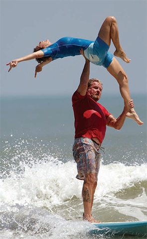
For almost two years, they trained with an Olympic gymnast learning lifts.
“He would have us lie down on the mat and, over and over again, get up as fast as we could and go into a lift,” he says. “Florida waves are very short-lived. We worked like mad at that.”
Dance, Dance, Dance
Besides surfing every other day, Dr. Maki has taken 90-minute ballet classes twice a week for the past five years. He works with a trainer for an hour, also twice a week.
“Without bragging, I have to say I’m much better now than I was when I first started surfing back in 1960,” he says. “I do pushups, calisthenics, and use a ballet bar and a balancing training board called an indo board.”
In 2012, he and his tandem surfing partner went on the International Tandem Surf Association’s world tour. They surfed in contests in Virginia, California, Hawaii, Florida, and France, earning 11th place overall.
But this year, he’s taking time off. Not to worry, though. When he turns 70, he plans on returning to the World Tandem Tour.
The break, he says, will allow him to focus more on his ballet. For the past three holiday seasons, he has played the role of Herr Drosselmeyer in The Nutcracker at Cocoa Village Playhouse in Cocoa Village, Fla.
“I hope to be dancing ballet and tandem surfing until I can’t walk anymore, because they’re so much fun,” Dr. Maki says. “If you have a positive attitude and do your best to be happy with what you’re doing at work—some days can be brutal as a hospitalist—it carries over to your patients and they heal faster. You don’t get healed by medicine alone.”
Carol Patton is a freelance writer in Las Vegas.
Lance Maki, MD, has accomplished many things in his life. He joined the Air Force and flew KC-135 tankers as an aircraft commander, and he served as a flight surgeon and T-38 instructor pilot. As an OB/GYN physician, he worked in private practice. Now he is a bicoastal hospitalist and intimacy therapist. Still, it’s what he does in his spare time that attracts the most attention.
Dr. Maki is a tandem surfer and ballet dancer.
Tandem what? Ballet dancer? The kind who wears tights, stands on his tiptoes, and leaps into the air?
Make no mistake. At 5 feet, 10 inches and 190 pounds, this 68-year-old doctor is no weakling. Ballet requires the strength and coordination to leap high into the air while doing the splits. Tandem surfing demands even more skill and similar strength. The sport requires surfers to lift someone half their weight or more above their head and hold them in various poses while riding four- to six-foot high ocean waves on a surfboard less than two feet wide.
“We live in a crazy world,” says Dr. Maki, explaining that very little compares to surfing with dolphins and manatees. “When you enjoy life, you’re well-rounded and have that mind-body-spirit connection. You’re going to be a much better doctor and much more pleasant to be around.”
Practice, Persistence, and Prayers
Dr. Maki’s fascination with the ocean began in 1960, when his family vacationed in California. The following year, when he was in high school, they moved from his hometown in St. Johns, Mich., to La Mirada, Calif. During his senior year of high school, he says he surfed 150 days.
Back then, surfing was simply fun, nothing more. While attending California State University at Fullerton, he rarely surfed. There were too many things to do. In 1967, he married Kristine, now a nurse practitioner, and he joined the Air Force in 1972. He served as a pilot for the next 12 years.
The couple had six children from 1970 to 1982. Two years later, on an Air Force scholarship at age 37, he attended Texas Tech University Health Sciences Center School of Medicine in Lubbock.
After graduating from medical school in 1988, he returned to active duty and completed his OB/GYN residency at Wright State University and Miami Valley Hospital, which were affiliated with Wright-Patterson Air Force Base in Dayton, Ohio. He spent another four years as an OB/GYN doctor and flight surgeon at Griffiss Air Force base in upstate New York. After retiring from the Air Force in 1996, he moved his family to Tipton, Ind., where he started an OB/GYN private practice.
That same year, his 14-year-old son started exhibiting normal teenage behavioral problems. Before it got out of hand, Kristine suggested that Dr. Maki enroll him in a structured and positive activity like surf camp.
“I said there aren’t any oceans in Indiana. I can’t surf anymore,” recalls Dr. Maki, now a devout Catholic who prays for a good and safe surf once he gets past the breakers.
Still, Kristine persisted, so Dr. Maki found a surf camp in San Clemente, Calif. As it turned out, Michael didn’t care for surfing and, as Dr. Maki quickly discovered, surfing wasn’t like riding a bike. It takes a while to remember how to just stay on the board.
“I went surfing and was absolutely terrible,” he says. “I was ready to quit, but people encouraged me to get on a big, old, fat surfboard, and pushed me into a wave. All of a sudden, it was like I was back surfing in high school.”
Dr. Maki’s renewed interest in surfing quickly evolved into his favorite passion. The family moved again, to Florida in 2002. Dr. Maki has worked as a locum tenens hospitalist for Ob Hospitalist Group at various facilities in California and Florida.
Through his surfing network, he learned about tandem surfing. Although Kristine and his friends believed he was “too old” and “too much of a klutz,” he was determined. So, in 2007, he traveled to Hawaii and—at the age of 60—learned how to tandem surf. Ironically, Kristine found him the perfect tandem partner—a family friend who was five years his junior and half his size and weight.
For almost two years, they trained with an Olympic gymnast learning lifts.
“He would have us lie down on the mat and, over and over again, get up as fast as we could and go into a lift,” he says. “Florida waves are very short-lived. We worked like mad at that.”
Dance, Dance, Dance
Besides surfing every other day, Dr. Maki has taken 90-minute ballet classes twice a week for the past five years. He works with a trainer for an hour, also twice a week.
“Without bragging, I have to say I’m much better now than I was when I first started surfing back in 1960,” he says. “I do pushups, calisthenics, and use a ballet bar and a balancing training board called an indo board.”
In 2012, he and his tandem surfing partner went on the International Tandem Surf Association’s world tour. They surfed in contests in Virginia, California, Hawaii, Florida, and France, earning 11th place overall.
But this year, he’s taking time off. Not to worry, though. When he turns 70, he plans on returning to the World Tandem Tour.
The break, he says, will allow him to focus more on his ballet. For the past three holiday seasons, he has played the role of Herr Drosselmeyer in The Nutcracker at Cocoa Village Playhouse in Cocoa Village, Fla.
“I hope to be dancing ballet and tandem surfing until I can’t walk anymore, because they’re so much fun,” Dr. Maki says. “If you have a positive attitude and do your best to be happy with what you’re doing at work—some days can be brutal as a hospitalist—it carries over to your patients and they heal faster. You don’t get healed by medicine alone.”
Carol Patton is a freelance writer in Las Vegas.
Hospitalists Key Partners in Healthcare’s Future, Evolution
After a career working for hospitals, I am about to retire as president and CEO of the American Hospital Association (AHA), an organization that represents some 5,000 hospitals and health systems. This moment compels me to look at the past—what we have learned and how hospitals have changed—and consider the possibilities the future holds for hospitals and hospitalists.
I have watched as hospitals have triumphed over tragedies, from natural disasters to mass shootings. More recently, I saw hospitalists pour their hearts and souls into preparing for the possibility of Ebola. Time and time again, you have responded through your deep-seated commitment.
I have observed the journey toward operational excellence through a punishing recession, a government shutdown, and burdensome regulations that make day-to-day operations amazingly complicated. Yet costs have moderated in historic ways. In fact, hospitals are tackling the tough problems of quality and safety that have plagued us for generations, from preventable infections to disparities to system fragmentation, with a commitment that says to all: This is not acceptable. This will change. And the results show great improvements.
On a clinical level, we’ve made dramatic advances. New technologies and treatments mean that we routinely cure conditions in patients who would once have been without hope. We can also restore quality of life to patients who previously, after an illness or injury, would have spent the rest of their lives struggling with the tasks of everyday living.
But the most remarkable transformation that has taken place in America’s hospitals over my lifetime is in the culture. The focus on patient-centered care has required a new mindset that empowers patients to make decisions about their own care and pushes healthcare workers to much higher levels of coordination and communication. A significant factor in this has been the introduction, growth, and maturation of the hospitalist specialty.
One result of this transformation is that physicians, nurses, and other clinical staff, who once worked in separate silos, are increasingly working as teams. Hospitalists often lead these teams. Clinical integration is the catalyst for profound improvements in patient care. Team-based care is more efficient; sharing information about a patient lessens the chance of duplication of services and increases the use of protocols shown to improve patient outcomes. Clinical integration also helps hospitals develop and implement best practices, and that is making it possible to achieve dramatic progress in tackling some stubborn problems that have plagued these facilities for years, such as healthcare-associated infections.
Moving forward, hospitals are intensely focused on achieving three critical goals: improving the patient care experience, improving the overall health of the community, and reducing the per capita cost of health care. Accomplishing those goals means accelerating the pace of change even further.
For years, we’ve been moving toward a system that is more integrated, with hospitals, physicians, and post-acute care providers combining forces to make true systems of care available to patients. Payments are more at risk as we move away from the fee-for-service model we’ve had for decades to a system that rewards value and outcomes. Healthcare is also becoming far more accountable and transparent about quality and pricing. Hospitals that tried to do better with less are now trying to do less with less, demanding that expensive procedures lead to better patient outcomes. They are focusing more on prevention and less on intervention.
All of this has established the foundation for the next generation of transition. What will it look like?
Efficient, Value-Based Approaches
The clinical gains we have made in healthcare are associated with tremendous costs for specialized equipment and services. By combining in some fashion, rather than duplicating, these resources, hospitals can continue to provide patients with the most promising advances in treatment. As a result, more hospitals are part of health systems that share multiple resources in order to deliver the best care with the best value. More hospitals employ physicians and other clinicians. And this trend will accelerate.
Every hospital will need to determine the path that makes the best sense for itself and its community. Some hospitals will form strategic alliances with other healthcare providers, merging with or acquiring them to offer patients the best they have to offer. Expect to see more hospitals develop a health insurance function and still more to branch out into areas such as behavioral health, home health, or post-acute, long-term, or ambulatory care. Other hospitals will choose the opposite route—specialization in a single area where they can become a high-performing provider of essential services. Examples are children’s hospitals and rehabilitation centers.
No matter which route your hospital takes, expect to see it become increasingly involved in efforts to improve the health of the community it serves. Hospitals will define themselves less by the walls of their buildings and more by the health of their communities. They will actively seek the perspectives of patients and families on how they operate.
New Ideas Welcome
We have an aging population and a growing number of people of all ages with chronic conditions like diabetes and asthma. There’s a lot of room for improvement that will come about by engaging people in the prevention and management of chronic conditions and the employment of new technologies like telehealth. Some 40% of premature deaths stem from unhealthy behavior. By finding effective ways to help people stay healthy, hospitals can have a huge impact in controlling the growth of healthcare spending. Hospitals will also be working to engage patients and families in making decisions about treating advanced illness, including end-of-life care.
Health information technology and electronic health records, done right, will provide hospitals with new ways to improve the quality of care. With better information, we don’t have to guess. We are collecting, analyzing, and applying information—and transforming it into knowledge about what works, and what doesn’t, for patients. For example, by analyzing race, ethnicity, and language preference data, hospitals can address disparities in outcomes for certain populations. This adjustment is critical at a time when communities are changing and hospitals must change to reflect their needs. Better use of information will also allow hospitals to develop and share more evidence-based practices.
In short, hospitals will undergo nothing short of reformation in the years ahead. The demands are daunting, the excitement is contagious, and the commitment to communities is immense.
It has been a tremendous privilege to spend my career with the women and men of America’s hospitals, good people who are willing and able to do whatever it takes to deliver the highest quality care to the people who rely upon them.
Experimental Antibody May Reduce C. diff Recurrence
An experimental antibody developed by Merck & Co Inc was shown in pivotal studies to reduce by about 10 percentage points the risk that infection with Clostridium difficile will recur.
In the United States, C. difficile infects nearly half a million people each year and contributes to around 29,000 deaths. The infection is treated with standard antibiotics, which also wipe out healthy bacteria that normally keep C. difficile under control.
Merck said two Phase 3 studies found 12 weeks of treatment with antibiotics and a one-time infusion of bezlotoxumab, designed to block the ability of a toxin to bind to cells, reduced to about 15% the risk that C. difficile would recur. The studies found that the infection recurred in about 25% of patients treated with antibiotics and a placebo.
"We have therapies to treat the initial episode, but this infection comes back frequently - there is a 25% risk of recurrence after the first time, and that rises to 40% or even 60% after the second infection," said Nick Kartsonis, associate vice president in clinical research, infectious diseases at Merck.
The studies showed no benefit from a second experimental antibody, actoxumab, either alone or in combination with bezlotoxumab. Merck said the actoxumab arm was stopped for efficacy and safety reasons after an interim analysis.
The studies were presented September 20 at the Interscience Conference of Antimicrobial Agents and Chemotherapy (ICAAC) and International Congress of Chemotherapy and Infection (ICC) joint meeting in San Diego.
Bezlotoxumab is a selective, fully-human, monoclonal antibody designed to neutralize C. difficile toxin B.
The company said it plans to file before the end of the year for regulatory approval of bezlotoxumab, which it licensed from Massachusetts Biologic Laboratories and Medarex, now owned by Bristol-Myers Squibb.
Side effects, including nausea, diarrhea and urinary tract infection, occurred at similar rates for patients in both the treatment and placebo arms of the trials.
The incidence of C. difficile infection has risen sharply over the last two decades and is now a leading cause of healthcare-acquired infections in community hospitals in the United States, according to the U.S. Centers for Disease Control and Prevention.
Other companies are working on vaccines against C. difficile. Doctors are also treating patients with "stool transplants," which involves inserting fecal material from a healthy person into the gut of someone with severe diarrhea in order to restore friendly bacteria.
Hospitalists’ Code of Conduct Needed for Sick Day Callouts
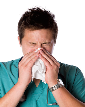
It is Tuesday morning, and I drag myself out of bed after a very restless night. It is day number three of a syndrome of fatigue, headache, and moderate productive cough. I have been on service for eight days of a two-week stretch; I am hoping to “make it to the end of the week.” I convince myself I am “not that sick” and head into work for a long day of rounds, after two cups of coffee and 600 mg of Motrin. Throughout the day, I try to hide my cough from my residents and students, and especially the nurses and my patients. I have a pocket full of cough drops and a cup of ice water at hand to stifle any coughing fits that could reveal how I actually feel. This is not the first time I have come to work only “half well.” I convince myself I am not contagious, as long as I wash my hands and control my cough. Without a fever, how could I possibly justify calling in a colleague to cover for me?
I am not alone in my psychological justifications for coming to work. A recent JAMA Pediatrics article found that 83% of clinicians admitted to coming to work while sick, while 95% admitted to knowing that it could be dangerous to their patients.1,2 The study surveyed approximately 500 attendings and 250 advanced practice providers at the Children’s Hospital of Philadelphia. A substantial minority of providers (9%) admitted to coming to work sick at least five times in the past year.
The reasons these providers gave for working in spite of being ill likely ring true with each and every hospitalist in the field: They were concerned about 1) letting down their patients or 2) hospital staffing in their absence. Most providers also expressed concern about the continuity of care for their patients in their absence. Most also admitted that they feared being ostracized by their colleagues and believed that there were unwritten but real expectations for them to work regardless of personal illness.
Historically, physicians and other healthcare providers have been widely believed to be relatively immune to mundane ailments, by themselves and by others. How incredibly rare it is to hear, “Sorry, your doctor is sick; we have to reschedule your visit.” Even when afflicted by physical impairments, physicians have long considered it more “honorable” to work through these infirmities than to resign to physical limitations and ask for help.
Misguided or Mishandled
This sense of duty starts early in medical training and continues throughout a physician’s early career. I discovered this firsthand during my internship after suffering a stress fracture in my foot. I woke up one morning with significant foot pain and swelling but hobbled through rounds without a word spoken about my limp. By the afternoon, I could hardly bear weight on my foot, so one of my fellow interns suggested I limp over to the orthopedic clinic; thankfully, they saw me the same day, diagnosed the stress fracture, and fitted me in a walking cast. The next day on rounds, when I asked my attending if we could take the elevator up the two floors to the next patient, he looked annoyed and said I could meet them there; they scurried up the stairs. For the next few weeks, I never missed a minute of work but kept trailing behind and missing key pieces of presentations and information from rounds, having to hobble back and forth to the elevator between floors.
The lesson I quickly learned back then was that if I was not “fit for duty” with any sort of physical ailment, it was clearly my problem to make up for my deficits, because the work expectations would go unchanged. Although a stress fracture did not put my patients at risk, the experience sent a strong message: Regardless of the impact on patients, it is always better to come to work than to stay home, whatever the type or degree of affliction.
The JAMA Pediatrics study did find substantial differences in the types of symptoms that would keep a provider at home: While 75% reported they would come to work with a cough and rhinorrhea, 30% would come with diarrhea, 16% would come with a fever, and only 5% would come with vomiting.
To be honest, this sounds about right in comparison to what my threshold would be, and it is about what I would accept as reasonable from a colleague. I do hope that if I were “really sick,” with fever and/or vomiting, I would have the good sense to stay home and ask for coverage, and I hope my colleagues and I would support each other in these decisions.
The study really gets at the sociocultural factors that steer physicians into making such decisions, based on the conditions for being excused that they think are socially acceptable. I suspect these are similar to those that other industries would also consider acceptable. But, of course, the difference is that workers in other industries are less likely to cause harm to large numbers of vulnerable and innocent “bystanders.” Adding to the problem, there is no good “definition” for what is “too sick”; although it is complicated and varies by person, the definition should at least take into account the level of potential contagion and risk to patients.
The authors suggest that, in order to remedy this longstanding situation, open dialogue needs to take place among physician groups to reduce the ambiguity about what is appropriate. A good start would be the generation of clear policies that restrict providers from coming to work with specifics signs/symptoms.
As hospitalists, we should all discuss the article within our groups and honestly determine in advance what our “code of conduct” should be for illnesses, based on our provider mix and our patient populations. (Decisions for ICU, medical-surgical, or oncology may vary.) This would reduce ambiguity and create new social norms about when to stay home. In addition, administrative and provider group leaders need to show strong leadership and support for such policies and ensure adequate staffing in the event of appropriate callouts. Such policies need to ensure that callouts are equitable and non-punitive. These relatively simple measures would go a long way in reducing the risk of illness among ourselves and our patients.

References
- Szymczak JE, Smathers S, Hoegg C, Klieger S, Coffin SE, Sammons JS. Reasons why physicians and advanced practice clinicians work while sick: A mixed methods analysis [published online ahead of print July 6, 2015]. JAMA Pediatr. doi:10.1001/jamapediatrics.2015.0684.
- Starke JR, Jackson MA. When the health care worker is sick: primum non nocere [published online ahead of print July 6, 2015]. JAMA Pediatr. doi:10.1001/jamapediatrics.2015.0994.
It is Tuesday morning, and I drag myself out of bed after a very restless night. It is day number three of a syndrome of fatigue, headache, and moderate productive cough. I have been on service for eight days of a two-week stretch; I am hoping to “make it to the end of the week.” I convince myself I am “not that sick” and head into work for a long day of rounds, after two cups of coffee and 600 mg of Motrin. Throughout the day, I try to hide my cough from my residents and students, and especially the nurses and my patients. I have a pocket full of cough drops and a cup of ice water at hand to stifle any coughing fits that could reveal how I actually feel. This is not the first time I have come to work only “half well.” I convince myself I am not contagious, as long as I wash my hands and control my cough. Without a fever, how could I possibly justify calling in a colleague to cover for me?
I am not alone in my psychological justifications for coming to work. A recent JAMA Pediatrics article found that 83% of clinicians admitted to coming to work while sick, while 95% admitted to knowing that it could be dangerous to their patients.1,2 The study surveyed approximately 500 attendings and 250 advanced practice providers at the Children’s Hospital of Philadelphia. A substantial minority of providers (9%) admitted to coming to work sick at least five times in the past year.
The reasons these providers gave for working in spite of being ill likely ring true with each and every hospitalist in the field: They were concerned about 1) letting down their patients or 2) hospital staffing in their absence. Most providers also expressed concern about the continuity of care for their patients in their absence. Most also admitted that they feared being ostracized by their colleagues and believed that there were unwritten but real expectations for them to work regardless of personal illness.
Historically, physicians and other healthcare providers have been widely believed to be relatively immune to mundane ailments, by themselves and by others. How incredibly rare it is to hear, “Sorry, your doctor is sick; we have to reschedule your visit.” Even when afflicted by physical impairments, physicians have long considered it more “honorable” to work through these infirmities than to resign to physical limitations and ask for help.
Misguided or Mishandled
This sense of duty starts early in medical training and continues throughout a physician’s early career. I discovered this firsthand during my internship after suffering a stress fracture in my foot. I woke up one morning with significant foot pain and swelling but hobbled through rounds without a word spoken about my limp. By the afternoon, I could hardly bear weight on my foot, so one of my fellow interns suggested I limp over to the orthopedic clinic; thankfully, they saw me the same day, diagnosed the stress fracture, and fitted me in a walking cast. The next day on rounds, when I asked my attending if we could take the elevator up the two floors to the next patient, he looked annoyed and said I could meet them there; they scurried up the stairs. For the next few weeks, I never missed a minute of work but kept trailing behind and missing key pieces of presentations and information from rounds, having to hobble back and forth to the elevator between floors.
The lesson I quickly learned back then was that if I was not “fit for duty” with any sort of physical ailment, it was clearly my problem to make up for my deficits, because the work expectations would go unchanged. Although a stress fracture did not put my patients at risk, the experience sent a strong message: Regardless of the impact on patients, it is always better to come to work than to stay home, whatever the type or degree of affliction.
The JAMA Pediatrics study did find substantial differences in the types of symptoms that would keep a provider at home: While 75% reported they would come to work with a cough and rhinorrhea, 30% would come with diarrhea, 16% would come with a fever, and only 5% would come with vomiting.
To be honest, this sounds about right in comparison to what my threshold would be, and it is about what I would accept as reasonable from a colleague. I do hope that if I were “really sick,” with fever and/or vomiting, I would have the good sense to stay home and ask for coverage, and I hope my colleagues and I would support each other in these decisions.
The study really gets at the sociocultural factors that steer physicians into making such decisions, based on the conditions for being excused that they think are socially acceptable. I suspect these are similar to those that other industries would also consider acceptable. But, of course, the difference is that workers in other industries are less likely to cause harm to large numbers of vulnerable and innocent “bystanders.” Adding to the problem, there is no good “definition” for what is “too sick”; although it is complicated and varies by person, the definition should at least take into account the level of potential contagion and risk to patients.
The authors suggest that, in order to remedy this longstanding situation, open dialogue needs to take place among physician groups to reduce the ambiguity about what is appropriate. A good start would be the generation of clear policies that restrict providers from coming to work with specifics signs/symptoms.
As hospitalists, we should all discuss the article within our groups and honestly determine in advance what our “code of conduct” should be for illnesses, based on our provider mix and our patient populations. (Decisions for ICU, medical-surgical, or oncology may vary.) This would reduce ambiguity and create new social norms about when to stay home. In addition, administrative and provider group leaders need to show strong leadership and support for such policies and ensure adequate staffing in the event of appropriate callouts. Such policies need to ensure that callouts are equitable and non-punitive. These relatively simple measures would go a long way in reducing the risk of illness among ourselves and our patients.
References
- Szymczak JE, Smathers S, Hoegg C, Klieger S, Coffin SE, Sammons JS. Reasons why physicians and advanced practice clinicians work while sick: A mixed methods analysis [published online ahead of print July 6, 2015]. JAMA Pediatr. doi:10.1001/jamapediatrics.2015.0684.
- Starke JR, Jackson MA. When the health care worker is sick: primum non nocere [published online ahead of print July 6, 2015]. JAMA Pediatr. doi:10.1001/jamapediatrics.2015.0994.
Hospital Groups Might Do Better Without Daytime Admission Shifts, Morning Meetings

You shouldn’t maintain things that do not deliver the value you anticipated when you first put them in place. For example, I thought Netflix streaming would be terrific, but I have used it so infrequently that it probably costs me $50 per movie or show watched. I should probably dump it.
Your hospitalist group might have some operational practices that are not as valuable as they seem and could be replaced with something better. For many groups, this might include doing away with a separate daytime admitter shift and a morning meeting to distribute the overnight admissions.
Daytime Admission Shift
My experience is that hospitalist groups with more than about five daytime doctors almost always have a day-shift person dedicated to seeing new admissions. In most cases, this procedure is implemented with the idea of reducing the stress of other day-shift doctors, who don’t have to interrupt rounds to admit a new patient. Some see a dedicated admitter as a tool to improve ED throughput, because this doctor isn’t tied up with rounds and can immediately start seeing a new admission.
I think an admitter shift does deliver both of these benefits, but its costs make it suboptimal in most settings. For example, a single admitter will impede ED throughput any time more than one new admission is waiting to be seen, and for most groups that will be much of the day. In fact, improved ED throughput is best achieved by having many hospitalists available for admissions, not just a single admitter. (There are many other factors influencing ED throughput, such as whether ED doctors simply send patients to their “floor” bed prior to being seen by a hospitalist. But for this article, I’m just considering the influence of a dedicated admitter.)
I think “silo-ing” work into different roles, such as separating rounding and admitting, makes it more difficult to ensure that each is always working productively. There are likely to be times when the admitter has little or nothing to do, even though the rounders are very busy. Or perhaps the rounders aren’t very busy, but the admitter has just been asked to admit four ED patients at the same time.
While protecting rounders from the stress of admissions is valuable, it comes at the cost of a net increase in hospitalist work, because a new doctor must get to know the patient on the day following admission. And this admitter-to-rounder handoff serves as another opportunity for errors—and probably lowers patient satisfaction.
I think most groups should consider moving the admitter shift into an additional rounder position, dividing admissions across all of the doctors working during the daytime. For example, a group that has six rounders and a separate admitter would change to seven rounders, each available to admit every seventh daytime admission. Each would bear the meaningful stress of having rounds interrupted to admit a new patient, but accepting every seventh daytime admission shouldn’t be too difficult on most days.
Don’t forget that eliminating the admitter means that the list of new patients you take on each morning will be shorter. Mornings may be a little less stressful.
A.M. Distribution
The daytime doctors at many hospitalist groups meet each morning to discuss how the new admissions from the prior night (or even the last 24 hours) will be distributed. Or perhaps one person, sometimes a nurse or clerical staff, arrives very early each day to do this.
Although it might take some careful planning, I think most groups that use this sort of morning distribution should abandon it for a better system. Consider a group in which all six daytime doctors spend an average of 20 minutes distributing patients each morning. Twenty minutes (0.33 hours) times six doctors times 365 days comes to 730 hours annually.
Assuming these doctors are compensated at typical rates, the practice is spending more than $100,000 annually just so the doctors can distribute patients each morning. On top of this, nurses and others at the hospital are usually delayed in learning which daytime hospitalist is caring for each patient. These costs seem unreasonably high.
An alternative is to develop a system by which any admitter, such as a night doctor, who will not be providing subsequent care to a patient can identify by name the doctor who will be providing that care. During the admission encounter, the admitter can tell patient/family, “Dr. Boswell will be taking over your care starting tomorrow. He’s a great guy and has been named one of Portland’s best doctors.” This seems so much better than saying, “One of my partners will be taking over tomorrow. I don’t know which of my partners it will be, but they’re all good doctors.” And Dr. Boswell’s name can be entered into the attending physician field of the EHR so that all hospital staff will know without delay.
MedAptus has recently launched software they call “Assign” that may be able to replace the morning meeting and automate assigning new admissions to each hospitalist. I haven’t seen it in operation, so I can’t speak for its effectiveness, but it might be worthwhile for some groups.
Practical Considerations
The changes I’ve described above might not be optimal for every group, and they may take meaningful work to implement. But I don’t think the difficulty of these things is the biggest barrier. The biggest barrier is probably just inertia in most cases, the same reason I’m still a Netflix streaming subscriber even though I almost never watch it. I did, however, really enjoy the Nexflix original series Lilyhammer.