User login
Myth of the Month: NPO good for people with pancreatitis?
A 60-year-old man presents to the emergency department with nausea and abdominal pain, and is admitted with pancreatitis due to alcohol. In the evening after receiving pain medication, his abdominal pain is diminished but still present. He has an appetite and asks for food.
What do you recommend?
A. Nil per os (NPO) until pain is resolved.
B. NPO until amylase/lipase have normalized.
C. Nasogastric tube placement.
D. Okay to start feeding.
Myth: Treatment of pancreatitis includes early avoidance of food.

The conventional management of acute pancreatitis involves an NPO regimen until the pain and nausea have resolved.1 This dogma is offered because of the concern that food intake will stimulate pancreatic enzyme release in an already inflamed/injured pancreas.
The approach of NPO and slowly reintroducing feeding after prolonged periods of being without food is associated with pain relapses and increased length of hospitalizations.2 Nasojejunal feedings have become well accepted in patients with severe pancreatitis requiring ICU care.3
Are there data to show that oral feeding of patients with mild pancreatitis causes worse outcomes?
Dr. Niels Teich and colleagues randomized 143 hospitalized patients with mild pancreatitis to eating when they felt ready to (69 patients) vs. a group that were kept NPO until lipase levels returned to normal.4 The patients who started eating when they were ready left the hospital a day earlier than the patients who were fed only when lipase levels normalized (7 days vs. 8 days). There was no difference in abdominal pain between the two groups.
Dr. Maxim Petrov and colleagues looked at whether nasogastric tube feeding was preferable to NPO in patients with mild to moderate pancreatitis.5 In a randomized trial of 35 patients with pancreatitis, 17 received nasogastric feedings within 24 hours of admission, and 18 were NPO. The patients who received early nasogastric feedings had lower pain scores at 72 hours, compared with the NPO group (1 vs. 3 on a visual analog 10-point scale, P = .036). The number of patients who did not require opiates at 48 hours was also significantly less in the nasogastric feeding group (9 vs. 3, P = .024).
I think the most striking difference was in patients’ ability to tolerate oral feeding. Patients in both groups received oral food at an average of 4 days; only 1 of 17 patients in the nasogastric feeding group could not tolerate feeding, compared with 9 of 18 patients in the NPO group.
Dr. Gunilla Eckerwall and colleagues studied the outcome of immediate oral feeding in patients with mild pancreatitis.6 Sixty patients with mild acute pancreatitis, defined by amylase greater than 3 times normal and APACHE scores less than 8, were randomized to either immediate oral feeding (30 patients) or fasting (30 patients). Key outcome measures in the study were amylase, systemic inflammatory response, and length of hospital stay.
There were no differences in amylase levels, labs measuring systemic inflammatory response, or gastrointestinal symptoms between the two groups. The immediate oral feeding group had a significantly shorter length of hospital stay than the fasting group (4 days vs. 6 days, P less than .05).
So, what does all this tell us about feeding patients with acute pancreatitis? For mild to moderate acute pancreatitis, the outcomes appear to be no worse when patients are fed early. There may be a trend to quicker hospital discharge in those who get fed earlier. The studies have all been small, and a large multicenter trial would be welcome.
References
1. Gastroenterology. 2007 May;132(5):2022-44.
3. Am J Gastroenterol. 2006 Oct;101(10):2379-400.
4. Pancreas. 2010 Oct;39(7):1088-92.
5. Clin Nutr. 2013 Oct;32(5):697-703.
6. Clin Nutr. 2007 Dec;26(6):758-63.
Dr. Paauw is professor of medicine in the division of general internal medicine at the University of Washington, Seattle, and he serves as third-year medical student clerkship director at the University of Washington. Contact Dr. Paauw at [email protected].
A 60-year-old man presents to the emergency department with nausea and abdominal pain, and is admitted with pancreatitis due to alcohol. In the evening after receiving pain medication, his abdominal pain is diminished but still present. He has an appetite and asks for food.
What do you recommend?
A. Nil per os (NPO) until pain is resolved.
B. NPO until amylase/lipase have normalized.
C. Nasogastric tube placement.
D. Okay to start feeding.
Myth: Treatment of pancreatitis includes early avoidance of food.
The conventional management of acute pancreatitis involves an NPO regimen until the pain and nausea have resolved.1 This dogma is offered because of the concern that food intake will stimulate pancreatic enzyme release in an already inflamed/injured pancreas.
The approach of NPO and slowly reintroducing feeding after prolonged periods of being without food is associated with pain relapses and increased length of hospitalizations.2 Nasojejunal feedings have become well accepted in patients with severe pancreatitis requiring ICU care.3
Are there data to show that oral feeding of patients with mild pancreatitis causes worse outcomes?
Dr. Niels Teich and colleagues randomized 143 hospitalized patients with mild pancreatitis to eating when they felt ready to (69 patients) vs. a group that were kept NPO until lipase levels returned to normal.4 The patients who started eating when they were ready left the hospital a day earlier than the patients who were fed only when lipase levels normalized (7 days vs. 8 days). There was no difference in abdominal pain between the two groups.
Dr. Maxim Petrov and colleagues looked at whether nasogastric tube feeding was preferable to NPO in patients with mild to moderate pancreatitis.5 In a randomized trial of 35 patients with pancreatitis, 17 received nasogastric feedings within 24 hours of admission, and 18 were NPO. The patients who received early nasogastric feedings had lower pain scores at 72 hours, compared with the NPO group (1 vs. 3 on a visual analog 10-point scale, P = .036). The number of patients who did not require opiates at 48 hours was also significantly less in the nasogastric feeding group (9 vs. 3, P = .024).
I think the most striking difference was in patients’ ability to tolerate oral feeding. Patients in both groups received oral food at an average of 4 days; only 1 of 17 patients in the nasogastric feeding group could not tolerate feeding, compared with 9 of 18 patients in the NPO group.
Dr. Gunilla Eckerwall and colleagues studied the outcome of immediate oral feeding in patients with mild pancreatitis.6 Sixty patients with mild acute pancreatitis, defined by amylase greater than 3 times normal and APACHE scores less than 8, were randomized to either immediate oral feeding (30 patients) or fasting (30 patients). Key outcome measures in the study were amylase, systemic inflammatory response, and length of hospital stay.
There were no differences in amylase levels, labs measuring systemic inflammatory response, or gastrointestinal symptoms between the two groups. The immediate oral feeding group had a significantly shorter length of hospital stay than the fasting group (4 days vs. 6 days, P less than .05).
So, what does all this tell us about feeding patients with acute pancreatitis? For mild to moderate acute pancreatitis, the outcomes appear to be no worse when patients are fed early. There may be a trend to quicker hospital discharge in those who get fed earlier. The studies have all been small, and a large multicenter trial would be welcome.
References
1. Gastroenterology. 2007 May;132(5):2022-44.
3. Am J Gastroenterol. 2006 Oct;101(10):2379-400.
4. Pancreas. 2010 Oct;39(7):1088-92.
5. Clin Nutr. 2013 Oct;32(5):697-703.
6. Clin Nutr. 2007 Dec;26(6):758-63.
Dr. Paauw is professor of medicine in the division of general internal medicine at the University of Washington, Seattle, and he serves as third-year medical student clerkship director at the University of Washington. Contact Dr. Paauw at [email protected].

Crisaborole’s safety holds up in long-term atopic dermatitis trial
SCOTTSDALE, ARIZ. – The phosphodiesterase-4 inhibitor crisaborole was well tolerated over 6 to 12 months, yielding no major safety signals during a multicenter, open-label extension study of patients with mild-to-moderate atopic dermatitis.
These safety results held up across age groups and over time, said Dr. Lawrence Eichenfield, a dermatologist at Children’s Hospital, San Diego, and at the University of San Diego School of Medicine. “The majority of treatment-emergent adverse events were considered mild to moderate and not related to treatment. There were no reports of long-term cutaneous reactions, such as atrophy or telangiectasia,” he and his associates added.
Atopic dermatitis has lacked widely accepted treatment options. Despite attempts to educate patients and parents about topical steroids, many are afraid to use them, and topical calcineurin inhibitors have a black box warning for cancer risk. Not surprisingly, therefore, a 2% ointment of crisaborole made headlines in 2015 after meeting its efficacy and safety endpoints in two pivotal phase III trials of patients with mild-to-moderate atopic dermatitis. Based on those results, Anacor Pharmaceuticals filed a new drug application for the novel boron-based small molecule in January 2016.
The pivotal trials lasted just 28 days, so to assess long-term safety, Dr. Eichenfield and his associates enrolled a subgroup of 517 patients aged 2 to 72 years into a single-arm, open-label, 48-week extension study of crisaborole. About 31% of participants had received the control vehicle during the pivotal trials, while the rest had received crisaborole and tolerated it well enough to continue using it. Patients applied crisaborole twice daily during treatment cycles of 28 days, and were evaluated on days 1, 8, and 29 for up to 12 treatment cycles. Patients whose skin became clear or almost clear went off treatment, but they were still assessed for adverse effects at the same frequency.
In all, 396 patients used crisaborole for at least 6 months, and 271 completed 12 months of treatment, the researchers reported at the annual meeting of the Society for Investigative Dermatology. Only nine (1.7%) patients stopped treatment during the extension study because of treatment-emergent adverse effects. A total of 65% of patients had at least one treatment-emergent adverse event during the initial phase III trials, the extension study, or both. These were usually mildly or moderately severe and included nasopharyngitis, upper respiratory infections, cough, and/or fever, all of which were considered unrelated to treatment.
Treatment-related adverse events included flares of atopic dermatitis, burning or stinging at the application site, and application site infection, which affected 3.1%, 2.3%, and 1.2%, respectively, of patients in the extension study. None of these events were considered serious. Notably, 11% of patients experienced atopic dermatitis flares in the original phase III trials, the researchers reported. Patients who could not tolerate crisaborole were excluded from the extension study, which might help explain the lower flare rate (3%) with long-term treatment.
“Crisaborole topical ointment, 2%, demonstrated a favorable long-term safety profile for the treatment of patients aged 2 years and older with mild-to-moderate atopic dermatitis,” the researchers concluded. The Food and Drug Administration accepted the new drug application in March.
Anacor Pharmaceuticals makes crisaborole and funded the study. Dr. Eichenfield has served as an investigator and consultant to Anacor. Three coinvestigators also reported affiliations with Anacor.
AT THE 2016 SID ANNUAL MEETING
Key clinical point: The topical phosphodiesterase-4 inhibitor crisaborole was safe and well tolerated for up to 48 weeks in patients with mild-to-moderate atopic dermatitis.
Major finding: The most common treatment–related adverse events were atopic dermatitis flare (3%), stinging and burning at the application site (2%), and application site infection (1%). None were serious.
Data source: A single-arm, multicenter, open-label, 48-week extension study of 517 patients with mild-to-moderate atopic dermatitis.
Disclosures: Anacor Pharmaceuticals makes crisaborole and funded the study. Dr. Eichenfield has served as an investigator and consultant to Anacor. Three coinvestigators also reported affiliations with Anacor.

EC approves FC fusion protein for hemophilia B

Photo courtesy of Biogen
The European Commission (EC) has approved eftrenonacog alfa (Alprolix) to treat hemophilia B.
This recombinant factor IX Fc fusion protein therapy is indicated for both on-demand and prophylactic treatment in patients of all ages.
Eftrenonacog alfa is developed by fusing factor IX to the Fc portion of immunoglobulin G subclass 1. This enables eftrenonacog alfa to use a naturally occurring pathway to prolong the time the therapy remains in the body.
Prophylactically, eftrenonacog alfa can be administered with an initial dose every 7 days or every 10 days, and the dosing interval can be adjusted based on individual response.
Sobi and Biogen are collaborators in the development and commercialization of eftrenonacog alfa for hemophilia B.
Sobi said it is working to make the drug available in Europe as quickly as possible.
The EC’s decision to approve eftrenonacog alfa was based on results from two phase 3 trials: the B-LONG study and the Kids B-LONG study.
B-LONG study
The B-LONG study included 123 male subjects with severe hemophilia B who were 12 years of age or older. They had no current or previous factor IX inhibitors and a history of 100 or more documented prior exposure days to factor IX products.
Patients received eftrenonacog alfa in 1 of 4 treatment arms:
- Weekly prophylaxis starting at 50 IU/kg, with pharmacokinetic (PK)-driven dose adjustments (n=63)
- Individualized interval prophylaxis starting at 100 IU/kg every 10 days, with PK-driven interval adjustments (n=29)
- On-demand treatment at 20 IU/kg to 100 IU/kg (n=27)
- Perioperative management (n=12, including 8 from arms 1-3).
Researchers assessed control of bleeding in all patients who experienced a bleeding episode while on study. In total, 90.4% of bleeding episodes were controlled by a single injection of eftrenonacog alfa.
The overall median annualized bleeding rates (ABRs)—including spontaneous and traumatic bleeds—were 2.95 in the weekly prophylaxis arm, 1.38 in the individualized interval prophylaxis arm, and 17.69 in the episodic treatment arm.
The perioperative management arm consisted of 12 patients undergoing 14 major surgical procedures. The treating physicians rated the hemostatic efficacy of eftrenonacog alfa as “excellent” or “good” in all surgeries.
Eftrenonacog alfa was considered generally well-tolerated. None of the patients developed inhibitors, and none reported anaphylaxis.
The most common adverse events—with an incidence of 5% or greater—occurring outside of the perioperative management arm were nasopharyngitis, influenza, arthralgia, upper respiratory infection, hypertension, and headache.
One serious adverse event may have been drug-related. The patient experienced obstructive uropathy in the setting of hematuria. However, he continued to receive eftrenonacog alfa, and the event resolved with medical management.
Kids B-LONG
In Kids B-LONG, researchers tested eftrenonacog alfa in 30 previously treated children younger than 12 who had severe hemophilia B. Patients had at least 50 prior exposure days to factor IX therapies.
Children who received eftrenonacog alfa prophylactically had an overall median ABR of 1.97. The median ABR for spontaneous joint bleeds was 0.
Approximately 33% of patients did not experience any bleeding episodes. About 92% of bleeding episodes were controlled by 1 or 2 injections of eftrenonacog alfa.
None of the patients developed inhibitors. Researchers said there were no treatment-related serious adverse events and no cases of serious allergic reactions or vascular thrombotic events.
None of the patients discontinued the study due to an adverse event. One adverse event—decreased appetite occurring in 1 patient—was considered related to eftrenonacog alfa treatment.
The pattern of treatment-emergent adverse events in this study was generally consistent with results seen in adolescents and adults in the B-LONG study. ![]()
Pediatric Dermatology Consult - May 2016
By Ellen S. Haddock and Lawrence F. Eichenfield, MD
Terra firma-forme dermatosis
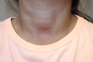
The easy removal of this patient’s persistent skin discoloration with an alcohol wipe confirms the diagnosis of terra firma-forme dermatosis. Terra firma-forme dermatosis is a benign skin condition in which dirt-like plaques develop on the skin despite normal hygiene. They cannot be removed with soap and water, but are removed easily with alcohol.
Terra firma means “solid earth” in Latin and refers to the dirt-like appearance of the lesion.1,2 The condition typically presents with brown or black hyperkeratotic plaques or papules. The hyperpigmented lesions may be papillomatous, verrucous (warty), or have a reticulated (net-like) distribution.3 The condition is usually asymptomatic but occasionally itchy.1 In the largest published series of 31 patients by Berk et al., the neck, ankles, and face were most commonly affected, but the condition can occur anywhere on the body.4 Lesions may be single or multiple and tend to be symmetrically distributed.4 The condition seems to be most common in young adults and adolescents, but can occur at any age and affects both genders equally.2,5
It seems to occur more commonly in darker skin, heavier patients, and concave body areas.3,6 The condition can last for months (median duration of 4 months in Berk et al.’s series), and by the time patients present to a physician they usually have tried aggressive scrubbing with multiple different soaps.4 As in this case, it is not uncommon for patients to receive an unnecessary work-up for diabetes due to concern for acanthosis nigricans.4 This work-up can be avoided if practitioners attempt removal with 70% isopropyl alcohol before ordering the diabetic work-up.
Biopsies are not typically performed (and should be abandoned if the lesion disappears when the site is cleansed with alcohol in preparation for biopsy), but hematoxylin and eosin staining shows lamellar hyperkeratosis, orthokeratotic whorls, and increased melanin in the basal layer and hyperkeratotic areas. Keratin globules are seen throughout the stratum corneum.7
Electron microscopy shows that the keratin lamellae is immature in some places, with incomplete keratinization and retention of desmosomal attachments.7 Overall, the histology is nonspecific and may be indistinguishable from other benign papillomatous conditions including confluent and reticulated papillomatosis (CARP), acanthosis nigricans, and epidermal nevi.6
Differential diagnosis
Clinically, the lesion may appear very similar to the brown velvety plaques of acanthosis nigricans, but the latter can be quickly ruled out if the lesion disappears when cleansed with alcohol. The differential diagnosis also includes dermatosis neglecta, CARP, hyperpigmented tinea versicolor, and dirty neck syndrome of atopic dermatitis.4,7
Dermatosis neglecta is caused by poor hygiene with insufficient skin cleaning. In contrast with terra firma-forme dermatitis, it can be removed with soap and water. Patients with dermatosis neglecta lack a history of aggressive attempts at removal by scrubbing. Dermatosis neglecta looks similar to terra firma-forme dermatosis, but may have a waxy “cornflake-like” scale.2 It is most common in the elderly, but also can be seen in children, especially if they have had a cast or brace that prevented washing an area.8
CARP may present as brown hyperkeratotic or verrucous papules, but typically occurs on the trunk and has a reticulated pattern, which is less common in terra firma-forme dermatosis. Typically, it cannot be removed completely with alcohol10-12 and tends to respond best to treatment with minocycline.11
Hyperpigmented tinea versicolor lesions are usually more discrete and have fine scale, which becomes more prominent when the lesions are scraped. When analyzed with KOH under a microscope, hyphae and spores looking like spaghetti and meatballs can be seen.
Dirty neck syndrome is acquired hyperpigmentation that may develop on the neck of patients with atopic dermatitis. It cannot be removed with alcohol.4 It may result from frictional melanosis with melanin incontinence and post-inflammatory hyperpigmentation.12 The hyperpigmentation is patchy or rippled,12 whereas terra firma-forme dermatosis more often presents as plaques.
Etiology
The cause of terra firma-forme dermatosis is uncertain. It is thought to be a disorder of keratinocyte retention, in which delayed keratinocyte maturation causes prolonged cell-to-cell adhesion that impairs shedding.7,8 Discoloration may be due to retained melanin as well as sebum and dirt that build up along with excess keratinocytes.7-9 Alcohol is a better solvent for this retained material than soap and water. Patients using heavy emollients or oily soaps for conditions like atopic dermatitis may be relatively predisposed to the condition if emollients or oily soaps are not removed completely by bathing; the combination of underlying scale and retained skin care products may make skin more adhesive, disrupting normal keratinocyte shedding and allowing dirt and sebum to accumulate.1 Atopic dermatitis was the most common comorbid condition for terra firma-forme dermatosis in Berk et al.’s series (12 of 31 patients).4
Treatment
Wiping with 70% isopropyl alcohol is both diagnostic and therapeutic, removing the lesion completely. Recurrence is uncommon but possible.3 In rare instances of regularly recurrent lesions, the area can be wiped prophylactically with alcohol weekly.1
References
- Indian J Dermatol Venereol Leprol. 2012 May-Jun;78(3):358-60.
- Pediatr Dermatol. 2011 Jan-Feb;28(1):79-81.
- Dermatol Pract Concept. 2015 Jul; 5(3): 29-33.
- Pediatr Dermatol. 2012 May-Jun;29(3):297-300.
- Eur J Intern Med. 2016 Feb. doi: 10.1016/j.ejim.2016.02.009.
- J Cutan Pathol. 2012 Feb;39(2):300-1.
- Arch Dermatol. 1987;123(5):567-9.
- Pediatr Dermatol. 2015;32(2):e50-3.
- Arch Dermatol. 2010;146(6):679-80.
- Arch Dermatol. 2011 Feb;147(2):247-8.
- Am J Clin Dermatol. 2006;7(5):305-313.
- Dermatology. 2014;229:174-182.
Ms. Haddock is a medical student at the University of California, San Diego, and a research associate at Rady Children’s Hospital–San Diego. Dr. Eichenfield is chief of pediatric and adolescent dermatology at Rady Children’s Hospital–San Diego and professor of dermatology and pediatrics at the University of California, San Diego. Dr. Eichenfield and Ms. Haddock state they have no relevant financial disclosures. Email them at [email protected].
By Ellen S. Haddock and Lawrence F. Eichenfield, MD
Terra firma-forme dermatosis
The easy removal of this patient’s persistent skin discoloration with an alcohol wipe confirms the diagnosis of terra firma-forme dermatosis. Terra firma-forme dermatosis is a benign skin condition in which dirt-like plaques develop on the skin despite normal hygiene. They cannot be removed with soap and water, but are removed easily with alcohol.
Terra firma means “solid earth” in Latin and refers to the dirt-like appearance of the lesion.1,2 The condition typically presents with brown or black hyperkeratotic plaques or papules. The hyperpigmented lesions may be papillomatous, verrucous (warty), or have a reticulated (net-like) distribution.3 The condition is usually asymptomatic but occasionally itchy.1 In the largest published series of 31 patients by Berk et al., the neck, ankles, and face were most commonly affected, but the condition can occur anywhere on the body.4 Lesions may be single or multiple and tend to be symmetrically distributed.4 The condition seems to be most common in young adults and adolescents, but can occur at any age and affects both genders equally.2,5
It seems to occur more commonly in darker skin, heavier patients, and concave body areas.3,6 The condition can last for months (median duration of 4 months in Berk et al.’s series), and by the time patients present to a physician they usually have tried aggressive scrubbing with multiple different soaps.4 As in this case, it is not uncommon for patients to receive an unnecessary work-up for diabetes due to concern for acanthosis nigricans.4 This work-up can be avoided if practitioners attempt removal with 70% isopropyl alcohol before ordering the diabetic work-up.
Biopsies are not typically performed (and should be abandoned if the lesion disappears when the site is cleansed with alcohol in preparation for biopsy), but hematoxylin and eosin staining shows lamellar hyperkeratosis, orthokeratotic whorls, and increased melanin in the basal layer and hyperkeratotic areas. Keratin globules are seen throughout the stratum corneum.7
Electron microscopy shows that the keratin lamellae is immature in some places, with incomplete keratinization and retention of desmosomal attachments.7 Overall, the histology is nonspecific and may be indistinguishable from other benign papillomatous conditions including confluent and reticulated papillomatosis (CARP), acanthosis nigricans, and epidermal nevi.6
Differential diagnosis
Clinically, the lesion may appear very similar to the brown velvety plaques of acanthosis nigricans, but the latter can be quickly ruled out if the lesion disappears when cleansed with alcohol. The differential diagnosis also includes dermatosis neglecta, CARP, hyperpigmented tinea versicolor, and dirty neck syndrome of atopic dermatitis.4,7
Dermatosis neglecta is caused by poor hygiene with insufficient skin cleaning. In contrast with terra firma-forme dermatitis, it can be removed with soap and water. Patients with dermatosis neglecta lack a history of aggressive attempts at removal by scrubbing. Dermatosis neglecta looks similar to terra firma-forme dermatosis, but may have a waxy “cornflake-like” scale.2 It is most common in the elderly, but also can be seen in children, especially if they have had a cast or brace that prevented washing an area.8
CARP may present as brown hyperkeratotic or verrucous papules, but typically occurs on the trunk and has a reticulated pattern, which is less common in terra firma-forme dermatosis. Typically, it cannot be removed completely with alcohol10-12 and tends to respond best to treatment with minocycline.11
Hyperpigmented tinea versicolor lesions are usually more discrete and have fine scale, which becomes more prominent when the lesions are scraped. When analyzed with KOH under a microscope, hyphae and spores looking like spaghetti and meatballs can be seen.
Dirty neck syndrome is acquired hyperpigmentation that may develop on the neck of patients with atopic dermatitis. It cannot be removed with alcohol.4 It may result from frictional melanosis with melanin incontinence and post-inflammatory hyperpigmentation.12 The hyperpigmentation is patchy or rippled,12 whereas terra firma-forme dermatosis more often presents as plaques.
Etiology
The cause of terra firma-forme dermatosis is uncertain. It is thought to be a disorder of keratinocyte retention, in which delayed keratinocyte maturation causes prolonged cell-to-cell adhesion that impairs shedding.7,8 Discoloration may be due to retained melanin as well as sebum and dirt that build up along with excess keratinocytes.7-9 Alcohol is a better solvent for this retained material than soap and water. Patients using heavy emollients or oily soaps for conditions like atopic dermatitis may be relatively predisposed to the condition if emollients or oily soaps are not removed completely by bathing; the combination of underlying scale and retained skin care products may make skin more adhesive, disrupting normal keratinocyte shedding and allowing dirt and sebum to accumulate.1 Atopic dermatitis was the most common comorbid condition for terra firma-forme dermatosis in Berk et al.’s series (12 of 31 patients).4
Treatment
Wiping with 70% isopropyl alcohol is both diagnostic and therapeutic, removing the lesion completely. Recurrence is uncommon but possible.3 In rare instances of regularly recurrent lesions, the area can be wiped prophylactically with alcohol weekly.1
References
- Indian J Dermatol Venereol Leprol. 2012 May-Jun;78(3):358-60.
- Pediatr Dermatol. 2011 Jan-Feb;28(1):79-81.
- Dermatol Pract Concept. 2015 Jul; 5(3): 29-33.
- Pediatr Dermatol. 2012 May-Jun;29(3):297-300.
- Eur J Intern Med. 2016 Feb. doi: 10.1016/j.ejim.2016.02.009.
- J Cutan Pathol. 2012 Feb;39(2):300-1.
- Arch Dermatol. 1987;123(5):567-9.
- Pediatr Dermatol. 2015;32(2):e50-3.
- Arch Dermatol. 2010;146(6):679-80.
- Arch Dermatol. 2011 Feb;147(2):247-8.
- Am J Clin Dermatol. 2006;7(5):305-313.
- Dermatology. 2014;229:174-182.
Ms. Haddock is a medical student at the University of California, San Diego, and a research associate at Rady Children’s Hospital–San Diego. Dr. Eichenfield is chief of pediatric and adolescent dermatology at Rady Children’s Hospital–San Diego and professor of dermatology and pediatrics at the University of California, San Diego. Dr. Eichenfield and Ms. Haddock state they have no relevant financial disclosures. Email them at [email protected].
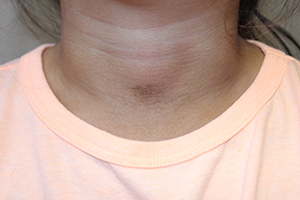
A 4-year-old girl presented for evaluation of a localized hyperpigmented rash of 4 months duration. The family noticed a localized, brown discoloration on her anterior neck. It was asymptomatic. Her mother tried scrubbing the area with several different soaps and baby wipes, but noted no change. Her medical history is notable for atopic dermatitis, well controlled with intermittent topical corticosteroids, and reactive airway disease. There is a family history of atopic dermatitis and type 2 diabetes. On physical exam, the patient is a non-obese (BMI 16 kg/m2) female with a 4 x 3–cm hyperpigmented, rough, slightly elevated plaque on her anterior neck. (See Before photo.) On close inspection, the plaque appears to be composed of hyperpigmented rugations. Viewed through a dermatoscope, the lesion’s polygonal brownish pigmentation looks like cobblestones (Can Med Assoc J. 2016;188[4]:285).The patient had no discoloration on the posterior neck, axillae, groin, or other locations. Blood work, ordered by a practitioner concerned about possible acanthosis nigricans, revealed: • CBC: within normal limits • Glucose: 82 mg/dL (normal 60-110) • Insulin: 3 mU/mL (normal less than 17) • Cholesterol: 136 mg/dL (normal less than 200) • Triglycerides: 79 mg/dL (normal 32-116) • Thyroid-stimulating hormone: 1.72 uIU/mL (normal 0.35-5) Rubbing the hyperpigmented area with a 70% isopropyl alcohol swab in clinic removed it completely.

Mastering MACRA: How to thrive under new payment models
As MACRA makes quality-based care the law of the land, don’t just glide under the new expectations, thrive. That advice comes from accountable care experts who are seeing firsthand the tools leading to success in the new payment landscape.
Rule No. 1: Step up to the plate, said Julian D. “Bo” Bobbitt, a Raleigh, N.C.–based health law attorney and accountable care organization (ACO) specialist.

“MACRA changes everything,” he said in an interview. “This is massive. Indecision will not stop your placement in the value-based payment system. Why not control your destiny to achieve your professional and financial goals?”
On May 9, the Centers for Medicare & Medicaid Services published a proposed final rule that outlines key payment provisions of the Medicare Access and CHIP Reauthorization Act of 2015 (MACRA). The proposal establishes parameters for the new Quality Payment Program, which includes the Merit-based Incentive Payment System (MIPS) and advanced Alternative Payment Models (APMs). Once final, the rule will consolidate three Medicare quality programs into MIPS: the Physician Quality Reporting System, the Value-Based Modifier Program; and the Meaningful Use program. CMS also proposes an APM pathway in which eligible clinicians can earn incentives.
The MACRA basics go as follows: From 2019 through 2024, well-performing physicians will be eligible for a bonus payment of up to 10% from a $500 million pool, according to CMS guidance released April 27. Poorly performing clinicians will see a pay cut of up to 4% in 2019, which increases to a max of 9% in 2022.

Taking small steps that focus on value now is key to excelling under MACRA, according to Jeb Dunkelberger, vice president for accountable care services at McKesson. “As an organization, ask: How can I get myself into a situation where I can maintain one foot on the boat and one foot on the dock, and be successful in the fee-for-service world, while starting to expose myself to fee-for-value?”
Starting a Medicare Shared Savings ACO is one way to accomplish this, Mr. Dunkelberger said in an interview. Track 1 of that program provides doctors with the potential for shared savings, while protecting them from financial risk. In his experience, practices have become successful after starting such ACOs, combined with a chronic care management initiative. Under this option, providers receive a fee-for-service payment, but they are also reaching out to patients and delivering preventive care, he said.
“You still have a traditional fee-for-service mechanism,” he said. “Your revenue cycle doesn’t change. Your coding doesn’t change. But at the same time, you’re simultaneously developing a competency that will be perceived as high-value in a futuristic world where we shift the location of care delivery and incentivize wellness and prevention more so than ever before.”
Joining an ACO sooner, rather than later, makes sense on many levels, Mr. Bobbitt added.
“Accountable care organizations seem to be an ideal vehicle to increase your value contribution and your reimbursement,” he said. “The law gives a 5% bump if you are in a qualifying ACO. There’s work involved and there’s infrastructure cost, but you can get into the plus side under MACRA and avoid the negative side, and you’re still open to the upside of the rewards for high performance.”

Transitioning early from volume to value has paid off for Dr. Grace E. Terrell and her large multispecialty group based in High Point, N.C. The group began adding components of value-based care in 2011 and is now part of an ACO with multiple payers and partners.
“We did this by changing the way we were providing care in specific care models and also investing substantially in information integration as well as changing our contracts so we were being paid based upon our outcomes, quality, and cost-effectiveness, rather than just fee-for-service,” said Dr. Terrell, the group’s president and CEO.
Since making the changes, the group has improved quality of care while reducing cost, said Dr. Terrell, who serves on the federal advisory committee for MACRA, officially known as the Physician-Focused Payment Model Technical Advisory Committee. Dr. Terrell said that her practice group had the sixth-highest quality score and the fourth-lowest cost among providers in the 2014 Medicare Shared Savings Program. The group has also launched a population health management company in collaboration with an academic medical center and a testing laboratory.
Maintaining a patient-focused viewpoint is essential to switching from volume to value, Dr. Terrell said. For example, her group focused on patients with severe chronic obstructive pulmonary disease and teamed them with a respiratory therapist, particularly after hospitalizations. Their efforts reduced 30-day hospital readmissions from 12% to 6%. They also created clinics for patients who have five or more chronic conditions; physicians are linked with nurse navigators, social workers, and other professionals to offer a more holistic approach.
“All of these different models focused foremost on patients,” Dr. Terrell said. “They also focused on teamwork. Even though physicians were leading the team, it involved integrated medicine. It also involved integration across the spectrum of care so we had to work very carefully with our hospital partners.”
Collaboration is a critical piece to quality-based success, Mr. Dunkelberger said, but he advises taking the time and effort to find the right partners. Thoroughly vet potential partners, he said. Ask for case studies and overall impact of work. Be wary of flashy flowcharts and “too good to be true” promises.
“Look for a partner and not [necessarily] a vendor,” Mr. Dunkelberger said. “Make sure their incentives align with yours.”
Take stock of readiness to participate in alternative payment models, advised Edith Coakley Stowe, a health care attorney in Washington. The ability to meet electronic health record expectations, a category under MACRA now called Advancing Care Information, is extremely important, she said. Equally important – especially in primary care – are strategies for managing patients between visits. Decide whether your practice should build, buy, or enter into a joint venture to achieve these goals.
“The good news is that what gets tested in alternative payment models generally finds its way into policies and programs applicable across the Medicare program,” Ms. Stowe said in an interview. “That means that participants in alternative payment models get a head start. Despite the blind corners and complexity of options facing physician groups right now, having a mentality of testing, trying, and continuously evaluating is going to stand them in the best stead.”
On Twitter @legal_med
As MACRA makes quality-based care the law of the land, don’t just glide under the new expectations, thrive. That advice comes from accountable care experts who are seeing firsthand the tools leading to success in the new payment landscape.
Rule No. 1: Step up to the plate, said Julian D. “Bo” Bobbitt, a Raleigh, N.C.–based health law attorney and accountable care organization (ACO) specialist.
“MACRA changes everything,” he said in an interview. “This is massive. Indecision will not stop your placement in the value-based payment system. Why not control your destiny to achieve your professional and financial goals?”
On May 9, the Centers for Medicare & Medicaid Services published a proposed final rule that outlines key payment provisions of the Medicare Access and CHIP Reauthorization Act of 2015 (MACRA). The proposal establishes parameters for the new Quality Payment Program, which includes the Merit-based Incentive Payment System (MIPS) and advanced Alternative Payment Models (APMs). Once final, the rule will consolidate three Medicare quality programs into MIPS: the Physician Quality Reporting System, the Value-Based Modifier Program; and the Meaningful Use program. CMS also proposes an APM pathway in which eligible clinicians can earn incentives.
The MACRA basics go as follows: From 2019 through 2024, well-performing physicians will be eligible for a bonus payment of up to 10% from a $500 million pool, according to CMS guidance released April 27. Poorly performing clinicians will see a pay cut of up to 4% in 2019, which increases to a max of 9% in 2022.
Taking small steps that focus on value now is key to excelling under MACRA, according to Jeb Dunkelberger, vice president for accountable care services at McKesson. “As an organization, ask: How can I get myself into a situation where I can maintain one foot on the boat and one foot on the dock, and be successful in the fee-for-service world, while starting to expose myself to fee-for-value?”
Starting a Medicare Shared Savings ACO is one way to accomplish this, Mr. Dunkelberger said in an interview. Track 1 of that program provides doctors with the potential for shared savings, while protecting them from financial risk. In his experience, practices have become successful after starting such ACOs, combined with a chronic care management initiative. Under this option, providers receive a fee-for-service payment, but they are also reaching out to patients and delivering preventive care, he said.
“You still have a traditional fee-for-service mechanism,” he said. “Your revenue cycle doesn’t change. Your coding doesn’t change. But at the same time, you’re simultaneously developing a competency that will be perceived as high-value in a futuristic world where we shift the location of care delivery and incentivize wellness and prevention more so than ever before.”
Joining an ACO sooner, rather than later, makes sense on many levels, Mr. Bobbitt added.
“Accountable care organizations seem to be an ideal vehicle to increase your value contribution and your reimbursement,” he said. “The law gives a 5% bump if you are in a qualifying ACO. There’s work involved and there’s infrastructure cost, but you can get into the plus side under MACRA and avoid the negative side, and you’re still open to the upside of the rewards for high performance.”
Transitioning early from volume to value has paid off for Dr. Grace E. Terrell and her large multispecialty group based in High Point, N.C. The group began adding components of value-based care in 2011 and is now part of an ACO with multiple payers and partners.
“We did this by changing the way we were providing care in specific care models and also investing substantially in information integration as well as changing our contracts so we were being paid based upon our outcomes, quality, and cost-effectiveness, rather than just fee-for-service,” said Dr. Terrell, the group’s president and CEO.
Since making the changes, the group has improved quality of care while reducing cost, said Dr. Terrell, who serves on the federal advisory committee for MACRA, officially known as the Physician-Focused Payment Model Technical Advisory Committee. Dr. Terrell said that her practice group had the sixth-highest quality score and the fourth-lowest cost among providers in the 2014 Medicare Shared Savings Program. The group has also launched a population health management company in collaboration with an academic medical center and a testing laboratory.
Maintaining a patient-focused viewpoint is essential to switching from volume to value, Dr. Terrell said. For example, her group focused on patients with severe chronic obstructive pulmonary disease and teamed them with a respiratory therapist, particularly after hospitalizations. Their efforts reduced 30-day hospital readmissions from 12% to 6%. They also created clinics for patients who have five or more chronic conditions; physicians are linked with nurse navigators, social workers, and other professionals to offer a more holistic approach.
“All of these different models focused foremost on patients,” Dr. Terrell said. “They also focused on teamwork. Even though physicians were leading the team, it involved integrated medicine. It also involved integration across the spectrum of care so we had to work very carefully with our hospital partners.”
Collaboration is a critical piece to quality-based success, Mr. Dunkelberger said, but he advises taking the time and effort to find the right partners. Thoroughly vet potential partners, he said. Ask for case studies and overall impact of work. Be wary of flashy flowcharts and “too good to be true” promises.
“Look for a partner and not [necessarily] a vendor,” Mr. Dunkelberger said. “Make sure their incentives align with yours.”
Take stock of readiness to participate in alternative payment models, advised Edith Coakley Stowe, a health care attorney in Washington. The ability to meet electronic health record expectations, a category under MACRA now called Advancing Care Information, is extremely important, she said. Equally important – especially in primary care – are strategies for managing patients between visits. Decide whether your practice should build, buy, or enter into a joint venture to achieve these goals.
“The good news is that what gets tested in alternative payment models generally finds its way into policies and programs applicable across the Medicare program,” Ms. Stowe said in an interview. “That means that participants in alternative payment models get a head start. Despite the blind corners and complexity of options facing physician groups right now, having a mentality of testing, trying, and continuously evaluating is going to stand them in the best stead.”
On Twitter @legal_med

Atopic dermatitis increases risk of ADHD, comorbidities add to it
Atopic dermatitis (AD) is associated with an increased risk for attention-deficit/hyperactivity disorder (ADHD) in children and adults, and a host of factors and comorbid conditions increase this risk, according to a study published in the British Journal of Dermatology.
Mark A. Strom and his associates at Northwestern University in Chicago examined data from 19 U.S. population-based surveys on 354,416 children ages 2-17 years and 34,613 adults over the age of 18 to determine if childhood and adult AD and the severity of AD were associated with ADHD. Additionally, the investigators sought to identify factors contributing to this association (Br J Dermatol. 2016 Apr 23. doi: 10.1111/bjd.14697).

The prevalence of ADHD was 9.4% in children with AD and 7.1% in children without AD. For children, the study results obtained from multivariate models adjusting for age, sex, sociodemographic variables, allergic disease, and health care utilization confirmed the previously established increase in risk for ADHD in those with AD for an adjusted odds ratio of 1.14. Severe AD and sleep disturbance were found to independently and synergistically contribute to the increased risk of ADHD. Also, the presence of AD in the absence of other allergic disease was associated with an increased risk for ADHD. Obesity, headaches, and anemia were found to further increase the risk for ADHD in those with AD.
Results from the adjusted multivariate models also established an increased risk for ADHD among adults with AD, for an adjusted odds ratio of 1.61. The presence of asthma, headaches, and insomnia increased this risk. The analysis additionally revealed that a body mass index indicative of underweight status in adults with AD was protective against the risk for developing ADHD.
Mr. Strom and his associates found a significant decrease in their initial odds ratios when they added outpatient health care visitation to their multivariate models, which resulted in an overall pooled odds ratio for the relationship between AD and ADHD just above statistical significance. They speculated that children with AD or ADHD would be more likely to visit health care providers, which could increase the likelihood for diagnosis of comorbid conditions.
Funding was provided by the Agency for Healthcare Research and Quality and the Dermatology Foundation. The authors reported no conflicts of interest.
Atopic dermatitis (AD) is associated with an increased risk for attention-deficit/hyperactivity disorder (ADHD) in children and adults, and a host of factors and comorbid conditions increase this risk, according to a study published in the British Journal of Dermatology.
Mark A. Strom and his associates at Northwestern University in Chicago examined data from 19 U.S. population-based surveys on 354,416 children ages 2-17 years and 34,613 adults over the age of 18 to determine if childhood and adult AD and the severity of AD were associated with ADHD. Additionally, the investigators sought to identify factors contributing to this association (Br J Dermatol. 2016 Apr 23. doi: 10.1111/bjd.14697).
The prevalence of ADHD was 9.4% in children with AD and 7.1% in children without AD. For children, the study results obtained from multivariate models adjusting for age, sex, sociodemographic variables, allergic disease, and health care utilization confirmed the previously established increase in risk for ADHD in those with AD for an adjusted odds ratio of 1.14. Severe AD and sleep disturbance were found to independently and synergistically contribute to the increased risk of ADHD. Also, the presence of AD in the absence of other allergic disease was associated with an increased risk for ADHD. Obesity, headaches, and anemia were found to further increase the risk for ADHD in those with AD.
Results from the adjusted multivariate models also established an increased risk for ADHD among adults with AD, for an adjusted odds ratio of 1.61. The presence of asthma, headaches, and insomnia increased this risk. The analysis additionally revealed that a body mass index indicative of underweight status in adults with AD was protective against the risk for developing ADHD.
Mr. Strom and his associates found a significant decrease in their initial odds ratios when they added outpatient health care visitation to their multivariate models, which resulted in an overall pooled odds ratio for the relationship between AD and ADHD just above statistical significance. They speculated that children with AD or ADHD would be more likely to visit health care providers, which could increase the likelihood for diagnosis of comorbid conditions.
Funding was provided by the Agency for Healthcare Research and Quality and the Dermatology Foundation. The authors reported no conflicts of interest.
FROM THE BRITISH JOURNAL OF DERMATOLOGY
Key clinical point: Several factors contribute to the increased risk for ADHD in children and adults with atopic dermatitis.
Major finding: Children and adults with atopic dermatitis had increased odds of having ADHD of 1.14, which were further increased by the presence of certain comorbidities.
Data sources: Nineteen U.S. population-based surveys on 354,416 children aged 2-17 and 34,613 adults over the age of 18.
Disclosures: Funding was provided by the Agency for Healthcare Research and Quality and the Dermatology Foundation. The authors reported no conflicts of interest.

FDG-PET/CT leads pack for small-cell lung cancer staging
For pretreatment staging of small-cell lung cancer (SCLC) the use of positron-emission tomography combined with CT was more sensitive compared with several other tests, according to a new report on a review of studies.
Overall, positron emission tomography using [F]-fluorodeoxyglucose as a radiotracer combined with CT (FDG-PET/CT) had greater sensitivity to detect osseous metastases than did bone scintigraphy or CT alone, according to Dr. Jonathan R. Treadwell, Ph.D., of ECRI Institute–Penn Medicine’s Evidence-based Practice Center in Plymouth Meeting, Pa., and colleagues. In addition, the researchers concluded that adding FDG-PET/CT to the protocol for patients who have undergone standard staging increased the sensitivity for detecting additional metastases. Data on endobronchial ultrasound were insufficient to draw any conclusions.
The findings generally line up with recent guidelines from the American College of Radiology (ACR) and American College of Chest Physicians (ACCP). In 2014, the ACR gave the highest rating of “usually appropriate” (with regard to staging SCLC) to FDG-PET/CT from skull base to mid-thigh, while bone scintigraphy was rated as “may be appropriate” and not necessary if PET/CT had been done, the researchers wrote. The 2013 ACCP guideline “suggested” FDG PET instead of bone scintigraphy for patients with limited disease, they added.
The researchers reviewed data from seven studies to assess the accuracy and effectiveness of several imaging modalities for the pretreatment staging of SCLC. The report was generated for the Agency for Healthcare Research and Quality (AHRQ) as part of its Comparative Effectiveness Review series, and is not an official AHRQ position, the researchers noted.
Combining FDG-PET with CT scanning has demonstrated even greater effectiveness at identifying malignant tumors and metabolically active metastases than has PET alone, because the CT allows for more localized anatomic detail, the researchers explained. “False negative scans usually result from non–metabolically active sites of tumor or from suboptimal quality studies,” they said, while false positives using FDG-PET are usually attributed to inflammation or metabolically active infection.
The meta-analysis included data on endobronchial ultrasound, which involves ultrasound to view structures inside and adjacent to the airway; bone scintigraphy, a less expensive planar molecular imaging technique; and CT alone.
Comparative evidence on pretreatment staging for SCLC is limited, according to the researchers. The data did not allow them to determine how FDG-PET/CT compared to other imaging in terms of specificity, and any type of imaging can yield false positives, they said. However, higher sensitivity alone can benefit patients in terms of improving patient selection for optimal therapy, sparing patients chemotherapy if not needed, and improving the prediction value of ongoing research, they noted.
“Although high-quality evidence may not be voluminous, I think most physicians would agree with the conclusion that a bone scan is not mandatory in the work-up of possible SCLC, if a PET/CT has been done,” Dr. W. Michael Alberts of the Moffitt Cancer Center in Tampa, Fla., said in an interview.
Cost might play a role in why the guidelines are being issued at this time, he noted, because “the initial work-up of the patient with suspected SCLC may prove to be quite expensive, and the elimination of a superfluous test may be a fiscal winner.” However, more research is needed in this area, particularly in the areas of including the order of pretreatment testing and the incorporation of new procedures and imaging modalities, he added. “Perhaps more intellectually challenging, however, might be the question of why SCLC is becoming less common, or why has improvement in treatment been so slow compared to NSCLC,” he added.
The researchers had no financial conflicts to disclose.
More connective tissue disease–associated PAH seen in older patients
Patient age contributes to significant differences in the characteristics and etiology of pulmonary arterial hypertension seen in randomized, controlled trials, including more frequent connective tissue disease–associated disease in older patients, according to a post-hoc analysis of trials.
Additionally, older age was associated with worse baseline functional status, worse outcomes in the 6-minute walk distance, and an overall reduced response to treatment, while hemodynamic severity was higher in younger patients.
“Although registry data have shown that idiopathic PAH [pulmonary arterial hypertension] is increasingly recognized in older populations, our analysis shows that idiopathic etiology was less frequent in the older group. In contrast, CTD [connective tissue disease]–associated PAH accounted for a higher proportion of PAH etiology in the oldest age group,” wrote Jonathan A. Rose of Case Western Reserve University, Cleveland, and his colleagues (Chest. 2016;149[5]:1234-44. doi: 10.1016/j.chest.2015.11.008).
The researchers analyzed seven multicenter, randomized, double-blind, placebo-controlled treatment trials for PAH conducted by United Therapeutics and one open-label extension study that involved following patients being treated with subcutaneous treprostinil for 4 additional years. Data from the open-label extension trial and one of the trials were combined and reported as one trial. All trials excluded patients with a history of left-sided heart disease, pulmonary artery wedge pressure (PAWP) greater than 15 mmHg, or pulmonary vascular resistance (PVR) less than 3 Woods units. The researchers categorized the 2,627 patients included in the trials in the following three age groups: 50 years or younger, 51-64 years, and 65 years or older.
Between 53% and 74% of patients in all trials across all age groups had idiopathic PAH, but older patients comprised a significantly smaller proportion of the patients with idiopathic PAH in three of the trials (P = .004) and a significantly higher percentage of the patients with CTD-associated PAH in all of the trials (P less than .001). Across the trials, CTD-associated PAH occurred in 15%-21% of patients 50 years or younger, 25%-40% of those aged 51-64 years, and 27%-49% of those aged 65 years or older.
From baseline to the end in three of the studies, a smaller change in the 6-minute walk distance was seen in older patients and a higher proportion of older patients had an overall decrease in total 6-minute walk distance. Older age remained associated with this outcome measure when only data from treatment-naive patients or the subgroup of patients with CTD-associated PAH were included.
The association between age and lower baseline functional status was observed in four of the trials; A lower proportion of patients in the oldest age group were classified as being of the World Health Organization functional classes I and II, with 9%-32% of patients aged 65 years or older, 10%-33% in those aged 51-64 years, and 16%-43% of those aged 50 years or younger.
Hemodynamic severity was among the areas in which older patients performed better than younger patients, with the oldest age group having lower baseline mean pulmonary artery pressure (mPAP) and PVR in the two trials that measured hemodynamics.
“The difference in the clinical function and hemodynamics in older patients may reflect different mechanisms of disease. Older patients may have diminished cardiopulmonary reserve, and similar degrees of hemodynamic severity manifest with a larger functional impairment. Alternatively, additional comorbidities in older patients may contribute to a larger extent, and clinical function may not be as directly related to hemodynamic severity,” according to the researchers.
Mortality was generally small in these studies and not significantly different among age groups, except for in the open-label extension study of Subcutaneous Infusion of Treprostinil in Patients with PAH (SC-TRE) and the Study of Intravenous Remodulin in Patients in India with PAH (TRUST) trials, which were combined and reported on as one trial (P = .0004).
Nausea was the only adverse effect found to be associated with age in more than one trial, and the relationship between it and age in two trials were inconsistent. While a higher incidence of nausea was found in the oldest age group in the Oral Treprostinil as Monotherapy for the Treatment of Pulmonary Arterial Hypertension (FREEDOM-M) trial (P = .03), a lower incidence of nausea was found in the oldest age group in the Treprostinil Sodium Inhalation Used in the Management of Pulmonary Arterial Hypertension (TRIUMPH) trial (P = .02).
The analysis was supported by a grant from the National Institutes of Health. Two of the authors, Jody M. Cleveland and Youlan Rao, Ph.D., reported being employees of United Therapeutics, while Dr. Omar A. Minai, another author of the report, serves on the scientific advisory board of United Therapeutics. The other authors declared no conflicts.
FROM CHEST
FDA issues warning about counterfeit BiCNU
A counterfeit version of BiCNU has been detected in foreign countries, the Food and Drug Administration reports.
BiCNU is approved to treat different types of brain cancer, multiple myeloma, and lymphoma (Hodgkin’s and non-Hodgkin’s), manufactured by Emcure Pharmaceuticals, and distributed by Heritage Pharmaceuticals.
There has been no counterfeit BiCNU detected in the United States, but the FDA encourages health care professionals to diligently inspect BiCNU vials before administering the drug to patients.
“While the [National Drug Code] on the outer package of the authentic and counterfeit version might match, the best way to distinguish a counterfeit is to look at the BiCNU vial inside the packaging,” the FDA reported in a written statement, which includes a list of the counterfeit lots.
To report sales solicitation of suspect drugs call the FDA’s office of criminal investigations at 800-551-3989 or e-mail [email protected]. To report adverse events related to suspect medications, submit a report online at www.fda.gov/medwatch/report.htm.
On Twitter @jess_craig94
WATCH: Mentoring in Hospital Medicine
Drs. Vineet Arora and Hyung "Harry" Cho offer insight on how mentorship—giving, and receiving—is an essential part of all stages of an hospitalist career, in academic or community-based HM.
The video associated with this article is no longer available on this site. Please view all of our videos on the MDedge YouTube channel