User login
Attend SVS PAC Reception in Washington, D.C.
When meeting near Washington, D.C., meeting with members of Congress is a natural choice.

The SVS Political Action Committee Reception will be held from 5:30 to 7 p.m. Wednesday, June 8, on Capitol Hill, at 2261 Rayburn House Office Building. Round-trip transportation will be provided from the Gaylord National’s Maryland Ballroom bus loop entrance on Level 2. The shuttle bus will depart promptly at 4:45 p.m.
The reception is for Capitol Club contributors, those SVS members who have contributed $1,000 or more since Jan. 1, 2015, including those who contribute during the 2016 Vascular Annual Meeting, up until June 8. Attendees can meet members of Congress and thank them for their support of vascular surgery issues.
PAC contributions allow SVS members and staff to gain important access to members of Congress by attending meetings and discussing issues that have a major impact on vascular surgery.
See vsweb.org/PAC to donate and secure a spot at the reception – and to invest in the future.
When meeting near Washington, D.C., meeting with members of Congress is a natural choice.
The SVS Political Action Committee Reception will be held from 5:30 to 7 p.m. Wednesday, June 8, on Capitol Hill, at 2261 Rayburn House Office Building. Round-trip transportation will be provided from the Gaylord National’s Maryland Ballroom bus loop entrance on Level 2. The shuttle bus will depart promptly at 4:45 p.m.
The reception is for Capitol Club contributors, those SVS members who have contributed $1,000 or more since Jan. 1, 2015, including those who contribute during the 2016 Vascular Annual Meeting, up until June 8. Attendees can meet members of Congress and thank them for their support of vascular surgery issues.
PAC contributions allow SVS members and staff to gain important access to members of Congress by attending meetings and discussing issues that have a major impact on vascular surgery.
See vsweb.org/PAC to donate and secure a spot at the reception – and to invest in the future.
Earn CME, Maintenance of Certification Credits
Beyond learning what’s new in the world of vascular surgery, paid physicians also can earn both Continuing Medical Education credits and Maintenance of Certification Part 2 Lifelong Learning and Self-Assessment credits.
The Society for Vascular Surgery is accredited by the Accreditation Council for Continuing Medical Education to provide continuing medical education for physicians. SVS has designated the 2016 Vascular Annual Meeting for a maximum of 29.25 AMA PRA Category 1 Credits™.
Physicians should claim only the credits commensurate with the extent of their participation in the activity.
Of the 29.25 credits, 13.25 AMA PRA Category 1 Credits™ meet the requirements for American Board of Surgery MOC Part 2 self-assessment.
To claim credits, physicians must attend that session and then complete the appropriate self-assessment exam with a passing score of 75%. Access to the exams will be available via a link in the 2016 VAM Mobile App and a link on the SVS website. New this year is the ability to have access to the exams immediately after a session.
Attendees may access the exams and claim CME and self-assessment credits through Dec. 31, 2016.
Separate certificates are issued for postgraduate courses (a total of nine credits are available and people may register for postgraduate courses only and not participate in VAM), the Registered Physician Vascular Interpretation exam course (3.25 CME and 3.25 MOC credits) and for VAM itself (up to 29.25 and 13.25 CME and MOC credits, respectively, depending on session participation). A grand total of 25.5 MOC credits is available to someone who takes three postgraduate courses, participates in two breakfast and all plenary sessions and also completes the RPVI review course.
Industry-Supported Satellite Symposia Offered at VAM
Vascular Annual Meeting attendees are invited to the satellite symposia offered by industry, including three breakfast sessions on Thursday, June 9. These sessions are not part of the ACCME-accredited portion of the Vascular Annual Meeting.
Thursday, June 9, 6:30 - 8 a.m.
B1: Complex SFA Disease: Maximizing Outcomes While Minimizing Costs
Presenting: Dr. Brian G. DeRubertis, Dr. Peter L. Faries
Sponsored by Abbott
B2: EndoAnchors in Practice: Treatment Algorithms from the Experts
Sponsored by Medtronic
Presenting: EndoAnchors: What Are They and How They Bring Value to Your Practice – Dr. Bart Muhs
How I Utilize ANCHOR Data to Make Clinically Relevant Treatment Decisions – Dr. Jeffrey Jim
How EndoAnchors Can be Used Safely and Effectively in the Treatment of Abdominal Aortic Aneurysms – Dr. Frank R. Arko
How EndoAnchors Can be Used Safely and Effectively in the Treatment of Thoracic Aortic Aneurysms – Dr. Jean M. Panneton
B3: New Techniques in Advanced Imaging and 3D Navigation for Complex Endovascular Procedures
Sponsored by Philips
Presenting: Dr. Marc L. Schermerhorn and Dr. Wayne K. Nelson
Thursday, June 9, 7:30 - 9 p.m.
“The Battle Continues ... The Battle of the Bulge: Debating the Future of Aortic Aneurysm Repair”
Sponsored by Endologix, Inc.
Moderator: Dr. Christopher J. Kwolek; Presenter: TBA
Friday, June 10, 6:30 - 8:30 p.m.
“Internal Iliac Artery Preservation Expert Panel”
Sponsored by Gore
Presenting: Moderator: Dr. Darren B. Schneider
Pros and Cons of Current Treatment Options – Dr. Jason T. Lee
GORE® EXCLUDER® Iliac Branch Device (IBE) and Procedural Overview – Dr. Gustavo Oderich
IBE Clinical Trial Outcomes and the Benefits of Internal Iliac Artery Preservation – Dr. Darren B. Schneider
Case Report: First Patient Treated and Two-Year Follow-up – Dr. Brian G. Peterson
Case Report: First U.S. Bilateral Case and One-Year Follow-up – Dr. Sharif Ellozy
Rapid-Fire Case Presentations
As of 5/6/16
Postgraduate Courses: Six Sessions Offered
The 2016 Vascular Annual Meeting is offering six postgraduate courses on Wednesday, with two held concurrently in three time slots. SVS members get in free with their registration, a $300 value. Registration is required.
Topics for both the postgraduate courses and the hands-on workshops (also held Wednesday; see story in the May Vascular Specialist) are selected based on member feedback and requests, said Dr. Kellie Brown, chair of the Postgraduate Education Committee that is responsible for the sessions. “We try to cover topics that are current, timely, and necessary for the majority of our membership,” she said.
That includes a broader focus this year on both dialysis access and thoracic outlet, for example. “We’ve always done something on access, but a lot of members asked for more,” she said. “Not only is dialysis access a common issue, particularly for those in community practice, but it is also an area of innovation.”
Another session covers management of the diabetic foot. “Vascular surgeons deal with this every single day,” Dr. Brown said. “It’s a really common problem – so let’s talk about the best way to manage it during a session at VAM.”
This session on diabetic foot management also introduces a new VAM partner, the American Podiatric Medical Association, which worked with SVS and the Society for Vascular Medicine on a comprehensive new guideline for managing the diabetic foot. “Having joint sessions – and we have several this year – adds an extra layer to VAM sessions,” Dr. Brown said. “We get to offer examination of other aspects of a particular topic, which adds not only more information but also a certain level of excitement.”
Last year, “Standing on the Shoulders of Giants” drew the most attendees. For 2016, “Giants” is back, with different experts and a continued focus on open surgery. “People are so comfortable with the endovascular side, but open surgery is becoming less common. This offers an opportunity to learn the techniques from the masters,” she said.
As for next year, Dr. Brown advised anyone who has a topic idea to submit it with his or her VAM evaluation.
Session 1: 7 – 10 a.m.
P1: Controversies and Management of Lower Extremity Arterial Occlusive Disease
Moderators: Dr. Christopher J. Abularrage and Dr. Niten Singh
P2: Complete Management of Venous Disease: From Superficial to Deep
Moderators: Dr. Ruth L. Bush and Dr. Mark Meissner
Session 2: 10:15 a.m. – 1:15 p.m.
P3: SVS/APMA Joint Session: Management of the Diabetic Foot
Moderators: Dr. James R. Christina (APMA) and Dr. Matthew J. Eagleton (SVS)
P4: Hemodialysis: Challenges, Controversies and New Techniques
Moderators: Dr. Melissa L. Kirkwood and Dr. Jill Zink
Session 3: 2 – 5 p.m.
P5: Endovascular Strategies for Complex Aortic Scenarios
Moderators: Dr. Ali Azizzadeh and Dr. Edward Woo
P6: Standing on the Shoulders of Giants: Open Operative Techniques by the Masters
Moderators: Dr. Hasan Dosluoglu and Dr. Michael Rohrer
Vascular Live
The Vascular Live stage is located in Exhibit Halls A-C, on Level 1. On the Vascular Live stage, exhibitors will present new ideas, showcase breakthrough technologies and discuss the latest trends in vascular surgery.
Thursday, June 9
12:15 – 1:15 p.m.
Sponsored by Bard Peripheral Vascular, Inc.
“Latest Outcomes from Lutonix® Global SFA Registry and Head to Head Comparison between IN.PACT and LUTONIX® DCB”
Speakers: Dr. Yvonne Bausback, Dr. Chad Laurich, and Dr. Renu Virmani
3:30 – 4 p.m.
Sponsored by Abbott
“Treatment Strategies for Complex Femoropopliteal Disease”
Speaker: Dr. Niten Singh
5:30 – 6 p.m.
Sponsored by Philips
“Physician Perspective on Outpatient Based Labs (OBLs)”
6 – 6:30 p.m.
Sponsored by GE Healthcare
“Integrated Workflow for Endograft Sizing, 3D Fusion, and Assessment, Using New EVAR ASSIST”
Speaker: Dr. Stephan Haulon
Friday, June 10
9:30 – 10 a.m.
Sponsored by Silk Road Medical
“TransCarotid Artery Revascularization (TCAR): The Way Forward in Treating Carotid Artery Disease and Stroke Prevention”
Moderator: Dr. Sumaira Macdonald
Speakers: Dr. Vikram S. Kashyap, Dr. Peter A. Schneider, and Dr. Raghu L. Motaganahalli
12:30 – 1 p.m.
Sponsored by Gore
“Five-year Patency Rates for GORE® PROPATEN® Vascular Grafts vs. ePTFE Grafts Without Heparin. See the Results”
Speaker: Dr. Russell H. Samson
1 – 1:30 p.m.
Sponsored by Gore
“Understanding How Device Design Can Impact Clinical Outcomes in Type B Dissection”
Speaker: Dr. Alan B. Lumsden
“Durable TEVAR – Results from 100,000 Implants”
Speaker: Dr. Mark A. Farber
3 – 3:30 p.m.
Sponsored by Hansen Medical
“Flexible Robotics with Electromagnetic Tracking and the Impacts on Safety and Efficiency”
Speaker: Dr. Alan B. Lumsden
Vascular Live presentations are not eligible for CME credit.
Homans Lecture: A Look Ahead in Vascular Surgery
Vascular surgery has a rich, varied and enormously interesting history, filled with innovators and creators whose inventions changed the course of patient care. This history has been well-documented innumerable times.
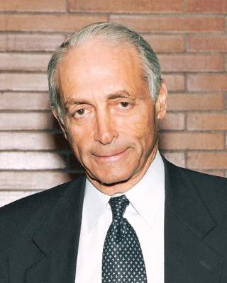
Dr. Frank J. Veith is a part of that storied history. However, during the John Homans Distinguished Lecture Thursday morning, he will look ahead, and not back.
During “A Look at the Future of Vascular Surgery,” he plans to discuss the implications of future changes in the field, not to mention the world of medical care, and “what vascular surgery has to do to survive,” he said.
In mid-April he was working on the VEITHsymposium he created more than 40 years ago and so had not yet finalized his thoughts on his lecture.
But at that time he believed he would revisit his long-ago Presidential Address, “Charles Darwin and Vascular Surgery,” delivered in June 1996 and which also discussed the future of vascular surgery.
He noted then that it was a “time of enormous upheaval in American medicine, and holding this office has also forced me to focus on the future of Vascular Surgery and how well it will survive these turbulent times.” As Darwin studied extinct and contemporary species, his thoughts on survival could be correlated to that of the subspecialty itself, Dr. Veith said.
The endovascular revolution had begun and he was convinced – and stated – that vascular surgeons would need to learn endovascular skills. “People thought I was crazy when I said that endovascular was going to replace most of what we did,” he said. “I turned out to be very right.”
Some of his other pronouncements that day turned out to be incorrect, he said. But because change is once again part of the field of medicine, he wants to address issues he sees and possible solutions to those issues.
“I want it to be interesting and provocative,” he said. “I don’t want the listeners to fall asleep.”
Dr. Veith is professor of surgery at New York University Medical Center and at the Cleveland Clinic Lerner College of Medicine of Case Western Reserve University. He holds the William J. von Liebig chair in Vascular Surgery at the Cleveland Clinic Foundation and has lectured throughout the world.
He is a pioneer in the area of limb-salvage surgery and endovascular grafting for traumatic, aneurysmal, and occlusive arterial disease. Each year he hosts the VEITHsymposium on what is new and important in the treatment of vascular disease.
Vascular surgery has a rich, varied and enormously interesting history, filled with innovators and creators whose inventions changed the course of patient care. This history has been well-documented innumerable times.
Dr. Frank J. Veith is a part of that storied history. However, during the John Homans Distinguished Lecture Thursday morning, he will look ahead, and not back.
During “A Look at the Future of Vascular Surgery,” he plans to discuss the implications of future changes in the field, not to mention the world of medical care, and “what vascular surgery has to do to survive,” he said.
In mid-April he was working on the VEITHsymposium he created more than 40 years ago and so had not yet finalized his thoughts on his lecture.
But at that time he believed he would revisit his long-ago Presidential Address, “Charles Darwin and Vascular Surgery,” delivered in June 1996 and which also discussed the future of vascular surgery.
He noted then that it was a “time of enormous upheaval in American medicine, and holding this office has also forced me to focus on the future of Vascular Surgery and how well it will survive these turbulent times.” As Darwin studied extinct and contemporary species, his thoughts on survival could be correlated to that of the subspecialty itself, Dr. Veith said.
The endovascular revolution had begun and he was convinced – and stated – that vascular surgeons would need to learn endovascular skills. “People thought I was crazy when I said that endovascular was going to replace most of what we did,” he said. “I turned out to be very right.”
Some of his other pronouncements that day turned out to be incorrect, he said. But because change is once again part of the field of medicine, he wants to address issues he sees and possible solutions to those issues.
“I want it to be interesting and provocative,” he said. “I don’t want the listeners to fall asleep.”
Dr. Veith is professor of surgery at New York University Medical Center and at the Cleveland Clinic Lerner College of Medicine of Case Western Reserve University. He holds the William J. von Liebig chair in Vascular Surgery at the Cleveland Clinic Foundation and has lectured throughout the world.
He is a pioneer in the area of limb-salvage surgery and endovascular grafting for traumatic, aneurysmal, and occlusive arterial disease. Each year he hosts the VEITHsymposium on what is new and important in the treatment of vascular disease.

Schedule-At-A-Glance
Wednesday, June 8
6:00 a.m. – 6:30 p.m.
Registration
7:00 – 10:00 a.m.
Postgraduate Courses
P1: Controversies and Management of Lower Extremity Arterial Occlusive Disease
P2: Complete Management of Venous Disease: From Superficial to Deep
8:00 – 11:30 a.m.
V1: VESS Paper Session 1
8:00 a.m. – 5:00 p.m.
VQI Annual Meeting
8:30 – 10:00 a.m.
Hands-on Workshops
W2: Tricks for Pedal, Tibial, Radial and Brachial Access
W3: Techniques for Challenging Embolization
W5: Atherectomy Techniques
10:15 a.m. – 1:15 p.m.
Postgraduate Courses
P3: SVS/APMA Joint Session: Management of the Diabetic Foot
P4: Hemodialysis: Challenges, Controversies and New Techniques
10:30 a.m. – 12:00 noon
Hands-on Workshops
W7: Technical Tips for Crossing CTO
W10: IVUS: An Essential Tool for the Endovascular Specialist
12:30 – 2:45 p.m.
FT: International Fast Talk
12:30 - 4:00 p.m.
V2: VESS Paper Session 2
1:30 – 3:00 p.m.
Hands-on Workshops
W14: IVC Filters: Techniques in Deployment and Recovery
W15: Non-Thermal Techniques for Venous Ablation
W16: Percutaneous Mechanical Thrombectomy: Challenges, Advances and New Techniques
W17: How to Make Carotid Stenting Competitive with CEA
W18: FEVAR Tips & Tricks: From Sizing to Implantation
2:00 – 5:00 p.m.
Postgraduate Courses
P5: Endovascular Strategies for Complex Aortic Scenarios
P6: Standing on the Shoulders of Giants: Open Operative Techniques by the Masters
2:00 – 6:00 p.m.
General Surgery Resident/Medical Student Scholarship Program – Open and Endovascular Simulation Training
3:00 – 6:00 p.m.
C1: International Forum (included among Concurrent Sessions)
3:30 – 5:00 p.m.
Hands-on Workshops
W19: DEB/DES Techniques & Applications
W22: Percutaneous Mechanical Thrombectomy: Challenges, Advances and New Techniques
W23: How to Make Carotid Stenting Competitive with CEA
W24: FEVAR Tips & Tricks: From Sizing to Implantation
4:00 – 5:00 p.m.
C2: SVS New Member Session (included among Concurrent Sessions)
5:00 – 5:30 p.m.
SVS New Member Reception
5:00 – 6:30 p.m.
Concurrent Sessions
C3: Tools to Navigate the Changing Vascular Landscape
C4: Connections and Conversations for Young to Mid-Level Vascular Surgeon Researchers
5:30 – 7:00 p.m.
SVS PAC Reception
6:15 – 7:15 p.m.
International Guest Reception
6:45 – 7:45 p.m.
General Surgery Resident/Medical Student
Program – Welcome Reception
Thursday, June 9
6:00 a.m. – 6:00 p.m.
Registration
6:30 – 8:00 a.m.
Satellite Symposia Breakfast Sessions
B1: Complex SFA Disease: Maximizing Outcomes While Minimizing Costs;* sponsored by Abbott
B2: EndoAnchors in Practice: Treatment Algorithms from the Experts;* sponsored by Medtronic
B3: New Techniques in Advanced Imaging and 3D Navigation for Complex Endovascular Procedures; sponsored by Philips;*
*not eligible for CME credit
6:30 – 8:00 a.m.
General Surgery Resident Program Breakfast
6:30 – 8:00 a.m.
Medical Student Program Breakfasts
MS1/MS2
MS3/MS4
8:00 – 8:30 a.m.
Opening Ceremony
8:30 – 10:00 a.m.
S1: William J. von Liebig Forum
10:00 – 10:30 a.m.
E1: John Homans Lecture
10:30 a.m. – 12:00 noon
F1: E. Stanley Crawford Critical Issues Forum
12:00 noon – 1:30 p.m.
Lunch and Vascular Live Presentations in Exhibit Hall
12:00 noon – 1:30 p.m.
Meet the Leaders Luncheon
General Surgery Resident/Medical Student Program – Diversity Medical Student Scholarship Luncheon
12:00 noon - 6:30 p.m.
Exhibition
1:30 – 2:50 p.m.
S2: Plenary Session 2
2:50 – 3:00 p.m.
SVS Awards Ceremony
3:00 – 3:30 p.m.
E2: Roy Greenberg Distinguished Lecture
3:30 – 4:00 p.m.
Coffee Break and Vascular Live Presentation
4:00 – 5:30 p.m.
S3: Plenary Session 3
5:30 – 6:30 p.m.
Opening Reception and Vascular Live Presentations
5:30 – 6:30 p.m.
IP: Interactive Poster Session
6:30 – 7:30 p.m.
Networking Reception for Women, Diversity and Young Surgeons7:30 p.m.
7:30 – 9:30 p.m.
Satellite Symposia
The Battle of the Bulge: Debating the Future of Aortic Aneurysm Repair;* sponsored by Endologix, Inc.
Friday, June 10
6:00 a.m. – 5:30 p.m.
Registration
6:30 – 8:00 a.m.
Breakfast Sessions
B4: Radiation Safety for the Vascular Surgeon
B5: Pediatric Vascular Surgery
B6: Treatment of Complex Iliocaval-Iliofemoral Deep Venous Thrombosis and Obstruction
6:30 – 8:00 a.m.
General Surgery Resident/Medical Student Program – Mock Interviews Practice Session
8:00 – 9:30 a.m.
S4: Plenary Session 4
9:30 – 10:00 a.m.
Coffee Break and Vascular Live Presentation
9:30 a.m. – 4:30 p.m.
Exhibition
10:00 – 11:00 a.m.
S5: Plenary Session 5
11:00 a.m. – 12:15 p.m.
Presidential Address: “We Care” (includes 15-min introduction by President-Elect)
12:15 – 1:30 p.m.
Vascular Surgery Trainee Luncheon
12:15 – 1:30 p.m.
VA Surgeons Meeting
12:15 – 1:30 p.m.
Lunch and Vascular Live Presentations in Exhibit Hall
1:30 – 3:00 p.m.
S6: Plenary Session 6
3:00 – 3:30 p.m.
Coffee Break and Vascular Live Presentation
3:30 – 5:00 p.m.
Concurrent Sessions
C5: SVS/ESVS Joint Debate Session
C6: SVS/STS Joint Session: Aortic Controversies
C7: SVS/SVM Joint Session: Medical Management of Vascular Disease
C8: Poster Competition
5:00 – 6:30 p.m.
General Surgery Resident/Medical Student Program – Residency Fair
6:30 – 8:30 p.m.
Satellite Symposia
Internal Iliac Artery Preservation Expert Panel;* sponsored by Gore
8:30 – 9:30 p.m.
Special Event: Capitol Steps Performance
Saturday, June 11
6:00 a.m. – 5:00 p.m.
Registration
6:30 – 8:00 a.m.
Breakfast Sessions
B7: Controversies in Thoracic Outlet Syndrome
B8: Hemodialysis Access: Issues and Challenges
B9: Quality Improvement and Reporting: A Survival Guide for Your Practice
8:00 – 10:00 a.m.
S7: Plenary Session 7/Late-Breaking
9:00 a.m. – 1:00 p.m.
Exhibition
9:30 – 10:00 a.m.
Coffee Break
10:30 a.m. – 12:00 noon
F2: Beyond the Journal of Vascular Surgery: “Top Ten” Papers Relevant to Vascular Surgery
10:30 a.m. – 12:00 noon
Concurrent Sessions
C9: Device Development: From Idea to IDE
C10: Essential Tools for Young Academic and Private Practice Surgeons
C11: Politics in Vascular Surgery: What Practicing Surgeons Need to Know about Power and Influence in Washington, DC
12:00 noon – 1:00 p.m.
Non-Member Lunch in Exhibit Hall
12:00 noon – 1:30 p.m.
SVS Member Business Luncheon
1:30 – 3:30 p.m.
VH: “How I Do It” Video Session
3:30 – 4:30 p.m.
F3: Poster Runoff: Championship Round
1:30 – 5:00 p.m.
Ultrasound Physics and Vascular Test Interpretation – Physician Vascular Interpretation Examination Review
*not eligible for CME credit
Concurrent Sessions Provide Younger Surgeons Information
From learning how to make research a career focus to learning what tools are needed to help adapt to the changing world of vascular surgery, the Vascular Annual Meeting has concurrent sessions designed to offer invaluable information for younger surgeons.
“C4: Connections and Conversations for Young to Mid-Level Vascular Surgeon Researchers” will be held from 5 to 6:30 p.m. Wednesday. “C10: Essential Tools for Young Academic and Private Practice Surgeons” will be held at the other end of VAM, from 10:30 a.m. to 12 p.m. Saturday.
Younger surgeons comprise a large percentage of the SVS membership as well as VAM attendees, said Dr. Grace Wang, chair of the Young Surgeons Committee. “We wanted sessions that are of interest to both private practice and academic surgeons.”
C4: Connections and Conversations
This session is cosponsored by the SVS Research Council, SVS Research and Education Committee, SVS Young Surgeons Committee, and the SVS Clinical and Comparative Effectiveness Research Committee.
“Research” now includes several disparate arenas, each with its own sets of challenges, said Dr. Wang. Accordingly, planners divided the session to include four main research areas. Experts in those fields will provide brief overviews and then facilitate small-group discussions with time allowed for both general and specific questions.
Speakers include Dr. Alan Dardik, “Basic Science and Translational Research;” Dr. Michael S. Conte, “Clinical Research” (including clinical trials); Dr. Philip P. Goodney, “Patient-Centered Research;” and Dr. Marc L. Schermerhorn, for “Database Research.” Dr. Wang will co-moderate the session with Dr. Misty D. Humphries.
Approximately 15 additional SVS member-researchers well-known for one or more of the particular topic areas will serve as small group facilitators. “I believe that this session is broadly applicable for all surgeons,” Dr. Wang said. Ensuring quality outcomes, for example, is a concern for providers at both community and academic hospitals Industry-sponsored trials also span community practice and academic hospitals. For physician-researchers who have not yet decided what specific kind of research appeals to them or what will be the main focus of their career, “this session can help refine their thinking, and allow them to connect with established mentors in that field” she added.
C10: Essential Tools
This session offers a combination of various broad topics that would be of use to both private practice and academic surgeons. Dr. Wang will co-moderate with Dr. Bradley G. Thomas, who is in private practice.
Dr. Russell Samson will advise participants about “Developing a niche and brand to build a loyal referral system,” Dr. Gilbert R. Upchurch will offer suggestions on “Developing a Robust Aortic Aneurysm Practice” and Dr. Julie A. Freischlag will discuss utilizing time management to make the most of each day.
Because a number of vascular procedures can be performed by cardiologists and/or interventional radiologists as well as vascular surgeons, Dr. Frank J. Veith will enlighten the audience on the important topic of ”Managing and Resolving Turf Battles.”
And given the changing health care system and economy, planners turned to Dr. Bhagwan Satiani, who also holds an MBA, to cover the “Top Ten Things to Understand about the Business Side of Medicine.”
An additional topic is returning to school for an advanced degree. Dr. Ruth L. Bush, one of the panelists, received not only her master’s in public health but also a law degree after getting her medical degree, and Dr. Louis Nguyen already had his MBA before he also earned his MPH. “Both obtained degrees after coming on staff, and we thought their viewpoints on advanced training would be valuable,” Dr. Wang said. “It suggests that training does not necessarily end with fellowship.”
Dr. Wang is enthused about these and other VAM sessions – plus other programming, grants, and opportunities for younger members, students, residents, and fellows that the SVS offers throughout the year.
“Our trainees and young surgeons are the future of vascular surgery, and it is exciting to see programming at the VAM that is geared toward them” she said.
The Vascular and Endovascular Surgery Society Welcomes You
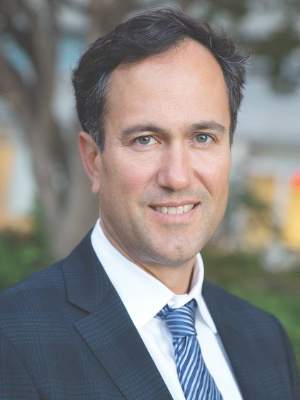
I would like to invite all Vascular Annual Meeting attendees to join us for the Spring Meeting of the Vascular and Endovascular Surgery Society (VESS; formerly Peripheral Vascular Surgery Society). This year’s program will be held on Wednesday, June 8 at 8:00 a.m. and conclude at 4:00 p.m. Dr. Bernadette Aulivola and her Program Committee have selected over 25 outstanding research projects for presentation.
The format of the meeting allows great discussion between the presenters and attendees. As in previous years, the VESS and SVS Program Committees combined to select the best scientific papers for the Vascular Annual Meeting. I am sure you will be delighted with this year’s Vascular Annual Meeting.
Founded in 1976 as a club for young and recently graduated Vascular Surgeons, the society has grown to a national organization and international society boasting well over 1,000 members. Focusing on young vascular surgeons in both academic and community practice, our organization offers excellent educational opportunities and networking for career growth.
This year we are excited to introduce the VESS Women and Diversity Meet the Leaders Program, which matches young surgeons with a leader based on the needs of the young surgeon and the experience of the leader to create a mentoring relationship.
VESS has two important scientific meetings yearly. The Spring Meeting is held in conjunction with the Vascular Annual Meeting. The Annual Meeting is held in the winter and is a highlight for both new and old members.
These meetings are a special time to share research, contribute ideas, learn new technologies, and make lifelong friends. The next Annual Meeting will take place February 2-5, 2017 at the Steamboat Grand in Steamboat, Colo. The Annual Meeting will feature interesting interactive programs, engaging speakers, and exciting social events. With nearly 3,000 skiable acres, and featuring night skiing too, it will be an outstanding venue for both education and relaxation. I encourage you to take advantage of the unique setting the Annual Meeting offers. Don’t miss this great vascular meeting in a great setting.
Our members strive to promote basic and clinical research that directly improves outcomes for our vascular and endovascular surgery patients. If you are not a member, please consider joining. You won’t be disappointed. Our new website is www.vesurgery.org. I look forward to seeing you at one of our meetings and encourage you to get involved in this terrific Society!
Thomas Maldonado, MD
VESS President

Patients Who Don't Speak English are Likely to Return to the Emergency Room
(Reuters Health) - Patients in the emergency room who don't speak English well are slightly more likely to return within days, suggesting their care the first time was not as good as it could have been, researchers say.
In a study in one New York hospital, about 4 percent of English speakers made an unplanned return to the ER within three days, compared to 5 percent of people with limited English.
Low use of professional translators may partly explain the disparity in care, the researchers report in the Annals of Emergency Medicine.
"There's a necessary but not sufficient step to providing care for people with low English proficiency . . . having a good interpreter or healthcare provider who can speak to them in their language," said Dr. Elizabeth Jacobs of the University of Wisconsin-Madison, who was not part of the new study.
The study team, led by Dr. Ka Ming Ngai of the Icahn School of Medicine at Mount Sinai in New York, analyzed 2012 data from the Mount Sinai emergency department. More than 32,000 adult patients and 45,000 ER visits were included. The study did not include patients with psychiatric or substance-related
complaints, those who were nonverbal or had altered mental status, and those with a history of frequent ER visits.
Almost 3,000 patients had limited English proficiency, and in about half of cases someone served as an interpreter. Usually, this was a family member or an ER staff member. Only 527 visits in this group, 24 percent, involved a professional interpreter.
More than a quarter of patients were admitted to the hospital and 1,380 patients had an unplanned return to the ER within three days.
After accounting for age, sex, insurance, race, ethnicity, triage category and other health problems, having limited English proficiency was not tied to greater risk of being admitted to the hospital.
But those with limited English proficiency were about 24 percent more likely to return to the ER unexpectedly.
Ngai told Reuters Health by email that he has been studying the problem of language barriers for the past six years and over time has seen some improvements.
"New medical students are now routinely educated to use interpreter phones during their clinical simulation . . .however, there are still many barriers including access to interpreters and interpreter phones, time constraints, and (doctors) trying to 'get by' with their own language skills," he said.
Ngai said regulatory bodies require hospitals to make language services available. In New York State, for example, upon a request to the hospital administration by the patient, the patient's family or representative, or the provider of medical care, hospitals must provide translation services in inpatient and outpatient settings within 20 minutes and in emergency settings with 10 minutes.
Most New York Hospitals use an interpreter phone service, he said.
Patients who struggle to speak the local language are "a really important population to study and think about how we can improve their care," Jacobs said.
A 5 percent rather than 4 percent rate of return to the ER is not a large difference, but that could be due to the large number of patients excluded from the study, and because there was no validated measure of English proficiency, Jacobs said.
"That might be why we didn't see large differences, if some people considered low English proficiency actually spoke English well, or were getting good interpretive services," she said. "If you took them out, the difference might be larger."
Patients who do not speak English may struggle in other areas of the health system more than at the ER, she added.
But even having an interpreter at the hospital won't help patients deal with insurance providers, she noted.
"There are contextual issues that you may not be able to fully adequately address unless you can understand the nuances," Jacobs said.
"When dealing with immigrant population, it is almost always more than 'just' language," Ngai agreed. There can be cultural issues, too.
In addition, people with low English proficiency may also be less able to take days off of work, and to agree to be admitted to the hospital when necessary, than others, Jacobs said.
We've made tremendous progress in assuring interpreters are more available," in person, over the phone or by video, Jacobs said.
But, she said, "we are very imperfect at getting patients the services they need. It's important for providers to be educated on these issues and to understand how to access these services."
It would be ideal to try to match patients with providers by language and culture, but in the meantime, "language is a good start," Ngai said.