User login
Host RNA biosignatures distinguish bacterial from viral fever
RNA-expression biosignatures derived from the patient’s peripheral blood distinguish bacterial from viral causes of fever in young children, according to two separate preliminary studies published online Aug. 23 in JAMA.
Several studies have suggested that the source of infection in febrile children might be identified by examining the pattern of host genes that are either activated or suppressed during the body’s inflammatory response. Distinguishing the relatively few but potentially life-threatening bacterial infections from the more common but milder, self-resolving viral infections is difficult, and current practice is to admit “ill-appearing” febrile children to the hospital and administer parenteral antibiotics while awaiting the results of blood and tissue cultures. Those results are often ambiguous, and the whole process represents a large burden on health care resources as well as contributing to inappropriate antibiotic treatment.

Two multinational research groups developed different techniques for detecting RNA biosignatures in patients’ blood samples, then assessed the accuracy of those tests in validation cohorts. One group focused on ruling out bacterial infection as the source of fever in young children (median age, 19 months), while the other investigated whether the host responses of the youngest children (aged 60 days and younger), who have immature immune systems, are robust enough to allow detection of RNA biosignatures.
In the discovery phase of the first study, analysis of RNA gene expression was performed on blood samples obtained from 240 children at admission to hospitals in the United Kingdom, Spain, and the United States during a 4-year period. A total of 8,565 RNA transcript signatures were identified as potential biomarkers to discriminate between viral and bacterial infection. This was narrowed down to 38 transcript signatures, and then to only 2 – IFI44L and FAM89A – that were used to devise a Disease Risk Score (DRS) for each patient, said Jethro A. Herberg, PhD, of the division of infectious diseases, Imperial College London, and his associates.
IFI44L expression was increased in patients who had viral infection, while FAM89A expression was increased in those who had bacterial infection, as compared with healthy children. (In previous studies, IFI44L was reported to be up-regulated in interferon-mediated antiviral responses and FAM89A was reported to be elevated among children with septic shock.)
The DRS showed 90% sensitivity in distinguishing viral from bacterial infection in the discovery cohort. It then showed 96.4% sensitivity in a validation cohort of 130 febrile children (mean age, 17 months). The DRS also identified bacterial infection in a validation cohort of 24 children with meningococcal infection (91.7% sensitivity and 96.0% specificity), and distinguished it from inflammatory conditions in another cohort of 30 children with juvenile idiopathic arthritis and 18 with Henoch-Schönlein purpura (90.0% sensitivity and 95.8% specificity).
The DRS discriminated among viral, bacterial, and inflammatory diseases including systemic lupus erythematosus in a further validation cohort, a published dataset from children and adults who had all three types of illness. It was accurate regardless of the severity of infection and regardless of the duration of infection, as well as in cases where patients were coinfected with both virus and bacteria, the investigators said (JAMA. 2016 Aug 23. doi:10.1001/jama.2016.11236).
“The DRS signature, distinguishing viral from bacterial infections with only two transcripts, has potential to be translated into a clinically applicable test using current technology. Furthermore, new methods for rapid detection of nucleic acids, including nanoparticles and electrical impedance, have potential for low-cost, rapid analysis of multitranscript signatures,” Dr. Herberg and his associates noted.
Further research is needed to assess the accuracy and clinical utility of this technique in different settings, they added.
In the second study, RNA gene expression was analyzed from blood samples from 1,883 febrile infants (median age, 37 days) “who posed diagnostic quandaries” at admission to 22 emergency departments during a 2-year period. The discovery phase involved 89 of these infants who ultimately were found to have bacterial infections (bacteremia or UTIs), 190 who didn’t have bacterial infections (enterovirus, influenza, or other viruses), and 19 healthy control infants, said Prashant Mahajan, MD, division chief and research director, pediatric emergency medicine, Children’s Hospital of Michigan, Detroit, and his associates.

The investigators identified 3,753 RNA transcript signatures that could potentially identify or rule out bacterial sources of infection, which they then narrowed down to 66. This set of 66 signatures showed 82% sensitivity and 88% specificity in the discovery cohort and 87% sensitivity and 89% specificity in a validation cohort.
“The bacterial RNA biosignature was notably more predictive of bacterial infection than clinical examination” and use of the Yale Observation Score, and it “added significantly to prediction beyond the YOS alone,” Dr. Mahajan and his associates said (JAMA. 2016 Aug 23. doi: 10.1001/jama.2016.9207).
“Despite the young age of the febrile infants evaluated, they carried robust RNA biosignatures and demonstrated that regardless of the etiology of the infections, their immune systems are programed to respond not only with shared elements induced by common microbes but also with specific patterns that allow discrimination by class of pathogen,” they noted.
Further research is needed to confirm and refine these preliminary results. “As technology advances, RNA biosignatures may prove to be an alternative and accurate method to identify infants with bacterial infections. This would help clinicians target evaluation and therapy when they are needed and avoid invasive procedures, antibiotics, and hospitalizations when they are not,” Dr. Mahajan and his associates said.
Dr. Herberg’s study was supported by the Imperial College Comprehensive Biomedical Research Center, the National Institutes of Health, the European Union’s Seventh Framework Program, and numerous other groups. Dr. Herberg reported having no relevant financial disclosures. Dr. Mahajan’s study was supported by the Health Resources and Services Administration, Emergency Services for Children, the Eunice Kennedy Shriver National Institute of Child Health and Human Development, and the National Institutes of Health. Dr. Mahajan reported having no relevant financial disclosures.
The work by Herberg et al. and Mahajan et al. represents an important advance: the potential of genetics to help in the evaluation of febrile children.

|
Dr. Howard Bauchner |
Clearly RNA sequencing and other methods for RNA quantification are in the early days, and clinical applications must await further replication and refinement of these results in rigorous studies. But the day may soon arise when a parent of a febrile child may do a laboratory test at home, call a physician, and mutually decide whether the child should be seen for further evaluation.
Howard Bauchner, MD, is JAMA Editor in Chief. He reported having no relevant financial disclosures. Dr. Bauchner made these remarks in an editorial accompanying the two reports on RNA biosignatures (JAMA 2016;316:824-5).
The work by Herberg et al. and Mahajan et al. represents an important advance: the potential of genetics to help in the evaluation of febrile children.
|
|
Dr. Howard Bauchner |
Clearly RNA sequencing and other methods for RNA quantification are in the early days, and clinical applications must await further replication and refinement of these results in rigorous studies. But the day may soon arise when a parent of a febrile child may do a laboratory test at home, call a physician, and mutually decide whether the child should be seen for further evaluation.
Howard Bauchner, MD, is JAMA Editor in Chief. He reported having no relevant financial disclosures. Dr. Bauchner made these remarks in an editorial accompanying the two reports on RNA biosignatures (JAMA 2016;316:824-5).
RNA-expression biosignatures derived from the patient’s peripheral blood distinguish bacterial from viral causes of fever in young children, according to two separate preliminary studies published online Aug. 23 in JAMA.
Several studies have suggested that the source of infection in febrile children might be identified by examining the pattern of host genes that are either activated or suppressed during the body’s inflammatory response. Distinguishing the relatively few but potentially life-threatening bacterial infections from the more common but milder, self-resolving viral infections is difficult, and current practice is to admit “ill-appearing” febrile children to the hospital and administer parenteral antibiotics while awaiting the results of blood and tissue cultures. Those results are often ambiguous, and the whole process represents a large burden on health care resources as well as contributing to inappropriate antibiotic treatment.
Two multinational research groups developed different techniques for detecting RNA biosignatures in patients’ blood samples, then assessed the accuracy of those tests in validation cohorts. One group focused on ruling out bacterial infection as the source of fever in young children (median age, 19 months), while the other investigated whether the host responses of the youngest children (aged 60 days and younger), who have immature immune systems, are robust enough to allow detection of RNA biosignatures.
In the discovery phase of the first study, analysis of RNA gene expression was performed on blood samples obtained from 240 children at admission to hospitals in the United Kingdom, Spain, and the United States during a 4-year period. A total of 8,565 RNA transcript signatures were identified as potential biomarkers to discriminate between viral and bacterial infection. This was narrowed down to 38 transcript signatures, and then to only 2 – IFI44L and FAM89A – that were used to devise a Disease Risk Score (DRS) for each patient, said Jethro A. Herberg, PhD, of the division of infectious diseases, Imperial College London, and his associates.
IFI44L expression was increased in patients who had viral infection, while FAM89A expression was increased in those who had bacterial infection, as compared with healthy children. (In previous studies, IFI44L was reported to be up-regulated in interferon-mediated antiviral responses and FAM89A was reported to be elevated among children with septic shock.)
The DRS showed 90% sensitivity in distinguishing viral from bacterial infection in the discovery cohort. It then showed 96.4% sensitivity in a validation cohort of 130 febrile children (mean age, 17 months). The DRS also identified bacterial infection in a validation cohort of 24 children with meningococcal infection (91.7% sensitivity and 96.0% specificity), and distinguished it from inflammatory conditions in another cohort of 30 children with juvenile idiopathic arthritis and 18 with Henoch-Schönlein purpura (90.0% sensitivity and 95.8% specificity).
The DRS discriminated among viral, bacterial, and inflammatory diseases including systemic lupus erythematosus in a further validation cohort, a published dataset from children and adults who had all three types of illness. It was accurate regardless of the severity of infection and regardless of the duration of infection, as well as in cases where patients were coinfected with both virus and bacteria, the investigators said (JAMA. 2016 Aug 23. doi:10.1001/jama.2016.11236).
“The DRS signature, distinguishing viral from bacterial infections with only two transcripts, has potential to be translated into a clinically applicable test using current technology. Furthermore, new methods for rapid detection of nucleic acids, including nanoparticles and electrical impedance, have potential for low-cost, rapid analysis of multitranscript signatures,” Dr. Herberg and his associates noted.
Further research is needed to assess the accuracy and clinical utility of this technique in different settings, they added.
In the second study, RNA gene expression was analyzed from blood samples from 1,883 febrile infants (median age, 37 days) “who posed diagnostic quandaries” at admission to 22 emergency departments during a 2-year period. The discovery phase involved 89 of these infants who ultimately were found to have bacterial infections (bacteremia or UTIs), 190 who didn’t have bacterial infections (enterovirus, influenza, or other viruses), and 19 healthy control infants, said Prashant Mahajan, MD, division chief and research director, pediatric emergency medicine, Children’s Hospital of Michigan, Detroit, and his associates.
The investigators identified 3,753 RNA transcript signatures that could potentially identify or rule out bacterial sources of infection, which they then narrowed down to 66. This set of 66 signatures showed 82% sensitivity and 88% specificity in the discovery cohort and 87% sensitivity and 89% specificity in a validation cohort.
“The bacterial RNA biosignature was notably more predictive of bacterial infection than clinical examination” and use of the Yale Observation Score, and it “added significantly to prediction beyond the YOS alone,” Dr. Mahajan and his associates said (JAMA. 2016 Aug 23. doi: 10.1001/jama.2016.9207).
“Despite the young age of the febrile infants evaluated, they carried robust RNA biosignatures and demonstrated that regardless of the etiology of the infections, their immune systems are programed to respond not only with shared elements induced by common microbes but also with specific patterns that allow discrimination by class of pathogen,” they noted.
Further research is needed to confirm and refine these preliminary results. “As technology advances, RNA biosignatures may prove to be an alternative and accurate method to identify infants with bacterial infections. This would help clinicians target evaluation and therapy when they are needed and avoid invasive procedures, antibiotics, and hospitalizations when they are not,” Dr. Mahajan and his associates said.
Dr. Herberg’s study was supported by the Imperial College Comprehensive Biomedical Research Center, the National Institutes of Health, the European Union’s Seventh Framework Program, and numerous other groups. Dr. Herberg reported having no relevant financial disclosures. Dr. Mahajan’s study was supported by the Health Resources and Services Administration, Emergency Services for Children, the Eunice Kennedy Shriver National Institute of Child Health and Human Development, and the National Institutes of Health. Dr. Mahajan reported having no relevant financial disclosures.
FROM JAMA
Key clinical point: RNA biosignatures derived from the patient’s peripheral blood distinguish bacterial from viral causes of fever in young children.
Major finding: Study 1. The DRS showed 96.4% sensitivity in a validation cohort of 130 febrile children. Study 2. A set of 66 RNA transcript signatures showed 82% sensitivity and 88% specificity in the discovery cohort and 87% sensitivity and 89% specificity in a validation cohort.
Data source: Two separate preliminary studies developing and validating tests of host responses to infection, involving 240 and 279 patients, respectively.
Disclosures: Dr. Herberg’s study was supported by the Imperial College Comprehensive Biomedical Research Center, the National Institutes of Health, the European Union’s Seventh Framework Program, and numerous other groups. Dr. Herberg reported having no relevant financial disclosures. Dr. Mahajan’s study was supported by the Health Resources and Services Administration, Emergency Services for Children, the Eunice Kennedy Shriver National Institute of Child Health and Human Development, and the National Institutes of Health. Dr. Mahajan reported having no relevant financial disclosures.

Treatment may allow HSCT without radiation, chemotherapy
A new therapy combining an anti-c-Kit monoclonal antibody with a CD47 blocker allowed hematopoietic stem cell engraftment in immunocompetent mice without the need for toxic preconditioning using radiation or chemotherapy, according to a report published in Science Translational Medicine.
Until now, hematopoietic stem cell transplantation has required rigorous conditioning regimens to clear out the host’s bone marrow, which can cause lifelong complications. So the procedure has been reserved for patients whose life-threatening disorders justified such toxicity. “Safer and more targeted conditioning protocols could both improve the safety of transplantation and extend the existing clinical utility of this powerful form of cell therapy,” said Akanksha Chhabra, PhD, of the department of blood and marrow transplantation, Stanford (Calif.) University, and her associates.
They assessed the new combined treatment in a series of laboratory and mouse studies. The opsonizing anti-c-Kit monoclonal antibodies induced robust depletion of functional hematopoietic stem cells in immunocompetent mice, which allowed donor stem cells to engraft in these hosts. Adding the T-cell–depleting CD47-antagonists further facilitated immune ablation of host stem cells and progenitor cells. Combined, the two agents eliminated more than 99% of host hematopoietic stem cells in the bone marrow and enabled strong engraftment of the donor stem cells, while avoiding radiation- and chemotherapy-related adverse effects.
The main toxicities that occurred in treated mice were, as expected, reductions in hematologic parameters, especially red blood cell indices. This may be related to a factor in mouse physiology that is not present in humans. But if such toxicities do develop in human subjects, they can be mitigated by careful monitoring and occasional supportive transfusions, Dr. Chhabra and her associates said (Sci Transl Med. 2016;8:351ra105).
These two types of antibodies are already being investigated separately in early-phase clinical trials. If the combined treatment proves effective and safe in humans – a question that awaits further clinical studies – hematopoietic stem cell transplantation might be extended to nonmalignant conditions such as inherited immunodeficiency, inborn errors of metabolism, and hemoglobinopathies. It might also be adapted for use in solid-organ transplants, the researchers added.
This work was supported by the Virginia and D.K. Ludwig Fund for Cancer Research and several other nonprofit organizations, the California Institute for Regenerative Medicine, and the National Institutes of Health. Dr. Chhabra is a coinventor on a patent described in this article, and her associates are cofounders of Forty Seven, the company that licensed the technology for radiation- and chemotherapy-free stem-cell transplantation. Two associates also serve as advisors for Alexo Therapeutics, which develops CD47-based treatments.
FROM SCIENCE TRANSLATIONAL MEDICINE
Key clinical point: A new treatment allowed hematopoietic stem cell engraftment in immunocompetent mice without the need for toxic preconditioning using radiation or chemotherapy.
Major finding: The combined therapy eliminated more than 99% of host hematopoietic stem cells.
Data source: A series of laboratory and mouse studies of combined treatment with anti-c-Kit monoclonal antibodies plus CD47 blockers.
Disclosures: This work was supported by the Virginia and D.K. Ludwig Fund for Cancer Research and several other nonprofit organizations, the California Institute for Regenerative Medicine, and the National Institutes of Health. Dr. Chhabra is a coinventor on a patent described in this article, and her associates are cofounders of Forty Seven, the company that licensed the technology for radiation- and chemotherapy-free stem-cell transplantation. Two associates also serve as advisors for Alexo Therapeutics, which develops CD47-based treatments.
Hemophilia carriers are at risk for abnormal bleeding
ORLANDO – The traditional view that women who are hemophilia carriers are unaffected by the disease may not be accurate, because many hemophilia carriers experience abnormal bleeding, a hemophilia specialist contends.
“We do know that hemophilia carriers have increased bleeding scores compared to controls,” said Michelle Sholzberg, MD, a hematologist and medical director of the coagulation laboratory at St. Michael’s Hospital in Toronto.

Bleeding in carriers is predominantly mucocutaneous bleeding, and may include epistaxis, heavy menstrual bleeding, bleeding with interventional procedures, and postpartum hemorrhage, she said at the World Federation of Hemophilia World Congress.
The majority of women who carry a factor VIII or factor IX mutation on one X chromosome have factor levels ranging from 0.40 to 0.60 IU/mL, which are generally considered to be adequate for hemostasis.
Yet, “we know that many carriers bleed, and in fact there are carriers who bleed with higher factor levels that are truly in the normal range,” Dr. Sholzberg said.
Normal coagulation factor levels in the general population range from about 0.50 to 1.50 IU/mL. By this standard, approximately 30% of hemophilia carriers have low factor levels, she said.
Joint damage
In addition, 14%-19% of hemophilia A carriers report hemarthrosis, and there is an association in factor VIII or IX deficiencies among hemophilia carriers and reduced joint range of motion. In one study, this decreased range of motion was evident as early as the preteen years, and was suggestive of subclinical musculoskeletal bleeding among carriers. In a separate study, investigators found evidence that hemophilia A carriers have pathologic and radiologic evidence of structural joint damage.
Variability in factor levels among carriers may be caused by lyonization (the inactivation of one of the X-chromosomes), ABO blood type, the presence of mutations in genes encoding for von Willebrand factor, compound heterozygosity or homozygosity, or Turner’s mosaicism.
Clotting factor levels can also change with pregnancy, with hemophilia A carriers experiencing an increase in mean factor VIII levels from 0.46 IU/mL prepregnancy to 1.21 IU/mL in the third trimester, and hemophilia B carriers having a corresponding rise in factor IX levels from 0.31 IU/mL to 0.48 IU/mL, and many carriers still have suboptimal factor levels at pregnancy, Dr. Sholzberg said.
Postpartum hemorrhagic complications among carriers can lead to iron-deficiency anemia. The Centers for Disease Control and Prevention recommends testing all nonpregnant women for anemia every 5-10 years throughout their childbearing years, and annual objective testing for women with risk factors, she noted.
“I think we can all appreciate now that the multidisciplinary approach is important for women with bleeding disorders, and carriers of hemophilia can be safely cared for at HTCs [hemophilia treatment centers],” she said.
Carriers should be treated with a multimodal approach that enhances patient education and awareness, with an emphasis on self-report of symptoms and communication with health care providers.
“It’s also critical never to start a conversation with a woman who has a bleeding disorder with ‘Do you have heavy menstrual bleeding?’ I think we all know that the answer is almost always ‘No, I don’t,’ and that’s because if she has bled heavily for her entire life, she doesn’t know what normal is,” Dr. Sholzberg said.
She described a typical comprehensive care plan for a pregnant hemophilia A carrier. The plan will include information about the diagnosis, recommendations to the obstetricians to avoid the use of invasive instrumentation, anesthesia recommendations, recommendations for care of the mother regarding hemostatic agents, postpartum tranexamic acid, and factor levels, and hematology recommendations for the newborn.
Dr. Sholzberg disclosed research support and/or honoraria from Shire (previously Baxter, Baxalta), Octapharma, CSL Behring, Pfizer, and Novo Nordisk, and advisory committee activity for Shire.
ORLANDO – The traditional view that women who are hemophilia carriers are unaffected by the disease may not be accurate, because many hemophilia carriers experience abnormal bleeding, a hemophilia specialist contends.
“We do know that hemophilia carriers have increased bleeding scores compared to controls,” said Michelle Sholzberg, MD, a hematologist and medical director of the coagulation laboratory at St. Michael’s Hospital in Toronto.
Bleeding in carriers is predominantly mucocutaneous bleeding, and may include epistaxis, heavy menstrual bleeding, bleeding with interventional procedures, and postpartum hemorrhage, she said at the World Federation of Hemophilia World Congress.
The majority of women who carry a factor VIII or factor IX mutation on one X chromosome have factor levels ranging from 0.40 to 0.60 IU/mL, which are generally considered to be adequate for hemostasis.
Yet, “we know that many carriers bleed, and in fact there are carriers who bleed with higher factor levels that are truly in the normal range,” Dr. Sholzberg said.
Normal coagulation factor levels in the general population range from about 0.50 to 1.50 IU/mL. By this standard, approximately 30% of hemophilia carriers have low factor levels, she said.
Joint damage
In addition, 14%-19% of hemophilia A carriers report hemarthrosis, and there is an association in factor VIII or IX deficiencies among hemophilia carriers and reduced joint range of motion. In one study, this decreased range of motion was evident as early as the preteen years, and was suggestive of subclinical musculoskeletal bleeding among carriers. In a separate study, investigators found evidence that hemophilia A carriers have pathologic and radiologic evidence of structural joint damage.
Variability in factor levels among carriers may be caused by lyonization (the inactivation of one of the X-chromosomes), ABO blood type, the presence of mutations in genes encoding for von Willebrand factor, compound heterozygosity or homozygosity, or Turner’s mosaicism.
Clotting factor levels can also change with pregnancy, with hemophilia A carriers experiencing an increase in mean factor VIII levels from 0.46 IU/mL prepregnancy to 1.21 IU/mL in the third trimester, and hemophilia B carriers having a corresponding rise in factor IX levels from 0.31 IU/mL to 0.48 IU/mL, and many carriers still have suboptimal factor levels at pregnancy, Dr. Sholzberg said.
Postpartum hemorrhagic complications among carriers can lead to iron-deficiency anemia. The Centers for Disease Control and Prevention recommends testing all nonpregnant women for anemia every 5-10 years throughout their childbearing years, and annual objective testing for women with risk factors, she noted.
“I think we can all appreciate now that the multidisciplinary approach is important for women with bleeding disorders, and carriers of hemophilia can be safely cared for at HTCs [hemophilia treatment centers],” she said.
Carriers should be treated with a multimodal approach that enhances patient education and awareness, with an emphasis on self-report of symptoms and communication with health care providers.
“It’s also critical never to start a conversation with a woman who has a bleeding disorder with ‘Do you have heavy menstrual bleeding?’ I think we all know that the answer is almost always ‘No, I don’t,’ and that’s because if she has bled heavily for her entire life, she doesn’t know what normal is,” Dr. Sholzberg said.
She described a typical comprehensive care plan for a pregnant hemophilia A carrier. The plan will include information about the diagnosis, recommendations to the obstetricians to avoid the use of invasive instrumentation, anesthesia recommendations, recommendations for care of the mother regarding hemostatic agents, postpartum tranexamic acid, and factor levels, and hematology recommendations for the newborn.
Dr. Sholzberg disclosed research support and/or honoraria from Shire (previously Baxter, Baxalta), Octapharma, CSL Behring, Pfizer, and Novo Nordisk, and advisory committee activity for Shire.
EXPERT ANALYSIS FROM WFH 2016 WORLD CONGRESS

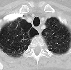
NAF1 gene mutations predispose to pulmonary fibrosis, emphysema
Rare frameshift mutations in the NAF1 gene were discovered to cause a telomere-shortening syndrome which, among other adverse effects, predisposes carriers to develop pulmonary fibrosis (PF) and emphysema, according to a report published in Science Translational Medicine.
“Our findings here ... highlight how telomere shortening is a relevant mechanism for PF-emphysema susceptibility in a subset of patients beyond those with mutations in the telomerase core components. It is thus possible that efforts to reverse the telomere defect, or other regenerative approaches, will influence the natural history of these progressive pathologies in patients with telomere-mediated lung disease,” said Susan E. Stanley, an MD-PhD candidate in the department of oncology, Johns Hopkins University, Baltimore, and her associates.

Pulmonary fibrosis and emphysema cluster in some families, but the genetic basis of such cases is poorly understood. Both PF and emphysema have been linked to premature aging of lung tissue and to abnormalities in the maintenance of telomere length. In addition, at least half of patients with familial and sporadic PF, and many with emphysema, have the clinical features of a short-telomere syndrome, including bone marrow failure/myelodysplastic syndrome, liver disease, and infertility.
The diagnosis of a short-telomere syndrome, as opposed to isolated PF-emphysema, is essential for appropriate treatment because if the defect is systemic, patients will “show exquisite sensitivity to otherwise tolerated medications and procedures, especially in the setting of lung transplantation,” the investigators said (Sci Transl Med. 2016;8:351ra107).
To explore the genetic basis of familial PF-emphysema, the researchers performed a series of studies, beginning with whole-genome sequencing on peripheral blood samples from five unrelated probands in familial PF-emphysema pedigrees. These participants had abnormally short telomeres and extrapulmonary features of short-telomere syndrome. Three of them who had low levels of the telomerase RNA component TR were selected for a candidate gene search, which revealed the NAF1 mutations.
The mutations were then found to be present in 2 of 30 (7%) affected members of a prevalence cohort but in none of 134 unaffected control subjects (0%), and in none of 9,006 samples from a public database of unaffected people (0%). Further genetic laboratory and mouse studies were performed to link the mutations with specific pathologies and to trace their functional effects. Their results led the researchers to conclude that these rare NAF1 variants interfere with RNA biogenesis, causing short telomeres resulting in lung disease and other abnormalities.
This work was supported by the National Institutes of Health, the Commonwealth Foundation, and the American Cancer Society. Ms. Stanley and her associates reported having no relevant financial disclosures.
Rare frameshift mutations in the NAF1 gene were discovered to cause a telomere-shortening syndrome which, among other adverse effects, predisposes carriers to develop pulmonary fibrosis (PF) and emphysema, according to a report published in Science Translational Medicine.
“Our findings here ... highlight how telomere shortening is a relevant mechanism for PF-emphysema susceptibility in a subset of patients beyond those with mutations in the telomerase core components. It is thus possible that efforts to reverse the telomere defect, or other regenerative approaches, will influence the natural history of these progressive pathologies in patients with telomere-mediated lung disease,” said Susan E. Stanley, an MD-PhD candidate in the department of oncology, Johns Hopkins University, Baltimore, and her associates.
Pulmonary fibrosis and emphysema cluster in some families, but the genetic basis of such cases is poorly understood. Both PF and emphysema have been linked to premature aging of lung tissue and to abnormalities in the maintenance of telomere length. In addition, at least half of patients with familial and sporadic PF, and many with emphysema, have the clinical features of a short-telomere syndrome, including bone marrow failure/myelodysplastic syndrome, liver disease, and infertility.
The diagnosis of a short-telomere syndrome, as opposed to isolated PF-emphysema, is essential for appropriate treatment because if the defect is systemic, patients will “show exquisite sensitivity to otherwise tolerated medications and procedures, especially in the setting of lung transplantation,” the investigators said (Sci Transl Med. 2016;8:351ra107).
To explore the genetic basis of familial PF-emphysema, the researchers performed a series of studies, beginning with whole-genome sequencing on peripheral blood samples from five unrelated probands in familial PF-emphysema pedigrees. These participants had abnormally short telomeres and extrapulmonary features of short-telomere syndrome. Three of them who had low levels of the telomerase RNA component TR were selected for a candidate gene search, which revealed the NAF1 mutations.
The mutations were then found to be present in 2 of 30 (7%) affected members of a prevalence cohort but in none of 134 unaffected control subjects (0%), and in none of 9,006 samples from a public database of unaffected people (0%). Further genetic laboratory and mouse studies were performed to link the mutations with specific pathologies and to trace their functional effects. Their results led the researchers to conclude that these rare NAF1 variants interfere with RNA biogenesis, causing short telomeres resulting in lung disease and other abnormalities.
This work was supported by the National Institutes of Health, the Commonwealth Foundation, and the American Cancer Society. Ms. Stanley and her associates reported having no relevant financial disclosures.
FROM SCIENCE TRANSLATIONAL MEDICINE
Key clinical point: Certain rare mutations in the NAF1 gene were discovered to predispose carriers to develop pulmonary fibrosis and emphysema.
Major finding: The rare NAF1 mutations were detected in 2 of 30 (7%) family members in an affected pedigree but in 0 of 134 controls.
Data source: A series of genetic sequencing and other studies involving five affected probands, 30 unrelated but affected patients, and 134 control subjects.
Disclosures: This work was supported by the National Institutes of Health, the Commonwealth Foundation, and the American Cancer Society. Ms. Stanley and her associates reported having no relevant financial disclosures.
No VTE prophylaxis needed after joint surgery in patients with hemophilia
ORLANDO – In patients with hemophilia who have therapeutic factor levels at the time of joint replacement surgery, prophylaxis against venous thromboembolism (VTE) may be unnecessary.
In a cohort study of patients with hemophilia A or B who underwent total joint replacement surgery while being in proper hemostasis with therapeutic factor levels, there were no clinically evident episodes of venous thromboembolism, even though none of the patients had received perioperative anticoagulant prophylaxis, reported investigators from the National Hemophilia Center and Institute of Thrombosis and Hemostasis at the Sheba Medical Center in Tel Hashomer, Israel.

The data should be reassuring to clinicians whose patients with hemophilia require major orthopedic procedures, said lead author Dr. Anna Seltser, an orthopedic resident at Sheba Medical Center, in an interview.
“We have a lot of hemophilia patients who are not well treated because they live in the desert or distant communities, and we also sometimes treat patients from the Palestinian side of the Gaza Strip who don’t have access to care and need this type of surgery,” she said.
“We collected what I think is the biggest series of patients until now, we didn’t give any of them VTE prophylaxis, and none of them had any DVT [deep vein thrombosis], PE [pulmonary embolism], or similar complication,” she said.
Skip the heparin?
VTE prophylaxis with low-molecular-weight heparin, warfarin, or other anticoagulant agents is a common practice following orthopedic surgery in patients without bleeding disorders. But for patients with severe hemophilia, who often require major joint replacement surgery following years of bleeding-induced arthropathy, it’s unclear whether perioperative anticoagulation is beneficial, the investigators noted in a scientific poster at the World Federation of Hemophilia World Congress.
Dr. Seltser and colleagues therefore conducted a prospective cohort study of 50 patients with hemophilia A or B treated with major joint surgery and subsequent revisions from 1988 through 2015 at their center. In all, 47 patients had severe hemophilia A, 2 had mild hemophilia A, and 1 had hemophilia B.
The authors analyzed data on demographics, comorbidities, type of surgery, use of factor concentrates therapy around the time of surgery, and complications during follow-up, including massive hemorrhage, infections, implant loosening, DVT, and PE.
The patients underwent a total of 74 primary joint replacements (16 hips, 52 knees, and 6 ankles) and 23 revision surgeries.
As noted, there were no episodes of either DVT or PE among any of the patients. All but one complication occurred among patients undergoing total knee replacement. These included three cases of hemarthrosis, three limited-range-of-motion cases requiring closed manipulations, four soft-tissue hematomas, and one case each of superficial wound infection, urinary tract infection, pneumonia, and Candida infection of the tongue.
The only other complication was a case of disseminated intravascular coagulation, sepsis, and hemorrhagic shock in a patient who had undergone a revision (original procedure unspecified).
“Despite the concern that proper replacement factor therapy, applied before and after the surgery, may increase the risk for thromboembolic complications in patients with hemophilia undergoing joint replacement, our data show that prophylactic anticoagulation in this group of patients is not necessary,” the investigators concluded.
The study was internally funded. The investigators reported no conflicts of interest.
ORLANDO – In patients with hemophilia who have therapeutic factor levels at the time of joint replacement surgery, prophylaxis against venous thromboembolism (VTE) may be unnecessary.
In a cohort study of patients with hemophilia A or B who underwent total joint replacement surgery while being in proper hemostasis with therapeutic factor levels, there were no clinically evident episodes of venous thromboembolism, even though none of the patients had received perioperative anticoagulant prophylaxis, reported investigators from the National Hemophilia Center and Institute of Thrombosis and Hemostasis at the Sheba Medical Center in Tel Hashomer, Israel.
The data should be reassuring to clinicians whose patients with hemophilia require major orthopedic procedures, said lead author Dr. Anna Seltser, an orthopedic resident at Sheba Medical Center, in an interview.
“We have a lot of hemophilia patients who are not well treated because they live in the desert or distant communities, and we also sometimes treat patients from the Palestinian side of the Gaza Strip who don’t have access to care and need this type of surgery,” she said.
“We collected what I think is the biggest series of patients until now, we didn’t give any of them VTE prophylaxis, and none of them had any DVT [deep vein thrombosis], PE [pulmonary embolism], or similar complication,” she said.
Skip the heparin?
VTE prophylaxis with low-molecular-weight heparin, warfarin, or other anticoagulant agents is a common practice following orthopedic surgery in patients without bleeding disorders. But for patients with severe hemophilia, who often require major joint replacement surgery following years of bleeding-induced arthropathy, it’s unclear whether perioperative anticoagulation is beneficial, the investigators noted in a scientific poster at the World Federation of Hemophilia World Congress.
Dr. Seltser and colleagues therefore conducted a prospective cohort study of 50 patients with hemophilia A or B treated with major joint surgery and subsequent revisions from 1988 through 2015 at their center. In all, 47 patients had severe hemophilia A, 2 had mild hemophilia A, and 1 had hemophilia B.
The authors analyzed data on demographics, comorbidities, type of surgery, use of factor concentrates therapy around the time of surgery, and complications during follow-up, including massive hemorrhage, infections, implant loosening, DVT, and PE.
The patients underwent a total of 74 primary joint replacements (16 hips, 52 knees, and 6 ankles) and 23 revision surgeries.
As noted, there were no episodes of either DVT or PE among any of the patients. All but one complication occurred among patients undergoing total knee replacement. These included three cases of hemarthrosis, three limited-range-of-motion cases requiring closed manipulations, four soft-tissue hematomas, and one case each of superficial wound infection, urinary tract infection, pneumonia, and Candida infection of the tongue.
The only other complication was a case of disseminated intravascular coagulation, sepsis, and hemorrhagic shock in a patient who had undergone a revision (original procedure unspecified).
“Despite the concern that proper replacement factor therapy, applied before and after the surgery, may increase the risk for thromboembolic complications in patients with hemophilia undergoing joint replacement, our data show that prophylactic anticoagulation in this group of patients is not necessary,” the investigators concluded.
The study was internally funded. The investigators reported no conflicts of interest.
AT WFH 2016 WORLD CONGRESS
Key clinical point: Prophylaxis against thromboembolic events after orthopedic surgery in patients with hemophilia may not be necessary.
Major finding: There were no thromboembolic events after joint surgery without anticoagulant prophylaxis in patients with hemophilia A or B.
Data source: Cohort study of 50 patients with hemophilia A or B undergoing major joint replacement surgery.
Disclosures: The study was internally funded. The investigators reported no conflicts of interest.

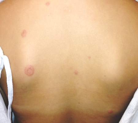
CANDLE syndrome case highlights key features of this type 1 interferonopathy
BOSTON – Annular erythematous plaques in a child with fever and dense, atypical, mixed mononuclear and neutrophilic dermal infiltrate on biopsy could signal the recently described autoinflammatory disorder known as CANDLE Syndrome, according to Raegan Hunt, MD.
CANDLE, which stands for chronic atypical neutrophilic dermatosis with lipodystrophy and elevated temperature, is a proteasome-associated autoinflammatory syndrome characterized by dysregulation of type 1 interferon signaling, Dr. Hunt of Baylor College of Medicine and Texas Children’s Hospital said during a presentation at the American Academy of Dermatology summer meeting.
She described a case involving a 12-year-old girl with erythematous, warm, tender, nonpruritic papules and plaques on the superior chest, neck, and upper back. Most had an annular configuration with central clearing and occasionally central duskiness and ulceration.
The child also sometimes had violaceous swelling of the eyelid, nodules on the ear, and figurate erythematous plaques on the upper arm.
“She’s been having these since she was an infant in periodic bursts, and carried a diagnosis of annular erythema of infancy,” Dr. Hunt said.
Other symptoms included recurrent fevers, myalgias, transient and migratory arthritis, elevated C-reactive protein and erythrocyte sedimentation rate, and aseptic meningitis requiring hospitalization.
She had no family history of autoimmune disease, immune deficiency, or other genetic diseases, Dr. Hunt said.
“She was treated with methotrexate and prednisone, as well as IVIG [intravenous immunoglobulin] every 4 weeks. Her prednisone was never weaned successfully below 0.8 mg/kg. She had complications of chronic corticosteroid disease, including many fractures,” she said.
A biopsy showed a dense infiltrate of atypical, mixed mononuclear and neutrophilic dermal infiltrate.
“These taken together are suggestive of ... CANDLE,” she said.
Genetic testing confirmed the presence of a compound heterozygous mutation in the proteasome subunit beta type 9, or PSMB8 gene.
Key features of CANDLE syndrome, first described in a 2010 article and further described by Liu, et al in 2012, include early disease onset, recurrent febrile episodes, skin lesions – including recurrent attacks of erythematous annular plaques and violaceous swelling of the eyelids, delayed physical development, progressive lipodystrophy, arthralgias, systemic inflammation, and aseptic meningitis episodes.
“Our patient did have very, very subtle lipodystrophy, but overall not as quick onset as some other [cases in children] that have been described,” Dr. Hunt said.
The type 1 interferonopathy is of autosomal recessive inheritance, and involves either homozygous or compound heterozygous mutations in PSMB8, as found in this patient. PSMB8 is an immunoproteasome unit involved in proteolysis and maintenance of cell homeostasis, she explained.
Treatment of the syndrome involves high-dose corticosteroids (usually 1-2 mg/kg day), which helps improve cutaneous eruptions, joint pain, and fever, but it is important to remember that disease flares can occur with tapering, Dr. Hunt noted.
Response is typically poor in patients treated with steroid-sparing agents, such as methotrexate, cyclosporine, azathioprine, or IVIG, as well as with tumor necrosis factor–alpha inhibitors and interleukin-1 receptor antagonists.
“But there is an interesting molecule on the horizon for possibly treating this more specifically and that’s JAK inhibitors,” she said, noting that a National Institutes of Health compassionate use clinical research trial is evaluating the JAK 1/2 inhibitor, baricitinib.
Dr. Hunt reported having no relevant disclosures.
EXPERT ANALYSIS FROM THE AAD SUMMER ACADEMY 2016
Key clinical point: Annular erythematous plaques in a child with fever and dense, atypical, mixed mononuclear and neutrophilic dermal infiltrate on biopsy could signal the recently described autoinflammatory disorder known as CANDLE syndrome.
Major finding: Genetic testing confirmed the presence of a compound heterozygous mutation in the proteasome subunit beta type 9, or PSMB8 gene.
Data source: Expert analysis – case description.
Disclosures: Dr. Hunt reported having no disclosures.

Flu vaccine prevented hospitalizations in patients 50 and older
The seasonal influenza vaccination reduced flu-related hospitalizations by 56.8% among people aged 50 and older during a recent flu season, according to a report published in Clinical Infectious Diseases.
Even in the oldest age group – the population with the highest risk of developing flu complications and perhaps the weakest immune response – influenza vaccination prevented serious complications, said Fiona P. Havers, MD, of the influenza division, Centers for Disease Control and Prevention, Atlanta, and her associates.

Data on vaccine efficacy in older adults are sparse, and randomized, placebo-controlled trials to gather evidence would be unethical. Dr. Havers and her colleagues studied the issue using a case-control design, focusing on community-dwelling adults aged 50 years and older during the 2010-2011 flu season. They identified 368 patients across 10 states who were hospitalized for polymerase chain reaction–confirmed influenza and matched them for age and county of residence with 773 control subjects.
Hospitalized case-patients were less likely to have been vaccinated (55%) than were control subjects (63%). Thus, the flu vaccine reduced the risk of hospitalization for influenza by 56.8% overall.

Vaccination reduced hospitalization for influenza by 63.9% in the youngest age group (50-64 years), by 61.0% in the intermediate age group (65-74 years), and by 57.3% in the oldest age group (75 years and older).
These results are similar to those reported in other studies assessing the same time period, including one that evaluated vaccine efficacy in ambulatory adults in the United States and Europe. They also are consistent with the results of observational studies performed during different flu seasons, the investigators said (Clin Infect Dis. 2016 Aug 2. doi: 10.1093/cid/ciw512).
Compared with control subjects, case-patients were more likely to be of nonwhite race, to be of Hispanic ethnicity, to have a lower income, to have had fewer years of education, to have two or more chronic health conditions, to have required recent hospitalization for respiratory problems, to have impaired mobility, and to have lower functional status.
“These findings support current U.S. recommendations for annual influenza vaccination in older adults, especially in adults aged 65 and older who are at higher risk of influenza-associated complications,” Dr. Havers and her associates said.
The Centers for Disease Control and Prevention supported the study. Dr. Havers reported having no relevant financial disclosures; one of her associates reported ties to Genentech, Merck, Novavax, and Pfizer.
The seasonal influenza vaccination reduced flu-related hospitalizations by 56.8% among people aged 50 and older during a recent flu season, according to a report published in Clinical Infectious Diseases.
Even in the oldest age group – the population with the highest risk of developing flu complications and perhaps the weakest immune response – influenza vaccination prevented serious complications, said Fiona P. Havers, MD, of the influenza division, Centers for Disease Control and Prevention, Atlanta, and her associates.
Data on vaccine efficacy in older adults are sparse, and randomized, placebo-controlled trials to gather evidence would be unethical. Dr. Havers and her colleagues studied the issue using a case-control design, focusing on community-dwelling adults aged 50 years and older during the 2010-2011 flu season. They identified 368 patients across 10 states who were hospitalized for polymerase chain reaction–confirmed influenza and matched them for age and county of residence with 773 control subjects.
Hospitalized case-patients were less likely to have been vaccinated (55%) than were control subjects (63%). Thus, the flu vaccine reduced the risk of hospitalization for influenza by 56.8% overall.
Vaccination reduced hospitalization for influenza by 63.9% in the youngest age group (50-64 years), by 61.0% in the intermediate age group (65-74 years), and by 57.3% in the oldest age group (75 years and older).
These results are similar to those reported in other studies assessing the same time period, including one that evaluated vaccine efficacy in ambulatory adults in the United States and Europe. They also are consistent with the results of observational studies performed during different flu seasons, the investigators said (Clin Infect Dis. 2016 Aug 2. doi: 10.1093/cid/ciw512).
Compared with control subjects, case-patients were more likely to be of nonwhite race, to be of Hispanic ethnicity, to have a lower income, to have had fewer years of education, to have two or more chronic health conditions, to have required recent hospitalization for respiratory problems, to have impaired mobility, and to have lower functional status.
“These findings support current U.S. recommendations for annual influenza vaccination in older adults, especially in adults aged 65 and older who are at higher risk of influenza-associated complications,” Dr. Havers and her associates said.
The Centers for Disease Control and Prevention supported the study. Dr. Havers reported having no relevant financial disclosures; one of her associates reported ties to Genentech, Merck, Novavax, and Pfizer.
FROM CLINICAL INFECTIOUS DISEASES
Key clinical point: Seasonal influenza vaccination reduced flu-related hospitalizations by 56.8% in people aged 50 years and older.
Major finding: Vaccination reduced hospitalization for influenza by 63.9% in people aged 50-64 years, by 61.0% in those aged 65-74 years, and by 57.3% in those aged 75 years and older.
Data source: A retrospective case-control study involving 368 cases and 773 matched controls assessed during a single recent flu season.
Disclosures: The Centers for Disease Control and Prevention supported the study. Dr. Havers reported having no relevant financial disclosures; one of her associates reported ties to Genentech, Merck, Novavax, and Pfizer.
Extended-release granisetron superior antiemetic for cisplatin chemotherapy
SAN DIEGO – An investigational extended-release formulation of granisetron outperformed ondansetron as a component of triple-drug antiemetic therapy for patients on cisplatin-based highly emetogenic chemotherapy, Dr. Lee Schwartzberg reported at the annual meeting of the Society of Gynecologic Oncology.
He presented a post hoc subgroup analysis drawn from the previously reported pivotal phase III 942-patient MAGIC trial of extended-release granisetron (Sustol), formerly known as APF530, versus ondansetron (Zofran) as part of the triple-drug antiemetic regimen recommended in National Community Cancer Network guidelines for the prevention of chemotherapy-induced nausea and vomiting (CINV) in patients on highly emetogenic chemotherapy .

Dr. Schwartzberg and coinvestigators focused on the 251 MAGIC participants on high-dose cisplatin-based chemotherapy. The reason? Few studies have examined the management of CINV in patients on platinum-based regimens, commonly used in treating gynecologic cancers, explained Dr. Schwartzberg, medical director of the West Clinic in Memphis.
Sustol consists of 2% granisetron in a proprietary slow-release vehicle. A single 500-mg subcutaneous dose containing 10 mg of granisetron maintains therapeutic blood levels of granisetron for 5 days or longer. These pharmacokinetics make APF530 an attractive strategy for preventing both acute and delayed chemotherapy-induced nausea and vomiting. The delayed phase, which affects many cancer patients 2-5 days following administration of chemotherapy, has traditionally been a particularly difficult management challenge.
National Community Cancer Network guidelines recommend that patients on highly emetogenic chemotherapy receive a potent three-drug antiemetic regimen consisting of a 5-hydroxytryptoamine-3 receptor antagonist – ondansetron and granisetron fall within this class – along with a neurokinin-1 receptor antagonist and dexamethasone.
MAGIC was a double-blind, prospective, multicenter trial in which 942 patients on highly emetogenic chemotherapy received either 500 mg of subcutaneous extended-release granisetron or intravenous ondansetron at 0.15 mg/kg. In addition, all participants concomitantly got 150 mg of the neurokinin-1 receptor antagonist fosaprepitant (Emend) plus 12 mg of IV dexamethasone on day 1, followed by 8 mg of oral dexamethasone once on day 2 and 8 mg twice daily on days 3 and 4.
In the overall trial, extended-release granisetron significantly outperformed ondansetron. The primary endpoint – complete response defined as no emesis and no use of rescue medication for CINV during the delayed phase – was achieved in 64.7% of the Sustol group, compared with 56.6% on ondansetron.
Results in Dr. Schwartzberg’s post hoc analysis confined to the 251 randomized patients on high-dose cisplatin-based chemotherapy paralleled those in the overall trial: a 64.8% complete response rate in the delayed phase in the granisetron group versus 56.3% with ondansetron. That absolute 8.5% difference favoring the investigational agent translates to a number needed to treat (NNT) of 12.
In the 112 women on highly emetogenic cisplatin-based chemotherapy, the complete response rates in the delayed phase were 61.6% with extended-release granisetron and 53.2% with ondansetron, for an absolute 10.3% difference and an NNT of 10.
Heron Therapeutics, which is developing extended-release granisetron, is awaiting word from the Food and Drug Administration regarding the company’s application for marketing approval.
The MAGIC trial was sponsored by Heron Therapeutics. Dr. Schwartzberg is a consultant to the company.
SAN DIEGO – An investigational extended-release formulation of granisetron outperformed ondansetron as a component of triple-drug antiemetic therapy for patients on cisplatin-based highly emetogenic chemotherapy, Dr. Lee Schwartzberg reported at the annual meeting of the Society of Gynecologic Oncology.
He presented a post hoc subgroup analysis drawn from the previously reported pivotal phase III 942-patient MAGIC trial of extended-release granisetron (Sustol), formerly known as APF530, versus ondansetron (Zofran) as part of the triple-drug antiemetic regimen recommended in National Community Cancer Network guidelines for the prevention of chemotherapy-induced nausea and vomiting (CINV) in patients on highly emetogenic chemotherapy .
Dr. Schwartzberg and coinvestigators focused on the 251 MAGIC participants on high-dose cisplatin-based chemotherapy. The reason? Few studies have examined the management of CINV in patients on platinum-based regimens, commonly used in treating gynecologic cancers, explained Dr. Schwartzberg, medical director of the West Clinic in Memphis.
Sustol consists of 2% granisetron in a proprietary slow-release vehicle. A single 500-mg subcutaneous dose containing 10 mg of granisetron maintains therapeutic blood levels of granisetron for 5 days or longer. These pharmacokinetics make APF530 an attractive strategy for preventing both acute and delayed chemotherapy-induced nausea and vomiting. The delayed phase, which affects many cancer patients 2-5 days following administration of chemotherapy, has traditionally been a particularly difficult management challenge.
National Community Cancer Network guidelines recommend that patients on highly emetogenic chemotherapy receive a potent three-drug antiemetic regimen consisting of a 5-hydroxytryptoamine-3 receptor antagonist – ondansetron and granisetron fall within this class – along with a neurokinin-1 receptor antagonist and dexamethasone.
MAGIC was a double-blind, prospective, multicenter trial in which 942 patients on highly emetogenic chemotherapy received either 500 mg of subcutaneous extended-release granisetron or intravenous ondansetron at 0.15 mg/kg. In addition, all participants concomitantly got 150 mg of the neurokinin-1 receptor antagonist fosaprepitant (Emend) plus 12 mg of IV dexamethasone on day 1, followed by 8 mg of oral dexamethasone once on day 2 and 8 mg twice daily on days 3 and 4.
In the overall trial, extended-release granisetron significantly outperformed ondansetron. The primary endpoint – complete response defined as no emesis and no use of rescue medication for CINV during the delayed phase – was achieved in 64.7% of the Sustol group, compared with 56.6% on ondansetron.
Results in Dr. Schwartzberg’s post hoc analysis confined to the 251 randomized patients on high-dose cisplatin-based chemotherapy paralleled those in the overall trial: a 64.8% complete response rate in the delayed phase in the granisetron group versus 56.3% with ondansetron. That absolute 8.5% difference favoring the investigational agent translates to a number needed to treat (NNT) of 12.
In the 112 women on highly emetogenic cisplatin-based chemotherapy, the complete response rates in the delayed phase were 61.6% with extended-release granisetron and 53.2% with ondansetron, for an absolute 10.3% difference and an NNT of 10.
Heron Therapeutics, which is developing extended-release granisetron, is awaiting word from the Food and Drug Administration regarding the company’s application for marketing approval.
The MAGIC trial was sponsored by Heron Therapeutics. Dr. Schwartzberg is a consultant to the company.
AT THE ANNUAL MEETING ON WOMEN’S CANCER
Key clinical point: Investigational agent outperforms ondansetron during delayed phase of highly emetogenic cisplatin-based chemotherapy-induced nausea and vomiting.
Major finding: Complete response rate during delayed phase was 64.8% when triple antiemetic therapy included extended-release granisetron and 56.3% when the regimen included ondansetron.
Data source: This post hoc subgroup analysis of the 942-patient pivotal phase III MAGIC trial focused specifically on the 251 participants on highly emetogenic cisplatin-based chemotherapy.
Disclosures: The study presenter reported serving as a consultant to Heron Therapeutics, which sponsored the MAGIC trial.
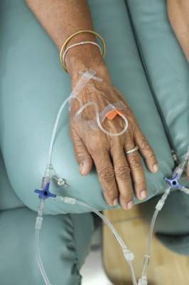
VIDEO: Varenicline underused for smoking cessation
ROME – “Unfounded” concerns about cardiovascular effects have contributed to underuse of varenicline for smoking cessation, Kornelia Kotseva, MD, said at the annual congress of the European Society of Cardiology.
In an exclusive video interview, Dr. Kotseva of Imperial College, London, told our reporter Bruce Jancin that meta-analyses have shown no significant difference in serious cardiovascular events between varenicline-treated patients and those on placebo, regardless of whether the patients had underlying cardiovascular disease.
Varenicline use for smoking cessation reduces the risk of cardiovascular events by 36%, she reported, more than reductions seen with either nicotine replacement therapy or bupropion.
The video associated with this article is no longer available on this site. Please view all of our videos on the MDedge YouTube channel
AT THE ESC CONGRESS 2016
Thinking through the State of Hospital Medicine Report
sSHM’s 2016 State of Hospital Medicine Report (SoHM) is now available, and it’s unquestionably the best source of detail regarding how hospital medicine groups are configured and operated.1
The SoHM is published in even years and combines data from two sources:
- Hospitalist data from Medical Group Management Association’s Physician Compensation and Productivity Survey. Within the SoHM, you will find the same figures for hospitalist compensation, production, and a few related metrics that are from the MGMA survey report.
- SHM’s survey of hospital medicine groups. This survey drills into significant detail on things like scope of clinical practice, staffing levels, work schedules, bonus metrics, CPT code distribution, roles for NPs and PAs, and the amount of financial support provided to the group.
There are several new topics in this year’s SoHM, including CME allowances, utilization of prolonged service codes, and charge capture methodologies being used by hospital medicine groups. My colleague, Leslie Flores, has been very involved in the survey for 10 years and has written a blog with more details.
One Caveat …
The mix of survey respondents varies and includes a much larger portion of hospital medicine groups employed by multi-state management companies than prior surveys. Even if a parameter hasn’t changed for any hospitalist group, the fact that responses come from different contingents of the hospitalist workforce can result in a different result from one survey to the next. It is difficult to be certain if variations across successive surveys reflect a real change in the marketplace or are a function of variation in the respondent population.
Now let’s review and analyze some of this year’s survey findings for hospital medicine groups caring for adults:
Financial Support Stayed Flat
The amount of financial support provided to a hospital medicine group per FTE has increased significantly in every prior survey. This money typically comes from the hospital that the hospital medicine group serves and is sometimes referred to as the “subsidy.” For hospital medicine groups serving adults, it was $139,000 in 2012 and $156,000 in 2014.
The current survey showed a median of $157,500, essentially unchanged from two years prior. This is either an aberration in the survey (e.g., a result of a different survey population) or an indicator that this amount has begun to level off. Clearly, there is an upper limit to the amount of financial support the marketplace can support, but from my experience working with hospitalist groups around the country, I haven’t seen evidence that we’ve reached that point. I suspect it is an aberration and future surveys will show a continued rising trend, though perhaps not as rapidly as in years past.
Compensation Method Is Evolving
A mean of 14.7% of compensation was tied to production, up from around 10% in prior surveys. And the portion tied to performance (e.g., patient satisfaction, quality metrics) was unchanged at 6%. It’s interesting that despite proliferation of pay-for-performance programs and increasing emphasis on quality and value, it is the productivity portion of compensation that increased. It’s hard to know if that is a meaningful trend.
Compensation Amount Continues to Increase
For hospitalists caring for adults, the median amount of compensation rose to $278,746, up from $253,000 in 2014, $234,000 in 2013, and $221,000 in 2011. These figures come from the MGMA survey, and the financial support figures above come from the separate SHM survey. That means it’s impossible to make firm conclusions about how the numbers do or don’t interrelate.
Don’t forget that surveys report all forms of compensation, including base, production, bonus, extra shifts, and other elements. This year’s $278,746 includes all the bonus dollars earned by each hospitalist in the survey. We can make a very rough guess at the bonus by multiplying the portion of total compensation tied to performance in the SHM survey (6%) by the total compensation ($278,746) from the MGMA survey, which comes to $13,397. But we still don’t know the portion of the total bonus dollars available that represents. My experience is that the total bonus dollars available is around $20,000 or more at most hospital medicine groups. Therefore, a doctor who earned $13,397 presumably didn’t meet all performance goals.
A Deeper Dive into Hospital Medicine Group Finances
It is really interesting to ponder where the dollars come from to fund higher hospitalist compensation if the financial support provided per FTE hasn’t increased. Perhaps hospitalists are generating more encounters, work relative value units (wRVUs), or professional fee collections?
Median professional fee collections were $213,000 this year, up from $151,000 in the prior survey two years ago. This increase could, in theory, fully fund the higher hospitalist compensation without the need for an increase in other sources of revenue.
So why are collections up? It could be because hospitalists are coding the average visit at a higher level: 2.02 wRVUs per encounter this year compared to 1.97 in 2014 and 1.91 in 2012. The survey can’t help distinguish whether this increase is because we’re seeing more complex patients or whether we’re improving our documentation to catch up with the complexity of the patients we’ve been seeing all along. I suspect it is both.
The increase in wRVUs per encounter, however, is offset by a continued downward trend in numbers of encounters: 1,684 this year compared to 1,850 in 2014 and 2,078 in 2012. The total wRVUs generated per hospitalist in a year stayed about the same at 4,247 compared to 4,298 in 2014.
The best explanation for why total collections are up would be that payor rates have increased. But Medicare, which accounts for about 60%–65% of the payor mix for most hospital medicine groups, hasn’t increased rates enough to explain this, and I’m not aware of other payor classes that have increased significantly. Another explanation could be that hospital medicine groups are simply doing a better job with billing and collections and other revenue-cycle management activities, resulting in increased revenue.
I guess it shouldn’t be surprising that some of the survey results don’t seem internally consistent. The data come from two different surveys, the response rate for each question varies, and other issues mean the survey just can’t provide that level of precision. We also need to keep in mind that analyses like I’ve provided here are only very rough explanations. But I think they’re still valuable to think about even if they don’t provide definitive answers. TH
Reference
- 2016 State of Hospital Medicine Report. Society of Hospital Medicine website. Accessed August 9, 2016.