User login
Identifying and targeting malignant aging in sAML

Photo courtesy of the University
of California San Diego
Researchers say they have identified RNA-based biomarkers that distinguish normal, aged hematopoietic stem and progenitor cells (HSPCs) from leukemia stem cells (LSCs) associated with secondary acute myeloid leukemia (sAML).
The team believes their discovery may provide a new way to predict leukemic relapse and identify targets for drug development.
Catriona Jamieson, MD, PhD, of the University of California San Diego in La Jolla, and her colleagues described the discovery in Cell Stem Cell.
The researchers noted that age-related hematopoietic stem cell exhaustion and myeloid-lineage skewing promote the transformation of hematopoietic progenitors into therapy-resistant LSCs in sAML. However, the contribution of RNA processing alterations to HSPC aging and LSC generation remains unclear.
With this study, Dr Jamieson and her colleagues discovered RNA splice isoform expression patterns that distinguished normal, aged HSPCs from sAML LSCs.
The team said the aged HSPCs displayed pro-apoptotic BCL2 splice isoform switching, while sAML LSCs favored pro-survival expression of BCL2L1 (BCL-XL).
“These splicing signatures could potentially be used as clinical biomarkers to detect blood stem cells that show signs of early aging or leukemia and to monitor patient responses to treatment,” said study author Leslie Crews, PhD, of the University of California San Diego.
“By being able to distinguish benign from malignant aging based on distinctive RNA splicing patterns, we can develop therapeutic strategies that selectively target leukemia stem cells while sparing normal hematopoietic stem cells,” Dr Jamieson added.
In fact, she and her colleagues were able to show that treatment with a pharmacologic splicing modulator known as 17S-FD-895 could eradicate sAML LSCs while sparing normal HSPCs. The compound reversed pro-survival splice isoform switching and impaired LSC maintenance.
“Our findings show that RNA splicing is a unique therapeutic vulnerability for secondary AML,” Dr Jamieson said. “RNA-splicing-targeted therapies may be a potent and selective way to clear leukemia stem cells and prevent relapse.” ![]()
Combo extends PFS in previously treated MM

Photo courtesy of Janssen
Results of the phase 3 CASTOR trial suggest that adding daratumumab to treatment with bortezomib and dexamethasone can improve progression-free survival (PFS) in patients with previously treated multiple myeloma (MM).
Compared to patients who received only bortezomib and dexamethasone, those who received daratumumab as well had a higher response rate and longer PFS, but they also had a higher incidence of grade 3/4 adverse events (AEs).
These results were recently published in NEJM. They were previously presented at the 2016 ASCO Annual Meeting. The CASTOR trial was funded by Janssen Research & Development.
The trial enrolled 498 patients with relapsed or relapsed and refractory MM. Patients were randomized to receive bortezomib (1.3 mg/m2) and dexamethasone (20 mg) alone (control arm) or in combination with daratumumab (16 mg/kg).
Baseline characteristics were well balanced between the treatment arms. Across both groups, the median patient age was 64 (range, 30 to 88), the median time since MM diagnosis was 3.8 years, and the patients had received a median of 2 (range, 1 to 10) previous lines of therapy.
Efficacy
The overall response rate was significantly higher in the daratumumab arm than the control arm—82.9% and 63.2%, respectively (P<0.001). The rate of complete response or better was higher in the daratumumab arm as well—19.2% and 9.0%, respectively (P=0.001).
After a median follow-up of 7.4 months, the median PFS was not reached in the daratumumab arm and was 7.2 months in the control arm (hazard ratio=0.39, P<0.001). The estimated 12-month PFS was 60.7% and 26.9%, respectively.
Overall survival was not reached in either treatment arm.
This trial was unblinded after meeting its primary endpoint of improved PFS in this interim analysis. Based on the recommendation of an independent data monitoring committee, patients in the control arm were offered the option to receive daratumumab following confirmed disease progression.
Safety
AEs occurred in 98.8% of patients in the daratumumab arm and 95.4% of patients in the control arm. Grade 3/4 AEs occurred in 76.1% and 62.4%, respectively.
There was a higher incidence of the following AEs in the daratumumab arm than the control arm—thrombocytopenia (58.8% vs 43.9%), neutropenia (17.7% vs 9.3%), lymphopenia (13.2% vs 3.8%), peripheral sensory neuropathy (47.3% vs 37.6%), bleeding events (7.0% vs 3.8%), and secondary primary cancers (2.5% vs 0.4%).
Infusion-related reactions associated with daratumumab were reported in 45.3% of patients. These reactions were mostly grade 1 or 2.
The percentage of patients who discontinued treatment because of at least 1 AE was similar between the daratumumab and control arms (7.4% and 9.3%, respectively). ![]()
Long-acting all-injectable HIV therapy successful in phase 2b
DURBAN, SOUTH AFRICA – Long-acting injectable antiretroviral therapy with cabotegravir and rilpivirine in nanosuspension successfully suppressed HIV-infected patients’ plasma viral load to fewer than 50 copies/mL for 48 weeks as maintenance therapy in the LATTE-2 trial, David A. Margolis, MD, reported at the 21st International AIDS Conference.
The intramuscular gluteal injections given every 4 or 8 weeks also proved safe and extremely well tolerated. At week 48, more than 97% of patients on injectable maintenance antiretroviral therapy (ART) reported a high degree of satisfaction with their treatment regimen on a structured questionnaire and expressed a willingness to continue on injectable therapy in the future, according to Dr. Margolis of Viiv Healthcare in Research Triangle Park, N.C.

Cabotegravir is an HIV integrase inhibitor, rilpivirine a nonnucleoside reverse transcriptase inhibitor. Rilpivirine is already approved as Edurant, a once-daily 25-mg tablet for oral therapy. Cabotegravir is under development as a 30-mg once-daily tablet. As oral agents, each has a half-life of roughly 40 hours. The two-drug oral combination showed a high degree of safety and efficacy through 96 weeks of follow-up in the LATTE-1 study, previously reported by Dr. Margolis (Lancet Infect Dis. 2015 Oct;15[10]):1145-55) .
In addition, the two antiretroviral agents are being developed as long-acting injectables with half-lives of 20-90 days. LATTE-2 was a phase IIb, open-label, multicenter study that began with 309 HIV-positive, ART-naive subjects who participated in a 20-week induction phase during which they received 30 mg/day of oral cabotegravir plus abacavir/lamivudine to induce HIV viral suppression. During the last 4 weeks of the induction phase, participants also received oral rilpivirine at 25 mg once daily to ensure they could tolerate the drug.
At the end of the 20-week induction phase, 228 patients with an HIV viral load below 50 copies/mL were randomized 2:2:1 to maintenance therapy with intramuscular injections of long-acting cabotegravir at 200 mg/mL and long-acting rilpivirine at 300 mg/mL in nanosuspension every 4 or 8 weeks or to the oral once-daily versions of the two drugs.
After 48 weeks of maintenance therapy, an HIV RNA load of less than 50 copies/mL was present in 92% of subjects on injections every 8 weeks, 91% with injections every 4 weeks, and 89% of those on oral daily therapy. Moreover, of the eight virologic nonresponders at 48 weeks in the 8-week-injection schedule, six remained in the study and five of them have consistently achieved a viral load below 50 copies/mL in subsequent visits through week 72.
Viral failure defined as an HIV RNA viral load greater than 200 copies/mL on two occasions occurred in 1% of patients on the 8-week interval schedule, 1% of those on oral therapy, and no one on IM injections every 4 weeks. One patient on every-8-week schedule developed treatment-emergent viral resistance.
More than 80% of patients in both IM injection study arms experienced painful injection site reactions after their first treatment session, 82% of which were mild and 17% moderate. Ninety percent of these reactions resolved within 7 days; median duration was 3 days. Two patients withdrew from the study as a result of these reactions. After the first round of injections, the incidence of injection site reactions fell to 25%-30%.
Other adverse events consisted of fever, fatigue, headache, rash, and flu-like symptoms, each with an incidence of 2%-4%.
Based upon the LATTE-2 outcomes, the once-per-month injection regimen has been selected for the forthcoming pivotal phase III randomized clinical trials of long-acting cabotegravir/rilpivirine for ART, Dr. Margolis said.
The pharmaceutical industry is pursuing long-acting injectable ART both for pre-exposure prophylaxis (PrEP) and for treatment of HIV-infected patients. Clinicians and patients alike are eager for this option, especially for PrEP, where adherence to daily oral therapy has been suboptimal in many studies.
But there is a fly in the ointment. One of the most talked about studies presented at AIDS 2016 was Dr. Ian McGowan’s report that low levels of rilpivirine were found in plasma and female genital tract fluids more than 18 months after a single IM 1,200-mg dose of rilpivirine in seven participants in a long-acting PrEP study.
“There is certainly the possibility that extended periods of drug availability at perhaps subtherapeutic concentrations might increase the risk of ART resistance in individuals who seroconvert after exposure to long-acting PrEP,” cautioned Dr. McGowan, professor of medicine at the University of Pittsburgh.
No similar studies have as yet been done with other candidate long-acting injectables, he said.
“It’s clear, I think, that as we move forward with this exciting field of long-acting injectables we really need to better characterize the terminal half-life of these products so that we can better inform management of the pharmacokinetic tail and hopefully avoid the potential for ART resistance,” Dr. McGowan added.
His study was funded by the Bill and Melinda Gates Foundation.
Dr. Margolis is an employee of Viiv Healthcare, the LATTE-2 study sponsor.
DURBAN, SOUTH AFRICA – Long-acting injectable antiretroviral therapy with cabotegravir and rilpivirine in nanosuspension successfully suppressed HIV-infected patients’ plasma viral load to fewer than 50 copies/mL for 48 weeks as maintenance therapy in the LATTE-2 trial, David A. Margolis, MD, reported at the 21st International AIDS Conference.
The intramuscular gluteal injections given every 4 or 8 weeks also proved safe and extremely well tolerated. At week 48, more than 97% of patients on injectable maintenance antiretroviral therapy (ART) reported a high degree of satisfaction with their treatment regimen on a structured questionnaire and expressed a willingness to continue on injectable therapy in the future, according to Dr. Margolis of Viiv Healthcare in Research Triangle Park, N.C.
Cabotegravir is an HIV integrase inhibitor, rilpivirine a nonnucleoside reverse transcriptase inhibitor. Rilpivirine is already approved as Edurant, a once-daily 25-mg tablet for oral therapy. Cabotegravir is under development as a 30-mg once-daily tablet. As oral agents, each has a half-life of roughly 40 hours. The two-drug oral combination showed a high degree of safety and efficacy through 96 weeks of follow-up in the LATTE-1 study, previously reported by Dr. Margolis (Lancet Infect Dis. 2015 Oct;15[10]):1145-55) .
In addition, the two antiretroviral agents are being developed as long-acting injectables with half-lives of 20-90 days. LATTE-2 was a phase IIb, open-label, multicenter study that began with 309 HIV-positive, ART-naive subjects who participated in a 20-week induction phase during which they received 30 mg/day of oral cabotegravir plus abacavir/lamivudine to induce HIV viral suppression. During the last 4 weeks of the induction phase, participants also received oral rilpivirine at 25 mg once daily to ensure they could tolerate the drug.
At the end of the 20-week induction phase, 228 patients with an HIV viral load below 50 copies/mL were randomized 2:2:1 to maintenance therapy with intramuscular injections of long-acting cabotegravir at 200 mg/mL and long-acting rilpivirine at 300 mg/mL in nanosuspension every 4 or 8 weeks or to the oral once-daily versions of the two drugs.
After 48 weeks of maintenance therapy, an HIV RNA load of less than 50 copies/mL was present in 92% of subjects on injections every 8 weeks, 91% with injections every 4 weeks, and 89% of those on oral daily therapy. Moreover, of the eight virologic nonresponders at 48 weeks in the 8-week-injection schedule, six remained in the study and five of them have consistently achieved a viral load below 50 copies/mL in subsequent visits through week 72.
Viral failure defined as an HIV RNA viral load greater than 200 copies/mL on two occasions occurred in 1% of patients on the 8-week interval schedule, 1% of those on oral therapy, and no one on IM injections every 4 weeks. One patient on every-8-week schedule developed treatment-emergent viral resistance.
More than 80% of patients in both IM injection study arms experienced painful injection site reactions after their first treatment session, 82% of which were mild and 17% moderate. Ninety percent of these reactions resolved within 7 days; median duration was 3 days. Two patients withdrew from the study as a result of these reactions. After the first round of injections, the incidence of injection site reactions fell to 25%-30%.
Other adverse events consisted of fever, fatigue, headache, rash, and flu-like symptoms, each with an incidence of 2%-4%.
Based upon the LATTE-2 outcomes, the once-per-month injection regimen has been selected for the forthcoming pivotal phase III randomized clinical trials of long-acting cabotegravir/rilpivirine for ART, Dr. Margolis said.
The pharmaceutical industry is pursuing long-acting injectable ART both for pre-exposure prophylaxis (PrEP) and for treatment of HIV-infected patients. Clinicians and patients alike are eager for this option, especially for PrEP, where adherence to daily oral therapy has been suboptimal in many studies.
But there is a fly in the ointment. One of the most talked about studies presented at AIDS 2016 was Dr. Ian McGowan’s report that low levels of rilpivirine were found in plasma and female genital tract fluids more than 18 months after a single IM 1,200-mg dose of rilpivirine in seven participants in a long-acting PrEP study.
“There is certainly the possibility that extended periods of drug availability at perhaps subtherapeutic concentrations might increase the risk of ART resistance in individuals who seroconvert after exposure to long-acting PrEP,” cautioned Dr. McGowan, professor of medicine at the University of Pittsburgh.
No similar studies have as yet been done with other candidate long-acting injectables, he said.
“It’s clear, I think, that as we move forward with this exciting field of long-acting injectables we really need to better characterize the terminal half-life of these products so that we can better inform management of the pharmacokinetic tail and hopefully avoid the potential for ART resistance,” Dr. McGowan added.
His study was funded by the Bill and Melinda Gates Foundation.
Dr. Margolis is an employee of Viiv Healthcare, the LATTE-2 study sponsor.
AT AIDS 2016
Key clinical point: The era of all-injectable, long-acting antiretroviral therapy for HIV may be drawing closer.
Major finding: More than 90% of HIV-infected patients maintained a viral load below 50 copies/mL throughout 48 weeks of maintenance therapy with intramuscular injections of cabotegravir and rilpivirine regardless of whether given every 4 or 8 weeks.
Data source: LATTE-2 was a phase IIb, open-label, multicenter study in which 228 HIV-infected patients with an HIV RNA viral load below 50 copies/mL on oral therapy at baseline were randomized to long-acting injectable antiretroviral therapy either every 4 or 8 weeks or to daily oral therapy.
Disclosures: The presenter is an employee of ViiV Healthcare, the study sponsor.
Antibiotics overprescribed during asthma-related hospitalizations
Antibiotics are overprescribed in asthma-related hospitalizations, even though guidelines recommend against prescribing antibiotics during exacerbations of asthma in the absence of concurrent infection, reported Peter K. Lindenauer, MD, MSc, of Baystate Medical Center in Springfield, Mass., and his colleagues.
They examined the hospitalization records of 51,951 individuals admitted to 577 hospitals in the United States between 2013 and 2014 with a principal diagnosis of either asthma or acute respiratory failure combined with asthma as a secondary diagnosis. Each patient type and the timing of antibiotic therapy was noted.

A total of 30,226 of the 51,951 patients (58.2%) were prescribed antibiotics at some point during their hospitalization, while 21,248 (40.9%) were prescribed antibiotics on the first day of hospitalization, without “documentation of an indication for antibiotic therapy.”
Macrolides were most commonly prescribed, given to 9,633 (18.5%) of patients, followed by quinolones (8,632, 16.1%), third-generation cephalosporins (4,420, 8.5%), and tetracyclines (1,858, 3.6%). After adjustment for risk variables, chronic obstructive asthma hospitalizations were found to be those most highly associated with receiving antibiotics (odds ratio 1.6, 95% confidence interval 1.5-1.7).
“Possible explanations for this high rate of potentially inappropriate treatment include the challenge of differentiating bacterial from nonbacterial infections, distinguishing asthma from chronic obstructive pulmonary disease in the acute care setting, and gaps in knowledge about the benefits of antibiotic therapy,” the authors posited, adding that these findings “suggest a significant opportunity to improve patient safety, reduce the spread of resistance, and lower spending through greater adherence to guideline recommendations.”
The National Heart, Lung, and Blood Institute and Veterans Affairs Health Services Research and Development funded the study. Dr. Lindenauer and his coauthors did not report any relevant financial disclosures.
Antibiotics are overprescribed in asthma-related hospitalizations, even though guidelines recommend against prescribing antibiotics during exacerbations of asthma in the absence of concurrent infection, reported Peter K. Lindenauer, MD, MSc, of Baystate Medical Center in Springfield, Mass., and his colleagues.
They examined the hospitalization records of 51,951 individuals admitted to 577 hospitals in the United States between 2013 and 2014 with a principal diagnosis of either asthma or acute respiratory failure combined with asthma as a secondary diagnosis. Each patient type and the timing of antibiotic therapy was noted.
A total of 30,226 of the 51,951 patients (58.2%) were prescribed antibiotics at some point during their hospitalization, while 21,248 (40.9%) were prescribed antibiotics on the first day of hospitalization, without “documentation of an indication for antibiotic therapy.”
Macrolides were most commonly prescribed, given to 9,633 (18.5%) of patients, followed by quinolones (8,632, 16.1%), third-generation cephalosporins (4,420, 8.5%), and tetracyclines (1,858, 3.6%). After adjustment for risk variables, chronic obstructive asthma hospitalizations were found to be those most highly associated with receiving antibiotics (odds ratio 1.6, 95% confidence interval 1.5-1.7).
“Possible explanations for this high rate of potentially inappropriate treatment include the challenge of differentiating bacterial from nonbacterial infections, distinguishing asthma from chronic obstructive pulmonary disease in the acute care setting, and gaps in knowledge about the benefits of antibiotic therapy,” the authors posited, adding that these findings “suggest a significant opportunity to improve patient safety, reduce the spread of resistance, and lower spending through greater adherence to guideline recommendations.”
The National Heart, Lung, and Blood Institute and Veterans Affairs Health Services Research and Development funded the study. Dr. Lindenauer and his coauthors did not report any relevant financial disclosures.
FROM JAMA INTERNAL MEDICINE
Key clinical point: Antibiotics are overprescribed in asthma-related hospitalizations.
Major finding: Among patients hospitalized for asthma, 58.2% had received antibiotics without any documentation or indication for such therapy.
Data source: Retrospective study of 51,951 patients in 577 U.S. hospitals from 2013 to 2014.
Disclosures: The National Heart, Lung, and Blood Institute and Veterans Affairs Health Services Research and Development funded the study. The researchers reported no relevant financial disclosures.
CASTOR study shows daratumumab efficacy in myeloma
Daratumumab significantly improved survival when added to the current two-drug regimen for multiple myeloma, according to published data from a phase III study.
Patients treated with the anti-CD38 antibody in addition to the current standard treatment combination of bortezomib and dexamethasone had a 61% progression-free survival rate compared with a 27% rate seen in controls who received only bortezomib and dexamethasone.
The study results were presented initially at the annual meeting of the American Society of Hematology in 2015.
After an average follow-up of 7 months, 67 disease-progression events or deaths occurred in the daratumumab group, compared with 122 in the control group. Overall treatment response rates also were significantly higher in the daratumumab group compared with controls (83% vs. 63%), reported Antonio Palumbo, MD, of the University of Turin, Italy, and his associates in the CASTOR study.
The multicenter, randomized trial included 251 multiple myeloma patients in the daratumumab group 247 patients in the control group. Demographics were similar between the groups; the median patient age was 64 years.
Although more than 95% of patients in each group reported at least one adverse event, fewer than 10% of patients in each group discontinued treatment as a result. The most common adverse events associated with discontinuation were peripheral sensory neuropathy and pneumonia (N Engl J Med 2016;375:754-66).
The study was funded by Janssen Research and Development.
FROM NEW ENGLAND JOURNAL OF MEDICINE

Major depressive disorder increases acute MI risk in HIV
Major depressive disorder is associated with a significant increase in the risk of acute myocardial infarction in adults infected with HIV, even after accounting for existing cardiovascular and HIV-related risk factors, according to research published online Aug. 24 in JAMA Cardiology.
Researchers conducted a retrospective analysis of data from 26,144 HIV-infected veterans participating in the U.S. Department of Veterans Affairs Veterans Aging Cohort Study. Among these veterans, 4,853 (19%) had major depressive disorder and 2,296 (9%) had dysthymic disorder.
After adjustment for cardiovascular risk factors such as hypertension and lipid levels, HIV-infected individuals with major depressive disorder had a 29% higher risk of acute MI compared with HIV-infected individuals without major depressive disorder.
This association remained at the same level but lost its statistical significance after adjustment for hepatitis C infection, renal disease, and alcohol or cocaine dependence (JAMA Cardiology 2016, Aug 24. doi: 10.1001/jamacardio.2016.2716).
Acute myocardial event risk was not significantly increased in HIV-infected individuals with dysthymic disorder, although the hazard ratios themselves were only slightly smaller than those of people with major depressive disorder.
Depression is a known independent risk factor for cardiovascular disease; the authors cited one meta-analysis of 16 studies in the general population that suggested a 57% increase in cardiovascular risk associated with depression.
“Similar to the general population, MDD may be independently associated with incident atherosclerotic CVD in the HIV infected population,” wrote Tasneem Khambaty, PhD, of the University of Miami, and coauthors.
“Given the greater risk for CVD of HIV-infected adults and adults with depression separately and the high prevalence (24%-40%) of depressive disorders in those with HIV, a key remaining question is the following: Is depression independently associated with incident atherosclerotic CVD in the HIV infected population?” the authors asked.
The same mechanisms that increase the risk of cardiovascular disease with depression in the general population also appear to be at play in individuals with HIV, the authors said.
Certain HIV medications such as efavirenz have been independently associated with depression, suicidality, and an increased risk of acute MI events, although researchers said this would not have accounted for the increase in risk observed in this study.
The Veterans Aging Cohort Study was funded by the National Institute on Alcohol Abuse and Alcoholism, and Veterans Health Administration Public Health Strategic Health Core Group, and this analysis was partly supported by funding from the National Institutes of Health. Two authors declared grants and other funding from pharmaceutical companies, two declared grants from the National Institutes of Health, and one author disclosed a grant from General Electric. No other conflicts of interest were disclosed.
FROM JAMA Cardiology
Key clinical point: Major depressive disorder is associated with a significant increase in the risk of acute myocardial infarction in adults infected with HIV, even after accounting for existing cardiovascular risk factors.
Major finding: HIV-infected individuals with major depressive disorder have a 29% increased risk of acute MI compared to HIV-infected individuals without major depressive disorder.
Data source: Analysis of data from 26,144 HIV-infected veterans participating in the U.S. Department of Veterans Affairs Veterans Aging Cohort Study.
Disclosures: The Veterans Aging Cohort Study was funded by the National Institute on Alcohol Abuse and Alcoholism, and Veterans Health Administration Public Health Strategic Health Core Group, while this analysis was partly supported by funding from the National Institutes of Health. Two authors declared grants and other funding from pharmaceutical companies, two declared grants from the National Institutes of Health, and one author declared a grant from General Electric.
CPAP fell short for preventing cardiovascular events
Adults with moderate to severe sleep apnea and coronary or cerebrovascular disease had about the same frequency of cardiovascular events whether they received continuous positive airway pressure (CPAP) therapy or usual care alone, according to a large randomized trial.
But CPAP was used for only 3.3 hours per night by these patients and might have been “insufficient to provide the level of effect on cardiovascular outcomes that had been hypothesized,” Dr. Doug McEvoy of the Adelaide Institute for Sleep Health, Flinders University, Adelaide, Australia and his associates reported at the annual congress of the European Society of Cardiology. Their study was simultaneously published in the New England Journal of Medicine (N Engl J Med. 2016 Aug 28. doi: 10.1056/NEJMoa1606599).
Notably, CPAP did show a trend toward significance in a prespecified subgroup analysis that matched 561 patients who used CPAP for a longer period – more than 4 hours a night – with the same number of controls (hazard ratio, 0.8; 95% CI, 0.6 to 1.1; P = .1). Dr. McEvoy discussed the implications of prolonged CPAP use in a video interview with Bruce Jancin, our reporter at the ESC Congress in Rome.
Obstructive sleep apnea causes episodic hypoxemia, sympathetic nervous system activation; intrathoracic pressure swings strain the heart and great vessels, and increases markers of oxidative stress, hypercoagulation, and inflammation. Randomized trials have linked CPAP therapy to lower systolic blood pressure measures and improved endothelial function and insulin sensitivity. Observational studies suggest that CPAP might help prevent cardiovascular events and death if used consistently, the investigators noted.
Because cardiovascular disease and obstructive sleep apnea often co-occur, the researchers carried out a secondary prevention trial, Sleep Apnea Cardiovascular Endpoints (SAVE), to quantify rates of major cardiovascular events among 2,717 adults aged 45-75 years with obstructive sleep apnea and established coronary or cerebrovascular disease. Patients were randomly assigned to receive CPAP therapy plus usual care, or usual care alone. The primary endpoint was a composite of cardiovascular death, myocardial infarction, stroke, or hospitalization from unstable angina, transient ischemic attack, or heart failure. The researchers also looked at other cardiovascular outcomes, snoring symptoms, mood, daytime sleepiness, and health-related quality of life. They used a 1-week run-in period of sham CPAP (administered at subtherapeutic pressure) to ensure what they considered an adequate level of adherence.
The average apnea-hypopnea index (that is, the average number of apnea or hypopnea events recorded per hour) was 29 at baseline and 3.7 after initiating CPAP, the investigators said. At a mean of 3.7 years of follow-up, 17% of CPAP users (220 patients) and 15.4% of controls had a cardiovascular event, for a hazard ratio of 1.1 (95% confidence interval, 0.9 to 1.3; P = 0.3).
Not only did CPAP fail to meet the composite primary endpoint, but it did not significantly affect any cause-specific cardiovascular outcome, the researchers said. However, CPAP users did improve significantly more than controls on measures of daytime sleepiness (the Epworth Sleepiness Scale), anxiety and depression (Hospital Anxiety and Depression Scale), self-reported physical and mental health (Short-Form Health Survey), and quality of life (European Quality of Life-5 Dimensions questionnaire). They also missed fewer days of work than did controls.
Study funders included the National Health and Medical Research Council of Australia, Respironics Sleep and Respiratory Research Foundation, and Phillips Respironics. Dr. McEvoy reported receiving research equipment for the study from AirLiquide. Several coinvestigators reported other ties to industry.
The video associated with this article is no longer available on this site. Please view all of our videos on the MDedge YouTube channel
This trial raises several issues. One major issue is whether the results were negative because obstructive sleep apnea does not have clinically significant adverse cardiovascular effects or because the patients did not use CPAP for a long enough duration each night to derive cardiovascular benefits. Given the substantial human and animal data that have consistently documented links between obstructive sleep apnea and cardiovascular health, we suspect that mean CPAP duration may have been inadequate at 3.3 hours per night, which is probably less than half the time the patient was asleep.
What do these results mean for clinical practice? We believe that symptomatic patients with obstructive sleep apnea should be offered a trial of CPAP therapy. However, on the basis of results from the SAVE trial, prescribing CPAP with the sole purpose of reducing future cardiovascular events in asymptomatic patients with obstructive sleep apnea and established cardiovascular disease cannot be recommended. Ongoing clinical trials will shed further light on the effects of CPAP therapy in nonsleepy patients with obstructive sleep apnea and acute coronary syndromes.
Babak Mokhlesi, MD, is with the Sleep Disorders Center at the University of Chicago. Najib Ayas, MD, is with the Sleep Disorders Program at the University of British Columbia, Vancouver. The remarks are excerpted from their editorial (N Engl J Med. 2016 Aug 28. doi: 10.1056/NEJMe1609704).
Adults with moderate to severe sleep apnea and coronary or cerebrovascular disease had about the same frequency of cardiovascular events whether they received continuous positive airway pressure (CPAP) therapy or usual care alone, according to a large randomized trial.
But CPAP was used for only 3.3 hours per night by these patients and might have been “insufficient to provide the level of effect on cardiovascular outcomes that had been hypothesized,” Dr. Doug McEvoy of the Adelaide Institute for Sleep Health, Flinders University, Adelaide, Australia and his associates reported at the annual congress of the European Society of Cardiology. Their study was simultaneously published in the New England Journal of Medicine (N Engl J Med. 2016 Aug 28. doi: 10.1056/NEJMoa1606599).
Notably, CPAP did show a trend toward significance in a prespecified subgroup analysis that matched 561 patients who used CPAP for a longer period – more than 4 hours a night – with the same number of controls (hazard ratio, 0.8; 95% CI, 0.6 to 1.1; P = .1). Dr. McEvoy discussed the implications of prolonged CPAP use in a video interview with Bruce Jancin, our reporter at the ESC Congress in Rome.
Obstructive sleep apnea causes episodic hypoxemia, sympathetic nervous system activation; intrathoracic pressure swings strain the heart and great vessels, and increases markers of oxidative stress, hypercoagulation, and inflammation. Randomized trials have linked CPAP therapy to lower systolic blood pressure measures and improved endothelial function and insulin sensitivity. Observational studies suggest that CPAP might help prevent cardiovascular events and death if used consistently, the investigators noted.
Because cardiovascular disease and obstructive sleep apnea often co-occur, the researchers carried out a secondary prevention trial, Sleep Apnea Cardiovascular Endpoints (SAVE), to quantify rates of major cardiovascular events among 2,717 adults aged 45-75 years with obstructive sleep apnea and established coronary or cerebrovascular disease. Patients were randomly assigned to receive CPAP therapy plus usual care, or usual care alone. The primary endpoint was a composite of cardiovascular death, myocardial infarction, stroke, or hospitalization from unstable angina, transient ischemic attack, or heart failure. The researchers also looked at other cardiovascular outcomes, snoring symptoms, mood, daytime sleepiness, and health-related quality of life. They used a 1-week run-in period of sham CPAP (administered at subtherapeutic pressure) to ensure what they considered an adequate level of adherence.
The average apnea-hypopnea index (that is, the average number of apnea or hypopnea events recorded per hour) was 29 at baseline and 3.7 after initiating CPAP, the investigators said. At a mean of 3.7 years of follow-up, 17% of CPAP users (220 patients) and 15.4% of controls had a cardiovascular event, for a hazard ratio of 1.1 (95% confidence interval, 0.9 to 1.3; P = 0.3).
Not only did CPAP fail to meet the composite primary endpoint, but it did not significantly affect any cause-specific cardiovascular outcome, the researchers said. However, CPAP users did improve significantly more than controls on measures of daytime sleepiness (the Epworth Sleepiness Scale), anxiety and depression (Hospital Anxiety and Depression Scale), self-reported physical and mental health (Short-Form Health Survey), and quality of life (European Quality of Life-5 Dimensions questionnaire). They also missed fewer days of work than did controls.
Study funders included the National Health and Medical Research Council of Australia, Respironics Sleep and Respiratory Research Foundation, and Phillips Respironics. Dr. McEvoy reported receiving research equipment for the study from AirLiquide. Several coinvestigators reported other ties to industry.
The video associated with this article is no longer available on this site. Please view all of our videos on the MDedge YouTube channel
FROM THE ESC CONGRESS 2016
Key clinical point: About 3.3. hours a night of continuous positive airway pressure (CPAP) therapy did not prevent more serious cardiovascular events than usual care alone for adults with moderate to severe obstructive sleep apnea and established cardiovascular or cerebrovascular disease.
Major finding: At 3.7 years of follow-up, 17% of CPAP patients and 15.4% of controls had experienced a major cardiovascular event (hazard ratio, 1.1; P = .3).
Data source: An international, multicenter, randomized, parallel-group, open-label trial of 2,717 adults with blinded endpoint assessment.
Disclosures: Study funders included the National Health and Medical Research Council of Australia, Respironics Sleep and Respiratory Research Foundation, and Phillips Respironics. Dr. McEvoy reported receiving research equipment for the study from AirLiquide. Several coinvestigators reported a number of other ties to industry.

VIDEO: NOACs cut intracranial bleeds in real-world atrial fib patients
ROME – The new oral anticoagulants performed as advertised in a real-world, Danish registry of more than 40,000 patients with atrial fibrillation.
During the first year on anticoagulant treatment, patients who received a new oral anticoagulant (NOAC) had an ischemic stroke rate similar to that of patients who received the traditional oral anticoagulant, warfarin, but a significantly reduced rate of intracranial hemorrhage, Laila Stærk, MD, reported at the annual congress of the European Society of Cardiology.
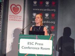
These results “reinforce what we have seen in the clinical trials, but with the strength of looking in the entire Danish population,” said Dan Atar, MD, a cardiologist and professor of medicine at the University of Oslo.
“It is enlightening and very reassuring to have these real-world, unselected, registry data. They provide reassurance about safety and efficacy” when prescribing a NOAC, Dr. Atar said in an interview.
The study reported by Dr. Stærk and her associates included 43,299 Danish patients who were recently diagnosed with nonvalvular atrial fibrillation and started on treatment with an oral anticoagulant during the period August 2011 (when the first NOAC, dabigatran, became available for routine use in Denmark) through December 2015. During this period, 42% of these patients received warfarin, 29% received dabigatran (Pradaxa), 16% received apixaban (Eliquis) and 13% received rivaroxaban (Xarelto).
In a propensity-score type of analysis that controlled for baseline differences in clinical and demographic parameters, the results showed that the rate of ischemic stroke during the first year on treatment ranged from 2.0% to 2.5% in the four subgroups based on the anticoagulant received with no statistically-significant differences among the four subgroups. In other words, all three NOACs had efficacy profiles similar to those of warfarin, said Dr. Stærk, a cardiology researcher at Herlev and Gentofte University Hospitals in Hellerup, Denmark.
But on the safety side, all three NOACs were linked with lower rates of intracranial hemorrhages during the 1-year follow-up compared with the patients who received warfarin. In the cases of dabigatran and apixaban, the reduced intracranial hemorrhage rates were statistically significant, with a 0.6% rate among the patients on warfarin and rates that were reduced by a relative 34% for patients who received dabigatran and by a relative 20% among those on apixaban. Rivaroxaban linked with a 13% relative risk reduction in intracranial hemorrhage that was not statistically significant.
Dr. Atar said he would not make comparisons among the three NOACs based on these data, but rather interpreted the finding as showing that collectively the three NOACs assessed had comparable efficacy but better safety compared with warfarin.
He also noted that the Danish registry data document the transition that occurred during 2011 to 2015 in anticoagulant prescribing that shifted from warfarin to NOACs, with 57% of atrial fibrillation patients receiving a NOAC. In Norway, NOAC prescriptions for atrial fibrillation patients recently pulled ahead of warfarin prescription rates, Dr. Atar said. Reassuring data such as those in this report will help to further drive the shift from warfarin to NOACs, and he predicted that soon NOACs will be the anticoagulants used to treat the overwhelming majority of patients with nonvalvular atrial fibrillation.
Dr. Stærk has received research funding from Boehringer Ingelheim, the company that markets dabigatran (Pradaxa). Dr. Atar said that he has been a consultant to and has received research funding from several drug companies.
The video associated with this article is no longer available on this site. Please view all of our videos on the MDedge YouTube channel
On Twitter @mitchelzoler
ROME – The new oral anticoagulants performed as advertised in a real-world, Danish registry of more than 40,000 patients with atrial fibrillation.
During the first year on anticoagulant treatment, patients who received a new oral anticoagulant (NOAC) had an ischemic stroke rate similar to that of patients who received the traditional oral anticoagulant, warfarin, but a significantly reduced rate of intracranial hemorrhage, Laila Stærk, MD, reported at the annual congress of the European Society of Cardiology.
These results “reinforce what we have seen in the clinical trials, but with the strength of looking in the entire Danish population,” said Dan Atar, MD, a cardiologist and professor of medicine at the University of Oslo.
“It is enlightening and very reassuring to have these real-world, unselected, registry data. They provide reassurance about safety and efficacy” when prescribing a NOAC, Dr. Atar said in an interview.
The study reported by Dr. Stærk and her associates included 43,299 Danish patients who were recently diagnosed with nonvalvular atrial fibrillation and started on treatment with an oral anticoagulant during the period August 2011 (when the first NOAC, dabigatran, became available for routine use in Denmark) through December 2015. During this period, 42% of these patients received warfarin, 29% received dabigatran (Pradaxa), 16% received apixaban (Eliquis) and 13% received rivaroxaban (Xarelto).
In a propensity-score type of analysis that controlled for baseline differences in clinical and demographic parameters, the results showed that the rate of ischemic stroke during the first year on treatment ranged from 2.0% to 2.5% in the four subgroups based on the anticoagulant received with no statistically-significant differences among the four subgroups. In other words, all three NOACs had efficacy profiles similar to those of warfarin, said Dr. Stærk, a cardiology researcher at Herlev and Gentofte University Hospitals in Hellerup, Denmark.
But on the safety side, all three NOACs were linked with lower rates of intracranial hemorrhages during the 1-year follow-up compared with the patients who received warfarin. In the cases of dabigatran and apixaban, the reduced intracranial hemorrhage rates were statistically significant, with a 0.6% rate among the patients on warfarin and rates that were reduced by a relative 34% for patients who received dabigatran and by a relative 20% among those on apixaban. Rivaroxaban linked with a 13% relative risk reduction in intracranial hemorrhage that was not statistically significant.
Dr. Atar said he would not make comparisons among the three NOACs based on these data, but rather interpreted the finding as showing that collectively the three NOACs assessed had comparable efficacy but better safety compared with warfarin.
He also noted that the Danish registry data document the transition that occurred during 2011 to 2015 in anticoagulant prescribing that shifted from warfarin to NOACs, with 57% of atrial fibrillation patients receiving a NOAC. In Norway, NOAC prescriptions for atrial fibrillation patients recently pulled ahead of warfarin prescription rates, Dr. Atar said. Reassuring data such as those in this report will help to further drive the shift from warfarin to NOACs, and he predicted that soon NOACs will be the anticoagulants used to treat the overwhelming majority of patients with nonvalvular atrial fibrillation.
Dr. Stærk has received research funding from Boehringer Ingelheim, the company that markets dabigatran (Pradaxa). Dr. Atar said that he has been a consultant to and has received research funding from several drug companies.
The video associated with this article is no longer available on this site. Please view all of our videos on the MDedge YouTube channel
On Twitter @mitchelzoler
AT THE ESC CONGRESS 2016
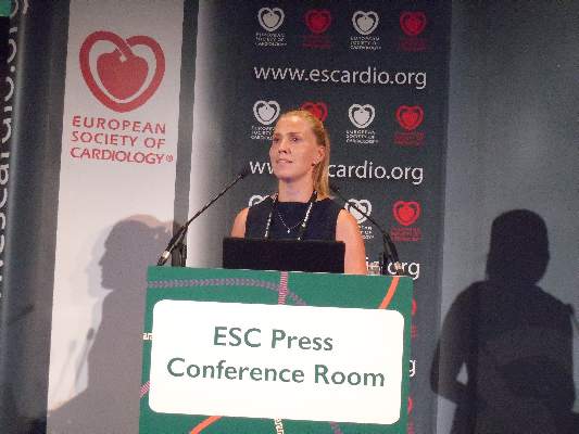
Medical Student Guides Aspiring Physicians into Hospital Medicine
Editor’s note: As SHM celebrates the “Year of the Hospitalist,” we’re putting the spotlight on some of our most active members who are making substantial contributions to hospital medicine. You can get involved, too! Log on to www.hospitalmedicine.org/yoth for more information on how you can join the yearlong celebration and help SHM improve the care of hospitalized patients.

This month, The Hospitalist spotlights Aram Namavar, MS, a recipient of SHM’s Student Hospitalist Scholar Grant and a second-year medical student at Loyola University Chicago’s Stritch School of Medicine. Namavar is one of the first students to sit on an SHM committee as part of the Physicians in Training (PIT) Committee, which is focused on ensuring the successful transition of medical students and residents into the practice of hospital medicine. He recently started a hospital medicine special interest group at his medical school and is playing an active role in the launch of SHM’s new Students Community on the Hospital Medicine Exchange (HMX).
What piqued your interest in hospital medicine as you were choosing a specialty?

The laundry room was located right next to the training room, so I had the opportunity to meet the orthopedic surgeons and training staff. After spending some time in this program, I felt that there were aspects to a career in orthopedics that did not fit with my personality, such as what I felt was limited patient interaction and ability to impact multiple domains of their care outside of surgery. I pivoted and began volunteering at UCLA Health and was exposed to a plethora of medical specialties and patient populations. Through the self-discovery of my potential career path, I also discovered myself and my values. I realized that I desired flexibility and versatility in my career to engage in clinical, leadership, mentorship, research, education, and advocacy roles, all of which are possible in hospital medicine. I am now certain that my career in medicine will lead me to become an academic hospitalist.
Can you tell us about your quality improvement project that you initiated as a result of winning the SHM Student Hospitalist Scholar Grant?
This summer has been a very enriching experience for my personal and professional development. My main project is focused on patient-centered readmissions and has three components. I am promoting a patient-centered approach to readmissions through examination of the role that decisional conflict plays in hospital readmissions.
In shared decision-making models, decisional conflict is a measure of uncertainty, readiness, and comfort level in making a decision. Aware of the plight of diverse populations in accessing healthcare and having higher readmission rates, I have widened the scope of my project. A second arm of my study is investigating which social determinants of health may be a root cause for why Hispanic patients are being readmitted at an increased rate compared to their non-readmitted counterparts. The third arm of my study is elucidating patient-centered views of the cause and preventability of readmission for Hispanic versus non-Hispanic patients.
What inspired you to become involved with the PIT Committee?
Working with undergraduate and medical students and resident physicians, I have always had a passion for inspiring the future of medicine. Even in hospital medicine’s 20 years of existence, I knew there were many opportunities for me to engage trainees. For this reason, I applied for a seat on the PIT Committee and was accepted as the only medical student to sit on an SHM committee.
Having founded the first hospital medicine interest group at a medical school in the U.S. at Loyola, my work on the PIT Committee is focused on enhancing our ability to engage medical students and residents. I am a member of the Student Interest Group Task Force that is creating a toolkit for medical schools from across the nation to gain recognition from SHM for creating a hospital medicine interest group. The blueprint we used at Loyola is being adopted and incorporated in our toolkit. Another avenue of engagement is through the new Students Community on HMX, SHM’s online member engagement platform. This new community, which I helped to launch, will serve as an important channel for us to connect with trainees nationally and encourage them to consider hospital medicine as a specialty.
What opportunities do you see for hospitalists as the medical landscape continues to evolve?
I see hospitalists playing important roles in value-based care and population health. Hospitalists have carved their niche in quality improvement within health systems, and I believe they will be best suited for spearheading projects to enhance the value of medical care. Hospitalists will also no longer have an impact within the confines of a hospital as the landscape is shifting toward population health; therefore, hospitalists will be charged with helping to devise methods to proactively identify care gaps in their patients to promote preventive care and chronic disease management.
What would you tell medical students about choosing hospital medicine as a career?
I believe that a career in hospital medicine is one of the most versatile. If you are someone who must be continually stimulated by various work settings, then hospital medicine is right for you. I enjoy multidimensional work, and as a future academic hospitalist, I know that my scope of practice will include mentorship, education, research, leadership, and clinical duties. The ability to have such a versatile career will be extremely fulfilling.
What’s next for you in your medical career?
As a second-year medical student, my primary focus is on succeeding academically and adequately preparing for my USMLE Step 1 exam to match at my desired internal medicine residency program. Outside of my studies, I am continuing to develop my analytical skills and leadership acumen so that I can become a major player in hospital medicine once I am an academic hospitalist. After residency, I will be pursuing a fully employed MBA program where I will be equipped with the necessary skills to realize my professional goals. TH
Editor’s note: As SHM celebrates the “Year of the Hospitalist,” we’re putting the spotlight on some of our most active members who are making substantial contributions to hospital medicine. You can get involved, too! Log on to www.hospitalmedicine.org/yoth for more information on how you can join the yearlong celebration and help SHM improve the care of hospitalized patients.
This month, The Hospitalist spotlights Aram Namavar, MS, a recipient of SHM’s Student Hospitalist Scholar Grant and a second-year medical student at Loyola University Chicago’s Stritch School of Medicine. Namavar is one of the first students to sit on an SHM committee as part of the Physicians in Training (PIT) Committee, which is focused on ensuring the successful transition of medical students and residents into the practice of hospital medicine. He recently started a hospital medicine special interest group at his medical school and is playing an active role in the launch of SHM’s new Students Community on the Hospital Medicine Exchange (HMX).
What piqued your interest in hospital medicine as you were choosing a specialty?
The laundry room was located right next to the training room, so I had the opportunity to meet the orthopedic surgeons and training staff. After spending some time in this program, I felt that there were aspects to a career in orthopedics that did not fit with my personality, such as what I felt was limited patient interaction and ability to impact multiple domains of their care outside of surgery. I pivoted and began volunteering at UCLA Health and was exposed to a plethora of medical specialties and patient populations. Through the self-discovery of my potential career path, I also discovered myself and my values. I realized that I desired flexibility and versatility in my career to engage in clinical, leadership, mentorship, research, education, and advocacy roles, all of which are possible in hospital medicine. I am now certain that my career in medicine will lead me to become an academic hospitalist.
Can you tell us about your quality improvement project that you initiated as a result of winning the SHM Student Hospitalist Scholar Grant?
This summer has been a very enriching experience for my personal and professional development. My main project is focused on patient-centered readmissions and has three components. I am promoting a patient-centered approach to readmissions through examination of the role that decisional conflict plays in hospital readmissions.
In shared decision-making models, decisional conflict is a measure of uncertainty, readiness, and comfort level in making a decision. Aware of the plight of diverse populations in accessing healthcare and having higher readmission rates, I have widened the scope of my project. A second arm of my study is investigating which social determinants of health may be a root cause for why Hispanic patients are being readmitted at an increased rate compared to their non-readmitted counterparts. The third arm of my study is elucidating patient-centered views of the cause and preventability of readmission for Hispanic versus non-Hispanic patients.
What inspired you to become involved with the PIT Committee?
Working with undergraduate and medical students and resident physicians, I have always had a passion for inspiring the future of medicine. Even in hospital medicine’s 20 years of existence, I knew there were many opportunities for me to engage trainees. For this reason, I applied for a seat on the PIT Committee and was accepted as the only medical student to sit on an SHM committee.
Having founded the first hospital medicine interest group at a medical school in the U.S. at Loyola, my work on the PIT Committee is focused on enhancing our ability to engage medical students and residents. I am a member of the Student Interest Group Task Force that is creating a toolkit for medical schools from across the nation to gain recognition from SHM for creating a hospital medicine interest group. The blueprint we used at Loyola is being adopted and incorporated in our toolkit. Another avenue of engagement is through the new Students Community on HMX, SHM’s online member engagement platform. This new community, which I helped to launch, will serve as an important channel for us to connect with trainees nationally and encourage them to consider hospital medicine as a specialty.
What opportunities do you see for hospitalists as the medical landscape continues to evolve?
I see hospitalists playing important roles in value-based care and population health. Hospitalists have carved their niche in quality improvement within health systems, and I believe they will be best suited for spearheading projects to enhance the value of medical care. Hospitalists will also no longer have an impact within the confines of a hospital as the landscape is shifting toward population health; therefore, hospitalists will be charged with helping to devise methods to proactively identify care gaps in their patients to promote preventive care and chronic disease management.
What would you tell medical students about choosing hospital medicine as a career?
I believe that a career in hospital medicine is one of the most versatile. If you are someone who must be continually stimulated by various work settings, then hospital medicine is right for you. I enjoy multidimensional work, and as a future academic hospitalist, I know that my scope of practice will include mentorship, education, research, leadership, and clinical duties. The ability to have such a versatile career will be extremely fulfilling.
What’s next for you in your medical career?
As a second-year medical student, my primary focus is on succeeding academically and adequately preparing for my USMLE Step 1 exam to match at my desired internal medicine residency program. Outside of my studies, I am continuing to develop my analytical skills and leadership acumen so that I can become a major player in hospital medicine once I am an academic hospitalist. After residency, I will be pursuing a fully employed MBA program where I will be equipped with the necessary skills to realize my professional goals. TH

Discovery in mice may have implications for HSCT

Preclinical research has shown how a cell surface molecule, Lymphotoxin β receptor, controls the entry of T cells into the thymus.
Researchers believe this might represent a pathway that could be targeted to reboot the immune system after hematopoietic stem cell transplant (HSCT).
Graham Anderson, PhD, of the University of Birmingham in the UK, and his colleagues conducted this research and reported their findings in the Journal of Immunology.
“The thymus is often something of an ignored organ, but it plays a crucial role in maintaining an effective immune system,” Dr Anderson said.
“Post-transplantation, T-cell progenitors derived from the bone marrow transplant can struggle to enter the thymus, as if the doorway to the thymus is closed. Identifying molecular regulators that can ‘prop open’ the door and allow these cells to enter and mature could well be a means to help reboot the immune system.”
Conducting experiments in mice, Dr Anderson and his colleagues found that Lymphotoxin β receptor was required to allow the entry of T-cell progenitors to the thymus both in a healthy state and during immune recovery after HSCT.
When the researchers used an antibody to stimulate Lymphotoxin β receptor after HSCT, they observed enhanced thymus recovery and an increase in the number of transplant-derived T cells, which correlated with increased adhesion molecule expression by thymic stroma.
The team said this study has revealed a novel link between Lymphotoxin β receptor and thymic stromal cells in thymus colonization and highlights its potential as an immunotherapeutic target to boost T-cell reconstitution after HSCT.
“This is just one piece of the puzzle,” said study author Beth Lucas, PhD, also of the University of Birmingham.
“It may be that there are adverse effects to opening the door to the thymus, but identifying a pathway that regulates this process is a significant step.”
For their next step, the researchers plan to study in vitro samples of the human thymus to examine the role Lymphotoxin β receptor might play in regulating thymus function in humans. ![]()