User login
Infliximab biosimilar posts mostly reassuring data in Norway’s NOR-SWITCH study
WASHINGTON – Data from the first randomized trial of switching from an originator biologic to a biosimilar of the originator indicate that the infliximab biosimilar Remsima is no different from the infliximab originator Remicade in the rate of disease worsening over 1 year across a combination of all its approved indications.
The outcomes of the Norwegian, double-blind, noninferiority trial, called NOR-SWITCH, indicate similar rates of disease worsening across patients switched to Remsima and those who stayed on Remicade. However, exploratory group analyses conducted on the different disease subgroups in the trial (Crohn’s disease, ulcerative colitis, spondyloarthritis, rheumatoid arthritis, psoriasis, and psoriatic arthritis) showed a potentially concerning level of disease worsening among Crohn’s disease patients on Remsima with a confidence interval that nearly fell entirely within the range favoring Remicade.
In the United States, Remsima, also known as CT-P13, is marketed by Pfizer as Inflectra.

The trial randomized 482 patients who were on stable treatment with Remicade for at least 6 months for any of the six indications for which Remicade and Remsima are approved to either stay on Remicade or switch to Remsima with the same dosing regimen for 52 weeks. Overall, patients had a mean age of about 48 years and 36%-41% were female. They had a mean disease duration of about 17 years and had been taking Remicade for a mean of nearly 7 years.
The primary endpoint was disease worsening during follow-up, according to worsening in disease-specific composite measures and/or a consensus between an investigator and a patient that led to a major change in treatment. The investigators made an assumption of 30% disease worsening across all the indications for the trial’s power calculation, based on available literature and observational data.
Disease worsening occurred in 26.2% of patients who stayed on Remicade and 29.6% of patients who switched to Remsima, based on a per-protocol analysis of 202 Remicade and 206 Remsima patients. The 95% confidence interval of the adjusted treatment difference of –4.4% was –12.7% to 3.9%, which was within the pre-specified noninferiority margin of 15%.
Exploratory subgroup analyses of the different disease subgroups showed no statistically significant differences between the two treatments in disease worsening. However, in Crohn’s disease patients, who formed the largest subgroup in the study at 155 patients, the adjusted treatment difference was –14.3% (21.2% with disease worsening for Remicade and 36.5% for Remsima) with a 95% CI of –29.3% to 0.7%.
It’s difficult to discern whether the 95% confidence interval seen in the Crohn’s disease subgroup is a part of the natural variation one would expect to see in a subgroup analysis of different diseases or if there might be a true signal for disease worsening in the Crohn’s disease patients who took Remsima. “The problem is that it’s in the largest subgroup that has no other data. If this had been in rheumatoid arthritis, that would be different,” coauthor Inge C. Olsen, PhD, a biostatistician at Diakonhjemmet Hospital, said in an interview. “All the registry trials were done in RA and spondyloarthritis patients. ... That’s an issue, but with regards to the [NOR-SWITCH] study, it’s very clear that you have no power to show anything in the subgroup analysis, and they are exploratory analyses and are not answering any hypothesis.” Currently, there are no plans to follow up on these results in another study, he said.
Other issues that the NOR-SWITCH study does not answer are the outcomes of switching back and forth between Remicade and Remsima, switching from one infliximab biosimilar to another infliximab biosimilar, and switching from other originator biologics to their biosimilars.
“Is that feasible? Is that safe? Will it retain efficacy? We don’t know. There’s a real need for those studies to be done,” Dr. Goll said.
In Norway, the remaining patients who had not switched yet from Remicade to Remsima are now doing so based on the trial’s results, Dr. Goll said. The cost of Remsima in Norway was about 75% less than Remicade in 2015 and about 60% less in 2016, she noted.
It’s still an open question what the results of the NOR-SWITCH trial might indicate for how clinicians in the United States will use Inflectra and other biosimilars, according to John J. Cush, MD, professor of medicine and rheumatology at Baylor University, Dallas.
“I think the real problem here is that it’s nice to know that [CT-P13] wasn’t inferior, but when you get into the weeds and you look at the details, those of us who may not have a lot of certainty about this might worry about this, especially when there are three new biosimilars approved in the United States: Amjevita, which is an adalimumab biosimilar; Erelzi, which is an etanercept biosimilar; and Inflectra’s about to be launched as an infliximab biosimilar,” Dr. Cush said during a session reviewing selected abstracts from the meeting. “When this NOR-SWITCH study was done in Norway, it’s a 70% savings over the original product. The new ones being introduced over here [in the United States] start at about 15%. I’m less motivated with that degree of savings to want to take some chances on my patients. So we need a little bit more certainty; we need to feel better about the cost savings to patients and health care overall. Confidence in biosimilars is what’s going to sell biosimilars. We’re a long way from confidence still.”
NOR-SWITCH was funded by the Norwegian government. Some of the investigators disclosed relationships with Pfizer and/or Celltrion, which separately market CT-P13 in different parts of the world.
[email protected]
WASHINGTON – Data from the first randomized trial of switching from an originator biologic to a biosimilar of the originator indicate that the infliximab biosimilar Remsima is no different from the infliximab originator Remicade in the rate of disease worsening over 1 year across a combination of all its approved indications.
The outcomes of the Norwegian, double-blind, noninferiority trial, called NOR-SWITCH, indicate similar rates of disease worsening across patients switched to Remsima and those who stayed on Remicade. However, exploratory group analyses conducted on the different disease subgroups in the trial (Crohn’s disease, ulcerative colitis, spondyloarthritis, rheumatoid arthritis, psoriasis, and psoriatic arthritis) showed a potentially concerning level of disease worsening among Crohn’s disease patients on Remsima with a confidence interval that nearly fell entirely within the range favoring Remicade.
In the United States, Remsima, also known as CT-P13, is marketed by Pfizer as Inflectra.
The trial randomized 482 patients who were on stable treatment with Remicade for at least 6 months for any of the six indications for which Remicade and Remsima are approved to either stay on Remicade or switch to Remsima with the same dosing regimen for 52 weeks. Overall, patients had a mean age of about 48 years and 36%-41% were female. They had a mean disease duration of about 17 years and had been taking Remicade for a mean of nearly 7 years.
The primary endpoint was disease worsening during follow-up, according to worsening in disease-specific composite measures and/or a consensus between an investigator and a patient that led to a major change in treatment. The investigators made an assumption of 30% disease worsening across all the indications for the trial’s power calculation, based on available literature and observational data.
Disease worsening occurred in 26.2% of patients who stayed on Remicade and 29.6% of patients who switched to Remsima, based on a per-protocol analysis of 202 Remicade and 206 Remsima patients. The 95% confidence interval of the adjusted treatment difference of –4.4% was –12.7% to 3.9%, which was within the pre-specified noninferiority margin of 15%.
Exploratory subgroup analyses of the different disease subgroups showed no statistically significant differences between the two treatments in disease worsening. However, in Crohn’s disease patients, who formed the largest subgroup in the study at 155 patients, the adjusted treatment difference was –14.3% (21.2% with disease worsening for Remicade and 36.5% for Remsima) with a 95% CI of –29.3% to 0.7%.
It’s difficult to discern whether the 95% confidence interval seen in the Crohn’s disease subgroup is a part of the natural variation one would expect to see in a subgroup analysis of different diseases or if there might be a true signal for disease worsening in the Crohn’s disease patients who took Remsima. “The problem is that it’s in the largest subgroup that has no other data. If this had been in rheumatoid arthritis, that would be different,” coauthor Inge C. Olsen, PhD, a biostatistician at Diakonhjemmet Hospital, said in an interview. “All the registry trials were done in RA and spondyloarthritis patients. ... That’s an issue, but with regards to the [NOR-SWITCH] study, it’s very clear that you have no power to show anything in the subgroup analysis, and they are exploratory analyses and are not answering any hypothesis.” Currently, there are no plans to follow up on these results in another study, he said.
Other issues that the NOR-SWITCH study does not answer are the outcomes of switching back and forth between Remicade and Remsima, switching from one infliximab biosimilar to another infliximab biosimilar, and switching from other originator biologics to their biosimilars.
“Is that feasible? Is that safe? Will it retain efficacy? We don’t know. There’s a real need for those studies to be done,” Dr. Goll said.
In Norway, the remaining patients who had not switched yet from Remicade to Remsima are now doing so based on the trial’s results, Dr. Goll said. The cost of Remsima in Norway was about 75% less than Remicade in 2015 and about 60% less in 2016, she noted.
It’s still an open question what the results of the NOR-SWITCH trial might indicate for how clinicians in the United States will use Inflectra and other biosimilars, according to John J. Cush, MD, professor of medicine and rheumatology at Baylor University, Dallas.
“I think the real problem here is that it’s nice to know that [CT-P13] wasn’t inferior, but when you get into the weeds and you look at the details, those of us who may not have a lot of certainty about this might worry about this, especially when there are three new biosimilars approved in the United States: Amjevita, which is an adalimumab biosimilar; Erelzi, which is an etanercept biosimilar; and Inflectra’s about to be launched as an infliximab biosimilar,” Dr. Cush said during a session reviewing selected abstracts from the meeting. “When this NOR-SWITCH study was done in Norway, it’s a 70% savings over the original product. The new ones being introduced over here [in the United States] start at about 15%. I’m less motivated with that degree of savings to want to take some chances on my patients. So we need a little bit more certainty; we need to feel better about the cost savings to patients and health care overall. Confidence in biosimilars is what’s going to sell biosimilars. We’re a long way from confidence still.”
NOR-SWITCH was funded by the Norwegian government. Some of the investigators disclosed relationships with Pfizer and/or Celltrion, which separately market CT-P13 in different parts of the world.
[email protected]
AT THE ACR ANNUAL MEETING
Key clinical point:
Major finding: Disease worsening occurred in 26.2% of patients who stayed on Remicade and 29.6% of patients who switched to Remsima, based on a per-protocol analysis.
Data source: The multicenter, double-blind, randomized NOR-SWITCH trial of 482 patients.
Disclosures: The trial was funded by the Norwegian government. Some of the investigators disclosed relationships with Pfizer and/or Celltrion, which separately market CT-P13 in different parts of the world.
TRUE-AHF: Urgent vasodilator therapy in acute HF provides no long-term benefit
NEW ORLEANS – An investigational synthetic natriuretic peptide given early during hospitalization for acute decompensated heart failure didn’t produce any of the hoped-for intermediate- or long-term clinical benefits in the phase III TRUE-AHF study, Milton Packer, MD, reported at the American Heart Association scientific sessions.
The failure of this investigational vasodilator, ularitide, to influence downstream cardiovascular mortality or early readmission for heart failure closes the door on the once-promising hypothesis that myocardial microinjury occurring during ADHF is due to ventricular distension, observed Dr. Packer, the Distinguished Scholar in Cardiovascular Science at Baylor University Medical Center, Dallas. “Ularitide did exactly what we expected it to do while we were giving it: we caused intravascular decompression, we reduced cardiac wall stress, but we did not affect cardiac microinjury, and we didn’t change long-term cardiovascular mortality or any of our secondary endpoints, including and in particular the 30-day risk of rehospitalization for heart failure,” he said.

During a median follow-up of 15 months there were 236 cardiovascular deaths in the ularitide group and 225 in the control group, a nonsignificant difference. Nor were there any differences between the two groups in secondary endpoints including length of stay in the ICU during the index hospitalization, rehospitalization for ADHF within 30 days of discharge, or the composite of all-cause mortality or cardiovascular hospitalization within 6 months, which occurred in 40.7% of the ularitide group and 37.2% of controls.
The explanation for this lack of long-term benefit lies in the finding that myocardial microinjury wasn’t prevented by the rapid reduction of cardiac distension produced by ularitide. This was evident in the therapy’s inability to dampen the rise in high-sensitivity cardiac troponin T which occurred in the initial 48 hours of the study.
“The trial demonstrated the effects and safety of ularitide. However, to gain long-term benefits on hospitalizations and death in patients following a hospital admission for heart failure, physicians must focus on the drugs that patients take as an outpatient rather than the drugs they receive as an inpatient,” Dr. Packer concluded.

“Readmission rates remain stubbornly at 20% within 30 days and near 50% at 6 months. Acute decompensation is an inflection point in the natural history of heart failure with subsequent 1-year mortality rates consistently approximating 25%. Clearly there is something about the hospitalization that is a herald event which speaks to much worse outcomes, compared with chronic ambulatory heart failure,” said Dr. Yancy, professor of medicine and chief of cardiology at Northwestern University in Chicago.
He agreed with Dr. Packer that in light of the TRUE-AHF results, what Dr. Yancy termed “the early injury hypothesis” isn’t worth further pursuit.
Ularitide thus joins a long list of failed therapies for ADHF. Treatments that have convincingly been shown to have no significant impact on mortality and at best only modest impact on morbidity include continuous IV infusion of loop diuretics in the DOSE trial; the arginine vasopressin antagonists, which failed to impress in EVEREST, SECRET, and TACTICS; nesiritide (Natrecor) in the ASCEND-HF trial; and levosimendan (Simdax), which proved disappointing in the SURVIVE and REVIVE II studies, according to Dr. Yancy.
The jury is still out on serelaxin, he added. The drug showed a favorable signal in the RELAX-AHF trial. The results of RELAX II are awaited with interest.
“Today we still don’t have an effective single intervention for acute decompensated heart failure other than process-of-care improvement,” the cardiologist noted.
What holds promise for improved long-term outcomes in ADHF at this point? Dr. Yancy said sacubitril/valsartan (Entresto) is intriguing based upon the results of the PARADIGM-HF trial (N Engl J Med. 2014;371:993-1004). But the drug needs to be studied prospectively in patients in the throes of ADHF before it can appropriately be recommended for the purpose of changing the natural history of this disorder, Dr. Yancy stressed.
Devices such as the implantable pulmonary catheter are under study as a promising means of altering the natural history of ADHF by identifying actionable signals of impending decompensation weeks beforehand, he added.
The TRUE-AHF trial was sponsored by Cardiorentis. Dr. Packer reported serving as a consultant to that company and more than a dozen other pharmaceutical and medical device companies.
[email protected]
NEW ORLEANS – An investigational synthetic natriuretic peptide given early during hospitalization for acute decompensated heart failure didn’t produce any of the hoped-for intermediate- or long-term clinical benefits in the phase III TRUE-AHF study, Milton Packer, MD, reported at the American Heart Association scientific sessions.
The failure of this investigational vasodilator, ularitide, to influence downstream cardiovascular mortality or early readmission for heart failure closes the door on the once-promising hypothesis that myocardial microinjury occurring during ADHF is due to ventricular distension, observed Dr. Packer, the Distinguished Scholar in Cardiovascular Science at Baylor University Medical Center, Dallas. “Ularitide did exactly what we expected it to do while we were giving it: we caused intravascular decompression, we reduced cardiac wall stress, but we did not affect cardiac microinjury, and we didn’t change long-term cardiovascular mortality or any of our secondary endpoints, including and in particular the 30-day risk of rehospitalization for heart failure,” he said.
During a median follow-up of 15 months there were 236 cardiovascular deaths in the ularitide group and 225 in the control group, a nonsignificant difference. Nor were there any differences between the two groups in secondary endpoints including length of stay in the ICU during the index hospitalization, rehospitalization for ADHF within 30 days of discharge, or the composite of all-cause mortality or cardiovascular hospitalization within 6 months, which occurred in 40.7% of the ularitide group and 37.2% of controls.
The explanation for this lack of long-term benefit lies in the finding that myocardial microinjury wasn’t prevented by the rapid reduction of cardiac distension produced by ularitide. This was evident in the therapy’s inability to dampen the rise in high-sensitivity cardiac troponin T which occurred in the initial 48 hours of the study.
“The trial demonstrated the effects and safety of ularitide. However, to gain long-term benefits on hospitalizations and death in patients following a hospital admission for heart failure, physicians must focus on the drugs that patients take as an outpatient rather than the drugs they receive as an inpatient,” Dr. Packer concluded.
“Readmission rates remain stubbornly at 20% within 30 days and near 50% at 6 months. Acute decompensation is an inflection point in the natural history of heart failure with subsequent 1-year mortality rates consistently approximating 25%. Clearly there is something about the hospitalization that is a herald event which speaks to much worse outcomes, compared with chronic ambulatory heart failure,” said Dr. Yancy, professor of medicine and chief of cardiology at Northwestern University in Chicago.
He agreed with Dr. Packer that in light of the TRUE-AHF results, what Dr. Yancy termed “the early injury hypothesis” isn’t worth further pursuit.
Ularitide thus joins a long list of failed therapies for ADHF. Treatments that have convincingly been shown to have no significant impact on mortality and at best only modest impact on morbidity include continuous IV infusion of loop diuretics in the DOSE trial; the arginine vasopressin antagonists, which failed to impress in EVEREST, SECRET, and TACTICS; nesiritide (Natrecor) in the ASCEND-HF trial; and levosimendan (Simdax), which proved disappointing in the SURVIVE and REVIVE II studies, according to Dr. Yancy.
The jury is still out on serelaxin, he added. The drug showed a favorable signal in the RELAX-AHF trial. The results of RELAX II are awaited with interest.
“Today we still don’t have an effective single intervention for acute decompensated heart failure other than process-of-care improvement,” the cardiologist noted.
What holds promise for improved long-term outcomes in ADHF at this point? Dr. Yancy said sacubitril/valsartan (Entresto) is intriguing based upon the results of the PARADIGM-HF trial (N Engl J Med. 2014;371:993-1004). But the drug needs to be studied prospectively in patients in the throes of ADHF before it can appropriately be recommended for the purpose of changing the natural history of this disorder, Dr. Yancy stressed.
Devices such as the implantable pulmonary catheter are under study as a promising means of altering the natural history of ADHF by identifying actionable signals of impending decompensation weeks beforehand, he added.
The TRUE-AHF trial was sponsored by Cardiorentis. Dr. Packer reported serving as a consultant to that company and more than a dozen other pharmaceutical and medical device companies.
[email protected]
Key clinical point:
Major finding: Early administration of ularitide during hospitalization for acute decompensated heart failure failed to achieve any long-term clinical benefits.
Data source: The TRUE-AHF trial was a double-blind, placebo-controlled, randomized trial including 2,157 patients hospitalized for acute decompensated heart failure at 156 centers in 23 countries.
Disclosures: The study was sponsored by Cardiorentis. The presenter reported serving as a consultant to that company and more than a dozen other pharmaceutical and medical device companies.
Cardiac rehab also slashes stroke risk
ROME – Cardiac rehabilitation programs have a previously unappreciated benefit: participants enjoy a 60% reduction in the risk of stroke, Gijs van Halewijn, MD, reported at the annual congress of the European Society of Cardiology.
“I think cardiologists are really focused on cardiovascular deaths, especially from MI. But we’ve shown that cardiac rehabilitation also has an effect on cerebrovascular events,” said Dr. van Halewijn of Erasmus University in Rotterdam (the Netherlands).

He presented a meta-analysis of randomized controlled trials of cardiac rehab conducted during 2010-2015. The purpose was to assess the value provided by cardiac rehab in the contemporary era of acute coronary syndrome management featuring primary percutaneous coronary intervention, drug-eluting stents, and potent medications for secondary cardiovascular prevention. That hadn’t previously been looked at systematically.
“The standard meta-analyses cited in the field include randomized trials from as far back as just after World War II,” Dr. van Halewijn noted in an interview.
He employed the same search and analytic methods utilized by the Cochrane Collaboration in evaluating 18 randomized controlled trials of lifestyle- or exercise-based cardiac rehab, compared with usual care, in a total of 7,691 participants.
The results of the meta-analysis indicate cardiac rehab provides powerful secondary prevention benefits above and beyond those obtained through contemporary interventional procedures and preventive medications.
Cardiovascular mortality was reduced by 58% in cardiac rehab participants compared with usual care controls. The risk of acute MI was decreased by 30%. And in a new observation, cerebrovascular events were reduced by 60% in the four randomized trials in which that was an endpoint. All of these differences were highly statistically significant.
“Interestingly, the number needed to treat was 45 for MI, so if you have 45 patients included in your cardiac rehabilitation program, you can prevent one MI. And you can prevent one cerebrovascular event with 82 participants,” according to Dr. van Halewijn.
Cardiac rehab had no effect on all-cause mortality in the overall meta-analysis. However, in the trials involving comprehensive cardiac rehab programs targeting six or more of the components of secondary cardiovascular prevention described by the British Association for Cardiac Prevention and Rehabilitation (Heart. 2013 Aug;99[15]:1069-71), participation was associated with a 37% reduction in the risk of all-cause mortality, compared with usual care.
Those components are smoking, blood pressure, cholesterol, HbA1c, exercise training, counseling about the importance of exercise, stress management, and checking medications.
In addition, Dr. van Halewijn continued, cardiac rehab programs in which a physician or nurse made sure participants were on guideline-directed cardiovascular medications had a 65% reduction in all-cause mortality, compared with usual care.
“Cardiac rehabilitation’s opportunity is to evolve into comprehensive programs addressing all aspects of lifestyle, risk factor management, and prescription of medications to reduce death and nonfatal events,” the physician concluded.
This study was supported by Erasmus University Medical Center, Imperial College London, and the Dutch Heart Foundation. Dr. van Halewijn reported having no financial conflicts of interest.
[email protected]
ROME – Cardiac rehabilitation programs have a previously unappreciated benefit: participants enjoy a 60% reduction in the risk of stroke, Gijs van Halewijn, MD, reported at the annual congress of the European Society of Cardiology.
“I think cardiologists are really focused on cardiovascular deaths, especially from MI. But we’ve shown that cardiac rehabilitation also has an effect on cerebrovascular events,” said Dr. van Halewijn of Erasmus University in Rotterdam (the Netherlands).
He presented a meta-analysis of randomized controlled trials of cardiac rehab conducted during 2010-2015. The purpose was to assess the value provided by cardiac rehab in the contemporary era of acute coronary syndrome management featuring primary percutaneous coronary intervention, drug-eluting stents, and potent medications for secondary cardiovascular prevention. That hadn’t previously been looked at systematically.
“The standard meta-analyses cited in the field include randomized trials from as far back as just after World War II,” Dr. van Halewijn noted in an interview.
He employed the same search and analytic methods utilized by the Cochrane Collaboration in evaluating 18 randomized controlled trials of lifestyle- or exercise-based cardiac rehab, compared with usual care, in a total of 7,691 participants.
The results of the meta-analysis indicate cardiac rehab provides powerful secondary prevention benefits above and beyond those obtained through contemporary interventional procedures and preventive medications.
Cardiovascular mortality was reduced by 58% in cardiac rehab participants compared with usual care controls. The risk of acute MI was decreased by 30%. And in a new observation, cerebrovascular events were reduced by 60% in the four randomized trials in which that was an endpoint. All of these differences were highly statistically significant.
“Interestingly, the number needed to treat was 45 for MI, so if you have 45 patients included in your cardiac rehabilitation program, you can prevent one MI. And you can prevent one cerebrovascular event with 82 participants,” according to Dr. van Halewijn.
Cardiac rehab had no effect on all-cause mortality in the overall meta-analysis. However, in the trials involving comprehensive cardiac rehab programs targeting six or more of the components of secondary cardiovascular prevention described by the British Association for Cardiac Prevention and Rehabilitation (Heart. 2013 Aug;99[15]:1069-71), participation was associated with a 37% reduction in the risk of all-cause mortality, compared with usual care.
Those components are smoking, blood pressure, cholesterol, HbA1c, exercise training, counseling about the importance of exercise, stress management, and checking medications.
In addition, Dr. van Halewijn continued, cardiac rehab programs in which a physician or nurse made sure participants were on guideline-directed cardiovascular medications had a 65% reduction in all-cause mortality, compared with usual care.
“Cardiac rehabilitation’s opportunity is to evolve into comprehensive programs addressing all aspects of lifestyle, risk factor management, and prescription of medications to reduce death and nonfatal events,” the physician concluded.
This study was supported by Erasmus University Medical Center, Imperial College London, and the Dutch Heart Foundation. Dr. van Halewijn reported having no financial conflicts of interest.
[email protected]
AT THE ESC CONGRESS 2016
Key clinical point:
Major finding: The number of patients who need to participate in a cardiac rehabilitation program following an ACS in order to prevent one cerebrovascular event is 85.
Data source: This was a meta-analysis of 18 randomized trials carried out in 2010-2015 comparing contemporary cardiac rehabilitation to usual care in 7,691 patients.
Disclosures: This study was supported by Erasmus University (Rotterdam, the Netherlands) Medical Center, Imperial College London, and the Dutch Heart Foundation. The presenter reported having no financial conflicts of interest.
First JAK inhibitor in psoriatic arthritis achieves results similar to adalimumab
WASHINGTON – Tofacitinib demonstrated efficacy comparable to adalimumab in patients with active psoriatic arthritis (PsA) and an inadequate response to conventional disease-modifying antirheumatic drugs in the phase III OPAL Broaden study.
“This is the first study to demonstrate efficacy of a JAK [Janus kinase] inhibitor in PsA,” said lead author Philip J. Mease, MD, of Swedish Medical Center, Seattle. “The primary endpoints were achieved with tofacitinib [Xeljanz] versus placebo. Onset of efficacy of tofacitinib on the ACR20 [the American College of Rheumatology 20% improvement criteria] was observed as early as week 2. Radiographic non-progression rates were low and similar to placebo. Improvement was seen in enthesitis and dactylitis as well as in joint and skin symptoms, and efficacy was maintained through month 12. Tofacitinib is a potential future option for the treatment of PsA.”

The study enrolled 422 patients with active PsA diagnosed within the last 6 months or longer, and all also had plaque psoriasis at screening. The primary endpoint was ACR20 response at 3 months.
Patients were randomized to receive tofacitinib 5 mg twice daily, tofacitinib 10 mg twice daily, adalimumab (Humira) 40 mg subcutaneously every 2 weeks, or placebo. At 3 months, placebo patients were re-randomized to tofacitinib 5 mg or 10 mg twice daily. The trial was completed by 372 patients (88.4%).
At baseline, groups had similar demographic and disease characteristics. Patients had a median age just under 50 years, tended to be overweight, and had disease duration that ranged from 5 to 7 years. The majority of patients had enthesitis and dactylitis, and all were on background disease-modifying antirheumatic drugs.
All of the active treatment arms achieved statistically significant rates versus placebo on the primary endpoint of ACR20 response at 3 months: 50% in the tofacitinib 5-mg group, 61% in the tofacitinib 10-mg group, and 52% in the adalimumab group, compared with 33% of placebo-treated patients. ACR20 responses increased out to 12 months in 68%, 70%, and 60%, respectively in the three active treatment arms. At 3 months, ACR50 responses in the three active treatment arms were 28%, 40%, and 33%, respectively, and for ACR70 were 17%, 14%, and 19%.
Health Assessment Questionnaire Disability Index (HAQ-DI) responses were statistically significant for all three active dosing arms, compared with placebo.
“Placebo patients re-randomized to tofacitinib also had good ACR responses at month 12,” Dr. Mease said.
The rate of radiographic non-progression at month 12 was similar in all treatment arms at about 95%, including former placebo patients who switched to tofacitinib.
Skin responses, according to Psoriasis Area and Severity Index (PASI) 75, were 43% for tofacitinib 5 mg twice daily, 44% for tofacitinib 10 mg twice daily, and 39% for adalimumab. At 12 months, the percentage of patients achieving PASI 75 increased for all three treatment arms.
The same pattern was observed for enthesitis and dactylitis.
Minimal disease activity was seen at 3 months in about 25% of all the active treatment arms, and the rates improved at month 12.
There were few discontinuations due to adverse events in any group. Adverse event rates were similar across all treatment arms at month 3, including the placebo arm. The rate of serious adverse events reported at month 12 was 5% for tofacitinib 5 mg twice daily, 2.9% for tofacitinib 10 mg twice daily, 7.8% for adalimumab, and 6% in placebo patients switched to either dose of tofacitinib at month 3.
Laboratory changes were similar to those reported with tofacitinib monotherapy in rheumatoid arthritis.
The study was sponsored by Pfizer. Dr. Mease’s financial disclosures included Pfizer as well as a long list of companies that make conventional and biologic disease-modifying antirheumatic drugs. Another four authors disclosed financial ties to Pfizer and other pharmaceutical companies. Five coauthors were employees of Pfizer.
WASHINGTON – Tofacitinib demonstrated efficacy comparable to adalimumab in patients with active psoriatic arthritis (PsA) and an inadequate response to conventional disease-modifying antirheumatic drugs in the phase III OPAL Broaden study.
“This is the first study to demonstrate efficacy of a JAK [Janus kinase] inhibitor in PsA,” said lead author Philip J. Mease, MD, of Swedish Medical Center, Seattle. “The primary endpoints were achieved with tofacitinib [Xeljanz] versus placebo. Onset of efficacy of tofacitinib on the ACR20 [the American College of Rheumatology 20% improvement criteria] was observed as early as week 2. Radiographic non-progression rates were low and similar to placebo. Improvement was seen in enthesitis and dactylitis as well as in joint and skin symptoms, and efficacy was maintained through month 12. Tofacitinib is a potential future option for the treatment of PsA.”
The study enrolled 422 patients with active PsA diagnosed within the last 6 months or longer, and all also had plaque psoriasis at screening. The primary endpoint was ACR20 response at 3 months.
Patients were randomized to receive tofacitinib 5 mg twice daily, tofacitinib 10 mg twice daily, adalimumab (Humira) 40 mg subcutaneously every 2 weeks, or placebo. At 3 months, placebo patients were re-randomized to tofacitinib 5 mg or 10 mg twice daily. The trial was completed by 372 patients (88.4%).
At baseline, groups had similar demographic and disease characteristics. Patients had a median age just under 50 years, tended to be overweight, and had disease duration that ranged from 5 to 7 years. The majority of patients had enthesitis and dactylitis, and all were on background disease-modifying antirheumatic drugs.
All of the active treatment arms achieved statistically significant rates versus placebo on the primary endpoint of ACR20 response at 3 months: 50% in the tofacitinib 5-mg group, 61% in the tofacitinib 10-mg group, and 52% in the adalimumab group, compared with 33% of placebo-treated patients. ACR20 responses increased out to 12 months in 68%, 70%, and 60%, respectively in the three active treatment arms. At 3 months, ACR50 responses in the three active treatment arms were 28%, 40%, and 33%, respectively, and for ACR70 were 17%, 14%, and 19%.
Health Assessment Questionnaire Disability Index (HAQ-DI) responses were statistically significant for all three active dosing arms, compared with placebo.
“Placebo patients re-randomized to tofacitinib also had good ACR responses at month 12,” Dr. Mease said.
The rate of radiographic non-progression at month 12 was similar in all treatment arms at about 95%, including former placebo patients who switched to tofacitinib.
Skin responses, according to Psoriasis Area and Severity Index (PASI) 75, were 43% for tofacitinib 5 mg twice daily, 44% for tofacitinib 10 mg twice daily, and 39% for adalimumab. At 12 months, the percentage of patients achieving PASI 75 increased for all three treatment arms.
The same pattern was observed for enthesitis and dactylitis.
Minimal disease activity was seen at 3 months in about 25% of all the active treatment arms, and the rates improved at month 12.
There were few discontinuations due to adverse events in any group. Adverse event rates were similar across all treatment arms at month 3, including the placebo arm. The rate of serious adverse events reported at month 12 was 5% for tofacitinib 5 mg twice daily, 2.9% for tofacitinib 10 mg twice daily, 7.8% for adalimumab, and 6% in placebo patients switched to either dose of tofacitinib at month 3.
Laboratory changes were similar to those reported with tofacitinib monotherapy in rheumatoid arthritis.
The study was sponsored by Pfizer. Dr. Mease’s financial disclosures included Pfizer as well as a long list of companies that make conventional and biologic disease-modifying antirheumatic drugs. Another four authors disclosed financial ties to Pfizer and other pharmaceutical companies. Five coauthors were employees of Pfizer.
AT THE ACR ANNUAL MEETING
Key clinical point:
Major finding: ACR20 was achieved at 3 months by 50% in the tofacitinib 5 mg group, 61% in the tofacitinib 10 mg group, and 52% of the adalimumab group versus 33% of placebo patients, and responses persisted out to Month 12.
Data source: OPAL Broaden was a Phase III trial that included 422 patients with active psoriatic arthritis in both joints and skin.
Disclosures: The study was sponsored by Pfizer. Dr. Mease’s financial disclosures included Pfizer as well as a long list of companies that make conventional and biologic disease-modifying antirheumatic drugs. Another four authors disclosed financial ties to Pfizer and other pharmaceutical companies. Five coauthors were employees of Pfizer.
Large-scale tumor profiling deemed feasible, but challenges remain

Photo courtesy of the
National Institute of
General Medical Sciences
New research suggests large-scale genomic profiling is technically feasible in a broad population of cancer patients.
However, the study also revealed challenges and barriers to widespread implementation of precision medicine, according to researchers.
Specifically, half of the patients studied did not receive results of genomic profiling due to insufficient samples or sequencing failure.
Most patients who did receive results did not see a change in their care.
However, genomic profiling provided an accurate diagnosis and changed treatment for a handful of the patients studied.
Lynette M. Sholl, MD, of Brigham and Women’s Hospital in Boston, Massachusetts, and her colleagues reported these findings in JCI Insight.
The report contains data on pediatric and adult patients with a range of malignancies.
Patient samples were analyzed using OncoPanel. This platform sequences hundreds of known cancer-related genes to look for alterations that drive tumors and might be helpful in guiding treatment choice or enrolling the patient in an appropriate clinical trial.
This study began with 7397 patients, but many of these individuals did not have specimens adequate for sequencing. This left 3892 patients (53%) to undergo genomic profiling, but sequencing failed in 165 (4%) of them. So sequencing was successful in 3727 patients, or 50% of the overall population.
Of the 3727 patients in whom sequencing was successful, 73% had at least 1 genetic alteration that was considered “clinically actionable or informative” by the researchers.
This included 54% of patients with alterations that might be used to inform diagnosis or recommend enrollment in a clinical trial. It also included 19% of patients who had an alteration that “would inform standard-of-care therapeutic decision-making,” according to the researchers.
The team provided several examples of how genomic testing clarified or changed a patient’s diagnosis, which, in turn, altered treatment and prognosis.
One example was a patient who was originally diagnosed with peripheral T-cell lymphoma, which was later revised to myeloid sarcoma. Sequencing results suggested the patient actually had FIP1L1-PDGFRA-driven acute myeloid leukemia, which predicted responsiveness to imatinib.
The patient was treated with imatinib and experienced a “dramatic and sustained clinical response.” He then proceeded to allogeneic transplant and had no evidence of disease at 1 year of follow-up.
The researchers concluded that genomic sequencing results may alter the management of cancer patients in some cases, but certain barriers must be overcome to enable precision cancer medicine on a large scale. ![]()
Tumor markers may predict anti–PD-L1 treatment response
NATIONAL HARBOR, MD. – Density in tumors of two key immune cells may be a biomarker for response to an inhibitor of programmed death–ligand 1 (PD-L1) among patients with non–small cell lung cancer (NSCLC).
Patients with NSCLC whose tumors bore high densities of both CD8-positive cytotoxic T lymphocytes and PD-L1 had significantly better outcomes when treated with the investigational PD-L1 inhibitor durvalumab than patients with high densities of either cell type alone, reported Sonja Althammer, PhD, a scientist at Definiens AG, maker of the anti–PD-L1 compound.

Dr. Althammer and her colleagues assessed the use of an automated image analysis and pattern recognition system to determine whether tumor-infiltrating CD8-positive cytotoxic T lymphocytes and PD-L1 densities could identify patients most likely to respond to the investigational PD-L1 inhibitor durvalumab.
“The hypothesis that we had in mind was that the interaction between both cell populations is important, so both cell populations should be present,” she said.
The investigators analyzed archived or fresh tumor biopsy samples from 163 patients with untreated or previously treated NSCLC (median 3 prior lines of therapy) who were enrolled in a nonrandomized phase I/II trial evaluating durvalumab in advanced NSCLC and other solid tumors.
They matched CD8 and PD-L1 immunohistochemistry-stained sections from tissues blocks. High PD-L1 expression was defined as a 25% or higher proportion of PD-L1–positive tumor cells with membrane staining at any intensity.
The images were then evaluated with an automated system, with results matched to clinical outcomes based on the densities of CD8-positive and PD-L1–positive cells, and PD-L1 alone from pre-treatment biopsy samples using a discovery set (84 samples) and validation set (79). The datasets were matched on baseline PD-L1 status, histology, Eastern Cooperative Oncology Group performance status, lines of therapy, and response.
In a training set looking at overall response rate (ORR), they found evidence to suggest that the combination of CD8 and PD-L1 positivity was associated with a higher ORR: 42%, compared with 31% for CD8-positive expression alone, 27% for PD-L1–positive alone, and 7% for CD8-positive PD-L1–negative (less than 25% expression) alone.
They then examined the predictive ability of the combined markers, and found that over approximately 28 months of follow-up, patients with CD8-positive and PD-L1–positive dense tumors had better overall survival (median overall survival, 24.3 months), compared with PD-L1–positive only patients (median overall survival, 17.1 months), CD8-positive only patients (median overall survival, 17.8 months), or the entire patient sample (median overall survival, 11.1 months).
Similarly, progression-free survival was also better among patients with high expression of both markers (respective median progression-free survival was 7.3, 3.6, 5.3, and 2.8 months).
Dr. Althammer acknowledged that the findings were preliminary and needed to be confirmed independently in larger studies.
The study was supported by MedImmune. Sonja Althammer is an employee of Definiens AG, which is a subsidiary of MedImmune/AstraZeneca.
NATIONAL HARBOR, MD. – Density in tumors of two key immune cells may be a biomarker for response to an inhibitor of programmed death–ligand 1 (PD-L1) among patients with non–small cell lung cancer (NSCLC).
Patients with NSCLC whose tumors bore high densities of both CD8-positive cytotoxic T lymphocytes and PD-L1 had significantly better outcomes when treated with the investigational PD-L1 inhibitor durvalumab than patients with high densities of either cell type alone, reported Sonja Althammer, PhD, a scientist at Definiens AG, maker of the anti–PD-L1 compound.
Dr. Althammer and her colleagues assessed the use of an automated image analysis and pattern recognition system to determine whether tumor-infiltrating CD8-positive cytotoxic T lymphocytes and PD-L1 densities could identify patients most likely to respond to the investigational PD-L1 inhibitor durvalumab.
“The hypothesis that we had in mind was that the interaction between both cell populations is important, so both cell populations should be present,” she said.
The investigators analyzed archived or fresh tumor biopsy samples from 163 patients with untreated or previously treated NSCLC (median 3 prior lines of therapy) who were enrolled in a nonrandomized phase I/II trial evaluating durvalumab in advanced NSCLC and other solid tumors.
They matched CD8 and PD-L1 immunohistochemistry-stained sections from tissues blocks. High PD-L1 expression was defined as a 25% or higher proportion of PD-L1–positive tumor cells with membrane staining at any intensity.
The images were then evaluated with an automated system, with results matched to clinical outcomes based on the densities of CD8-positive and PD-L1–positive cells, and PD-L1 alone from pre-treatment biopsy samples using a discovery set (84 samples) and validation set (79). The datasets were matched on baseline PD-L1 status, histology, Eastern Cooperative Oncology Group performance status, lines of therapy, and response.
In a training set looking at overall response rate (ORR), they found evidence to suggest that the combination of CD8 and PD-L1 positivity was associated with a higher ORR: 42%, compared with 31% for CD8-positive expression alone, 27% for PD-L1–positive alone, and 7% for CD8-positive PD-L1–negative (less than 25% expression) alone.
They then examined the predictive ability of the combined markers, and found that over approximately 28 months of follow-up, patients with CD8-positive and PD-L1–positive dense tumors had better overall survival (median overall survival, 24.3 months), compared with PD-L1–positive only patients (median overall survival, 17.1 months), CD8-positive only patients (median overall survival, 17.8 months), or the entire patient sample (median overall survival, 11.1 months).
Similarly, progression-free survival was also better among patients with high expression of both markers (respective median progression-free survival was 7.3, 3.6, 5.3, and 2.8 months).
Dr. Althammer acknowledged that the findings were preliminary and needed to be confirmed independently in larger studies.
The study was supported by MedImmune. Sonja Althammer is an employee of Definiens AG, which is a subsidiary of MedImmune/AstraZeneca.
AT SITC 2016
Key clinical point: Tumor expression of two cell types may predict responses to anti–PD-L1 immunotherapy.
Major finding: High tumor densities of CD8-postive cytotoxic T lymphocytes and the programmed death-ligand 1 were associated with improved overall response rates and survival in patients with advanced non–small cell lung cancer treated with a PD-L1 inhibitor.
Data source: Imaging study of samples from 163 patients enrolled in a nonrandomized phase I/II trial.
Disclosures: The study was supported by MedImmune. Sonja Althammer is an employee of Definiens AG, which is a subsidiary of MedImmune/AstraZeneca.
Discharging select diverticulitis patients from the ED found to be acceptable
CORONADO, CALIF. – Among patients diagnosed with diverticulitis via CT scan in the emergency department who were discharged home, only 13% required a return visit to the hospital, results from a long-term retrospective analysis demonstrated.
“In select patients whose assessment includes a CT scan, discharge to home from the emergency department with treatment for diverticulitis is safe,” study author Anne-Marie Sirany, MD, said at the annual meeting of the Western Surgical Association.

A few years ago, researchers conducted a randomized trial to evaluate the treatment of uncomplicated diverticulitis (Ann Surg. 2014;259[1]:38-44). Patients were diagnosed with diverticulitis in the emergency department and randomized to either hospital admission or outpatient management at home. The investigators found no significant differences between the readmission rates of the inpatient and outpatient groups, but the health care costs were three times lower in the outpatient group. Dr. Sirany and her associates set out to compare the outcomes of patients diagnosed with and treated for diverticulitis in the emergency department who were discharged to home, versus those who were admitted to the hospital. They reviewed the medical records of 240 patients with a primary diagnosis of diverticulitis by CT scan who were evaluated in the emergency department at one of four hospitals and one academic medical center from September 2010 to January 2012. The primary outcome was hospital readmission or return to the emergency department within 30 days, while the secondary outcomes were recurrent diverticulitis or surgical resection for diverticulitis.
The mean age of the 240 patients was 59 years, 45% were men, 22% had a Charlson Comorbidity Index (CCI) of greater than 2, and 7.5% were on steroids or immunosuppressant medications. More than half (62%) were admitted to the hospital, while the remaining 38% were discharged home on oral antibiotics. Compared with patients discharged home, those admitted to the hospital were more likely to be older than age 65 (43% vs. 24%, respectively; P = .003), have a CCI of 2 or greater (28% vs. 13%; P = .007), were more likely to be on immunosuppressant or steroid medications (11% vs. 1%; P = .003), show extraluminal air on CT (30% vs. 7%; P less than .0001), or show abscess on CT (19% vs. 1%; P less than .0001). “Of note: We did not have any patients who had CT scan findings of pneumoperitoneum who were discharged home, and 48% of patients admitted to the hospital had uncomplicated diverticulitis,” she said.
After a median follow-up of 37 months, no significant differences were observed between patients discharged to home and those admitted to the hospital in readmission or return to the emergency department (13% vs. 14%), recurrent diverticulitis (23% in each group), or in colon resection at subsequent encounter (16% vs. 19%). “Among patients discharged to home, only one patient required emergency surgery, and this was 20 months after their index admission,” Dr. Sirany said. “We think that the low rate of readmission in patients discharged home demonstrates that this is a safe approach to management of patients with diverticulitis, when using information from the CT scan.”
Closer analysis of patients who were discharged home revealed that six patients had extraluminal air on CT scan, three of whom returned to the emergency department or were admitted to the hospital. In addition, 11% of those with uncomplicated diverticulitis returned to the emergency department or were admitted to the hospital.
Dr. Sirany acknowledged certain limitations of the study, including its retrospective design, a lack of complete follow-up for all patients, and the fact that it included patients with recurrent diverticulitis. “Despite the limitations, we recommend that young, relatively healthy patients, with uncomplicated findings on CT scan, can be discharged to home and managed as an outpatient,” she said. “In an era where there’s increasing attention to health care costs, we need to think more critically about which patients need to be admitted for management of uncomplicated diverticulitis.” She reported having no financial disclosures.
[email protected]
CORONADO, CALIF. – Among patients diagnosed with diverticulitis via CT scan in the emergency department who were discharged home, only 13% required a return visit to the hospital, results from a long-term retrospective analysis demonstrated.
“In select patients whose assessment includes a CT scan, discharge to home from the emergency department with treatment for diverticulitis is safe,” study author Anne-Marie Sirany, MD, said at the annual meeting of the Western Surgical Association.
A few years ago, researchers conducted a randomized trial to evaluate the treatment of uncomplicated diverticulitis (Ann Surg. 2014;259[1]:38-44). Patients were diagnosed with diverticulitis in the emergency department and randomized to either hospital admission or outpatient management at home. The investigators found no significant differences between the readmission rates of the inpatient and outpatient groups, but the health care costs were three times lower in the outpatient group. Dr. Sirany and her associates set out to compare the outcomes of patients diagnosed with and treated for diverticulitis in the emergency department who were discharged to home, versus those who were admitted to the hospital. They reviewed the medical records of 240 patients with a primary diagnosis of diverticulitis by CT scan who were evaluated in the emergency department at one of four hospitals and one academic medical center from September 2010 to January 2012. The primary outcome was hospital readmission or return to the emergency department within 30 days, while the secondary outcomes were recurrent diverticulitis or surgical resection for diverticulitis.
The mean age of the 240 patients was 59 years, 45% were men, 22% had a Charlson Comorbidity Index (CCI) of greater than 2, and 7.5% were on steroids or immunosuppressant medications. More than half (62%) were admitted to the hospital, while the remaining 38% were discharged home on oral antibiotics. Compared with patients discharged home, those admitted to the hospital were more likely to be older than age 65 (43% vs. 24%, respectively; P = .003), have a CCI of 2 or greater (28% vs. 13%; P = .007), were more likely to be on immunosuppressant or steroid medications (11% vs. 1%; P = .003), show extraluminal air on CT (30% vs. 7%; P less than .0001), or show abscess on CT (19% vs. 1%; P less than .0001). “Of note: We did not have any patients who had CT scan findings of pneumoperitoneum who were discharged home, and 48% of patients admitted to the hospital had uncomplicated diverticulitis,” she said.
After a median follow-up of 37 months, no significant differences were observed between patients discharged to home and those admitted to the hospital in readmission or return to the emergency department (13% vs. 14%), recurrent diverticulitis (23% in each group), or in colon resection at subsequent encounter (16% vs. 19%). “Among patients discharged to home, only one patient required emergency surgery, and this was 20 months after their index admission,” Dr. Sirany said. “We think that the low rate of readmission in patients discharged home demonstrates that this is a safe approach to management of patients with diverticulitis, when using information from the CT scan.”
Closer analysis of patients who were discharged home revealed that six patients had extraluminal air on CT scan, three of whom returned to the emergency department or were admitted to the hospital. In addition, 11% of those with uncomplicated diverticulitis returned to the emergency department or were admitted to the hospital.
Dr. Sirany acknowledged certain limitations of the study, including its retrospective design, a lack of complete follow-up for all patients, and the fact that it included patients with recurrent diverticulitis. “Despite the limitations, we recommend that young, relatively healthy patients, with uncomplicated findings on CT scan, can be discharged to home and managed as an outpatient,” she said. “In an era where there’s increasing attention to health care costs, we need to think more critically about which patients need to be admitted for management of uncomplicated diverticulitis.” She reported having no financial disclosures.
[email protected]
AT WSA 2016
Key clinical point:
Major finding: After a median follow-up of 37 months, no significant differences were observed between patients discharged to home and those admitted to the hospital in readmission or return to the emergency department (13% vs. 14%, respectively).
Data source: A retrospective review of 240 patients with a primary diagnosis of diverticulitis by CT scan who were evaluated in the emergency department at one of four hospitals and one academic medical center from September 2010 to January 2012.
Disclosures: Dr. Sirany reported having no financial disclosures.

Emergent colon cancer resection does not negatively affect patient outcomes
CORONADO, CALIF. – With the exception of patients that present with perforation, emergent resection of colon cancers does not appear to adversely affect operative outcomes or patient survival, a 3-year analysis of data showed.
At the annual meeting of the Western Surgical Association, Jason W. Smith, MD, said that of the estimated 106,100 new cases of colon cancer each year, 6%-30% of patients have symptoms or late complications related to the disease that require an emergency intervention, often leading to dismal outcomes. “The problem with many existing studies of emergent colon cancer resections is that they tend to throw everybody into one large group, making it difficult to compare some of these patients,” said Dr. Smith, a trauma surgeon in the department of surgery at the University of Louisville (Ky.) School of Medicine. “Our thought was, if we provide an appropriate oncologic resection at the time of our initial management in these patients when they come to the emergency department, can we affect the similar rate of overall outcomes for these patients with regard to their cancer prognosis?”

Of the 117 patients in the emergent group, 35 had a perforation and 82 had an emergent resection. In an unmatched analysis comparing perforation, emergent resection, and elective resection, the patients who presented with a perforation had a much higher Charlson Comorbidity Index (CCI) score and a higher American Society of Anesthesiologists (ASA) class. They tended to be on vasopressors or suffering from inflammatory response related to their perforation, they had lower levels of blood pressure and hemoglobin, and they had much higher rates of 30-day mortality and overall 30-day morbidity, compared with their counterparts in the other two groups. Of the eight deaths that occurred in patients with colon perforation, four were related to sepsis and multiple organ failure, one to respiratory failure/acute respiratory distress syndrome, one to acute MI, one to exacerbation of chronic lung disease, and one to transition to palliative care due to cancer diagnosis. “So the overall predominance of the deaths associated in the first 30 days were related to the inflammatory responses associated with that perforation, not specific to the cancer itself,” Dr. Smith said. At the same time, the ASA and CCI scores were not different between those with morbidity/mortality and those who survived. “So it’s difficult to identify these patients out of the gate,” he said.
When the researchers more closely examined data from patients with a perforation, 27 of 35 (77%) survived at 30 days. Survival at 1, 2, and 3 years was 78%, 57%, and 43%, respectively. “This is a mixture of stage II and stage IV patients, so they’re difficult to compare and difficult to standardize across the board,” Dr. Smith noted. “But what you see is that their survival is not significantly different related to their disease if you discount the inflammatory process. Our initial thought was that for these perforated cancers, what we really need to do is provide the appropriate oncologic resection management [in order to] get the same oncologic outcomes.”
Next, the researchers compared the 82 patients who presented without a perforation but required an emergent operation with 82 of the elective surgery patients, matched for age, gender, the CCI, ASA class, oncology stage, and body mass index. There were no differences between the two groups in terms of R0 resection, the number of lymph nodes sampled, or estimated blood loss. However, compared with patients in the elective resection group, those in the emergent resection group had higher rates of ostomy placement (30% vs. 10%, respectively; P = .01), and a longer hospital length of stay (an average of 18 vs. 12 days; P = .0007). “Most of that difference occurred on the front end of hospital stays,” Dr. Smith said. “Their postoperative days were not significantly different.”
As for long-term outcomes, more than 90% of all patients in both groups received chemotherapy within the first year postprocedure, and overall time to initiation of chemotherapy was not significantly different in the emergent vs. elective groups (6.6 vs. 5.5 weeks, respectively; P = .43). However, patients suffering postsurgical complications had an increased risk of delayed chemotherapy.
In a risk-adjusted analysis, overall survival at 3 years was not different between the emergent and elective operation groups (hazard ratio, 1.1; P = .54). Similarly, disease-free survival was not different at 3 years between the two groups (HR, 1.06; P = .84). Independent predictors of poor long-term outcome included age greater than 70 (HR, 1.45; P less than 0.03); elevated ASA class (HR, 2.99 for class III vs. class I-II; P = .08; and HR, 7.45 for ASA class IV vs. I-II; P = .03); presence of residual disease (HR, 3.08; P less than .001), and advanced cancer stage.
He acknowledged certain limitations of the study, including the fact that it was a blinded retrospective cohort with the potential for unrecognized bias, and that it measured 3-year survival instead of 5-year survival data.
“Emergent resection of nonperforated colon cancers does not appear to adversely affect operative outcomes or patient survival when proper oncologic principles are applied to their initial management,” Dr. Smith concluded. “Outcome differences in patients suffering perforation may correlate with the physiologic derangements associated with the perforation rather than the oncologic disease; thus, every effort should be made to provide an appropriate oncologic operation.” He reported having no financial disclosures.
[email protected]
CORONADO, CALIF. – With the exception of patients that present with perforation, emergent resection of colon cancers does not appear to adversely affect operative outcomes or patient survival, a 3-year analysis of data showed.
At the annual meeting of the Western Surgical Association, Jason W. Smith, MD, said that of the estimated 106,100 new cases of colon cancer each year, 6%-30% of patients have symptoms or late complications related to the disease that require an emergency intervention, often leading to dismal outcomes. “The problem with many existing studies of emergent colon cancer resections is that they tend to throw everybody into one large group, making it difficult to compare some of these patients,” said Dr. Smith, a trauma surgeon in the department of surgery at the University of Louisville (Ky.) School of Medicine. “Our thought was, if we provide an appropriate oncologic resection at the time of our initial management in these patients when they come to the emergency department, can we affect the similar rate of overall outcomes for these patients with regard to their cancer prognosis?”
Of the 117 patients in the emergent group, 35 had a perforation and 82 had an emergent resection. In an unmatched analysis comparing perforation, emergent resection, and elective resection, the patients who presented with a perforation had a much higher Charlson Comorbidity Index (CCI) score and a higher American Society of Anesthesiologists (ASA) class. They tended to be on vasopressors or suffering from inflammatory response related to their perforation, they had lower levels of blood pressure and hemoglobin, and they had much higher rates of 30-day mortality and overall 30-day morbidity, compared with their counterparts in the other two groups. Of the eight deaths that occurred in patients with colon perforation, four were related to sepsis and multiple organ failure, one to respiratory failure/acute respiratory distress syndrome, one to acute MI, one to exacerbation of chronic lung disease, and one to transition to palliative care due to cancer diagnosis. “So the overall predominance of the deaths associated in the first 30 days were related to the inflammatory responses associated with that perforation, not specific to the cancer itself,” Dr. Smith said. At the same time, the ASA and CCI scores were not different between those with morbidity/mortality and those who survived. “So it’s difficult to identify these patients out of the gate,” he said.
When the researchers more closely examined data from patients with a perforation, 27 of 35 (77%) survived at 30 days. Survival at 1, 2, and 3 years was 78%, 57%, and 43%, respectively. “This is a mixture of stage II and stage IV patients, so they’re difficult to compare and difficult to standardize across the board,” Dr. Smith noted. “But what you see is that their survival is not significantly different related to their disease if you discount the inflammatory process. Our initial thought was that for these perforated cancers, what we really need to do is provide the appropriate oncologic resection management [in order to] get the same oncologic outcomes.”
Next, the researchers compared the 82 patients who presented without a perforation but required an emergent operation with 82 of the elective surgery patients, matched for age, gender, the CCI, ASA class, oncology stage, and body mass index. There were no differences between the two groups in terms of R0 resection, the number of lymph nodes sampled, or estimated blood loss. However, compared with patients in the elective resection group, those in the emergent resection group had higher rates of ostomy placement (30% vs. 10%, respectively; P = .01), and a longer hospital length of stay (an average of 18 vs. 12 days; P = .0007). “Most of that difference occurred on the front end of hospital stays,” Dr. Smith said. “Their postoperative days were not significantly different.”
As for long-term outcomes, more than 90% of all patients in both groups received chemotherapy within the first year postprocedure, and overall time to initiation of chemotherapy was not significantly different in the emergent vs. elective groups (6.6 vs. 5.5 weeks, respectively; P = .43). However, patients suffering postsurgical complications had an increased risk of delayed chemotherapy.
In a risk-adjusted analysis, overall survival at 3 years was not different between the emergent and elective operation groups (hazard ratio, 1.1; P = .54). Similarly, disease-free survival was not different at 3 years between the two groups (HR, 1.06; P = .84). Independent predictors of poor long-term outcome included age greater than 70 (HR, 1.45; P less than 0.03); elevated ASA class (HR, 2.99 for class III vs. class I-II; P = .08; and HR, 7.45 for ASA class IV vs. I-II; P = .03); presence of residual disease (HR, 3.08; P less than .001), and advanced cancer stage.
He acknowledged certain limitations of the study, including the fact that it was a blinded retrospective cohort with the potential for unrecognized bias, and that it measured 3-year survival instead of 5-year survival data.
“Emergent resection of nonperforated colon cancers does not appear to adversely affect operative outcomes or patient survival when proper oncologic principles are applied to their initial management,” Dr. Smith concluded. “Outcome differences in patients suffering perforation may correlate with the physiologic derangements associated with the perforation rather than the oncologic disease; thus, every effort should be made to provide an appropriate oncologic operation.” He reported having no financial disclosures.
[email protected]
AT WSA 2016
Key clinical point:
Major finding: In a risk-adjusted analysis, overall survival at 3 years was not different between the emergent and elective operation groups (HR, 1.1; P = .54).
Data source: A retrospective review of 548 elective and emergent colectomies for colon cancer performed at the University of Louisville (Ky.) from 2011 to 2015.
Disclosures: Dr. Smith reported having no financial disclosures.
Kaiser experience: A helping hand reduces COPD readmissions
LOS ANGELES – With a handful of common-sense steps, the Kaiser Permanente Los Angeles Medical Center reduced 30-day hospital readmissions for chronic obstructive pulmonary disease (COPD) from 17.4/1,000 in Dec. 2013 to 11.9/1,000 in Dec. 2015.
The 57 readmissions avoided in 2015 saved the medical center $700,359, according to a report at the annual meeting of the American College of Chest Physicians.

The quality improvement project – dubbed KP Breath – started in 2013 after staff realized their COPD readmission rates were significantly higher than other area hospitals, and likely to increase. “We knew we had a problem, and that if we did not address it, it was going to be out of control,” Mr. Cam said. There was also the risk of Centers for Medicare & Medicaid Services penalties for COPD readmissions.
Mr. Cam and his colleagues discovered several problems. “Leaving the hospital, [COPD patients] didn’t know what medication was for what, or their medication schedule. They didn’t know how to use their inhalers, and didn’t understand what the disease process was all about, and what it was doing to them,” he said.
There was little continuity of care after discharge; many patients didn’t even have a pulmonologist. Essentially, COPD patients were lost to follow-up until they returned to the emergency department with another exacerbation.
A rapid Plan, Do, Study, Act cycle was the first step; it identified solutions that would work based on COPD management guidelines and published studies. “They were all things that have been shown to reduce rehospitalizations,” said pulmonologist Luis Moreta-Sainz, MD, another key project member.
The team staggered their changes over 2 years. Pulmonary consults for acute exacerbation admissions shot up, and respiratory therapists started to stop by to educate almost every COPD patient about medication use, trigger avoidance, and other matters. Patients began watching educational videos from their bed.
Changes were made after discharge, too. “We felt strongly that pulmonary rehabilitation needed to be an integral part of care, and that patients had to be connected to the pulmonary clinic,” Dr. Moreta-Sainz said.
Patients were booked for a pulmonologist at the clinic soon after they left the hospital, and greeted there by their COPD navigator – a respiratory therapist operating at the top of their license – who bridged the gap between inpatient and outpatient care and oversaw their case, helping with medical, psychosocial, and palliative needs.
Patients were also channeled into pulmonary rehab, three sessions per week for 6-8 weeks, with additional sessions as needed. The outpatient education emphasized and expanded the inpatient lessons, and patients exercised on treadmills and other equipment. They learned how to use resistance bands at home to increase upper body strength and decrease disability. Kaiser increased the number of weekly pulmonary rehab slots from 8 to 64 to make it happen.
After rehab, patients were offered a pedometer to measure how many steps they walked, and a phone number to report it each day. Those who participated got a call from the navigator when they fell below targets.
It has all made a huge difference. Dr. Moreta-Sainz said he’d like to add in-home visits and family support groups, so caregivers know what to do if things head south.
The work was funded by Kaiser; Dr. Moreta-Sainz and Mr. Cam have no disclosures.
[email protected]
On a recent morning, two of my scheduled clinic patients were “no-shows.” Both of them were patients with COPD that I had recently cared for in-hospital for an exacerbation. While I know that snow may have played a role, there are other barriers to care, including lack of access to transportation, poor health literacy, and no effective health insurance.

On a recent morning, two of my scheduled clinic patients were “no-shows.” Both of them were patients with COPD that I had recently cared for in-hospital for an exacerbation. While I know that snow may have played a role, there are other barriers to care, including lack of access to transportation, poor health literacy, and no effective health insurance.
On a recent morning, two of my scheduled clinic patients were “no-shows.” Both of them were patients with COPD that I had recently cared for in-hospital for an exacerbation. While I know that snow may have played a role, there are other barriers to care, including lack of access to transportation, poor health literacy, and no effective health insurance.
LOS ANGELES – With a handful of common-sense steps, the Kaiser Permanente Los Angeles Medical Center reduced 30-day hospital readmissions for chronic obstructive pulmonary disease (COPD) from 17.4/1,000 in Dec. 2013 to 11.9/1,000 in Dec. 2015.
The 57 readmissions avoided in 2015 saved the medical center $700,359, according to a report at the annual meeting of the American College of Chest Physicians.
The quality improvement project – dubbed KP Breath – started in 2013 after staff realized their COPD readmission rates were significantly higher than other area hospitals, and likely to increase. “We knew we had a problem, and that if we did not address it, it was going to be out of control,” Mr. Cam said. There was also the risk of Centers for Medicare & Medicaid Services penalties for COPD readmissions.
Mr. Cam and his colleagues discovered several problems. “Leaving the hospital, [COPD patients] didn’t know what medication was for what, or their medication schedule. They didn’t know how to use their inhalers, and didn’t understand what the disease process was all about, and what it was doing to them,” he said.
There was little continuity of care after discharge; many patients didn’t even have a pulmonologist. Essentially, COPD patients were lost to follow-up until they returned to the emergency department with another exacerbation.
A rapid Plan, Do, Study, Act cycle was the first step; it identified solutions that would work based on COPD management guidelines and published studies. “They were all things that have been shown to reduce rehospitalizations,” said pulmonologist Luis Moreta-Sainz, MD, another key project member.
The team staggered their changes over 2 years. Pulmonary consults for acute exacerbation admissions shot up, and respiratory therapists started to stop by to educate almost every COPD patient about medication use, trigger avoidance, and other matters. Patients began watching educational videos from their bed.
Changes were made after discharge, too. “We felt strongly that pulmonary rehabilitation needed to be an integral part of care, and that patients had to be connected to the pulmonary clinic,” Dr. Moreta-Sainz said.
Patients were booked for a pulmonologist at the clinic soon after they left the hospital, and greeted there by their COPD navigator – a respiratory therapist operating at the top of their license – who bridged the gap between inpatient and outpatient care and oversaw their case, helping with medical, psychosocial, and palliative needs.
Patients were also channeled into pulmonary rehab, three sessions per week for 6-8 weeks, with additional sessions as needed. The outpatient education emphasized and expanded the inpatient lessons, and patients exercised on treadmills and other equipment. They learned how to use resistance bands at home to increase upper body strength and decrease disability. Kaiser increased the number of weekly pulmonary rehab slots from 8 to 64 to make it happen.
After rehab, patients were offered a pedometer to measure how many steps they walked, and a phone number to report it each day. Those who participated got a call from the navigator when they fell below targets.
It has all made a huge difference. Dr. Moreta-Sainz said he’d like to add in-home visits and family support groups, so caregivers know what to do if things head south.
The work was funded by Kaiser; Dr. Moreta-Sainz and Mr. Cam have no disclosures.
[email protected]
AT CHEST 2016

California bucks trend of rising U.S. maternal mortality
While the United States as a whole is seeing an unsettling rise in maternal mortality, California is on a divergent path.
Maternal mortality in the Golden State was tracking at a similar rate with national figures from 1999-2008 when the trend started to change. By 2013, the U.S. maternal mortality rate had grown to 22.0 deaths per 100,000 live births, while California’s rate had dropped to 7.3 per 100,000, according to data from the California Department of Public Health and the U.S. Centers for Disease Control and Prevention.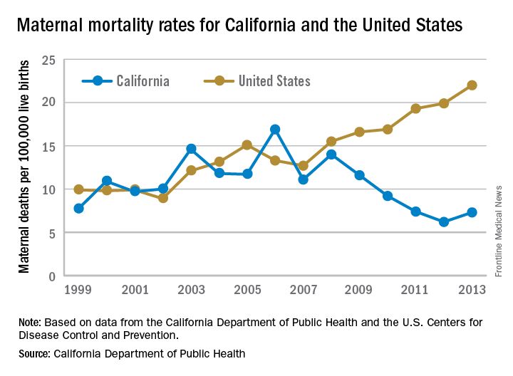
“We reviewed every maternal death for almost 10 years and through that process, we learned a lot about practices of care and the opportunities to really have intervened,” Elliott Main, MD, medical director of the Collaborative, said in an interview. “There were certain causes of death that had a pretty high chance of preventability and those were hemorrhage and preeclampsia.”
Identifying risk factors
The Collaborative’s research identified a number of risk factors that were significant contributors to maternal morbidity.

Dr. Main noted that obesity and older maternal age are both associated with an increased likelihood of having a cesarean delivery, which is associated with an increased risk of hemorrhage.
“So there are pathways that can get you into more trouble, but again if you are on top of those, you can be proactive and not necessarily have this high rate of complications,” he added.
With data in hand about what was contributing to the risks of maternal mortality, the Collaborative set out to build a series of toolkits or “bundles” to help guide hospitals in limiting complications and responding to emergencies. These toolkits are based on the state’s own data, as well as best practices identified in the medical literature and national guidelines from organizations such as the American College of Obstetricians and Gynecologists.
For example, the hypertension bundle includes information aimed at readiness, recognition and prevention, response, and reporting/system learning. Other bundles, which the Collaborative helped to develop and which are distributed through the Council on Patient Safety in Women’s Health Care, cover areas such as mental health, thromboembolism, hemorrhage, and safe reduction in primary cesarean births.
“It’s all about the implementation of those and that’s where we spent a lot of time in California with what are called quality collaboratives,” he said. “These are generally state-based [efforts], where you put together a consortium of providers, hospitals, and public health and patient advocates, and you work on improving the care for these certain conditions.”
The bundles are not meant to be cookbook medicine, Dr. Main noted, but rather are designed so that they can be customized based on the resources of an individual hospital, whether the facility handles 300 births a year or 5,000.
“There is no such thing as a national protocol for this,” Dr. Main said. “You have to have some flexibility and differences in the protocols but what we really are striving for is that for emergencies, people have standard protocols for the treatment of that emergency.”
Disparities remain
While California is a success story in terms of its overall drop in maternal mortality, racial disparities remain, particularly for African American women.
Maternal mortality among African Americans has dropped 50% in the state, mirroring the overall decline in the state during the 2008-2013 time period, but it is still three to four times higher than for other racial/ethnic groups.
“African American women do have more risk factors. There is more obesity. There is more hypertension and they have more social stresses and so forth, but none of those are reasons that they should die,” Dr. Main said. “They are reasons that they should have more intensive attention and care. If you have an older African American woman with hypertension, you’ve got to be on your toes when you are taking care of her and be a lot more responsive to warning signs than in a 25-year-old white woman that is perfectly healthy.”

“Minorities represent half of all U.S. persons, yet racial and ethnic minorities suffer higher rates of maternal mortality than whites in this country,” said Dr. Howell, who also chairs ACOG’s work group on reduction of peripartum racial disparities. “In fact, black and African Americans are three to four times more likely to die than whites. This is the largest disparity among population perinatal health measures.”
Native Americans, Asians, and some Latinas also have elevated rates of maternal mortality, compared with white women, she noted.
Depending on the city, those rates could be even higher, she said.
“In New York City, a recent publication by our Department of Health reviewed deaths from 2006 to 2010 and they found that black women were 10 times more likely to die than white women,” Dr. Howell said. “It’s also important to remember that for every maternal death, over 100 women experience severe obstetric morbidity or a life-threatening diagnosis or undergo a lifesaving procedure during their delivery hospitalization.”
But there are care tools available to help address this issue, too. The Alliance for Innovation on Maternal Health, which includes the California collaborative and ACOG, has developed a safety bundle that focuses on themes of shared decision making, implicit bias, continuity of care, provider and patient education, and care fragmentation. It also recommends implementation of a disparity dashboard, meaning that hospitals and health systems would stratify their quality results by race and ethnicity to identify and address gaps in care, Dr. Howell said.
[email protected]
While the United States as a whole is seeing an unsettling rise in maternal mortality, California is on a divergent path.
Maternal mortality in the Golden State was tracking at a similar rate with national figures from 1999-2008 when the trend started to change. By 2013, the U.S. maternal mortality rate had grown to 22.0 deaths per 100,000 live births, while California’s rate had dropped to 7.3 per 100,000, according to data from the California Department of Public Health and the U.S. Centers for Disease Control and Prevention.
“We reviewed every maternal death for almost 10 years and through that process, we learned a lot about practices of care and the opportunities to really have intervened,” Elliott Main, MD, medical director of the Collaborative, said in an interview. “There were certain causes of death that had a pretty high chance of preventability and those were hemorrhage and preeclampsia.”
Identifying risk factors
The Collaborative’s research identified a number of risk factors that were significant contributors to maternal morbidity.
Dr. Main noted that obesity and older maternal age are both associated with an increased likelihood of having a cesarean delivery, which is associated with an increased risk of hemorrhage.
“So there are pathways that can get you into more trouble, but again if you are on top of those, you can be proactive and not necessarily have this high rate of complications,” he added.
With data in hand about what was contributing to the risks of maternal mortality, the Collaborative set out to build a series of toolkits or “bundles” to help guide hospitals in limiting complications and responding to emergencies. These toolkits are based on the state’s own data, as well as best practices identified in the medical literature and national guidelines from organizations such as the American College of Obstetricians and Gynecologists.
For example, the hypertension bundle includes information aimed at readiness, recognition and prevention, response, and reporting/system learning. Other bundles, which the Collaborative helped to develop and which are distributed through the Council on Patient Safety in Women’s Health Care, cover areas such as mental health, thromboembolism, hemorrhage, and safe reduction in primary cesarean births.
“It’s all about the implementation of those and that’s where we spent a lot of time in California with what are called quality collaboratives,” he said. “These are generally state-based [efforts], where you put together a consortium of providers, hospitals, and public health and patient advocates, and you work on improving the care for these certain conditions.”
The bundles are not meant to be cookbook medicine, Dr. Main noted, but rather are designed so that they can be customized based on the resources of an individual hospital, whether the facility handles 300 births a year or 5,000.
“There is no such thing as a national protocol for this,” Dr. Main said. “You have to have some flexibility and differences in the protocols but what we really are striving for is that for emergencies, people have standard protocols for the treatment of that emergency.”
Disparities remain
While California is a success story in terms of its overall drop in maternal mortality, racial disparities remain, particularly for African American women.
Maternal mortality among African Americans has dropped 50% in the state, mirroring the overall decline in the state during the 2008-2013 time period, but it is still three to four times higher than for other racial/ethnic groups.
“African American women do have more risk factors. There is more obesity. There is more hypertension and they have more social stresses and so forth, but none of those are reasons that they should die,” Dr. Main said. “They are reasons that they should have more intensive attention and care. If you have an older African American woman with hypertension, you’ve got to be on your toes when you are taking care of her and be a lot more responsive to warning signs than in a 25-year-old white woman that is perfectly healthy.”
“Minorities represent half of all U.S. persons, yet racial and ethnic minorities suffer higher rates of maternal mortality than whites in this country,” said Dr. Howell, who also chairs ACOG’s work group on reduction of peripartum racial disparities. “In fact, black and African Americans are three to four times more likely to die than whites. This is the largest disparity among population perinatal health measures.”
Native Americans, Asians, and some Latinas also have elevated rates of maternal mortality, compared with white women, she noted.
Depending on the city, those rates could be even higher, she said.
“In New York City, a recent publication by our Department of Health reviewed deaths from 2006 to 2010 and they found that black women were 10 times more likely to die than white women,” Dr. Howell said. “It’s also important to remember that for every maternal death, over 100 women experience severe obstetric morbidity or a life-threatening diagnosis or undergo a lifesaving procedure during their delivery hospitalization.”
But there are care tools available to help address this issue, too. The Alliance for Innovation on Maternal Health, which includes the California collaborative and ACOG, has developed a safety bundle that focuses on themes of shared decision making, implicit bias, continuity of care, provider and patient education, and care fragmentation. It also recommends implementation of a disparity dashboard, meaning that hospitals and health systems would stratify their quality results by race and ethnicity to identify and address gaps in care, Dr. Howell said.
[email protected]