User login
VIDEO: Tune in to psoriasis patients’ quality of life
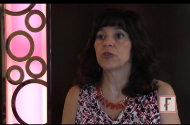
LAS VEGAS – Physicians often fail to predict the impact of disease on quality of life in their psoriasis patients, which can help guide treatment, Joel Gelfand, MD, said in a video interview at Skin Disease Education Foundation’s annual Las Vegas dermatology seminar.
“This is true across all medical conditions,” said Dr. Gelfand, professor of dermatology, at the University of Pennsylvania, Philadelphia. For example, some patients with psoriasis may have extensive disease, but it doesn’t bother them, and therefore they may need less treatment, he pointed out.
In his practice, patients with psoriasis are asked to rate physical symptoms (including flaking and itching) and emotional symptoms (including anxiety and depression) related to their disease on a scale of 0 to 10, with 10 being the worst. “The higher those scores are, the more aggressive I’ll be in treating them,” he said. Patient scores can be tracked over time, to review progress with their chosen treatment, he noted.
Dr. Gelfand disclosed relationships with multiple companies including AbbVie, Janssen, Lilly, Novartis, Celgene, Merck, Sanofi, Pfizer, and Valeant.
SDEF and this news organization are owned by the same parent company.
The video associated with this article is no longer available on this site. Please view all of our videos on the MDedge YouTube channel
At SDEF LAS VEGAS DERMATOLOGY SEMINAR

Which Treatments Effectively Prevent Pediatric Migraine?
Amitriptyline and topiramate, the most commonly used medications for preventing pediatric migraine, are no more effective than placebo, according to trial results published online ahead of print October 27 in the New England Journal of Medicine. The drugs are, however, associated with higher rates of adverse events.
While the researchers were conducting this study, the FDA approved topiramate for the treatment of episodic migraine in adolescents between ages 12 and 17. “Although our trial included patients outside this age range and included those with either episodic or chronic migraine, the trial results suggest that prevention medication for pediatric migraine might be reexamined,” said Scott W. Powers, PhD, Codirector of the Headache Center at Cincinnati Children’s, and colleagues.

The FDA has not approved any migraine-prevention medication for children younger than 12, and clinical practice guidelines for this indication are based on consensus, rather than evidence. Dr. Powers and colleagues conducted the Childhood and Adolescent Migraine Prevention (CHAMP) trial to compare the efficacy of common medications in children and adolescents between ages 8 and 17 with migraine. They randomized 361 patients to amitriptyline (1 mg/kg/day), topiramate (2 mg/kg/day), or placebo in a 2:2:1 ratio.
The trial included a 28-day baseline period, an eight-week dose-escalation period, and a 16-week maintenance phase. The primary outcome was a reduction of 50% or more in the number of headache days during the last 28 days of the trial, compared with baseline. Secondary outcomes included headache-related disability, headache days, number of trial completers, and serious adverse events.
The baseline characteristics of the three treatment groups were similar. The population’s mean age was 14. About 68% of the population was female, and 70% was white.
After a planned interim analysis, the investigators ended the trial early for futility. They found no significant between-group differences in the primary outcome. Approximately 52% of patients receiving amitriptyline, 55% of those receiving topiramate, and 61% of controls had a reduction in headache days of at least 50%. Headache-related disability, headache days, and the rate of trial completion also did not differ between groups.
Patients who received amitriptyline or topiramate had higher rates of several adverse events than those receiving placebo, including fatigue (30% vs 14%) and dry mouth (25% vs 12%) in the amitriptyline group and paresthesia (31% vs 8%) and weight loss (8% vs 0%) in the topiramate group. Three patients receiving amitriptyline had serious adverse events of altered mood, and one patient receiving topiramate attempted suicide.
“We see this [study] as an important opportunity for health care providers, scientists, children, and families because our findings suggest a paradigm shift,” said Dr. Powers. “First-line prevention treatment will involve a multidisciplinary team approach and focus on nonpharmacologic aspects of care. The good news is we can help children with migraines get better.”
—Erik Greb
Suggested Reading
Powers SW, Coffey CS, Chamberlin LA, et al. Trial of amitriptyline, topiramate, and placebo for pediatric migraine. N Engl J Med. 2016 Oct 27 [Epub ahead of print].
Amitriptyline and topiramate, the most commonly used medications for preventing pediatric migraine, are no more effective than placebo, according to trial results published online ahead of print October 27 in the New England Journal of Medicine. The drugs are, however, associated with higher rates of adverse events.
While the researchers were conducting this study, the FDA approved topiramate for the treatment of episodic migraine in adolescents between ages 12 and 17. “Although our trial included patients outside this age range and included those with either episodic or chronic migraine, the trial results suggest that prevention medication for pediatric migraine might be reexamined,” said Scott W. Powers, PhD, Codirector of the Headache Center at Cincinnati Children’s, and colleagues.
The FDA has not approved any migraine-prevention medication for children younger than 12, and clinical practice guidelines for this indication are based on consensus, rather than evidence. Dr. Powers and colleagues conducted the Childhood and Adolescent Migraine Prevention (CHAMP) trial to compare the efficacy of common medications in children and adolescents between ages 8 and 17 with migraine. They randomized 361 patients to amitriptyline (1 mg/kg/day), topiramate (2 mg/kg/day), or placebo in a 2:2:1 ratio.
The trial included a 28-day baseline period, an eight-week dose-escalation period, and a 16-week maintenance phase. The primary outcome was a reduction of 50% or more in the number of headache days during the last 28 days of the trial, compared with baseline. Secondary outcomes included headache-related disability, headache days, number of trial completers, and serious adverse events.
The baseline characteristics of the three treatment groups were similar. The population’s mean age was 14. About 68% of the population was female, and 70% was white.
After a planned interim analysis, the investigators ended the trial early for futility. They found no significant between-group differences in the primary outcome. Approximately 52% of patients receiving amitriptyline, 55% of those receiving topiramate, and 61% of controls had a reduction in headache days of at least 50%. Headache-related disability, headache days, and the rate of trial completion also did not differ between groups.
Patients who received amitriptyline or topiramate had higher rates of several adverse events than those receiving placebo, including fatigue (30% vs 14%) and dry mouth (25% vs 12%) in the amitriptyline group and paresthesia (31% vs 8%) and weight loss (8% vs 0%) in the topiramate group. Three patients receiving amitriptyline had serious adverse events of altered mood, and one patient receiving topiramate attempted suicide.
“We see this [study] as an important opportunity for health care providers, scientists, children, and families because our findings suggest a paradigm shift,” said Dr. Powers. “First-line prevention treatment will involve a multidisciplinary team approach and focus on nonpharmacologic aspects of care. The good news is we can help children with migraines get better.”
—Erik Greb
Suggested Reading
Powers SW, Coffey CS, Chamberlin LA, et al. Trial of amitriptyline, topiramate, and placebo for pediatric migraine. N Engl J Med. 2016 Oct 27 [Epub ahead of print].

Infants with congenital Zika born without microcephaly still can still develop it
Infants born with laboratory-confirmed congenital Zika virus but who show no signs of microcephaly at birth may still experience a reduction in cranial size as they grow older, according to the Centers for Disease Control and Prevention’s latest Morbidity and Mortality Weekly Report.
“These findings demonstrate the importance of early neuroimaging for infants exposed to Zika virus prenatally and the need for comprehensive medical and developmental follow-up,” wrote Vanessa van der Linden, MD, of the Association for Assistance of Disabled Children in Recife, Brazil, and her coauthors.
Dr. van der Linden and her coinvestigators examined 13 infants, all of whom were born in Brazil between October 2015 and January 2016, who had confirmed brain abnormalities at birth despite having a normal head size. These abnormalities included ventriculomegaly, subcortical calcifications, cortical malformations, and decreased brain volume. Investigators defined microcephaly as being “head circumference (HC) [that’s] more than 2 [standard deviations] below the mean for gestational age and sex.”
Nine of the infants were male, four were female. Eleven of the infants were born within 37-41 weeks’ of gestation. The remaining two were born at 35-36 weeks’ of gestation, considered “preterm” by the investigators. All infants tested positive for Zika via immunoglobulin M testing of cerebrospinal fluid, serum, or both. Only six of the mothers reported having a rash while pregnant; four reported experiencing it during the first trimester, while the other two said it occurred in the second.
All 13 infants showed a decrease in HC to what was defined as microcephaly within 1 year of birth (October 2016). Neuroimaging showed that all but one had decreased brain volume, all had malformations of cortical development, four had cerebellum or brain-stem hypoplasia, ten had ventriculomegaly, and three had increased extra-axial CSF space.
“More than 60% of infants in this series had epilepsy (likely related to the cortical malformations), and all had significant motor disabilities consistent with mixed cerebral palsy,” the authors noted, adding that the “pathogenesis of postnatal microcephaly from congenital Zika virus infections is [still] not known.”
FROM THE MMWR
Update on New Drugs in Dermatology
CenterWatch (http://www.centerwatch.com/) is an online resource that provides directories, analysis, and market research of medications that are either under clinical evaluation or available for use in patients. A list of currently approved drugs by the US Food and Drug Administration (FDA) also is available by specialty. It is important for dermatologists in-training to know about recently approved drugs and those that are in the pipeline, as these treatments may benefit patients who are unresponsive to other previously used medications. New drugs also may be useful for physicians who have a difficult time getting insurance to cover prescriptions for their patients, as most new medications have built-in patient assistance.
New Drugs in Dermatology
Actinic Keratosis
Ameluz (aminolevulinic acid hydrochloride)(Biofrontera AG) is a new drug that was approved in May 2016 for treatment of mild to moderate actinic keratosis on the face and scalp.1 It is only intended for in-office use on patients who may not be candidates for other treatment options for actinic keratosis. The product is a gel formulation that should be applied to cover the lesions and approximately 5 mm of the surrounding area with a film of approximately 1-mm thickness. The entire treatment area is then illuminated with a red light source, either with a narrow spectrum around 630 nm with a light dose of approximately 37 J/cm2 or a broader and continuous spectrum in the range of 570 to 670 nm with a light dose between 75 and 200 J/cm2.1 Similar to the previously used aminolevulinic acid treatment method for actinic keratosis, the patient may experience a burning stinging sensation throughout the treatment and the skin will then proceed to peel.
Psoriasis and Psoriatic Arthritis
Taltz (ixekizumab)(Eli Lilly and Company) was approved by the FDA in March 2016 for the treatment of moderate to severe plaque psoriasis.2 It is a humanized IL-17A antagonist that works when IgG4 monoclonal antibodies selectively bind with IL-17A cytokines and inhibit their interaction with the IL-17 receptor. Although this injectable medication is approved for the treatment of psoriasis, it also can potentially be used off label for the treatment of psoriatic arthritis and rheumatoid arthritis. The approved dosage is 160 mg (two 80-mg injections) at week 0, followed by 80 mg at weeks 2, 4, 6, 8, 10, and 12, then 80 mg every 4 weeks.2 Injectable immunomodulatory medications such as ixekizumab are ideal for patients in whom topical treatments and light therapy failed and they continue to have serious psoriatic discomfort as well as for those who have substantial body surface area coverage.
In January 2015, Cosentyx (secukinumab)(Novartis Corporation) was approved by the FDA.3 Similar to ixekizumab, this injectable is an IgG1 monoclonal antibody that selectively binds to the IL-17A cytokine and inhibits its interaction with the IL-17 receptor. It is approved for the treatment of moderate to severe plaque psoriasis and psoriatic arthritis. The approved dosage for plaque psoriasis is 300 mg (two 150-mg subcutaneous injections) at weeks 0 through 4 followed by 300 mg every 4 weeks as needed until clearance.3 Similar to ixekizumab, secukinumab may be used for the treatment of recalcitrant psoriasis or psoriasis with substantial body surface area involvement.
Melanoma
Cotellic (cobimetinib)(Genentech USA, Inc) was FDA approved in November 2015.4 Cobimetinib is a reversible inhibitor of mitogen-activated protein kinase (MAPK)/extracellular signal regulated kinase 1. Mitogen-activated protein kinase MEK1 and MEK2 are regulators of the extracellular signal-related kinase pathway, which promotes cellular proliferation. This pathway is key, as melanomas that have a BRAF V600E and kinase mutation continue to proliferate due to the constitutive activation of MEK1 and MEK2, further promoting cellular proliferation. Cobimetinib is approved for the treatment of melanoma in patients with unresectable or metastatic melanoma with a BRAF V600E or V600K mutation, in conjunction with vemurafenib. Zelboraf (vemurafenib)(Genentech USA, Inc), another inhibitor of BRAF V600E, also is used for the treatment of unresectable melanomas and was initially approved in 2011.5
BRAF is a serine/threonine protein kinase. When unregulated, it results in the deregulation of cell proliferation. According to Ascierto et al,6 50% of melanomas have a BRAF mutation, with nearly 90% of them with a V600E mutation. Hence, since the advent of direct chemotherapeutic agents such as BRAF inhibitors, clinical trials have shown notable reduction in mortality and morbidity of melanoma patients with BRAF mutations.6
Imlygic (talimogene laherparepvec)(Amgen, Inc) is a modified oncolytic viral therapy.7 This treatment was approved by the FDA in 2015 and replicates within tumors to produce granulocyte-macrophage colony-stimulating factor protein, which promotes an antitumor immune response within unresectable cutaneous, subcutaneous, and nodal melanoma lesions. Although it is not a gene-directed therapy, the melanoma does not require a specific mutation for treatment. Again, this medication is better served in conjunction with other melanoma chemotherapeutic and surgical interventions.
Submental Fat
Kybella (deoxycholic acid)(Allergan) is a nonhuman, nonanimal, synthetically created compound that is naturally found within the human body for the breakdown and absorption of dietary fat.8 This drug was FDA approved in 2015 for the improvement of the appearance of moderate subcutaneous fat under the chin. Patients are evaluated in clinic to determine if the submental fat would be responsive to an injectable or require more radical surgical intervention based on desired outcomes. The treatment is administered as 0.2-mL injections (up to a total of 10 mL) spaced 1-cm apart and ideally is repeated at regular intervals to evaluate for efficacy.
Basal Cell Carcinoma
Odomzo (sonidegib)(Novartis Corporation) was FDA approved in 2015 for locally advanced basal cell carcinoma.9 Odomzo is a smoothened antagonist that inhibits the hedgehog signaling pathway. Smoothened is a transmembrane protein that allows for signal transduction of hedgehog proteins.10 Protein patched homolog 1 binds to smoothened protein and prevents the signal transduction through the cell for Gli family zinc factor 1 to continue protein translation; however, when PTCH is mutated and can no longer bind to smoothened, tumor formation results, specifically basal cell carcinoma. Hence, sonidegib is for the treatment of basal cell carcinomas that have persisted despite radiation treatment and/or surgery as well as for patients who have multiple basal cell carcinomas that can no longer be treated with surgery or radiation.
Final Thoughts
Overall, although there are several medications that can be used in conjunction for treatment of dermatological conditions, it always is recommended to know what is in the pipeline as FDA-approved medications for dermatology.
- Ameluz [package insert]. Leverkusen, Germany: Biofrontera Bioscience GmbH; 2016.
- Taltz [package insert]. Indianapolis, IN: Eli Lilly and Company; 2016.
- Cosentyx [package insert]. East Hanover, NJ: Novartis Corporation; 2015.
- Cotellic [package insert]. San Francisco, CA: Genentech, Inc; 2016.
- Zelboraf [package insert]. San Francisco, CA: Genentech, Inc; 2016.
- Ascierto PA, Kirkwood JM, Grob JJ, et al. The role of BRAF V600 mutation in melanoma. J Transl Med. 2012;10:85.
- Imlygic (talimogene laherparepvec). Thousand Oaks, CA: Amgen Inc; 2015.
- Kybella [package insert]. West Lake Village, CA: Kythera Biopharmaceuticals, Inc; 2015.
- Odomzo [package insert]. East Hanover, NJ: Novartis Pharmaceuticals Corporation; 2015.
- Villavicencio EH, Walterhouse DO, Iannaccone PM. The sonic hedgehog-patched-gli pathway in human development and disease. Am J Hum Genet. 2000;67:1047-1054.
CenterWatch (http://www.centerwatch.com/) is an online resource that provides directories, analysis, and market research of medications that are either under clinical evaluation or available for use in patients. A list of currently approved drugs by the US Food and Drug Administration (FDA) also is available by specialty. It is important for dermatologists in-training to know about recently approved drugs and those that are in the pipeline, as these treatments may benefit patients who are unresponsive to other previously used medications. New drugs also may be useful for physicians who have a difficult time getting insurance to cover prescriptions for their patients, as most new medications have built-in patient assistance.
New Drugs in Dermatology
Actinic Keratosis
Ameluz (aminolevulinic acid hydrochloride)(Biofrontera AG) is a new drug that was approved in May 2016 for treatment of mild to moderate actinic keratosis on the face and scalp.1 It is only intended for in-office use on patients who may not be candidates for other treatment options for actinic keratosis. The product is a gel formulation that should be applied to cover the lesions and approximately 5 mm of the surrounding area with a film of approximately 1-mm thickness. The entire treatment area is then illuminated with a red light source, either with a narrow spectrum around 630 nm with a light dose of approximately 37 J/cm2 or a broader and continuous spectrum in the range of 570 to 670 nm with a light dose between 75 and 200 J/cm2.1 Similar to the previously used aminolevulinic acid treatment method for actinic keratosis, the patient may experience a burning stinging sensation throughout the treatment and the skin will then proceed to peel.
Psoriasis and Psoriatic Arthritis
Taltz (ixekizumab)(Eli Lilly and Company) was approved by the FDA in March 2016 for the treatment of moderate to severe plaque psoriasis.2 It is a humanized IL-17A antagonist that works when IgG4 monoclonal antibodies selectively bind with IL-17A cytokines and inhibit their interaction with the IL-17 receptor. Although this injectable medication is approved for the treatment of psoriasis, it also can potentially be used off label for the treatment of psoriatic arthritis and rheumatoid arthritis. The approved dosage is 160 mg (two 80-mg injections) at week 0, followed by 80 mg at weeks 2, 4, 6, 8, 10, and 12, then 80 mg every 4 weeks.2 Injectable immunomodulatory medications such as ixekizumab are ideal for patients in whom topical treatments and light therapy failed and they continue to have serious psoriatic discomfort as well as for those who have substantial body surface area coverage.
In January 2015, Cosentyx (secukinumab)(Novartis Corporation) was approved by the FDA.3 Similar to ixekizumab, this injectable is an IgG1 monoclonal antibody that selectively binds to the IL-17A cytokine and inhibits its interaction with the IL-17 receptor. It is approved for the treatment of moderate to severe plaque psoriasis and psoriatic arthritis. The approved dosage for plaque psoriasis is 300 mg (two 150-mg subcutaneous injections) at weeks 0 through 4 followed by 300 mg every 4 weeks as needed until clearance.3 Similar to ixekizumab, secukinumab may be used for the treatment of recalcitrant psoriasis or psoriasis with substantial body surface area involvement.
Melanoma
Cotellic (cobimetinib)(Genentech USA, Inc) was FDA approved in November 2015.4 Cobimetinib is a reversible inhibitor of mitogen-activated protein kinase (MAPK)/extracellular signal regulated kinase 1. Mitogen-activated protein kinase MEK1 and MEK2 are regulators of the extracellular signal-related kinase pathway, which promotes cellular proliferation. This pathway is key, as melanomas that have a BRAF V600E and kinase mutation continue to proliferate due to the constitutive activation of MEK1 and MEK2, further promoting cellular proliferation. Cobimetinib is approved for the treatment of melanoma in patients with unresectable or metastatic melanoma with a BRAF V600E or V600K mutation, in conjunction with vemurafenib. Zelboraf (vemurafenib)(Genentech USA, Inc), another inhibitor of BRAF V600E, also is used for the treatment of unresectable melanomas and was initially approved in 2011.5
BRAF is a serine/threonine protein kinase. When unregulated, it results in the deregulation of cell proliferation. According to Ascierto et al,6 50% of melanomas have a BRAF mutation, with nearly 90% of them with a V600E mutation. Hence, since the advent of direct chemotherapeutic agents such as BRAF inhibitors, clinical trials have shown notable reduction in mortality and morbidity of melanoma patients with BRAF mutations.6
Imlygic (talimogene laherparepvec)(Amgen, Inc) is a modified oncolytic viral therapy.7 This treatment was approved by the FDA in 2015 and replicates within tumors to produce granulocyte-macrophage colony-stimulating factor protein, which promotes an antitumor immune response within unresectable cutaneous, subcutaneous, and nodal melanoma lesions. Although it is not a gene-directed therapy, the melanoma does not require a specific mutation for treatment. Again, this medication is better served in conjunction with other melanoma chemotherapeutic and surgical interventions.
Submental Fat
Kybella (deoxycholic acid)(Allergan) is a nonhuman, nonanimal, synthetically created compound that is naturally found within the human body for the breakdown and absorption of dietary fat.8 This drug was FDA approved in 2015 for the improvement of the appearance of moderate subcutaneous fat under the chin. Patients are evaluated in clinic to determine if the submental fat would be responsive to an injectable or require more radical surgical intervention based on desired outcomes. The treatment is administered as 0.2-mL injections (up to a total of 10 mL) spaced 1-cm apart and ideally is repeated at regular intervals to evaluate for efficacy.
Basal Cell Carcinoma
Odomzo (sonidegib)(Novartis Corporation) was FDA approved in 2015 for locally advanced basal cell carcinoma.9 Odomzo is a smoothened antagonist that inhibits the hedgehog signaling pathway. Smoothened is a transmembrane protein that allows for signal transduction of hedgehog proteins.10 Protein patched homolog 1 binds to smoothened protein and prevents the signal transduction through the cell for Gli family zinc factor 1 to continue protein translation; however, when PTCH is mutated and can no longer bind to smoothened, tumor formation results, specifically basal cell carcinoma. Hence, sonidegib is for the treatment of basal cell carcinomas that have persisted despite radiation treatment and/or surgery as well as for patients who have multiple basal cell carcinomas that can no longer be treated with surgery or radiation.
Final Thoughts
Overall, although there are several medications that can be used in conjunction for treatment of dermatological conditions, it always is recommended to know what is in the pipeline as FDA-approved medications for dermatology.
- Ameluz [package insert]. Leverkusen, Germany: Biofrontera Bioscience GmbH; 2016.
- Taltz [package insert]. Indianapolis, IN: Eli Lilly and Company; 2016.
- Cosentyx [package insert]. East Hanover, NJ: Novartis Corporation; 2015.
- Cotellic [package insert]. San Francisco, CA: Genentech, Inc; 2016.
- Zelboraf [package insert]. San Francisco, CA: Genentech, Inc; 2016.
- Ascierto PA, Kirkwood JM, Grob JJ, et al. The role of BRAF V600 mutation in melanoma. J Transl Med. 2012;10:85.
- Imlygic (talimogene laherparepvec). Thousand Oaks, CA: Amgen Inc; 2015.
- Kybella [package insert]. West Lake Village, CA: Kythera Biopharmaceuticals, Inc; 2015.
- Odomzo [package insert]. East Hanover, NJ: Novartis Pharmaceuticals Corporation; 2015.
- Villavicencio EH, Walterhouse DO, Iannaccone PM. The sonic hedgehog-patched-gli pathway in human development and disease. Am J Hum Genet. 2000;67:1047-1054.

VIDEO: Challenging case – consider HSV with erythema multiforme
LAS VEGAS – In a presentation at Skin Disease Education Foundation’s annual Las Vegas dermatology seminar, Miriam S. Bettencourt, MD, shared a challenging diagnostic case of erythema multiforme associated with herpes simplex, in a patient who presented with blisters all over his body.
“We know that 90% of cases of erythema multiforme are related to herpetic infections,” but this patient had no recent history of herpes simplex outbreaks, Dr. Bettencourt, of the University of Nevada, Las Vegas, said in a video interview.
‘“Let’s remember that HSV [herpes simplex virus] ... can be associated with erythema multiforme even if a patient does not have any flares,” Dr. Bettencourt said. A consult with a rheumatologist can be helpful, as in this case, if a patient has a positive antinuclear antibody test, which could not be explained, she added, noting that the patient is doing well after 6 months of therapy.
Dr. Bettencourt disclosed relationships with AbbVie, Allergan, Aqua, Celgene, Janssen, IntraDerm, Leo, Promius, and Valeant.
SDEF and this news organization are owned by the same parent company.
The video associated with this article is no longer available on this site. Please view all of our videos on the MDedge YouTube channel
AT SDEF LAS VEGAS DERMATOLOGY SEMINAR
VIDEO: For facial resurfacing, revisit CO2 lasers
LAS VEGAS – Patients can enjoy positive results from facial resurfacing with fractionated lasers, but they don’t always yield the same benefits as the traditional CO2 laser, Christopher Zachary, M.D., said at Skin Disease Education Foundation’s annual Las Vegas dermatology seminar.
“A lot of us are going back to using traditional laser resurfacing” for the patients who need it, such as those with many wrinkles, crepey skin, and extensive sun damage, Dr. Zachary, professor and chair of the department of dermatology at the University of California, Irvine, said in a video interview.
“Those patients are not going to have an optimal result, even with the most aggressive of fractionated ablative lasers, as compared to the traditional laser resurfacing,” he added.
Dr. Zachary disclosed relationships with multiple companies, including Solta, Zeltiq, Scion, Amway, and Candela. SDEF and this news organization are owned by the same parent company.
The video associated with this article is no longer available on this site. Please view all of our videos on the MDedge YouTube channel
AT SDEF LAS VEGAS DERMATOLOGY SEMINAR
A look at HIV-related cancers: incidence, screening, and stem transplantation
HIV-related lymphoma rate remains sky-high despite ART
Key clinical point ART has had a major impact on the incidence of HIV-related non-Hodgkin lymphoma but no effect on Hodgkin lymphoma. Major finding The overall incidence of non-Hodgkin lymphoma in HIV-positive adults on ART is 287 cases per 100,000 person-years, varying by location and route of HIV acquisition. Data source A longitudinal analysis of non-Hodgkin lymphoma incidence in more than 210,000 HIV-infected adults on combination ART on 4 continents. Disclosures The European Union and the National Institutes of Health. The presenter reported having no financial conflicts of interest.
Click on the PDF icon at the top of this introduction to read the full article.
Palliative concurrent chemoradiation for gastrostomy site metastasis
Patients with head and neck squamous cell carcinoma typically present with dysphagia, odynophagia, and weight loss. Treatment of the disease with surgery or concurrent chemoradiation often results in local inflammation and limits further oral intake. Percutaneous endoscopic gastrostomy has been a common and effective means of nutritional support in these patients.
Click on the PDF icon at the top of this introduction to read the full article.
VIDEO: Bulk matters in body sculpting
LAS VEGAS – Both heating and cooling techniques can provide effective results for patients seeking to improve their appearance with body sculpting, Christopher Zachary, MD, said at Skin Disease Education Foundation’s annual Las Vegas dermatology seminar.
Whether the clinician chooses devices that use radiofrequency, laser, or cryolipolysis to target fat, the key is bulk treatment, Dr. Zachary, professor and chair of the department of dermatology at the University of California, Irvine, said in a video interview.
When cooling or heating the fat, “it has to been done in bulk; it has to be done for a certain length of time,” he said, noting that treatment times vary with devices, from 5 to 60 minutes. “I can’t stress enough the importance of bulk cooling or bulk heating,” which induce a chronic reaction “that results in localized fat reduction,” he added.
Dr. Zachary disclosed relationships with multiple companies, including Solta, Zeltiq, Scion, Amway, and Candela. SDEF and this news organization are owned by the same parent company.
The video associated with this article is no longer available on this site. Please view all of our videos on the MDedge YouTube channel
AT SDEF LAS VEGAS DERMATOLOGY SEMINAR
Acute-onset hypokalemic paralysis with arsenic trioxide therapy in patient with acute promyelocytic leukemia
Acute myeloid leukemia (AML) is characterized by clonal proliferation of myeloid precursors with a reduced capacity to differentiate into mature cellular components.1 Acute promyeloctic leukemia (APL; previously called AML-M3), a subtype of AML, is further characterized by a balanced translocation t(15;17) (q24.1;q21.1). It is an interesting model in cancer research because it responds to the differentiation and apoptosis induction therapy using arsenic trioxide (ATO) and all-trans retinoic acid (ATRA).2
Click on the PDF icon at the top of this introduction to read the full article.