User login
Study supports palliative care in HSCT recipients
![]()
Photo by Chad McNeeley
Palliative care can be beneficial for patients undergoing hematopoietic stem cell transplant (HSCT) to treat hematologic malignancies, according to research published in JAMA.
The single-center study suggested that palliative care can improve HSCT recipients’ quality of life, relieve symptoms associated with the procedure, and reduce depression and anxiety.
Researchers observed such benefits during hospitalization for HSCT and a few months later.
In addition, caregivers of patients receiving palliative care experienced less depression and were better at coping with the stress associated with the illness of their loved one.
“Palliative care clinicians are increasingly asked to help care for patients with solid tumors but are rarely consulted for patients with hematologic malignancies, especially those receiving therapy designed to cure their disease,” said study author Areej El-Jawahri, MD, of Massachusette General Hospital in Boston.
“The physical and psychological symptoms associated with HSCT are sometimes regarded as expected and unavoidable, which, combined with the persistent misperception that equates palliative care with end-of-life care, has contributed to a lack of involvement of palliative care clinicians in the care of these patients.”
Intervention
Dr El-Jawahri and her colleagues studied 160 patients who underwent autologous or allogeneic HSCT to treat a variety of hematologic malignancies from August 2014 into January 2016.
Participants were randomized to receive either standard care (n=79) or the palliative care intervention (n=81).
Within 3 days of their admission to the hospital, patients in the intervention group had an initial meeting with a palliative care clinician—a physician or advance practice nurse—who continued to meet with them at least twice a week during their hospitalization.
At the meetings, which could be attended by a family member or friend of the patient, clinicians first focused on establishing a rapport with patients and their caregivers.
Clinicians addressed ways of managing the physical and psychological symptoms patients were experiencing and provided support and strategies for coping with distress. Patients received an average of 8 palliative care visits during their hospitalizations, which lasted on average 20 days.
At the outset of the study and 2 weeks into the process, a time when symptoms tend to be at their worst, patients in both groups and participating caregivers completed questionnaires assessing their mood and quality of life.
Patients also completed questionnaires asking about symptoms of their illness and those associated with the procedure. Patients completed additional assessments 3 months after HSCT as well.
Results
The study’s primary endpoint was change in quality of life from baseline to week 2. Patients receiving the palliative care intervention had significantly better quality of life scores at week 2 than patients in the control group.
Also at the 2-week mark, patients receiving the palliative care intervention reported lower levels of depression, anxiety, and symptoms than the control group, but there was no significant difference between the groups with regard to fatigue.
At 3 months, patients receiving the palliative care intervention still had higher quality of life scores and less depression than controls, but there were no significant between-group differences in anxiety, fatigue, or symptom burden.
Caregivers attended 42% of the palliative care sessions. At the 2-week assessment, caregivers in the intervention group were found to have fewer depressive symptoms and improved coping skills, compared with caregivers in the control group.
“Caregivers play a crucial role in supporting patients during the transplant process, and they are substantially impacted as they watch their loved ones struggle with side effects that can be emotionally challenging,” Dr El-Jawahri said.
She and her colleagues noted that additional, larger studies are needed to assess caregiver impacts more completely, to replicate patient results at centers with more diverse patient populations, to assess the inclusion of more complete palliative care teams, to collect cost data, and to adapt the palliative care intervention to assist patients receiving other potentially curative treatment for hematologic or other cancers. ![]()
EC approves nivolumab for relapsed/refractory cHL
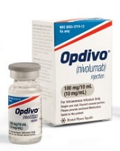
Photo from Business Wire
The European Commission (EC) has approved nivolumab (Opdivo) for the treatment of adults with relapsed or refractory classical Hodgkin lymphoma (cHL) who have already received an autologous hematopoietic stem cell transplant (auto-HSCT) and treatment with brentuximab vedotin (BV).
Nivolumab is the first PD-1 inhibitor approved in the European Economic Area as a treatment for a hematologic malignancy.
The EC previously approved nivolumab to treat advanced melanoma, non-small cell lung cancer, and renal cell carcinoma. In Europe, nivolumab is marketed by Bristol-Myers Squibb.
Trials in cHL
The EC’s approval of nivolumab in cHL is based on an integrated analysis of data from 2 trials—the phase 1 CheckMate -039 trial and the phase 2 CheckMate -205 trial.
In CheckMate -039, researchers evaluated nivolumab in patients with cHL, non-Hodgkin lymphoma, and multiple myeloma. Results from this trial were presented at the 13th International Congress on Malignant Lymphoma in June 2015.
In CheckMate -205, researchers are evaluating nivolumab in 4 cohorts of cHL patients. Cohort A includes patients who previously received auto-HSCT and were BV-naïve at enrollment (n=63). Cohort B includes patients who previously received auto-HSCT followed by BV (n=80).
Cohort C includes patients who previously received BV before and/or after auto-HSCT (n=100). And cohort D, which is currently enrolling, is an evaluation of nivolumab in combination with chemotherapy in newly diagnosed, advanced-stage cHL patients who are treatment-naïve (n=50).
Results from cohort B were presented at the 21st Congress of the European Hematology Association in June 2016. Results from cohort C were presented at the 10th International Symposium on Hodgkin Lymphoma last month.
Integrated analysis
The analysis included cHL patients from CheckMate -205 and -039 who had received auto-HSCT and BV.
In the efficacy population (n=95), the objective response rate was 66%. The percentage of patients with a complete response was 6%. Twenty-three percent of patients had stable disease.
The median time to response was 2.0 months (range, 0.7-11.1), and the median duration of response was 13.1 months (range, 0.0+, 23.1+). At 12 months, the progression-free survival rate was 57%.
The safety of nivolumab in cHL was evaluated in 263 patients from CheckMate -205 (n=240) and CheckMate -039 (n=23). Serious adverse events (AEs) occurred in 21% of these patients.
The most common serious AEs (reported in at least 1% of patients) were infusion-related reactions, pneumonia, pleural effusion, pyrexia, rash, and pneumonitis.
The most common AEs (reported in at least 20% of patients) were fatigue (32%), upper respiratory tract infection (28%), pyrexia (24%), diarrhea (23%), and cough (22%).
Twenty-three percent of patients had a dose delay resulting from an AE, and 4.2% of patients discontinued treatment due to AEs.
Forty patients went on to allogeneic HSCT after nivolumab, and 6 of these patients died from complications of the transplant. The 40 patients had a median follow-up from allogeneic HSCT of 2.9 months (range, 0-22).
Because of these deaths, the US Food and Drug Administration asked Bristol-Myers Squibb to study the safety of allogeneic HSCT after nivolumab. ![]()
Hospitalists Stretched as their Responsibilities Broaden
The very nature of America’s hospitals is changing. At one time in the not too distant past, hospitals could charge “cost-plus,” tacking on a profit above their actual expenses. Hospitals generated most of their revenue from procedures on horizontal patients with long stays in house. Physicians viewed the hospital as a swap meet, with each physician having an autonomous booth and not caring much what went on elsewhere in the facility.

Today, hospitals are under tough cost pressures, with changes in payments from Medicare, Medicaid, and private insurers. Many hospitals now get more than 50% of their revenue from vertical patients from what was previously considered the outpatient segment of healthcare. Physicians have moved from being revenue providers to being potential competitors or, in the best-case scenario, active partners and teammates with their hospital.
And hospitalists are right in the middle of this changing dynamic.
Because the hospital and the healthcare system are rapidly evolving, it should not surprise anyone that the very nature of hospital medicine is changing rapidly. Some would say too rapidly.
At a strategic planning session I led almost 20 years when the National Association of Inpatient Physicians (NAIP), the precursor to SHM, was just starting out, the prevailing consensus was that hospitalists might take over inpatient services for 50% of family physicians and 25% of internists. Obviously, the penetrance of hospital medicine into almost every hospital in the U.S. and the transfer of the acute-care management of most of the inpatients previously handled by family physicians and internists are just part of the growth in hospital medicine.
Even more innovative and disruptive has been the almost relentless scope creep as hospitalists now actively comanage many surgical and subspecialty patients. As the neurologists have given up most of their acute-care duties, hospitalists are now the de facto inpatient neurologists. Hospitalists also now manage the majority of inpatient senior citizens and have become the inpatient geriatricians without the formal training. In-hospital procedures (e.g., central line, ultrasound, intubation, etc.) previously done by surgeons or critical-care or primary-care physicians now are done by default by hospitalists.
But these expansions of hospitalist scope pale in comparison with the continued broadening of responsibilities that continues to stretch even the most well-trained hospitalists beyond their training or capacity.
Palliative Care
There are not enough trained and certified palliative-care physicians to allocate one of them to each hospital. Yet treatment and survival of cancer and other serious diseases as well as the aging of the population demand that hospitals be prepared to provide the most compassionate and up-to-date palliative approach possible. Palliative care is more than just end-of-life care. It involves hospice as well as pain and symptom management. It is aimed at improvement in quality of life and is used in the presence or absence of curative strategies.
Hospitalists have been thrust into the breach and are being asked more and more to provide palliative-care services. SHM has recognized the gap between the increasing demand on hospitalists and the inadequate training we all receive in residency. That’s why we’re working with palliative-care societies and experts to develop educational and training initiatives to close these gaps.
Critical Care
Our hospitals are becoming increasingly critical care intensive as simpler cases are treated as outpatients and only the very ill come to be admitted to hospital. This has created an increasing demand for more physicians trained in critical care at a time when older intensivists are retiring or going into sleep medicine and younger physicians, who might have chosen a career in critical care, are becoming hospitalists. The shortage of trained critical-care providers is reaching a crisis point in many American hospitals, with hospitalists being asked to be the critical-care extender.
Over the years, SHM has partnered with the Society of Critical Care Medicine (SCCM) to propose innovative training options (e.g., one-year critical-care fellowship obtained midcareer), but the boards and others in the critical-care establishment have not been supportive. SHM plans to continue to work with open-minded critical-care thought leaders to develop and promote additional training in critical-care skills for hospitalists, who continue to be thrust into this role at their local hospitals.
Post-Acute Care
For many of hospital medicine’s larger national and regional companies, the management of the care in the post-acute-care space of skilled nursing facilities, long-term acute-care facilities, and the like has been the fastest-growing part of their business in the last few years. Skills and process improvement that have helped improve effectiveness and efficiency in our nation’s hospitals are being applied to post-acute-care facilities. Once again, hospitalists are finding themselves being asked to perform at a high level in environments that are new to them.
In this arena, the hospitalist’s ability to impact care is evident in managing transfers and information as well as providing leadership in patient safety. Determining the correct postdischarge disposition is the largest driver of costs in the acute-care and post-acute-care setting. Hospitalists and the hospital medicine organizations are providing key direction.
Preoperative Care
Many may not know that bundled into the anesthesia fee is the funding to cover pre-op assessment and post-op management as well as the intraoperative oversight of anesthesia and vital signs for the surgical patient. In reality, the role of perioperative management has fallen for many years initially to internists and more recently to hospitalists.
Hospitalists have been active in optimizing the patient for surgery and medically clearing the patient. Hospitalists work with surgeons to manage comorbidities; prevent complications, such as infections, DVTs, and pulmonary emboli; and help with pain management and transitions to discharge from the hospital. Hospitalists have worked with surgeons to create efficiencies like reduced length of stay and prevention of readmission as well as to help the patient return to function postoperatively.
SHM’s Perioperative Care Work Group is publishing a set of Perioperative Care Guidelines in the Journal of Hospital Medicine. SHM is actively working with the American College of Surgeons on a teamwork approach to the surgical patient as well as innovative alternative payment models with bundling at the level of the individual surgical patients, which the Centers for Medicare & Medicaid Services is currently evaluating.
Working through a Dilemma
The one thing all these expansions of scope have in common is that there is an unfilled need and hospitalists are being thrust onto the front lines, thrown into the deep water without the benefit of thorough training that should be requisite with the responsibilities. This is not a turf battle where we have stolen someone’s cheese. This is pure and simple where need is trumping training, and if not done properly, the patient may suffer, and hospitalists will bear the uncomfortable feeling of being asked to do more than we should.
SHM and our national hospitalist thought leaders see this dilemma. We are working diligently with other professional medical societies and key specialty educators and thought leaders to create training pathways to support the expansion of the hospitalist’s scope. This is building the boat while you are going down a rapidly moving river. It is not easy stuff. But our patients and our hospitalists demand this, and SHM will step up. Help is on the way.
Larry Wellikson, MD, MHM, is CEO of the Society of Hospital Medicine.
The very nature of America’s hospitals is changing. At one time in the not too distant past, hospitals could charge “cost-plus,” tacking on a profit above their actual expenses. Hospitals generated most of their revenue from procedures on horizontal patients with long stays in house. Physicians viewed the hospital as a swap meet, with each physician having an autonomous booth and not caring much what went on elsewhere in the facility.
Today, hospitals are under tough cost pressures, with changes in payments from Medicare, Medicaid, and private insurers. Many hospitals now get more than 50% of their revenue from vertical patients from what was previously considered the outpatient segment of healthcare. Physicians have moved from being revenue providers to being potential competitors or, in the best-case scenario, active partners and teammates with their hospital.
And hospitalists are right in the middle of this changing dynamic.
Because the hospital and the healthcare system are rapidly evolving, it should not surprise anyone that the very nature of hospital medicine is changing rapidly. Some would say too rapidly.
At a strategic planning session I led almost 20 years when the National Association of Inpatient Physicians (NAIP), the precursor to SHM, was just starting out, the prevailing consensus was that hospitalists might take over inpatient services for 50% of family physicians and 25% of internists. Obviously, the penetrance of hospital medicine into almost every hospital in the U.S. and the transfer of the acute-care management of most of the inpatients previously handled by family physicians and internists are just part of the growth in hospital medicine.
Even more innovative and disruptive has been the almost relentless scope creep as hospitalists now actively comanage many surgical and subspecialty patients. As the neurologists have given up most of their acute-care duties, hospitalists are now the de facto inpatient neurologists. Hospitalists also now manage the majority of inpatient senior citizens and have become the inpatient geriatricians without the formal training. In-hospital procedures (e.g., central line, ultrasound, intubation, etc.) previously done by surgeons or critical-care or primary-care physicians now are done by default by hospitalists.
But these expansions of hospitalist scope pale in comparison with the continued broadening of responsibilities that continues to stretch even the most well-trained hospitalists beyond their training or capacity.
Palliative Care
There are not enough trained and certified palliative-care physicians to allocate one of them to each hospital. Yet treatment and survival of cancer and other serious diseases as well as the aging of the population demand that hospitals be prepared to provide the most compassionate and up-to-date palliative approach possible. Palliative care is more than just end-of-life care. It involves hospice as well as pain and symptom management. It is aimed at improvement in quality of life and is used in the presence or absence of curative strategies.
Hospitalists have been thrust into the breach and are being asked more and more to provide palliative-care services. SHM has recognized the gap between the increasing demand on hospitalists and the inadequate training we all receive in residency. That’s why we’re working with palliative-care societies and experts to develop educational and training initiatives to close these gaps.
Critical Care
Our hospitals are becoming increasingly critical care intensive as simpler cases are treated as outpatients and only the very ill come to be admitted to hospital. This has created an increasing demand for more physicians trained in critical care at a time when older intensivists are retiring or going into sleep medicine and younger physicians, who might have chosen a career in critical care, are becoming hospitalists. The shortage of trained critical-care providers is reaching a crisis point in many American hospitals, with hospitalists being asked to be the critical-care extender.
Over the years, SHM has partnered with the Society of Critical Care Medicine (SCCM) to propose innovative training options (e.g., one-year critical-care fellowship obtained midcareer), but the boards and others in the critical-care establishment have not been supportive. SHM plans to continue to work with open-minded critical-care thought leaders to develop and promote additional training in critical-care skills for hospitalists, who continue to be thrust into this role at their local hospitals.
Post-Acute Care
For many of hospital medicine’s larger national and regional companies, the management of the care in the post-acute-care space of skilled nursing facilities, long-term acute-care facilities, and the like has been the fastest-growing part of their business in the last few years. Skills and process improvement that have helped improve effectiveness and efficiency in our nation’s hospitals are being applied to post-acute-care facilities. Once again, hospitalists are finding themselves being asked to perform at a high level in environments that are new to them.
In this arena, the hospitalist’s ability to impact care is evident in managing transfers and information as well as providing leadership in patient safety. Determining the correct postdischarge disposition is the largest driver of costs in the acute-care and post-acute-care setting. Hospitalists and the hospital medicine organizations are providing key direction.
Preoperative Care
Many may not know that bundled into the anesthesia fee is the funding to cover pre-op assessment and post-op management as well as the intraoperative oversight of anesthesia and vital signs for the surgical patient. In reality, the role of perioperative management has fallen for many years initially to internists and more recently to hospitalists.
Hospitalists have been active in optimizing the patient for surgery and medically clearing the patient. Hospitalists work with surgeons to manage comorbidities; prevent complications, such as infections, DVTs, and pulmonary emboli; and help with pain management and transitions to discharge from the hospital. Hospitalists have worked with surgeons to create efficiencies like reduced length of stay and prevention of readmission as well as to help the patient return to function postoperatively.
SHM’s Perioperative Care Work Group is publishing a set of Perioperative Care Guidelines in the Journal of Hospital Medicine. SHM is actively working with the American College of Surgeons on a teamwork approach to the surgical patient as well as innovative alternative payment models with bundling at the level of the individual surgical patients, which the Centers for Medicare & Medicaid Services is currently evaluating.
Working through a Dilemma
The one thing all these expansions of scope have in common is that there is an unfilled need and hospitalists are being thrust onto the front lines, thrown into the deep water without the benefit of thorough training that should be requisite with the responsibilities. This is not a turf battle where we have stolen someone’s cheese. This is pure and simple where need is trumping training, and if not done properly, the patient may suffer, and hospitalists will bear the uncomfortable feeling of being asked to do more than we should.
SHM and our national hospitalist thought leaders see this dilemma. We are working diligently with other professional medical societies and key specialty educators and thought leaders to create training pathways to support the expansion of the hospitalist’s scope. This is building the boat while you are going down a rapidly moving river. It is not easy stuff. But our patients and our hospitalists demand this, and SHM will step up. Help is on the way.
Larry Wellikson, MD, MHM, is CEO of the Society of Hospital Medicine.
Examining the No. 1 Preventable Cause of Cancer
When tobacco and cancer are mentioned, many people automatically assume lung cancer. But the 70-plus carcinogens identified in tobacco smoke cause at least 12 types of cancer, according to a Vital Signs report, including cancers of the stomach, kidney and renal pelvis, urinary bladder, and cervix. “Smokeless” products such as chewing tobacco carry 28 identified carcinogens.
Related: Quitting Smoking During Substance Abuse Treatment
Researchers from the CDC and National Cancer Institute analyzed data from the U.S. Cancer Statistics 2004-2013 to assess incidence and death rates for cancers caused by tobacco use. During that time, the incidence of tobacco-related cancer went down 1.3% per year, and mortality dropped 1.6% each year. Tobacco-related cancer incidence declined significantly in 44 states. Rates were lowest in the West and dropped the slowest in the Midwest.
However, the burden remains high, the researchers note, and disparities persist among certain groups. For instance, the incidence of tobacco-related cancer rose with age: Two-fifths of deaths were among people aged ≥ 75 years. And the incidence of cancer and death rates were highest but decreased fastest among blacks.
Related: Impact of a Drop-in Group Medical Appointment on Tobacco Quit Rates
There’s no question that lung cancer remains the cancer most linked to smoking. Lung cancer accounted for about one-third of tobacco-related cancer cases and almost one-half of tobacco-related cancer deaths. Between 2009 and 2013, about 101,300 men and 65,700 women died each year of cancers attributable to cigarette smoking—most died of lung cancer. Exposure to secondhand smoke accounted for another 7,300 deaths by lung cancer among nonsmokers.
Roughly 6 million people may die prematurely of a tobacco-related cancer unless they can take advantage of more sustained, comprehensive, and evidence-based interventions, the researchers say. States that have invested in smoking prevention and control programs, they point out, have seen larger declines in youth and adult smoking, decreases in lung cancer, and reduced tobacco-related health care costs.
Related: CDC Media Campaign Helps Americans Quit Smoking
The researchers also emphasize the benefits of smoking-cessation counseling and treatment as well as screening for cervical, colorectal, and lung cancers, to help detect them at earlier more easily treatable stages. Vaccination against hepatitis B virus and human papillomavirus can prevent liver and cervix cancer, to which tobacco can contribute.
According to Vital Signs, the CDC funds 65 comprehensive cancer control programs to prevent cancer, increase access to early detection and care for tobacco-related cancers, help cancer survivors quit using tobacco, and improve cancer outcomes, especially in communities with higher tobacco-related rates of cancer and deaths. The CDC also urges health care providers to encourage patients to quit—or not start—smoking, to be aware of screenings, and to remember that there’s no “risk-free” level of secondhand smoke.
Source:
Henley SJ, Thomas CC, Sharapova SR, et al. MMWR. 2016;65(44):1212-1218.
The Overdose Education and Naloxone Distribution Program at a VA Hospital
Fatal opioid overdoses have quadrupled in the U.S. from 1999 to 2013.1 In 2013, 43,982 deaths in the U.S. were attributable to drug overdoses, and 37% of them involved opioids.1 Data from 2005 suggested that veterans are at an increased risk with a 2-fold rise in overdose observed.2 In 2013, there were 17,124 encounters at VA facilities for the treatment of opioid overdose.3 In response to this opioid epidemic, individual VA facilities began implementing Overdose Education and Naloxone Distribution (OEND) programs in 2013.4 In May 2014, the Under Secretary for Health mandated the implementation of OEND programs across all VA facilities, and the VA became the only health care system in the U.S. with a national OEND program.4
Naloxone
Naloxone is a pure opioid antagonist that competes with and displaces opioids at opioid receptors. In an overdose, opioids occupying opioid receptor sites can lead to respiratory depression, which can lead to death. Patients can experience rapid reversal of respiratory depression and opioid overdose with the first dose of naloxone 0.4 mg. Naloxone has a short half-life relative to opioids, whereas opioids can bind to opioid receptors once again after naloxone is eliminated, risking the reemergence of overdose symptoms. For this reason, a second dose of naloxone is often given. When administered appropriately with close monitoring, naloxone can provide a safe and effective way to reverse a potentially life-threatening opioid overdose.
Availability
In April 2014, Evzio, an intramuscular/subcutaneous formulation of naloxone, was FDA approved as an emergency treatment for known or suspected opioid overdose. Shortly after, in November 2015, Narcan, a nasal spray formulation of naloxone, also was approved. Each formulation has been instrumental in fulfilling an unmet medical need in the U.S. Both Evzio and Narcan have been used in opioid-overdose prevention programs to train health care professionals and lay persons to respond in the event of suspected overdose and administer naloxone.
Maxwell and colleagues implemented a naloxone distribution program in Chicago, Illinois, in January 2001 after a 4-fold increase in heroin-related overdose deaths was observed.5 Within a year, Chicago experienced a 20% decrease in heroin-related overdose deaths, and this trend continued with additional 10% reductions in heroin-related overdoses in 2002 and 2003. Similarly, Piper and colleagues implemented a naloxone distribution program and found that 82% of injection drug users felt “comfortable” to “very comfortable” administering naloxone in the event of an overdose, and 86% of subjects reported they would want naloxone in the event of an overdose.6
Naloxone is available for over-the-counter purchase in Rhode Island, which is a bordering state to VA Connecticut Healthcare System (VACT). In 2015 new legislation permitted the training and certification of Connecticut community pharmacists to prescribe naloxone. Furthermore, in May 2016, Connecticut passed legislation to permit qualified individuals to carry naloxone and administer it to another person that he or she, believes is experiencing an opioid-related drug overdose. Given the increase in naloxone availability both nationally and locally, VACT sought to implement a naloxone distribution program to offer the same access to veterans. The primary objective of this article is to describe the development, implementation, and preliminary results of this OEND program.
Development
In accordance with VA guidance, the VACT OEND program development and implementation process included 4 steps that required program coordinators to identify target populations, garner support, train staff members, and implement the OEND program.
A multidisciplinary team of representatives from pharmacy, mental health (MH), and substance abuse (SA) departments was established to draft hospital policy. The hospital policy detailed job functions related to the OEND program for prescribing, educating, verifying, and dispensing naloxone. This policy also served as a document to identify the target population.
With resources from the National VA Pharmacy Benefits Manager, the target population for VACT listed veterans with an opioid use disorder, prescription opioid use disorder, and injection opioid use disorder. Also listed were veterans at risk of an opioid overdose, such as those encountered in medication-assisted treatment programs for opioid use disorder, recent inpatient withdrawal management for opioid use disorder, HIV education/prevention programs, syringe access programs, outpatient and residential opioid use disorder treatment programs, community meetings/support group programs for opioid use disorder, emergency departments for opioid overdose or intoxication, domiciliary care or community-based treatment for homeless veterans, and primary health care for follow-up of recent opioid overdose or intoxication. The aim of the broad description of veterans was to ensure that naloxone access remained inclusive.
The program overview and hospital policy were presented to the VACT medication management committee (MMC), which approved a small pilot project for veterans engaged in SA treatment from January 2015 to April 2015. Barriers to implementation were encountered at this time, which included a belief that only MH or SA providers should prescribe naloxone rescue kits. Reasons for this were largely related to the belief that MH and SA providers are more familiar with substance use disorders. These barriers were addressed by increasing education to providers by detailing previous success stories regarding naloxone programs to highlight that the benefits outweighed any associated risks.
Implementation
Naloxone kits were assembled in preparation for program implementation. Each kit contained 2 vials of naloxone (each vial contains 1 dose), 2 needleless luer-lock syringes, and 1 atomizer with visual instructions for product assembly. Next, a template was created for entry into the veteran’s electronic health record (EHR) each time a naloxone kit was dispensed. The template tracked diagnosis, history of naloxone kit use, and aspects of the OEND program that were discussed with the veteran. Embedded within the template is a hyperlink to the Substance Abuse and Mental Health Services Administration Opioid Overdose Prevention Toolkit booklet, which serves as an education guide and take-home material for the veteran.
Once the naloxone kits were assembled and the template created, SA providers were able to engage in OEND program training. Training required providers to attend a 60-minute, 75-slide PowerPoint seminar, highlighting risk factors and prevention strategies for opioid overdose, proper identification and management of an opioid overdose, appropriate administration of naloxone, and monitoring parameters after administration. Providers were not screened for baseline knowledge, nor was a posttraining proficiency evaluation performed. However, providers were allowed to ask questions and request further clarification about any unclear aspects of training.
Each month, data were collected on the number of providers trained and naloxone kits dispensed. Implementation began in January 2015, and veterans were either educated during individual or group sessions led by a trained OEND provider. An institutional review board-approved quality improvement (QI) process ran concurrently that sought to improve the OEND program. Phase 1 of this QI project anonymously surveyed OEND providers to evaluate knowledge, comfort, attitude, and fear of consequences regarding naloxone distribution in an attempt to understand barriers to program implementation or success.7 In phase 2, veterans who received a naloxone kit will be interviewed regarding knowledge retention of OEND program education and subsequent opioid abuse and naloxone use after receiving a naloxone kit.
Results
From January 2015 through preliminary data analysis in April 2015, the facility trained 19 providers and dispensed naloxone kits to 49 veterans. In the first 4 months of program implementation, there were no reports of attempted opioid overdose reversals with naloxone in the 49 veterans who had received a naloxone kit. Of note, past studies have demonstrated that 1 death is prevented for every 227 naloxone kits distributed.8 Therefore, it is possible that the analysis was too early to detect an impact.
As of October 2016, VACT has dispensed 538 naloxone kits to 441 veterans, as some patients have received naloxone kit refills for various reasons (ie, lost, used, confiscated, stolen, etc). The OEND program implementation team has been made aware that at least 5 of the 441 veterans have used naloxone kits, though it is unclear whether naloxone kits were used on the veteran themselves or someone else. This is likely an underestimate of naloxone kit use, as veterans could be sent to alternative hospitals in the event of an opioid overdose, or veterans may not disclose actual use of naloxone kit to VACT providers. Phase 2 of the QI project is designed to extract an estimation of actual naloxone kit use in the VACT veteran population.
Discussion
Naloxone, a pure opioid antagonist, is readily available in many communities. In order to extend the same continuity of care to veterans, VACT implemented the OEND program to increase access to naloxone kits. This program serves as an opportunity to educate veterans regarding safe practices with opioids and provides a life-saving measure in the event of an overdose.
The 4-step approach to implementation was a feasible method of quickly implementing an OEND program. Despite ongoing prescriber interest in participating in the OEND program, barriers to implementation were encountered. The largest barrier was provider concerns related to an increase in opioid use given the availability of a reversal agent. However, the medical literature suggests that opioid use tends to decrease when naloxone programs are implemented. Additionally, OEND implementation barriers were internally analyzed in an effort to foster a successful OEND program with continuous evaluation and improvement.7
Clark and colleagues examined 19 published studies regarding community-based naloxone programs in order to assess whether naloxone distribution reduced fatal and nonfatal overdose rates.9 Naloxone was used successfully across 1,949 patients, and 8 studies reported survival rates of 83% to 96% after naloxone, while 11 studies reported 100% survival rate. The authors of a study of the Chicago Recovery Alliance program found an overall decrease in heroin overdose, and the initial downward trend was seen during the year of naloxone implementation.9
In addition, the cost of program implementation was minimal at the local level. Given that implementation of OENDs is a national VA mandate, all naloxone kits are provided to individual VA facilities at no cost from the Consolidated Mail Outpatient Pharmacy, which is the only foreseeable tangible cost. Therefore, intangible costs may include time spent implementing the OEND program, time spent maintaining naloxone inventory, time spent discussing and educating patients on naloxone, and time spent dispensing naloxone kits to patients. However, the intangible costs of the program are predicted to be reduced or offset by the cost-savings measure of reduced emergency department visits and hospitalizations for opioid overdose.
By implementing the OEND program at VACT, veterans are able to engage in primary prophylaxis via education regarding safe practices of opioids. These veterans are also able to obtain tertiary prophylaxis by receiving a naloxone kit and associated training. It is predicted that deaths due to opioid overdose will decrease in this veteran population with the expansion of the OEND program.
Scientific Significance
The OEND programs increase naloxone access and can potentially decrease the incidence of opioid misuse and fatal opioid overdose.
Conclusion
An OEND program can be easily implemented to dispense naloxone kits and deliver education regarding safe opioid use.
1. Chen LH, Hedegaard H, Warner M. Rates of deaths from drug poisoning and drug poisoning involving opioid analgesics—United States, 1999-2013. MMWR. 2015;64(1):32.
2. Bohnert AS, Ilgen MA, Galea S, McCarthy JF, Blow FC. Accidental poisoning mortality among patients in the Department of Veterans Affairs Health System. Med Care. 2011;49(4):393-396.
3. Bohnert AS, Ilgen MA, Trafton JA, et al. Trends and regional variation in opioid overdose mortality among Veterans Health Administration patients, fiscal year 2001 to 2009. Clin J Pain. 2014;30(7):605-612.
4. Oliva EM, Nevedal A, Lewis ET, et al. Patient perspectives on an opioid overdose education and naloxone distribution program in the U.S. Department of Veterans Affairs. Subst Abus. 2016;37(1):118-126.
5. Maxwell S, Bigg D, Stanczykiewicz K, Carlberg-Racich S. Prescribing naloxone to actively injecting heroin users: a program to reduce heroin overdose deaths. J Addict Dis. 2006;25(3):89-96.
6. Piper TM, Stancliff S, Rudenstine S, et al. Evaluation of a naloxone distribution and administration program in New York City. Subst Use Misuse. 2008;43(7):858-870.
7. Peckham AM, Niculete ME, Steinberg, HR, et al. A survey of a Veterans Administration Healthcare System to determine medication provider acceptance of the Opioid Overdose Education and Naloxone Distribution Program (OEND). Subst Abus. In press.
8. Coffin PO, Sullivan SD. Cost-effectiveness of distributing naloxone to heroin users for lay overdose reversal. Ann Intern Med. 2013;158(1):1-9.
9. Clark AK, Wilder CM, Winstanley EL. A systematic review of community opioid overdose prevention and naloxone distribution programs. J Addict Med. 2014;8(3):153-163.
Fatal opioid overdoses have quadrupled in the U.S. from 1999 to 2013.1 In 2013, 43,982 deaths in the U.S. were attributable to drug overdoses, and 37% of them involved opioids.1 Data from 2005 suggested that veterans are at an increased risk with a 2-fold rise in overdose observed.2 In 2013, there were 17,124 encounters at VA facilities for the treatment of opioid overdose.3 In response to this opioid epidemic, individual VA facilities began implementing Overdose Education and Naloxone Distribution (OEND) programs in 2013.4 In May 2014, the Under Secretary for Health mandated the implementation of OEND programs across all VA facilities, and the VA became the only health care system in the U.S. with a national OEND program.4
Naloxone
Naloxone is a pure opioid antagonist that competes with and displaces opioids at opioid receptors. In an overdose, opioids occupying opioid receptor sites can lead to respiratory depression, which can lead to death. Patients can experience rapid reversal of respiratory depression and opioid overdose with the first dose of naloxone 0.4 mg. Naloxone has a short half-life relative to opioids, whereas opioids can bind to opioid receptors once again after naloxone is eliminated, risking the reemergence of overdose symptoms. For this reason, a second dose of naloxone is often given. When administered appropriately with close monitoring, naloxone can provide a safe and effective way to reverse a potentially life-threatening opioid overdose.
Availability
In April 2014, Evzio, an intramuscular/subcutaneous formulation of naloxone, was FDA approved as an emergency treatment for known or suspected opioid overdose. Shortly after, in November 2015, Narcan, a nasal spray formulation of naloxone, also was approved. Each formulation has been instrumental in fulfilling an unmet medical need in the U.S. Both Evzio and Narcan have been used in opioid-overdose prevention programs to train health care professionals and lay persons to respond in the event of suspected overdose and administer naloxone.
Maxwell and colleagues implemented a naloxone distribution program in Chicago, Illinois, in January 2001 after a 4-fold increase in heroin-related overdose deaths was observed.5 Within a year, Chicago experienced a 20% decrease in heroin-related overdose deaths, and this trend continued with additional 10% reductions in heroin-related overdoses in 2002 and 2003. Similarly, Piper and colleagues implemented a naloxone distribution program and found that 82% of injection drug users felt “comfortable” to “very comfortable” administering naloxone in the event of an overdose, and 86% of subjects reported they would want naloxone in the event of an overdose.6
Naloxone is available for over-the-counter purchase in Rhode Island, which is a bordering state to VA Connecticut Healthcare System (VACT). In 2015 new legislation permitted the training and certification of Connecticut community pharmacists to prescribe naloxone. Furthermore, in May 2016, Connecticut passed legislation to permit qualified individuals to carry naloxone and administer it to another person that he or she, believes is experiencing an opioid-related drug overdose. Given the increase in naloxone availability both nationally and locally, VACT sought to implement a naloxone distribution program to offer the same access to veterans. The primary objective of this article is to describe the development, implementation, and preliminary results of this OEND program.
Development
In accordance with VA guidance, the VACT OEND program development and implementation process included 4 steps that required program coordinators to identify target populations, garner support, train staff members, and implement the OEND program.
A multidisciplinary team of representatives from pharmacy, mental health (MH), and substance abuse (SA) departments was established to draft hospital policy. The hospital policy detailed job functions related to the OEND program for prescribing, educating, verifying, and dispensing naloxone. This policy also served as a document to identify the target population.
With resources from the National VA Pharmacy Benefits Manager, the target population for VACT listed veterans with an opioid use disorder, prescription opioid use disorder, and injection opioid use disorder. Also listed were veterans at risk of an opioid overdose, such as those encountered in medication-assisted treatment programs for opioid use disorder, recent inpatient withdrawal management for opioid use disorder, HIV education/prevention programs, syringe access programs, outpatient and residential opioid use disorder treatment programs, community meetings/support group programs for opioid use disorder, emergency departments for opioid overdose or intoxication, domiciliary care or community-based treatment for homeless veterans, and primary health care for follow-up of recent opioid overdose or intoxication. The aim of the broad description of veterans was to ensure that naloxone access remained inclusive.
The program overview and hospital policy were presented to the VACT medication management committee (MMC), which approved a small pilot project for veterans engaged in SA treatment from January 2015 to April 2015. Barriers to implementation were encountered at this time, which included a belief that only MH or SA providers should prescribe naloxone rescue kits. Reasons for this were largely related to the belief that MH and SA providers are more familiar with substance use disorders. These barriers were addressed by increasing education to providers by detailing previous success stories regarding naloxone programs to highlight that the benefits outweighed any associated risks.
Implementation
Naloxone kits were assembled in preparation for program implementation. Each kit contained 2 vials of naloxone (each vial contains 1 dose), 2 needleless luer-lock syringes, and 1 atomizer with visual instructions for product assembly. Next, a template was created for entry into the veteran’s electronic health record (EHR) each time a naloxone kit was dispensed. The template tracked diagnosis, history of naloxone kit use, and aspects of the OEND program that were discussed with the veteran. Embedded within the template is a hyperlink to the Substance Abuse and Mental Health Services Administration Opioid Overdose Prevention Toolkit booklet, which serves as an education guide and take-home material for the veteran.
Once the naloxone kits were assembled and the template created, SA providers were able to engage in OEND program training. Training required providers to attend a 60-minute, 75-slide PowerPoint seminar, highlighting risk factors and prevention strategies for opioid overdose, proper identification and management of an opioid overdose, appropriate administration of naloxone, and monitoring parameters after administration. Providers were not screened for baseline knowledge, nor was a posttraining proficiency evaluation performed. However, providers were allowed to ask questions and request further clarification about any unclear aspects of training.
Each month, data were collected on the number of providers trained and naloxone kits dispensed. Implementation began in January 2015, and veterans were either educated during individual or group sessions led by a trained OEND provider. An institutional review board-approved quality improvement (QI) process ran concurrently that sought to improve the OEND program. Phase 1 of this QI project anonymously surveyed OEND providers to evaluate knowledge, comfort, attitude, and fear of consequences regarding naloxone distribution in an attempt to understand barriers to program implementation or success.7 In phase 2, veterans who received a naloxone kit will be interviewed regarding knowledge retention of OEND program education and subsequent opioid abuse and naloxone use after receiving a naloxone kit.
Results
From January 2015 through preliminary data analysis in April 2015, the facility trained 19 providers and dispensed naloxone kits to 49 veterans. In the first 4 months of program implementation, there were no reports of attempted opioid overdose reversals with naloxone in the 49 veterans who had received a naloxone kit. Of note, past studies have demonstrated that 1 death is prevented for every 227 naloxone kits distributed.8 Therefore, it is possible that the analysis was too early to detect an impact.
As of October 2016, VACT has dispensed 538 naloxone kits to 441 veterans, as some patients have received naloxone kit refills for various reasons (ie, lost, used, confiscated, stolen, etc). The OEND program implementation team has been made aware that at least 5 of the 441 veterans have used naloxone kits, though it is unclear whether naloxone kits were used on the veteran themselves or someone else. This is likely an underestimate of naloxone kit use, as veterans could be sent to alternative hospitals in the event of an opioid overdose, or veterans may not disclose actual use of naloxone kit to VACT providers. Phase 2 of the QI project is designed to extract an estimation of actual naloxone kit use in the VACT veteran population.
Discussion
Naloxone, a pure opioid antagonist, is readily available in many communities. In order to extend the same continuity of care to veterans, VACT implemented the OEND program to increase access to naloxone kits. This program serves as an opportunity to educate veterans regarding safe practices with opioids and provides a life-saving measure in the event of an overdose.
The 4-step approach to implementation was a feasible method of quickly implementing an OEND program. Despite ongoing prescriber interest in participating in the OEND program, barriers to implementation were encountered. The largest barrier was provider concerns related to an increase in opioid use given the availability of a reversal agent. However, the medical literature suggests that opioid use tends to decrease when naloxone programs are implemented. Additionally, OEND implementation barriers were internally analyzed in an effort to foster a successful OEND program with continuous evaluation and improvement.7
Clark and colleagues examined 19 published studies regarding community-based naloxone programs in order to assess whether naloxone distribution reduced fatal and nonfatal overdose rates.9 Naloxone was used successfully across 1,949 patients, and 8 studies reported survival rates of 83% to 96% after naloxone, while 11 studies reported 100% survival rate. The authors of a study of the Chicago Recovery Alliance program found an overall decrease in heroin overdose, and the initial downward trend was seen during the year of naloxone implementation.9
In addition, the cost of program implementation was minimal at the local level. Given that implementation of OENDs is a national VA mandate, all naloxone kits are provided to individual VA facilities at no cost from the Consolidated Mail Outpatient Pharmacy, which is the only foreseeable tangible cost. Therefore, intangible costs may include time spent implementing the OEND program, time spent maintaining naloxone inventory, time spent discussing and educating patients on naloxone, and time spent dispensing naloxone kits to patients. However, the intangible costs of the program are predicted to be reduced or offset by the cost-savings measure of reduced emergency department visits and hospitalizations for opioid overdose.
By implementing the OEND program at VACT, veterans are able to engage in primary prophylaxis via education regarding safe practices of opioids. These veterans are also able to obtain tertiary prophylaxis by receiving a naloxone kit and associated training. It is predicted that deaths due to opioid overdose will decrease in this veteran population with the expansion of the OEND program.
Scientific Significance
The OEND programs increase naloxone access and can potentially decrease the incidence of opioid misuse and fatal opioid overdose.
Conclusion
An OEND program can be easily implemented to dispense naloxone kits and deliver education regarding safe opioid use.
1. Chen LH, Hedegaard H, Warner M. Rates of deaths from drug poisoning and drug poisoning involving opioid analgesics—United States, 1999-2013. MMWR. 2015;64(1):32.
2. Bohnert AS, Ilgen MA, Galea S, McCarthy JF, Blow FC. Accidental poisoning mortality among patients in the Department of Veterans Affairs Health System. Med Care. 2011;49(4):393-396.
3. Bohnert AS, Ilgen MA, Trafton JA, et al. Trends and regional variation in opioid overdose mortality among Veterans Health Administration patients, fiscal year 2001 to 2009. Clin J Pain. 2014;30(7):605-612.
4. Oliva EM, Nevedal A, Lewis ET, et al. Patient perspectives on an opioid overdose education and naloxone distribution program in the U.S. Department of Veterans Affairs. Subst Abus. 2016;37(1):118-126.
5. Maxwell S, Bigg D, Stanczykiewicz K, Carlberg-Racich S. Prescribing naloxone to actively injecting heroin users: a program to reduce heroin overdose deaths. J Addict Dis. 2006;25(3):89-96.
6. Piper TM, Stancliff S, Rudenstine S, et al. Evaluation of a naloxone distribution and administration program in New York City. Subst Use Misuse. 2008;43(7):858-870.
7. Peckham AM, Niculete ME, Steinberg, HR, et al. A survey of a Veterans Administration Healthcare System to determine medication provider acceptance of the Opioid Overdose Education and Naloxone Distribution Program (OEND). Subst Abus. In press.
8. Coffin PO, Sullivan SD. Cost-effectiveness of distributing naloxone to heroin users for lay overdose reversal. Ann Intern Med. 2013;158(1):1-9.
9. Clark AK, Wilder CM, Winstanley EL. A systematic review of community opioid overdose prevention and naloxone distribution programs. J Addict Med. 2014;8(3):153-163.
Statins increase bleeding risk with dabigatran

Photo courtesy of
St. Michael’s Hospital
Two commonly used statins, lovastatin and simvastatin, can increase the risk of major hemorrhage in patients receiving the anticoagulant dabigatran etexilate, according to research published in the Canadian Medical Association Journal.
“We found no difference in the risk of stroke in patients receiving dabigatran who were prescribed lovastatin or simvastatin versus other statins,” said study author Tony Antoniou, PhD, of St. Michael’s Hospital in Toronto, Ontario, Canada.
“However, an increase in the risk of bleeding requiring hospital admission or emergency department visits was seen with lovastatin and simvastatin compared with the other statins.”
Dr Antoniou and his colleagues made these discoveries by conducting 2 population-based, nested case-control studies.
The studies included patients older than 65 years of age who started treatment with dabigatran etexilate between 2012 and 2014. All patients had nonvalvular atrial fibrillation and were receiving dabigatran etexilate for the prevention of stroke and systemic embolism.
In the first study, the cases were patients with ischemic stroke. In the second study, the cases were patients with major hemorrhage. Each case had up to 4 age- and sex-matched controls.
Both cases and controls received a statin. And the researchers set out to determine the association between each outcome and the use of simvastatin or lovastatin compared to other statins.
In the 45,991 patients studied, there were 397 cases of ischemic stroke and 1117 cases of major hemorrhage.
Multivariable analysis suggested that use of simvastatin or lovastatin was not associated with an increased risk of stroke or transient ischemic attack
relative to other statins. The adjusted odds ratio was 1.33.
However, the use of simvastatin and lovastatin was associated with an increased risk of major hemorrhage. The adjusted odds ratio was 1.46.
Dr Antoniou and his colleagues believe simvastatin and lovastatin increase the risk of bleeding by increasing the amount of dabigatran absorbed by the body.
The team noted that dabigatran etexilate is metabolized to dabigatran by carboxylesterase enzymes, intestinal absorption of the prodrug is opposed by P-glycoprotein, and simvastatin and lovastatin are potent inhibitors of P-glycoprotein and carboxylesterase.
The researchers said the fact that the statins inhibit P-glycoprotein appears to explain the increased risk of bleeding they observed. And the fact that simvastatin and lovastatin were not associated with an increased risk of stroke suggests carboxylesterase inhibition is of little clinical relevance in this setting.
On the other hand, the researchers also noted that the number of cases receiving lovastatin and simvastatin was small. This may have influenced the team’s power to detect an association between these drugs and stroke.
Regardless, the researchers said the results of these studies suggest there is a clinically important drug interaction between dabigatran etexilate and both simvastatin and lovastatin. Therefore, other statins should be considered in patients receiving dabigatran etexilate. ![]()
EC expands indication for arsenic trioxide in APL

Image courtesy of the Armed
Forces Institute of Pathology
The European Commission (EC) has extended the approved indication for arsenic trioxide (Trisenox®) in patients with acute promyelocytic leukemia (APL).
The drug is now approved for use in combination with all-trans-retinoic acid (ATRA) to induce remission and for consolidation in adults with newly diagnosed low- to intermediate-risk APL (white blood cell count, ≤ 10 x 103/μL) characterized by the presence of the t(15;17) translocation and/or the presence of the PML/RARα gene.
Arsenic trioxide was previously approved by the EC to induce remission and as consolidation in adults with relapsed/refractory APL, which is characterized by the presence of the t(15;17) translocation and/or the presence of the PML/RARα gene, whose previous treatment included a retinoid and chemotherapy.
“This [expanded] approval by the European Commission is good news for APL patients, as we now have access to a cure for an acute leukemia without using chemotherapy,” said Francesco Lo-Coco, MD, of University of Rome Tor Vergata in Italy.
“Moreover, this decision is a very positive endorsement by the European Commission, as it was made based solely on published academic research and studies.”
Arsenic trioxide is marketed by Teva Pharmaceutical Industries Ltd.
Phase 3 study results
The EC’s expanded approval of arsenic trioxide is based on results from the APL0406 Intergroup GIMEMA-AMLSG-SAL study. Previous results from this phase 3 study were published in NEJM in 2013.
Updated results include 276 adults (ages 18 to 71) with newly diagnosed, low- or intermediate-risk APL. Patients were randomized to receive ATRA plus arsenic trioxide or ATRA plus chemotherapy.
A total of 263 patients were evaluable for response to induction. One hundred percent of patients in the arsenic trioxide arm (127/127) achieved a complete response (CR), as did 97% (132/136) of patients in the chemotherapy arm (P=0.12).
After a median follow-up of 40.6 months, the event-free survival was 97.3% in the arsenic trioxide arm and 80% in the chemotherapy arm (P<0.001). The cumulative incidence of relapse was 1.9% and 13.9%, respectively (P=0.0013).
At 50 months, the overall survival was 99.2% in the arsenic trioxide arm and 92.6% in the chemotherapy arm (P=0.0073).
After induction, there were 2 relapses and 1 death in CR in the arsenic trioxide arm.
In the chemotherapy arm, there were 2 instances of molecular resistance after third consolidation, 15 relapses, 5 deaths in CR, and 2 patients who developed a therapy-related myeloid neoplasm. ![]()
When Losing Weight Leads to Gaining Weight
Just what someone with diabetes who’s successfully losing weight doesn’t want to hear: Losing weight may boost appetite significantly.
NIH researchers analyzed data from a year-long study of 242 people, of whom 153 were taking canagliflozin, a drug that increases the amount of glucose excreted in urine. That calorie loss led to a gradual weight loss that averaged 8 pounds. The patients were not “directly aware” of the calorie loss, the researchers say.
Related: A VA-Based, Multidisciplinary Weight Management Program
The participants could eat and drink without restriction. Using a math model, the researchers calculated the changes in the amount of calories consumed during the study. They found no long-term calorie intake changes in the 89 people on placebo. By contrast, for every pound of lost weight, the canagliflozin patients consumed about 50 calories more per day than they were eating before the study. After about 6 months, the extra appetite-fueled calories led to a plateau in weight loss.
The findings didn’t entirely surprise the researchers. In an earlier study of participants in a weight loss program not involving canagliflozin, something similar happened. Despite the dieters’ consistent efforts, increased appetite led to a calorie intake 3 times stronger than the changes in caloric expenditure that typically accompany weight loss, the researchers say—and a plateau.
Related: Keeping Diabetes at Bay
The study provides the “first quantification of the homeostatic control of energy intake in free-living humans,” the researchers say. While energy expenditure adaptations often are thought to be the main reasons for slowing of weight loss and subsequent regain, feedback control of energy intake plays an even larger role and helps explain why long-term maintenance of a reduced body weight is so difficult, the researchers say.
The conclusion? “Persistent effort is required to avoid weight gain,” the NIH report notes. Unfortunately, “weight regain is typical in the absence of heroic and vigilant efforts to maintain behavior changes in the face of an omnipresent obesogenic environment.”
Related: A Call to Action: Intensive Lifestyle Intervention Against Diabesity
Many premature infants receive reflux medication after NICU discharge
Three-quarters of premature infants who receive gastroesophageal reflux medications get those drugs after being discharged from neonatal intensive care units, despite questions about the safety and efficacy of the medications in premature infants.
In a retrospective study of 2,217 premature infants treated within the Children’s Hospital of Philadelphia primary care network from 2005 to 2009, 812 were treated with gastroesophageal reflux (GER) medications. Of this group, 77% were started on GER medication after neonatal intensive care unit (NICU) discharge, according to Jo Ann D’Agostino, DNP, CRNP, and her associates (Pediatrics. 2016 Nov 23. doi: 10.1542/peds.2016-1977).
Histamine-2 receptor antagonists were the most commonly prescribed GER medication, received by 90% of infants. Proton pump inhibitors were prescribed to 37% of infants, 22% received prokinetics, and 2% received cholinergics. During the first year of life, 40% of treated infants received multiple GER medications, with 73% of these infants receiving at least two medications simultaneously.
Risk factors associated with the use of multiple GER medications include a gestation period less than 32 weeks, feeding difficulty, tube feeding, a need for supplemental oxygen, and asthma.
“Because premature infants are a medically fragile group, the need for 1 acid suppression medication, let alone 2 or more in combination, should be given careful consideration. The potential impact of acid suppression on community-acquired illnesses has yet to be explored for this vulnerable population,” said Dr. D’Agostino of the department of pediatrics at the Children’s Hospital of Philadelphia, and her coauthors.
Infants who received GER treatment after NICU discharge were started on medication at a mean chronological age of 95 days and received medication for a mean of 294 days. Infants who started GER treatment while in the NICU received medication for a mean of 375 days.
A total of 743 infants were started on GER medications before the age of 6 months, and of this group, 43% were still being treated at the age of 1 year. Extended medication usage was associated with a gestational age under 32 weeks, chronic lung disease, airway malacia, and reactive airways disease.
A gestation period of less than 32 weeks was associated with a 31% increase in GER medication duration, compared with infants with a gestation period of 34-35 weeks, and a gestation period of less than 28 weeks was associated with a 50% increase in medication duration.
“Physiologic reflux symptoms are reported to peak at 4 months of age. Feeding issues are also common for premature infants. Whether this combination of issues is influencing the decision to start treatment, as opposed to actual GER disease, is an important distinction for providers to consider before starting medication,” Dr. D’Agostino and her associates noted.
“With uncertain evidence of efficacy, the rationale for using these medications in this high-risk population should be carefully evaluated,” they concluded.
The study was funded by the National Institutes of Health. The authors had no relevant financial disclosures.
“Medications are frequently used to treat gastroesophageal reflux in premature infants,” wrote P. Brian Smith, MD, but “a number of these medications have been associated with significant harm” and the “short- or long-term benefits of GER medications in this population are undocumented.”
Histamine-2 receptor blockers, the most common GER medications, induce alterations to the fecal microbiota of premature infants by lowering microbial diversity and promoting overgrowth of Proteobacteria. “These alterations weaken the gastrointestinal tract’s protective barrier and render very low birth weight infants, already predisposed to [necrotizing enterocolitis] and other infections, even more vulnerable,” he noted.
“Infants receiving antacid therapy are also at increased risk of bacteremia, lower respiratory tract infections, aspiration pneumonia, and death,” he added.
“Pediatrics has a long history of widespread use of medications for which the risks did not outweigh the benefits. All drugs should be shown to be both safe and effective before use. [This study] has documented widespread, long-term use of medications that are likely neither,” he said.
Dr. Smith is with Duke University Medical Center, Durham, N.C. These comments are taken from an accompanying editorial (Pediatrics. 2016 Nov 23. doi: 10.1542/peds.2016-2849). Dr. Smith is a consultant for Astellas Pharma and Abbvie and receives grant support from Cempra Pharmaceuticals and Shionogi. Dr Smith receives salary support from the National Institutes of Health and the U.S. Food and Drug Administration.
Three-quarters of premature infants who receive gastroesophageal reflux medications get those drugs after being discharged from neonatal intensive care units, despite questions about the safety and efficacy of the medications in premature infants.
In a retrospective study of 2,217 premature infants treated within the Children’s Hospital of Philadelphia primary care network from 2005 to 2009, 812 were treated with gastroesophageal reflux (GER) medications. Of this group, 77% were started on GER medication after neonatal intensive care unit (NICU) discharge, according to Jo Ann D’Agostino, DNP, CRNP, and her associates (Pediatrics. 2016 Nov 23. doi: 10.1542/peds.2016-1977).
Histamine-2 receptor antagonists were the most commonly prescribed GER medication, received by 90% of infants. Proton pump inhibitors were prescribed to 37% of infants, 22% received prokinetics, and 2% received cholinergics. During the first year of life, 40% of treated infants received multiple GER medications, with 73% of these infants receiving at least two medications simultaneously.
Risk factors associated with the use of multiple GER medications include a gestation period less than 32 weeks, feeding difficulty, tube feeding, a need for supplemental oxygen, and asthma.
“Because premature infants are a medically fragile group, the need for 1 acid suppression medication, let alone 2 or more in combination, should be given careful consideration. The potential impact of acid suppression on community-acquired illnesses has yet to be explored for this vulnerable population,” said Dr. D’Agostino of the department of pediatrics at the Children’s Hospital of Philadelphia, and her coauthors.
Infants who received GER treatment after NICU discharge were started on medication at a mean chronological age of 95 days and received medication for a mean of 294 days. Infants who started GER treatment while in the NICU received medication for a mean of 375 days.
A total of 743 infants were started on GER medications before the age of 6 months, and of this group, 43% were still being treated at the age of 1 year. Extended medication usage was associated with a gestational age under 32 weeks, chronic lung disease, airway malacia, and reactive airways disease.
A gestation period of less than 32 weeks was associated with a 31% increase in GER medication duration, compared with infants with a gestation period of 34-35 weeks, and a gestation period of less than 28 weeks was associated with a 50% increase in medication duration.
“Physiologic reflux symptoms are reported to peak at 4 months of age. Feeding issues are also common for premature infants. Whether this combination of issues is influencing the decision to start treatment, as opposed to actual GER disease, is an important distinction for providers to consider before starting medication,” Dr. D’Agostino and her associates noted.
“With uncertain evidence of efficacy, the rationale for using these medications in this high-risk population should be carefully evaluated,” they concluded.
The study was funded by the National Institutes of Health. The authors had no relevant financial disclosures.
FROM PEDIATRICS
Key clinical point:
Major finding: 37% of infants in the study received GER medication, with 77% of prescriptions occurring after NICU discharge.
Data source: Retrospective study of 2,217 preterm infants in the primary care network of the Children’s Hospital of Philadelphia.
Disclosures: The study was funded by the National Institutes of Health. The authors have no relevant financial disclosures.
Itchy rash in groin
The FP examined the rash closely and considered a diagnosis of inverse psoriasis. He looked at the patient’s nails for further clues and found that the patient had nail pitting and some onycholysis, which are both found in psoriasis. There were also some psoriatic plaques over the dorsum of his fingers. The remainder of the patient’s skin was clear.
Between the nail findings and the fact that the rash didn’t respond to antifungal medicine, the FP realized that this was truly inverse psoriasis and decided not to perform a potassium hydroxide (KOH) preparation.
The partial response to hydrocortisone supported the psoriasis diagnosis as well, but the FP knew that hydrocortisone was rarely potent enough to treat psoriasis, so he prescribed topical triamcinolone cream. While ointments are frequently more potent, the choice of the cream was made to avoid the greasiness that the patient would feel with an ointment in the groin.
Triamcinolone was chosen to avoid issues of atrophy that could occur with a higher potency steroid in an intertriginous area. However, if the triamcinolone was not effective, the FP was prepared to prescribe a stronger potency topical steroid for a short period of time until the psoriasis cleared. Then, the moderate potency triamcinolone could be used to prevent recurrence. The patient was also told he could use the triamcinolone on his fingers.
The physician counseled the patient on quitting smoking because smoking worsens psoriasis and, of course, has many health risks. The patient was not willing to completely stop, but said he would try to cut down. The FP encouraged him to have a long-term goal of complete smoking cessation.
At a follow-up visit one month later, the rash was 90% better, but the psoriasis over the fingers was only 50% better. The FP then prescribed clobetasol ointment for the fingers and told the patient to also use it in the inguinal area for one week. Two months later, the patient had 95% clearance in both areas. The FP told the patient that this is a lifelong disease that can often be controlled, but not cured.
Photos and text for Photo Rounds Friday courtesy of Richard P. Usatine, MD. This case was adapted from: Usatine R, Smith M. Tinea cruris. In: Usatine R, Smith M, Mayeaux EJ, et al, eds. Color Atlas of Family Medicine. 2nd ed. New York, NY: McGraw-Hill; 2013:795-798.
To learn more about the Color Atlas of Family Medicine, see: www.amazon.com/Color-Family-Medicine-Richard-Usatine/dp/0071769641/
You can now get the second edition of the Color Atlas of Family Medicine as an app by clicking on this link: usatinemedia.com