User login
Adding Pap to plasma testing boosted detection of ovarian tumors
NATIONAL HARBOR, MD. – Combining a liquid-based Pap smear and cell-free DNA plasma testing can boost the sensitivity of screening for primary ovarian tumors, based on the results of a prospective study of 201 patients.
The approach, however, was most sensitive when women already had late-stage disease, Amanda Nickles Fader, MD, said at the annual meeting of the Society of Gynecologic Oncology. Nonetheless, she called combined Pap and plasma testing “a promising step toward a broadly applicable screening methodology for the early detection of ovarian cancer.”
Ovarian cancer is a “legal malignancy,” and detecting it at “earlier, more curable stages is a clinical imperative,” said Dr. Fader of the Johns Hopkins Kelly Gynecologic Oncology Service in Baltimore, Md.
Routine screening with ultrasound or the CA-125 test has not been shown to reduce deaths from ovarian cancer and can lead to unnecessary diagnostic surgeries. Therefore, population-based screening is not recommended by the U.S. Preventive Services Task Force, the American Cancer Society, the American Congress of Obstetricians and Gynecologists, or the National Comprehensive Cancer Network, Dr. Fader noted.
This paradigm, however, is starting to change with the advent of DNA-based tests for ovarian cancer, including the PapGene test and assays for circulating DNA released by apoptotic tumor cells (ctDNA), Dr. Fader said. In a previous pilot study, Pap testing identified oncogenic driver mutations in 41% of primary ovarian tumors. In another study, polymerase chain reaction (PCR) testing detected ctDNA in more than 75% of metastatic solid tumors.
Might combining Pap and plasma testing for ctDNA further boost sensitivity? To test this idea, Dr. Fader and her associates performed liquid-based Pap tests and collected plasma from patients just before they underwent surgery for primary ovarian cancer.
They purified DNA from the Pap tests and used a Safe SeqS assay to amplify for 18 genes with known driver mutations in ovarian cancer: AKT1, APC, BRAF, CDKN2A, CTNNB1, EGFR, FBXW7, FGFR2, KRAS, MAPK1, NRAS, PIK3CA, PIK3R1, POLE, PPP2R1A, PTEN, RN F43, and TP53. Plasma DNA was purified and amplified for 16 genes of interest: AKT1, APC, BRAF, CDKN2A, CTNNB1, EGFR, FBXW7, FGFR2, GNAS, HRAS, KRAS, NRAS, PIK3CA, PPP2R1A, PTEN, and TP53. Tests were considered positive if they identified at least one driver mutation.
By itself, the Pap test identified only 34% of cases of stage I or stage II ovarian cancer and only 37% of cases of stage III/IV cancers, for an overall sensitivity of 36%, Dr. Fader reported. Likewise, ctDNA plasma testing yielded sensitivity values of 34% in stage I/II disease and 54% in stage III/IV disease. But combining these assays yielded sensitivities of 64% for stage I/II ovarian cancer and 97% for stage III/IV cancer, for an overall sensitivity of 76%.
Driver mutations most often involved the TP53 gene, followed by PIK3CA and CDKN2A. Rarer mutations included those of PTEN, KRAS, NRAS, APC, POLE, and PPP2RIA, Dr. Fader said. Study participants were 20-79 years old, with a median age of 61, and about 70% had stage III or stage IV disease. Nearly three-quarters of tumors were serous adenocarcinomas, of which 65% were high grade. Other tumors were mucinous (10%), clear cell (5%), germ cell (3%), carcinosarcomas (2%), or sex cord stromal (1%).
There was less detection of early-stage disease in this study, even though the investigators used a sensitive test that identified relatively rare driver mutations, Dr. Fader said. So far, ctDNA testing seems to be more effective for detecting metastatic ovarian cancer, she said. Future studies should further refine DNA detection by adding Tao Brush testing and should evaluate Pap and plasma testing in a larger cohort of women with early-stage ovarian cancer or precursor lesions, including carriers of BRCA mutations, she added.
Dr. Fader did not acknowledge external funding sources. She reported having no conflicts of interest.
AT THE ANNUAL MEETING ON WOMEN’S CANCER
Key clinical point: Adding a liquid-based Pap smear to cell-free DNA plasma testing increased the sensitivity of screening for primary ovarian cancer.
Major finding: This combined approach detected 64% of stage I/II cases and 97% of stage III/IV cases, for an overall sensitivity of 76%. In the same cohort, Pap or plasma testing alone detected between 34% and 54% of cases.
Data source: A prospective study of 201 patients undergoing surgery for primary ovarian cancer.
Disclosures: Dr. Fader did not acknowledge external funding sources. She reported having no conflicts of interest.
New guidance: Bone health is front and center in functional hypothalamic amenorrhea care
Evaluate bone health no longer than 6 months after onset of functional hypothalamic amenorrhea (FHA), according to new guidance on its diagnosis and management.
The preferred measure of bone mineral density (BMD) is dual-energy x-ray absorptiometry (DXA), guidance lead author Catherine M. Gordon, MD, of Cincinnati Children’s Hospital Medical Center noted in an interview.
“Our group has tried to raise awareness with our guideline about bone health,” said Dr. Gordon. “Bone is unfortunately detrimentally affected in adolescents and adult women with FHA, [so] our guideline now formally recommends bone density screening after 6 months of amenorrhea.”
Among the available therapies to preserve or restore bone density is short-term use of transdermal E2 therapy, with cyclic oral progestin, only after conventional intervention with nurtritional and exercise modification has been attempted for a “reasonable” amount of time but menstrual cycles have not yet been reestablished. Bisphosphonates, denosumab, testosterone, and leptin also can be used to improve bone mineral density (BMD), with recombinant parathyroid hormone 1-34 (rPTH) recommended in rare cases of extremely low BMD.
“Patients should not use oral contraceptives as a way to induce menstruation or improve BMD,” she said.
“We anticipate that some clinicians will be surprised to see that combined oral contraceptive pills do not provide bone protection for patients with FHA,” Dr. Gordon explained. “Our guideline reviews data on this point, which led to our recommendation that short-term transdermal estrogen may be helpful in restoring menses for select patients with longstanding amenorrhea despite efforts to correct their ‘energy deficit.’ ”
Clinicians also should be aware that FHA can mask the signs and symptoms of polycystic ovary syndrome in some young women. It’s therefore important for providers to understand that a patient could carry both diagnoses: Such patients should have a baseline BMD measurement taken, along with “clinical monitoring for hyper-response in those treated with exogenous gonadotropins for infertility,” according to the guidelines.
In general, “FHA is a ‘diagnosis of exclusion,’ meaning that underlying anatomic or organic pathologies must first be ruled out. It is important to consider other etiologies first before the amenorrhea is attributed to inadequate intake, overexercise, or stress,” Dr. Gordon said.
According to the guidelines, patients should only undergo evaluation for FHA if menstrual cycle interval exceeds 45 days on a consistent basis, or if they present with amenorrhea for at least 3 months. Patients with suspected FHA should be screened for psychological stressors that could be inducing anovulation.
When trying to make a diagnosis of FHA, providers should be “obtaining a detailed personal history with a focus on diet; eating disorders; exercise and athletic training; attitudes, such as perfectionism and high need for social approval; ambitions and expectations for self and others; weight fluctuations; sleep patterns; stressors; mood; menstrual pattern; fractures; and substance abuse,” the guidelines state.
Family histories also should be evaluated, and patients should undergo a full physical. Once a diagnosis of FHA is made, however, clinicians should help educate patients about different menstrual cycles while letting women know that an irregular cycle does not necessarily prevent them from conceiving.
The most important aspect of managing FHA is to take a multidiscipinary approach, according to Dr. Gordon and her colleagues (J Clin Endocrinol Metab. May 2017;102[5]:1-27).
“We emphasize that the mainstay of therapy for these adolescents and women is close attention to nutrition, exercise, and alleviating stressors through psychological support, best achieved through a multidisciplinary team,” Dr. Gordon stated.
Any patient diagnosed with FHA who also develops severe bradycardia, hypotension, orthostasis, or electrolyte imbalance should be treated as an inpatient. Energy imbalances can affect the hypothalamic-pituitary-ovarian (HPO) axis function, so correcting that should be considered a priority.
Adolescents and women for whom pregnancy has been excluded are recommended to undergo a progestin challenge, which should indicate chronic estrogen exposure via induced withdrawal bleeding. A brain MRI also can be conducted if the patient complains about chronic headaches, vomiting, or problems with vision or thirst.
FHA patients who wish to conceive have a number of options outlined by the guidelines. Pulsatile gonadotropin-releasing hormone (GnRH) should be the first treatment, followed by gonadotropin therapy, which must be administered as carefully as possible. Clomiphene citrate also can be used in certain situations, and cognitive behavior therapy is also a viable option, although the guidelines note that only one relatively small study offered any evidence of its efficacy. Kisspeptin and leptin should not be used as a treatment for infertility under any circumstances.
These guidelines were created in conjunction with the American Society for Reproductive Medicine, the European Society of Endocrinology, and the Pediatric Endocrine Society. An evidence-based approach was undertaken by a panel of eight experts, a methodologist, and a medical writer.
“There is currently a lack of consistency in clinical practice regarding the evaluation for patients with FHA,” said Dr. Gordon. “Our guideline attempts to clarify for clinicians what the appropriate general, endocrine, and imaging work-up would entail.”
The creation of these guidelines was funded by the Endocrine Society.
FROM THE JOURNAL OF CLINICAL ENDOCRINOLOGY AND METABOLISM
Peptide vaccine shows early promise in ovarian, endometrial cancers
NATIONAL HARBOR, MD. – The folate-binding protein vaccine E39+GM-CSF was well tolerated and exhibited a statistically significant, dose-dependent effect on recurrence and disease-free survival among patients with remitted primary ovarian or endometrial cancer, according to the results of a small prospective controlled phase I/IIa trial.
After a median follow-up of 12 months, cancer recurred in 41% of all vaccine recipients and 55% of controls (P = .41), G. Larry Maxwell, MD, reported at the annual meeting of the Society of Gynecologic Oncology. However, cancer recurred in only 13% of patients who received the highest (1,000 mcg) dose of peptide in the vaccine (P = .01 compared with the control group). A closer look showed that this survival benefit was limited to patients with primary disease, indicating that this vaccine has potential as an adjuvant to standard therapy for primary endometrial or ovarian cancer, he added.
Mortality from these cancers continues to rise in the United States despite conventional treatment with chemotherapy and radiation, noted Dr. Maxwell, who is chairman of the department of obstetrics and gynecology of Inova Fairfax Hospital in Annandale, Va.
“Targeted therapies have been evaluated, but durable response remains limited. Novel agents are needed,” he emphasized. He and his coinvestigators have focused on folate-binding protein, which is overexpressed by 20- to 80-fold in endometrial and ovarian tumors, compared with healthy tissue. To develop the vaccine, they combined E39, an immunogenic peptide of folate receptor 1 that amplifies the lymphocytic tumor response, with the immune adjuvant, granulocyte macrophage-colony stimulating factor (GM-CSF).
The trial included 51 patients, of whom 40 had primary ovarian or endometrial cancer and 11 had recurrent cancer. The 29 patients who were HLA-A2 positive were allocated to the vaccine group, receiving six intradermal inoculations of either 100-mcg, 500-mcg, or 1,000-mcg E39 plus 250-mcg GM-CSF, spaced by 21-28 days. Fifteen of these patients received 1,000-mcg E39, while 14 received 500- or 100-mcg doses. The treatment group also received two booster vaccines spaced 6 months apart. The 22 HLA-A2–negative patients were followed as controls. The intervention and control groups resembled each other clinically and demographically, Dr. Maxwell said.
Estimated rates of 2-year disease-free survival were 77% for patients in the 1,000 mcg–dose group, 44% for controls (P = .05), and 23% for patients who received less than 1,000 mcg vaccine (P = .005). Adverse events mainly included grade 1 or grade 2 myalgias, headaches, or reactions at the vaccination site. Mild adverse events were significantly more common at the 1,000-mcg E39 dose than at lower doses (P = .04). There was one grade 3 toxicity, and no grade 4 or 5 adverse events.
Delayed-type hypersensitivity reactions were more pronounced after vaccination, compared with baseline (5.7 ± 1.5 mm versus 10.3 ± 3.0 mm; P = .06), particularly in the 1,000-mcg group (3.8 ± 2.0 mm vs. 9.5 ± 3.5 mm, P = .03), Dr. Maxwell reported. Among patients whose cancer did not recur, delayed-type hypersensitivity was markedly higher after vaccination than at baseline (P = .06). “Our functional immunologic data show that vaccination is associated with delayed type hypersensitivity, but more important, it is associated with clinical outcome,” Dr. Maxwell said.
Low levels of folate-binding protein expression correlated with better disease-free survival, he also reported. “Possibly, this is because high levels of expression are associated with disease aggressiveness, which may outpace the immune response,” he said.
A phase Ib trial of the E39 folate-binding protein peptide vaccine is underway. Dr. Maxwell did not cite external funding sources and reported having no conflicts of interest.
AT THE ANNUAL MEETING ON WOMEN’S CANCER
Key clinical point: The folate-binding protein vaccine E39+GM-CSF was well tolerated and exhibited a statistically significant, dose-dependent effect on recurrence and disease-free survival among patients with remitted primary ovarian or endometrial cancer.
Major finding: Most adverse events were of grade 1 or grade 2 severity and were local, not systemic. After a median follow-up of 12 months, cancer recurred in 13% of patients who received the highest (1,000 mcg) dose of peptide in the vaccine, versus 55% controls (P = .01 compared with the control group).
Data source: A prospective controlled phase I/IIa trial of 51 patients with primary or recurrent ovarian or endometrial cancer.
Disclosures: Dr. Maxwell did not cite external funding sources and reported having no conflicts of interest.
CHMP recommends drug for relapsed/refractory cHL
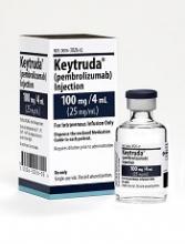
The European Medicines Agency’s Committee for Medicinal Products for Human Use (CHMP) has recommended approval for the anti-PD-1 therapy pembrolizumab (Keytruda) as a treatment for patients with relapsed or refractory classical Hodgkin lymphoma (cHL).
The recommendation pertains specifically to adults with cHL who have failed autologous hematopoietic stem cell transplant (auto-HSCT) and treatment with brentuximab vedotin (BV) or adults with cHL who are transplant-ineligible and have failed treatment with BV.
The CHMP’s recommendation will be reviewed by the European Commission, which is expected to make a decision about the drug in the second quarter of 2017.
Pembrolizumab is already approved for use in the European Union as a treatment for melanoma and non-small-cell lung cancer.
The CHMP’s positive opinion of pembrolizumab for cHL was based on data from the KEYNOTE-087 and KEYNOTE-013 trials. Results from both trials were presented at ASH 2016 (abstract 1107 and abstract 1108).
KEYNOTE-087
KEYNOTE-087 is a phase 2 trial in which researchers evaluated pembrolizumab (a 200 mg fixed dose every 3 weeks) in patients with relapsed or refractory cHL across 3 cohorts:
- Cohort 1: Patients who progressed after auto-HSCT and subsequent treatment with BV
- Cohort 2: Patients who failed salvage chemotherapy, were ineligible for a transplant, and progressed after BV
- Cohort 3: Patients who progressed after auto-HSCT and did not receive BV after transplant.
Across all 210 enrolled patients, the overall response rate (ORR) was 69.0%, and the complete response (CR) rate was 22.4%.
In Cohort 1 (n=69), the ORR was 73.9%. The CR rate was 21.7%, the partial response (PR) rate was 52.2%, 15.9% of patients had stable disease (SD), and 7.2% progressed. In 82.2% of responders, the response lasted 6 months or more.
In Cohort 2 (n=81), the ORR was 64.2%. The CR rate was 24.7%, the PR rate was 39.5%, 12.3% of patients had SD, and 21.0% progressed. In 70.0% of responders, the response lasted 6 months or more.
In Cohort 3 (n=60), the ORR was 70.0%. Twenty percent of patients had a CR, 50.0% had a PR, 16.7% had SD, and 13.3% progressed. In 75.6% of responders, the response lasted 6 months or more.
Results also included an analysis of patients with primary refractory disease (n=73), which was defined as failure to achieve CR or PR with first-line treatment. In this patient population, the ORR was 79.5%.
An ORR of 67.8% was reported in patients who relapsed after 3 or more lines of prior therapy (99/146).
The most common treatment-related adverse events (AEs) were hypothyroidism (12.4%), pyrexia (10.5%), fatigue (9.0%), rash (7.6%), diarrhea (7.1%), headache (6.2%), nausea (5.7%), cough (5.7%), and neutropenia (5.2%).
The most common grade 3/4 treatment-related AEs were neutropenia (2.4%), diarrhea (1.0%), and dyspnea (1.0%). Immune-mediated AEs included pneumonitis (2.9%), hyperthyroidism (2.9%), colitis (1.0%), and myositis (1.0%).
There were 9 discontinuations because of treatment-related AEs and no treatment-related deaths.
KEYNOTE-013
KEYNOTE-013 is a phase 1b trial that has enrolled 31 patients with relapsed or refractory cHL who failed auto-HSCT and subsequent BV or who were transplant-ineligible.
Patients received pembrolizumab at 10 mg/kg every 2 weeks. The median duration of follow-up was 29 months.
The ORR was 58%. Nineteen percent of patients achieved a CR, 39% had a PR, and 23% had SD.
The median duration of response had not been reached at last follow-up (range, 0.0+ to 26.1+ months), and 70% of responding patients had a response lasting 12 months or more.
The median progression-free survival (PFS) was 11.4 months (range, 4.9-27.8 months). The six-month PFS rate was 66%, and the 12-month PFS rate was 48%.
The median overall survival was not reached. Six-month and 12-month overall survival rates were 100% and 87%, respectively.
The most common treatment-related AEs were diarrhea (19%), hypothyroidism (13%), pneumonitis (13%), nausea (13%), fatigue (10%), and dyspnea (10%).
The most common grade 3/4 treatment-related AEs were colitis (3%), axillary pain (3%), AST increase (3%), joint swelling (3%), nephrotic syndrome back pain (3%), and dyspnea (3%).
AEs leading to discontinuation were nephrotic syndrome (grade 3), interstitial lung disease (grade 2), and pneumonitis (grade 2). There were no treatment-related deaths. ![]()
Perioperative statin associated with reduction in all-cause perioperative mortality in noncardiac surgery
CLINICAL QUESTION: Does perioperative statin use reduce 30-day mortality in noncardiac surgery?
BACKGROUND: Current perioperative guidelines focus on continuation of existing therapy in long-term statin users with weak recommendations of potential efficacy in reducing perioperative complications.
STUDY DESIGN: Retrospective, observational cohort analysis.
SETTING: Veterans’ Affairs Hospitals.
SYNOPSIS: Using the Veterans Affairs Surgical Quality Improvement Program database, 96,486 patients were studied who were undergoing elective or emergent noncardiac surgery (vascular, general, orthopedic, neurosurgery, otolaryngology, and urology). 96.3% were men. Patients who died the day of the surgery or the day after were excluded, as were patients with multiple surgeries during the assessment period. Statin exposure on the day of or the day after surgery was compared with no statin use. The primary outcome was 30-day mortality and the secondary outcomes were significant reduction in any other complication.
Statin exposure was associated with reduced 30-day all-cause mortality with a marginally favorable effect with longer-term statin use (6 months to 1 year before admission). For the secondary outcomes, there was significant risk reduction in cardiac, infectious, respiratory, and renal complications but no significant change in central nervous system or nonatherosclerotic thrombotic complications.
Statin exposure may be associated with adherence to medical treatment and follow-up thus causing a selection bias.
BOTTOM LINE: Perioperative statin use was associated with a reduction in 30-day mortality and other complications.
CITATIONS: London MJ, Schwartz GG, Hur K, Henderson WG. Association of perioperative statin use with mortality and morbidity after major noncardiac surgery. JAMA Intern Med. 2017 Feb 1;177(2):231-42.
Dr. Dietsche is a clinical instructor, Division of Hospital Medicine, University of Colorado School of Medicine, Aurora.
Male vs. female hospitalists, a comparison in mortality and readmission rate for Medicare patients
Clinical Question: Does physician sex affect hospitalized patient outcomes?
Background: Previous studies had suggested different practice patterns between male and female physicians in process measure of quality. No prior evaluation of patient outcomes examining those differences was studied in the past.
Setting: U.S. national sample (20%) of Medicare beneficiaries aged 65 years or older, hospitalized with acute medical conditions.
Synopsis: This observational study assessed the difference in patients’ outcomes that were treated by a male or female physician. 30-days mortality rate was analyzed from 1,583,028 hospitalizations. The mortality rate of patients cared for by female physicians was lower and statistically significant: 11.07% vs. 11.49% (adjusted risk difference, –0.43%; 95% CI, –0.57% to –0.28%; P less than .001). The difference did not change after considering patient and physician characteristics as well as when looking at hospital fixed effects (that is, hospital indicators). In order to prevent one death, a female physician needs to treat 233 patients.
Also, 30-day readmission rate, after adjustment readmissions (from 1,540,797 hospitalizations) was 15.02% vs. 15.57% (adjusted risk difference, –0.55%; 95% confidence interval, –0.71% to 0.39%; P less than .001) showing that the care provided by a female physician can reduce one readmission when treating 182 patients.
Bottom line: Patients older than 65 years have lower 30-day mortality and readmission rates when receiving inpatient care from a female internist, compared with care by a male internist.
Citations: Tsugawa Y, Jena AB, Figueroa JF, et al. Comparison of hospital mortality and readmission rates for Medicare patients treated by male vs. female physicians. JAMA Intern Med. 2017 Feb;177(2):206-13.
Dr. Orjuela is assistant professor of neurology at the University of Colorado School of Medicine, Aurora.
End-of-rotation resident transition in care and mortality among hospitalized patients
Clinical Question: Are hospitalized patients experiencing an increased mortality risk at the end-rotation resident transition in care and is this association related to the Accreditation Council for Graduate Medical Education (ACGME) 2011 duty-hour regulations?
Background: Prior studies of physicians’ transitions in care were associated with potential adverse patient events and outcomes. A higher mortality risk was suggested among patients with a complex hospital course or prolonged length of stay in association to house-staff transitions of care.
Setting: 10 University-affiliated U.S. Veterans Health Administration hospitals.
Synopsis: 230,701 patient discharges (mean age, 65.6 years; 95.8% male sex; median length of stay, 3 days) were included. The transition group included patients admitted at any time prior to an end-of-rotation who were either discharged or deceased within 7 days of transition. All other discharges were considered controls.
The primary outcome was in-hospital mortality rate; secondary outcomes included 30-day and 90-day mortality and readmission rates. An absolute increase of 1.5% to 1.9% in a unadjusted in-hospitality risk was found. The 30-day and 90-day mortality odds ratios were 1.10 and 1.21, respectively. A possible stronger association was found among interns’ transitions in care and the in-hospital and after-discharge mortality post-ACGME 2011 duty hour regulations. The latter raises questions about the interns’ inexperience and their amount of shift-to-shift handoffs. An adjusted analysis of the readmission rates at 30-day and 90-day was not significantly different between transition vs. control patients.
Bottom line: Elevated in-hospital mortality was seen among patients admitted to the inpatient medicine service at the end-of-rotation resident transitions in care. The association was stronger after the duty-hour ACGME (2011) regulations.
Citations: Denson JL, Jensen A, Saag HS, et al. Association between end-of-rotation resident transition in care and mortality among hospitalized patients. JAMA. 2016 Dec 6;316(21):2204-13.
Dr. Orjuela is assistant professor of neurology at the University of Colorado School of Medicine, Aurora.
VIDEO: Picowave laser uses are expanding beyond tattoo removal
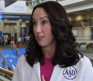
ORLANDO – The applications for picowavelength lasers are expanding, with emerging data on their uses for cosmetic indications other than tattoo removal, according to Anne Chapas, MD, who is in private practice in New York.
First introduced and cleared by the Food and Drug Administration for tattoo removal, “picowave devices ... are now being studied in multiple different cosmetic conditions, including their use in acne scars, fine lines and wrinkles, and melasma,” Dr. Chapas said in a video interview at the annual meeting of the American Academy of Dermatology.
The No. 1 thing dermatologists need to know is that these types of lasers are delivering energy extremely quickly,” at 1,000 times faster than nanosecond lasers, she said. Another difference between the two is that “the laser tissue interaction between the two types of devices is completely different.”
In the interview, she highlighted other important points about picowave lasers, including less downtime after treatment.
At the meeting, Dr. Chapas spoke during a session entitled “the Science Behind New Devices in Dermatology.”
Her disclosures include serving as a consultant and investigator for Syneron and Candela.
The video associated with this article is no longer available on this site. Please view all of our videos on the MDedge YouTube channel
AT AAD 17
VIDEO: Internet-based intervention shows antihypertensive efficacy
WASHINGTON – Patients who regularly accessed 30 minute, Internet-based behavioral counseling videos cut their systolic blood pressure, compared with baseline over 1 year by an average 4 mm Hg more than control patients in a randomized, phase II study with 264 patients.
Electronic counseling (e-counseling) “enhanced the efficacy of usual care for hypertension,” Robert P. Nolan, Ph.D., said at the annual meeting of the American College of Cardiology.
“We hope to optimize the efficacy of medical treatments with a behavioral intervention,” said Dr. Nolan, who added that the magnitude of the added benefit from the e-counseling program was “like adding an additional antihypertensive medication.”
“We know antihypertensive treatments work, but compliance is a huge challenge” for health care providers, commented E. Magnus Ohman, MBBS, a professor and cardiologist at Duke University in Durham, N.C. “Having a new way to enhance compliance would be fantastic,” he said.
The Internet-based counseling program devised by Dr. Nolan and his associates consisted of a year-long series of 28 videos, each about 30 minutes long, that participants in the active arm accessed over the Internet. During the study, participants received a series of emailed messages that sent links to the videos on a set schedule over 12 months: During the first 4 months they received an emailed link weekly, during the next 4 months they received an emailed link to a new video every other week, and during the final 4 months of the intervention participants received emailed links once a month. Patients could access each video more than once if they wished, and they were free to share the links with any family members or friends who helped the patients with lifestyle management of their hypertension.
Patients in the e-counseling intervention arm received links to videos that focused on motivational messages and teaching cognitive behavioral skills. Patients in the control arm received emails with generic messages on blood pressure management and without links to videos.
The REACH (E-Counseling Promotes Blood Pressure Reduction and Therapeutic Lifestyle Change in Hypertension) study ran at four Canadian sites. It sent invitations to participate to 609 patients with stage 1 or 2 hypertension, with a blood pressure prior to treatment of 140/90-180/110 mm Hg. Among the invited patients, 264 elected to participate; 100 patients in the e-counseling arm and 97 control patients completed the 1-year program. Participants averaged 58 years of age, their average body mass index was 31 kg/m2, and 9% smoked. Their blood pressure at entry averaged 141/87 mm Hg, their average pulse pressure was about 54 mm Hg, and their average 10-year risk for a cardiovascular disease event, measured by the Framingham Risk Score, was about 16%. At entry, patients in the study received an average of 1.5 antihypertensive drugs each.
At the end of the 1-year program, systolic blood pressure fell by an average of 6 mm Hg from baseline among patients who completed the control program, and by an average of 10.1 mm Hg among the patients who completed the e-counseling arm, a statistically significant difference for one of the study’s primary endpoints. Change in pulse pressure from baseline showed an average 1.5–mm Hg drop in the control patients and an average 4.3–mm Hg decline in the e-counseling patients, another statistically significant difference for a second primary endpoint, reported Dr. Nolan, a clinical psychologist and director of the cardiac eHealth program at the University of Toronto.
A third primary endpoint was change in the Framingham Risk Score, which fell by an average of 1.9% after 12 months in the e-counseling patients and rose by an average of 0.2% among the controls.
The final primary endpoint was the change in diastolic blood pressure from baseline, which showed a better than 4–mm Hg incremental decline in the men who received e-counseling, compared with controls, but among women in the study, the drop in diastolic blood pressure from baseline was nearly the same – about 6 mm Hg – in both the controls and e-counseling patients.
“This tells us that we need to better tailor the [e-counseling] intervention to men and to women,” Dr. Nolan said in a video interview. He also envisions better tailoring of the e-counseling videos to various socioeconomic and ethnic groups. He plans to continue testing of a revised version of the e-counseling intervention in a larger, phase III study, but he also hopes that the intervention videos can soon be available at no charge for use in routine practice.
REACH received no commercial funding. Dr. Nolan had no disclosures.
The video associated with this article is no longer available on this site. Please view all of our videos on the MDedge YouTube channel
[email protected]
On Twitter @mitchelzoler
I love the REACH study. Hypertension is incredibly prevalent among the patients I see, and they often need three different antihypertensive drugs to control their blood pressure. Lifestyle interventions can be very effective at helping to lower blood pressure, but during the 10-minute visits I have with most of my patients, it’s hard for me to have much impact on their behavior.
What I especially like about the Internet-based counseling used in this study was its application of evidence-based approaches to change patient behavior. This was a well-designed and exciting trial.
Sandra J. Lewis, MD, chief of cardiology at Legacy Good Samaritan Hospital in Portland, Ore., made these comments during a press conference. She had no disclosures.
WASHINGTON – Patients who regularly accessed 30 minute, Internet-based behavioral counseling videos cut their systolic blood pressure, compared with baseline over 1 year by an average 4 mm Hg more than control patients in a randomized, phase II study with 264 patients.
Electronic counseling (e-counseling) “enhanced the efficacy of usual care for hypertension,” Robert P. Nolan, Ph.D., said at the annual meeting of the American College of Cardiology.
“We hope to optimize the efficacy of medical treatments with a behavioral intervention,” said Dr. Nolan, who added that the magnitude of the added benefit from the e-counseling program was “like adding an additional antihypertensive medication.”
“We know antihypertensive treatments work, but compliance is a huge challenge” for health care providers, commented E. Magnus Ohman, MBBS, a professor and cardiologist at Duke University in Durham, N.C. “Having a new way to enhance compliance would be fantastic,” he said.
The Internet-based counseling program devised by Dr. Nolan and his associates consisted of a year-long series of 28 videos, each about 30 minutes long, that participants in the active arm accessed over the Internet. During the study, participants received a series of emailed messages that sent links to the videos on a set schedule over 12 months: During the first 4 months they received an emailed link weekly, during the next 4 months they received an emailed link to a new video every other week, and during the final 4 months of the intervention participants received emailed links once a month. Patients could access each video more than once if they wished, and they were free to share the links with any family members or friends who helped the patients with lifestyle management of their hypertension.
Patients in the e-counseling intervention arm received links to videos that focused on motivational messages and teaching cognitive behavioral skills. Patients in the control arm received emails with generic messages on blood pressure management and without links to videos.
The REACH (E-Counseling Promotes Blood Pressure Reduction and Therapeutic Lifestyle Change in Hypertension) study ran at four Canadian sites. It sent invitations to participate to 609 patients with stage 1 or 2 hypertension, with a blood pressure prior to treatment of 140/90-180/110 mm Hg. Among the invited patients, 264 elected to participate; 100 patients in the e-counseling arm and 97 control patients completed the 1-year program. Participants averaged 58 years of age, their average body mass index was 31 kg/m2, and 9% smoked. Their blood pressure at entry averaged 141/87 mm Hg, their average pulse pressure was about 54 mm Hg, and their average 10-year risk for a cardiovascular disease event, measured by the Framingham Risk Score, was about 16%. At entry, patients in the study received an average of 1.5 antihypertensive drugs each.
At the end of the 1-year program, systolic blood pressure fell by an average of 6 mm Hg from baseline among patients who completed the control program, and by an average of 10.1 mm Hg among the patients who completed the e-counseling arm, a statistically significant difference for one of the study’s primary endpoints. Change in pulse pressure from baseline showed an average 1.5–mm Hg drop in the control patients and an average 4.3–mm Hg decline in the e-counseling patients, another statistically significant difference for a second primary endpoint, reported Dr. Nolan, a clinical psychologist and director of the cardiac eHealth program at the University of Toronto.
A third primary endpoint was change in the Framingham Risk Score, which fell by an average of 1.9% after 12 months in the e-counseling patients and rose by an average of 0.2% among the controls.
The final primary endpoint was the change in diastolic blood pressure from baseline, which showed a better than 4–mm Hg incremental decline in the men who received e-counseling, compared with controls, but among women in the study, the drop in diastolic blood pressure from baseline was nearly the same – about 6 mm Hg – in both the controls and e-counseling patients.
“This tells us that we need to better tailor the [e-counseling] intervention to men and to women,” Dr. Nolan said in a video interview. He also envisions better tailoring of the e-counseling videos to various socioeconomic and ethnic groups. He plans to continue testing of a revised version of the e-counseling intervention in a larger, phase III study, but he also hopes that the intervention videos can soon be available at no charge for use in routine practice.
REACH received no commercial funding. Dr. Nolan had no disclosures.
The video associated with this article is no longer available on this site. Please view all of our videos on the MDedge YouTube channel
[email protected]
On Twitter @mitchelzoler
AT ACC 17
Key clinical point:
Major finding: The Internet-based program reduced systolic blood pressure from baseline by an average additional 4.1 mm Hg, compared with controls.
Data source: REACH, a multicenter, randomized trial with 264 hypertensive patients.
Disclosures: REACH received no commercial funding. Dr. Nolan had no disclosures.
VIDEO: Looking at keloids from a different perspective
ORLANDO – It may be time to start considering new options for treating keloids, according to Amy McMichael, MD, professor of dermatology at Wake Forest University, Winston-Salem, N.C.
At the annual meeting of the American Academy of Dermatology, Dr. McMichael discussed one of the highlights of the annual symposium of the Skin of Color Society, held right before the annual meeting, a presentation by Michael Tirgan, MD, who has treated patients with keloids for about 10 years.
Dr. Tirgan, an oncologist based in New York, has a large database and registry of patients and shared some interesting data at the symposium. “What he’s found is that those who come with the supermassive and massive keloids are those who have had the most surgery on their keloid,” Dr. McMichael said in a video interview at the meeting.
His findings shed light on what she described as “a new perspective in the way that we think about keloids” and a new approach to treatment – considering keloids as tumors – not just a scar.
Dr. McMichael had no relevant disclosures.
The video associated with this article is no longer available on this site. Please view all of our videos on the MDedge YouTube channel
AT AAD 17
